Article

Molecular Dynamics Simulation and Cryo-Electron Microscopy Investigation of AOT Surfactant Structure at the Hydrated Mica Surface

by
Daniel M. Long
1,†,‡,
Jeffery A. Greathouse
2,*,
Guangping Xu
3 and
Katherine L. Jungjohann
1,§
1
Center for Integrated Nanotechnologies, Sandia National Laboratories, Albuquerque, NM 87123, USA
2
Nuclear Waste Disposal Research and Analysis Department, Sandia National Laboratories, Albuquerque, NM 87185, USA
3
Geochemistry Department, Sandia National Laboratories, Albuquerque, NM 87185, USA
*
Author to whom correspondence should be addressed.
Current Address: Materials and Manufacturing Directorate, Air Force Research Laboratory, WPAFB, Dayton, OH 45433, USA.
Current Address: UES Inc., Beavercreek, OH 45432, USA.
§
Current Address: Analytical Microscopy and Imaging Sciences, National Renewable Energy Laboratory, Golden, CO 80401, USA.
Minerals 2022, 12(4), 479; https://doi.org/10.3390/min12040479
Submission received: 15 March 2022 / Revised: 6 April 2022 / Accepted: 7 April 2022 / Published: 14 April 2022
(This article belongs to the Special Issue Feature Papers in Clays and Engineered Mineral Materials)

Abstract

:
Structural properties of the anionic surfactant dioctyl sodium sulfosuccinate (AOT or Aerosol-OT) adsorbed on the mica surface were investigated by molecular dynamics simulation, including the effect of surface loading in the presence of monovalent and divalent cations. The simulations confirmed recent neutron reflectivity experiments that revealed the binding of anionic surfactant to the negatively charged surface via adsorbed cations. At low loading, cylindrical micelles formed on the surface, with sulfate head groups bound to the surface by water molecules or adsorbed cations. Cation bridging was observed in the presence of weakly hydrating monovalent cations, while sulfate groups interacted with strongly hydrating divalent cations through water bridges. The adsorbed micelle structure was confirmed experimentally with cryogenic electronic microscopy, which revealed micelles approximately 2 nm in diameter at the basal surface. At higher AOT loading, the simulations reveal adsorbed bilayers with similar surface binding mechanisms. Adsorbed micelles were slightly thicker (2.2–3.0 nm) than the corresponding bilayers (2.0–2.4 nm). Upon heating the low loading systems from 300 K to 350 K, the adsorbed micelles transformed to a more planar configuration resembling bilayers. The driving force for this transition is an increase in the number of sulfate head groups interacting directly with adsorbed cations.

Graphical Abstract

1. Introduction

Surface wettability plays an important role in many energy-related applications, including energy recovery [1,2], sequestration [3], and water treatment [4,5]. Complex fluids consisting of both polar and nonpolar components are typically in contact with surfaces in these applications [6]. Wettability therefore depends on both fluid–fluid and fluid–surface interactions [7], but the additivity of these intermolecular forces at complex interfaces is not well understood. Surfactants are widely used in subsurface energy applications to modify the interfacial tension in complex fluids [8]. Through modification of surface wettability, the distribution and rheological properties of these fluids can be better controlled for more efficient processes.
In this work, our focus is the adsorption of the anionic surfactant dioctyl sodium sulfosuccinate (AOT or Aerosol-OT) on the mica surface. Surfactant interactions with mineral surfaces have been studied with molecular simulation [9,10,11,12,13,14] and laboratory experiments [15,16]. While the adsorption of cationic surfactants on negatively charged surfaces has been well studied [15,17], the adsorption of an anionic species via cation bridging mechanisms is less commonly studied. The mechanism by which an anionic surfactant such as AOT adsorbs on a negatively charged surface has been attributed to cation bridging [18], which is thought to play a key role in the binding of polar organic species to mineral surfaces [19], and in the process of enhanced oil recovery through low salinity waterflooding [20].
In the presence of certain cations, recent neutron reflectivity (NR) experiments by Clarke and co-workers have revealed details of the AOT-mica interface [21]. AOT is thought to adsorb to the negatively charged mica surface in the form of AOT bilayers, with AOT molecules coordinated to the surface through cation bridging. This bridging is facilitated with divalent cations (Ca2+ and Mg2+) [22,23]; however, adsorption also occurs in the presence of weakly hydrating monovalent cations K+ and Cs+ [21,23]. With a thickness of around 2 nm, the bilayer consists of ordered AOT chains [24]. NR studies have also been used to investigate AOT adsorption on sapphire (Al2O3) in the presence of Na+ [25,26]. Na-AOT forms a bilayer with a thickness of 3.3 nm which is composed of Na-AOT pairs [25]. This layer thickness is consistent with a bilayer of “extended AOT molecules” or distorted micelles, although an interdigitated bilayer would have a reduced layer thickness of 2.2 nm [21]. The structure of adsorbed Na-AOT depends on concentration: separated pairs on the surface at lower concentration, continuous bilayers at higher concentrations, and multiple lamellar layers at concentrations above the critical micelle concentration (CMC) [26].
NR has also been utilized to show that both Na-AOT and Ca-AOT adsorb on calcite, although the slow kinetics of Na-AOT adsorption indicates structural conversion, and similar absorption, to Ca-AOT due to exchange with calcium ions desorbed from the surface [27]. Additionally, NR and attenuated total reflectance Fourier transform infrared (ATR-FTIR) spectroscopy was used to investigate Na-AOT and Ca-AOT adsorption on negatively charged silica [18]. The results indicated that an AOT bilayer or micelles near the surface were only seen in the presence of divalent calcium ions. Importantly, micelle self-assembly can occur at surfaces at low AOT concentration before bilayers form at the surface [28].
Molecular dynamics (MD) simulations have also provided insight into binding mechanisms of polar species on oxide surfaces, including surfactant interactions with solid surfaces [15,29]. Utilizing MD, the adsorption of functional groups of organic surfactants on an iron oxyhydroxide surface was shown to be controlled by the degree of surface interaction [30]. Another MD study showed that varying the concentration of ionic surfactants with sulfate head groups on alumina altered the form of the surfactant aggregate [31]. It has also been demonstrated that cation bridging mediates adsorption of anionic surfactant sodium dodecyl sulfate on rutile and anatase TiO2 surfaces [32]. Additionally, adsorption and morphology of neutral and anionic surfactants on silica were shown to be a function of the charge distribution on the silica surface [9].
One imaging tool that has not seen widespread use for surfactant interfaces is cryogenic electron microscopy (cryo-EM). Since its inception in the life sciences [33,34,35], cryogenic microscopy has been instrumental in advancing our knowledge of hydrated matter. Historically utilized to study organic molecules, biological matter, and nanoparticles in solution [36,37,38,39,40], cryogenic microscopy has only recently taken root in materials science. Recent advances in cryo-EM in materials science, specifically site-specific investigation of buried solid/liquid interfaces in energy storage materials [41,42,43,44,45,46,47,48,49,50,51,52,53,54], have revolutionized experiment design and characterization. This emerging use of cryo-EM in materials science inherently includes new method development which in turn leads to faster utilization of the technique. Surfactant/mica systems [55,56] have been investigated with cryogenic microscopy in prior literature; however, these studies focused on general liquid-solid interactions and did not investigate the nanoscale structure of the interface.
Here we perform extensive MD simulations to provide nanoscale detail of AOT adsorption on a mineral surface (mica). The presence of adsorbed micelles from MD simulations are confirmed using cryo-EM. We utilize the mica/AOT material system to develop a reproducible cryogenic process to investigate the mineral/liquid/surfactant interface at the nanometer scale. Mica (muscovite) was chosen as our mineral phase since it can be easily cleaved, yielding an atomically flat surface for characterization and adsorption studies [57]. Additionally, it is representative of clay phases present in many geologic shale formations containing oil and gas reservoirs [58].

2. Methods

2.1. MD Simulation

Interaction parameters used in the simulation are a combination of Clayff [59], flexible SPC water [60] with corresponding cation-water parameters [61,62,63], and the all-atom OPLS-AA force field [64] for the AOT anion. The combination of Clayff and OPLS-AA has previously been used to simulate organic species at clay mineral interfaces [65,66,67,68,69]. Parameters for the AOT anion are based on the OPLS-AA force field for ionic liquids [70] and have been used to simulate electrical double layer properties in ionic liquids [71]. Arithmetic mixing rules were used to generate Lennard–Jones parameters for unlike atom types. Although Clayff enables flexibility of mineral phases, for computational efficiency atoms in the mica layer were not allowed to move during the simulations.
The simulation supercell consisted of an aqueous phase in contact with a single mica layer on one side and a thin vacuum region on the other side, terminated by a Lennard–Jones 9-3 wall. The vacuum varies in thickness with system size (5–15 Å). A similar supercell was used previously to model cation adsorption on the mica surface [72]. The molecular model for mica was taken from the muscovite crystal structure [73]. After orthogonalizing, an expanded 8 × 4 supercell in the xy plane (35.87 Å × 41.46 Å) was created, and 32 negative charge sites on each basal surface were created by replacing Si atoms with Al atoms in agreement with Lowenstein’s rule [74]. The resulting layer charge of −1.0 e per unit cell (e is the elementary charge) was balanced by 32 potassium ions on the lower surface and aqueous cations on the upper surface (32 monovalent or 16 divalent cations).
The aqueous phase consisted of the appropriate number of AOT anions (32, 48, or 64), cations to balance the negative charge due to the mica layer and AOT anions, and water molecules. The bulk AOT concentrations are significantly greater than the CMC of AOT solutions. Instead, they correspond to loadings for monolayer and bilayer surface coverage. The lowest AOT loading (32) corresponds to one AOT anion per (monovalent) surface cation, although fewer AOT anions would likely be needed for monolayer coverage. The highest AOT loading (64) corresponds to bilayer coverage with one AOT anion per surface cation. Both monovalent (Na+, K+, Cs+) and divalent (Mg2+, Ca2+) cations were simulated (cation mixtures were not considered), for a total of 15 model systems. The AOT anions were randomly placed in the aqueous region. Cations were initially located at the upper mica surface or near an AOT sulfate group. Except in the K-AOT systems, the supercells contained no potassium cations in the aqueous region, corresponding to complete cation exchange at the external surface. Water molecules were initially placed in a grid pattern in the aqueous region, and waters in close contact with AOT anions or cations were removed. The final tally of water molecules in the supercells was 2483 (32 AOT), 2006 (48 AOT), and 2311 (64 AOT).
MD simulations were performed using the LAMMPS code [75] in the canonical ensemble with a thermostat temperature of 300 K. A Nose–Hoover thermostat was used with a relaxation time of 100 fs. Short-range interactions were evaluated every 0.5 fs with a cutoff distance of 10.0 Å, while long-range electrostatic interactions were evaluated every 1.0 fs using a particle-mesh Ewald algorithm [76]. Supercells were periodic in all three dimensions, but special slab boundary conditions [77] imposed a (virtual) vacuum region in the vertical (z) direction with thickness 3 Lz between adjacent cells (Lz is the supercell z-dimension), which removed long-range electrostatic interactions between slabs. The values of Lz are 81 Å (32 and 48 AOT) and 94 Å (64 AOT). A random configuration of the aqueous phase was generated by simulating the initial configuration over four 0.2-ns segments in which the systems were heated to 1000 K and then cooled back to 300 K. Each system was then simulated for an additional 110 ns, during which time an equilibrium surfactant structure was obtained. Figure 1 shows an MD snapshot of an equilibrated AOT structure with sodium counterions. Structural properties were analyzed over the final 20 ns of simulation. Each system with 32 AOT anions was subsequently heated to 350 K and simulated for an additional 40 ns to investigate the effect of elevated temperature on interfacial structure.

2.2. Sample Preparation and Cryogenic Electron Microscopy
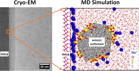
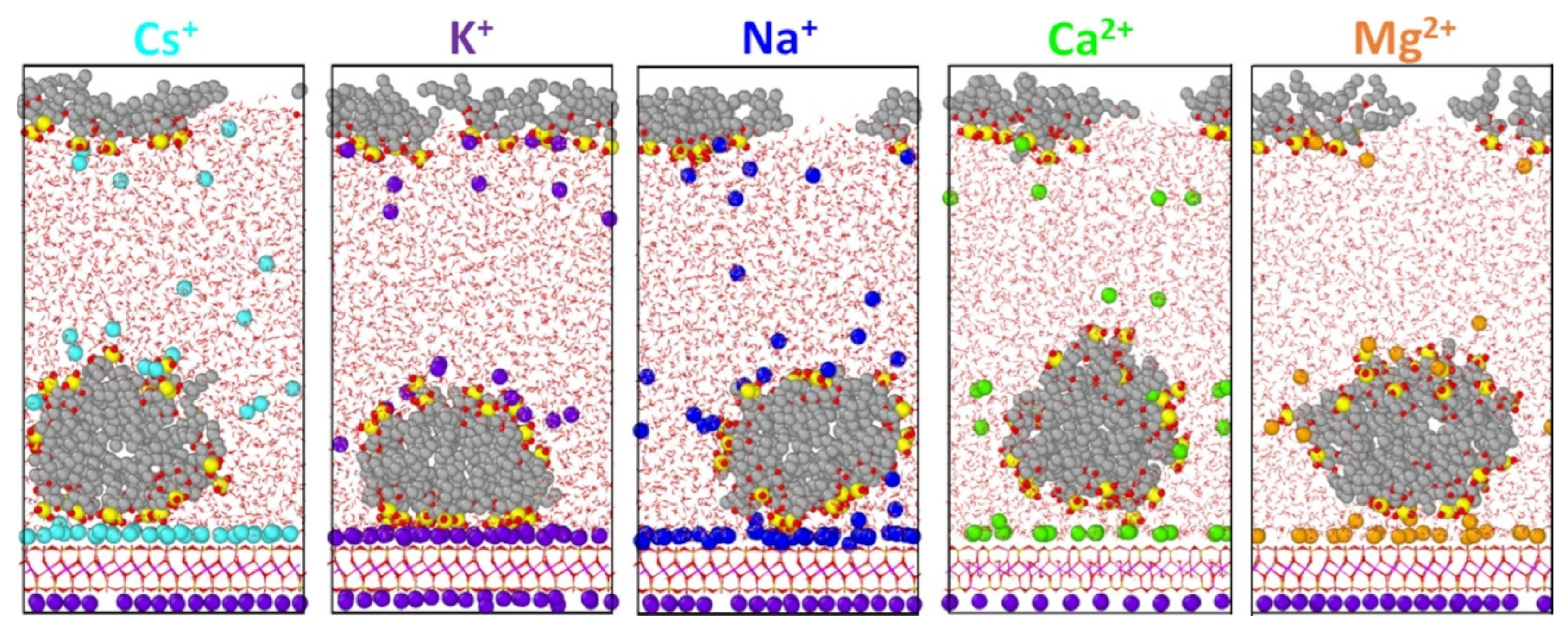
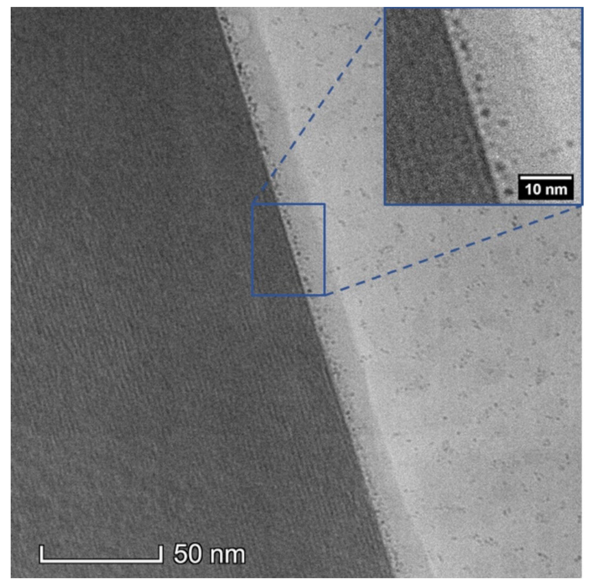
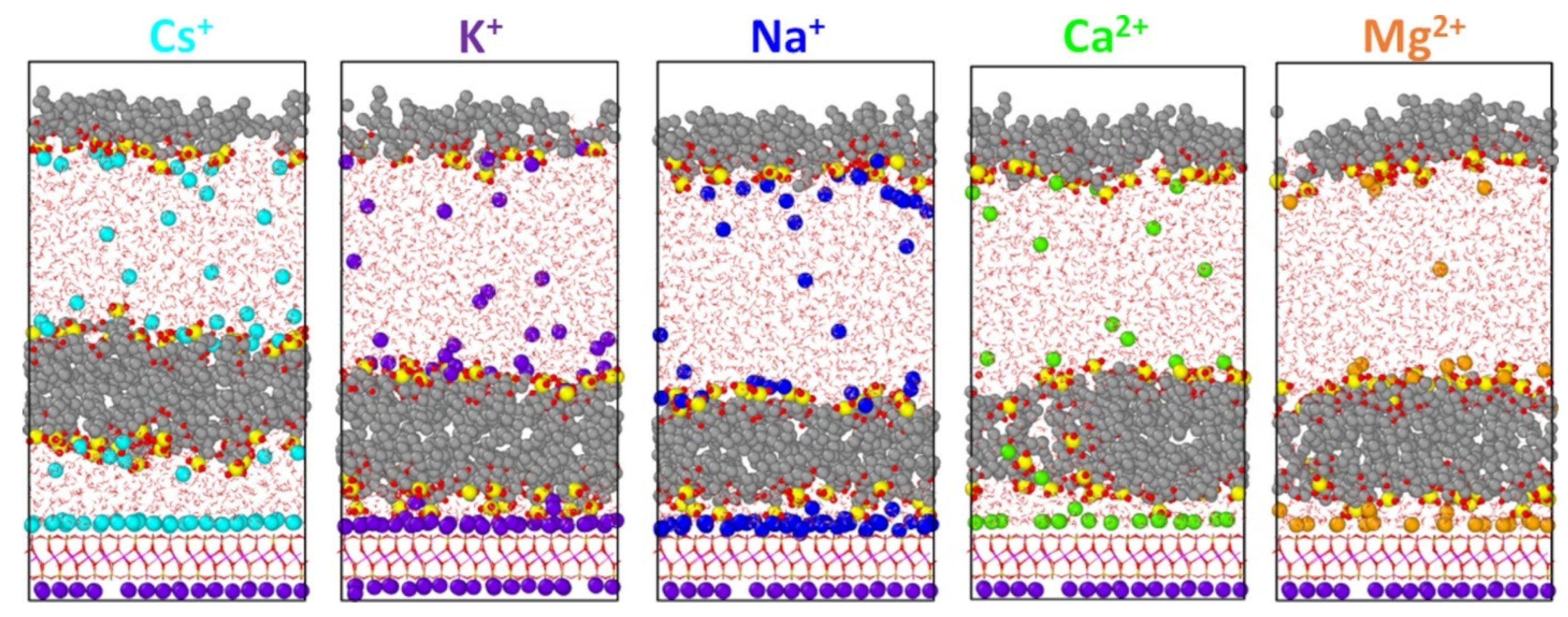
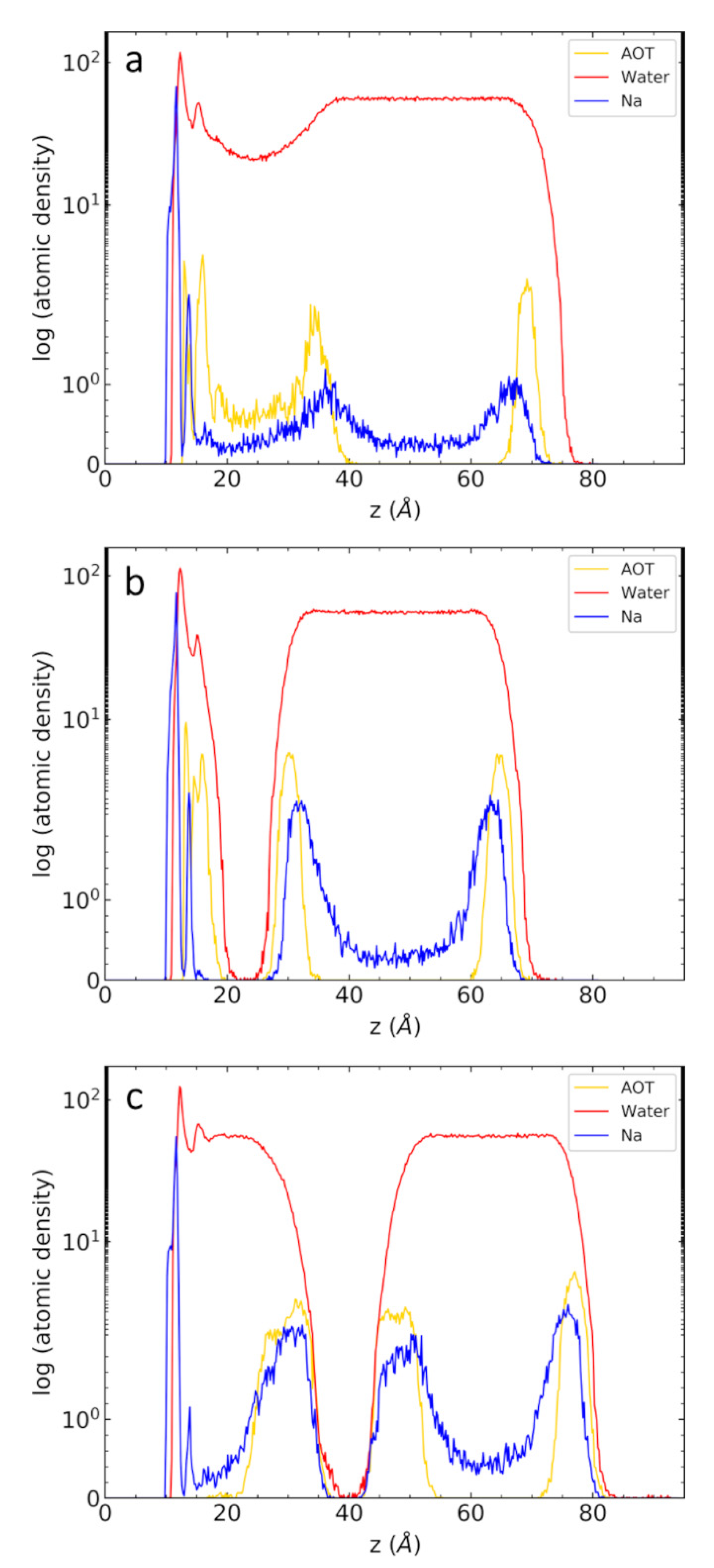
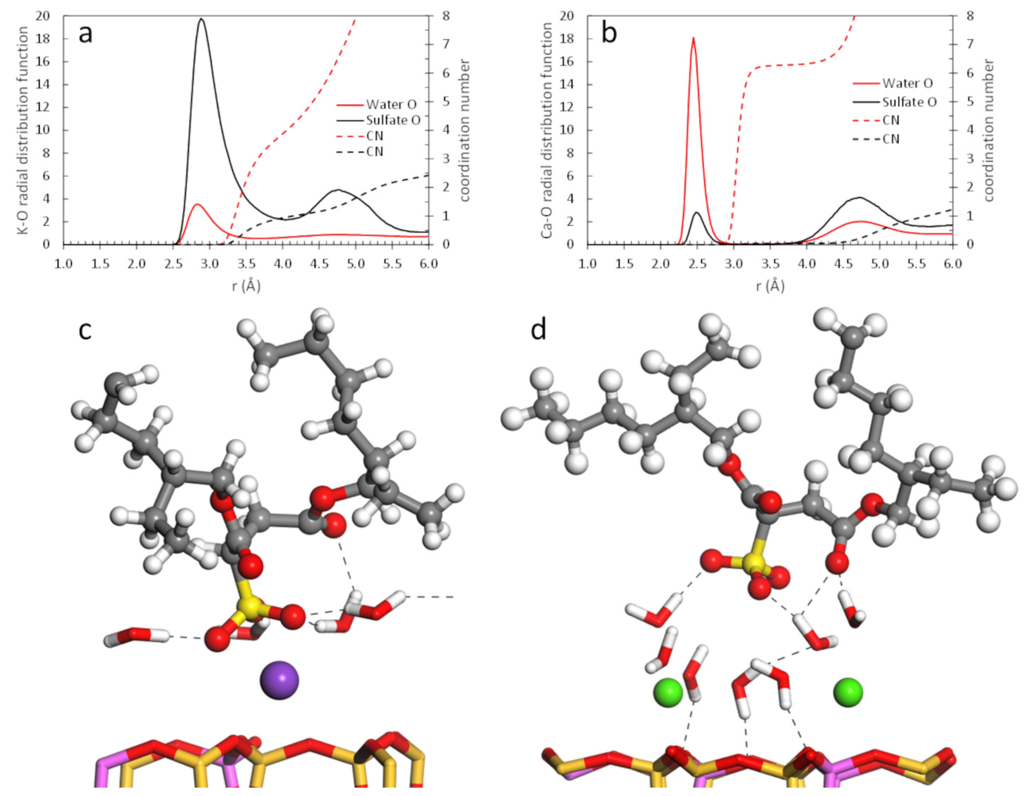
Investigating the mica/Na-AOT solution interface requires methodical preparation. Lamella for cryogenic transmission electron microscopy(cryo-TEM) were prepared via cryogenic focused ion beam (cryo-FIB) lift-out, which allowed for precise imaging of the mica/AOT solution interface across an atomically flat mica basal plane. AOT solutions [2.29 mM Na-AOT (0.92 CMC); 40 mM Na-AOT (16 CMC)] were made by combining pure Na-AOT (CAS Number 577-11-7, Sigma Aldrich, St. Louis, MO, USA) and 18.2 megohm deionized H2O and sonicating for 60 min and then left on a shaker for several days. The 0.92 CMC Na-AOT solution also contained 0.032 mM MgCl2 and 0.025 mM KCl, whereas the 16 CMC Na-AOT solution did not contain any additional ions. High purity muscovite mica sheets (MTI, Bernalillo, NM, USA) were adhered to a standard scanning electron microscope (SEM) pin stub via carbon tape and mechanically exfoliated to a few microns thickness to minimize the thermal mass of the sample during vitrification and reduce charging during cryo-FIB lamella preparation. One microliter of Na-AOT solution was immediately pipetted onto the mica sheet and the entire sample, including the SEM stub, was plunge frozen in liquid ethane or slush nitrogen. The sample was kept below −140 °C during transfer through a Leica cryogenic-EM workflow with a loading station (Leica Microsystems Inc., Buffalo Grove, IL, USA), sputter-coater (ACE 600), inert cryo transfer shuttle (VCT) and a Scios 2 dual-beam FIB (Thermo Fisher Scientific, Waltham, MA, USA) outfitted with liquid nitrogen-cooled SEM stage, lift-out needle and anticontamination shield. The vitrified sample was coated with 10 nm of Pt, and then transferred into the FIB for cryo-lift-out.
General cryo-FIB lift-out lamella preparation proceeded as described in prior literature [35,43,46,78,79,80,81,82,83,84] and included the use of lower accelerating voltages and beam currents, and the use of a condensed Pt precursor for the protective cap. Additionally, the side of the TEM lamella visible to the ion beam was coated with redeposition material prior to performing the J-cut and lift-out by running a pattern on the trench surface for a few minutes. This protective layer reduces the beam flux on the highly beam-sensitive lift-out in a manner similar to the use of electron beam assisted Pt deposition [85] prior to the standard ion beam assisted Pt deposition. Connecting the lift-out needle to the lamella and then the lamella to the TEM grid was done by bringing the two connecting surfaces into contact and milling into one or both of the surfaces to create a material bridge with redeposited material. Ion beam parameters were kept below 16 kV and 1 nA for trenching, were dropped to 8 kV for final thinning, and then set to 2 kV to minimize surface damage.
TEM imaging was performed on a Thermofisher Scientific Talos L120C operated at 120 kV and equipped with a CMOS detector optimized for lower kV electron detection. The sample was held at −180 °C during imaging in a Gatan 626 cryogenic TEM holder. The mica/solution interface was brought parallel to the electron beam by tilting the sample until the mica basal diffraction spots were symmetric to the undiffracted beam. Total beam dose to the sample was on the order of 1000 e2. Several lower-dose images were collected, drift corrected, and integrated to minimize the influence of thermal drift. While AOT (0.45 kDa) and AOT micelles (15–30 kDa) are too small for atomic reconstruction [86], we can still resolve the general shape of the micelle using low-voltage transmission electron microscopy.

3. Results

The MD simulations reveal that AOT micelles and bilayers form at the interface, depending on surface loading. At the lowest loading (32 AOT), AOT anions are partitioned between cylindrical micelles formed at the mica surface and a thin layer of excess anions at the vacuum-water interface (Figure 1). This interfacial structure was seen in the presence of both monovalent and divalent cations (Figure 2). The presence of a partial AOT monolayer at the vacuum-water interface suggests that the adsorbed structure (micelle vs. bilayer) depends on initial configuration. For comparison, a 32 AOT system was simulated with an AOT bilayer initially placed at the mica surface rather than a random configuration. The bilayer persisted during the entire simulation (Supplementary Material Figure S1), but the local binding environment and AOT layer thickness are approximately the same. It is possible that the adsorbed bilayer is a long-lasting but metastable structure. The timescale of AOT structural transitions at the mica surface is likely well beyond what is available with MD simulation.
The positioning of AOT micelles relative to the surface—as well as micelle shape—depend on AOT-counterion interactions. In particular, K+ counterions show a strong tendency to form inner-sphere complexes with AOT sulfate groups. This cation bridging serves to anchor more AOT anions to the mica surface compared with the other counterions, resulting in a more distorted micelle (or distorted bilayer). One would expect that the geometry of AOT-counterion biding would correlate with cation hydration energy, resulting in similar distorted micelles in the presence of Cs+ and possibly Na+ cations. However, there is a reduced tendency for Cs+ and Na+ cations to form inner-sphere complexes with sulfate groups, which may be a consequence of cation size as well as cation-AOT force field parameters. Molecular-level details of the binding between AOT sulfate groups and adsorbed cations will be discussed below.
Careful cryo-FIB preparation allows for clear imaging of the mica/liquid interface nanostructure as shown in the TEM images in Figure 3, Figure S2 and Figure S3. The mica basal planes (bottom portion of Figure 3) are poorly defined which indicate our microscope operating conditions yield a spatial resolution slightly above 1 nm. Images captured on a higher resolution microscope (Figure S2) indicate the basal plane spacing is 1.0 nm, in good agreement with typical mica basal plane spacing. The micrograph from the 0.92 CMC Na-AOT sample in Figure 3 shows a ~5 nm thick layer of disordered micelles at the interface between the solution and mica. The micelles have diameters of about 2 nm, are spherical in shape, and many appear to have a gap of ice between them and the terminal mica basal plane, in excellent agreement with the MD simulations. Micelles are also evident within the bulk of the vitrified ice which indicates that our 2.3 mM solution was likely above the CMC. Micelles in solution may be formed due to the presence of Mg2+ and K+ [87]. Previous NR experiments indicate that Ca-AOT bilayers form at the mica surface at 0.1 CMC [23]. It has been seen in other studies [88] that adsorbed species can have a preferential orientation at interfaces which can influence particle reconstruction. We see the same spherical micelle shape at the mica interface and within the solution, which indicates the micelles are indeed spherical in shape.
While it is encouraging that micelles are seen at the mica interface, organics [89] and AOT [90] do tend to become trapped at the air–water interface during sample preparation, which is also seen in the MD simulations. Darker contrast in the 15 nm of ice adjacent to the interface is due to a slight ice thickness gradient and varies across the interface (Figure 3). The micrograph of the 16 CMC solution (Figure S3) shows no discrete micelle structure at the mica interface; however, contrast in the vitreous phase is difficult to interpret due to densely packed micelles.
AOT bilayers formed at the mica surface during simulations at higher AOT loadings. The location of the bilayer relative to the mica surface depends significantly on surface loading with little dependence on AOT-counterion interactions. With 48 AOT anions, bilayers adsorbed to the surface except in the presence of Cs+, in which the bilayer was separated from the surface by a water layer approximately 1 nm thick (Figure 4). For both Na+ and K+, the bilayers are closer to the mica surface due to direct binding with adsorbed cations. For the divalent cations, the bilayers are slightly farther from the surface since the sulfate head groups bind to the cations via water bridging. More detail is provided with the radial distribution functions below. Increasing the AOT loading to 64 anions results in less structured bilayers (Figure S4). Except in the presence of K+, bilayers in the 64 AOT systems were also separated from the mica surface by water layers ranging in thickness from about 1 nm (Na+) to about 2 nm (Cs+, Ca2+, Mg2+). The size of the micelles at lower loading and the existence of a water layer between the mica and micelle agree well with the cryo-TEM results. Because water transport through the AOT bilayers is significantly hindered, it is possible that the presence of water between the bilayers and the mica surface in the 48 AOT and 64 AOT simulations could be a consequence of our heating and cooling method during the simulations.
The range of surfactant adsorption structures seen in our simulations is consistent with previous work. The adsorption of cylindrical micelles (Figure 1) was also seen in MD simulations of a cationic surfactant on mica [13]. Additionally, AFM images of phospholipid aggregates adsorbed on a negatively charged clay surface indicate the presence of adsorbed micelles at low concentration and bilayers at higher concentration [14]. Accompanying MD simulations showed that both adsorbed micelles and bilayers can form, depending on phospholipid melting temperature [14].
Atomic density profiles allow details of AOT layer thickness and surface coverages to be calculated. Profiles from simulations of Na-AOT at all three loadings are shown in Figure 5 as examples. Profiles for all cations and AOT loadings are shown in Figures S5–S7. The structure of adsorbed micelles (32 AOT) is characterized by a broad peak of sulfur atoms between 12–40 Å (Figure 5a and Figure S5). Bilayers are characterized by two sulfur peaks on either side of a thin hydrophobic region devoid of sulfur atoms. The hydrophobic region consists primarily of the aliphatic tails of the surfactants with almost no water or cations (Figure 5b,c, Figure S6 and Figure S7). The bilayer location relative to the mica surface depends on AOT content. At lower AOT loading, the bilayer is adsorbed on the mica surface with only a thin layer of hydrated cations between sulfate head groups and the surface (Figure 5b and Figure S6). At higher AOT loading, bilayers are separated from the mica surface by water layers approximately 10–20 Å thick (Figure 5c and Figure S7). The only outliers in these trends are for 48 Cs-AOT (desorbed bilayer) and 64 K-AOT systems (adsorbed bilayer). Both of these bilayer locations were used to fit the NR data for K-AOT and Ca-AOT layers on mica [23]. The four-layer model used to fit the Mg-AOT data cannot be reproduced in our simulations since there are insufficient numbers of AOT to form two bilayers.
Next, we estimated the thickness and surface coverage of the AOT nanostructures from the MD density profiles. As seen in Table 1, micelles are considerably thicker than bilayers for Na+ and the divalent cations. Micelles for K+ and Cs+ have greater surface coverage than for the other cations, resembling bilayers. The bilayer thicknesses are in good agreement with those measured experimentally at the mica surface (2.2 nm) [21,23]. They are considerably thinner than the length of two AOT molecules placed end-to-end (36 Å) [21], indicating significant interdigitation of the aliphatic chains. The AOT surface density from the 48 AOT systems are also shown in Table 1. In all cases the AOT surface coverage is far lower than the mica charge density (45 Å2/e) since steric effects prevent additional AOT adsorption. The AOT areas are consistent with estimates for Cs-AOT coverage on mica (60–82 Å2/AOT) [21], Na-AOT on calcite (86 Å2/AOT) [27] and silica (70 Å2/AOT) [18]. Interestingly, there are fewer sulfur atoms present in the first monolayer when divalent cations are present, resulting in much larger AOT coverage areas (Table 1). The snapshots in Figure 4 show incomplete bilayer formation for the divalent cations, with some sulfur atoms seen within the bilayer.
Molecular-level detail of the mica-AOT interface indicates that counterion properties control the binding mechanism between sulfate groups and adsorbed cations. Cation-oxygen radial distribution functions (RDFs) and accompanying simulation snapshots (Figure 6) represent the range of AOT binding environments seen in the simulations. Water molecules are easily exchanged in the first shell of K+, allowing direct binding of K+ to both mica and sulfate oxygen atoms (Figure 6a,c). As a result, the K-Osulfate coordination number is slightly larger than 1.0. Because the K-O RDFs in Figure 6 were calculated over the entire aqueous region, most (but not all) AOT sulfate groups form bidentate complexes with adsorbed K+ (i.e., K-O coordination number of 2). In contrast, adsorbed Ca2+ cations remain fully hydrated with very little contribution from Osulfate in the first coordination shell (Figure 6b,d). A comparison of cation-oxygen RDFs from all of the 32 AOT simulations (Figure S8) indicate that significant binding of sulfate groups by adsorbed cations only occurs with K+ and to a lesser extent by Cs+, Na+, and Mg2+. These results vary slightly from previous MD simulations of organic acid binding to mica by various cations, in which organic anions adsorbed via cation bridging with K+ and Ca2+ and water bridging with Na+ and Mg2+ [91]. However, another MD simulation study showed that anionic polymers adsorb on Ca-montmorillonite via indirect Ca-water binding as well as direct interaction with polar functional groups on the surface [92]. These subtle differences could be due to the cation-water force field parameters used in the simulations, and from the use of mixing rules to obtain organic-cation interaction parameters (i.e., specific organic-cation parameters were not derived). Results for Na-AOT using two different Na-mica interaction parameters [93] result in only minor differences in Na+ coordination (Figures S5 and S8).
The effect of increased temperature on micelle structure and mobility at the interface was examined by simulating the 32 AOT systems at 350 K. This temperature represents an upper limit seen in shale reservoirs [94]. MD snapshots comparing AOT micelles at 300 K and 350 K are shown in Figures S9 and S10. Heating results in a conversion of AOT micelles to a flatter structure, although there are not enough AOT anions at the surface to form complete bilayers. This conversion is caused by an increase in the number of cation-sulfate inner-sphere complexes, which increases the number of sulfate groups at the surface while reducing the number of sulfate groups along the sides of the micelles. Except in the presence of Ca2+ (which remain fully hydrated even at 350 K), the increased thermal energy allows more sulfate groups to replace water molecules in the cation hydration shells. Additionally, there was no movement of AOT anions from the vacuum–water interface to the mica surface at 350 K, indicating that a significant energy barrier exists for AOT anions to enter the aqueous phase from either interface.
The mobility of AOT micelles at the mica surface at both temperatures can be seen from trajectories in the lateral (xy) plane (Figures S11 and S12). Except in the presence of Ca2+, the partial bilayers at 350 K are slightly more mobile than micelles at 300 K, although no significant surface translation was observed during the 20-ns trajectories. AOT mobility is noticeably reduced in the presence of K+, Na+, and Mg2+ due to the presence of inner-sphere cation-sulfate complexes. The effect of cation-mica interaction parameters can be seen in Figure S13, in which weaker Na-Osurface interactions result in significantly increased AOT mobility.
The absence of Ca-sulfate inner-sphere complexes results in high surface mobility of the AOT micelle in this system at either temperature (Figure S12). In fact, during the heating stage from 300 K to 350 K, significant rearrangement of the Ca-AOT micelle occurs, resulting in a 90° rotation of the micelle orientation relative to the surface (Figure 7). During the transition, formation of a partial bilayer occurs, but with insufficient AOT molecules available, micelles with the new orientation eventually form. At 300 K, micelles in the 32 AOT systems were only oriented along either the x or y axes (Figure 2), but we have not attempted to correlate micelle orientation with crystallographic features of the mica phases. Likewise, determination of the energy barrier associated with such a transition is beyond the scope of the current work.

4. Discussion

The MD simulations give credence to the cryo-EM findings that micelle adsorption of AOT on mica does occur; however, we must fully explore plausible reasons why Na-AOT adsorption on mica has not been detected in past NR studies [22,23]. MD simulations of AOT solutions near the CMC (2.3 mM) are not practical, so our MD systems correspond to much larger AOT concentrations (but equivalent surface loadings) as experiment. However, our cryo-EM results clearly show micelles at the mica interface. Cryo-EM and NR experiments and their techniques are very different from one another. Cryo-EM is able to directly image vitrified structures at the nanoscale; however, it may suffer from improper sample preparation or imaging conditions. NR is extremely sensitive to interfaces and thin films, but it is a spatially integrated technique and thus sensitive to surface coverage. Here we discuss plausible reasons for the discrepancy between this study and the published NR literature.
The most obvious technical difficulty with cryo-EM is vitrification of the solution and the maintenance of that state. Surface adsorption is an exothermic process and lower solution temperatures will increase the probability of adsorption [95]. If the rate of freezing did not vitrify the solution, it would only yield the ordering we see if, and only if, the freezing rate was slow enough to allow the combination of remnant fluid mobility and the enhanced tendency of adsorption to yield structure changes. Both ion beam milling and electron beam imaging, if done improperly, could increase sample temperature and lead to devitrification (crystallization) and structural distortion.
Another possible explanation for the discrepancy lies within the AOT solution used in the experiment. Not only are micelles present in the bulk, indicating the local environment was prepared above the CMC, but the micelles are clustered together. Micelles in the bulk solution could be due to several factors, including the presence of Mg2+ and K+, dehydration/evaporation of the solution, vitrification-driven micelle formation, or imaging near the edge of the droplet or the droplet itself. Regardless of the rise in AOT concentration, it has been shown in similar systems that micelles first form in solution then adsorb, rather than surfactant adsorption followed by surface aggregation [28]. Previous NR studies of Ca-AOT at the mica surface demonstrate how increasing concentration can push the adsorption into the detectable range [22]. The NR studies [22,23] only analyzed up to 1 CMC of Na-AOT, thereby potentially missing any weakly adsorbed micelle behavior because the Na-AOT system was only utilized to prove the efficacy of NR.
Careful inspection of all micrographs (Figure 3, Figure S2 and Figure S3), display regions of darker contrast, where the ice is slightly thicker. These are caused by ion beam damage and correlate with the micelle clusters in the bulk. This indicates that “melting” or beam damage effects may cause substantial fluid mobility in the ice and lead to the micelle clustering and possibly even the interface clustering.
Lastly, one of the main reasons we believe the discrepancy between our cryo-EM and previous NR studies exists is because of the difference between the two methods. NR is very sensitive to thin surface films, but its output data are an integration of the signal across the entire region of interest, making it sensitive to surface coverage. Since AOT micelle adsorption is reversible, no long-lived surface structure would be seen over the long timescale of neutron experiments. Cryo-EM can image nanoscale features, as demonstrated here, but image formation is through the entire thickness of the lamella. If a lamella is ~100 nm thick with sporadically placed micelles near the interface, then imaging though the thickness of the sample can appear as if a dense micelle population exists at the interface. A high density of micelles at the interface would look more like a bilayer in the microscope, which is not what is seen in Figure 3. Therefore, we suggest that our cryo-EM image (Figure 3) reveals weakly bound micelles, which would be difficult or impossible to detect with NR. Tomography cannot help to clarify the environment because of the micelle size and proximity to the interface.

5. Conclusions

For the first time, the interfacial structure of AOT surfactant at the aqueous mica interface was investigated with MD simulation and validated by cryo-EM. Cryo-EM provided a snapshot of the mica-AOT interface, revealing the presence of adsorbed AOT as micelles. The MD simulations confirmed the presence of adsorbed AOT micelles at low surface coverage, while AOT bilayers formed at higher coverage.
MD simulations were performed for several monovalent and divalent cations to compare the effect of cation properties such as size, charge density, and hydration energy on interfacial structure and mobility. Binding of AOT anions to the negatively charged mica surface occurred via cation bridging directly to polar sulfate groups, or via water bridging between adsorbed cations and sulfate groups. Cation bridging is more prevalent in the presence of weakly hydrating monovalent cations, while water bridging is more prevalent in the presence of strongly hydrating divalent cations and large Cs+ cations. Simulations at an increased temperature (350 K) resulted in more cation bridging accompanied by a conversion from micelle to partial bilayer structure at the mica surface.
This work provides the foundation for additional multidisciplinary studies of complex fluid behavior at mineral surfaces and in mineral nanopores. Understanding the properties of such fluids in subsurface environments will enable breakthroughs in designing complex fluids for energy–water systems.

Supplementary Materials

The Supplementary Material is available free of charge at https://www.mdpi.com/article/10.3390/min12040479/s1, Additional simulation and cryo-EM results. Figure S1: Snapshots from MD simulations from 32 AOT systems at 300 K with K+ counterions, Figure S2: Micrograph of a mica/ice interface at higher resolution showing the mica basal plane spacing of 1.0 nm, Figure S3: Micrograph from 16 CMC Na-AOT with no discrete micelle structure at the mica interface, Figure S4: Snapshots from MD simulations of 64 AOT systems at 300 K showing the formation of bilayers at the mica-water interface, Figure S5: One-dimensional atomic density profiles from MD simulations of 32 AOT systems at 300 K, Figure S6: One-dimensional atomic density profiles from MD simulations of 48 AOT systems at 300 K, Figure S7: One-dimensional atomic density profiles from MD simulations of 64 AOT systems at 300 K, Figure S8: Cation-oxygen radial distribution functions and running coordination numbers from MD simulations of 32 AOT systems at 300 K, Figure S9: Snapshots from MD simulations of 32 AOT systems with monovalent counterions at 300 K and 350 K, Figure S10: Snapshots from MD simulations of 32 AOT systems with divalent counterions at 300 K and 350 K, Figure S11: MD trajectories in the xy plane comparing the effect of temperature on AOT micelle mobility on the mica surface, Figure S12: MD trajectories in the xy plane comparing the effect of temperature on AOT micelle mobility on the mica surface, Figure S13: MD trajectories in the xy plane comparing the effect of cation-mica interaction potential on AOT micelle mobility on the mica surface.

Author Contributions

Conceptualization, J.A.G., K.L.J. and G.X.; methodology, J.A.G., D.M.L. and G.X.; software, J.A.G.; validation, J.A.G., D.M.L. and G.X.; formal analysis, J.A.G.; investigation, J.A.G., D.M.L. and G.X.; resources, J.A.G., K.L.J. and G.X.; data curation, J.A.G., D.M.L. and G.X.; writing—original draft preparation, J.A.G., D.M.L. and G.X.; writing—review and editing, J.A.G., D.M.L. and G.X.; visualization, J.A.G. and D.M.L.; supervision, J.A.G. and K.L.J.; project administration, J.A.G.; funding acquisition, J.A.G., K.L.J. and G.X. All authors have read and agreed to the published version of the manuscript.

Funding

This research was funded by the Laboratory Directed Research and Development Program at Sandia National Laboratories.

Data Availability Statement

Data not contained within the article or Supplementary Materials are available on request from the corresponding author.

Acknowledgments

We would like to thank Yifeng Wang and the anonymous reviewers for their careful reading of our manuscript and their comments and suggestions. This work was performed, in part, at the Center for Integrated Nanotechnologies, an Office of Science User Facility operated for the U.S. Department of Energy (DOE) Office of Science. This work was supported by the Laboratory Directed Research and Development Program at Sandia National Laboratories. Sandia National Laboratories is a multi-mission laboratory managed and operated by National Technology and Engineering Solutions of Sandia, LLC, a wholly owned subsidiary of Honeywell International, Inc., for the U.S. Department of Energy’s National Nuclear Security Administration under contract DE-NA0003525. The views expressed in this document are those of the authors and do not necessarily reflect the official policy or position of the U.S. DOE or the U.S. Government.

Conflicts of Interest

The authors declare no conflict of interest.

References

  1. Sun, X.F.; Zhang, Y.Y.; Chen, G.P.; Gai, Z.Y. Application of Nanoparticles in Enhanced Oil Recovery: A Critical Review of Recent Progress. Energies 2017, 10, 345. [Google Scholar] [CrossRef] [Green Version]
  2. Zhang, L.; Xie, L.; Cui, X.W.; Chen, J.S.; Zeng, H.B. Intermolecular and surface forces at solid/oil/water/gas interfaces in petroleum production. J. Colloid Interface Sci. 2019, 537, 505–519. [Google Scholar] [CrossRef]
  3. Liang, Y.; Tsuji, S.; Jia, J.; Tsuji, T.; Matsuoka, T. Modeling CO2–Water–Mineral Wettability and Mineralization for Carbon Geosequestration. Acc. Chem. Res. 2017, 50, 1530–1540. [Google Scholar] [CrossRef]
  4. Zhang, S.N.; Huang, J.Y.; Chen, Z.; Yang, S.; Lai, Y.K. Liquid mobility on superwettable surfaces for applications in energy and the environment. J. Mater. Chem. A 2019, 7, 38–63. [Google Scholar] [CrossRef]
  5. Deshmukh, A.; Boo, C.; Karanikola, V.; Lin, S.H.; Straub, A.P.; Tong, T.Z.; Warsinger, D.M.; Elimelech, M. Membrane distillation at the water-energy nexus: Limits, opportunities, and challenges. Energy Environ. Sci. 2018, 11, 1177–1196. [Google Scholar] [CrossRef]
  6. Israelachvili, J.; Ruths, M. Brief History of Intermolecular and Intersurface Forces in Complex Fluid Systems. Langmuir 2013, 29, 9605–9619. [Google Scholar] [CrossRef]
  7. Kumar, G.; Prabhu, K.N. Review of non-reactive and reactive wetting of liquids on surfaces. Adv. Colloid Interface Sci. 2007, 133, 61–89. [Google Scholar] [CrossRef]
  8. Khandoozi, S.; Sharifi, A.; Riazi, M. Enhanced Oil Recovery Using Surfactants. In Chemical Methods; Sarapardeh, A.H., Schaffie, M., Ranjbar, M., Dong, M., Li, Z.L., Eds.; Gulf Professional Publishing: Houston, TX, USA, 2021; pp. 95–139. [Google Scholar]
  9. Tummala, N.R.; Shi, L.; Striolo, A. Molecular dynamics simulations of surfactants at the silica-water interface: Anionic vs. nonionic headgroups. J. Colloid Interface Sci. 2011, 362, 135–143. [Google Scholar] [CrossRef]
  10. Zhou, Q.; Shen, W.; Zhu, J.X.; Zhu, R.L.; He, H.P.; Zhou, J.H.; Yuan, P. Structure and dynamic properties of water saturated CTMA-montmorillonite: Molecular dynamics simulations. Appl. Clay Sci. 2014, 97–98, 62–71. [Google Scholar] [CrossRef]
  11. Peng, C.L.; Min, F.F.; Liu, L.Y. Effect of pH on the adsorption of dodecylamine on montmorillonite: Insights from experiments and molecular dynamics simulations. Appl. Surf. Sci. 2017, 425, 996–1005. [Google Scholar] [CrossRef]
  12. Bai, S.X.; Kubelka, J.; Piri, M. Wettability Reversal on Dolomite Surfaces by Divalent Ions and Surfactants: An Experimental and Molecular Dynamics Simulation Study. Langmuir 2021, 37, 6641–6649. [Google Scholar] [CrossRef]
  13. Tsagkaropoulou, G.; Allen, F.J.; Clarke, S.M.; Camp, P.J. Self-assembly and adsorption of cetyltrimethylammonium bromide and didodecyldimethylammonium bromide surfactants at the mica-water interface. Soft Matter 2019, 15, 8402–8411. [Google Scholar] [CrossRef] [Green Version]
  14. Kessenich, B.L.; Pokhrel, N.; Nakouzi, E.; Newcomb, C.J.; Flury, M.; Maibaum, L.; De Yoreo, J.J. Connecting wettability, topography, and chemistry in a simple lipid-montmorillonite system. J. Colloid Interface Sci. 2019, 555, 498–508. [Google Scholar] [CrossRef]
  15. Paria, S.; Khilar, K.C. A review on experimental studies of surfactant adsorption at the hydrophilic solid-water interface. Adv. Colloid Interface Sci. 2004, 110, 75–95. [Google Scholar] [CrossRef]
  16. Liu, Z.L.; Zhao, G.; Brewer, M.; Lv, Q.C.; Sudholter, E.J.R. Comprehensive review on surfactant adsorption on mineral surfaces in chemical enhanced oil recovery. Adv. Colloid Interface Sci. 2021, 294, 23. [Google Scholar] [CrossRef]
  17. Atkin, R.; Craig, V.S.J.; Wanless, E.J.; Biggs, S. Mechanism of cationic surfactant adsorption at the solid-aqueous interface. Adv. Colloid Interface Sci. 2003, 103, 219–304. [Google Scholar] [CrossRef]
  18. Wang, X.F.; Lee, S.Y.; Miller, K.; Welbourn, R.; Stocker, I.; Clarke, S.; Casford, M.; Gutfreund, P.; Skoda, M.W.A. Cation Bridging Studied by Specular Neutron Reflection. Langmuir 2013, 29, 5520–5527. [Google Scholar] [CrossRef]
  19. Sposito, G. The Surface Chemistry of Soils; Oxford University Press: New York, NY, USA, 1984. [Google Scholar]
  20. Tang, G.Q.; Morrow, N.R. Influence of Brine Composition and Fines Migration on Crude Oil/Brine/Rock Interactions and Oil Recovery. J. Pet. Sci. Eng. 1999, 24, 99–111. [Google Scholar] [CrossRef]
  21. Allen, F.J.; Griffin, L.R.; Alloway, R.M.; Gutfreund, P.; Lee, S.Y.; Truscott, C.L.; Welbourn, R.J.L.; Wood, M.H.; Clarke, S.M. An Anionic Surfactant on an Anionic Substrate: Monovalent Cation Binding. Langmuir 2017, 33, 7881–7888. [Google Scholar] [CrossRef]
  22. Griffin, L.R.; Browning, K.L.; Lee, S.Y.; Skoda, M.W.A.; Rogers, S.; Clarke, S.M. Multilayering of Calcium Aerosol-OT at the Mica/Water Interface Studied with Neutron Reflection: Formation of a Condensed Lamellar Phase at the CMC. Langmuir 2016, 32, 13054–13064. [Google Scholar] [CrossRef] [Green Version]
  23. Allen, F.J.; Truscott, C.L.; Gutfreund, P.; Welbourn, R.J.L.; Clarke, S.M. Potassium, Calcium, and Magnesium Bridging of AOT to Mica at Constant Ionic Strength. Langmuir 2019, 35, 5753–5761. [Google Scholar] [CrossRef]
  24. Hensel, J.K.; Carpenter, A.P.; Ciszewski, R.K.; Schabes, B.K.; Kittredge, C.T.; Moore, F.G.; Richmond, G.L. Molecular characterization of water and surfactant AOT at nanoemulsion surfaces. Proc. Natl. Acad. Sci. USA 2017, 114, 13351–13356. [Google Scholar] [CrossRef] [Green Version]
  25. Hellsing, M.S.; Rennie, A.R.; Hughes, A.V. Effect of Concentration and Addition of Ions on the Adsorption of Aerosol-OT to Sapphire. Langmuir 2010, 26, 14567–14573. [Google Scholar] [CrossRef]
  26. Hellsing, M.S.; Rennie, A.R.; Hughes, A.V. Adsorption of Aerosol-OT to Sapphire: Lamellar Structures Studied with Neutrons. Langmuir 2011, 27, 4669–4678. [Google Scholar] [CrossRef]
  27. Stocker, I.N.; Miller, K.L.; Welbourn, R.J.L.; Clarke, S.M.; Collins, I.R.; Kinane, C.; Gutfreund, P. Adsorption of Aerosol-OT at the calcite/water interface—Comparison of the sodium and calcium salts. J. Colloid Interface Sci. 2014, 418, 140–146. [Google Scholar] [CrossRef]
  28. Holmberg, K.; Jonsson, B.; Kronberg, B.; Lindman, B. Adsorption of Surfactants at Solid Surfaces. In Surfactants and Polymers in Aqueous Solution; John Wiley & Sons, Ltd.: Hoboken, NJ, USA, 2002; pp. 357–387. [Google Scholar]
  29. Striolo, A.; Grady, B.P. Surfactant Assemblies on Selected Nanostructured Surfaces: Evidence, Driving Forces, and Applications. Langmuir 2017, 33, 8099–8113. [Google Scholar] [CrossRef]
  30. Santos-Carballal, D.; Du, Z.M.; King, H.E.; de Leeuw, N.H. A computational study of the interaction of organic surfactants with goethite alpha-FeO(OH) surfaces. RSC Adv. 2016, 6, 91893–91903. [Google Scholar] [CrossRef] [Green Version]
  31. Liu, Z.; Yu, J.G.; O’Rear, E.A.; Striolo, A. Aqueous Dual-Tailed Surfactants Simulated on the Alumina Surface. J. Phys. Chem. B 2014, 118, 9695–9707. [Google Scholar] [CrossRef]
  32. Darkins, R.; Sushko, M.L.; Liu, J.; Duffy, D.M. Adhesion of Sodium Dodecyl Sulfate Surfactant Mono layers with TiO2 (Rutile and Anatase) Surfaces. Langmuir 2013, 29, 11609–11614. [Google Scholar] [CrossRef]
  33. Stewart, M.; Vigers, G. Electron microscopy of frozen-hydrated biological material. Nature 1986, 319, 631–636. [Google Scholar] [CrossRef]
  34. Dubochet, J.; Adrian, M.; Lepault, J.; McDowall, A.W. Cryo-electron microscopy of vitrified biological specimens. Trends Biochem. Sci. 1985, 10, 143–146. [Google Scholar] [CrossRef]
  35. Marko, M.; Hsieh, C.; Schalek, R.; Frank, J.; Mannella, C. Focused-ion-beam thinning of frozen-hydrated biological specimens for cryo-electron microscopy. Nat. Methods 2007, 4, 215–217. [Google Scholar] [CrossRef]
  36. Lin, Z.; Hill, R.M.; Davis, H.T.; Scriven, L.E.; Talmonx, Y. Cryo Transmission Electron Microscopy Study of Vesicles and Micelles in Siloxane Surfactant Aqueous Solutions. Langmuir 1994, 10, 1008–1011. [Google Scholar] [CrossRef]
  37. Wang, K.; Karlsson, G.; Almgren, M.; Asakawa, T. Aggregation Behavior of Cationic Fluorosurfactants in Water and Salt Solutions. A CryoTEM Survey. J. Phys. Chem. B 1999, 103, 9237–9246. [Google Scholar] [CrossRef]
  38. Almgren, M.; Edwards, K.; Karlsson, G. Cryo transmission electron microscopy of liposomes and related structures. Colloids Surf. A Physicochem. Eng. Asp. 2000, 174, 3–21. [Google Scholar] [CrossRef]
  39. Newcomb, C.J.; Moyer, T.J.; Lee, S.S.; Stupp, S.I. Advances in cryogenic transmission electron microscopy for the characterization of dynamic self-assembling nanostructures. Curr. Opin. Colloid Interface Sci. 2012, 17, 350–359. [Google Scholar] [CrossRef] [Green Version]
  40. De Yoreo, J.J.; Sommerdijk, N.A.J.M. Investigating materials formation with liquid-phase and cryogenic TEM. Nat. Rev. Mater. 2016, 1, 16035. [Google Scholar] [CrossRef]
  41. Li, Y.; Li, Y.; Pei, A.; Yan, K.; Sun, Y.; Wu, C.-L.; Joubert, L.-M.; Chin, R.; Koh, A.L.; Yu, Y.; et al. Atomic structure of sensitive battery materials and interfaces revealed by cryo–electron microscopy. Science 2017, 358, 506–510. [Google Scholar] [CrossRef] [Green Version]
  42. Li, Y.; Huang, W.; Li, Y.; Pei, A.; Boyle, D.T.; Cui, Y. Correlating Structure and Function of Battery Interphases at Atomic Resolution Using Cryoelectron Microscopy. Joule 2018, 2, 2167–2177. [Google Scholar] [CrossRef] [Green Version]
  43. Schreiber, D.K.; Perea, D.E.; Ryan, J.V.; Evans, J.E.; Vienna, J.D. A method for site-specific and cryogenic specimen fabrication of liquid/solid interfaces for atom probe tomography. Ultramicroscopy 2018, 194, 89–99. [Google Scholar] [CrossRef]
  44. Zachman, M.J.; Tu, Z.; Archer, L.A.; Kourkoutis, L.F. Nanoscale Elemental Mapping of Intact Solid–Liquid Interfaces and Reactive Materials in Energy Devices Enabled by Cryo-FIB/SEM. ACS Energy Lett. 2020, 5, 1224–1232. [Google Scholar] [CrossRef]
  45. Zachman, M.J.; Tu, Z.; Choudhury, S.; Archer, L.A.; Kourkoutis, L.F. Cryo-STEM mapping of solid-liquid interfaces and dendrites in lithium-metal batteries. Nature 2018, 560, 345–349. [Google Scholar] [CrossRef]
  46. Zachman, M.J.; Asenath-Smith, E.; Estroff, L.A.; Kourkoutis, L.F. Site-Specific Preparation of Intact Solid-Liquid Interfaces by Label-Free In Situ Localization and Cryo-Focused Ion Beam Lift-Out. Microsc. Microanal. 2016, 22, 1338–1349. [Google Scholar] [CrossRef] [Green Version]
  47. Huang, W.; Boyle, D.T.; Li, Y.; Li, Y.; Pei, A.; Chen, H.; Cui, Y. Nanostructural and Electrochemical Evolution of the Solid-Electrolyte Interphase on CuO Nanowires Revealed by Cryogenic-Electron Microscopy and Impedance Spectroscopy. ACS Nano 2019, 13, 737–744. [Google Scholar] [CrossRef]
  48. Fang, C.; Li, J.; Zhang, M.; Zhang, Y.; Yang, F.; Lee, J.Z.; Lee, M.-H.; Alvarado, J.; Schroeder, M.; Yang, Y.; et al. Quantifying inactive lithium in lithium metal batteries. Nature 2019, 572, 511–515. [Google Scholar] [CrossRef] [Green Version]
  49. Lee, J.Z.; Wynn, T.A.; Schroeder, M.A.; Alvarado, J.; Wang, X.; Xu, K.; Meng, Y.S. Cryogenic Focused Ion Beam Characterization of Lithium Metal Anodes. ACS Energy Lett. 2019, 4, 489–493. [Google Scholar] [CrossRef] [Green Version]
  50. Li, Y.; Huang, W.; Li, Y.; Chiu, W.; Cui, Y. Opportunities for Cryogenic Electron Microscopy in Materials Science and Nanoscience. ACS Nano 2020, 14, 9263–9276. [Google Scholar] [CrossRef]
  51. Nishikawa, K.; Shinoda, K. Characterization of Electrodeposited Li Metal by Cryo-Scanning Transmission Electron Microscopy/Electron Energy Loss Spectroscopy. J. Phys. Chem. Lett. 2021, 12, 3922–3927. [Google Scholar] [CrossRef]
  52. Jungjohann, K.L.; Gannon, R.N.; Goriparti, S.; Randolph, S.J.; Merrill, L.C.; Johnson, D.C.; Zavadil, K.R.; Harris, S.J.; Harrison, K.L. Cryogenic Laser Ablation Reveals Short-Circuit Mechanism in Lithium Metal Batteries. ACS Energy Lett. 2021, 6, 2138–2144. [Google Scholar] [CrossRef]
  53. Harrison, K.L.; Goriparti, S.; Merrill, L.C.; Long, D.M.; Warren, B.; Roberts, S.A.; Perdue, B.R.; Casias, Z.; Cuillier, P.; Boyce, B.L.; et al. Effects of Applied Interfacial Pressure on Li-Metal Cycling Performance and Morphology in 4 M LiFSI in DME. ACS Appl. Mater. Interfaces 2021, 13, 31668–31679. [Google Scholar] [CrossRef]
  54. Harrison, K.L.; Merrill, L.C.; Long, D.M.; Randolph, S.J.; Goriparti, S.; Christian, J.; Warren, B.; Roberts, S.A.; Harris, S.J.; Perry, D.L.; et al. Cryogenic electron microscopy reveals that applied pressure promotes short circuits in Li batteries. iScience 2021, 24, 103394. [Google Scholar] [CrossRef]
  55. Li, X.; Guo, Y.; Scriven, L.E.; Davis, H.T. Stabilization of aqueous clay suspensions with AOT vesicular solutions. Colloids Surf. A Physicochem. Eng. Asp. 1996, 106, 149–159. [Google Scholar] [CrossRef]
  56. Sahai, N.; Kaddour, H.; Dalai, P.; Wang, Z.; Bass, G.; Gao, M. Mineral Surface Chemistry and Nanoparticle-aggregation Control Membrane Self-Assembly. Sci. Rep. 2017, 7, 43418. [Google Scholar] [CrossRef] [Green Version]
  57. Schlegel, M.L.; Nagy, K.L.; Fenter, P.; Cheng, L.; Sturchio, N.C.; Jacobsen, S.D. Cation sorption on the muscovite (001) surface in chloride solutions using high-resolution X-ray reflectivity. Geochim. Cosmochim. Acta 2006, 70, 3549–3565. [Google Scholar] [CrossRef]
  58. Bryndzia, L.T.; Braunsdorf, N.R. From Source Rock to Reservoir: The Evolution of Self-Sourced Unconventional Resource Plays. Elements 2014, 10, 271–276. [Google Scholar] [CrossRef]
  59. Cygan, R.T.; Liang, J.-J.; Kalinichev, A.G. Molecular Models of Hydroxide, Oxyhydroxide, and Clay Phases and the Development of a General Force Field. J. Phys. Chem. B 2004, 108, 1255–1266. [Google Scholar] [CrossRef]
  60. Teleman, O.; Jonsson, B.; Engstrom, S. A Molecular Dynamics Simulation of a Water Model with Intramolecular Degrees of Freedom. Mol. Phys. 1987, 60, 193–203. [Google Scholar] [CrossRef]
  61. Aqvist, J. Ion-Water Interaction Potentials Derived from Free Energy Perturbation Simulations. J. Phys. Chem. 1990, 94, 8021–8024. [Google Scholar] [CrossRef]
  62. Smith, D.R.; Dang, L.X. Computer Simulations of NaCl Association in Polarizable Water. J. Chem. Phys. 1994, 100, 3757–3766. [Google Scholar] [CrossRef]
  63. Smith, D.E.; Dang, L.X. Computer-Simulations of Cesium Water Clusters—Do Ion Water Clusters Form Gas-Phase Clathrates. J. Chem. Phys. 1994, 101, 7873–7881. [Google Scholar] [CrossRef]
  64. Jorgensen, W.L.; Madura, J.D.; Swenson, C.J. Optimized intermolecular potential functions for liquid hydrocarbons. J. Am. Chem. Soc. 1984, 106, 6638–6646. [Google Scholar] [CrossRef]
  65. Greathouse, J.A.; Hart, D.B.; Ochs, M.E. Alcohol and Thiol Adsorption on (Oxy)hydroxide and Carbon Surfaces: Molecular Dynamics Simulation and Desorption Experiments. J. Phys. Chem. C 2012, 116, 26756–26764. [Google Scholar] [CrossRef]
  66. Greathouse, J.A.; Geatches, D.I.; Pike, D.Q.; Greenwell, H.C.; Johnston, C.T.; Wilcox, J.; Cygan, R.T. Methylene Blue Adsorption on the Basal Surfaces of Kaolinite: Structure and Thermodynamics from Quantum and Classical Molecular Simulation. Clays Clay Miner. 2015, 63, 185–198. [Google Scholar] [CrossRef]
  67. Szczerba, M.; Kalinichev, A.G. Intercalation of Ethylene Glycol in Smectites: Several Molecular Simulation Models Verified by X-ray Diffraction Data. Clays Clay Miner. 2016, 64, 488–502. [Google Scholar] [CrossRef] [Green Version]
  68. Fazelabdolabadi, B.; Alizadeh-Mojarad, A. A molecular dynamics investigation into the adsorption behavior inside {001} kaolinite and {1014} calcite nano-scale channels: The case with confined hydrocarbon liquid, acid gases, and water. Appl. Nanosci. 2017, 7, 155–165. [Google Scholar] [CrossRef] [Green Version]
  69. Xu, R.G.; Leng, Y.S. Squeezing and stick-slip friction behaviors of lubricants in boundary lubrication. Proc. Natl. Acad. Sci. USA 2018, 115, 6560–6565. [Google Scholar] [CrossRef] [Green Version]
  70. Lopes, J.N.C.; Padua, A.A.H.; Shimizu, K. Molecular force field for ionic liquids IV: Trialkylimidazolium and alkoxycarbonyl-imidazolium cations; alkylsulfonate and alkylsulfate anions. J. Phys. Chem. B 2008, 112, 5039–5046. [Google Scholar] [CrossRef]
  71. Mao, X.W.; Brown, P.; Červinka, C.; Hazell, G.; Li, H.; Ren, Y.; Chen, D.; Atkin, R.; Eastoe, J.; Grillo, I.; et al. Self-assembled nanostructures in ionic liquids facilitate charge storage at electrified interfaces. Nat. Mater. 2019, 18, 1350–1357. [Google Scholar] [CrossRef] [Green Version]
  72. Teich-McGoldrick, S.L.; Greathouse, J.A.; Cygan, R.T. Molecular Dynamics Simulations of Uranyl Adsorption and Structure on the Basal Surface of Muscovite. Mol. Simul. 2014, 40, 610–617. [Google Scholar] [CrossRef]
  73. Liang, J.J.; Hawthorne, F.C.; Swainson, I.P. Triclinic Muscovite: X-ray Diffraction, Neutron Diffraction and Photo-Acoustic FTIR Spectroscopy. Can. Mineral. 1998, 36, 1017–1027. [Google Scholar]
  74. Loewenstein, W. The Distribution of Aluminum in the Tetrahedra of Silicates and Aluminates. Am. Miner. 1954, 39, 92–96. [Google Scholar]
  75. Plimpton, S.J. Fast Parallel Algorithms for Short-Range Molecular Dynamics. J. Comp. Phys. 1995, 117, 1–19. [Google Scholar] [CrossRef] [Green Version]
  76. Plimpton, S.J.; Pollock, R.; Stevens, M. Particle-Mesh Ewald and rRESPA for Parallel Molecular Dynamics Simulations. In Proceedings of the Eighth SIAM Conference on Parallel Processing for Scientific Computing, Minneapolis, MN, USA, 14–17 March 1997. [Google Scholar]
  77. Yeh, I.C.; Berkowitz, M.L. Ewald summation for systems with slab geometry. J. Chem. Phys. 1999, 111, 3155–3162. [Google Scholar] [CrossRef]
  78. Rubino, S.; Akhtar, S.; Melin, P.; Searle, A.; Spellward, P.; Leifer, K. A site-specific focused-ion-beam lift-out method for cryo Transmission Electron Microscopy. J. Struct. Biol. 2012, 180, 572–576. [Google Scholar] [CrossRef]
  79. Strunk, K.M.; Wang, K.; Ke, D.; Gray, J.L.; Zhang, P. Thinning of large mammalian cells for cryo-TEM characterization by cryo-FIB milling. J. Microsc. 2012, 247, 220–227. [Google Scholar] [CrossRef]
  80. Mahamid, J.; Schampers, R.; Persoon, H.; Hyman, A.A.; Baumeister, W.; Plitzko, J.M. A focused ion beam milling and lift-out approach for site-specific preparation of frozen-hydrated lamellas from multicellular organisms. J. Struct. Biol. 2015, 192, 262–269. [Google Scholar] [CrossRef]
  81. Parmenter, C.D.; Nizamudeen, Z.A. Cryo-FIB-lift-out: Practically impossible to practical reality. J. Microsc. 2021, 281, 157–174. [Google Scholar] [CrossRef]
  82. Parmenter, C.D.; Fay, M.W.; Hartfield, C.; Eltaher, H.M. Making the practically impossible “Merely difficult”—Cryogenic FIB lift-out for “Damage free” soft matter imaging. Microsc. Res. Tech. 2016, 79, 298–303. [Google Scholar] [CrossRef]
  83. He, J.; Hsieh, C.; Wu, Y.; Schmelzer, T.; Wang, P.; Lin, Y.; Marko, M.; Sui, H. Cryo-FIB specimen preparation for use in a cartridge-type cryo-TEM. J. Struct. Biol. 2017, 199, 114–119. [Google Scholar] [CrossRef]
  84. Chang, Y.; Lu, W.; Guénolé, J.; Stephenson, L.T.; Szczpaniak, A.; Kontis, P.; Ackerman, A.K.; Dear, F.F.; Mouton, I.; Zhong, X.; et al. Ti and its alloys as examples of cryogenic focused ion beam milling of environmentally-sensitive materials. Nat. Commun. 2019, 10, 942. [Google Scholar] [CrossRef]
  85. Bassim, N.D.; De Gregorio, B.T.; Kilcoyne, A.L.D.; Scott, K.; Chou, T.; Wirick, S.; Cody, G.; Stroud, R.M. Minimizing damage during FIB sample preparation of soft materials. J. Microsc. 2012, 245, 288–301. [Google Scholar] [CrossRef]
  86. Henderson, R. The Potential and Limitations of neutrons, electrons and X-Rays for atomic resolution microscopy of unstained biological molecules. Q. Rev. Biophys. 1995, 28, 171–193. [Google Scholar] [CrossRef] [Green Version]
  87. Xie, L.L. Thermodynamics of AOT Micelle Formation in Ethylammonium Nitrate. J. Dispers. Sci. Technol. 2009, 30, 100–103. [Google Scholar] [CrossRef]
  88. Chem, J.; Noble, A.J.; Kang, J.Y.; Darst, S.A. Eliminating effects of particle adsorption to the air/water interface in single-particle cryo-electron microscopy: Bacterial RNA polymerase and CHAPSO. J. Struct. Biol. X 2019, 1, 100005. [Google Scholar]
  89. Noble, A.J.; Dandey, V.P.; Wei, H.; Brasch, J.; Chase, J.; Acharya, P.; Tan, Y.Z.; Zhang, Z.; Kim, L.Y.; Scapin, G.; et al. Routine single particle CryoEM sample and grid characterization by tomography. elife 2018, 7, e34257. [Google Scholar] [CrossRef]
  90. Le, T.T.-Y.; Tsay, R.-Y.; Lin, S.-Y. A study on the dynamic surface tension of surfactant solutions at dilute concentrations. J. Mol. Liq. 2021, 324, 115112. [Google Scholar] [CrossRef]
  91. Kobayashi, K.; Liang, Y.F.; Murata, S.; Matsuoka, T.; Takahashi, S.; Amano, K.; Nishi, N.; Sakka, T. Stability Evaluation of Cation Bridging on Muscovite Surface for Improved Description of Ion-Specific Wettability Alteration. J. Phys. Chem. C 2017, 121, 9273–9281. [Google Scholar] [CrossRef]
  92. Sun, W.Y.; Zeng, H.B.; Tang, T. Enhanced Adsorption of Anionic Polymer on Montmorillonite by Divalent Cations and the Effect of Salinity. J. Phys. Chem. A. 2021, 125, 1025–1035. [Google Scholar] [CrossRef]
  93. Ho, T.A.; Criscenti, L.J.; Greathouse, J.A. Revealing Transition States During the Hydration of Clay Minerals. J. Phys. Chem. Lett. 2019, 10, 3704–3709. [Google Scholar] [CrossRef]
  94. Barry, P.H.; Lawson, M.; Meurer, W.P.; Cheng, A.; Ballentine, C.J. Noble Gases in Deepwater Oils of the U.S. Gulf of Mexico. Geochem. Geophys. Geosyst. 2018, 19, 4218–4235. [Google Scholar] [CrossRef]
  95. Belhaj, A.F.; Elraies, K.A.; Mahmood, S.M.; Zulkifli, N.N.; Akbari, S.; Hussien, O.S. The effect of surfactant concentration, salinity, temperature, and pH on surfactant adsorption for chemical enhanced oil recovery: A review. J. Pet. Explor. Prod. Technol. 2020, 10, 125–137. [Google Scholar] [CrossRef] [Green Version]
Figure 1. Snapshot from MD simulation of a 32 AOT system at 300 K with sodium counterions showing an adsorbed cylindrical AOT micelle in the yz plane (left) and a rotated view (right). The simulation cell boundaries are shown as black lines along with approximate cell dimensions. The mica layer is shown as sticks (red = O, dark yellow = Si, magenta = Al), AOT anions as spheres (red = O, gray = C, yellow = S), aqueous cations as large spheres (purple = K+, blue = Na+), and water atoms as red sticks. Hydrogen atoms in the mica layer and AOT anions have been omitted for clarity.
Minerals 12 00479 g001
Figure 2. Snapshots from MD simulations of 32 AOT systems at 300 K showing adsorbed cylindrical AOT micelles in the yz plane for different counterions. Atom rendering and coloring is the same as Figure 1 with aqueous cations shown as large spheres (purple = K+, light blue = Cs+, blue = Na+, green = Ca2+, dark orange = Mg2+).
Minerals 12 00479 g002
Figure 3. Cryo-TEM micrograph of the solid/liquid interface with the 0.92 CMC Na-AOT solution containing 2.29 mM Na-AOT, 0.032 mM MgCl2 and 0.025 mM KCl. The sample was tilted so that the mica basal planes were parallel to the illumination for clear imaging of the interface. The inset shows a magnified view of the interface where spherical micelles are visible.
Minerals 12 00479 g003
Figure 4. Snapshots from MD simulations of 48 AOT systems at 300 K showing the formation of bilayers at the mica–water interface. Atom rendering and coloring is the same as Figure 1 and Figure 2.
Minerals 12 00479 g004
Figure 5. One-dimensional atomic density profiles from MD simulations with (a) 32, (b) 48, and (c) 64 AOT with Na+ at 300 K. Profiles are shown for water oxygen atoms (red), AOT (sulfur atoms, yellow), and Na+ (blue).
Minerals 12 00479 g005
Figure 6. Results from MD simulations of 32 AOT systems at 300 K showing (a,b) average cation-oxygen RDFs (solid lines) and running coordination numbers (CN, dashed lines) for aqueous K+ and Ca2+, and (c,d) snapshots showing representative AOT surface complexes (bottom). Atom coloring is the same as Figure 1 except that water and AOT hydrogen atoms are colored white. Hydrogen bonds are shown as black dashed lines.
Minerals 12 00479 g006
Figure 7. Snapshots from MD simulations of 32 AOT with Ca2+ at (a) 300 K, (b) immediately after heating to 350 K, and (c) equilibrium at 350 K. A 2 × 2 repeat of the simulation cell is shown in the xy plane (looking down on the mica surface). The simulation cell is indicated by the blue box. Atoms not representing the AOT micelle or upper muscovite layer have been removed for clarity.
Minerals 12 00479 g007
Table 1. AOT Layer Thickness and Surface Coverage from MD Simulations.
CounterionMicelle Thickness a (Å)Bilayer Thickness b (Å)Area per Molecule (Å2/AOT) c
Cs+26.624.588
K+22.521.978
Na+26.120.493
Ca2+30.023.2107
Mg2+26.922.8106
a Distance between minima in the sulfur density profiles (Figure S5) for 32 AOT systems. b Distance between minima in the sulfur density profiles (Figure S6) for 48 AOT systems. c Calculated from the first monolayer of sulfur atoms in an adsorbed bilayer (48 AOT).
Publisher’s Note: MDPI stays neutral with regard to jurisdictional claims in published maps and institutional affiliations.

