Towards Iron-Titanium Oxide Nanostructures from Ecuadorian Black Mineral Sands

 ,
,  ,
,  ,
,  ,
,  and
and
Abstract
1. Introduction
2. Materials and Methods
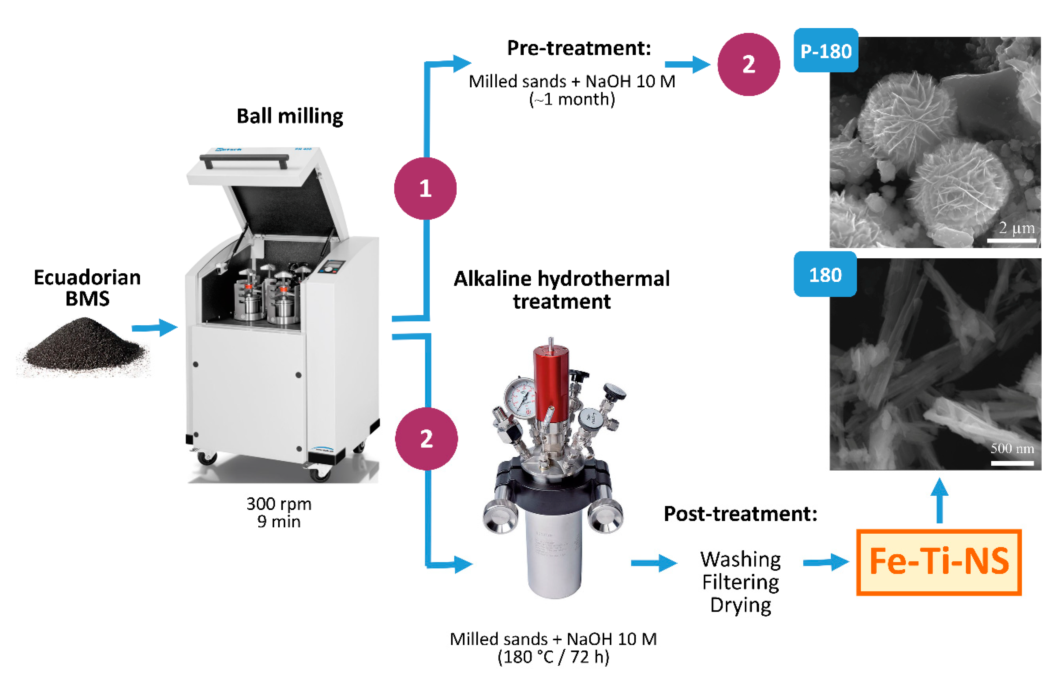
2.1. Synthesis of Fe-Ti-NS from Ecuadorian BMS
2.2. Characterization of Fe-Ti-NS
3. Results
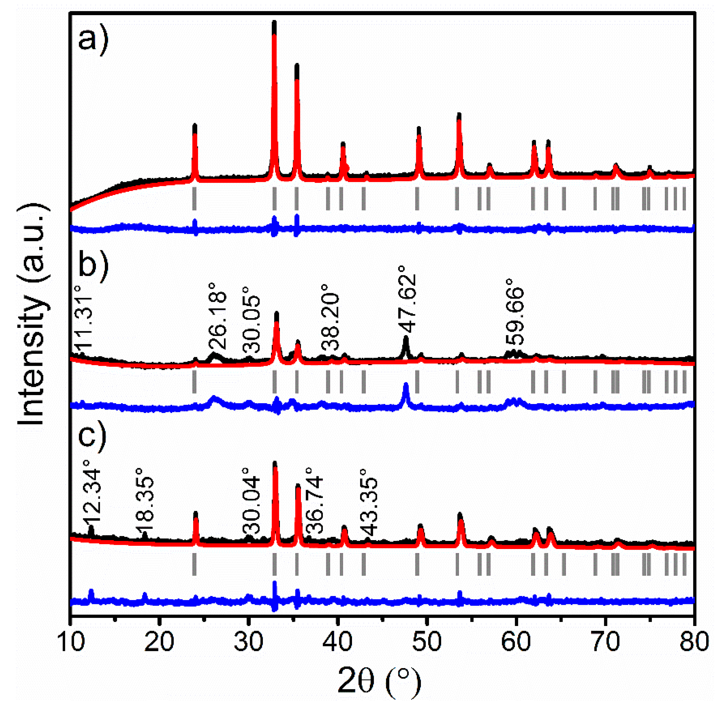
3.1. XRPD Analysis
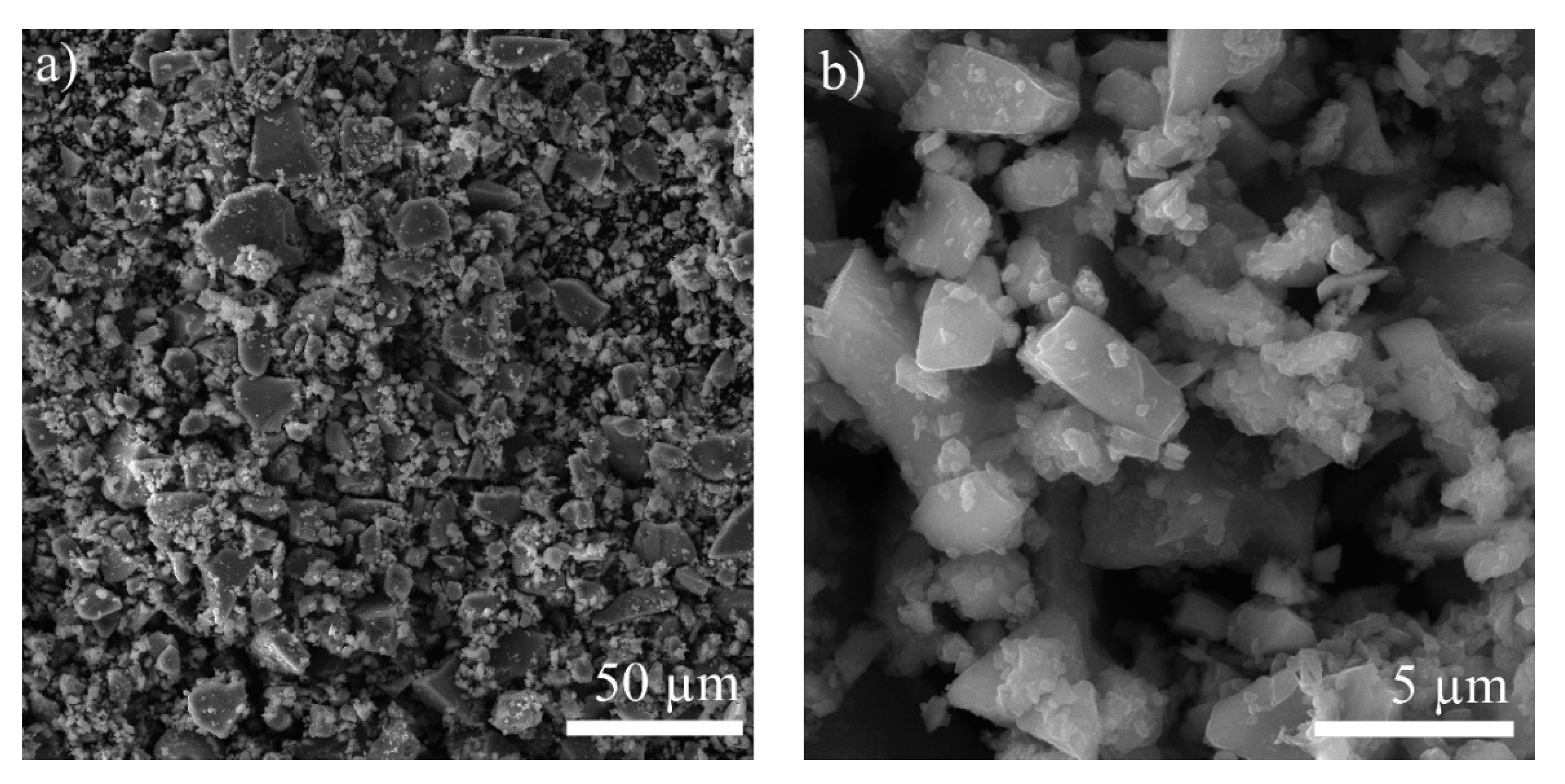
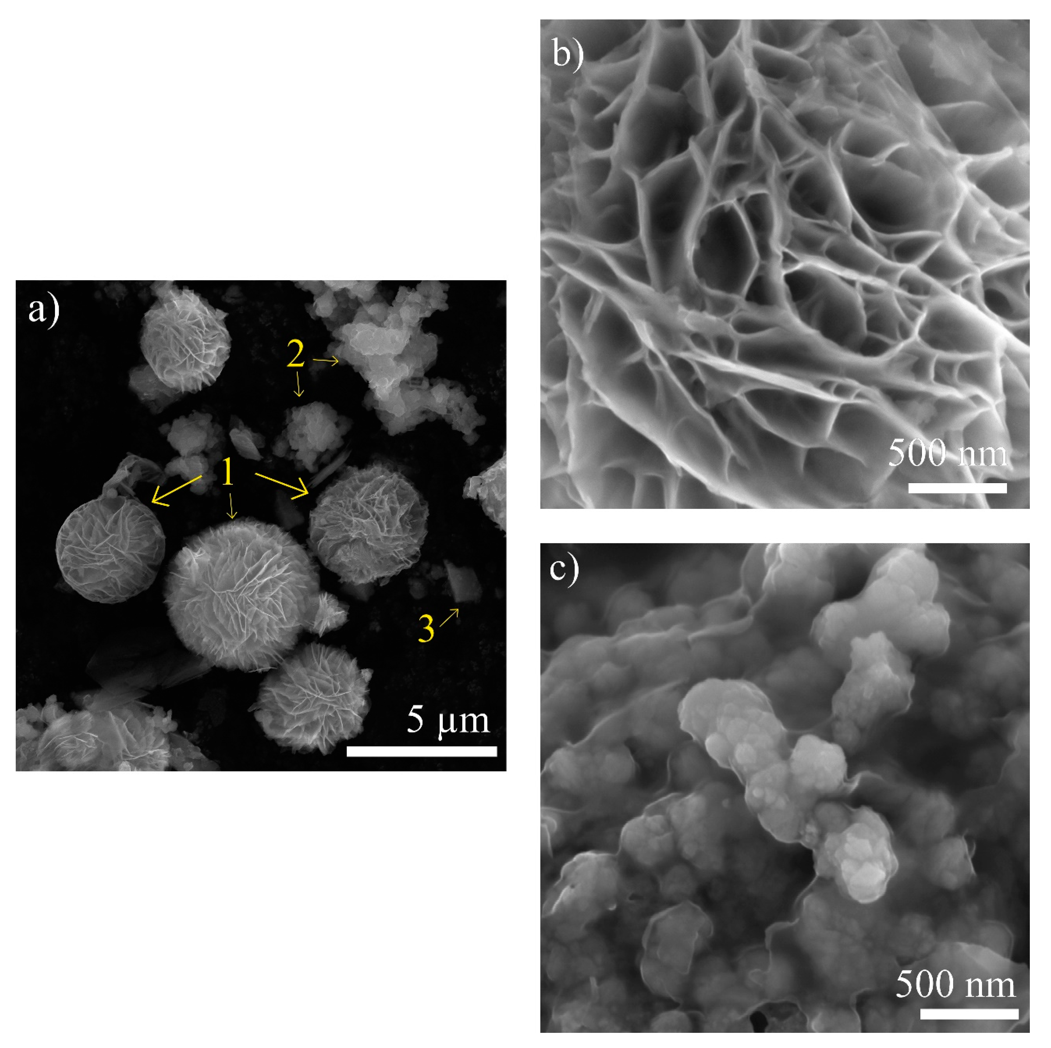
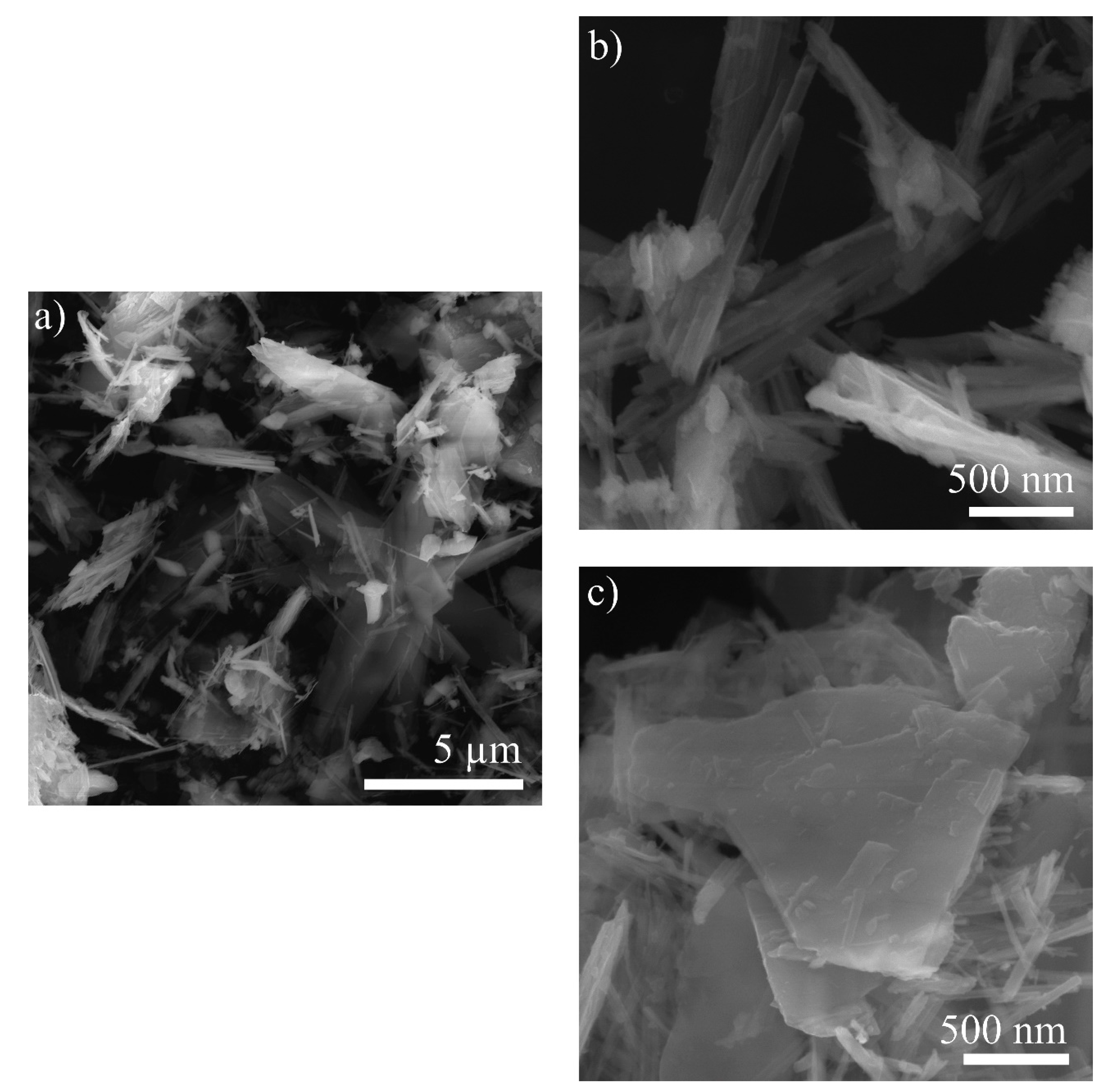
3.2. SEM-EDS Analysis
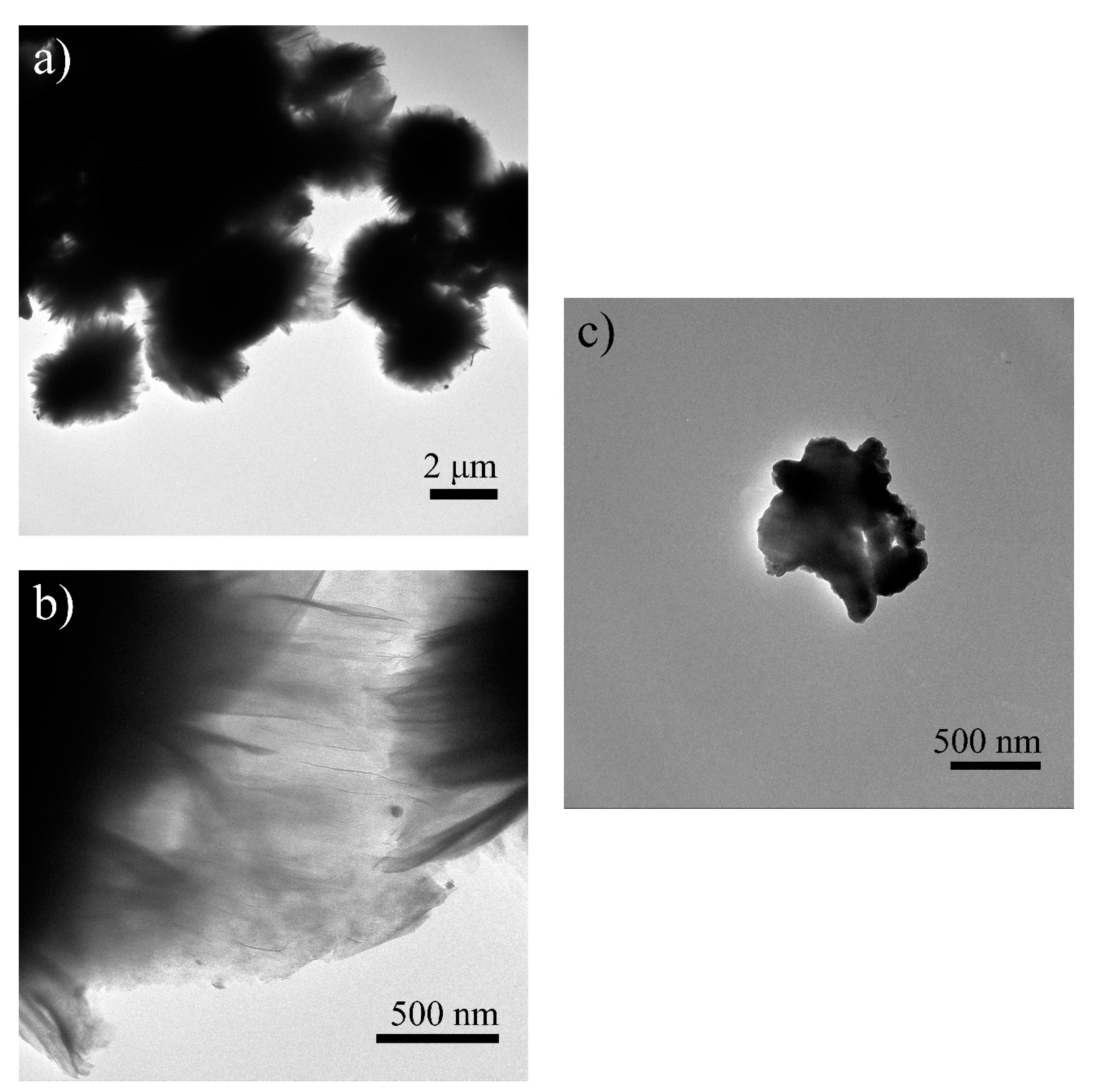
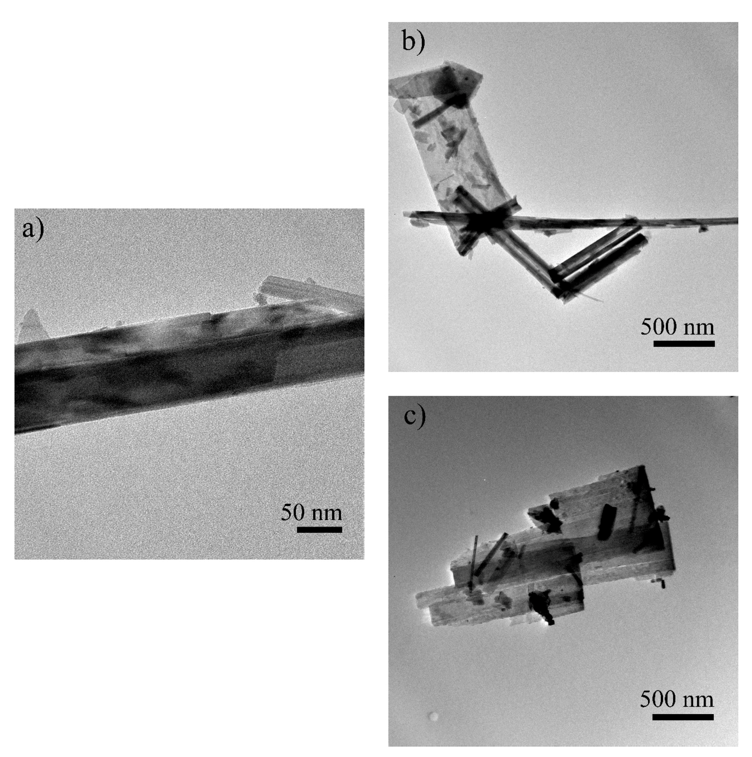
3.3. TEM Analysis
3.4. Gas Sorption Analysis
4. Discussion
5. Conclusions
Supplementary Materials
Author Contributions
Funding
Data Availability Statement
Acknowledgments
Conflicts of Interest
References
- Hou, B.; Keeling, J.; Van Gosen, B.S. Geological and Exploration Models of Beach Placer Deposits, Integrated from Case-Studies of Southern Australia. Ore Geol. Rev. 2017, 80, 437–459. [Google Scholar] [CrossRef]
- Da Silva, M.A.M. Provenance of heavy minerals in beach sands, southeastern Brazil: From Rio Grande to Chui (Rio Grande do Sul State). Sediment. Geol. 1979, 24, 133–148. [Google Scholar] [CrossRef]
- Abdel-Karim, A.A.M.; Zaid, S.M.; Moustafa, M.I.; Barakat, M.G. Mineralogy, chemistry and radioactivity of the heavy minerals in the black sands, along the northern coast of Egypt. J. Afr. Earth Sci. 2016, 123, 10–20. [Google Scholar] [CrossRef]
- Sundararajan, M.; Bhat, K.H.; Velusamy, S.; Babu, N.; Janaki, M.E.K.; Sasibhooshanan, S.; Das, P.N.M. Characterization of Ilmenite from Kerala Coastline, India: Implications in the Production of Synthetic Rutile. J. Miner. Mater. Charact. Eng. 2009, 8, 427–438. [Google Scholar] [CrossRef]
- Lagos, K.J.; Marinkovic, B.A.; Dosen, A.; Guamán, M.V.; Guerrero, V.H.; Pardo, E.; Pontón, P.I. Data on phase and chemical compositions of black sands from “El Ostional” beach situated in Mompiche, Ecuador. Data Br. 2020, 32, 106214. [Google Scholar] [CrossRef]
- Gázquez, M.J.; Bolívar, J.P.; Garcia-Tenorio, R.; Vaca, F. A Review of the Production Cycle of Titanium Dioxide Pigment. Mater. Sci. Appl. 2014, 5, 441–458. [Google Scholar] [CrossRef]
- Fray, D.J. Novel methods for the production of titanium. Int. Mater. Rev. 2008, 53, 317–325. [Google Scholar] [CrossRef]
- Muwanguzi, A.J.B.; Karasev, A.V.; Byaruhanga, J.K.; Jönsson, P.G. Characterisation of the Physical and Metallurgical Properties of Natural Iron Ore for Iron Production. ISRN Mater. Sci. 2012, 2012, 1–9. [Google Scholar] [CrossRef]
- Yousef, L.A. Uranium Adsorption Using Iron-Titanium Mixed Oxides Separated from Ilmenite Mineral, Black Sands, Rosetta, Egypt. Arab J. Nucl. Sci. Appl. 2017, 50, 43–57. [Google Scholar]
- Cañas-Martínez, D.M.; Gauthier, G.H.; Pedraza-Avella, J.A. Photo-oxidative and photo-reductive capabilities of ilmenite-rich black sand concentrates using methyl orange as a probe molecule. Photochem. Photobiol. Sci. 2019, 18, 912–919. [Google Scholar] [CrossRef]
- Yu, J.; Chen, Y.; Glushenkov, A.M. Titanium Oxide Nanorods Extracted from Ilmenite Sands. Cryst. Growth Des. 2009, 9, 1240–1244. [Google Scholar] [CrossRef]
- Jardim, P.M.; Mancic, L.; Marinkovic, B.A.; Milosevic, O.; Rizzo, F. Nax−yHyTi2−xFexO4·nH2O nanosheets with lepidocrocite-like layered structure synthesized by hydrothermal treatment of ilmenite sand. Cent. Eur. J. Chem. 2011, 9, 415–421. [Google Scholar] [CrossRef]
- Marinkovic, B.A.; Mancic, L.; Jardim, P.M.; Milosevic, O.; Rizzo, F. Hydrothermal synthesis of NaxFexTi2-xO4 from natural ilmenite sand: A CaFe2O4 structure type compound. Solid State Commun. 2008, 145, 346–350. [Google Scholar] [CrossRef]
- Costa, A.M.L.M.; Marinkovic, B.A.; Suguihiro, N.M.; Smith, D.J.; Da Costa, M.E.H.M.; Paciornik, S. Fe-doped nanostructured titanates synthesized in a single step route. Mater. Charact. 2015, 99, 150–159. [Google Scholar] [CrossRef]
- Sangaiya, P.; Jayaprakash, R. A Review on Iron Oxide Nanoparticles and Their Biomedical Applications. J. Supercond. Nov. Magn. 2018, 31, 3397–3413. [Google Scholar] [CrossRef]
- Sharma, G.; Kumar, A.; Sharma, S.; Naushad, M.; Prakash Dwivedi, R.; ALOthman, Z.A.; Mola, G.T. Novel development of nanoparticles to bimetallic nanoparticles and their composites: A review. J. King Saud Univ. Sci. 2019, 31, 257–269. [Google Scholar] [CrossRef]
- Haider, A.J.; Jameel, Z.N.; Al-Hussaini, I.H.M. Review on: Titanium Dioxide Applications. Energy Procedia 2019, 157, 17–29. [Google Scholar] [CrossRef]
- Nasirian, M.; Lin, Y.P.; Bustillo-Lecompte, C.F.; Mehrvar, M. Enhancement of photocatalytic activity of titanium dioxide using non-metal doping methods under visible light: A review. Int. J. Environ. Sci. Technol. 2018, 15, 2009–2032. [Google Scholar] [CrossRef]
- Çeşmeli, S.; Biray Avci, C. Application of titanium dioxide (TiO2) nanoparticles in cancer therapies. J. Drug Target. 2019, 27, 762–766. [Google Scholar] [CrossRef]
- Bhateria, R.; Singh, R. A review on nanotechnological application of magnetic iron oxides for heavy metal removal. J. Water Process Eng. 2019, 31, 100845. [Google Scholar] [CrossRef]
- Nasr, M.; Eid, C.; Habchi, R.; Miele, P.; Bechelany, M. Recent Progress on Titanium Dioxide Nanomaterials for Photocatalytic Applications. ChemSusChem 2018, 11, 3023–3047. [Google Scholar] [CrossRef]
- Simpraditpan, A.; Wirunmongkol, T.; Pavasupree, S.; Pecharapa, W. Simple hydrothermal preparation of nanofibers from a natural ilmenite mineral. Ceram. Int. 2013, 39, 2497–2502. [Google Scholar] [CrossRef]
- Simpraditpan, A.; Wirunmongkol, S.; Pavasupree, S.; Pecharapa, W. Preparation and Characterization of Titanate Nanofibers from Low-Cost Natural Ilmenite Sand. In Proceedings of the 10th Eco-Energy and Materials Science and Engineering Symposium, Muang, Ubon-ratchathani, Thailand, 5–8 December 2012. [Google Scholar]
- Kotova, O.B.; Ponaryadov, A.V.; Gömze, L.A. Hydrothermal synthesis of TiO2 nanotubes from concentrate of titanium ore of Pizhemskoe deposit (Russia). Vestn. Inst. Geol. Komi Sci. Cent. Ural Branch RAS 2016, 1, 34–36. [Google Scholar] [CrossRef]
- Suzuki, Y.; Pavasupree, S.; Yoshikawa, S.; Kawahata, R. Natural rutile-derived titanate nanofibers prepared by direct hydrothermal processing. J. Mater. Res. 2005, 20, 1063–1070. [Google Scholar] [CrossRef]
- Suzuki, Y.; Pavasupree, S.; Yoshikawa, S.; Kawahata, R. Direct Hydrothermal Processing of Long Titanate Nanofibers from Natural Rutile. Key Eng. Mater. 2006, 317–318, 243–246. [Google Scholar] [CrossRef]
- Pavasupree, S.; Suzuki, Y.; Yoshikawa, S.; Kawahata, R. Synthesis of titanate, TiO2 (B), and anatase TiO2 nanofibers from natural rutile sand. J. Solid State Chem. 2005, 178, 3110–3116. [Google Scholar] [CrossRef]
- Mancic, L.T.; Marinkovic, B.A.; Jardim, P.M.; Milosevic, O.B.; Rizzo, F. Precursor Particle Size as the Key Parameter for Isothermal Tuning of Morphology from Nanofibers to Nanotubes in the Na2−xHxTinO2n+1 System through Hydrothermal Alkali Treatment of Rutile Mineral Sand. Cryst. Growth Des. 2009, 9, 2152–2158. [Google Scholar] [CrossRef]
- Kawahata, R.; Suzuki, Y. Mineral sand as a low cost source of nanomaterials. Key Eng. Mater. 2006, 317–318, 739–742. [Google Scholar] [CrossRef]
- Tao, T.; Glushenkov, A.M.; Liu, H.; Liu, Z.; Dai, X.J.; Chen, H.; Ringer, S.P.; Chen, Y. Ilmenite FeTiO3 nanoflowers and their pseudocapacitance. J. Phys. Chem. C 2011, 115, 17297–17302. [Google Scholar] [CrossRef]
- Ministerio de Minería del Ecuador. Plan Nacional de Desarrollo Del Sector Minero. Available online: http://historico.mineria.gob.ec/plan-nacional-de-desarrollo-del-sector-minero (accessed on 8 December 2020).
- Marinkovic, B.A.; Pontón, P.I.; Resende, J.M.; Letichevsky, S.; Habran, M.; Viol, J.B.; Pandoli, O.; Mancic, L. Lepidocrocite-like ferrititanate nanosheets and their full exfoliation with quaternary ammonium compounds. Mater. Des. 2015, 85, 197–204. [Google Scholar] [CrossRef]
- Ginley, D.S.; Butler, M.A. The photoelectrolysis of water using iron titanate anodes. J. Appl. Phys. 1977, 48, 2019–2021. [Google Scholar] [CrossRef]
- Brown, N.E.; Navrotsky, A.; Nord, G.L.; Banerjee, S.K. Hematite-ilmenite (Fe2O3-FeTiO3) solid solutions: Determinations of Fe-Ti order from magnetic properties. Am. Mineral. 1993, 78, 941–951. [Google Scholar]
- Morgado, E.; De Abreu, M.A.S.; Pravia, O.R.C.; Marinkovic, B.A.; Jardim, P.M.; Rizzo, F.C.; Araújo, A.S. A study on the structure and thermal stability of titanate nanotubes as a function of sodium content. Solid State Sci. 2006, 8, 888–900. [Google Scholar] [CrossRef]
- Walter, D.; Berges, M.; Kreyling, W.; Korinth, G.; Drexler, H.; Landsiedel, R.; Heinrich, U.; Hartwig, A.; Schins, R.; Pauluhn, J.; et al. Nanomaterials; Wiley-VCH: Bonn, Germany, 2013; Chapter 1; pp. 19–24. [Google Scholar]
- Tada, H.; Jin, Q.; Nishijima, H.; Yamamoto, H.; Fujishima, M.; Okuoka, S.; Hattori, T.; Sumida, Y.; Kobayashi, H. Titanium(IV) Dioxide Surface-Modified with Iron Oxide as a Visible Light Photocatalyst. Angew. Chem. 2011, 123, 3563–3567. [Google Scholar] [CrossRef]
- Chen, Y.H.; Li, F.A. Kinetic study on removal of copper(II) using goethite and hematite nano-photocatalysts. J. Colloid Interface Sci. 2010, 347, 277–281. [Google Scholar] [CrossRef]
- Müller, M.; Villalba, J.C.; Mariani, F.Q.; Dalpasquale, M.; Lemos, M.Z.; Huila, M.F.G.; Anaissi, F.J. Synthesis and characterization of iron oxide pigments through the method of the forced hydrolysis of inorganic salts. Dye Pigments 2015, 120, 271–278. [Google Scholar] [CrossRef]
- El-Sayed Abdo, A.; El-Sarraf, M.A.; Gaber, F.A. Utilization of ilmenite/epoxy composite for neutrons and gamma rays attenuation. Ann. Nucl. Energy 2003, 30, 175–187. [Google Scholar] [CrossRef]
- Longo, A.; Wang, X.L.; Ruotolo, A.; Peluso, A.; Carotenuto, G.; Lortz, R. Effect of the polymeric matrix on the structural and magnetic properties of hematite/polymer composites. J. Nanopart. Res. 2012, 14, 1314. [Google Scholar] [CrossRef]
- Walter, D. Characterization of synthetic hydrous hematite pigments. Thermochim. Acta 2006, 445, 195–199. [Google Scholar] [CrossRef]
- Abdou, M.I.; Ayad, M.I.; Diab, A.S.M.; Hassan, I.A.; Fadl, A.M. Studying the corrosion mitigation behavior and chemical durability of FeTiO3/melamine formaldehyde epoxy composite coating for steel internal lining applications. Prog. Org. Coat. 2019, 133, 325–339. [Google Scholar] [CrossRef]
- Abdou, M.I.; Ayad, M.I.; Diab, A.S.M.; Hassan, I.A.; Fadl, A.M. Influence of surface modified ilmenite/melamine formaldehyde composite on the anti-corrosion and mechanical properties of conventional polyamine cured epoxy for internal coating of gas and oil transmission pipelines. Prog. Org. Coat. 2017, 113, 1–14. [Google Scholar] [CrossRef]
- Tjong, S.C. Structural and mechanical properties of polymer nanocomposites. Mater. Sci. Eng. R 2006, 53, 73–197. [Google Scholar] [CrossRef]


| Precursor | Synthesis Conditions | Post-Treatment | Synthesized Product | Ref. | |||||
|---|---|---|---|---|---|---|---|---|---|
| Origin | Crystalline Phase(s) | Fe/Ti Mass Ratio | Pre-Treatment | Chemical Formula | Crystalline Phase(s) | Morphology | |||
| Thailand | Rutile | 0.42 | None | NaOH 10 M 120 °C 72 h (Stirring) | Washing with HCl and distilled water; drying with hot air | H2TixO2x+1 | Trititanate (H2Ti3O7) | Layered nanostructures: Fe-doped titanate nanofibers (diameters: 20–90 nm; length: 2–7 mm) | [22,23] |
| Russia | Rutile and quartz (non-magnetic fraction) | 0.03 | Magnetic separation and milling of non-magnetic fraction up to 20–40 µm | NaOH 10 M 110 °C 24 h | Washing with distilled water and HCl; drying at 90 °C for 12 h | NaxH2−xTi3O7 (Fe/Ti ratio = 0.09) and SiO2 | Trititanate and quartz | Layered nanostructures: Titanate nanotubes (diameter: 70–100 nm; length: up to 4500 nm) | [24] |
| Australia | Rutile and anatase | 0.01 | None | NaOH 10 M 150 °C 120 h (Stirring) | Washing with HCl and distilled water; drying RT | Na0.4H1.6Ti3 with (Na,H)2Ti6O13 type-defect | Trititanate with hexatitanate-type defect | Layered nanostructures: Titanate nanofibers (diameter: 20–50 nm; length: 10–500 µm) | [25,26] |
| Australia | Rutile and anatase | Not specified | None | NaOH 10 M 150 °C 72 h (Stirring) | Washing with HCl and distilled water; freeze drying | H2TixO2x+1 (H2Ti3O7) | Tritanate | Solid nanofibers (diameter: 20–50 nm; length: hundreds of nanometers) | [27] |
| Australia | Ilmenite | 1.20 | Magnetic ball milling | NaOH 2 M 120 °C 2 h (Stirring) | Washing; drying at 90 °C for 4 h | FeTiO3 | Ilmenite | Nanoflowers (diameter: 1–2 µm; petal thickness: 5–20 nm; petal wide: 100–200 nm) | [30] |
| Brazil | Ilmenite, rutile, pseudorutile, and quartz | 0.81 | Milling up to 0.25 µm | NaOH 10 M 130 °C 70 h (Stirring) | Washing with distilled water; drying at 80 °C for 5 h | Nax−yHyTi2−x FexO4·nH2O and Fe2O3 | Lepidocrocite-like titanate and small amounts of hematite | Layered nanostructures: Leaf-like titanate nanosheets (thickness <30 nm; length <1 µm), and hematite nanoparticles | [12] |
| Brazil | Ilmenite, rutile | Not specified | Not specified | NaOH 10 M 190 °C 70 h (Stirring) | Washing with distilled water; drying at 80 °C for 5 h | NaxFexTi2−xO4 (92%) Fe3O4 (7%) x = 0.76–0.79 (Fe/Ti ratio = 0.71) | CaFe2O4 structure type group and magnetite | Submicron to micron crystals with well-defined facets | [13] |
| Brazil | Pseudorutile, ilmenite, hematite, rutile and quartz | 0.90 | Milling up to 0.8 µm | NaOH 10 M 110, 130, 150, 170, 190 °C 70 h (Stirring) | Washing with distilled water; drying at 80 °C for 5 h | Nax−yHyTi2−xFexO4 and Fe2O3 | Lepidocrocite-like titanate and hematite | Layered nanostructures: Fe-doped titanate nanosheets (110, 130 and 150 °C) Titanate nanoribbons (Fe-free) + Fe-doped titanate nanosheets and hematite nanoparticles (170 and 190 °C) | [14] |
| Brazil | Not specified | Not specified | Milling | NaOH 10 M 130 °C 70 h (Stirring) | Washing with distilled water and HCl; drying at 80 °C for 5 h | HxTi2−xFexO4·nH2O | Lepidocrocite-like titanate | Layered nanostructures: Fe-doped titanate nanosheets, composed of ~10 stacked layers (thickness of each layer <1 nm) | [32] |
| Brazil | Rutile, anatase, and zircon | 0.02 | Milling 0, 30, 60, 90 min | NaOH 10 M 140 °C 70 h (Stirring) | Washing with distilled water; drying at 80 °C for 5 h | Na2−xHxTinO2n+1 (Fe/Ti ratio = 0.01) | Trititanate (60 min); pentatitanate and trititanate (0, 30, 90 min) | Layered nanostructures: Nanotubes (60 min) (diameter <10 nm; length: several hundreds of nm) Nanofibers (0, 30, 90 min) (diameter: 20–50 nm; length: several hundreds of nm) | [28] |
| Sample | Unit-Cell Volume (Å3) | Mean Crystallite Size (nm) |
|---|---|---|
| Milled BMS | 308.51(7) | 58.80(0.9) |
| P-180 | 305.76(7) | 25.03(1.9) |
| 180 | 306.47(7) | 101.01(7.6) |
| Samples | Ilmenite Molar Fraction (xilm) | Hematite Molar Fraction (xhem) | Estimated Fe/Ti Mass Ratio |
|---|---|---|---|
| Milled BMS | 0.53 | 0.47 | 3.2 |
| P-180 | 0.34 | 0.66 | 5.7 |
| 180 | 0.40 | 0.60 | 4.7 |
| Samples | Morphology | Fe/Ti Mass Ratio |
|---|---|---|
| Milled BMS | Micron-size agglomerates | 3.1 |
| P-180 | Flower-like microparticles | 4.3 |
| Aggregates of nanoparticles | 0.7 | |
| Remaining precursor | 2.7 | |
| 180 | Nanobelts | 1.6 |
| Plate-like particles | 4.6 | |
| Remaining precursor | 3.0 |
Publisher’s Note: MDPI stays neutral with regard to jurisdictional claims in published maps and institutional affiliations. |
© 2021 by the authors. Licensee MDPI, Basel, Switzerland. This article is an open access article distributed under the terms and conditions of the Creative Commons Attribution (CC BY) license (http://creativecommons.org/licenses/by/4.0/).
Share and Cite
Lagos, K.J.; Marinkovic, B.A.; Debut, A.; Vizuete, K.; Guerrero, V.H.; Pardo, E.; Pontón, P.I. Towards Iron-Titanium Oxide Nanostructures from Ecuadorian Black Mineral Sands. Minerals 2021, 11, 122. https://doi.org/10.3390/min11020122
Lagos KJ, Marinkovic BA, Debut A, Vizuete K, Guerrero VH, Pardo E, Pontón PI. Towards Iron-Titanium Oxide Nanostructures from Ecuadorian Black Mineral Sands. Minerals. 2021; 11(2):122. https://doi.org/10.3390/min11020122
Chicago/Turabian StyleLagos, Karina J., Bojan A. Marinkovic, Alexis Debut, Karla Vizuete, Víctor H. Guerrero, Emilio Pardo, and Patricia I. Pontón. 2021. "Towards Iron-Titanium Oxide Nanostructures from Ecuadorian Black Mineral Sands" Minerals 11, no. 2: 122. https://doi.org/10.3390/min11020122
APA StyleLagos, K. J., Marinkovic, B. A., Debut, A., Vizuete, K., Guerrero, V. H., Pardo, E., & Pontón, P. I. (2021). Towards Iron-Titanium Oxide Nanostructures from Ecuadorian Black Mineral Sands. Minerals, 11(2), 122. https://doi.org/10.3390/min11020122