Article

Novel Strategy for Improvement of the Bioleaching Efficiency of Acidithiobacillus ferrooxidans Based on the AfeI/R Quorum Sensing System

State Key Laboratory of Microbial Technology, Shandong University, No. 72 Binhai Road, Qingdao 266237, China
*
Authors to whom correspondence should be addressed.
Minerals 2020, 10(3), 222; https://doi.org/10.3390/min10030222
Submission received: 27 January 2020 / Revised: 16 February 2020 / Accepted: 26 February 2020 / Published: 29 February 2020
(This article belongs to the Special Issue Microorganisms–Minerals Interactions in Aquatic Environments)

Abstract

:
Acidithiobacillus ferrooxidans is an acidophilic and chemolithotrophic sulfur- and iron-oxidizing bacterium that has been widely used in the bioleaching process for extracting metals. Extracellular polymeric substances (EPS) are essential for bacteria-ore interactions, and the regulation of EPS synthesis could be an important way of influencing the efficiency of the bioleaching process. Therefore, exploring and utilizing the regulatory pathways of EPS synthesis to improve the bacterial bioleaching capability have posed a challenge in the study and application of bioleaching bacteria. Here, several engineering strains were constructed using genetic manipulation methods. And we revealed the regulatory function of the AfeI/R quorum sensing (QS) system in EPS synthesis and biofilm formation of A. ferrooxidans, and the AfeI/R-mediated EPS synthesis could influence bacteria-substrate interactions and the efficiency of bioleaching. Finally, an AfeI/R-mediated bioleaching model was proposed to illustrate the role of QS system in this process. This study provided new insights into and clues for developing highly efficient bioleaching bacteria and modulating the bioleaching process.

1. Introduction

Bioleaching refers to the direct or indirect actions dissolution of the metal in the ores by microorganisms, and this technology has been rapidly developed and widely used in the production of nonferrous metals (gold, copper, uranium, etc.) over the past few decades [1,2,3]. The use of microorganisms in ore processing has some distinct advantages compared to traditional physicochemical methods [4,5,6]. On the one hand, bioleaching reduces production costs, because it does not require a large amount of energy during the roasting or smelting process [4,5,6]. On the other hand, bioleaching is significantly advantageous in extracting metals from certain low-grade ores [4,5,6]. Moreover, the application of bioleaching technology has been extended from biomining to the treatment of metal-containing wastes, such as sewage sludge, mine tailings, and printed circuit boards [7,8,9]. Therefore, bioleaching has become an efficient and promising green biotechnology for mining mineral resources and alleviating environmental pollution. However, due to the insufficient understanding of bioleaching bacteria, the conditions of most bioleaching plants are far from optimal, so the study of the physiological metabolism of bioleaching bacteria is necessary [10,11].
Acidithiobacillus ferrooxidans (A. ferrooxidans) is an acidophilic and chemolithotrophic sulfur- and iron-oxidizing bacterium [4,12,13]. A. ferrooxidans is a prevalent and dominant species in acid mine environments [11,12]. It has been extensively studied and widely used in the bioleaching industry for the extraction of nonferrous metals from sulfide minerals and heavy metals from waste materials, as well as in the biodesulfurization technique to treat sulfur containing gas and coals [2,14,15]. The wide distribution and application of A. ferrooxidans could be attributable to its versatile metabolic characteristics. A. ferrooxidans can obtain energy from the metabolism of inorganic sulfur, iron and hydrogen [12,13]. Moreover, A. ferrooxidans has developed a distinguished capability to grow on sulfide minerals, making it a model bacterium for studying bacteria-ore interactions and bioleaching mechanisms [16,17].
The process of bioleaching requires the consortium of microorganisms and the formation of the reaction space “surface conditioning layer” between the ores and the bacteria [18,19,20]. A. ferrooxidans can secrete extracellular polymeric substances (EPS) and form biofilm on the surface of ores [21,22,23]. Thus, EPS synthesis and biofilm formation are considered to be essential for bacteria-ore interaction, and they determine the rate of mineral dissolution in natural environments and the efficiency of industrial operations [3,22,23,24,25]. However, the co-growth and aggregation of A. ferrooxidans to form biofilms in extreme environments and thus participate in the complex bioleaching processes and to adapt to the environments is unclear.
Quorum sensing (QS) is a ubiquitous phenomenon that enables bacteria to establish cell-cell communication by producing, releasing, and detecting signal molecules (also known as auto-inductor (AI) molecule) [26,27]. Quorum sensing (QS) has been found to play a role in the regulation of EPS synthesis and biofilm formation in many bacteria [28,29]. A LuxI/R-like QS system (AfeI/R, namely qs-I), encoded by the afeI-orf3-afeR operon, was found in A. ferrooxidans [30,31]. The afeI gene encodes N-acyl homoserine lactones (acyl-HSLs) synthetase and AfeR is a regulator that can recognize and bind specific acyl-HSLs to regulate gene expression [31,32]. The function of the orf gene remains unknown. In addition, a second acyl-HSLs production system has been identified but this QS system operon (glyS-glyQ-gph-act, namely qs-II) lacked a signal receptor making its effectiveness tentative [33]. Nine different acyl-HSLs with acyl chains ranging from 8 to 16 carbons with diverse C-3 substituents (oxo or hydroxy) and different length were detected in A. ferrooxidans cultures with different energy substrates [31]. Exogenous addition of acyl-HSLs could stimulate the EPS synthesis, biofilm formation, and cell attachment, indicating the significance of QS regulation in the interaction of bacteria and substrates [10,34,35,36]. However, the regulatory roles of the QS system in EPS and biofilm formation as well as bioleaching efficacy remain unclear due to the lack of mutation-gene function studies.
Gene deletion and overexpression have been proved to be an effective and direct means of studying the genes function and constructing engineering strains. However, the high genetic barrier for the molecular manipulation of A. ferrooxidans has become an important obstacle in understanding the role of the QS system in regulating bacteria-substrate interaction and the bioleaching process [37]. In this study, we focused on the genetic manipulation of A. ferrooxidans, and successfully developed the afeI/R deletion and overexpression strains. The regulation effect of AfeI/R on the EPS synthesis and biofilm formation was detected using sulfur-coupon assays, and the bioerosion abilities of different strains to elemental sulfur was monitored using the scanning electron microscope (SEM). Further, the influence of AfeI/R on the growth and genes transcription of A. ferrooxidans was investigated. Bioleaching experiments of different strains to pyrites were carried out to evaluate the role of AfeI/R in regulating the leaching efficiency of A. ferrooxidans. Finally, an AfeI/R-regulating model of A. ferrooxidans was proposed to illustrate the important role of AfeI/R in the modulation of the bioleaching process.

2. Material and Methods

2.1. Bacteria and Growth Conditions

The bacteria and plasmids used in this study were listed in Table 1. Cells of A. ferrooxidans were grown in 9K media with 0.8% (w/v) sulfur at 30 °C on a rotary shaker of 180 rpm, and the solid media used for A. ferrooxidans growth was Starkey-Na2S2O3 media [38]. E. coli were cultured in LB media and shaken at 30 °C and 180 rpm [39]. The concentration of streptomycin (Sm) and kanamycin (km) used was 100 μg/mL for E. coli and 200 μg/mL for A. ferrooxidans. All cultures were initiated with 4 × 10⁶ cells unless otherwise noted. The cell growth was detected every two days.

2.2. Development of Knockout and Overexpression Strains of qs-I

The principle of markerless gene deletion was based on the principle of homologous recombination and the method was described previously [38]. Firstly, a suicide plasmid pSDUDI-HFqs-I containing the upstream and downstream homology arms of qs-I gene cluster was developed and identified by sequencing. Then, the plasmid pSDUDI-HFqs-I was conjugated to A. ferrooxidans. After the first homologous recombination, the single crossover strains were selected on Starkey-Na2 S2O3 solid plates and verified by PCR using the primers P2F/R. Then, the plasmid pMSD-I-secI was conjugated to the single crossover strains to stimulate the second homologous recombination by creating double-stranded breaks on the chromosome. △qs-I mutant was verified by PCR using the primers P1F/R, P2F/R, P3F/R and the DNA fragments amplified from the Δqs-I strain using the primer pairs P1F/R was confirmed by sequencing (Sangon Biotech, China). The plasmid pMSD-I-secI in the △qs-I strains was depleted by subculture using the free-antibiotic media. Finally, △qs-I, the markerless genes deletion strains, were obtained. All the primers used are listed in Table S1.
The qs-I operons were amplified and then cloned into the expression vector backbone, pJRD215, to obtain pJRD215-qs-I plasmids. The qs-I overexpression strain WT(pJRD215-qs) were acquired after the pJRD215-qs-I plasmids were transferred into the wild-type strain through conjugation. The WT(pJRD215-qs) strain were screened and identified by PCR using the primers P4F/R. All the primers used are listed in Table S1.

2.3. Identification of SO42− in the Culture Media Through Ion Chromatography

The concentration of SO42− was measured using ion chromatography (ICS-1100AR, DIONEX, Sunnyvale, CA, USA) [43]. Samples for ion chromatography were prepared from liquid culture during bacterial growth. The bacteria and sulfur power were removed by centrifugation at 12,000× g for 5 min at 4 °C, and the supernatants were added to three volumes of HPLC-grade acetonitrile for protein precipitation, and then centrifuged at 12,000× g for 5 min at 4 °C. The supernatants were used for detection. Before ion chromatography, samples were filtered through a 0.22 μm filter. The concentration of SO42− in the samples was obtained based on the peak area of the ion chromatography. All the samples were set with three biological replicates and performed three times.

2.4. Determination of Extracellular Polymeric Substances

Extracellular polymeric substances (EPS) were extracted as described previously [44,45]. Briefly, a variety of reagents were used for the EPS extraction, including TNE buffer (10 mM Tris, 100 mM NaCl 5 mM EDTA), 0.1% SDS (dissolved in the TNE buffer), and 50 mM Tris (pH 7.5). Cells were collected by centrifugation at 12000× g for 5 min at 4 °C and adjusted to the final concentration (OD600 nm = 1). Then, 1 mL of this cell suspension was collected at 12,000× g for 1 min at 4 °C. Afterwards, the pellets were resuspended in 4 mL of ice-cold TNE buffer and centrifuged at 12,000× g for 10 min at 4 °C. 0.1% SDS was added and reacted for 5 min at room temperature. Then, the samples were centrifuged at 12,000× g for 10 min at 4 °C to obtain EPS extracts. Finally, the extracts were washed three times with TNE buffer and dissolved with 50 mM Tris.
The total number of polysaccharides of the EPS was determined using the anthrone-sulfuric method [46]. Briefly, the 300 μL samples were reacted with 900 μL 0.2% anthrone-sulfuric acid for 20 min at 98 °C and then OD625 nm was measured. The protein level of the EPS extracts was determined using the Modified Bradford Protein Assay Kit (Songon Biotech, Shanghai, China) according to the product manual.
All the samples were set with three biological replicates and performed three times.

2.5. Extraction of Total Ribonucleic Acid (RNA) and Real-Time Quantitative PCR (RT-qPCR)

The samples used to extract RNA were cultured until they attained the mid-log phase. All the samples were set with three biological replicates and performed three times. The total amount of RNA extraction was performed according to the instructions outlined in the RNAprep Pure Cell/Bacteria Kit (Tiangen, China). The quality of the extracted RNA was determined using formaldehyde degeneration electrophoresis. Quality-tested RNA was reverse-transcribed into complementary DNA (cDNA) based on the instructions stipulated in the PrimeScriptTM RT Reagent Kit (TaKaRa, China). RT-qPCR was performed using the enzyme SYBRPremix Ex TaqTM (TaKaRa, China) on Roche LightCycler480 (Roche, Chicago, IL, USA). The amount of cDNA used in each reaction system was 200 ng and alaS was used as the reference gene [47]. The 2△△Ct method was used to analyze relative changes in gene expression, where [△Ct = (Ct target − Ct alaS) was applied for each sample, ΔΔCt = ΔCt engineering strain − ΔCt control strain] [48]. The RT-qPCR data are listed in Table 2. All the primers used are listed in Table S1.

2.6. Preparation of Sulfur and Pyrite Coupons and Scanning Electron Microscope Observation

Sulfur coupons were successfully fabricated using the previously-described method [34]. The sulfur powder was heated to melting point and then liquid sulfur was poured and solidified on the cover glass to make sulfur coupons. Pyrite was produced in Guangdong Province, China, and supported by the YunFu Pyrite Company (Yunfu, China). The ore was processed and cut into cubes with a side length of 0.5 mm before use. The prepared sulfur and pyrite coupons were co-cultured with A. ferrooxidans in static culture. Cultures with sulfur coupons (0.8%, w/v) were initiated with 1 × 10⁷ cells, while cultures with pyrite (250%, w/v) were initiated with 5 × 10⁷ cells.
The prepared sulfur and pyrite coupons were co-cultured with A. ferrooxidans for eight days. Three biological replicates were set for all the samples and independent experiments performed three times. Materials (sulfur and pyrite coupons) for scanning electron microscope (SEM) observation were fixed with 2.5% glutaraldehyde for 4 h at 4 °C. Then, the samples were sequentially dehydrated in 30%, 50%, 70%, 80%, 90%, 95%, and 100% ethanol for 15 min each. After drying with CO2 at critical point and sputtering gold, the samples were observed using SEM (Quanta 250 FEG, FEI) [20,49].

2.7. Biofilm Formation on Sulfur Coupons and Pyrite

The prepared sulfur coupons and pyrite were co-cultured with A. ferrooxidans for eight and 15 days, respectively. Three biological replicates were set for all the samples and independent experiments performed three times. Biofilm formation was determined as described previously [50]. Materials (sulfur coupons and pyrite) for the analysis of biofilm were washed five times with water and thoroughly dried in a dry oven. Five milliliters of crystal violet was added to soak for 15 min under a dark condition and then the crystal violet was washed with water and dried. To dissolve the biofilm-bound crystal violet, 1 mL of 30% acetic acid was added and measured at OD595 nm [50].

2.8. Identification of the Metal Elements in the Leachate

The prepared pyrite was initiated with 5 × 10⁷ cells and co-cultured for 15 days in static culture at 30 °C. Cells and pyrite were removed by centrifugation at 12,000× g for 5 min at 4 °C, and the supernatants were filtered through a 0.22 μm filter. Then, the metal elements in the samples were identified by inductively coupled plasma mass spectrometry (ICP-MS) (iCAP, RQ, Thermo, Waltham, MA, USA) [51].

2.9. Statistical Analysis

All experiments were performed three times and three biological replicates were set for all experimental samples. Students’ t test using a GraphPad Prism (version 7.0) was used to conduct a statistical analysis. Additionally, **** indicates P < 0.0001, ***indicates P < 0.001, **indicates P < 0.01, and *indicates P < 0.05.

3. Results

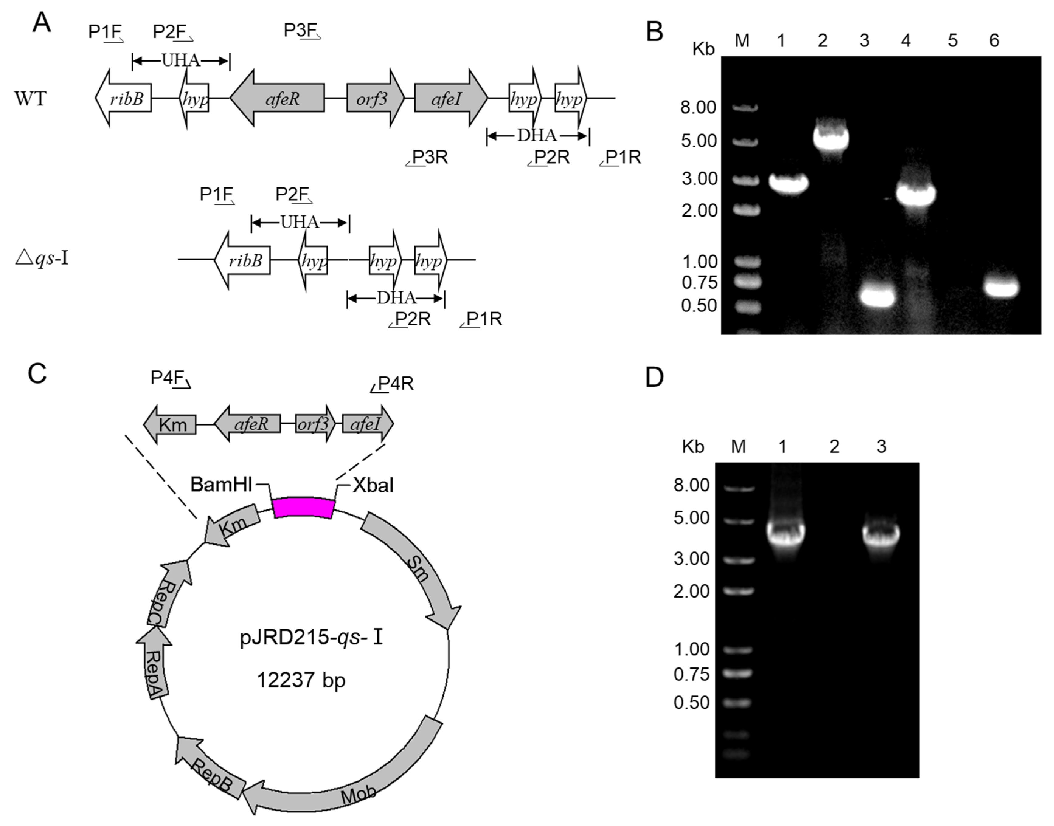
3.1. Development of the qs-I Operon Deletion and Overexpression Strains

To explore the function of the AfeI/R system in A. ferrooxidans, the qs-Ⅰ operon including AFE_1999 (coding the autoinducer synthetase, AfeI), AFE_1998 (coding hypothetical protein) and AFE_1997 (coding receptor protein, AfeR), was selected to generate qs-Ⅰ operon deletion strain and overexpression strain. The markerless deletion of the qs-Ⅰ operon was performed using the principle of homologous recombination as described previously [38]. In order to verify knockout strains, we designed three pairs of primers, specific to the lateral homology arm (P1F/R), the homology arm (P2F/R), and the qs-Ⅰ operon sequence (P3F/R), respectively, as shown in Figure 1A. Because of the lack of qs-Ⅰ operon, different fragments were amplified from Δqs-Ⅰ strain compared to the wild type. As shown in the electrophoresis gel results (Figure 1B), the DNA fragments amplified from the genome of Δqs-Ⅰ were smaller than those amplified from the wide type strain using the primer pairs, P1F/R and P2F/R. Furthermore, no DNA fragment could be amplified from Δqs-Ⅰ using the primers specific to the qs-Ⅰ operon (Figure 1B). The qs-Ⅰ expression plasmid (pJRD215-qs-Ⅰ) and the blank plasmid (pJRD215) were respectively conjugated to wild type strain, generating overexpression strain WT (pJRD215-qs) and control strain WT(pJRD215). The overexpression strains were confirmed by PCR amplification using primers that were specified for the pJRD215-qs-Ⅰ plasmid (Figure 1D). Only strains containing the pJRD215-qs-Ⅰ showed a 3700-bp band (Figure 1D, lane 1 and lane 3), while the wild-type did not exhibit the corresponding size band (Figure 1D, lane 2).

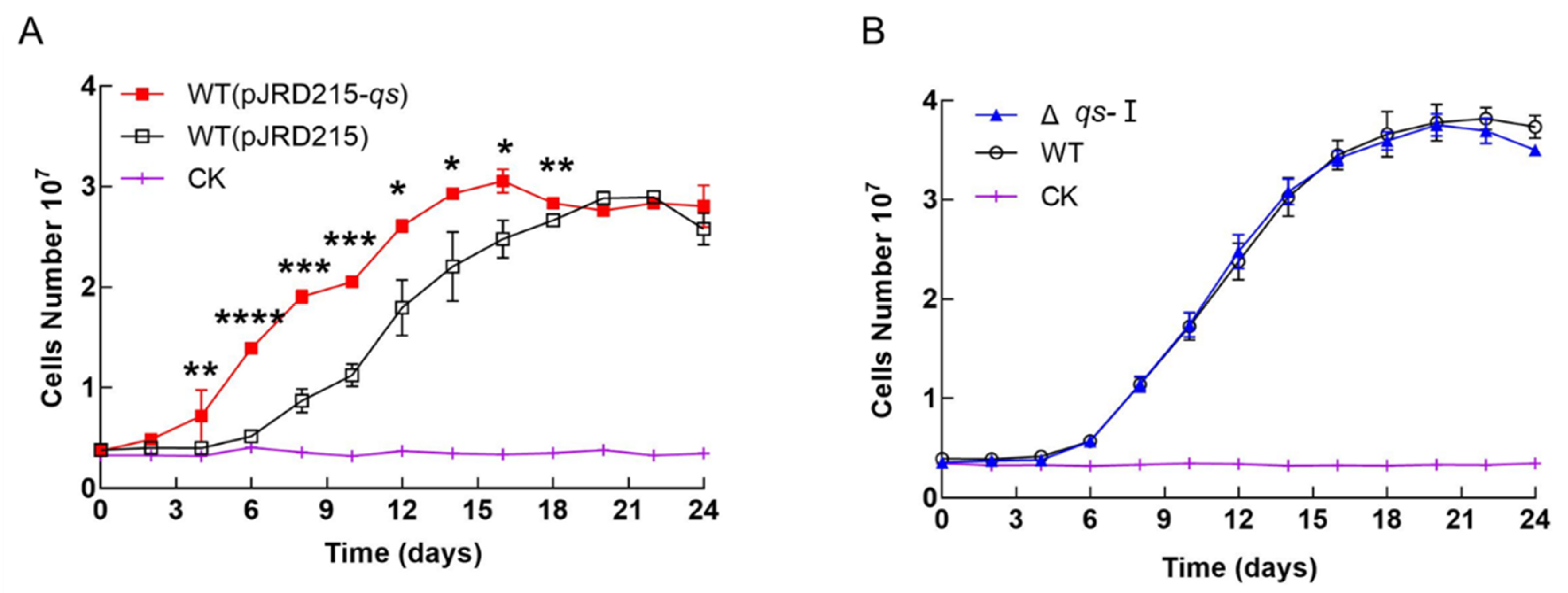
3.2. Effect of qs-I on the Cell Growth of A. ferrooxidans

It is worth noting that the lag period of qs-Ⅰ overexpression strain was shortened, and the growth of qs-Ⅰ overexpression strain was superior to that of the control strain in the exponential growth phase (Figure 2A). However, cell growth of qs-Ⅰ overexpression in the stationary phase alleviates the growth advantage (Figure 2A). In addition, the deletion of the qs-Ⅰ operon did not result in obvious difference in growth during the S0-cultivating process (Figure 2B). Thus, the results indicated that the overexpression of qs-Ⅰ could give the strain an obvious growth advantage in the lag and exponential phases.

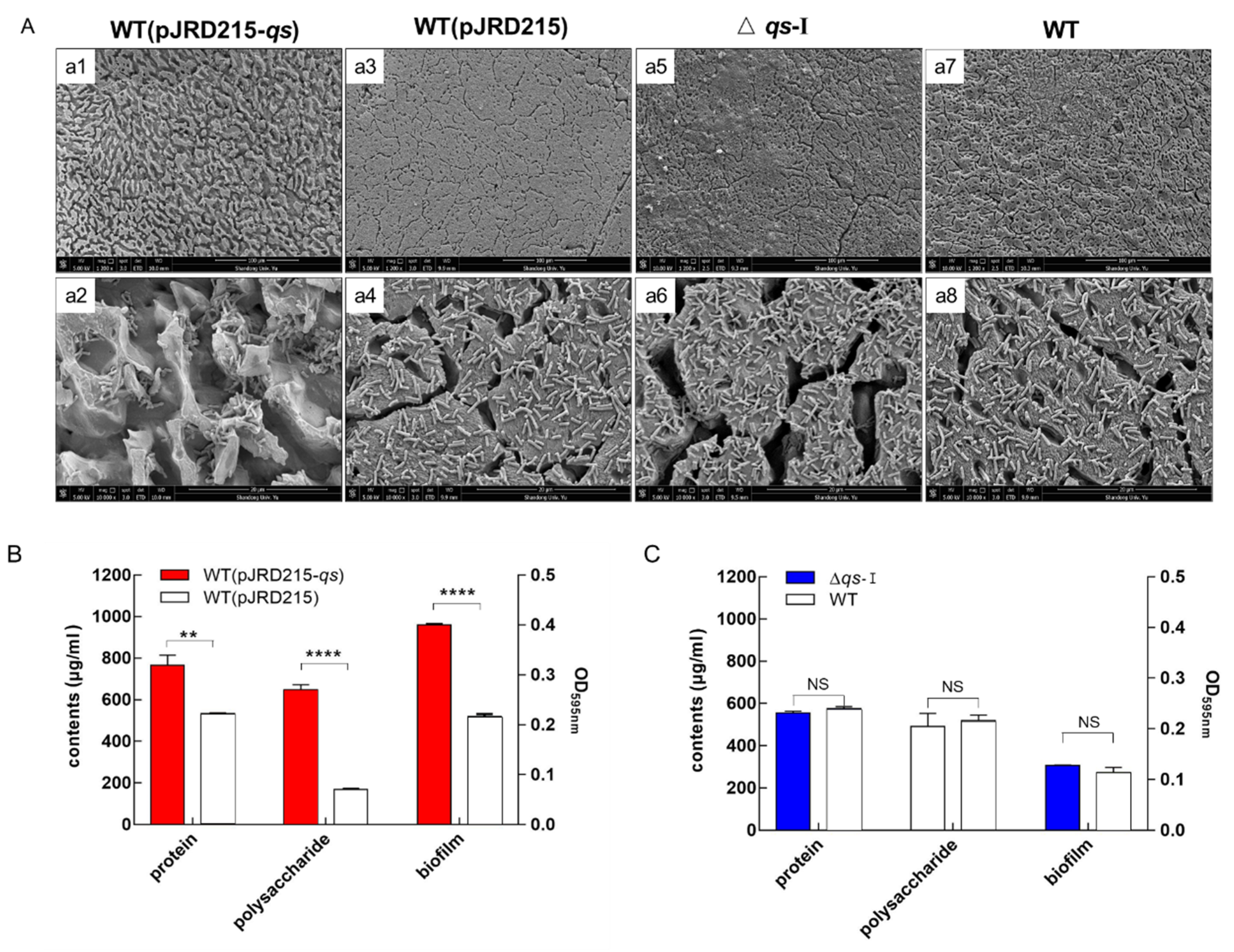
3.3. Influence of qs-I on Sulfur Metabolism and EPS Synthesis of A. ferrooxidans

Metabolite analysis showed that the sulfate yield of qs-Ⅰ overexpression strain increased significantly at the lag and exponential phases, but the difference was disappeared at the stationary phase (Figure 3A). The effect of qs-Ⅰ overexpression on EPS production was similar to that on sulfur metabolism (Figure 3A,C). The main constituent polysaccharide and protein of the EPS of qs-Ⅰ overexpression strain showed an obvious increase at the exponential phase and no difference at the stationary phase, compared to that of the control strain (Figure 3C). The deletion of qs-Ⅰ did not give rise to significant phenotypic changes during sulfur metabolism and EPS synthesis (Figure 3B,D). The above results indicated that the overexpression of qs-Ⅰ could promote EPS production and accelerate the sulfur-metabolic process of A. ferrooxidans.
To further investigate the regulatory effects of AfeI/R on the genes involved in sulfur metabolism and EPS synthesis, transcriptional analysis was carried out using RT-qPCR. As shown in Table 2, the deletion of the qs-Ⅰ operon did not result in the differential expression of the EPS-synthesis related and sulfur-metabolic genes, However, there was a significant upregulation of these genes, including sulfur metabolism genes (rhd2, resA, sbp1 and tsd1 in doxDA operon, AFE_2552, AFE_2554, dsrE, and tusA in hdr operon), EPS synthetic gene (pgm), and EPS transport related genes (oprD and TonB) in the overexpression strain (Table 2). Thus, the upregulation of these genes in the qs-Ⅰ overexpression strain suggested that the high expression of qs-Ⅰ could stimulate the activities of sulfur oxidation and EPS synthesis.

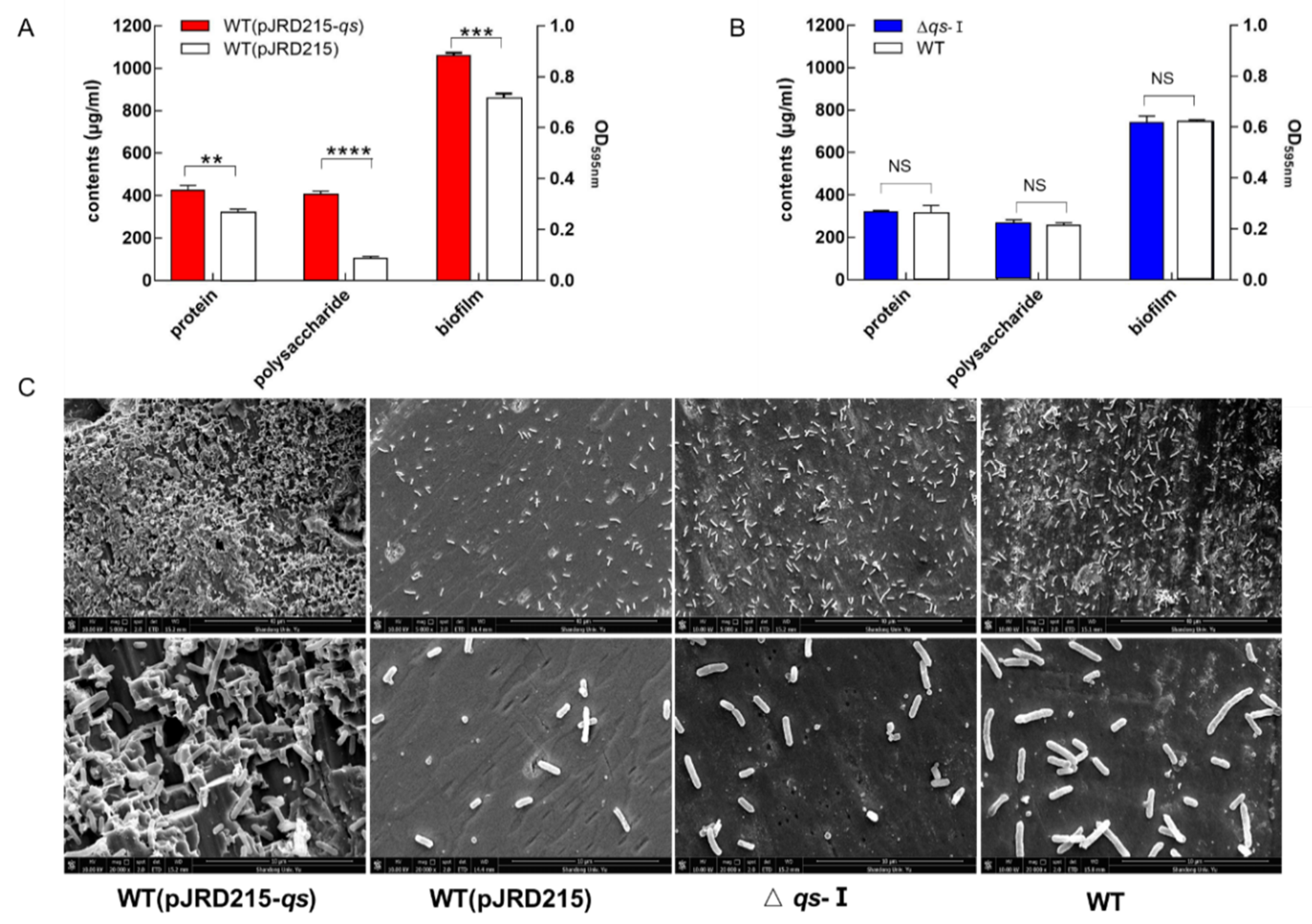
3.4. Effect of qs-I on the Bacteria-Substrate Interactions

The sulfur-coupon-cultivating assay was designed to investigate the role of AfeI/R in bacteria-substrate interactions. After an 8-day cultivation, the surface of the sulfur coupon cultivated by the qs-Ⅰ overexpression strains was quite uneven and bumpy (a1 and a2 in Figure 4A), while a smooth surface was observed from the sulfur coupons of WT(pJRD215), △qs-Ⅰ, and WT stains (a3–a8 in Figure 4A). Moreover, the overexpression of the qs-Ⅰ operon resulted in a remarkable increase in the EPS production of the planktonic cells and biofilm formation on the surface of elemental sulfur, while the deletion of this operon had no obvious effect on these aspects (Figure 4B,C). The protein and polysaccharide in the EPS of qs-Ⅰ overexpression strain were 1.5 and 3.8 times more than those in the control strain (WT(pJRD215)), respectively (Figure 4B). The biofilm on the sulfur coupons were detected using the staining method of crystal violet [50], showing that the amount of biofilm formed in qs-Ⅰ overexpression strain was twice as much as that formed in the control strain (Figure 4B). Therefore, it can be inferred that the significant increase in EPS and biofilm caused by the overexpression of qs-Ⅰ could significantly enable the bioerosion of the strain on the elemental sulfur during the bacteria-substrate interaction.

3.5. Effect of AfeI/R on the Bioleaching Process

To determine the importance of EPS synthesis and biofilm formation in the biological oxidation of ores, a mineral leaching experiment was performed using fabricated strains. Results showed that the EPS of the planktonic cells and the biofilm formed on the pyrite from the qs-Ⅰ overexpression strain were significantly higher than those from the control strain (Figure 5A,B). In particular, the polysaccharide component was 3.9 times that of the control strain (Figure 5A). Moreover, the SEM observation showed that the overexpression of qs-Ⅰ significantly increased the bacterial bioerosion ability on the pyrite (Figure 5C). Finally, the ICP-MS analysis showed that the concentrations of Cu, Zn, Fe, Mn, As, and Cr in the leachate of the qs-Ⅰ overexpression strain were 40%, 25%, 25%, 23%, 30%, and 54% more than those from the control strain, respectively (Figure 6A). However, no significant difference was detected in the generation of EPS and biofilm, the bioerosion ability, and leaching efficiency from the qs-Ⅰ deletion strain (Figure 5 and Figure 6). Therefore, the results suggested that the high expression of the qs-Ⅰ operon could effectively enhance bacterial oxidation to the ore and improve the leaching efficiency of metals.

4. Discussion

The synthesis of EPS and biofilm formation play a pivotal role in bacteria-ore interactions and the bioleaching of metals, thus the molecular mechanisms and the regulatory pathways involved in these biological processes have been of great significance in the research studies of bioleaching bacteria and the improvement of bioleaching efficiency in the industrial application [22,24]. Previous research studies revealed that QS systems play a role in the regulation of EPS synthesis and biofilm formation [52]. The addition of acyl-HSLs could enhance the biofilm formation of A. ferrooxidans on sulfur and pyrite surfaces, providing a promising strategy of improving the bioleaching efficiency and metal recovery in the biomining industry [10,34,35,36]. The development of genetic engineering and synthetic biology have shown that genetic manipulation is a powerful tool for strengthening and modifying the performance of microorganisms. In this study, using fabricated qs-I knockout and overexpression strains, we confirmed the regulatory role of AfeI/R in bacteria-substrate interactions and the bioleaching of minerals, revealing that the afeI/R-operon-dependent genetic modification is an effective means of developing highly efficient leaching bacteria.
EPS synthesis and biofilm formation are complex processes that are related to several metabolic and regulatory pathways [53,54]. Sixty genes involved in the biofilm formation of A. ferrooxidans were differentially expressed using an addition of a tetrazolic acyl-HSL analog (tetrazole 9c) [35]. Our study found that qs-Ⅰ regulates the synthesis of EPS in two aspects. Firstly, qs-Ⅰ regulated the synthesis of EPS. Overexpression of the qs-Ⅰ operon caused the upregulation of pgm (AFE_2324) transcription (Table 2), which is part of the gal operon (luxA-galE-galK-pgm-galM), encoding phosphoglucosidase and converting glucose-1-phosphate to glucose-6-phosphate [55,56]. It has been reported that the gal operon was involved in the formation of EPS precursors via the Leloir pathway [57,58]. Upregulation of the pgm transcription increases the synthesis of EPS. Moreover, qs-Ⅰ regulated the secretion and transport of EPS. Overexpression of qs-Ⅰ gene operon caused the upregulation of tonB (AFE_1991) and orpD (AFE_1497) genes transcription (Table 2). tonB and orpD genes involved in the transportation of EPS secretions have been reported [59]. Therefore, qs-Ⅰ regulated EPS synthesis and biofilm formation by regulating the synthesis and secretion of EPS in A. ferrooxidans.
EPS-mediated attachment is a prerequisite for the utilization of the solid-state energy substrates by A. ferrooxidans [22,24,36]. The EPS layer between cells and ores provides a pivotal microenvironment where metal sulfides are oxidized and minerals are dissolved [22,24]. Moreover, by secreting EPS and forming biofilms on mineral surfaces, sessile cells extend their reactive space and obtain significantly higher amounts of substrate than planktonic cells [52,60]. Thus, the increase of EPS could increase bioerosion capacity of the overexpression strain on the substrates (Figure 4 and Figure 5) and enhance the efficiency of the bioleaching process (Figure 6). These results further confirmed the crucial role played by EPS in the interactions between bacteria and solid-state substrates. They also revealed that AfeI/R-mediated regulation in EPS synthesis and biofilm formation could be an important control strategy for coordinating the metabolism and population of A. ferrooxidans in the growth environments.
The regulatory effects of QS in cell growth have been observed in many bacteria [61,62,63]. For instance, the addition of acyl-HSLs enhanced the growth of E. coli and C. jettenia, and the deletion of QS genes decreased the population density of B. glumae and B. Pseudomallei [61,62,63]. In this study, the enhance effects in the AfeI/R-mediated cell growth from the overexpression strain was detected (Figure 2A), indicating the AfeI/R-mediated regulation effects in A. ferrooxidans. However, the absence of this system did not significantly influence cell growth, EPS synthesis, biofilm formation, and bacterial bioerosion (Figure 2, Figure 3, Figure 4, Figure 5 and Figure 6). The mechanism of the QS system depends on the production, release and accumulation of the signal molecules [26]. Signal molecules were produced by two synthetases (AfeI and Act) in A. ferrooxidans and when the entire cluster of qs-Ⅰ was deleted, the generation of acyl-HSLs from AfeI was blocked, but the Act could still synthesize acyl-HSLs [31,33].
A. ferrooxidans has a highly efficient capacity to oxidize various reduced inorganic sulfur compounds (RISCs) and elemental sulfur to enable the growth of autotrophs [4]. Sulfur metabolism is critical for biohydrometallurgical technologies as well as providing acidic environment for the solubilization of metal sulfides [4,19,64]. The sulfur metabolizing process in A. ferrooxidans is achieved by the different sulfur- oxidizing enzymes located in different cellular compartments, including the activation and oxidation of extracellular element sulfur in the cellular outer membrane region, the thiosulfate metabolic enzymes/pathways at the core of the sulfur oxidization system in the periplasmic space, and the cytoplasmic sulfur metabolic proteins [65,66]. The function of the heterodisulfide reductase (Hdr)-like system is to oxidize the disulfide intermediate to sulfite and produce electrons in the cytoplasm [67]. DsrE / TusA is a core component of cytosolic sulfur transport and may transport sulfan sulfide to the Hdr sulfur-oxidation system [68]. The doxDA gene cluster is involved in the thiosulfate oxidation pathway [66]. The upregulation of the majority of sulfur-oxidizing genes, including rhd2, resA, sbp1, and tsd1 in doxDA operon, AFE_2552, AFE_2554, dsrE, and tusA in hdr operon, suggested that the sulfur-metabolism activity and the cell growth were promoted in the qs-Ⅰ overexpression strain (Figure 3A and Table 2).
Based on the enhancement of the cell growth, sulfur-metabolism activity, EPS synthesis, biofilm formation, and bioleaching efficiency by qs-Ⅰ, a model was outlined to illustrate the role of AfeI/R in the A. ferrooxidans-driven leaching process of minerals (Figure 7). The elevation of the qs-I expression level could promote the synthesis and secretion of EPS and cell growth, which stimulates the formation of biofilm on the surface of ores. The planktonic cells could be easily transformed to the sessile state through EPS, which gave the cells a growth advantage in the reactive space and the substrate. Simultaneously, the formation of biofilm could accelerate bacterial metabolism and mineral oxidation. As a result, the bioleaching efficiency could be enhanced by overexpressing the qs-I operon. Therefore, AfeI/R could function as a positive regulator to modulate leaching efficiency during the bioleaching of minerals.

5. Conclusions

We confirmed the regulation of AfeI/R in the EPS synthesis of A. ferrooxidans and revealed the significance of AfeI/R-mediated regulation of bacteria-substrate interactions and bioleaching process. Overexpression of qs-Ⅰ resulted in an obvious increase in EPS synthesis, biofilm formation, bioerosion capacity and bioleaching efficiency, while the deletion of qs-Ⅰ did not affect these above traits. Thus, the gene modification to increase the expression level of qs-Ⅰ could be an effective means to improve bacterial bioleaching efficiency. The proposal of the positive regulatory role of AfeI/R in the bioleaching process provides new insights into and clues for future theoretical and applied research studies.

Supplementary Materials

The following are available online at https://www.mdpi.com/2075-163X/10/3/222/s1, Table S1: List of the primers used in this study.

Author Contributions

X.-Y.G. performed the experiments. X.-Y.G. and L.-X.C. analyzed the data and wrote the manuscript. J.-Q.L. (Jian-Qiang Lin) revised the manuscript. X.-J.L., C.-A.F. and X.-F.G. did preliminary data analysis. J.-Q.L. (Jian-Qun Lin), X.-M.L. and X.P. designed the experiments. All authors conceived this study. All authors have read and agreed to the published version of the manuscript.

Funding

This work was supported by grants from the National Natural Science Foundation of China (31570036, 31370138, 31872621, 31570041), the Key Scientific and Technological Project of Shandong Province (2019GSF109032), the Project of Taishan Industry Leading Talent in Shandong province (LJNY201603), the State Key Laboratory of Microbial Technology Foundation (M2017-01).

Acknowledgments

We thank the supports from Core Facilities for Life and Environmental Sciences (State Key Laboratory of Microbial Technology, Shandong University), including Sen Wang for SEM technical assistance, Zhi-Feng Li for RT-qPCR instruction, Wang Rui for spectrometry analysis, Cheng-Jia Zhang and Nan-nan Dong for providing bacteriological incubators.

Conflicts of Interest

The authors declare that the research was conducted in the absence of any commercial or financial relationships that could be construed as a potential conflict of interest.

References

  1. Bosecker, K. Bioleaching: Metal solubilization by microorganisms. Fems Microbiol. Rev. 1997, 20, 591–604. [Google Scholar] [CrossRef]
  2. Rohwerder, T.; Gehrke, T.; Kinzler, K.; Sand, W. Bioleaching review part A: Progress in bioleaching: Fundamentals and mechanisms of bacterial metal sulfide oxidation. Appl. Microbiol. Biotechnol. 2003, 63, 239. [Google Scholar] [CrossRef] [PubMed]
  3. Li, Q.; Yang, B.; Zhu, J.; Jiang, H.; Li, J.; Zhang, R.; Sand, W. Comparative analysis of attachment to chalcopyrite of three mesophilic iron and/or sulfur-oxidizing acidophiles. Minerals 2018, 8, 406. [Google Scholar] [CrossRef] [Green Version]
  4. Rawlings, D.E. Heavy metal mining using microbes. Ann. Rev. Microbiol. 2002, 56, 65. [Google Scholar] [CrossRef] [PubMed]
  5. Olson, G.J.; Brierley, J.A.; Brierley, C.L. Bioleaching review part B: Progress in bioleaching: Applications of microbial processes by the minerals industries. Appl. Microbiol. Biotechnol. 2003, 63, 249–257. [Google Scholar] [CrossRef] [PubMed]
  6. Vardanyan, A.; Vardanyan, N.; Khachatryan, A.; Zhang, R.; Sand, W. Adhesion to mineral surfaces by cells of Leptospirillum, Acidithiobacillus and sulfobacillus from armenian sulfide ores. Minerals 2019, 9, 69. [Google Scholar] [CrossRef] [Green Version]
  7. Brierley, C.L. How will biomining be applied in future? Trans. Nonferrous Met. Soc. 2008, 18, 1302–1310. [Google Scholar] [CrossRef]
  8. Pathak, A.; Dastidar, M.G.; Sreekrishnan, T.R. Bioleaching of heavy metals from sewage sludge: A review. J. Environ. Manag. 2009, 90, 2343–2353. [Google Scholar] [CrossRef]
  9. Ma, Y.; Wang, H.; Song, Y.; Wu, Y.; Guo, Z. The synthesis of secondary iron minerals induced by quartz sand during the bioleaching process improves the dewaterability of municipal sewage sludge. Minerals 2018, 8, 419. [Google Scholar] [CrossRef] [Green Version]
  10. Gonzalez, A.; Bellenberg, S.; Mamani, S.; Ruiz, L.; Echeverria, A.; Soulere, L.; Doutheau, A.; Demergasso, C.; Sand, W.; Queneau, Y.; et al. AHL signaling molecules with a large acyl chain enhance biofilm formation on sulfur and metal sulfides by the bioleaching bacterium Acidithiobacillus ferrooxidans. Appl. Microbiol. Biotechnol. 2013, 97, 3729–3737. [Google Scholar] [CrossRef]
  11. Zhang, R.; Sun, C.; Kou, J.; Zhao, H.; Wei, D.; Xing, Y. Enhancing the leaching of chalcopyrite using Acidithiobacillus ferrooxidans under the induction of surfactant triton X-100. Minerals 2018, 9, 11. [Google Scholar] [CrossRef] [Green Version]
  12. Ohmura, N.; Sasaki, K.; Matsumoto, N.; Saiki, H. Anaerobic respiration using Fe(3+), S(0), and H(2) in the chemolithoautotrophic bacterium Acidithiobacillus ferrooxidans. J. Bacteriol. 2002, 184, 2081–2087. [Google Scholar] [CrossRef] [PubMed] [Green Version]
  13. Sugio, T.; Mizunashi, W.; Inagaki, K.; Tano, T. Purification and some properties of sulfur: Ferric ion oxidoreductase from Thiobacillus ferrooxidans. J. Bacteriol. 1987, 169, 4916–4922. [Google Scholar] [CrossRef] [Green Version]
  14. La Vars, S.M.; Newton, K.; Quinton, J.S.; Cheng, P.-Y.; Wei, D.-H.; Chan, Y.-L.; Harmer, S.L. Surface chemical characterisation of pyrite exposed to Acidithiobacillus ferrooxidans and associated extracellular polymeric substances. Minerals 2018, 8, 132. [Google Scholar] [CrossRef] [Green Version]
  15. Jafari, M.; Shafaei, S.Z.A.; Abdollahi, H.; Gharabaghi, M.; Chehreh Chelgani, S. A comparative study on the effect of flotation reagents on growth and iron oxidation activities of Leptospirillum ferrooxidans and Acidithiobacillus ferrooxidans. Minerals 2017, 7, 2. [Google Scholar] [CrossRef]
  16. Haghshenas, D.F.; Alamdari, E.K.; Bonakdarpour, B.; Darvishi, D.; Nasernejad, B. Kinetics of sphalerite bioleaching by Acidithiobacillus ferrooxidans. Hydrometallurgy 2009, 99, 202–208. [Google Scholar] [CrossRef]
  17. Zhang, S.; Yan, L.; Xing, W.; Chen, P.; Zhang, Y.; Wang, W. Acidithiobacillus ferrooxidans and its potential application. Extremophiles 2018, 22, 563–579. [Google Scholar] [CrossRef]
  18. Rodriguez-Leiva, M.; Tributsch, H. Morphology of bacterial leaching patterns by Thiobacillus ferrooxidans on synthetic pyrite. Arch. Microbiol. 1988, 149, 401–405. [Google Scholar] [CrossRef]
  19. Rohwerder, T.; Sand, W. Oxidation of Inorganic Sulfur Compounds in Acidophilic Prokaryotes. Eng. Life Sci. 2007, 7, 301–309. [Google Scholar] [CrossRef]
  20. Liu, H.L.; Chen, B.Y.; Lan, Y.W.; Cheng, Y.C. SEM and AFM images of pyrite surfaces after bioleaching by the indigenous Thiobacillus thiooxidans. Appl. Microbiol. Biotechnol. 2003, 62, 414–420. [Google Scholar] [CrossRef]
  21. Karamanev, D.G. Model of the biofilm structure of Thiobacillus ferrooxidans. J. Biotechnol. 1991, 20, 51–64. [Google Scholar] [CrossRef]
  22. Gehrke, T.; Telegdi, J.; Thierry, D.; Sand, W. Importance of extracellular polymeric substances from Thiobacillus ferrooxidans for bioleaching. Appl. Environ. Microbiol. 1998, 64, 2743–2747. [Google Scholar] [CrossRef] [PubMed] [Green Version]
  23. San Martín, F.; Aguilar, C. Study of the adhesion mechanism of Acidithiobacillus ferrooxidans to pyrite in fresh and saline water. Minerals 2019, 9, 306. [Google Scholar] [CrossRef] [Green Version]
  24. Harneit, K.; Göksel, A.; Kock, D.; Klock, J.H.; Gehrke, T.; Sand, W. Adhesion to metal sulfide surfaces by cells of Acidithiobacillus ferrooxidans, Acidithiobacillus thiooxidans and Leptospirillum ferrooxidans. Hydrometallurgy 2006, 83, 245–254. [Google Scholar] [CrossRef]
  25. Li, Q.; Wang, Q.; Zhu, J.; Zhou, S.; Gan, M.; Jiang, H.; Sand, W. Effect of extracellular polymeric substances on surface properties and attachment behavior of Acidithiobacillus ferrooxidans. Minerals 2016, 6, 100. [Google Scholar] [CrossRef] [Green Version]
  26. Parsek, M.R.; Greenberg, E.P. Acyl-homoserine lactone quorum sensing in Gram-negative bacteria: A signaling mechanism involved in associations with higher organisms. Proc. Natl. Acad. Sci. USA 2000, 97, 8789–8793. [Google Scholar] [CrossRef] [PubMed] [Green Version]
  27. Kai, P.; Bassler, B.L. Quorum sensing signal-response systems in Gram-negative bacteria. Nat. Rev. Microbiol. 2016, 14, 576. [Google Scholar] [CrossRef]
  28. An, D.; Danhorn, T.; Fuqua, C.; Parsek, M.R. Quorum sensing and motility mediate interactions between Pseudomonas aeruginosa and Agrobacterium tumefaciens in biofilm cocultures. Proc. Natl. Acad. Sci. USA 2006, 103, 3828–3833. [Google Scholar] [CrossRef] [Green Version]
  29. Davies, D.G.; Parsek, M.R.; Pearson, J.P.; Iglewski, B.H.; Costerton, J.W.; Greenberg, E.P. The involvement of cell-to-cell signals in the development of a bacterial biofilm. Science 1998, 280, 295–298. [Google Scholar] [CrossRef] [Green Version]
  30. Rivas, M.; Seeger, M.; Holmes, D.S.; Jedlicki, E. A Lux-like quorum sensing system in the extreme acidophile Acidithiobacillus ferrooxidans. Biol. Res. 2005, 38, 283. [Google Scholar] [CrossRef] [Green Version]
  31. Farah, C.; Vera, M.; Morin, D.; Haras, D.; Jerez, C.A.; Guiliani, N. Evidence for a functional quorum-sensing type AI-1 system in the extremophilic bacterium Acidithiobacillus ferrooxidans. Appl. Environ. Microbiol. 2005, 71, 7033–7040. [Google Scholar] [CrossRef] [PubMed] [Green Version]
  32. Banderas, A.; Guiliani, N. Bioinformatic prediction of gene functions regulated by quorum sensing in the bioleaching bacterium Acidithiobacillus ferrooxidans. Int. J. Mol. Sci. 2013, 14, 16901–16916. [Google Scholar] [CrossRef] [PubMed] [Green Version]
  33. Rivas, M.; Seeger, M.; Jedlicki, E.; Holmes, D.S. Second acyl homoserine lactone production system in the extreme acidophile Acidithiobacillus ferrooxidans. Appl. Environ. Microbiol. 2007, 73, 3225. [Google Scholar] [CrossRef] [PubMed] [Green Version]
  34. Bellenberg, S.; Diaz, M.; Noel, N.; Sand, W.; Poetsch, A.; Guiliani, N.; Vera, M. Biofilm formation, communication and interactions of leaching bacteria during colonization of pyrite and sulfur surfaces. Res. Microbiol. 2014, 165, 773–781. [Google Scholar] [CrossRef] [PubMed]
  35. Mamani, S.; Moinier, D.; Denis, Y.; Soulere, L.; Queneau, Y.; Talla, E.; Bonnefoy, V.; Guiliani, N. Insights into the quorum sensing regulon of the acidophilic Acidithiobacillus ferrooxidans revealed by transcriptomic in the presence of an acyl homoserine lactone superagonist analog. Front. Microbiol. 2016, 7, 1365. [Google Scholar] [CrossRef] [PubMed]
  36. Ruiz, L.M.; Valenzuela, S.; Castro, M.; Gonzalez, A.; Frezza, M.; Soulère, L.; Rohwerder, T.; Queneau, Y.; Doutheau, A.; Sand, W. AHL communication is a widespread phenomenon in biomining bacteria and seems to be involved in mineral-adhesion efficiency. Hydrometallurgy 2008, 94, 133–137. [Google Scholar] [CrossRef]
  37. Inaba, Y.; Banerjee, I.; Kernan, T.; Banta, S. Transposase-mediated chromosomal integration of exogenous genes in Acidithiobacillus ferrooxidans. Appl. Environ. Microbiol. 2018, 84, e01381-01318. [Google Scholar] [CrossRef] [Green Version]
  38. Wang, H.; Liu, X.; Liu, S.; Yu, Y.; Lin, J.; Lin, J.; Pang, X.; Zhao, J. Development of a markerless gene replacement system for Acidithiobacillus ferrooxidans and construction of a pfkB mutant. Appl. Environ. Microbiol. 2012, 78, 1826–1835. [Google Scholar] [CrossRef] [Green Version]
  39. Sambrook, J.; Fritsch, E.F.; Maniatis, T. Molecular Cloning: A Laboratory Manual; Cold Spring Harbor Laboratory: New York, NY, USA, 1982; pp. 895–909. [Google Scholar]
  40. Simon, R.; Priefer, U.; Pühler, A. A broad host range mobilization system for in vivo genetic engineering: Transposon mutagenesis in gram negative bacteria. Nat. Biotechnol. 1983, 1, 784–791. [Google Scholar] [CrossRef]
  41. Wang, Z.B.; Li, Y.Q.; Lin, J.Q.; Pang, X.; Liu, X.M.; Liu, B.Q.; Wang, R.; Zhang, C.J.; Wu, Y.; Lin, J.Q.; et al. The two-component system RsrS-RsrR regulates the tetrathionate intermediate pathway for thiosulfate oxidation in Acidithiobacillus caldus. Front. Microbiol. 2016, 7, 1755. [Google Scholar] [CrossRef] [Green Version]
  42. Davison, J.; Heusterspreute, M.; Chevalier, N.; Ha-Thi, V.; Brunel, F. Vectors with restriction site banks. V. pJRD215, a wide-host-range cosmid vector with multiple cloning sites. Gene 1987, 51, 275–280. [Google Scholar] [CrossRef]
  43. Miura, Y.; Kawaoi, A. Determination of thiosulfate, thiocyanate and polythionates in a mixture by ion-pair chromatography with ultraviolet absorbance detection. J. Chromatogr. A 2000, 884, 81–87. [Google Scholar] [CrossRef]
  44. More, T.T.; Yadav, J.S.S.; Yan, S.; Tyagi, R.D.; Surampalli, R.Y. Extracellular polymeric substances of bacteria and their potential environmental applications. J. Environ. Manag. 2014, 144, 1–25. [Google Scholar] [CrossRef] [PubMed]
  45. Xiao, Y.; Zhang, E.; Zhang, J.; Dai, Y.; Yang, Z.; Christensen, H.E.M.; Ulstrup, J.; Zhao, F. Extracellular polymeric substances are transient media for microbial extracellular electron transfer. Sci. Adv. 2017, 3, e1700623. [Google Scholar] [CrossRef] [Green Version]
  46. Grandy, A.S.; Erich, M.S.; Porter, G.A. Suitability of the anthrone–sulfuric acid reagent for determining water soluble carbohydrates in soil water extracts. Soil Biol. Biochem. 2000, 32, 725–727. [Google Scholar] [CrossRef]
  47. Nieto, P.A.; Covarrubias, P.C.; Jedlicki, E.; Holmes, D.S.; Quatrini, R. Selection and evaluation of reference genes for improved interrogation of microbial transcriptomes: Case study with the extremophile Acidithiobacillus ferrooxidans. BMC Mol. Biol. 2009, 10, 63. [Google Scholar] [CrossRef] [Green Version]
  48. Livak, K.J.; Schmittgen, T.D. Analysis of relative gene expression data using real-time quantitative PCR and the 2−ΔΔCT method. Methods 2001, 25, 402–408. [Google Scholar] [CrossRef]
  49. Zheng, Y.; Cosgrove, D.J.; Ning, G. High-resolution field emission scanning electron microscopy (FESEM) imaging of cellulose microfibril organization in plant primary cell walls. Microsc. Microanal. 2017, 23, 1048–1054. [Google Scholar] [CrossRef]
  50. Bret, L.; Di Martino, P. Effect of ceftazidime, amikacin and ciprofloxacin on biofilm formation by some enterobacterial clinical isolates. Chemotherapy 2004, 50, 255–259. [Google Scholar] [CrossRef]
  51. Rodrigues, J.; Nunes, J.; Batista, B.; Souza, S.; Barbosa, F. A fast method for the determination of 16 elements in hair samples by inductively coupled plasma mass spectrometry (ICP-MS) with tetramethylammonium hydroxide solubilization at room temperature. J. Anal. At. Spectrom. 2008, 23, 992. [Google Scholar] [CrossRef]
  52. Parsek, M.R.; Greenberg, E.P. Sociomicrobiology: The connections between quorum sensing and biofilms. Trends Microbiol. 2005, 13, 27–33. [Google Scholar] [CrossRef] [PubMed]
  53. Moormeier, D.E.; Bayles, K.W. Staphylococcus aureus biofilm: A complex developmental organism. Mol. Microbiol. 2017, 104, 365–376. [Google Scholar] [CrossRef] [PubMed] [Green Version]
  54. Marczak, M.; Dzwierzynska, M.; Skorupska, A. Homo- and heterotypic interactions between Pss proteins involved in the exopolysaccharide transport system in Rhizobium leguminosarum bv. trifolii. Biol. Chem. 2013, 394, 541–559. [Google Scholar] [CrossRef]
  55. Barreto, M.; Jedlicki, E.; Holmes, D.S. Identification of a gene cluster for the formation of extracellular polysaccharide precursors in the chemolithoautotroph Acidithiobacillus ferrooxidans. Appl. Environ. Microbiol. 2005, 71, 2902–2909. [Google Scholar] [CrossRef] [PubMed] [Green Version]
  56. Ciocchini, A.E.; Guidolin, L.S.; Casabuono, A.C.; Couto, A.S.; Iñón de Iannino, N.; Ugalde, R.A. A glycosyltransferase with a length-controlling activity as a mechanism to regulate the size of polysaccharides. Proc. Natl. Acad. Sci. USA 2007, 104, 16492–16497. [Google Scholar] [CrossRef] [PubMed] [Green Version]
  57. Holden, H.M.; Rayment, I.; Thoden, J.B. Structure and function of enzymes of the Leloir pathway for galactose metabolism. J. Biol. Chem. 2003, 278, 43885–43888. [Google Scholar] [CrossRef] [PubMed] [Green Version]
  58. Bettenbrock, K.; Alpert, C.A. The gal genes for the Leloir pathway of Lactobacillus casei 64H. Appl. Environ. Microbiol. 1998, 64, 2013–2019. [Google Scholar] [CrossRef] [Green Version]
  59. Zhang, Y.; Gao, J.; Wang, L.; Liu, S.; Bai, Z.; Zhuang, X.; Zhuang, G. Environmental adaptability and quorum sensing: Iron uptake regulation during biofilm formation by Paracoccus denitrificans. Appl. Environ. Microbiol. 2018, 84, e00865-00818. [Google Scholar] [CrossRef] [Green Version]
  60. Yang, H.; Luo, W.; Gao, Y. Effect of Acidithiobacillus ferrooxidans on humic-acid passivation layer on pyrite surface. Minerals 2018, 8, 422. [Google Scholar] [CrossRef] [Green Version]
  61. Goo, E.; Majerczyk, C.D.; An, J.H.; Chandler, J.R.; Seo, Y.S.; Ham, H.; Lim, J.Y.; Kim, H.; Lee, B.; Jang, M.S.; et al. Bacterial quorum sensing, cooperativity, and anticipation of stationary-phase stress. Proc. Natl. Acad. Sci. USA 2012, 109, 19775–19780. [Google Scholar] [CrossRef] [Green Version]
  62. An, J.H.; Goo, E.; Kim, H.; Seo, Y.S.; Hwang, I. Bacterial quorum sensing and metabolic slowing in a cooperative population. Proc. Natl. Acad. Sci. USA 2014, 111, 14912–14917. [Google Scholar] [CrossRef] [PubMed] [Green Version]
  63. Tang, X.; Guo, Y.; Wu, S.; Chen, L.; Tao, H.; Liu, S. Metabolomics uncovers the regulatory pathway of acyl-homoserine lactones based quorum sensing in anammox consortia. Environ. Sci. Technol. 2018, 52, 2206–2216. [Google Scholar] [CrossRef] [PubMed]
  64. Tributsch, H. Direct versus indirect bioleaching. Hydrometallurgy 2001, 59, 177–185. [Google Scholar] [CrossRef]
  65. Quatrini, R.; Appia-Ayme, C.; Denis, Y.; Ratouchniak, J.; Veloso, F.; Valdes, J.; Lefimil, C.; Silver, S.; Roberto, F.; Orellana, O.; et al. Insights into the iron and sulfur energetic metabolism of Acidithiobacillus ferrooxidans by microarray transcriptome profiling. Hydrometallurgy 2006, 83, 263–272. [Google Scholar] [CrossRef]
  66. Wang, R.; Lin, J.-Q.; Liu, X.-M.; Pang, X.; Zhang, C.-J.; Yang, C.-L.; Gao, X.-Y.; Lin, C.-M.; Li, Y.-Q.; Li, Y.; et al. Sulfur oxidation in the acidophilic autotrophic Acidithiobacillus spp. Front. Microbiol. 2019, 9. [Google Scholar] [CrossRef]
  67. Wagner, T.; Koch, J.; Ermler, U. Methanogenic heterodisulfide reductase (HdrABC-MvhAGD) uses two noncubane [4Fe-4S] clusters for reduction. Science 2017, 357, 699–703. [Google Scholar] [CrossRef] [Green Version]
  68. Liu, L.J.; Stockdreher, Y.; Koch, T.; Sun, S.T.; Fan, Z.; Josten, M.; Sahl, H.G.; Wang, Q.; Luo, Y.M.; Liu, S.J.; et al. Thiosulfate transfer mediated by DsrE/TusA homologs from acidothermophilic sulfur-oxidizing archaeon Metallosphaera cuprina. J. Biol. Chem. 2014, 289, 26949–26959. [Google Scholar] [CrossRef] [Green Version]
Figure 1. Conformation of qs-Ⅰ knockout and overexpression strains. (A) The graph of the qs-Ⅰ operon deletion strain and primer pairs were used for the verification of the mutant. UHA and DHA indicate the upstream and downstream homologous arms of the qs-Ⅰ operon. (B) Electrophoretogram to verify Δqs-Ⅰ. Lanes 1, 3, and 5 show PCR amplifications from Δqs-Ⅰ using the primers P1F/R, P2F/R, P3F/R, respectively. Lanes 2, 4, and 6 show PCR amplifications from wide type strain using primers P1F/R, P2F/R, P3F/R, respectively. (C) Graph of pJRD215-qs-Ⅰ plasmid. (D) Electrophoretogram to verify the qs-Ⅰ overexpression strains. Lanes 1, 2 and 3 show PCR amplifications using primers P4F/R from qs-Ⅰ overexpression strain, wide type and pJRD215-qs-Ⅰ plasmid, respectively.
Minerals 10 00222 g001
Figure 2. The growth curves of qs-Ⅰ overexpression (A) and knockout strains (B) of A. ferooxidans. CK indicates the blank control.
Minerals 10 00222 g002
Figure 3. Analyses of sulfur metabolism and extracellular polymeric substances (EPS) synthesis of engineered A. ferrooxidans strains. Sulfate production (A) and EPS synthesis (B) of qs-Ⅰ overexpression strains. Sulfate production (C) and EPS synthesis (D) of Δqs-Ⅰ strains. NS indicates no significant difference.
Minerals 10 00222 g003
Figure 4. Analysis of EPS synthesis and biofilm formation on the sulfur couples. Observation of cell attachment on sulfur coupons by SEM (A). Sulfur coupon cultivation of the qs-Ⅰ overexpression strain (a1 and a2), WT (pJRD215) strain (a3 and a4), Δqs-Ⅰ strain (a5 and a6), and wild-type strain (a7 and a8). The top row was magnified 1200× and the second row was magnified 10,000×. The protein and polysaccharide levels in EPS and the biofilm formation on the sulfur couples of the qs-Ⅰ overexpression strain (B) and Δqs-Ⅰ (C). NS indicates no significant difference.
Minerals 10 00222 g004
Figure 5. Analysis of EPS synthesis and biofilm formation in pyrite-cultivating. The protein and polysaccharide levels in EPS and the biofilm formation on the pyrite of the qs-Ⅰ overexpression strain (A) and Δqs-Ⅰ (B). Observation of cell attachment on pyrite by SEM (C). The top row was magnified 5000×, and the second row was magnified 20,000×. NS indicates no significant difference.
Minerals 10 00222 g005
Figure 6. Metal elements content in the leachate of qs-Ⅰ overexpression (A) and knockout strains (B) of A. ferooxidans. NS indicates no significant difference.
Minerals 10 00222 g006
Figure 7. Roles of qs-I mediated regulation in A. ferrooxidans bioleaching.
Minerals 10 00222 g007
Table 1. Bacteria and plasmids used in this study.
Strain and PlasmidsDescriptionSource
Strain
Acidithiobacillus ferrooxidans
ATCC 23270
Wild type strainATCC
WT (pJRD215)Wild type with the plasmid of pJRD215This study
qs-Iqs-I (afeR-orf3-afeI) gene cluster deletionThis study
WT (pJRD215-qs)overexpress qs-I gene cluster (afeR-orf3-afeI), wild type with the plasmid of pJRD215- qs-IThis study
Escherichia coli
DH5αF-φ80dlacZΔM15Δ(lacZYA-argF)U169 end A1 recA1 hsdR17(rk-,mk+) supE44λ-thi-1 gyr96 rela1 phoATransGen Biotech
SM10Kmrthi-1 thr leu tonA lacY supE recARP4-2-Tc::Mu[40]
Plasmids
pSDUDISuicide plasmid; Apr Kmr oiTRP4 multi-cloning sites[41]
pSDUDI-HFqs-ISuicide plasmid for Δqs-I construction, pSDUDI carrying both homologous fragments of qs-IThis study
pMSD1-I-SecIpMSD containing the I-SecI gene[38]
pJRD215Smr Kmr IncQ Mob+[42]
pJRD215-qs-IpJRD215 carrying the qs-I gene cluster (afeR-orf3-afeI) This study
Table 2. The relative transcription levels of genes involved in EPS synthesis and sulfur metabolism. This is the valid mean value of fold changes (FC) determined by RT-qPCR. FC ≥ 2, P ≤ 0.05, up-regulated; FC ≤ 0.5, P ≤ 0.05, down-regulated. doxDA operon includes genes AFE_0042-0050; hdr operon includes genes AFE_2550–2557. All experimental samples were set with three biological replicates and performed at three times. WT indicates the wide type of A. ferrooxidans.
Gene IDΔqs-I vs WTWT (pJRD215-qs) vs WT(pJRD215)Gene NameDescription
Sulfur metabolism genes
AFE_00421.24 ± 0.331.05 ± 0.03tsd2hypothetical protein
AFE_00430.94 ± 0.361.44 ± 0.13sbp2glycine/betaine ABC transporter substrate-binding protein
AFE_00441.00 ± 0.081.24 ± 0.01doxDA2quinol oxidase
AFE_00450.66 ± 0.241.52 ± 0.15rhd1rhodanese-like domain-containing protein
AFE_00460.58 ± 0.252.01 ± 0.50rhd2rhodanese-like domain-containing protein
AFE_00470.71 ± 0.352.56 ± 0.55resATlpA family protein disulfide reductase
AFE_00481.03 ± 0.321.11 ± 0.09doxDA1quinol oxidase
AFE_00491.01 ± 0.413.31 ± 0.42sbp1glycine/betaine ABC transporter substrate-binding protein
AFE_00501.09 ± 0.442.51 ± 0.32tsd1hypothetical protein
AFE_25500.69 ± 0.331.17 ± 0.08sdhCdisulfide reductase
AFE_25511.05 ± 0.411.69 ± 0.02hdrCheterodisulfide reductase subunit C
AFE_25520.98 ± 0.442.00 ± 0.37-hypothetical protein
AFE_25530.94 ± 0.031.41 ± 0.39hdrApyridine nucleotide-disulfide oxidoreductase
AFE_25541.07 ± 0.452.12 ± 0.13hdrBheterodisulfide reductase subunit B
AFE_25550.96 ± 0.151.28 ± 0.05hdrCheterodisulfide reductase subunit C
AFE_25561.00 ± 0.222.09 ± 0.26dsrENADH dehydrogenase
AFE_25570.93 ± 0.122.63 ± 0.57tusAsulfurtransferase TusA family protein
AFE_25580.85 ± 0.281.15 ± 0.35rhdrhodanese-like domain-containing protein
AFE_25861.27 ± 0.111.81 ± 0.06hdrBheterodisulfide reductase subunit B
AFE_00290.85 ± 0.110.54 ± 0.09tetHtetrathionate hydrolase
AFE_02670.61 ± 0.010.95 ± 0.18sqrpyridine nucleotide-disulfide oxidoreductase
AFE_17920.75 ± 0.071.62 ± 0.13sqrsulfide:quinone reductase
AFE_02691.46 ± 0.080.66 ± 0.06sdoMBL fold metallo-hydrolase
AFE_26440.75 ± 0.021.17 ± 0.23sdoMBL fold metallo-hydrolase
EPS related genes
AFE_23210.9 ± 0.100.66 ± 0.05luxAmonooxygenase, luciferase-like protein
AFE_23220.89 ± 0.040.71 ± 0.03galEconserved hypothetical protein
AFE_23230.98 ± 0.090.45 ± 0.10galKconserved hypothetical protein
AFE_23240.72 ± 0.132.55 ± 0.31pgmphosphoglucomutase, putative
AFE_23250.68 ± 0.191.45 ± 0.18galMhypothetical protein
AFE_14970.75 ± 0.315.34 ± 2.70orpDouter membrane porin, OprD family
AFE_19910.67 ± 0.3157.71 ± 6.94tonBmembrane protein, TonB family

