Article

Broken Mirrors: Multiple Circular Polarization and Inversion in the Ground and Photoexcited States of Mirror-Symmetric Helical Poly(di-iso-alkylsilane)s in Achiral Molecular Solvents

by
Michiya Fujiki
1,*,
Takashi Mori
1,
Julian R. Koe
2 and
Mohamed Mehawed Abdellatif
3,4
1
Graduate School of Advanced Science and Technology, Nara Institute of Science and Technology, 8916-5 Takayama-cho, Ikoma 630-0192, Nara, Japan
2
Department of Natural Sciences, International Christian University, 3-10-2 Osawa, Mitaka 181-8585, Tokyo, Japan
3
Department of Chemistry, Graduate School of Science, Tokyo Metropolitan University, 1-1 Minami-Osawa, Hachioji 192-0397, Tokyo, Japan
4
Chemistry of Tanning Materials and Leather Technology Department, Chemical Industries Research Institute, National Research Centre (NRC), 33 El Buhouth St., Dokki, Giza 12622, Egypt
*
Author to whom correspondence should be addressed.
Symmetry 2025, 17(9), 1544; https://doi.org/10.3390/sym17091544
Submission received: 3 August 2025 / Revised: 29 August 2025 / Accepted: 10 September 2025 / Published: 15 September 2025
(This article belongs to the Section Chemistry: Symmetry/Asymmetry)

Abstract

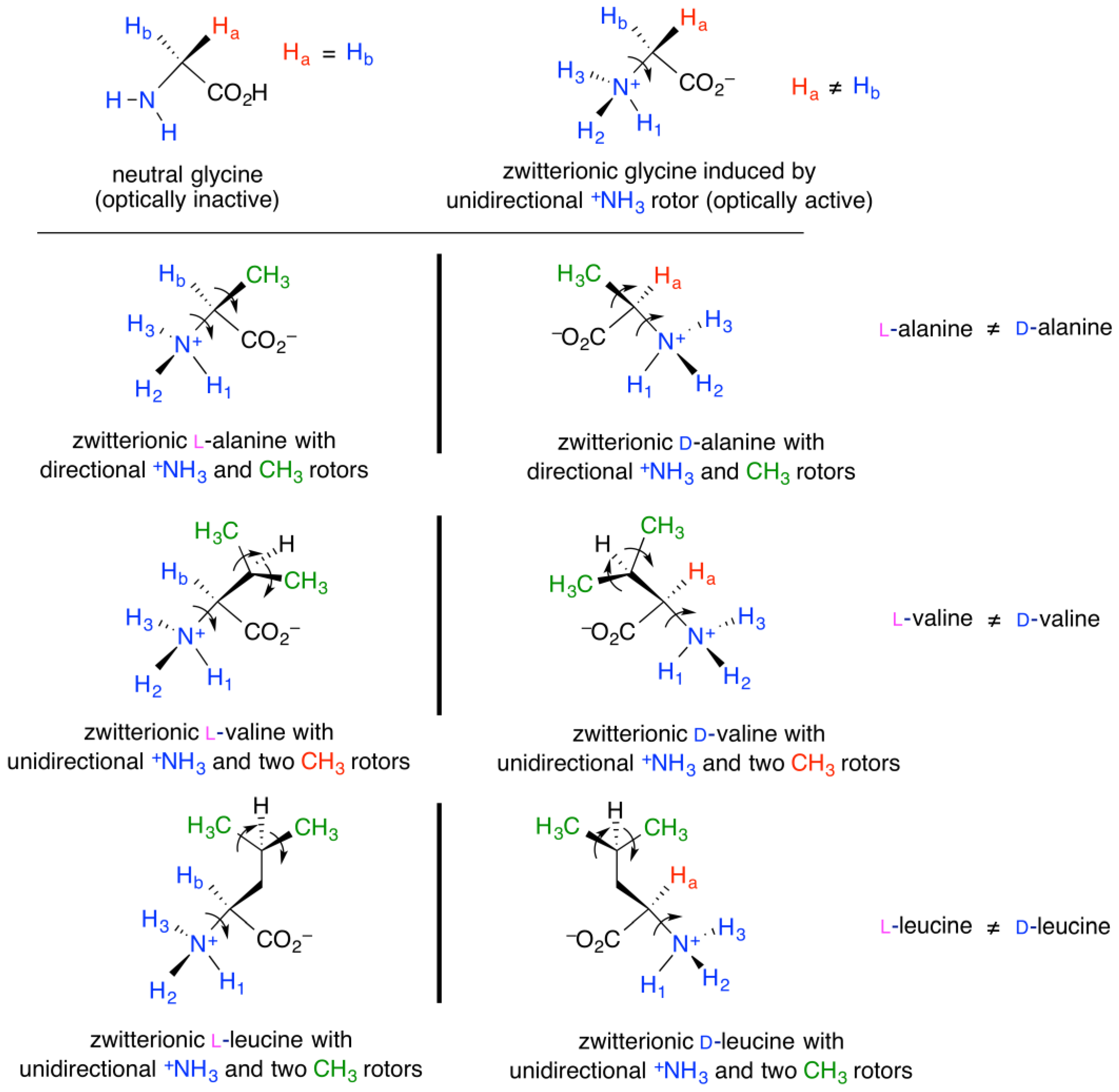
This paper comprehensively reports experimental proof of parity violation in the ground and photoexcited states of three mirror-symmetric Si–Si bond polymers in homogeneous solutions of achiral molecules under non-stirring conditions by analyzing 370 chiroptical datasets relating to multiple second-order helix–helix transitions in the circular dichroism (CD) of poly(di-i-butylsilane) (iBS), poly(di-i-pentylsilane) (iPS), and poly(di-i-hexylsilane) (iHS) in achiral alkanols and p-dioxane-h8/-d8. Particularly large (–)-CD of gabs = −3.1 × 10⁻² at 290 nm was found for iBS in i-pentanol at 25 °C. Notably, iPS in n-propanol at −5 °C generated (–)-CD with gabs = −0.48 × 10⁻² at 300 nm, but (+)-circularly polarized luminescence (CPL) with glum = +0.84 × 10⁻² at 326 nm. In contrast, iHS in n-octanol at 0 °C showed only very weak (–)-CD of gabs ~−0.03 × 10⁻² at 310 nm. The H/D isotopes of p-dioxane-h8/-d8 weakly affected the helix–helix transition characteristics of iBS. (–)-Sign vibrational CD signals assigned to the handed symmetric and asymmetric bending modes of the CH3 and CH2 groups of the solvents and other achiral molecules were observed. We assumed (i) three 1H nuclear-spin-1/2 induced handed motions of CH3 rotors at i-alkyl side chains and achiral alkanols, and (ii) helical main-chain Si atoms +) coordinated by handed lone pairs at oxygen ) in gauche-containing n- and i-alkanols induced by the CH3 rotors. A possible origin of biomolecular handedness is proposed based on the first observation of far-UV CD and UV spectra of zwitterionic glycine bearing H3N+ rotor in neutral H2O.

1. Introduction

Everything should be made as simple as possible, but not simpler (Einstein, 1933) [1]. This famous quote prompted theorists and experimentalists to make complexity easy to understand without oversimplifying it beyond common sense, leading to fantastic hypotheses, innovative theories, and gedanken-and-precision experiments. In physics, chemistry, biology, and cosmology, the most fundamental question is whether the seven symmetries of nature, namely, parity (P), charge conjugation (C), time reversal (T), and their combined CP, PT, CT, and CPT, are rigorously conserved, nearly conserved, or absolutely violated.
Until 1957, most prominent theorists believed that symmetry laws were rigorously conserved in the four fundamental electromagnetic force (EMF), weak nuclear force (WF), strong nuclear force (SF), and gravitational force (GF) [2,3,4]. In 1956, Lee and Yang pointed out that P-invariance in WF-caused events was not proven and proposed simple experiments using (i) β-decays radiating electrons (e), positrons (e+), neutrinos (ν), anti-neutrinos (anti-ν), and circularly polarized γ-rays, and (ii) decays in charged mesons and baryons [5]. Their proposals immediately prompted Wu et al. [6,7], Lederman et al. [8], and Schopper [9], who experimentally verified P-violating (PV) β decay in 60Co → 60Ni, β+ decay in 58Co → 58Fe, and emission of circularly polarized γ-rays from 60Co and 22Na nuclei [10].
Subsequently, Cronin and Fitch in 1964 discovered a tiny CP-violation (CPV) of 0.2% in the decay of neutral K-mesons [K0, comprising a down quark (d) and a strange anti-quark (anti-s)] to π-mesons by observing the parity-odd long-lived K0Lπππ (dominant) and parity-even short-lived K0Sππ (rare) [11]. In 2001, Belle and BaBar collaborations independently detected CPV events between neutral B-mesons (B0, comprising d and bottom anti-quark (anti-b)) and anti-B0 (anti-d and anti-b) [12,13]. In 2025, Large Hadron Collider beauty (LHCb) collaboration team reported the first CPV of baryons as high as 3.7% for Λb0 (up (u), d, b) relative to anti-Λb0 (anti-u, anti-d, anti-b) through Λb0pKπ+π and anti-Λb0 → anti-pK+ππ+ decays (p is a proton) [14]. This result sheds light on why matter (p (uud) and neutron (n, udd)) are dominant in our universe, but anti-matter (anti-p and anti-n) are very rare [15].
Since the 1960s, validating atomic P-violation (APV) hypothesis in physics has been challenging [16,17,18,19]. From the 1980s onward, the APV theory was experimentally confirmed using high-resolution optical rotation dispersion and luminescence-detective circular dichroism for collision-free gases [20,21,22,23] and spin-polarized neutron resonance scattering for solids [24,25,26,27,28]. Relativistic high-Z atoms (Z: atomic number), that is, 208Pb, 209Bi, 205Tl, 133Cs, 174Yb, and a dozen half-integer nuclear spin (HINS) high-Z isotopes, such as 117Sn, 139La, 81Br, 127I, etc., showed marked APV effects [20,21,22,23,24,25,26,27].
On the other hand, in 1887, modern stereochemistry has started with the theory of van’t Hoff who stated that the differences in the Gibbs free energy and enthalpy between mirror-image molecules (enantiomers) at T = 0 K and all other temperatures are exactly zero [28]. The notions indicated that P-conserving (PC)-EMF that mirror-image molecules strictly preserved energetic equality in the ground (S0) and photoexcited (S1 and T1) states of mirror-image molecules, supramolecules, polymers, aggregates, and crystals [29,30,31,32,33,34,35]. Recent circular dichroism (CD), vibrational circular dichroism (VCD), and Raman optical activity (ROA) spectrometers permit measurement of the minute differences in intensities between left and right circularly polarized light in absorption modes (CD and VCD) and scattering mode (ROA) in the S0 state of a broad range of chiral substances. Circularly polarized luminescence (CPL) spectroscopy complements the above by enabling the detection of subtle differences in intensity between left and right circularly polarized emission from the S1 and T1 states of various metastable chiral luminophores. For detailed theories, techniques, and applications of CD, VCD, ROA, and CPL, see two comprehensive books and a review [33,34,35]. If the modern stereochemistry is valid, mirror-image substances should exhibit ideal mirror-image CD, VCD, ROA, and CPL spectra with identical magnitudes at the same wavelengths and/or wavenumbers [28,29,30,31,32,33,34,35]. In other words, racemic compounds in a 50:50 mixture of enantiomers and achiral compounds should not exhibit any chiroptical signals, and are generally considered optically inactive [31,32].
Similar to the APV hypothesis, the molecular P-violation (MPV) hypothesis has been debated in connection with biomolecular handedness on Earth. From the 1960s onward, MPV theorists have predicted that the electroweak force (EWF) by unifying the PV-WF and PC-EMF causes a very small parity-violating energy difference (PVED), EPV, in the range from 10−10 eV to 10−21 eV for realistic and hypothetical mirror-image molecules [28,36,37,38,39,40,41,42,43,44,45,46,47,48,49,50,51,52,53,54,55,56,57]. The MPV effect arises from the handed weak neutral current (WNC) around the nuclei, which is mediated by Z0 boson (91.2 GeV). If the MPV hypothesis is valid, mirror-image molecules are no longer enantiomers and should be called diastereomers.
To amplify the tiny EPV to observable levels, six major scenarios have been theoretically proposed (see ref. [58] and references therein): (i) linear amplification model by Yamagata (EPVn, where n = repeat numbers of polymer chain and numbers of molecules in a crystal [36], and, presumably, a pure liquid can be included; (ii) non-linear amplification model revealing second-order phase transition discussed by Goldanskii et al. [45] and Salam [46]; (iii) tuning a tunneling splitting (ΔE±) connecting a barrier height (EB) in a double-well potential (DWP) [53,54]; (iv) EPVZ5 [39,42,43,44,55,56,57]; (v) EPV ∝ 1/ΔEST, where ΔEST = energy gap between S0 and T1 states, and, presumably, an energy gap between S1 and T1 states may be involved [43]; and (vi) half-integer nuclear spin (HINS) isotope induced anapole moment [51,59,60,61,62,63,64,65].
Historically, in 1898, Kipping and Pope were the first to investigate a possibility of the MPV effect using a spontaneous generation of chiral cubic crystal (space group P213) from an aqueous solution of achiral NaClO3 (point group C3v) comprising HINS 23Na and 35/37Cl isotopes under non-stirring (static) conditions (scenarios (i) and (vi)) [66]. They statistically analyzed the L-D preferences of 3137 chiral crystals obtained from 46 independent runs. They found a less obvious L-D preference (D-crystal 0.04% ee) from pure water, but clear L-D preferences in the presence of three hexoses: L-crystal 9.1% ee from D-glucose (25 runs), L-crystal 4.7% ee from D-mannitol (37 runs), and D-crystal 0.6% ee from L-rhamnose (16 runs). Later, Kipping was credited as the father of silicon chemistry, synthesizing the first optically active organosilicon compound, polysilanes, and polysiloxanes [67,68].
To date, several researchers have reported differences in the physicochemical properties, chiroptical spectra, and chemical reactions of mirror-image substances involving stereocenters in crystals [69,70,71,72,73,74,75,76,77,78], aggregates [79,80,81,82,83,84,85], pure liquids [75], and oligomers and polymers in dilute solutions [85,86,87,88]. Among mirror-image substances, optically active 73-helical polysilanes are wide-bandgap quantum wires, whose helicity is susceptible to the dihedral angle and stiffness of the Si–Si main chain, β-/γ-branched chiral side chains, temperature, and solvent molecules [85,86,88,89,90,91,92,93] (see the chemical structure in Figure S1, Supplementary Materials). However, these experimental results did not persuade most chemists who are skeptical of the MPV hypothesis because of the small EPV, non-100% ee sources, impurities, differences in the molecular weights of polymer, and lack of statistical analysis including reproducibility [94,95].
To overcome these concerns, we originally designed stereocenter-free non-rigid luminophores dissolved in achiral solvents in the absence of point, axial, and planar chirality (scenarios (i), (ii), (iii), and (v)) because luminophores exist as a racemic mixture of chiral rotamers (C2 and C1 symmetry) with a low EB in the S0 and S1 states [96,97,98]. From the CPL, photoluminescence (PL), CD, and UV-vis spectral datasets of more than 60 non-rigid π-conjugated luminophores [96,97,98], (–)-sign CPL polarization in the range of glum ~(−2 to −0.1) × 10−3 was observed without exception when the solvent viscosity at 25 °C increased. For comparison, four achiral D2h symmetric naphthalene, anthracene, tetracene, and pyrene exhibited less obvious CPL and CD spectra, and D- and L-camphor (C1) exhibited mirror-image CPL and CD spectra, as expected. Among the luminophores, pyrromethene 546 and 597 (C2v), coumarin 6 and 545 (Cs), and rhodamine B (Cs or C1) revealed a weak (–)-CD spectra of gabs ~−10−5 at 25 °C [97]. The (–)-CPL and (–)-CD spectra were ascribed to the handedness of the rotamers in the S1 and S0 states.
Recently, we investigated stereocenter-free helical poly(di-n-butylsilane) (nBS, Figure 1) and poly(diethylsilane) (ES, Figure 1) in achiral solvents using variable-temperature (VT) CD and CPL spectroscopy (scenarios (i), (ii), and (vi)). Interestingly, nBS in medium-chain n-alkanes spontaneously underwent second-order CD polarization at TC1 ~100 °C and inversion at TC2 ~30 °C due to temperature-driven 73-helix–83-helix and P-83-helix–M-83-helix transitions. Notably, the greatest (+)-CD and (+)-CPL magnitudes of nBS in n-C12H26 at −10 °C reached gabs ~+1.3 × 10⁻² and glum ~+2.0 × 10⁻², respectively. We proposed that the CH3 terminus, which is a three-identical 1H nuclear-spin-1/2 system in a triple-well potential (TWP), functions as a directionally hindered rotor induced by HINS PV forces [58] (and references therein). This hypothesis was further supported by the (–)-VCD bands assigned to symmetric and asymmetric bending modes of CH3 and CH2 groups in the n-alkanes and other achiral molecules carrying CH3 rotors [58]. However, it remains unclear whether these CD, CPL, and VCD characteristics are specific or generalizable to other mirror-symmetric stereocenter-free polysilanes and stereocenter-free molecular liquids.
To address this question, we highlighted three new stereocenter-free helical poly(di-i-butylsilane) (iBS), poly(di-i-pentylsilane) (iPS), and poly(di-i-hexylsilane) (iHS) (Figure 1), that carry β-, γ-, and δ-branched side chains, respectively. Per repeat unit, the side chains have four CH3 rotors with one, two, or three methylene (CH2) spacers. The effects of the achiral side chains allowed for elucidating how the rotational freedom of the CH2 spacers affected chiroptical characteristics in the S0 and S1 states of iBS, iPS, and iHS. In addition, a dozen n-, i-, and branched alkanols and, for comparison, several cyclic and acyclic alkyl ethers were chosen as stereocenter-free molecular liquids to examine whether these were optically inactive using VCD spectroscopy (Figure 2 and Figure S2, Supplementary Materials).
Here, we report the MPV effects of iBS, iPS, and iHS, that exhibited temperature-dependent four or three CD and CPL polarization and inversion characteristics in several achiral solvents by analyzing 333 experimental datasets (298 CD and UV, 12 CPL and PL, and 23 VCD and IR spectra) and 37 theoretical datasets (18 VCD and IR spectra and 19 Mulliken charges). In particular, iBS in i-pentanol at 25 °C generated the greatest (–)-CD with gabs = −3.1 × 10⁻² at 290 nm, whereas iHS in n-octanol at 0 °C showed a very weak (–)-CD with gabs = −0.03 × 10⁻² at 310 nm. Notably, iPS in n-propanol at −5 °C gave (–)-CD of gabs = −0.48 × 10⁻² at 300 nm, but (+)-CPL with glum = +0.84 × 10⁻² at 326 nm, indicating that iPS in the S0 and S1 states adopts opposite helical screw senses. The (–)-VCD bands were assigned to the symmetric and asymmetric bending modes of CH3 and CH2 groups in alkanols and alkyl ethers, respectively. With the aid of conformation-dependent VCD spectra and Mulliken charge calculations, we proposed that (i) the empty d-orbital of the Si atom with (+)-Mulliken charge is coordinated by one of the two lone pairs at the pseudo-stereocenter 16O atom with (–)-Mulliken charge, and/or (ii) 1H with (–)-electroweak (EW) charge is an attractive force with the handed lone pair of 16O with (+)-EW charge.

2. Materials and Methods

The synthesis and characterization of iBS (DPn = 15.9, PDI = 1.18), iPS (DPn = 22.6, PDI = 1.68), iHS (DPn = 20.1, PDI = 2.59), and their corresponding dialkyldichlorosilane monomers are summarized in Figures S4–S7 in Supplementary Materials. All solvents were of high purity and were used as received. Instrumentation and analysis of the NMR spectra (1H, 13C, and 29Si), VT CD and VT UV spectra (noise level |gabs| = 3 × 10⁻⁸, sensitivity 1.74 × 10⁻⁶ radians, and resolution at 300 nm Δλ/λ 6.7 × 10⁻⁴) with 0.2 nm interval ranging from −5 °C to 90 °C at 5 °C intervals, except for several samples with 20–25 °C intervals, CPL and PL spectra with 1.0 nm interval nm (resolution at 300 nm Δλ/λ = 3.0 × 10⁻³) at −5 °C upon excitation with depolarized light at 290 nm, and VCD and IR spectra (12 μm path length) with 4 cm−1 resolution at 23 °C are described in Supplementary Materials. The VCD and IR spectra and snapshots of displacement motions were obtained using Gaussian 09 (Rev.D.01) for calculations and GaussView 5 for visualization [99] based on the density functional theory (DFT) (B3LYP hybrid functional with 6-31G(d,p) basis set), as described in Supplementary Materials. Mulliken charges were obtained by second-order Møller–Plesset perturbation (MP2) with 6-31G(d,p) basis set, as described in Supplementary Materials. To avoid undesired chiroptical effects induced by hydrodynamic flow [45,58,100,101,102,103,104], and PV-GF [see ref. [58] and references therein], all CD, UV, CPL, and PL spectra at specified temperatures under static conditions were simultaneously recorded after thermal annealing at 60 °C for at least 15 min under static conditions [58]. The raw CD and UV spectra of the sample solutions were subtracted from those of the pure solvents at the same temperature because the JASCO J-820 is a single-beam spectropolarimeter [58]. For the CD, UV, CPL, and PL measurements, a homogeneous stock solution of polymer dissolved in i-octane ([conc]0 = (0.4–1.0) × 10−3 M per Si-repeating unit (Si-rpu) corresponding to [conc]0 = ca. (2–5) × 10−5 M per polymer chain) was diluted to a desired solvent in a rectangular quartz cuvette. Final concentration was [conc] = (0.7–1.7) × 10−5 M per Si-rpu, corresponding to ca. (3–8) × 10−7 M per polymer chain. The resulting absorbance of Siσ–Siσ* transition around ~300 nm was tuned in the range from ~0.2 to ~0.5 (at most). Detailed procedure was described in Section 3 in Supplementary Materials. Path length was 10.0 mm for most solvents except for 2.0 mm when using p-dioxane-h8 and -d8. Path length for a 1.0 mm was used for glycine in H2O solution.

3. Results

3.1. Solvent-Dependent CD, UV, CPL and PL Spectral Characteristics of iBS

First, to clarify the solvent-dependent effects of non-branching and branching positions between primary and secondary alkanols, n-propanol, n-butanol, i-propanol, i-butanol, i-pentanol, cyclopentanol, and 2-ethyl-1-butanol were chosen as achiral alkanols (Figure 2). To examine the H/D isotope effect, p-dioxane-h8 and -d8 were selected as the achiral alkyl ethers (Figure 2). The CD and UV spectral characteristics in the range from −5 °C to 90 °C, and the CPL and PL spectral characteristics at −5 °C of iBS in these homogeneous solutions are described below. Among the series of primary alkanols carrying one CH3 rotor that were preliminarily tested in this study, n-propanol was the shortest alkanol in which iBS was soluble (methanol and ethanol are non-solvents for iBS). Among the branched alkanols carrying two CH3 rotors, i-propanol is the shortest secondary alkanol providing good iBS solubility. In addition, iBS showed good solubility in the primary alkanols, such as i-butanol, i-pentanol, and 2-ethyl-1-butanol.

3.1.1. iBS in n-Propanol

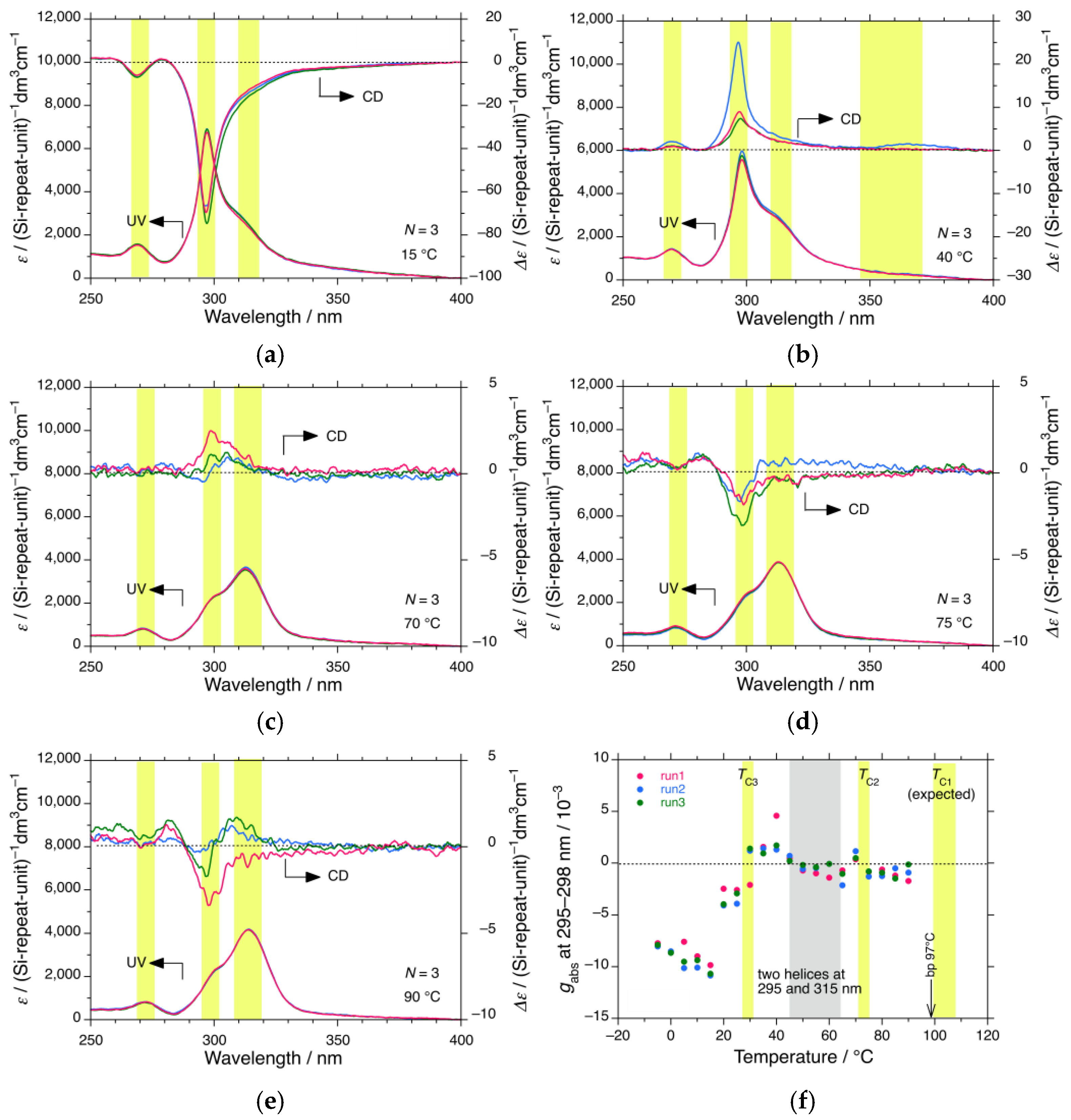
Figure 3a–e shows the changes in the CD and UV spectra obtained from three runs (N = 3) of iBS in n-propanol at 15 °C, 40 °C, 70 °C, 75 °C, and 90 °C, respectively. The yellow bars at ~270 nm, ~298 nm, and ~315 nm are assigned to three different helices, 114, 83, and 73, respectively, according to our previous TD-DFT and DFT calculations of ES [58]. All CD and UV spectra (N = 3) in the range from −5 °C to 90 °C at 5 °C intervals are shown in Figure S8 in Supplementary Materials.
Although the three helices coexist in n-propanol, their fractions and helical screw sense greatly depend on the solution’s temperature. From Figure 3a and Figure S8a–e in Supplementary Materials, iBS in n-propanol below 15 °C clearly reveals an intense (–)-CD band at 298 nm, a distinct (–)-CD band at 270 nm, and a clear (–)-CD band at 315 nm as a shoulder to the 298 nm (–)-CD band. Additionally, a broader weak UV band tail around 350 nm is assigned to the 157-helix. The temperature dependence of the multiple CD bands showing the same and opposite CD signs is a unique feature of iBS, iPS, and iHS in achiral solvents, as described in the following sections. Similar characteristics of nBS in n-alkanes and ES in n-propanol have been observed [58].
Figure 3f shows the temperature-dependent gabs values at 295–298 nm for the 298 nm CD band. The yellow bars indicate three second-order phase transitions of the 298 nm CD band at TC1 (>97 °C, onset temperature of preferential screw sense helicity, expected), TC2 (72 °C, helix–helix transition), and TC3 (29 °C, helix–helix transition), while the gray bar is due to the coexistence of two opposite helices at 298 nm and 315 nm, as revealed by the bisignate CD profile (Figure S8j–o, Supplementary Materials).
From Figure S8g–i, Supplementary Materials, iBS between 30 °C and 40 °C reveals three (+)-CD bands at 298, 315, and 270 nm, indicating a helix–helix transition at TC3. In addition, iBS between 70 °C and 75 °C reveals chiroptical inversion of the 298 nm CD bands, while the corresponding 298 nm UV bands weakened; conversely, the 315 nm UV bands increased. Notably, a small fraction of the 295 nm (–)-CD band remained at the shoulder of the 315 nm UV band, even at 85 °C and 90 °C, which is close to the boiling point (bp 97 °C) of n-propanol (Figure S8s–t, Supplementary Materials). The onset temperature of preferential screw sense helicity (TC1) is expected to be above 97 °C. Above TC1, the handed helicity is lost and becomes a racemic mixture of P-73- and M-73-helicity, which are called CD-silent and optically inactive states. Furthermore, the absolute magnitude of gabs, |gabs|, of the 298 nm CD band tended to decrease below 10 °C, indicating the occurrence of an additional helix–helix transition (TC4) below −20 °C. This suggests that iBS has a four helix-related phase-transition temperature. As shown in the following sections, iBS in p-dioxane-d8 and iPS in n-propanol have similar four transition temperatures, whereas nBS in n-alkanes has two transition temperatures.

3.1.2. iBS in i-Propanol

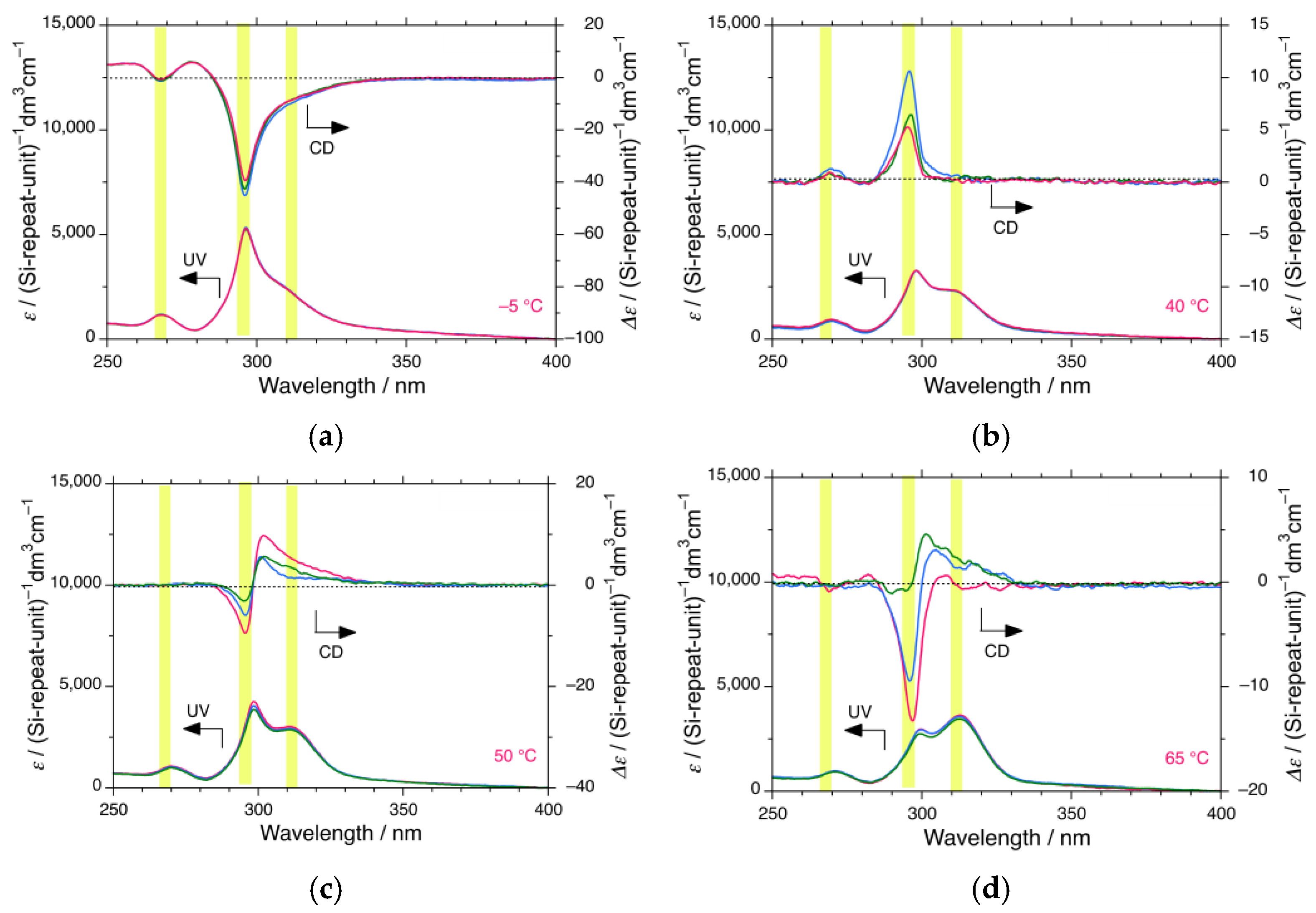
Figure 4a–e shows the changes in the CD and UV spectra (N = 3) of iBS in i-propanol at −5, 40, 50, 65 and 70 °C. The three yellow bars at ~270 nm, ~295 nm, and ~315 nm arise from the 114-, 83-, and 73-helices, respectively [58]. For clarity, all CD and UV spectra (N = 3) in the range from −5 °C to 90 °C at 5 °C intervals are shown in Figure S9 in Supplementary Materials. Similar to iBS in n-propanol, the fractions and helical screw sense of the three helices in i-propanol depend on temperature. From Figure 4a and Figure S9a–h in Supplementary Materials, iBS in i-propanol below 30 °C clearly reveals intense (–)-CD and UV bands at 298 nm with distinct (–)-CD and UV bands at 270 nm and obvious (–)-CD and UV bands at 315 nm. At temperatures above 35 °C, the 315 nm CD and UV bands become clear.
Figure 4f shows the temperature-dependent gabs values at 296 nm for the 298 nm CD band of iBS in i-propanol. The yellow bars show three second-order phase transitions for the 295 nm CD band at TC1 (>83 °C, onset temperature of preferential screw sense helicity, expected), TC2 (60 °C, helix–helix transition), and TC3 (32 °C, helix–helix transition), while the gray bar is due to two opposite screw sense helices at 295 nm and 315 nm coexisting between 50 °C and 65 °C, as shown by the bisignate CD profile (Figure 4c,d and Figure S9l–o in Supplementary Materials). The onset of preferential screw sense helicity temperature (TC1) is likely to be above 83 °C (bp of i-propanol). Above TC1, the handed helicity becomes a racemic mixture of P-73- and M-73-helicity, resulting in an optically inactive state. No significant differences in the CD and UV characteristics of iBS between n- and i-propanols were observed, although n- and i-propanols have one or two CH3 rotors, and are classified as primary and secondary alkanols, respectively.

3.1.3. iBS in i-Butanol

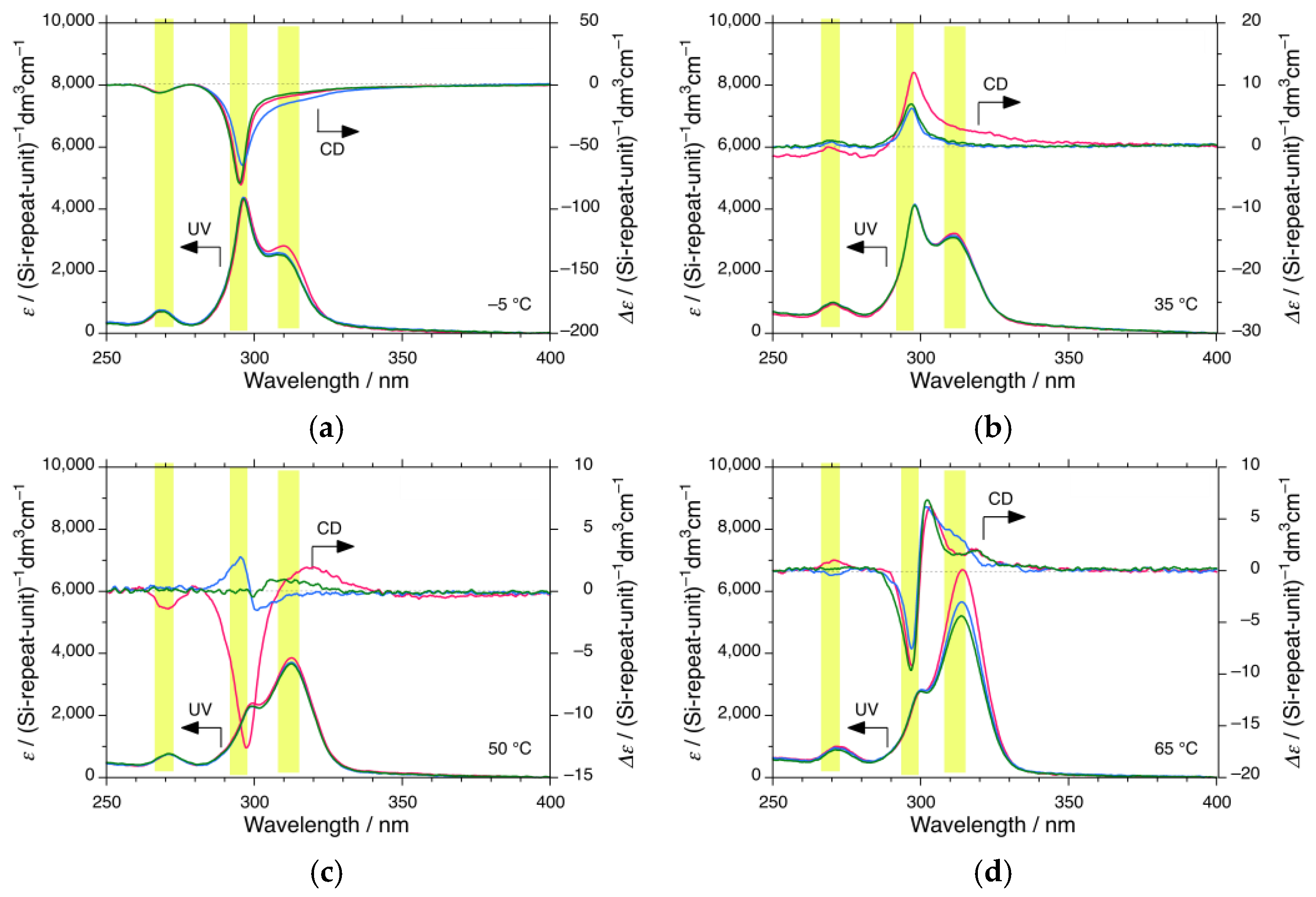
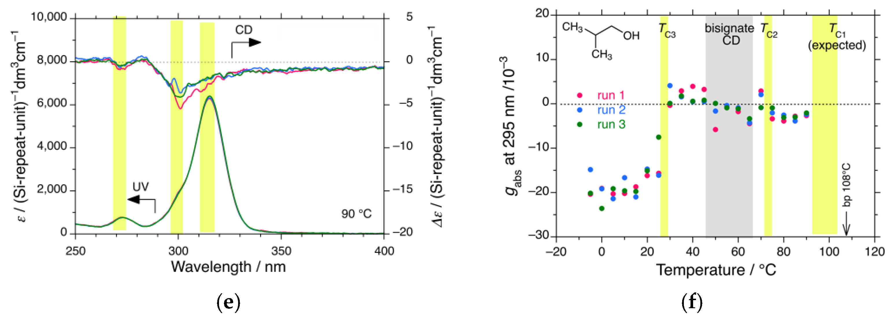
Figure 5a–e shows the CD and UV spectra (N = 3) of iBS in i-butanol at (a) −5 °C, (b) 35 °C, (c) 50 °C, (d) 65 °C, and (e) 90 °C, respectively. The three yellow bars at ~270 nm, ~295 nm, and ~315 nm are assigned to 114-, 83-, and 73-helices, respectively [58]. All CD and UV spectra (N = 3) in the range from −5 °C to 90 °C are shown in Figure S10 in Supplementary Materials.
Figure 5f plots the changes in the gabs values of the 295 nm CD band as a function of temperature. The yellow bars are due to three second-order phase transitions of the 295 nm CD band at TC1 (>100 °C, onset temperature of preferential screw sense helicity, expected), TC2 (72 °C, helix–helix transition), and TC3 (28 °C, helix–helix transition), while the gray bar is due to two opposite helices at 295 nm and 315 nm, as revealed by the bisignate CD profile in the range from 45 °C to 65 °C (Figure 5c,d and Figure S10k–o in Supplementary Materials).
Similarly, iBS in i-butanol below 25 °C showed intense (–)-CD and UV bands at 298 nm, distinct (–)-CD and UV bands at 270 nm, and a distinct UV band at 315 nm, with a less obvious (–)-CD band. Even at 80 °C and 90 °C, the 295 nm (–)-CD band at the shoulder of the 315 nm UV band is apparent, indicating that the handed 83-helix exists stably at elevated temperatures. The onset 83-helix generation temperature may be above 100 °C, which is close to the 108 °C bp of i-butanol.
There were no significant differences in the CD and UV characteristics of iBS in i-propanol and n-propanol, although they both carry two CH3 rotors and are classified as secondary and primary alkanols. The noticeable difference between i-propanol and i-butanol may be due to the relative magnitude of the 315 nm UV band compared to the 295 nm UV band: the 315 nm UV band in i-butanol appears more strongly in i-propanol band in the range from −5 °C to 70 °C.

3.1.4. iBS in i-Pentanol

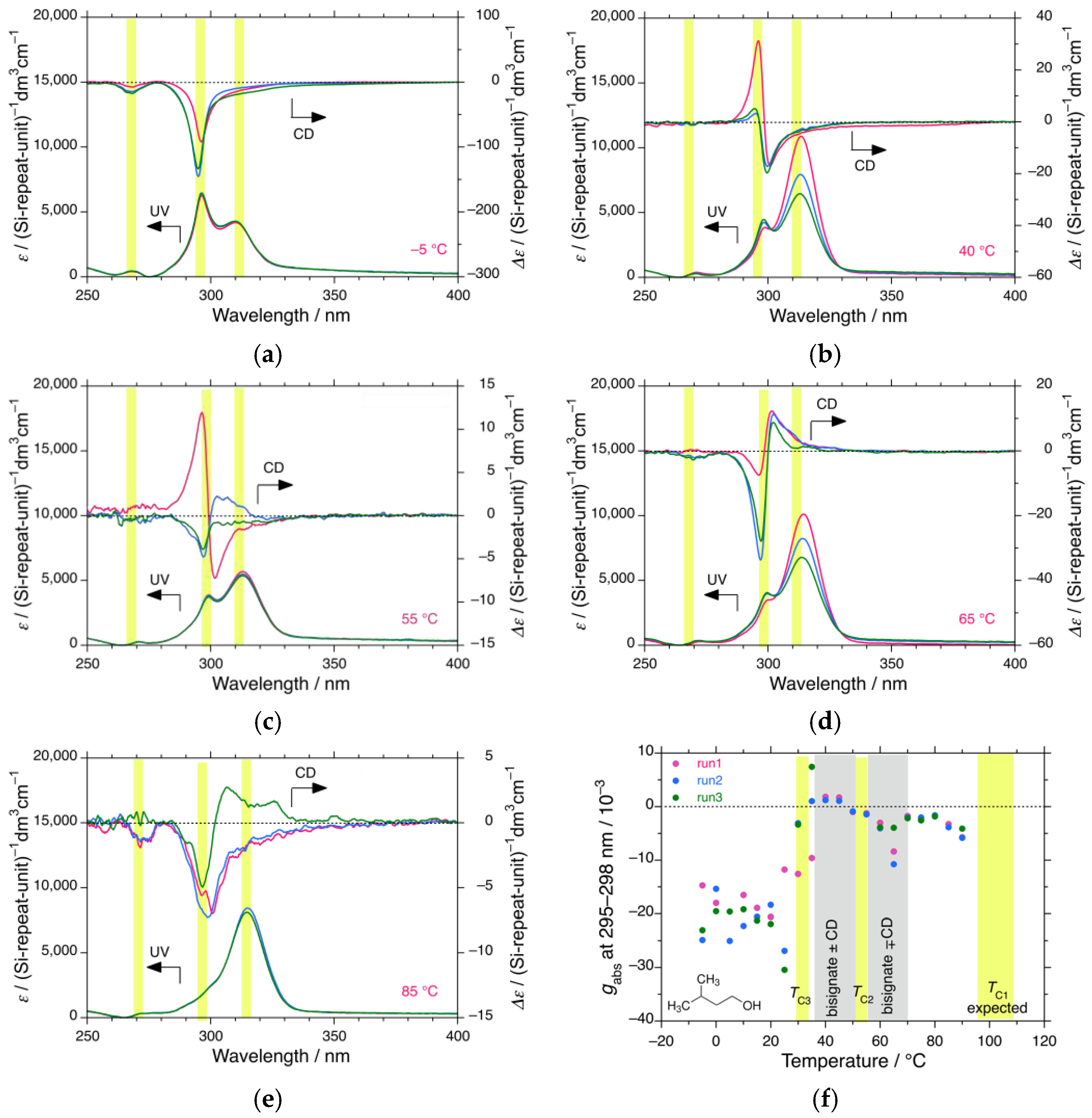
Figure 6a–e shows the changes in the CD and UV spectra (N = 3) of iBS in i-pentanol at (a) −5, (b) 40, (c) 55, (d) 65, and (e) 85 °C, respectively. The three yellow bars at ~270 nm, ~295 nm, and ~315 nm were assigned to 114-, 83-, and 73-helices, respectively [58]. All the CD and UV spectra (N = 3) in the range from −5 °C to 90 °C at 5 °C intervals are displayed in Figure S11 in Supplementary Materials.
Figure 6f shows the changes in the gabs values at 295 nm–298 nm of the 295 nm CD band as a function of temperature. The yellow bars are due to three second-order phase transitions of the 295 nm CD band at TC1 (>100 °C, onset temperature of preferential screw sense helicity, expected), TC2 (50 °C, helix–helix transition), and TC3 (25 °C, helix–helix transition), while the gray bars are due to two opposite helices at 295 nm and 315 nm from the bisignate CD profiles in the range from 35 °C to 70 °C (Figure 6b–d and Figure S11i–p in Supplementary Materials). Evidently, iBS in i-pentanol below 25 °C exhibited intense (–)-CD and UV bands at 298 nm, distinct (–)-CD and UV bands at 270 nm, and a distinct (–)-UV band at 270 nm with a (–)-CD shoulder band. On the other hand, even at 75–90 °C, the 295 nm (–)-CD band at the shoulder of the 315 nm UV band remains. The handed 83-helix is stable at elevated temperatures. The onset 83-helix generation temperature is assumed to be above 100 °C, which is close to 138 °C bp of i-pentanol.
There were no significant differences in the CD and UV characteristics of iBS between i-butanol and i-pentanol, although they both carry two CH3 rotors and are classified as primary alkanols. The noticeable difference between n-propanol, i-butanol, and i-propanol is the relative magnitude of the 315 nm UV band relative to the 295 nm UV band: the 315 nm UV band in i-butanol and i-propanol was more intense than that in n-propanol in the range from −5 °C to 70 °C.

3.1.5. iBS in n-Butanol, 2-Ethyl-1-butanol, and Cyclopentanol

Changes in the CD and UV spectra (N = 1) of iBS in the other three alkanols at three temperatures are shown in Figure S12 in Supplementary Materials: in n-butanol at −5, 20, and 40 °C, Figure S12a; in 2-ethyl-1-butanol at 0, 20, and 40 °C, Figure S12b; in cyclopentanol at −5, 20, and 40 °C, Figure S12c. Clearly, iBS in n-butanol below 40 °C shows very intense 295 nm (–)-CD and UV bands, 270 nm (–)-CD and UV bands, and a 315 nm UV band with a less-obvious 315 nm CD band. It is possible that a helix–helix transition occurs above 40 °C, which is higher than that in n-propanol. Although iBS in 2-ethyl-1-butanol at 0 °C has a 295 nm (–)-CD band at the tail of the 315 nm UV band, the 295 nm CD band completely disappears above 20 °C. 2-Ethyl-1-butanol was ineffective in inducing the 295 nm helix because of the bulky primary alkanol in this work. Cyclopentanol without a CH3 rotor can induce intense 295 nm (–)-CD and UV bands below −5 °C, but the CD band weakens at 20 °C and disappears at 40 °C.

3.1.6. iBS in p-Dioxane-h8 and p-Dioxane-d8—Less-Obvious H/D Isotope Effect

In a previous study [58], we observed notable H/D isotope effects in the temperature-dependent gabs characteristics of nBS in n-dodecane-d26 and -h26. n-Dodecane-d26 weakened the gabs values by one order of magnitude, induced oppositely signed CD spectra, and lowered the helix generation and helix–helix transition temperatures (TC1 and TC2). n-Dodecane-h26 and -d26, which are pure hydrocarbons carrying two CH3 and CD3 rotors at the termini, are unable to coordinate to the empty d-orbital of the Si atom. To further test the possible H/D isotope effect, p-dioxane-h8 and -d8, which are weakly polar cyclic ethers without CH3 rotors, were chosen as achiral solvents. It was expected that the two oxygen atoms of the ethers would be capable of coordinating to the empty d-orbital of the Si atom, as well as the oxygen atom of the alkanols.
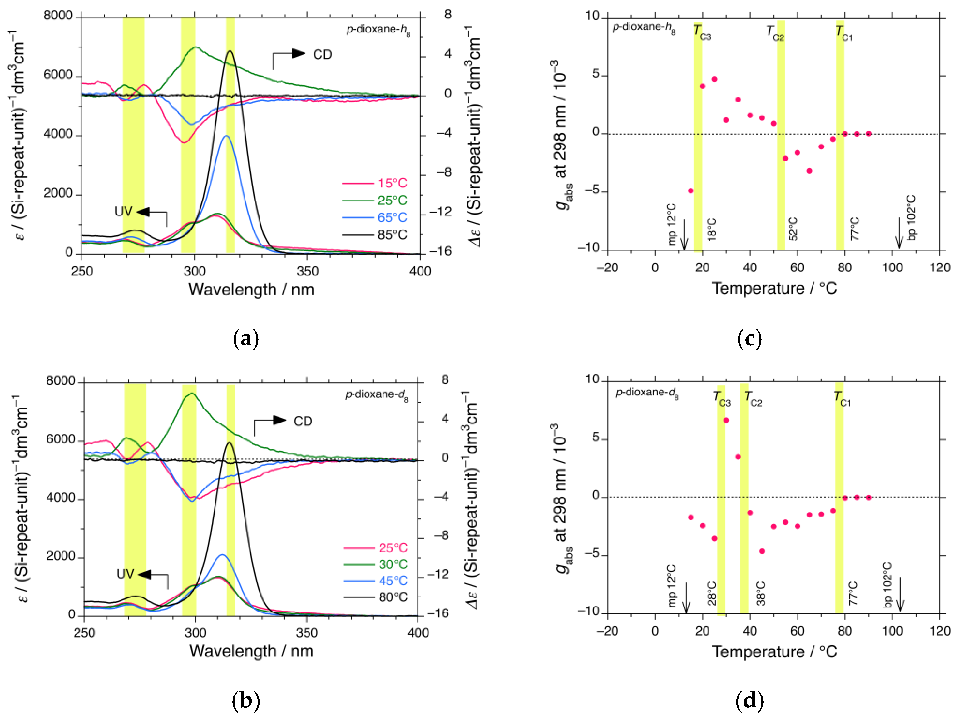
Figure 7a shows the changes in the CD and UV spectra (N = 1) of iBS in p-dioxane-h8 at 15, 25, 65, and 85 °C. For comparison, Figure 7b shows the CD and UV spectra (N = 1) of iBS in p-dioxane-d8 at 25, 30, 45, and 80 °C. All CD and UV spectra (N = 1) ranging from 15 °C to 90 °C at 5 °C intervals are displayed in Figures S13 and S14 in Supplementary Materials. It is evident that the three CD and UV bands at ~270, ~295, and ~315 nm, which are assigned to the 114-, 83-, and 73-helices, respectively, can be clearly seen. However, the 315 nm UV band relative to the 295 nm UV band below 30 °C is considerably more intense compared to that in n-propanol, i-propanol, i-butanol, and i-pentanol, reflecting the different coordination ability of oxygen atoms between ethers and alkanols.
Figure 7c shows the temperature dependence of the gabs values at 298 nm for the 295 nm CD band in p-dioxane-h8, which indicates three second-order phase transitions at TC1 (77 °C, onset temperature of preferential screw sense helicity), TC2 (52 °C, helix–helix transition), and TC3 (18 °C, helix–helix transition). For comparison, Figure 7d shows the temperature-dependent gabs values at 298 nm of the 295 nm CD band in p-dioxane-d8; three second-order phase transitions at TC1 (77 °C, onset temperature of preferential screw sense helicity), TC2 (38 °C, helix–helix transition), TC3 (28 °C, helix–helix transition), and TC4 (< 10 °C, helix–helix transition, expected).
As shown in Figure 7c,d, the three helix-related phase temperatures between p-dioxane-h8 and -d8 are very similar; TC1 between p-dioxane-h8 and -d8 is identical; TC2 in p-dioxane-h8 is 14 °C higher than that in p-dioxane-d8; conversely, TC3 in p-dioxane-h8 is 10 °C lower than that in p-dioxane-d8. As shown in Figure 7d, a fourth helix–helix transition temperature, TC4 < 10 °C, is likely to exist, and the H/D isotope effect between p-dioxane-h8 and -d8 is not obvious, possibly because of the lack of CH3 rotors and/or the weak coordination ability of the ethereal oxygen to the empty d-orbital of the Si atom.
These results led us to the ideas that (i) the temperature-dependent gabs characteristics of iBS in p-dioxane-h8 and -d8 partly arise from two i-butyl side chains carrying four independent directional CH3 rotors per repeating unit and (ii) the coordination capability of achiral solvent molecules to the empty d-orbital of the Si atom may be another important factor.

3.2. CD, UV, CPL, and PL Spectral Characteristics of iPS and iHS

Next, to determine the β-, γ-, and δ-branching effects at the i-alkyl side chains of the polysilanes, the temperature-dependent CD and UV spectra of iPS and iHS in n-propanol, n-pentanol, n-hexanol, n-octanol, i-propanol, i-butanol, and 2-ethyl-1-butanol as solvents were measured. The CD and UV characteristics of iPS and iHS are described below.

3.2.1. iPS in n-Propanol

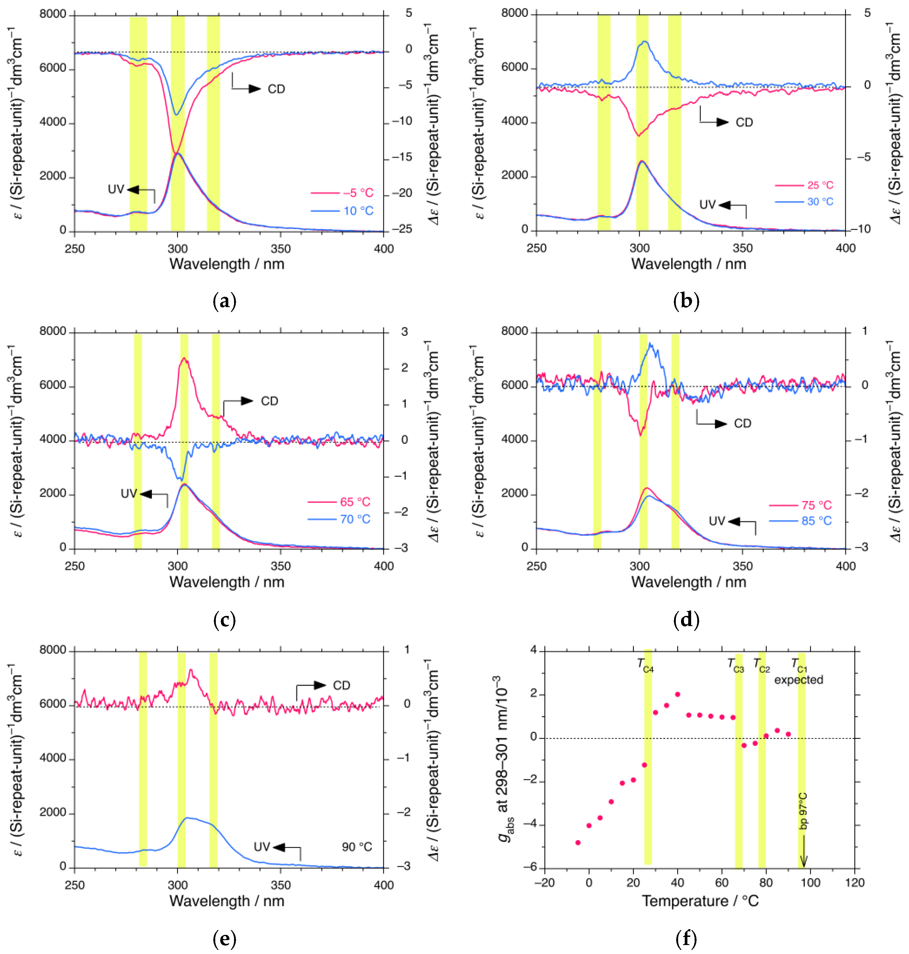
Figure 8a–e shows the CD and UV spectra (N = 1) of iPS in n-propanol at (a) –5 and 10 °C, (b) 25 and 40 °C, (c) 65 and 70 °C, (d) 75 and 85 °C, and (e) 90 °C. For further evidence, all the CD and UV spectra (N = 1) in the range from –5 °C to 90 °C at 5 °C intervals are shown in Figure S15 in Supplementary Materials. Figure 8f plots the temperature-dependent gabs values of iPS in n-propanol at 298–301 nm for the 295 nm CD band. The yellow bars are due to three second-order phase transitions of the 295 nm CD band at TC1 (>97 °C, onset temperature of preferential screw sense helicity, expected), TC2 (80 °C, helix–helix transition), TC3 (67 °C, helix–helix transition), and TC4 (27 °C, helix–helix transition). A fourth helix–helix transition temperature, TC4, is evident.
Similar to the series of iBS systems mentioned above, iPS in n-propanol reveals temperature-dependent CD generation and inversion characteristics, exhibiting four transition temperatures in the range from −5 °C to 90 °C. The gabs values of iPS and iBS in n-propanol at 0 °C were −0.5 × 10⁻² and −0.8 × 10⁻², respectively. The major difference is that while iPS exhibits a (+)-CD band, iBS conversely shows a (–)-CD band when the solution temperature is above TC2, possibly due to a difference in the rotational freedom of the CH2 spacers (two + two for iPS and one + one for iBS).

3.2.2. iPS in n-Pentanol, n-Hexanol, and n-Octanol, and iHS in n-Octanol

To determine the dependence of n-alkanols and the γ-/δ-branching position dependence of achiral alkyl side chains, the CD and UV characteristics of iPS and iHS in n-alkanols were tested. Figure S16a–c in Supplementary Materials, respectively, shows the CD and UV spectra (N = 1) of iPS in (a) n-pentanol at 0, 20, and 45 °C, (b) n-hexanol at 0, 20, and 45 °C, and (c) n-octanol at 0, 20, and 45 °C. Figure S16d in Supplementary Materials shows the changes in the CD and UV spectra (N = 1) of iHS in n-octanol at 0, 20, and 45 °C. As shown in Figure S16a–d in Supplementary Materials, iPS and iHS have three handed helices at ~275 nm, ~297 nm, and ~310 nm [58].
In n-pentanol and n-hexanol, iPS has the same (–)-CD band at 297 nm at 0 °C, but this is lost at 20 and 45 °C, whereas in n-octanol, iPS has 297 nm (–)-CD bands at 0 °C, none at 20 °C, and inverted (+)-CD bands at 45 °C. iHS in n-octanol showed a weak and broad 297 nm (–)-CD band at 0 °C, which was lost at 20 and 45 °C. In n-octanol at 0 °C, iHS and iPS have the same CD sign, but the magnitude of the iHS band is one-third of iPS. This arises from the rotational freedom of the CH2 spacers at the nearest neighbors of the CH3 rotors. The numbers of CH2 spacers in the branched side chains (one + one for iBS, two + two for iPS, and three + three for iHS) are critical factors in determining temperature-dependent gabs characteristics.

3.2.3. iPS in i-Propanol, i-Butanol, and 2-Ethyl-1-butanol

To determine the effect of branched alkanols, the CD and UV spectra of iPS for the three alkanols were measured. Figure S17a–c in Supplementary Materials, respectively, shows the CD and UV spectra (N = 1) of iPS in (a) i-propanol at 0, 20, and 40 °C, (b) i-butanol at 0, 20, and 40 °C, and (c) 2-ethyl-1-butanol at 0, 20, and 40 °C. The yellow bars in Figure S17a–c are due to the three helices at ~280 nm, ~300 nm, and ~315 nm [58]. In i-propanol, iPS showed a (–)-CD band of 297 nm at 0 °C, weakened at 20 °C, and inverted to a (+)-CD band at 40 °C. In i-butanol, iPS shows a 297 nm (–)-CD band at 0 °C, which weakens at 20 °C and is lost at 40 °C. Interestingly, in 2-ethyl-1-butanol, iPS showed a very weak (+)-CD band at 297 nm at the tail of the 315 nm UV band at 0, 20, and 40 °C. Among all the achiral alkanols tested, 2-ethyl-1-butanol is the bulkiest, which may inhibit coordination with the Si atom.

3.3. Solvent Effects in CD and UV Characteristics of iBS and iPS

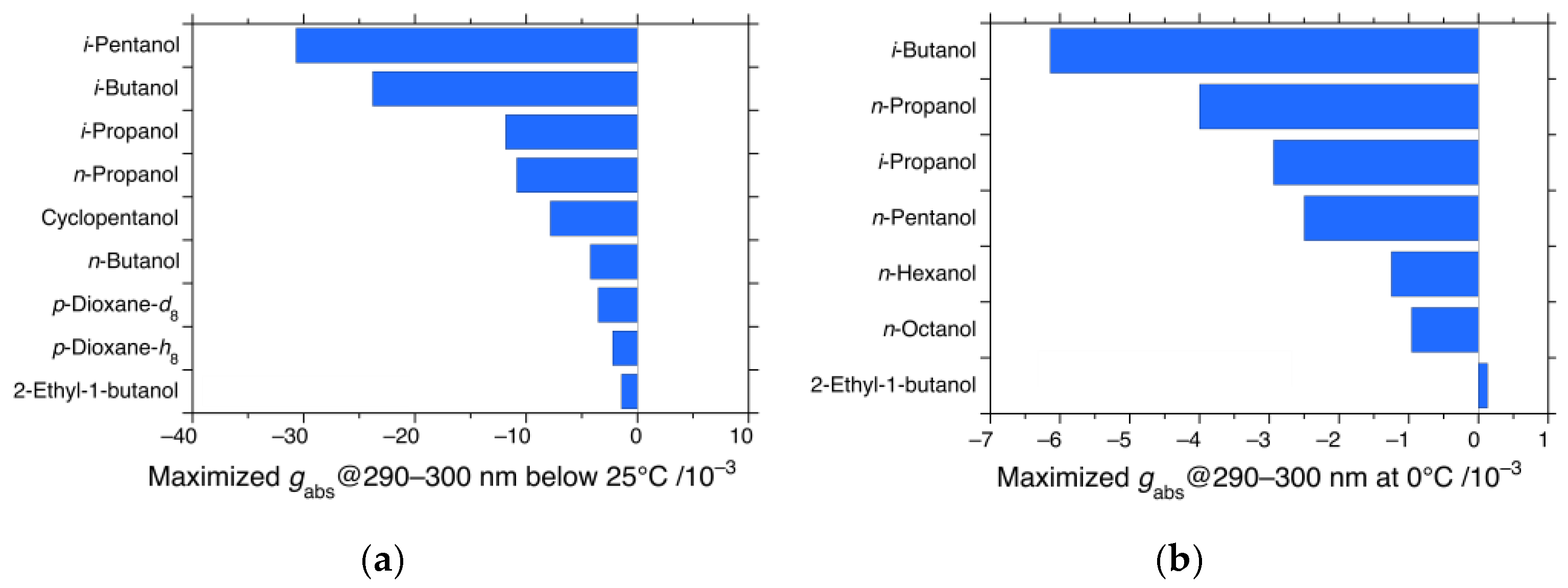
From the solvent-dependent gabs characteristics at the 290 nm CD and UV bands (Figure 3, Figure 4, Figure 5, Figure 6, Figure 7 and Figure 8 and Figures S8–S17 in Supplementary Materials), the maximized gabs values of iBS and iPS in various alkanols and p-dioxane below 25 °C are summarized in Figure 9a,b, respectively.
In iBS, i-alkanols induced intense gabs values compared to n-alkanols, except for sterically crowded 2-ethyl-1-butanol. However, p-dioxane-h8 and -d8 do not strongly induce gabs because of the absence of a CH3 rotor and/or ethereal oxygen atom. The presence of a CH3 rotor in the n- and i-alkanols is crucial. As shown in Figure 9a,b, the maximum gabs values of iPS were reduced to approximately one-fifth of those of iBS. Sterically compact alkanols such as i-butanol, n-propanol, i-propanol, and n-pentanol tend to provide considerably more intense gabs values. Furthermore, the gabs value of iHS was approximately one-third of that of iPS when n-octanol was used as the solvent (Figure S16c,d in Supplementary Materials). The relative magnitudes of the |gabs| values of iBS, iPS, and iHS were therefore evaluated as 1.0:0.2:0.07, respectively. From the relative magnitudes in the |gabs| values, the rotational freedom of CH2 spacers in the branched alkyl side chains of iBS, iPS, and iHS decreased in the order of i-butyl < i-pentyl < i-hexyl groups. The numbers of CH2 spacers are a crucial factor to regulate temperature-dependent CD generation and inversion characteristics.

3.4. CPL and CD Polarization and Inversion of iBS and iPS in n-Propanol at −5 °C

Although optically active polysilanes are chiral chromophores, they work as excellent luminophores because of the decay of the Wannier exciton (photogenerated electron-hole pair) in silicon quantum wires (Si-QWs) that are susceptible to metastable helicity in the S1 state. The absolute values of glum, |glum|, and helical screw sense in the S1 state often differ from those in the S0 state, as demonstrated in the following.
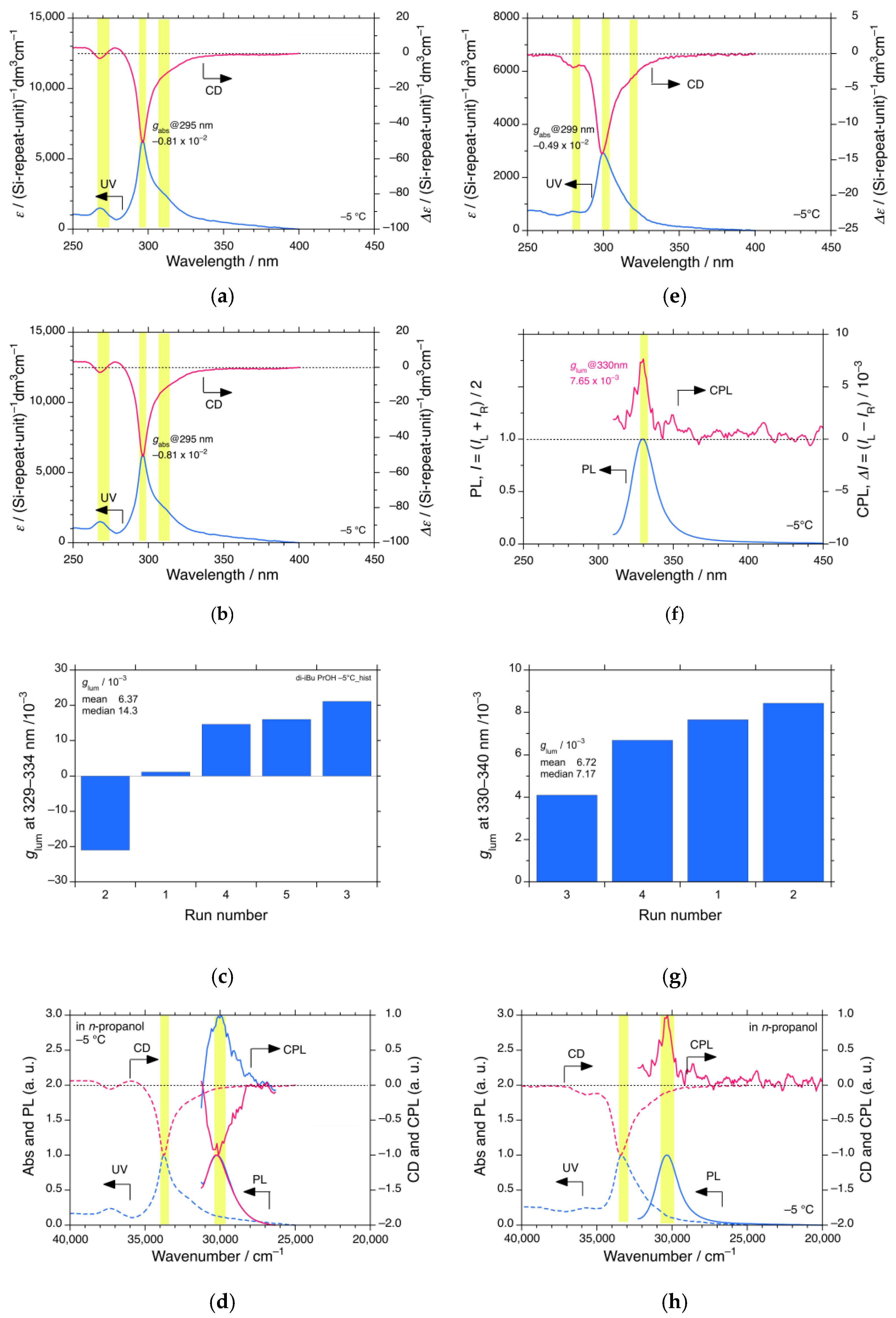
Figure 10a–d summarizes the CD and CPL characteristics of iBS in n-propanol at −5 °C: Figure 10a shows the CD and UV spectra with the greatest gabs values at 295 nm (among N = 3); Figure 10b shows the CPL and PL spectra with the greatest |glum| values at 340 nm with (+)- and (–)-signs; Figure 10c is its histogram (N = 5). Figure 10d shows a comparison of the normalized CD, UV, CPL, and PL spectra as a function of wavenumber. In addition, Figure S18a–d in Supplementary Materials shows the CD and CPL characteristics of iBS in i-butanol at −5 °C; Figure S18a shows the CD and UV spectra with the greatest gabs value at 295 nm (among N = 3); Figure S18b shows the CPL and PL spectra showing the greatest |glum| values at 330–335 nm with (+)- and (–)-signs; and Figure S18c is its histogram (N = 3). Figure S18d compares the normalized CD, UV, CPL, and PL spectra as a function of wavenumber.
Clearly, the CPL spectra of iBS in n-propanol and i-butanol oscillate between P- and M-83-helicity in DWP in the S1 state within the N = 3 and 5 experiments, although the corresponding (–)-CD spectra in the alkanols indicate a stable handed 83-helicity in the S0 state. The oscillation may be a characteristic of stochastic resonance. For further evidence, all CPL and PL spectra of iBS in n-propanol and i-butanol at −5 °C are shown in Figure S19a–h in Supplementary Materials.
The values of |glum| and |gabs| in n-propanol and i-butanol are compared: in n-propanol, |glum| = 2.10 × 10⁻² at 340 nm, which is 2.5 times greater than |gabs| = 0.86 × 10⁻² at 295 nm; and in i-butanol, |glum| = 2.00 × 10⁻² at 335 nm, which is very similar to |gabs| = 1.78 × 10⁻² at 295 nm. From Figure 10d and Figure S18d in Supplementary Materials, the apparent Stokes shifts between CD and CPL extrema in n-propanol and i-butanol are as large as 3590 cm−1 and 3640 cm−1, respectively. The large Stokes shifts, including the sign inversion between the S0 and S1 states imply great reorganization of the Si–Si main chain in the S1 state in the order of nanoseconds.
However, the CPL and CD inversion characteristics of iPS differ considerably from those of iBS. Figure 10e–g shows the CD and CPL characteristics of iPS in n-propanol at −5 °C; Figure 10e shows the CD and UV spectra with the greatest gabs value at 299 nm (N = 3); Figure 10f shows the CPL and PL spectra showing the greatest glum value with the (+)-sign at 330 nm; Figure 10g shows its histogram (N = 4); and Figure 10h shows the normalized CD, UV, CPL, and PL spectra as a function of wavenumber. Although the (+)-CPL and (–)-CD spectral profiles of iPS in n-propanol were inverted, all the (+)-CPL spectral profiles within the N = 4 experiments were unchanged. The helical screw sense of the 83-helicity in the S1 state is opposite to that in the S0 state. The |glum| = 0.77 × 10⁻² at 330 nm is approximately twice that of |gabs| = 0.45 × 10⁻² at 299 nm. A large Stokes shift of 3150 cm−1 (Figure 10h) is evidence that a significant structural reorganization of the Si–Si main chain occurs in the S1 state. For further evidence, all CPL and PL spectra of iPS in n-propanol at −5 °C are shown in Figure S19i–l in Supplementary Materials. The numbers of the CH2 spacers play a key role in regulating the CD and CPL polarization and inversion characteristics in n-propanol.

3.5. VCD and IR Spectral Characteristics of Achiral Alkanols and Alkyl Ethers

In a previous paper [58], we showed the (–)-VCD and IR spectra of a dozen of stereocenter-free molecules carrying CH3 and CF3 groups, that is, n-alkanes, perfluoro-n-alkanes, acetonitrile, and mono- and di-substituted benzene derivatives, although most chemists do not believe that these molecules are chiral and/or optically active. We hypothesized that because the CH3 and CF3 groups are representative of three identical nuclear-spin-1/2 systems made of three 1H and 19F atoms in TWP, the rotors are perpetually working as unidirectional hindered rotors because of the handedness of the PV-anapole moment [58]. This idea was applied to the behavior of CH3 termini at the two n-butyl side chains of nBS. In this work, we further investigated whether stereocenter-free alkanols and alkyl ethers carrying CH3 rotors reveal VCD signals, and for comparison also alkanols and alkyl ethers without CH3 rotors. VCD and IR spectral assignments of solvents and molecular liquids in this section were made based on comprehensive IR and Raman handbooks [105,106]. A further discussion of seven solvents (ethanol, n-propanol, n-butanol, i-propanol, i-butanol, p-dioxane, and THF), three molecular liquids (diglyme, 15-crown-5-ether (15CE5), and α-terpinene), etc. was given in the later Section 4.2.3 with proper references and Section 4.2.2 showing theoretical simulations of VCD and IR spectra for specific chiral and achiral rotamers.
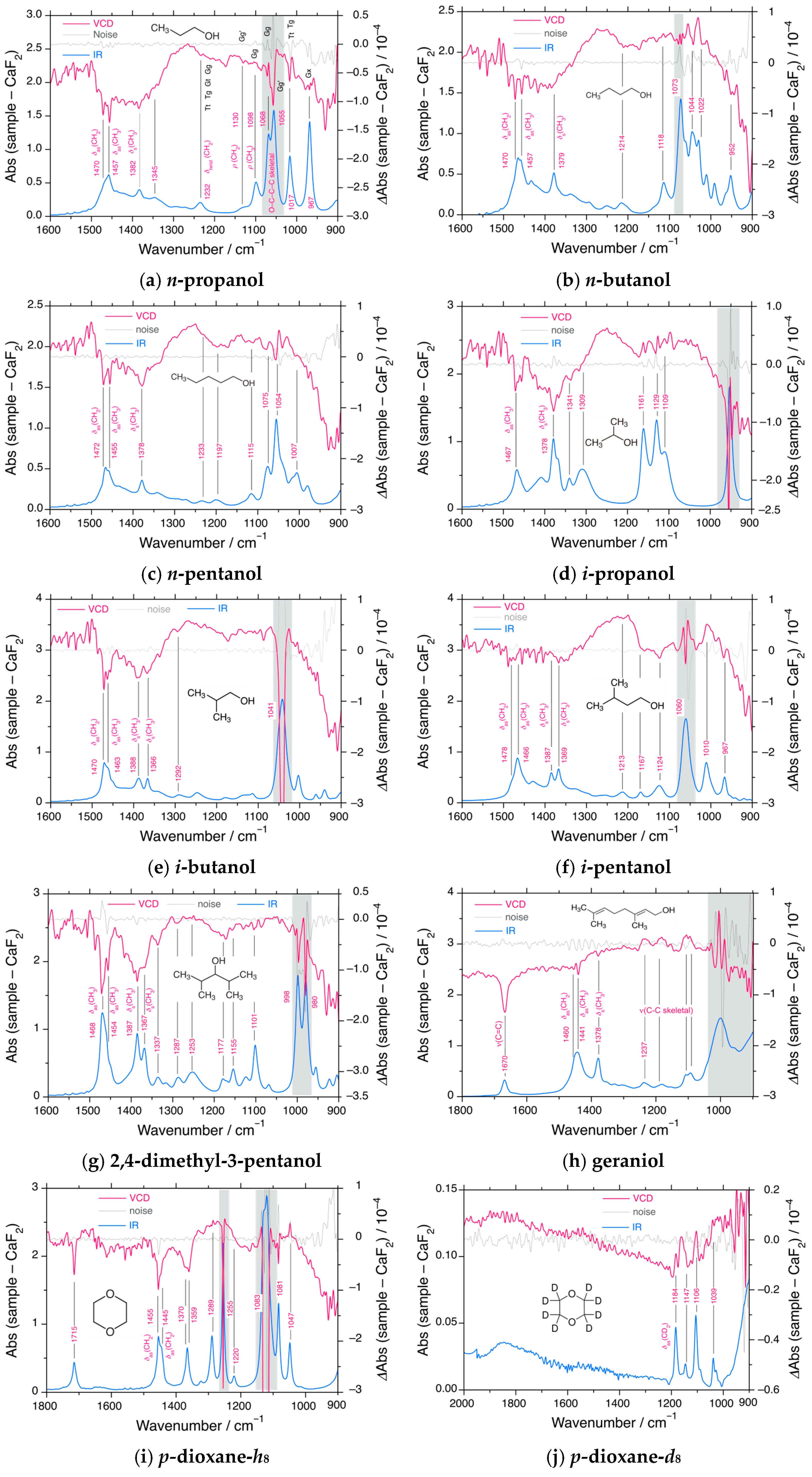
Figure 11a–j shows the VCD and IR spectra of various alkanols and p-dioxane: (a) n-propanol, (b) n-butanol, (c) n-pentanol, (d) i-propanol, (e) i-butanol, (f) i-pentanol, (g) 2,4-dimethyl-3-pentanol, (i) geraniol (trans-3,7-dimethyl-2,6-octadien-1-ol), (j) p-dioxane-h8, and (k) p-dioxane-d8. In addition, the VCD and IR spectra of other alkanols (methanol, ethanol, n-hexanol, n-octanol, cyclopentanol, 2-ethyl-1-butanol), cyclic alkyl ethers without CH3 rotors (tetrahydropyran (THP), THF, diglyme, and 15CE5), and acyclic alkyl ethers with CH3 rotors (diglyme = diethylene glycol dimethyl ether) are shown in Figure S20 in Supplementary Materials.
In a series of five n-alkanols carrying one CH3 rotor, that is, n-propanol, n-butanol, n-pentanol, n-hexanol, and n-octanol, three major (–)-VCD and IR bands at 1470–1473 cm−1, 1455–1457 cm−1, and 1455–1457 cm−1, which are assigned to CH2 scissor (δas(CH2)), CH3 asymmetric bending (δas(CH3)), and CH3 symmetric bending/umbrella) (δs(CH3)), respectively, were observed in each case (Figure 11a–c and Figure S20c,d in Supplementary Materials) [105,106]. However, the VCD bands at δas(CH3) and δs(CH3) for methanol and ethanol were less obvious (Figure S20a,b, Supplementary Materials) [105,106]. Although the intense IR signals at 1050 cm−1 and 1090 cm−1 are assigned to gauche and trans rotamers of skeletal C–C–O–H bonds [105,106], respectively, the corresponding intense (–)-VCD signals are ascribed to instrumental artifacts due to instrumental limitations when the IR absorbance exceeds 1.0 (smaller than 10% transmittance).
In the series of i-propanol, i-butanol, and i-pentanol with two CH3 rotors (Figure 11d–f), all the i-alkanols revealed δas(CH3)-origin (–)-VCD bands at 1463–1467 cm−1 and δs(CH3)-origin (–)-VCD bands at 1366–1387 cm−1 [105,106]. In addition, i-butanol and i-pentanol showed δas(CH2)-origin (–)-VCD bands at 1470–1478 cm−1, whereas δas(CH3)- and δs(CH3)-origin (–)-VCD bands were observed for i-propanol. The (–)-VCD and IR bands of δas(CH3) were spectroscopically similar to those of δas(CH2). Notably, 2,4-dimethyl-3-pentanol carrying four CH3 rotors and lacking a CH2 group revealed obvious δas(CH3)-origin (–)-VCD and IR signals at 1468 cm−1 and 1454 cm−1, and δs(CH3)-origin VCD and IR signals at 1387 cm−1 and 1367 cm−1 (Figure 11g) [105,106].
More interestingly, the isolated (–)-VCD and IR bands of geraniol (trans-3,7-dimethyl-2,6-octadien-1-ol) at 1670 cm−1, assigned to the stretching modes of the two C=C bonds were spectroscopically resolved, in addition to the (–)-VCD signals at 1441 cm−1 for δas(CH2) and δas(CH3), and 1378 cm−1 for δs(CH3) (Figure 11h) [105,106]. The two C=C bonds are likely to twist in one direction because of the handedness of the three CH3 rotors. Optically active geraniol is a realistic model of optically active ethylene twisted by 10°, induced by a handed WNC [42,43].
Cyclopentanol exhibited sharper (–)-VCD and IR signals assigned to δas(CH2) at 1473 cm−1, 1458 cm−1, and several other bands, that is, 1411 cm−1, 1342 cm−1, 1215 cm−1 (Figure S20e, Supplementary Materials) [105,106]. However, bulkier 2-ethyl-1-butanol with two CH3 rotors showed (–)-VCD and IR signals at 1468 cm−1 of δas(CH2), although signals due to δas(CH3) at 1460 cm−1 and δs(CH3) at 1377 cm−1 were less obvious (Figure S20f, Supplementary Materials) [105,106].
Regardless of the absence of a CH3 rotor, p-dioxane-h8 unexpectedly exhibited clear (–)-VCD and IR signals at 1715 cm−1, 1455 cm−1, 1445 cm−1, 1370 cm−1, 1359 cm−1, etc. (Figure 11i) [105,106]. It is known that, using Raman scattering spectroscopy, p-dioxane-h8 adopts three conformers: chair (D2d), 1,4-twisted boat (C1), and 2,5-twisted boat (C1) [107]. Our DFT calculations implied that the handed 2,5-twisted boat was responsible for the (–)-VCD and IR spectra. In contrast, p-dioxane-d8 showed very weak (+)-VCD and IR signals at 1000–1200 cm−1 (Figure 11j). The apparent sign inversion between p-dioxane-h8 and -d8 may be ascribed to the opposite sign in electroweak (EW) charge, Qw, between 1H (Qw = −0.108) and 2H (Qw = +0.892) [58].
Other cyclic alkyl ethers without a CH3 rotor (THP, THF, and 15CE5) exhibited multiple (–)-VCD and IR signals of δas(CH2): 1470 cm−1, 1455 cm−1, 1441 cm−1 for THP; 1460 cm−1 and 1449 cm−1 for THF; and 1476 cm−1 and 1454 cm−1 for 15CE5 (Figure S20g–i, Supplementary Materials) [105,106]. Diglyme, which is an acyclic alkyl ether carrying two CH3 rotors, shows (–)-VCD and IR bands at 1474 cm−1, 1455, cm−1, and 1354 cm−1, which are assigned to δas(CH2), δas(CH3), and δs(CH3), respectively (Figure S20j, Supplementary Materials) [105,106]. THP and THF have the cyclic frameworks of D-pyranose (a six-membered ring of hexose) and D-furanose (a five-membered ring of pentose), respectively. THP and THF may adopt optically active twisted frameworks with handedness in the absence of stereogenic centers. Previously, it was theoretically discussed that a C2-endo twisted THF molecule with handedness as a model of D-furanose rings is stabilized by EPV = 1 × 10⁻¹⁷ kcal mol−1 [49]. A large number of THF molecules, exceeding 1017 in a liquid cell, may therefore cooperatively amplify the EPV to detectable levels (scenarios (i), (ii), and (vi)).
As discussed in this section, achiral alkanols and alkyl ethers, which contain at least one oxygen atom, exhibit (–)-VCD and IR signals in the region of δas(CH2), δas(CH3), and δs(CH3) [105,106], regardless of the presence or absence of the CH3 rotor. This situation differs slightly from that of the CH3 rotor-bearing n-alkanes. Oxygen has three stable isotopes with natural abundances of 16O (99.8%), 17O (0.038%), and 18O (0.205%). The contribution of HINS 17O is negligibly small. The two lone pairs of 16O become unequal due to attractive PV-EW charges that are Qw = +6.216 for 16O and Qw = −0.108 for 1H by the Z0-WNC [58] and the opposite signs in their Mulliken charges.

4. Discussion

4.1. Helix–Helix Transition Temperatures of Mirror-Symmetric Polysilanes in Achiral Solvents

From the temperature- and solvent-dependent CD and UV characteristics, iBS and iPS revealed four (or three) sets of second-order phase transitions at TC1, TC2, TC3, and TC4 in the range from −5 °C to 90 °C, respectively. Despite equal populations of P-73- and M-73-helices at a UV λmax of 315–320 nm above TC1, affording so-called “CD-silent helices,” at temperatures below TC1, handed P-83 or M-83 helix at a UV λmax of 295–300 nm was observed. This structural transition occurs due to an imbalance in the P-and-M-73 and P-and-M-83 helices below TC1. In general, TC1 is called the onset temperature of preferential screw sense helicity, analogous to temperature-dependent second-order phase transitions (spontaneous polarization) in superconductivity, ferroelectricity, and ferromagnetism. Upon further cooling, the handed P-83 or M-83 helix underwent helix–helix transitions at TC2, TC3, and TC4, eventually dropping to the (–)-CD and UV bands at 295–300 nm as the most stable handed 83-helix below TC4. On the other hand, nBS in medium length n-alkanes revealing two helix-related phase transitions at TC1 and TC2 finally afford (+)-CD and UV bands at 295–300 nm as the most stable 83-helix below TC2. Although ES in n-propanol shows only TC1 at 30 °C, TC2 may exist at temperatures below −10 °C.
The phase transition characteristics of iBS, iPS, iHS, nBS, and ES and relevance of the numbers of CH3 rotors in the side chains and the solvents are summarized in Table 1. Here, iBS in the four alkanols, p-dioxane-h8/-d8, and iPS in n-propanol are likely to have four TC, whereas nBS in n-alkanes has two TC, regardless of the number of CH3 rotors (0 to 2) in the solvents. How many helical phase transitions exist is connected to the number of CH3 rotors in the side chains per repeating unit: i-butyl and i-pentyl side groups have four degrees of rotational freedom, whereas n-butyl side groups have only two. This implies that the rotational freedom in clockwise (CW) and counterclockwise (CCW) motions of four or two CH3 rotors in the side groups is key to determining the number of helical phase transitions. Temperature-dependent handed rotation of CH3 groups driven by quantum tunneling in a three-well potential may be a key in the multiple helix–helix transitions [58]. Inherent mechanisms are yet unresolved and a further theoretical understanding is necessary.
Temperature-dependent chiroptical codes are listed in Table 2. Here, the CW and CCW codes of each rotor are expressed as (+1) and (−1), respectively. A pair of ( + ) and ( + ) means disrotatory gear-like motions, while ( + ) and ( + ) are conrotatory anti-gear-like motions. In the case of iBS and iPS possessing four rotor freedoms, two CW and two CCW codes result in zero above TC1, while three CW and one CCW code are +2, and four CW codes give +4 below TC4. Conversely, one CW and three CCW codes are −2 and four CCW codes are −4. For nBS with two rotor freedoms, one CW and one CCW code result in zero above TC1, while two CW and two CCW codes give +2 and −2 below TC2, respectively. Decoding chiroptical codes makes it possible to change the temperature, solvents, and side chains.
In addition, the nature of the solvent structures is crucial for controlling the TC and chiroptical CD signs and magnitudes. Oxygen-atom-containing solvent molecules induce (–)-CD and UV bands below 25 °C. Conversely, solvents lacking oxygen atoms induce (+)-CD and UV bands below 25 °C [58]. The oxygen atoms in the solvents may determine the helical screw sense of iBS, iPS, iHS, and nBS. The coordination capability of lone pairs of oxygen atoms to empty d-orbitals of Si atoms is a key factor.

4.2. Rotamers in Alkanols, Alkyl Ethers, and Trisilane Derivatives

4.2.1. Mulliken Charges and Electroweak Charges

The question is thus whether (i) n-alkanols adopt extended achiral rotameric structures (point group Cs or C1), such as trans-planar bonds or pseudo-cyclic/folded rotameric structures, including gauche bonds, or whether (ii) i-alkanols and branched alkanols prefer chiral rotameric structures (point group Cs or C1) involving gauche bonds. Most chemists believe that n-alkanols predominantly adopt achiral trans-rotameric structures; thus, they are optically inactive because of the absence of stereocenters (Figure 2 and Figure S2 in Supplementary Materials). However, a pseudo-cyclic geometry becomes chiral due to the rotational C–C and C–O bonds, forcing the H(δ+)–C(δ) bond at the CH3 terminus to a position closer to the polar O(δ)–H(δ+) bond causing intramolecular attractive C(δ)–H(δ+)/O(δ) interactions [108,109] (Figure 12).
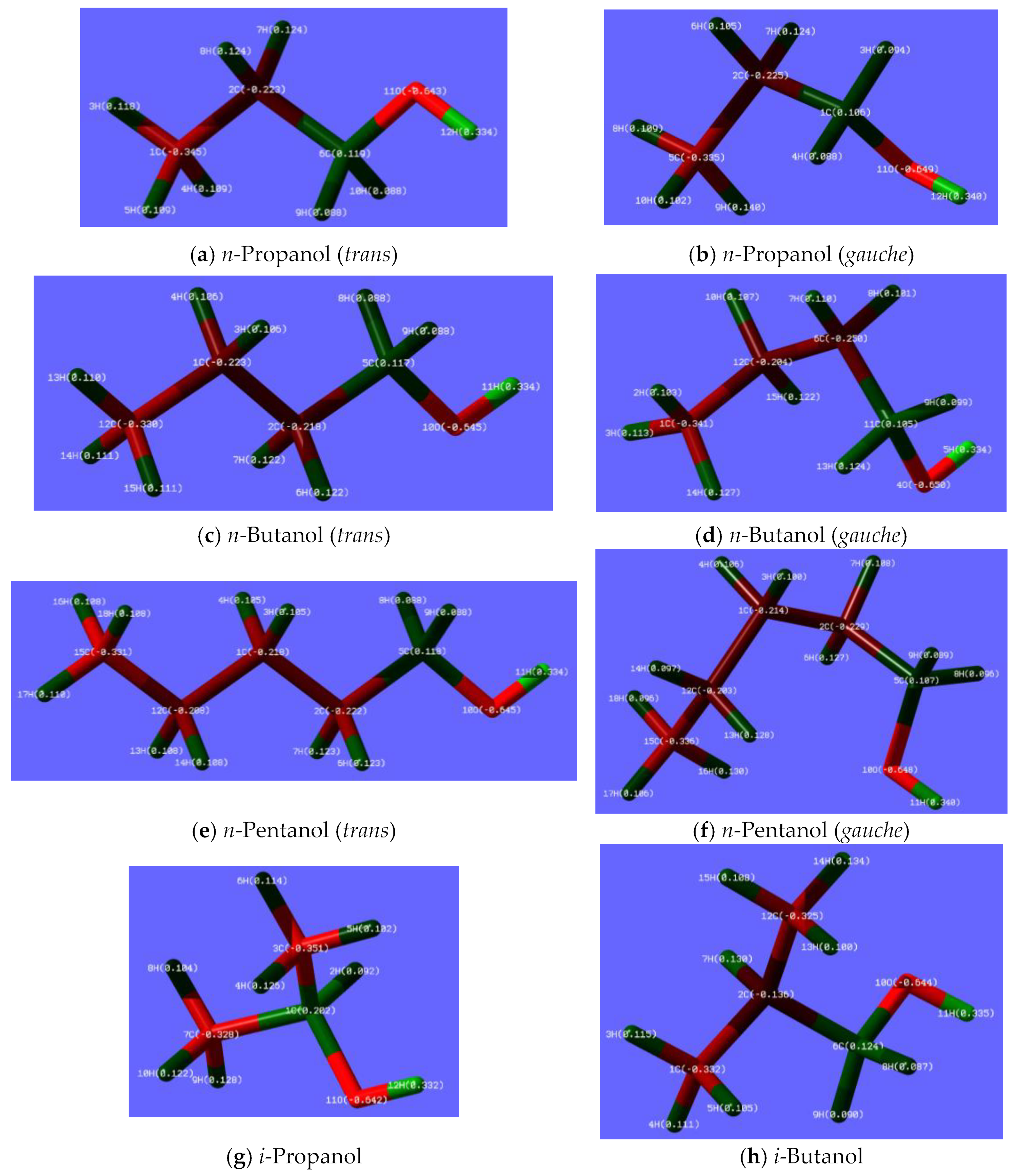
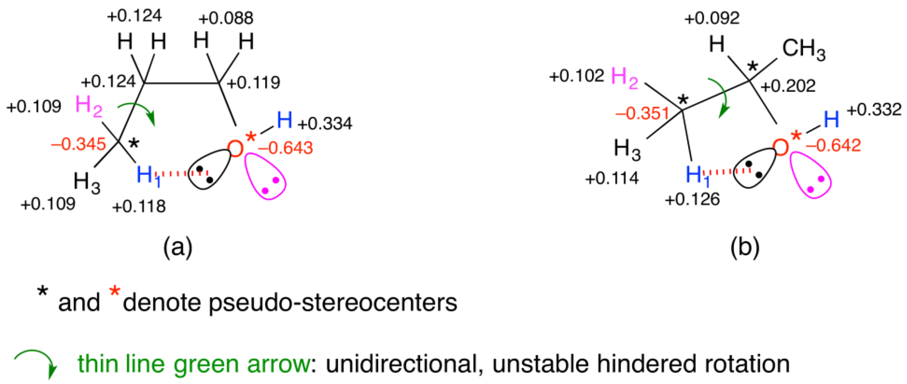
Optimized gauche n-propanol was found to be in chiral (C1) form, whereas optimized trans n-propanol maintained its achiral Cs form (Figure 13a,b). Mulliken charges in the gauche n-propanol are +0.109 (8H), +0.140 (9H), +0.102 (10H), −0.335 (5C) at the CH3 rotor and −0.649 (11O) and +0.340 (12H) at the O–H bond (Figure 13b). For comparison, the Mulliken charges in trans n-propanol were +0.118 (3H), +0.109 (4H), +0.109 (5H), and −0.345 (1C) for the CH3 rotor, and −0.643 (11O) and +0.334 (12H) for the O–H bond (Figure 13a). Clearly, the Mulliken charge of +0.140 at 9H in chiral n-propanol is the highest value among the three H atoms of CH3 because only 9H is located at the nearest neighbor of 11O with a Mulliken charge of −0.649.
Similarly, the optimized gauche n-butanol was in chiral (C1) form. Mulliken charges are +0.103 (2H), +0.113 (3H), +0.127 (14H), and −0.341 (1C) at the CH3 terminus and −0.650 (4O) and +0.334 (5H) at the O–H bond. Evidently, 14H exhibits the greatest Mulliken charge of +0.127 in CH3 because 14H is located at the nearest neighbor of 10O with a Mulliken charge of −0.645. Conversely, the three H atoms of CH3 in trans (Cs) n-butanol have nearly identical Mulliken charges, that is, +0.110 (13H), +0.111 (14H), and +0.111 (15H). Mulliken charges of −0.345 (12C), −0.645 (10O), and +0.334 (11H) in the chiral n-butanol are almost identical to those of the chiral n-propanol (Figure 13c,d). In trans n-pentanol, three H atoms of the CH3 terminus have nearly identical Mulliken charges ranging from +0.105 to +0.110 (Figure 13e). In gauche n-pentanol (C1), the Mulliken charges of the three H atoms of CH3 are very different; 16H, the nearest neighbor of 10O, is +0.130, whereas 17H and 18H are +0.106 and +0.096, respectively (Figure 13f). The Mulliken charges of two H atoms (13H and 14H) are very different; 13H is +0.128 and 14H is +0.097.
In a series of n-alkanols, the hypothetical chiral gauche geometries are considerably more stable relative to the achiral trans geometries: for n-propanol, gauche 0.00, trans 1.50; for n-butanol, gauche 0.00, trans 1.71; for n-pentanol, gauche 0.00, trans 1.01 (in kcal mol−1). Therefore, n-propanol, n-butanol, n-pentanol, n-hexanol, and n-octanol are likely to adopt pseudo-ring geometries with the aid of attractive intramolecular C(δ)–H(δ+)/O(δ) interactions [108,109].
Similarly, branched alkanols (C1) prefer pseudo-ring structures because of their attractive intramolecular C(δ)–H(δ+)/O(δ) interactions [108,109]: i-propanol, four-membered ring; i-butanol, five; i-pentanol, six; 2-ethyl-1-butanol, six; cyclopentanol, four. In i-propanol, 4H (+0.126), 9H (+0.128), and 10H (+0.122) have notably larger Mulliken charges among the six H atoms at the two CH3 rotors (Figure 13g). In i-butanol, 14H (+0.134) has the greatest Mulliken charge among the six H atoms at the two CH3 rotors (Figure 13h). Notably, in the case of i-pentanol, 17H (+0.186), which is closer to 9O (−0.634), has the greatest Mulliken charge among the six H atoms of the two CH3 rotors, whereas the Mulliken charges of the other five H atoms range from 0.085 to 0.109 (Figure S21a, Supplementary Materials), resulting in detectable VCD signals of δas(CH2), δas(CH3), and δs(CH3) (Figure 11f). The largest Mulliken charge of 17H at the CH3 rotor in the less bulky i-pentanol is likely to induce the greatest gabs value of iBS in i-pentanol due to significant intermolecular C(δ)–H(δ+)/O(δ) interaction (Figure 9a). In geraniol, 4H close to 7O has a large Mulliken charge of +0.167, although the six H atoms of the two CH3 termini have nearly identical Mulliken charges in the range from +0.117 to +0.123 (Figure S21b in Supplementary Materials). The handedness of the two through-space intramolecular C(δ)–H(δ+)/π(δ) interactions, 4H(+0.167)/15C(−0.150) and 16H(+0.128)/2C(−0.182), may be responsible for the (–)-VCD signals [108].
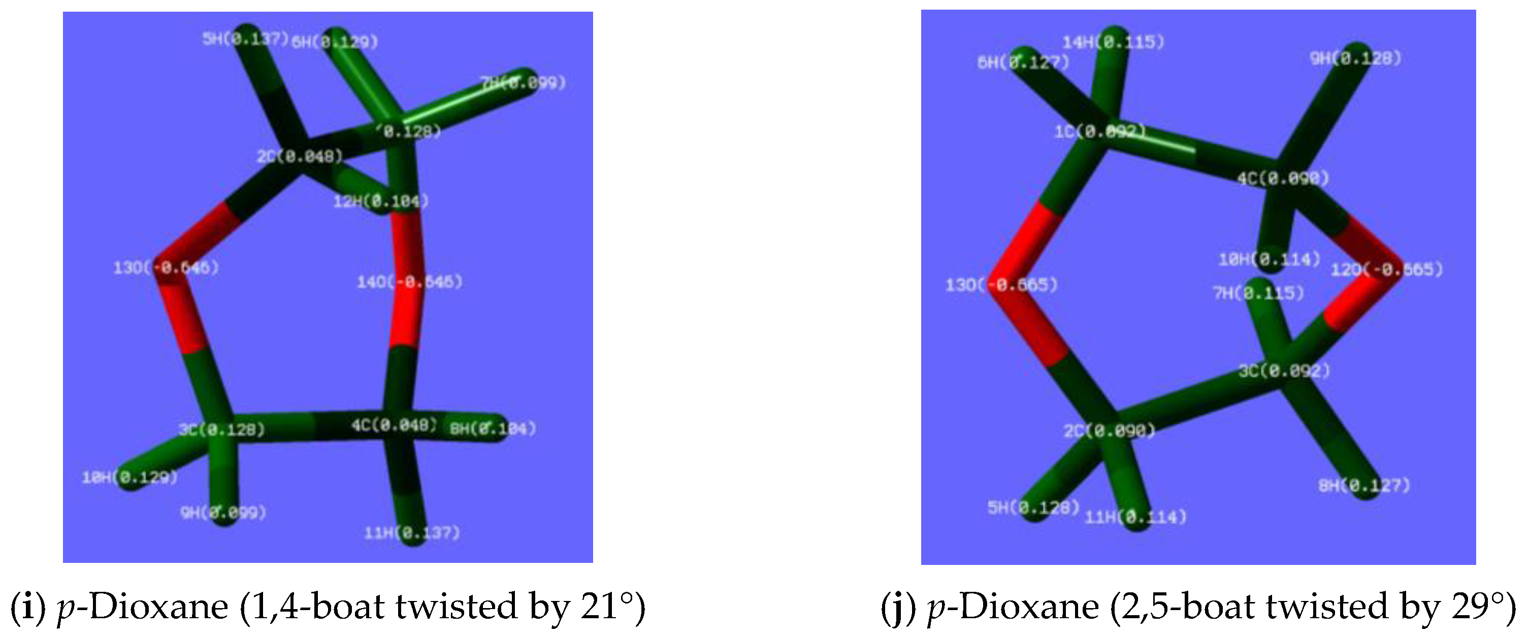
Among the three rotamers of p-dioxane, the 2,5-boat (C1) affords the largest Mulliken charges of +0.137 for 5H and 11H and the large Mulliken charges of +0.129 for 6H and 10H, although the 1,4-boat (C1) has larger Mulliken charge of +0.127 and +0.128 for 5H, 6H, 8H, and 9H (Figure 13i,j). The chair (D2h) consists of two equal sets of Mulliken charges: +0.128 for 6H, 8H, 10H, and 11H, and +0.108 for 5H, 7H, 9H, and 12H (Figure S21c, Supplementary Materials).
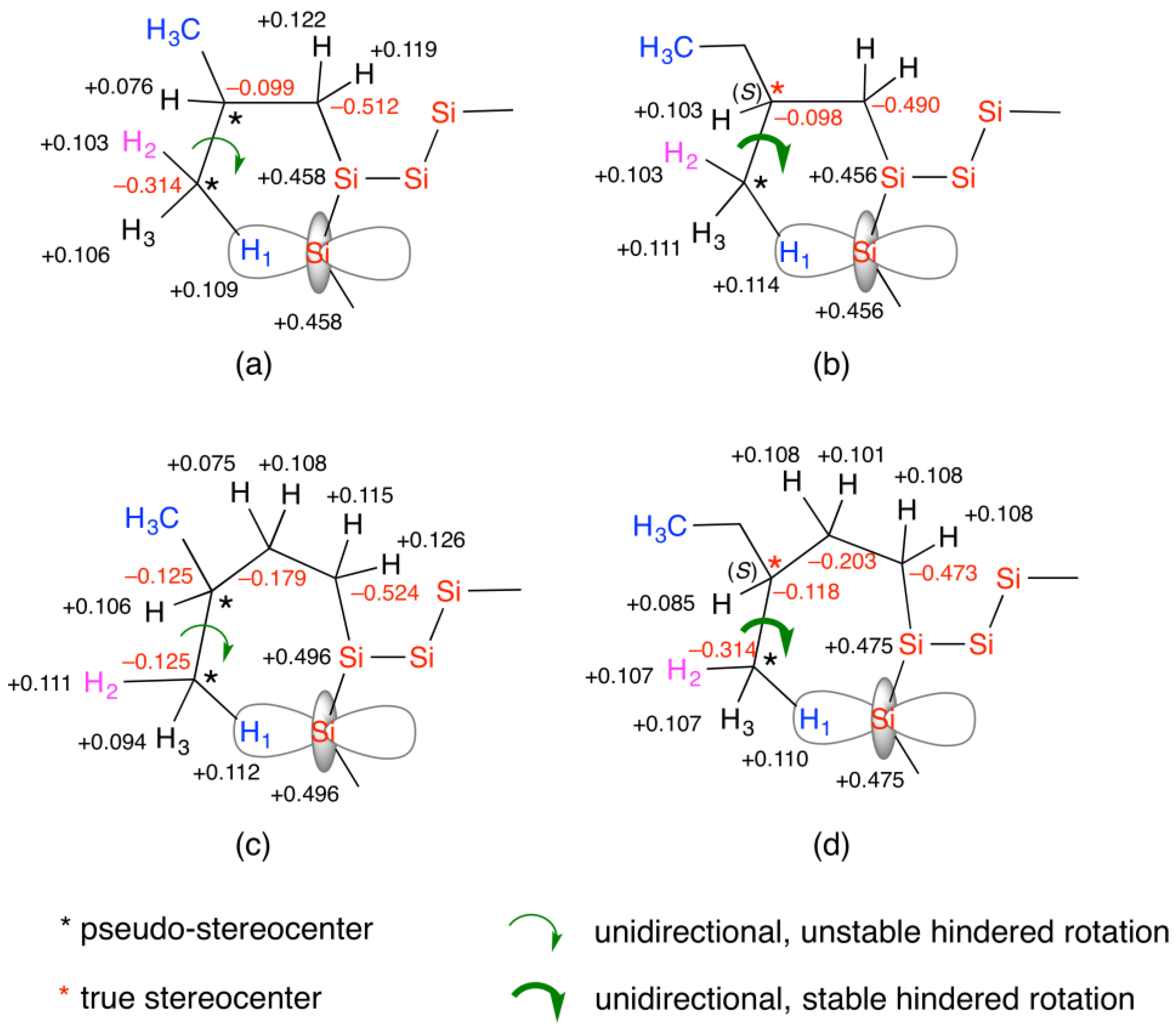
Mulliken charges of di-i-butylsilane with two trimethylsilyl (TMS) termini (iBS-TMS, Figure 1), di-i-pentylsilane with two TMS termini (iPS-TMS, Figure 1), and di-n-butylsilane with two TMS termini (nBS-TMS, Figure 1) are displayed in Figure S21d–f in Supplementary Materials. These trisilanes are optimized with MP2 and are simple models of iBS, iPS, and nBS. Large positive Mulliken charges at the central Si atom were observed: +0.458 for iBS-TMS, +0.496 for iPS-TMS, and +0.467 for nBS-TMS. The Si atoms are expected to experience attractive intermolecular C(δ+)–O(δ)/Si(δ+) interactions with the oxygen atoms of the alkanols and alkyl ethers. One of the two lone pairs at the (–)-Mulliken-charged oxygen atom can coordinate to an empty d-orbital of the (+)-Mulliken-charged Si atom. On the other hand, all H atoms, showing a weak positive Mulliken charge of ~+0.11 at the i-butyl, i-pentyl, and n-butyl side groups, resulted in attractive intermolecular C(δ)–H(δ+)/O(δ) interactions with the oxygen atoms of alkanols and alkyl ethers. Another explanation is that the 1H atom with (–)-electroweak (EW) charge, Qw, of −0.108 is attracted to the 16O atom with a (+)-EW charge of +6.216 and to three (+)-EW charged Si isotopes of +12.488 (28Si), +13.488 (29Si), and +15.488 (30Si) [58].

4.2.2. Inequality of the Two Lone Pairs at Oxygen Atom in Alkanols and Alkyl Ethers

In 1916, Lewis proposed the concept of a lone pair in which a pair of valence electrons is not involved in the covalent bond between atoms [110]. For example, the nitrogen atom in ammonia (NH3) has one lone pair, the oxygen atom in water (H2O) has two lone pairs, and the fluorine atom in CH3F has three lone pairs. Lone pairs have played a key role in modern chemistry [29,30,31,32]. In this paper, we hypothesize that in fact lone pairs play a key role as a hidden stereocenter with handedness, in addition to the Mulliken and EW charges.
From the discussion in Section 4.2.1, we are aware that the two lone pairs at the oxygen atom in gauche conformations of n- and i-alkanols are no longer equal. The oxygen carrying two lone pairs becomes a prochiral atom, as highlighted in red and black (Figure 14). In gauche n-propanol and i-propanol, the carbon atom of the CH3 rotor and the oxygen atom of the O–H group, respectively, become pseudo-stereocenters, [*] at the carbon atom, and [*] at the oxygen atom, associated with the Mulliken charges of H, C, and O atoms (Figure 14a,b). Equivalence between the left- and right-handed rotamers does not provide VCD spectral features. Handed rotamers are responsible for the experimental and theoretical VCD and IR spectra of gauche n-propanol and i-propanol (Figure 11a,j). Similarly, owing to the handedness of the chiral rotamers in gauche n-butanol, gauche n-pentanol, gauche n-hexanol, gauche n-octanol, i-butanol, i-pentanol, 2-ethyl-1-butanol, and cyclopentanol, the two lone pairs at the oxygen atom become inequivalent (Figure 12, Figure 13 and Figure 14). The theoretical (non-scaled) VCD and IR spectral shapes, including displacement vectors at specific vibration frequencies of gauche n-propanol, gauche n-butanol, gauche n-pentanol, i-propanol, i-butanol, 2,4-dimethyl-3-pentanol, and geraniol, are likely to reproduce the observed spectral shapes (Figures S22–S28 in Supplementary Materials). The snapshots of displacement vectors at a specific vibration frequency, though very complex, will help assign the vibration modes when the theoretical VCD and IR spectra are properly scaled from ~0.89 to ~0.96 to adjust to the observed spectra.
Even in p-dioxane-h8, THP, THF, and 15CE5, the two lone pairs on the oxygen atom become inequivalent due to the handedness of the chiral frameworks generated by the directional twisting motion of the C–C and C–O bonds. Among the chair (D2h), 1,4-boat (C1), and 2,5-boat (C1) forms of p-dioxane-h8, the theoretical VCD and IR spectra with displacement vectors of the optimized 2,5-boat twisted by 29° are in good agreement with the observed spectra rather than those of the optimized 1,4-boat twisted by 21° (Figure 11i and Figure S29, Supplementary Materials). Similarly, the calculated VCD and IR spectra with displacement vectors of an optimized chiral THP twisted by 15° partly agreed with the observed spectra (Figures S20g and S30, Supplementary Materials) and similarly for optimized 15° twisted THF (Figures S20h and S31, Supplementary Materials).
The solvent quantity of the pseudo-stereocenter oxygen and carbon atoms contributes to the chiroptical generation of iBS, iPS, iHS, nBS, and ES. Scenario (i) indicates that EPVn, where n is the number of solvent molecules in the system. The temperature-dependent CD polarization and inversion characteristics of iBS, iPS, iHS, and nBS were significantly influenced by the nature of the alkanols and alkyl ethers, as summarized in Table 1.

4.2.3. VCD and IR Spectra of Trisilanes Carrying Achiral and (S)-Chiral Substituents

The theoretical VCD and IR spectra including displacement vectors of iBS-TMS, iPS-TMS, and nBS-TMS, respectively (the trimeric models of iBS, iPS, and nBS), are given in Figures S32–S34, Supplementary Materials. For comparison, the theoretical VCD and IR spectra and displacement vectors of di-(S)-2-methylbutylsilane with TMS termini (S2MBS-TMS, Figure 1) and di-(S)-3-methylpentylsilane with TMS termini (S3MPS-TMS, Figure 1), respectively, were calculated as chiral molecular models of hypothetical poly[(S)-2-methylbutylsilane] and poly[(S)-3-methylpentylsilane] (Figures S35 and S36 in Supplementary Materials).
From Figures S32–S34 in Supplementary Materials, for iBS-TMS, the torsion bands τ(CH3) of the four CH3 rotors (#28, #31, #32, and #33) exist in the range of 238–294 cm−1. For iPS-TMS, τ(CH3) bands (#31, #33, #34, and #36) appeared in the range of 236–288 cm−1. For nBS-TMS, two τ(CH3) bands (#29 and #32) appeared at 235 cm−1 and 263 cm−1, respectively. In each case, three characteristic VCD and IR bands involving δas(CH2) at ~1520 cm−1, δas(CH3) at ~1480 cm−1, and δs(CH3) at ~1375 cm−1 are observed, regardless of the i-butyl and i-pentyl groups.
As shown in Figures S35 and S36 in Supplementary Materials, S2MBS-TMS provides several τ(CH3) bands of four CH3 rotors (#33, #34, #36, and #37) in the range of 251–291 cm−1. S3MPS-TMS has four τ(CH3) bands (#27, #35, #36, and #39) in the range of 206–295 cm−1. Three δas(CH2), δas(CH3), and δs(CH3) bands due to (S)-2-methylbutyl and (S)-3-methylpentyl groups are seen in the range of ~1510 ± 5 cm−1, ~1465 ± 10 cm−1, and ~1395 ± 10 cm−1, respectively.
The Mulliken charges of the four trisilanes bearing di-i-butyl, di-(S)-2-methylbutyl, di-i-pentyl, and di-(S)-3-methylpentyl substituents were compared (Figures S21d,e, S35f and S36f in Supplementary Materials). Almost identical (+)-Mulliken charges at the central Si atoms ranging from +0.458 to +0.496 in the two achiral and two (S)-chiral models are clear. Similarly, as illustrated in Figure 15, the data taken from Figures S21d,e, S35f and S36f in Supplementary Materials, very subtle differences in the Mulliken charges of the three H atoms at the CH3 terminus exist in the range from +0.103 to +0.112 for the achiral and (S)-chiral models. It is interesting to note that among the H1, H2, and H3 atoms of the CH3 rotor, the Mulliken charge of H1 (blue) closer to the Si atom is larger than that of H2 (pink) and H3 (black), regardless of the achiral and (S)-chiral models. The difference in the Mulliken charge of 0.011 between H1 (blue) and H2 (pink) in S2MBS-TMS was slightly larger than that of 0.006 between H1 (blue) and H2 (pink) in iBS-TMS. No such differences were observed in the Mulliken charges at H1 (blue), H2 (pink), and H3 (black) between S3MPS-TMS and iPS-TMS.
One may question (i) what is the major difference between the pseudo-stereocenters of the achiral substituents and the true stereocenters of the chiral substituents and (ii) what is the major role of the chiral substituents. A plausible answer is that true stereocenters play a major role in providing deterministic handedness in the chirality of molecules and Si–Si main-chain helicity. True stereocenters at the β-branched chiral substituents of rigid helical polysilanes carrying (S)- and (R)-2-methylbutyl side groups strongly enforce all C–C, Si–Si, and Si–C single bonds into handed rotational motions to resist thermal fluctuations. This results in the very stable P-73- and M-73-helicity of the Si–Si main chain, whose CD and UV spectra are insensitive to changes in temperature and the nature of the solvents [88,91,92,93]. As a result, the TC1 in i-octane is very high, possibly above 200 °C, and typically no helix–helix transition in the range from 100 °C to –100 °C [86,88,92,93]. For true stereocenters located in the γ-position of branched chiral substituents of semiflexible polysilanes carrying (S)- and (R)-3,7-dimethyloctyl side groups, the rotational freedom of C–C, Si–Si, and Si–C single bonds is maintained, allowing for a one-time helix–helix transition, TC2, in the range from 10 °C to −65 °C, although the TC1 is sufficiently high above 200 °C [86,88,92,93].
In contrast, the Si–Si main-chain helicity induced by the pseudo-stereocenters of the i-butyl and i-pentyl substituents is fragile and unpredictable toward unresolved fluctuations, physical forces, and chemical influences, involving changes in temperature, solvent nature, and hydrodynamic flow, as demonstrated in this study and in a previous work [58]. Multiple helix–helix transitions occur in the range from 80 °C to −20 °C, and the TC1 occurs at a lower temperature in the range from 80 °C to 110 °C (Table 1). The absolute magnitudes of the CD and CPL spectra in the S0 and S1 states of iBS and iPS fluctuated considerably, and their chiroptical signs often inverted upon photoexcitation with unpolarized light (Figure 3f, Figure 4f, Figure 5f, Figure 6f and Figure 11c,g). Swapping the preference of P-83-helicity or M-83-helicity evidenced by CD and CPL polarization and inversion was predominantly determined by the directional motions of the four CH3 rotors of the i-butyl and i-pentyl substituents (Table 1, Figure 10d,h, and Figure S18d in Supplementary Materials).
These dynamics are greatly affected by the handedness of the CH3 rotors in VCD-active n-, i-, and highly branched alkanols, and the handedness of the twisted C–C and C–O bonds of VCD-active p-dioxane-h8. Compared with n- and i-alkanols, the helix-inducing capability of p-dioxane-h8 is rather weak, probably because of the lack of a CH3 rotor. Although the handed rotational and twisting forces may be very weak, the control of solvent quantity enables strong amplification of a weak EPV by (a) the linear amplification scenario (i), obeying EPVx, where x is the number of solvent molecules in the system; (b) the non-linear amplification scenario (ii) based on EPVn, where n is the number of repeating units in a single polymer chain; and (c) an equilibrium shift by the solvent chirality transfer mechanism [111].
Finally, relevant to the stable and metastable conformations of n-/i-alkanols and cyclic/acyclic alkyl ethers, several studies in gas and liquid phases, and isolated molecules in Ar/Xe matrices at cryogenic temperatures, and a crystal have been reported to date. The relative stabilities of trans- and gauche-rotamers and between non-twisted and twisted conformers have been theoretically and experimentally characterized as follows: ethanol (in Ar matrix [112] and liquids [113,114]), n-propanol (gas and Ar matrix [115] and liquids [116]), i-propanol (in Ar matrix [117], gas and liquid phases [118,119]), n- and i-butanol (in CCl4 [120]), p-dioxane (liquids [107,121] and theory [122]), THF (a crystal [123]), diglyme (solids and liquids [124]), 15CE5 (theory [125]), and α-terpinene (in Xe matrix [126]). However, to the best of our knowledge, experimental and theoretical VCD studies on these stereocenter-free molecules have not yet been conducted. Most chemists believe that these molecules are optically inactive owing to the equal populations of left- and right-handed rotamers and twisted conformers. Among these molecules, THF is reported to adopt a P-twisted chiral conformation (point group C2) in a monoclinic crystal (space group C2/c) at −170 °C and −125 °C [123]; however, it is unclear as to whether the C2-structure is always P-handed and what is the most favorable conformation in the liquid phase at ambient temperature.

4.2.4. Scenarios of CD Polarization in iBS, iPS, and nBS with Alkanols and Alkyl Ethers

Based on the discussion above in Section 4.2.3, it is evident that the multiple generation and inversion of Si–Si main-chain helicity in iBS, iPS, and nBS are strongly influenced by the nature of the oxygen-containing alkanols and alkyl ethers when they are used in solvent quantities (Figure 3, Figure 4, Figure 5, Figure 6, Figure 7, Figure 8, Figure 9 and Figure 10 and Table 1). First, we considered attractive and repulsive intermolecular forces between the oxygen-containing molecules and iBS involving Mulliken and EW charges. The attractive forces result from a pair of opposite Mulliken charges as in C(δ)–H(δ+)/O(δ) and Si(δ+)/O(δ), or a pair of opposite sign EW charges as in H(Qw)/C(Qw+), H(Qw)/Si(Qw+), and H(Qw)/O(Qw+). The repulsive forces result from a pair of same-sign Mulliken charges as in C(δ)–H(δ+)/H(δ+)–C(δ) and Si(δ+)/H(δ+), or a pair of same-sign EW charges as in H(Qw)/H(Qw) and Si(Qw+)/O(Qw+).
The intermolecular attractive and repulsive interactions of iBS with the less-bulky gauche n-propanol and bulky 2-ethyl-1-butanol are schematically illustrated in Figure 16. Gauche n-propanol induces a large magnitude gabs with (–)-CD sign at 295 nm, and the 295 nm CD spectral profile is nearly identical to the 295 nm UV profile (Figure 3). In contrast, 2-ethyl-1-butanol provided the weakest gabs with a (–)-CD sign at 295 nm located at the tail of the 310 nm UV band (Figure S12b, Supplementary Materials). In the bulkier i-pentyl group of iPS, 2-ethyl-1-butanol induces the weakest gabs with a (+)-CD sign at 300 nm, which appears at the tail end of the 315 nm UV band (Figure S17c, Supplementary Materials).
The significant discrepancy between n-propanol and 2-ethyl-1-butanol is ascribed to the degree of repulsive C–H(δ+)/H(δ+)–C interactions between the (solvent) CH3 and CH3 (i-butyl and i-pentyl) rotors. As illustrated in Figure 16 (left), in the case of two inequivalent oxygen lone pairs in gauche n-propanol, one lone pair (black) interacts with H1 (blue) and the other lone pair (pink) is strongly coordinated to an empty d-orbital of the Si atom (red). The repulsive C–H(δ+)/H(δ+)–C interactions between the two CH3 groups were minimal. In contrast, in 2-ethyl-1-butanol, as illustrated in Figure 16 (right), although one lone pair (black) mainly interacted with H1 (blue), the other lone pair (pink) was very weakly coordinated to the empty d-orbital of the Si atom (red) owing to the bulkiness of the alkanol. The repulsive C–H(δ+)/H(δ+)–C force between the 2-ethyl-1-butanol and i-pentyl groups of iPS thus increased. A pair of negative-sign EW charge 1H atoms with positive-sign EW charge C/O/Si atoms, that is, H(Qw)/C(Qw+), H(Qw)/O(Qw+), and H(Qw)/Si(Qw+), may contribute to Si–Si main-chain helicity [58].
The generality of the CH3 hindered rotor actions having temperature-dependent handedness allowed for generating right-handed and left-handed helical polysilanes in the absence of any chiral catalysts, chiral substituents, and chiral solvents. This might be called “absolute helix generation” or generally, “absolute asymmetric synthesis” by choosing appropriate achiral side groups, achiral alkanols and achiral alkyl ethers in specified temperatures.

5. Perspective—Possible Origin of Biomolecular Handedness

Strong evidence of the MPV effects detected in artificial stereocenter-free molecules and polymers in the absence of point, axial, and planar chirality is very important to fill a missing link regarding handedness in the hierarchy of the matter world versus the anti-matter, ranging from quarks, hadrons, baryons, leptons, atoms, natural amino acids, carbohydrates, nucleic acids, homochiral life, spiral galaxies, and dextrorotatory rotation embedded into the cosmological microwave background which is a record of the Big Bang 13.8 billion years ago [14,15,21,22,23,24,25,26,27,28,55,56,127,128,129,130,131,132].
Biomolecules are classified into proteins, carbohydrates, lipids, nucleic acids, and terpenes/terpenoids. Proteins are composed only of L-amino acids. However, D-amino acids exist, though very rarely, in ion channels. Carbohydrates, which include mono-/di-/polysaccharides, are made of D-pentoses and D-hexoses. L-Hexoses, such as L-arabinose. L-xylose, and L-rhamnose, are known, but rare. 2-Deoxy-L-ribose was then synthesized. Lipids, such as phospholipids and glycolipids, contain saturated fatty acids and cis-double bond-containing fatty acids. The number of carbons in the alkyl chains of the fatty acids was between 12 and 24. Nucleic acids are alternating copolymers of phosphoric acid and nucleosides composed of D-pentose-carrying achiral bases. Because artificial L-pentose is available, non-natural L-nucleic acids and mirror-image DNA are in principle possible. The mirror-image life may then become an enemy of the current biosphere. The L-D preference for amino acids and pentose/hexose appears to be inherent on Earth. Terpenes and terpenoids play crucial roles in plant defense, attracting pollinators, plant–plant communication, and pheromones. Thus far, two representative scenarios, the necessity model based on PV-EWF and the by-chance mechanisms relying on PC-EMF, have been hypothesized to explain biomolecular handedness [43,45,51,52,55,56]. Our results support the PV-EWF driven necessity model for the origin. The L-D preference on Earth is predetermined by the PV-EWF.
A series of MPV experimental results from our simple chemical systems, not involving stereocenters, strongly indicated spontaneous generation of handed helicity from mirror-symmetric helical σ-conjugated polysilanes (iBS, iPS, iHS, nBS, ES, etc.) [58], π-conjugated oligofluorenes, and π-conjugated poly-p-phenylene, in addition to the spontaneous generation of handed chirality in the S1 and S0 states of nearly 60 types of mirror-symmetric molecules [96,97,98]. However, the preferred handedness of helicity and chirality induced by PV-EWF appears unstable and uncertain toward thermal and other fluctuations, and can abruptly invert to the opposite helical screw sense and chirality in the S0 and S1 states by chance, eventually undergoing racemization at temperatures above ~100 °C. Such instability and uncertainty might have prevented or significantly delayed the first emergence of life. Possibly, to fix the preferred helicity and chirality, the nature of the molecular evolution era on early Earth used L-D chirality to embed L-amino acids and D-carbohydrates as the building blocks of the first life.
From VCD and IR spectroscopy, THF and THP as skeletons of L-furanose and L-pentose are likely to twist with handedness, despite the absence of stereocenters [49]. The inequality of the two lone pairs at the oxygen atoms of the twisted rings plays a crucial role. In addition, several n-hydrocarbons bearing two CH3 termini exhibited VCD signals induced by directional CH3 hindered rotors [58]. Hydrocarbons are major components of lipids. Lipid molecules on the surface of a two-dimensional bilayer membrane drift slowly using their own directional CH3 rotors. However, the CD3 rotor in deuterated hydrocarbons is inefficient in inducing main-chain helicity and can generate the opposite helix sense [58]. If the ocean and atmosphere on primordial Earth were covered by D2O, the L-D preference for the helicity and chirality of proteins, RNA, and DNA may be the opposite. Therefore, D2O may function as a toxic water molecule to attack the current homochiral life on Earth.
As a model unsaturated hydrocarbon, geraniol involving two C=C bonds, which is a stereocenter-free terpenoid, revealed clear VCD signals due to the ν(C=C) modes. Other terpenes and terpenoids (α-humulene, myrcene, α- and γ-terpinenes, o-, m-, and p-cymenes, thymol, and nerol), triterpene (squalene), and sesquiterpenoid (farnesol) (Figure S37, Supplementary Materials) may be optically active liquids, which can be validated by VCD and IR spectroscopy. In a previous study [58], an aromatic terpene p-cymene (4-isopropyltoluene) with three CH3 rotors, showed clear VCD and IR spectral characteristics. In this work, the VCD and IR spectra of α- and γ-terpinenes, which are cyclic diene terpenes carrying three CH3 rotors, were measured, as shown in Figure S38a,b in Supplementary Materials, and (–)-VCD and IR signals at 1658 cm−1 and 1659 cm−1 due to ν(C=C) were observed for both. α-Terpinene additionally shows multiple (+)-VCD and IR signals at 1466 cm−1, 1450 cm−1, and 1430 cm−1 due to δas(CH2), δas(CH3), and (+)-VCD and IR signals at 1481 cm−1 and 1430 cm−1, respectively due to δs(CH3). The corresponding VCD and IR signals of γ-terpinene were very weak. Notably, (–)-VCD and IR signals at 1753 cm−1 (α-terpinene) and 1771 cm−1 (γ-terpinene) were evident, although the origin of the VCD bands was not fully characterized. These VCD and IR spectral characteristics were very similar to those of p-cymene (Figure S38c in Supplementary Materials; data from Figure S20g–i in Supplementary Materials of ref. [58]).
Achiral glycine (H2N-CH2-CO2H) is the simplest amino acid and the two protons (Ha and Hb) of α-CH2 group are energetically equal (Figure 17, top). When neutral glycine turns into a zwitterion (H3N+-CH2-CO2), Ha and Hb become unequal due to the directional H3N+ hindered rotor. The +NH3 group belongs to three identical 1H nuclear-spin-1/2 systems in TWP as for the CH3 and CF3 rotors (Figure 17 and Figure 18) [58]. Other zwitterions of natural L-amino acids are regarded as alkyl and aralkyl derivatives substituted with the Ha atom of the glycine zwitterion. Similarly, non-natural D-amino acids are alkyl and aralkyl derivatives substituted with the Hb atom of the zwitterionic glycine. Previous studies on MPV effects were based on the idea that the CO2 group is treated as a hindered rotor in the zwitterions of glycine, alanine, valine, serine, and aspartate [43,127]. The zwitterionic pairs of L- and D-amino acids are no longer equal when the perpetual clockwise rotation of the directional +NH3 and CH3 rotors (L- and D-alanine), +NH3, and two CH3 rotors (L- and D-valine and L- and D-leucine), for example, occur, regardless of any L-D chirality [58] (Figure 17 and Figure 18).
Because iBS and leucine both have an i-butyl group, one can expect that four CH3 rotors at the i-butyl group of leucine perpetually work as unidirectional hindered rotors regardless of whether the leucine is L or D. C3v symmetric protonated water, O+H3, which belongs to the three identical 1H HINS nuclear-spin-1/2 systems in TWP, assists the directional rotors in the zwitterionic amino acids (Figure 17 and Figure 18). Pure neutral liquid H2O ([O+H3] = [OH] = 10−7 M) showed weak nuclear spin-induced optical rotation (NSOR) in the visible-near IR region, although further measurements under north-up/north-up and zero-magnetic fields may be required [133]. L- and D-Alanine and racemic alanine in aqueous solutions showed weak (–)-Verde constants by extrapolating [conc] = 0.0 g L−1 [134]. These results implied the presence of non-mirror L- and D-alanine in H2O. Therefore, non-mirror image vibrational spectra of the +NH3, CH3, and CO2 rotors in the four zwitterionic L- and D-amino acids are expected. This idea can be validated by ROA and VCD spectroscopy [33,34].
To validate the N+H3 rotor hypothesis, we measured the first CD and UV spectra of glycine in neutral H2O (1.00 × 10⁻² M with 1 mm path length) at 20 °C under atmospheric pressure by minimizing inherent absorptions of ozone and water in the far-UV region. From Figure 19a, glycine showed far-UV CD and UV spectra with gabs = +9.1 × 10⁻⁵ at 182.0 nm and gabs = −2.7 × 10⁻⁵ at 188.5 nm. Glycine having an isoelectric point (IEP) of 5.97 mostly exists as the zwitterionic species in H2O [135]. However, when glycine was dissolved in D2O, no CD or UV spectra due to zwitterionic D3N+-CH2-CO2 species were observed. This implies that the N+D3 group which is a three-identical integer nuclear-spin-1 (INS) system in TWP does not efficiently work as a unidirectional rotor [136]. Or same-sign repulsive EW charges of 2H (Qw = +0.892) and 16O (Qw = +6.216) [58] between N+D3 and CO2 rotors may be the reason. Solvation with H2O thus plays a critical role in generating optically active zwitterionic glycine [137,138,139], and possibly zwitterionic alanine and other α-amino acids [48,127,140,141].
The simulated chiroptical spectra of zwitterionic glycine were calculated using TD-DFT (B3LYP/6-31G(d,p)) and showed six singlet transitions with fwhm of 0.10 eV) when including the integral equation formalism for the polarizable continuum model (IEF-PCM) of H2O (Figure 19b). The experimental CD and UV spectra are qualitatively supported by the simulated spectra (Figure 19a,b). For visualization, two major molecular orbitals with an isovalue of 0.08 at the first LUMO (#21) and the first HOMO (#20) are displayed in Figure 19c,d. From the Mulliken charges of the zwitterionic glycine in H2O, the CO2 rotor experiences an electrostatic attraction force from the N+H3 rotor (Figure 19e). We propose that unidirectional motion (yellow arc) of the three-fold symmetric N+H3 rotor synchronously drives unidirectional motion (white arc) of the two-fold symmetric CO2 rotor as a chromophore responsible for the observed CD and UV spectra (Figure 19e), although the 12C16O2 group, which belongs to the INS system in DWP, may not work efficiently as a directional hindered rotor [43,127]. Further CD and UV spectra of other zwitterionic glycine oligomers (from dimer to hexamer) and three pairs of zwitterionic L- and D-alanine species (monomer, dimer, and trimer) in H2O were experimentally and theoretically investigated. The optically active zwitterionic glycine and its oligomers in liquid H2O may serve as an excellent platform for generating L-amino acids under mild conditions on Earth. Vacuum-UV and high-energy circularly polarized light sources [142,143,144,145], 14N nucleus with handed electron anti-neutrino (anti-νe) from supernova [146], HINS radioisotopes (15N, 13N, and 15O) generated by lightning in thunderclouds as natural particle accelerator [147], and chiral-induced spin selectivity (known as CISS) induced by north-up and south-up magnetic surface [148,149] are also candidates for the physical origins of natural L-amino acids regardless of terrestrial and extraterrestrial environments [54]. The results will be submitted elsewhere.

6. Conclusions

Although violations of parity (P) and charge-parity (CP) symmetries in fundamental physics have been established, the molecular P-violation (MPV) hypothesis in chemistry remains a matter of debate. Since the 1960s, MPV theorists have argued that an electroweak force (EWF) induces a very small parity-violating energy difference (PVED), EPV, in the range from 10−10 to 10−21 eV for mirror-image molecules. The PVED is endowed with handedness by the weak neutral current (WNC) and induced by a massive Z0 boson. The EWF is a unified P-violating (PV) weak nuclear force (WF) and P-conserving (PC) electromagnetic force (EMF). To validate the MPV hypothesis, six major theoretical scenarios were proposed: (i) linear amplification model (EPVn), where n is the number of polymers and the number of molecules in a crystal and a pure liquid; (ii) non-linear amplification model revealing second-order phase transition; (iii) tuning a tunneling splitting (ΔE±) connecting to the barrier height (EB) of mirror-image molecules in the double-well potential (DWP); (iv) EPVZ5, Z: atomic number; (v) EPV ∝ 1/ΔEST, ΔEST: difference in energy between S0 and T1 states; and (iv) half-integer nuclear spin (HINS) atom-induced chiral anapole moment.
To validate the MPV hypothesis, we designed poly(di-i-butylsilane) (iBS), poly(di-i-pentylsilane) (iPS), and poly(di-i-hexylsilane) (iHS) (Figure 1), that carry achiral β-, γ-, and δ-branched side chains, respectively. In addition, a dozen n-, i-, and branched alkanols and several cyclic and acyclic alkyl ethers were chosen as stereocenter-free solvents and stereocenter-free molecular liquids to study their VCD and IR spectral characteristics. We collected 333 experimental datasets comprising 298 CD and UV spectra, 12 CPL and PL spectra, and 23 VCD and IR spectra. In addition, we obtained 37 theoretical datasets, including 18 VCD and IR spectra and 19 Mulliken charges.
Analysis of the big datasets led to the conclusion that the clear MPV effects are reproducible: iBS, iPS, and iHS undergo multiple temperature-dependent CD and CPL polarizations and inversions at Siσ–Siσ* transitions in a homogeneous solution of achiral n-, i-, branched, and cyclic alkanols and p-dioxane-h8 and -d8 under non-stirring conditions. In particular, iBS in i-pentanol at 25 °C provided the greatest intensity (–)-CD band with gabs = −3.1 × 10⁻² at 290 nm, whereas iHS in n-octanol at 0 °C induced a weak (–)-CD band with gabs = −0.03 × 10⁻² at 310 nm. Notably, in n-propanol iPS adopted opposite helical screw senses in the ground and photoexcited states: at −5 °C a (–)-CD band with gabs = −0.49 × 10⁻² at 299 nm, was observed, but the CPL showed a (+)-CPL band with glum = +0.84 × 10⁻² at 336 nm. These MPV results were intimately connected to the (–)-VCD and IR bands in the handed symmetric and asymmetric bending modes of the CH3 and CH2 groups in the alkanols and alkyl ethers. The (–)-VCD and IR results of the molecules were presumably applicable to iBS and iPS.
Inequality between the two lone pairs at the pseudo-stereocenter O atom(s) of the gauche-bond-containing alkanols and twisted alkyl ethers led to handed intermolecular Si(δ+)/O(δ) and C(δ)–H(δ+)/O(δ) interactions, in which (i) the empty d-orbital of the Si atom with (+)-Mulliken charge is coordinated by a handed lone pair of the O atom with (–)-Mulliken charge in alkanols and alkyl ethers, and (ii) (+)-Mulliken-charged H atoms of the i-butyl and i-pentyl side groups in iBS and iPS experience attraction to the handed lone pair of (–)-Mulliken-charged O atoms.
We reconfirmed the importance of directional CH3 rotors in the i-butyl, i-pentyl, and n-butyl groups of polysilanes and n-/i-alkanols as solvent molecules. Four sets of CH3 rotors in the two i-butyl and i-pentyl side groups per repeating unit of iBS and iPS induced four mirror-symmetric breaking and second-order phase transitions to generate handed 83-helicity at TC1, TC2, TC3, and TC4 from mirror-symmetric P-73- and M-73-helicity above the TC1. Two sets of CH3 rotors at the two n-butyl groups of nBS induced two mirror-symmetric breaking and helix–helix transitions at TC1 and TC2. One or two CH3 rotors of n- and i-alkanols effectively modulated the mirror-symmetry breaking characteristics (CD and UV spectral profiles, gabs, glum, and their chiroptical signs) of 83-helicity. However, 83-helicity was inefficiently induced in p-dioxane-h8 and -d8, presumably because of the lack of CH3 and CD3 rotors, although p-dioxane-h8 exhibited clear VCD and IR spectra while the VCD and IR spectra of p-dioxane-d8 were less obvious.
The present MPV experiments with the aid of theoretical interpretation may shake the fundamentals of three-dimensional stereochemistry that have been accepted since 1887. Furthermore, the generality of the directional CH3 rotors allows for generating right-handed and left-handed helical polysilanes in the absence of chiral catalysts, chiral substituents, and chiral solvents—called “absolute helix generation” or generally, “absolute asymmetric synthesis”—by appropriate choice of achiral side groups, achiral alkanols and alkyl ethers, and temperature. The definitions of optical activity and inactivity, chirality and achirality, enantiomer and diastereomer, equality and inequality of the two lone pairs at the oxygen atom, handed vibrational motions of CH3 and CH2 groups, nuclear-spin-dependent helicity/chirality, differences in the photoexcited and ground states of helicity/chirality, and Kasha’s rule including Jablonski diagram will need to be reconsidered immediately. From the first observation of far-UV CD and UV spectra of zwitterionic glycine in neutral H2O, a possible origin of biomolecular L-amino acids on Earth was discussed.
The PV-EWF theory will thus bring to an end the long-standing mystery of the missing link connecting the handedness at all hierarchies in the matter-dominant (and rare anti-matter) universe, ranging from quarks, hadrons, baryons, leptons, atoms, amino acids, carbohydrates, nucleic acids, terpenes, terpenoids, homochiral life, spiral galaxies, and dextrorotatory rotation embedded into a cosmological microwave background, remnants of the Big Bang which occurred 13.8 billion years ago. The traditional rigid three-dimensional stereochemistry on Earth may become non-rigid spatiotemporal PV-EWF chemistry with handed dynamics when universal origin PV forces are fully validated in the future.

Supplementary Materials

The following supporting information can be downloaded at: https://www.mdpi.com/article/10.3390/sym17091544/s1. Synthesis and characterization of dichlorosilane monomers, iBS, iPS, and iHS. Instrumentation including NMR (1H, 13C, and 29Si), GPC, VT-CD, and UV spectra; CPL and PL spectra at −5 °C; VCD and IR spectra at 23 °C; chiroptical data management software (KaleidaGraph); and theoretical calculations of Mulliken charges and VCD and IR spectra including displacement vectors (Gaussian09 and GaussView5). Figure S1: Chemical structures of optically active 73-helical polysilanes carrying (S)-/(R)-3,7-dimethyloctyl and (S)-/(R)-2-methylbutyl side groups studied in the past; Figure S2: Chemical structures of methanol, ethanol, n- and i-alkanols, branched and cyclic alkanols, and cyclic and acyclic alkyl ethers measured by VCD and IR spectroscopy; Figure S3: 1H-, 13C-, and 29Si-NMR spectra of di-i-butyldichlorosilane in CDCl3; Figure S4: 1H-, 13C-, and 29Si-NMR spectra of di-i-pentyldichlorosilane in CDCl3; Figure S5: 1H-NMR spectrum of iBS in CDCl3; Figure S6: 1H-NMR spectrum of iPS in CDCl3; Figure S7: 1H-NMR spectrum of iHS in CDCl3; Figure S8: VT-CD and UV spectra (N = 3) of iBS in n-propanol ranging from −5 °C to 90 °C; Figure S9: VT-CD and UV spectra (N = 3) of iBS in i-propanol ranging from −5 °C to 70 °C; Figure S10: VT-CD and UV spectra (N = 3) of iBS in i-butanol ranging from −5 °C to 90 °C; Figure S11: VT-CD and UV spectra (N = 3) of iBS in i-pentanol ranging from −5 °C to 90 °C; Figure S12: VT-CD and UV spectra (N = 1) of iBS in n-butanol, 2-ethyl-1-butanol, and cyclopentanol at −5 (or 0), 20, and 40 °C; Figure S13: VT-CD and UV spectra (N = 1) of iBS in p-dioxane-h8 ranging from 15 °C to 90 °C; Figure S14: VT-CD and UV spectra (N = 1) of iBS in p-dioxane-d8 ranging from 15 °C to 90 °C; Figure S15: VT-CD and UV spectra (N = 1) of iPS in n-propanol ranging from −5 °C to 90 °C; Figure S16: VT-CD and UV spectra (N = 1) of iPS in n-pentanol, n-hexanol, and n-octanol and of iHS in n-octanol at 0, 20, and 45 °C; Figure S17: VT-CD and UV spectra (N = 1) of iPS in i-propanol, i-butanol, and 2-ethyl-1-butanol at 0, 20, and 40 °C; Figure S18: CD and CPL characteristics of iBS in i-butanol at −5 °C; Figure S19: CPL and PL spectra (N = 3 to 5) of iBS in n-propanol and i-butanol and iPS in n-propanol at −5 °C; Figure S20: VCD and IR spectra of methanol, ethanol, n-hexanol, n-octanol, cyclopentanol, 2-ethyl-1-butanol, THP, THF, 15CE5, and diglyme; Figure S21: Mulliken charges (MP2/6-31G(d,p)) of i-pentanol, geraniol, p-dioxane (chair, D2h), iBS-TMS, iPS-TMS, and nBS-TMS; Figure S22: Theoretical VCD and IR spectra and displacement vectors of n-propanol; Figure S23: Theoretical VCD and IR spectra and displacement vectors of n-butanol; Figure S24: Theoretical VCD and IR spectra and displacement vectors of n-pentanol; Figure S25: Theoretical VCD and IR spectra and displacement vectors of i-pentanol; Figure S26: Theoretical VCD and IR spectra and displacement vectors of i-butanol; Figure S27: Theoretical VCD and IR spectra and displacement vectors of 2,4-dimethyl-3-pentanol; Figure S28: Theoretical VCD and IR spectra and displacement vectors of geraniol (trans-3,7-dimethyl-2,6-octadien-1-ol); Figure S29: Theoretical VCD and IR spectra and displacement vectors of p-dioxane-h8; Figure S30: Theoretical VCD and IR spectra and displacement vectors of THP; Figure S31: Theoretical VCD and IR spectra and displacement vectors of THF; Figure S32: Theoretical VCD and IR spectra and displacement vectors of iBS-TMS; Figure S33: Theoretical VCD and IR spectra and displacement vectors of iPS-TMS; Figure S34: Theoretical VCD and IR spectra and displacement vectors of nBS-TMS; Figure S35: Theoretical VCD and IR spectra, displacement vectors, and Mulliken charges of S2MBS-TMS; Figure S36: Theoretical VCD and IR spectra, displacement vectors, and Mulliken charges of S3MPS-TMS; Figure S37: Chemical structures of terpenes, terpenoids, triterpene, and sesquiterpenoid, that do not involve point chirality; Figure S38: Observed VCD and IR spectra of α-terpinene, γ-terpinene, and p-cymene.

Author Contributions

Conceptualization, M.F.; methodology, M.F. and T.M.; validation, M.F., T.M. and M.M.A.; formal analysis, M.F., T.M. and M.M.A.; investigation, T.M., M.F., J.R.K. and M.M.A.; resources, M.F. and T.M.; data curation, M.F. and T.M.; writing—original draft preparation, M.F.; writing—review and editing, J.R.K., M.F. and M.M.A.; visualization, M.F. and T.M.; supervision, M.F.; project administration, M.F.; funding acquisition, M.F. All authors have read and agreed to the published version of the manuscript.

Funding

This work was supported by the Japan Society for the Promotion of Science (JSPS)–KAKENHI Grants-in-Aid for Scientific Research [16655046 (FY2003–2005) and 22350052 (FY2010–2013)], the NAIST Foundation (FY2009), Japan Science and Technology Agency (JST): CREST program (designing hyperhelical polymers and understanding structure–property–functionality relationship, FY1998–2003), and an in-house proposal at the NTT Basic Research Laboratory (FY1994–1998).

Data Availability Statement

The data presented in this study are available upon reasonable request from the corresponding author.

Acknowledgments

The authors are grateful to the respected scholars, researchers, and students listed in the acknowledgements of refs. [96,97,98], particularly, Masaaki Ishikawa, Anubhav Saxena, Yoshihiro Kimura, Fumiko Ichiyanagi, Yasuko Nakamura, Yoko Nakano, Makoto Taguchi, Ayako Nakao, Nozomu Suzuki, Puhup Puneet, Nanami Ogata, and Seiko Amazumi in their days as students and researchers (FY 2002–2019) when they joined the MPV validation projects at NAIST. M.F. is thankful to Takashi Takakuwa, Yoshirou Kondo, Koushi Nagamori, and Nobuyuki Sakayanagi at JASCO for technical advice over three decades. Special thanks are given to Yasuo Nakanishi and Yoshio Morikage at the JASCO Engineering Service (Osaka, Japan), who maintained the chiroptical spectrometers for decades. M.F. and J.R.K. express their gratitude to the late Hideki Sakurai (1931–2024, Emeritus, Tohoku University) and the late Robert West (1928–2022, Emeritus, University of Wisconsin–Madison) for stimulating discussion around the helical polysilane projects over many years. M.F. is grateful to the late Meir Shinitzky (1939–2015, Emeritus, Weizmann Institute of Science) for sharing the MPV hypothesis. M.M.A. and M.F. thank Kotohiro Nomura (Tokyo Metropolitan University) for valuable comments. M.F. acknowledges Shu Seki (Kyoto University) and Michinori Suginome (Kyoto University) for fruitful discussion on this topic. Finally, M.F. enjoyed a fruitful discussion on the homochirality question with Kenji Ikehara (Emeritus, Nara Woman’s University), who proposed the glycine-alanine-aspartic acid-valine (GADV)-protein world hypothesis for the origin of the first life on Earth via abiotic routes [129].

Conflicts of Interest

The authors declare no conflicts of interest.

References

  1. Einstein, A. The Ultimate Quotable Einstein; Calaprice, A., Ed.; Princeton University Press: Princeton, NJ, USA, 2010; pp. 397, 475, ISBN-13 978-0-691-13817-6. [Google Scholar]
  2. Myneni, K. Symmetry Destroyed: The Failure of Parity, History of Science: Parity Violation. 1984. Available online: https://ccreweb.org/documents/parity/parity.html (accessed on 1 August 2025).
  3. Gardner, M. The New Ambidextrous Universe: Symmetry and Asymmetry from Mirror Reflections to Superstrings, 3rd rev. ed.; Dover: Mineola, NY, USA, 2005; ISBN-13 978-0486442440. [Google Scholar]
  4. Brading, K.; Castellani, E.; Teh, N. Symmetry and symmetry breaking. In Stanford Encyclopedia of Philosophy; Metaphysics Research Lab, Stanford University: Stanford, CA, USA, 2023; pp. 1–53. Available online: https://plato.stanford.edu/entries/symmetry-breaking/ (accessed on 1 August 2025).
  5. Lee, T.D.; Yang, C.N. Question of parity violation in weak interactions. Phys. Rev. 1956, 104, 254–258. [Google Scholar] [CrossRef]
  6. Wu, C.S.; Ambler, E.; Hayward, R.W.; Hoppes, D.D.; Hudson, R.P. Experimental test of parity conservation in beta decay. Phys. Rev. 1957, 105, 1413–1415. [Google Scholar] [CrossRef]
  7. Ambler, E.; Hayward, R.W.; Hoppes, D.D.; Hudson, R.P.; Wu, C.S. Further experiments on β decay of polarized nuclei. Phys. Rev. 1957, 106, 1361–1363. [Google Scholar] [CrossRef]
  8. Garwin, R.L.; Lederman, L.M.; Weinrich, M. Observations of the failure of conservation of parity and charge conjugation in meson decays: The magnetic moment of the free muon. Phys. Rev. 1957, 105, 1415–1417. [Google Scholar] [CrossRef]
  9. Schopper, H. Circular polarization of γ-rays: Further proof for parity failure in β decay. Phil. Mag. 1957, 2, 710–713. [Google Scholar] [CrossRef]
  10. Rodberg, L.S.; Weisskopf, V.F. Fall of parity—Recent discoveries related to symmetry of laws of nature. Science 1957, 125, 627–632. [Google Scholar] [CrossRef] [PubMed]
  11. Christenson, J.H.; Cronin, J.W.; Fitch, V.L.; Turlay, R. Evidence for the 2π decay of the K20 meson. Phys. Rev. Lett. 1964, 13, 138–140. [Google Scholar] [CrossRef]
  12. Abe, K.; Abe, R.; Adachi, I.; Ahn, B.S.; Aihara, H.; Akatsu, M.; Alimonti, G.; Asai, K.; Asai, M.; Asano, Y.; et al. Observation of large CP violation in the neutral B meson system. Phys. Rev. Lett. 2001, 87, 091802. [Google Scholar] [CrossRef]
  13. Aubert, B.; Boutigny, D.; Gaillard, J.-M.; Hicheur, A.; Karyotakis, Y.; Lees, J.P.; Robbe, P.; Tisserand, V.; Palano, A.; Chen, G.P.; et al. Observation of CP violation in the B0 meson system. Phys. Rev. Lett. 2001, 87, 091801. [Google Scholar] [CrossRef]
  14. LHCb Collaboration. Observation of charge–parity symmetry breaking in baryon decays. Nature 2025, 643, 1223–1228. [Google Scholar] [CrossRef]
  15. Baryon. Available online: https://en.wikipedia.org/wiki/Baryon (accessed on 1 August 2025).
  16. Michel, F.C. Parity nonconservation in nuclei. Phys. Rev. 1964, 133, B329–B349. [Google Scholar] [CrossRef]
  17. Stodolsky, L. Neutron optics and weak currents. Phys. Lett. B 1974, 50, 352–356. [Google Scholar] [CrossRef]
  18. Bouchiat, M.A.; Bouchiat, C. Weak neutral currents in atomic physics. Phys. Lett. B 1974, 48, 111–114. [Google Scholar] [CrossRef]
  19. Bouchiat, M.A.; Bouchiat, C.I. Parity violation induced by weak neutral currents in atomic physics. J. Phys. 1974, 35, 899–927. [Google Scholar] [CrossRef]
  20. Fortson, E.N.; Wilets, L. Parity nonconservation in atoms: Status of theory and experiment. Adv. Atom. Mol. Phys. 1980, 16, 319–373. [Google Scholar] [CrossRef]
  21. Fortson, E.N.; Lewis, L.L. Atomic parity nonconservation experiments. Phys. Rep. 1984, 113, 289–344. [Google Scholar] [CrossRef]
  22. Bouchiat, M.A.; Guena, J.; Hunter, L.; Pottier, L. Observation of a parity violation in cesium. Phys. Lett. B 1982, 117, 358–364. [Google Scholar] [CrossRef]
  23. Bouchiat, M.-A.; Bouchiat, C. Parity violation in atoms. Rep. Prog. Phys. 1997, 60, 1351–1396. [Google Scholar] [CrossRef]
  24. Forte, M.; Heckle, B.R.; Ramsey, N.F.; Green, K.; Ramsey, G.L.; Byrne, J.; Pendlebury, J.M. First measurement of parity-nonconserving neutron-spin rotation: The tin isotopes. Phys. Rev. Lett. 1980, 45, 2088–2091. [Google Scholar] [CrossRef]
  25. Alfimenkov, V.P.; Borzakov, S.B.; Van Thuan, V.; Mareev, Y.D.; Pikelner, L.B.; Khrykin, A.S.; Sharapov, E. Parity nonconservation in neutron resonances. Nucl. Phys. A 1983, 398, 93–106. [Google Scholar] [CrossRef]
  26. Mitchell, G.E.; Bowman, J.D.; Weidenmüller, H.A. Parity violation in the compound nucleus. Rev. Mod. Phys. 1999, 71, 445–457. [Google Scholar] [CrossRef]
  27. Smith, D.A.; Bowman, D.; Crawford, B.E.; Grossmann, C.A.; Haseyama, T.; Johnson, M.B.; Masaike, A.; Matsuda, Y.; Mitchell, G.E.; Nazarenko, V.A.; et al. Parity violation in neutron resonances of 117Sn. Phys. Rev. C 2001, 64, 015502. [Google Scholar] [CrossRef]
  28. Quack, M. On biomolecular homochirality as a quasi-fossil of the evolution of life. Adv. Chem. Phys. 2014, 157, 249–291. [Google Scholar] [CrossRef]
  29. Eliel, E.L.; Wilen, S.H.; Mander, L.N. Stereochemistry of Organic Compounds; Wiley-Interscience: New York, NY, USA, 1994; ISBN 978-0471016700. [Google Scholar]
  30. Mislow, K. Introduction to Stereochemistry; Dover: Mineola, NY, USA, 2003; ISBN 978-0486425306. [Google Scholar]
  31. Anslyn, E.V.; Dougherty, D.A. Chapter 6: Stereochemistry. In Modern Physical Organic Chemistry; University Science Books: Sausalito, CA, USA, 2004; pp. 297–351. ISBN 978-1-891389-31-3. [Google Scholar]
  32. Vollhardt, P.; Schore, N.E. Chapter 5: Stereoisomers. In Organic Chemistry: Structure and Function, 7th ed.; W.H. Freeman: New York, NY, USA, 2015; pp. 167–210, ISBN-13: 978-1-4641-2027-5. [Google Scholar]
  33. Berova, N.; Polavarapu, P.; Nakanishi, K.; Woody, R.W. Comprehensive Chiroptical Spectroscopy, Volume 1: Instrumentation, Methodologies, and Theoretical Simulations; Wiley: Hoboken, NJ, USA, 2012; ISBN 978-1118012932. [Google Scholar]
  34. Berova, N.; Polavarapu, P.; Nakanishi, K.; Woody, R.W. Comprehensive Chiroptical Spectroscopy, Volume 2: Applications in Stereochemical Analysis of Synthetic Compounds, Natural Products, and Biomolecules; Wiley: Hoboken, NJ, USA, 2012; ISBN 978-1118012925. [Google Scholar]
  35. Zhang, X.; Xu, Y.; Valenzuela, C.; Zhang, X.; Wang, L.; Feng, W.; Li, Q. Liquid crystal-templated chiral nanomaterials: From chiral plasmonics to circularly polarized luminescence. Light Sci. Appl. 2022, 1, 223. [Google Scholar] [CrossRef] [PubMed]
  36. Yamagata, Y. A hypothesis for the asymmetric appearance of biomolecules on Earth. J. Theor. Biol. 1966, 11, 495–498. [Google Scholar] [CrossRef]
  37. Rein, D.W. Some remarks on parity violating effects of intramolecular interactions. J. Mol. Evol. 1974, 4, 15–22. [Google Scholar] [CrossRef]
  38. Letokhov, V.S. On difference of energy levels of left and right molecules due to weak interactions. Phys. Lett. A 1975, 53, 275–276. [Google Scholar] [CrossRef]
  39. Zel’dovich, Y.B.; Saakyan, D.B.; Sobel’man, I.I. Energy difference between right–hand and left-hand molecules, due to parity nonconservation in weak interactions of electrons with nuclei. Sov. Phys. JETP Lett. 1977, 25, 94–97. Available online: http://jetpletters.ru/ps/1388/article_21066.pdf (accessed on 1 August 2025).
  40. Keszthelyi, L. Origin of the asymmetry of biomolecules and weak interaction. Orig. Life 1977, 8, 299–340. [Google Scholar] [CrossRef]
  41. Harris, R.A.; Stodolsky, L. Quantum beats in optical activity and weak interactions. Phys. Lett. B 1978, 78, 313–317. [Google Scholar] [CrossRef]
  42. Hegstrom, R.A.; Rein, D.W.; Sandars, P.G.H. Calculation of the parity nonconverging energy difference between mirror-image molecules. J. Chem. Phys. 1980, 73, 2329–2341. [Google Scholar] [CrossRef]
  43. Mason, S.F.; Tranter, G.E. The parity-violating energy difference between enantiomeric molecules. Mol. Phys. 1984, 53, 1091–1111. [Google Scholar] [CrossRef]
  44. Wiesenfeld, L. Effect of atomic number on parity-violating energy differences between enantiomers. Mol. Phys. 1988, 64, 739–745. [Google Scholar] [CrossRef]
  45. Avetisov, V.A.; Goldanskii, V.I.; Kuz’min, V.V. Handedness, origin of life and evolution. Phys. Today 1991, 44, 33–41. [Google Scholar] [CrossRef]
  46. Salam, A. The role of chirality in the origin of life. J. Mol. Evol. 1991, 33, 105–113. [Google Scholar] [CrossRef]
  47. Janoschek, R. Chapter 2: Theories on the origin of biomolecular homochirality. In Chirality: From Weak Bosons to the α–Helix; Janoschek, R., Ed.; Springer: Berlin/Heidelberg, Germany, 1991; pp. 18–33. ISBN 978-3642765711. [Google Scholar] [CrossRef]
  48. Laerdahl, J.K.; Wesendrup, R.; Schwerdtfeger, P. D- or L-alanine: That is the question. ChemPhysChem 2000, 1, 60–62. [Google Scholar] [CrossRef] [PubMed]
  49. Tranter, G.E.; MacDermott, A.J. Parity-violating energy differences between chiral conformations of tetrahydrofuran, a model system for sugars. Chem. Phys. Lett. 1986, 130, 120–122. [Google Scholar] [CrossRef]
  50. Zanasi, R.; Lazzeretti, P. On the stabilization of natural L-enantiomers of α-amino acids via parity-violating effects. Chem. Phys. Lett. 1998, 286, 240–242. [Google Scholar] [CrossRef]
  51. Wagnière, G.H. On Chirality and the Universal Asymmetry: Reflections on Image and Mirror Image; Wiley-VCH: Weinheim, Germany, 2007; ISBN 978-3906390383. [Google Scholar]
  52. Guijarro, A.; Yus, M. The Origin of Chirality in the Molecules of Life: A Revision from Awareness to the Current Theories and Perspectives of this Unsolved Problem; RSC: Cambridge, UK, 2008; ISBN 978-0854041565. [Google Scholar]
  53. Bargueño, P.; Gonzalo, I.; de Tudela, R.P. Detection of parity violation in chiral molecules by external tuning of electroweak optical activity. Phys. Rev. A 2009, 80, 012110. [Google Scholar] [CrossRef]
  54. Macdermott, A.J. Chiroptical signatures of life and fundamental physics. Chirality 2012, 24, 764–769. [Google Scholar] [CrossRef]
  55. Quack, M.; Seyfang, M.G.; Wichmann, G. Perspectives on parity violation in chiral molecules: Theory, spectroscopic experiment and biomolecular homochirality. Chem. Sci. 2022, 13, 10598–10643. [Google Scholar] [CrossRef]
  56. Sallembien, Q.; Bouteiller, L.; Crassous, J.; Raynal, M. Possible chemical and physical scenarios towards biological homochirality. Chem. Soc. Rev. 2022, 51, 3436–3476. [Google Scholar] [CrossRef] [PubMed]
  57. Fiechter, M.R.; Haase, P.A.B.; Saleh, N.; Soulard, P.; Tremblay, B.; Havenith, R.W.A.; Timmermans, R.G.E.; Schwerdtfeger, P.; Crassous, J.; Darquié, B.; et al. Toward detection of the molecular parity violation in chiral Ru(acac)3 and Os(acac)3. J. Phys. Chem. Lett. 2022, 13, 10011–10017. [Google Scholar] [CrossRef] [PubMed]
  58. Fujiki, M.; Mori, T.; Koe, J.R.; Abdellatif, M.M. Nuclear-spin-dependent chirogenesis: Hidden symmetry breaking of poly(di-n-butylsilane) in n-alkanes. Symmetry 2025, 17, 433. [Google Scholar] [CrossRef]
  59. Sushkov, O.P.; Flambaum, V.V.; Khriplovich, I.B. Possibility of investigating P- and T-odd nuclear forces in atomic and molecular experiments. Sov. Phys. JETP 1984, 60, 873–883. Available online: http://www.jetp.ras.ru/cgi-bin/dn/e_060_05_0873.pdf (accessed on 1 August 2025).
  60. Barra, A.L.; Robert, J.B.; Wiesenfeld, L. Possible observation of parity nonconservation by high-resolution NMR. Europhys. Lett. 1988, 5, 217–222. [Google Scholar] [CrossRef]
  61. Khriplovich, I.B.; Pospelov, M.E. Anapole moment of a chiral molecule. Z. Phys. 1990, 17, 81–84. [Google Scholar] [CrossRef]
  62. Laerdahl, J.K.; Schwerdtfeger, P. Fully relativistic ab initio calculations of the energies of chiral molecules including parity-violating weak interactions. Phys. Rev. A 1999, 60, 4439–4453. [Google Scholar] [CrossRef]
  63. DeMille, D.; Cahn, S.B.; Murphree, D.; Rahmlow, D.A.; Kozlov, M.G. Using molecules to measure nuclear spin-dependent parity violation. Phys. Rev. Lett. 2008, 100, 023003. [Google Scholar] [CrossRef]
  64. Fabian, H.; Berger, R.; Stohner, J. Investigation of parity violation in nuclear spin-rotation interaction of fluorooxirane. Mol. Phys. 2013, 111, 2345–2362. [Google Scholar] [CrossRef]
  65. Eills, J.; Blanchard, J.W.; Bougas, L.; Kozlov, M.G.; Pines, A.; Budker, D. Measuring molecular parity nonconservation using nuclear-magnetic-resonance spectroscopy. Phys. Rev. A 2017, 96, 042119. [Google Scholar] [CrossRef]
  66. Kipping, F.S.; Pope, W.J. LXIIL—Enantiomorphism. J. Chem. Soc. Trans. 1898, 73, 606–617. [Google Scholar] [CrossRef]
  67. Challenger, F.; Kipping, F.S. LII.—Organic derivatives of silicon. Part XIII. Optically active compounds containing one asymmetric silicon group. J. Chem. Soc. Trans. 1910, 97, 755–773. [Google Scholar] [CrossRef]
  68. Kipping, F.S. CCCVIII.—Organic derivatives of silicon. Part XXX. Complex silicohydrocarbons [SiPh2]n. J. Chem. Soc. Trans. 1924, 125, 2291–2297. [Google Scholar] [CrossRef]
  69. Garay, A.S.; Keszthelyi, L.; Demeter, I.; Hraskó, P. Differences in the annihilation of positrons in optical isomers. Chem. Phys. Lett. 1973, 23, 549–552. [Google Scholar] [CrossRef]
  70. Thiemann, W.; Darge, W. Experimental attempts for the study of the origin of optical activity on earth. Orig. Life 1974, 5, 263–283. [Google Scholar] [CrossRef]
  71. Szabó-Nagy, A.; Keszthelyi, L. Demonstration of the parity-violating energy difference between enantiomers. Proc. Natl. Acad. Sci. USA 1999, 96, 4252–4255. [Google Scholar] [CrossRef] [PubMed]
  72. Wang, W.; Yi, F.; Ni, Y.; Zhao, Z.; Jin, X.; Tang, Y. Parity violation of electroweak force in phase transitions of single crystals of D-and L-alanine and valine. J. Biol. Phys. 2000, 26, 51–65. [Google Scholar] [CrossRef] [PubMed]
  73. Mahurin, S.; McGinnis, M.; Bogard, J.S.; Hulett, L.D.; Pagni, R.M.; Compton, R.N. Effect of beta radiation on the crystallization of sodium chlorate from water: A new type of asymmetric synthesis. Chirality 2001, 13, 636–640. [Google Scholar] [CrossRef] [PubMed]
  74. Vallazza, M.; Perbandt, M.; Klussmann, S.; Rypniewski, W.; Einspahr, H.M.; Erdmann, V.A.; Betzel, C. First look at RNA in L-configuration. Acta Cryst. D 2004, 60, 1–7. [Google Scholar] [CrossRef]
  75. Shinitzky, M. Space asymmetry as a possible global feature. Chirality 2013, 25, 308–311. [Google Scholar] [CrossRef]
  76. Belo, E.A.; Pereira, J.E.; Freire, P.T.; Argyriou, D.N.; Eckert, J.; Bordallo, H.N. Hydrogen bonds in crystalline D-alanine: Diffraction and spectroscopic evidence for differences between enantiomers. IUCrJ 2018, 5, 6–12. [Google Scholar] [CrossRef] [PubMed]
  77. Kozlova, S.; Ryzhikov, M.; Pishchur, D.; Mirzaeva, I. Overview of low-temperature heat capacity data for Zn2(C8H4O4)2C6H12N2 and the Salam hypothesis. Symmetry 2019, 11, 657. [Google Scholar] [CrossRef]
  78. Kiliszek, A.; Błaszczyk, L.; Bejger, M.; Rypniewski, W. Broken symmetry between RNA enantiomers in a crystal lattice. Nucleic Acids Res. 2021, 49, 12535–12539. [Google Scholar] [CrossRef] [PubMed]
  79. Shinitzky, M.; Haimovitz, R. Chiral surfaces in micelles of enantiomeric N-palmitoyl- and N-stearoylserine. J. Am. Chem. Soc. 1993, 115, 12545–12549. [Google Scholar] [CrossRef]
  80. Kodona, E.K.; Alexopoulos, C.; Panou-Pomonis, E.; Pomonis, P.J. Chirality and helix stability of polyglutamic acid enantiomers. J. Colloid Interface Sci. 2008, 319, 72–80. [Google Scholar] [CrossRef]
  81. Fujiki, M.; Suzuki, N.; Taguchi, M.; Zhang, W.; Abdellatif, M.M.; Nomura, K. Chiral optofluidics: Gigantic circularly polarized light enhancement of all-trans-poly(9,9-di-n-octylfluorene-2,7-vinylene) during mirror-symmetry-breaking aggregation by optically tuning fluidic media. RSC Adv. 2012, 2, 6663–6671. [Google Scholar] [CrossRef]
  82. Fujiki, M.; Yoshida, K.; Suzuki, N.; Zhang, J.; Zhang, W.; Zhu, X. Mirror symmetry breaking and restoration within μm-sized polymer particles in optofluidic media by pumping circularly polarised light. RSC Adv. 2013, 3, 5213–5219. [Google Scholar] [CrossRef]
  83. Dubey, M.; Kumar, A.; Dhavale, V.M.; Kurungot, S.; Pandey, D.S. Can enantiomer ligands produce structurally distinct homochiral MOFs? CrystEngComm 2015, 17, 8202–8206. [Google Scholar] [CrossRef]
  84. Fujiki, M.; Yoshida, K.; Suzuki, N.; Rahim, N.A.A.; Abd, J.J. Tempo-spatial chirogenesis. Limonene-induced mirror symmetry breaking of Si–Si bond polymers during aggregation in chiral fluidic media. J. Photochem. Photobiol. A Chem. 2016, 331, 120–129. [Google Scholar] [CrossRef]
  85. Fujiki, M.; Okazaki, S.; Rahim, N.A.A.; Yamada, T.; Nomura, K. Synchronization in non-mirror-symmetrical chirogenesis: Non-helical π–conjugated polymers with helical polysilane copolymers in co-colloids. Symmetry 2021, 13, 594. [Google Scholar] [CrossRef]
  86. Fujiki, M. Experimental tests of parity violation at helical polysilylene level. Macromol. Rapid Commun. 2001, 22, 669–674. [Google Scholar] [CrossRef]
  87. Scolnik, Y.; Portnaya, I.; Cogan, U.; Tal, S.; Haimovitz, R.; Fridkin, M.; Elitzur, A.C.; Deamer, D.W.; Shinitzky, M. Subtle differences in structural transitions between poly-L- and poly-D-amino acids of equal length in water. Phys. Chem. Chem. Phys. 2006, 8, 333–339. [Google Scholar] [CrossRef]
  88. Fujiki, M. Mirror symmetry breaking in helical polysilanes: Preference between left and right of chemical and physical origin. Symmetry 2010, 2, 1625–1652. [Google Scholar] [CrossRef]
  89. Teramae, H.; Takeda, K. Ab initio studies on silicon compounds. 2. On the gauche structure of the parent polysilane. J. Am. Chem. Soc. 1989, 111, 1281–1285. [Google Scholar] [CrossRef]
  90. Tachibana, H.; Matsumoto, M.; Tokura, Y.; Moritomo, Y.; Yamaguchi, A.; Koshihara, S.; Miller, R.D.; Abe, S. Spectra of one-dimensional excitons in polysilanes with various backbone conformations. Phys. Rev. B 1993, 47, 4363–4371. [Google Scholar] [CrossRef] [PubMed]
  91. Fujiki, M. Effect of main chain length in the exciton spectra of helical-rod polysilanes as a model of a 5 Å wide quantum wire. Appl. Phys. Lett. 1994, 65, 3251–3253. [Google Scholar] [CrossRef]
  92. Fujiki, M. Optically active polysilylenes: State-of-the-art chiroptical polymers. Macromol. Rapid Commun. 2001, 22, 539–563. [Google Scholar] [CrossRef]
  93. Fujiki, M. Mirror symmetry breaking of silicon polymers—From weak bosons to artificial helix. Chem. Rec. 2009, 9, 271–298. [Google Scholar] [CrossRef]
  94. Sullivan, R.; Pyda, M.; Pak, J.; Wunderlich, B.; Thompson, J.R.; Pagni, R.; Barnes, C.; Schwerdtfeger, P.; Compton, R. Search for electroweak interactions in amino acid crystals. II. The Salam hypothesis. J. Phys. Chem. A 2003, 107, 6674–6680. [Google Scholar] [CrossRef]
  95. Bürgia, H.-B.; Macchi, P. Comments on ‘Hydrogen bonds in crystalline D-alanine: Diffraction and spectroscopic evidence for differences between enantiomers’. IUCrJ 2018, 5, 654–657. [Google Scholar] [CrossRef]
  96. Fujiki, M.; Koe, J.R.; Mori, T.; Kimura, Y. Questions of mirror symmetry at the photoexcited and ground states of non-rigid luminophores raised by circularly polarized luminescence and circular dichroism spectroscopy: Part 1. Oligofluorenes, oligophenylenes, binaphthyls and fused aromatics. Molecules 2018, 23, 2606. [Google Scholar] [CrossRef] [PubMed]
  97. Fujiki, M.; Koe, J.R.; Amazumi, S. Questions of mirror symmetry at the photoexcited and ground states of non-rigid luminophores raised by circularly polarized luminescence and circular dichroism spectroscopy. Part 2: Perylenes, BODIPYs, molecular scintillators, coumarins, rhodamine B, and DCM. Symmetry 2019, 11, 363. [Google Scholar] [CrossRef]
  98. Puneet, P.; Singh, S.; Fujiki, M.; Nandan, B. Handed mirror symmetry breaking at the photo-excited state of π-conjugated rotamers in solutions. Symmetry 2021, 13, 272. [Google Scholar] [CrossRef]
  99. Frisch, M.J.; Trucks, G.W.; Schlegel, H.B.; Scuseria, G.E.; Robb, M.A.; Cheeseman, J.R.; Scalmani, G.; Barone, V.; Mennucci, B.; Petersson, G.A.; et al. Gaussian09 (Rev.D.01); Gaussian, Inc.: Wallingford, CT, USA, 2013. [Google Scholar]
  100. Ohno, O.; Kaizu, Y.; Kobayashi, H. J-aggregate formation of a water-soluble porphyrin in acidic aqueous media. J. Chem. Phys. 1993, 99, 4128–4139. [Google Scholar] [CrossRef]
  101. Ribó, J.M.; Crusats, J.; Sagués, F.; Claret, J.; Rubires, R. Chiral sign induction by vortices during the formation of mesophases in stirred solutions. Science 2001, 292, 2063–2066. [Google Scholar] [CrossRef]
  102. Okano, K.; Taguchi, M.; Fujiki, M.; Yamashita, T. Circularly polarized luminescence of rhodamine B in a supramolecular chiral medium formed by a vortex flow. Angew. Chem. Int. Ed. 2011, 50, 12474–12477. [Google Scholar] [CrossRef]
  103. Tang, X.; Ji, X.; Li, Y.; Li, B.; Yang, Y. Achiral polydialkylsilane aggregates that record stirring direction. Chem. Asian J. 2016, 11, 852–857. [Google Scholar] [CrossRef]
  104. Sun, J.; Li, Y.; Yan, F.; Liu, C.; Sang, Y.; Tian, F.; Feng, Q.; Duan, P.; Zhang, L.; Shi, X.; et al. Control over the emerging chirality in supramolecular gels and solutions by chiral microvortices in milliseconds. Nat. Commun. 2018, 9, 2599. [Google Scholar] [CrossRef]
  105. Lin-Vien, D.; Colthup, N.B.; Fateley, W.G.; Grasselli, J.G. Chapter 2: Alkanes. In The Handbook of Infrared and Raman Characteristic Frequencies of Organic Molecules; Elsevier: Amsterdam, The Netherlands, 1991; ISBN 978-0124511606. [Google Scholar]
  106. Socrates, G. Chapter 2: Alkane Group Residue: C–H Group. In Infrared and Raman Characteristic Group Frequencies: Tables and Charts, 3rd ed.; Wiley: Chichester, UK, 2001; ISBN 978-0470093078. [Google Scholar]
  107. Wada, R.; Kato, M. A Raman spectroscopic study of the conformations of 1,4-dioxane in the pure liquid, methanol, and aqueous solution. Chem. Phys. Lett. 2015, 641, 74–79. [Google Scholar] [CrossRef]
  108. Takahashi, O.; Kohno, Y.; Nishio, M. Relevance of weak hydrogen bonds in the conformation of organic compounds and bioconjugates: Evidence from recent experimental data and high-level ab initio MO calculations. Chem. Rev. 2010, 110, 6049–6076. [Google Scholar] [CrossRef] [PubMed]
  109. Desiraju, G.R.; Steiner, T. The Weak Hydrogen Bond in Structural Chemistry and Biology; Oxford Science Press: New York, NY, USA, 1999; ISBN-13 978-0198502524. [Google Scholar]
  110. Lewis, G.N. The atom and the molecule. J. Am. Chem. Soc. 1916, 38, 762–785. [Google Scholar] [CrossRef]
  111. Fujiki, M.; Jalilah, A.J.; Koe, J.R.; Nomura, K.; Suzuki, N.; Rahim, N.A.A.; Zhang, W. Chapter 17: Ultraweak intermolecular interactions in chirogenesis from noncharged CPL-/CD-silent molecules, oligomers, and polymers endowed with noncharged chiral terpenes, mono-/polysaccharides, and helical polysilanes. In Chiral Luminescence: From Molecules to Materials and Devices; Akagi, K., Ed.; Wiley-VCH: Weinheim, Germany, 2024; pp. 381–415. ISBN 978-3527351800. [Google Scholar] [CrossRef]
  112. Coussan, S.; Bouteiller, Y.; Perchard, J.P.; Zheng, W.Q. Rotational isomerism of ethanol and matrix isolation infrared spectroscopy. J. Phys. Chem. A 1998, 102, 5789–5793. [Google Scholar] [CrossRef]
  113. Chen, L.; Zhu, W.; Lin, K.; Hu, N.; Yu, Y.; Zhou, X.; Yuan, L.-F.; Hu, S.-M.; Luo, Y. Identification of alcohol conformers by Raman spectra in the C−H stretching region. J. Phys. Chem. A 2015, 119, 3209–3217. [Google Scholar] [CrossRef]
  114. Balanay, M.P.; Kim, D.H.; Haiyan Fan, H. Revisiting the formation of cyclic clusters in liquid ethanol. J. Chem. Phys. 2016, 144, 154302. [Google Scholar] [CrossRef]
  115. Doroshenko, I.; Onuk, M.; Meyliev, L.; Kuyliev, B. Conformational composition of propanol in gaseous state and in matrix isolation. Mol. Cryst. Liq. Cryst. 2022, 747, 81–90. [Google Scholar] [CrossRef]
  116. Michniewicz, N.; Muszyński, A.S.; Wrzeszcz, W.; Czarnecki, M.A.; Golec, B.; Hawranek, J.P.; Mielke, Z. Vibrational spectra of liquid 1-propanol. J. Mol. Structr. 2008, 887, 180–186. [Google Scholar] [CrossRef]
  117. Dobrowolski, J.C.; Ostrowski, S.; Kołos, R.; Jamróz, M.H. Ar-matrix IR spectra of 2-propanol and its OD, D7 and D8 isotopologues. Vib. Spectrosc. 2008, 48, 82–91. [Google Scholar] [CrossRef]
  118. Yu, Y.; Wang, Y.; Hu, N.; Lin, K.; Zhou, X.; Liu, S. Overlapping spectral features and new assignment of 2-propanol in the C–H stretching region. J. Raman Spectrosc. 2014, 45, 259–265. [Google Scholar] [CrossRef]
  119. Salah, M.; Marakchi, K.; Komiha, N.; Senent, M.L. Theoretical Spectroscopic Study of Isopropyl Alcohol (CH3-CHOH-CH3). Astrophys. J. 2024, 963, 143. [Google Scholar] [CrossRef]
  120. Czarnecki, M.A.; Wojtków, D.; Haufa, K. Rotational isomerism of butanols: Infrared, near-infrared and DFT study. Chem. Phys. Lett. 2006, 431, 294–299. [Google Scholar] [CrossRef]
  121. Malherbe, F.E.; Bernstein, H.J. The infrared and Raman spectra of p-dioxane. J. Am. Chem. Soc. 1952, 74, 4438–4440. [Google Scholar] [CrossRef]
  122. Freeman, F.; Kasner, M.L.; Hehre, W.J. An ab initio theory and density functional theory (DFT) study of conformers of tetrahydro-2H-pyran. J. Phys. Chem. A 2001, 105, 10123–10132. [Google Scholar] [CrossRef]
  123. Luger, P.; Buschmann, J. Twist conformation of tetrahydrofuran in the crystal. Angew. Chem. Int. Ed. Engl. 1983, 22, 410. [Google Scholar] [CrossRef]
  124. Johansson, P.; Grondin, J.; Lassègues, J.-C. Structural and vibrational properties of diglyme and longer glymes. J. Phys. Chem. A 2010, 114, 10700–10705. [Google Scholar] [CrossRef]
  125. Gámez, F.; Martínez-Haya, B.; Blanco, S.; López, J.C.; Alonso, J.L. High-resolution rotational spectroscopy of a cyclic ether. J. Phys. Chem. Lett. 2012, 3, 482–485. [Google Scholar] [CrossRef]
  126. Marzec, K.M.; Reva, I.; Fausto, R.; Malek, K.; Proniewicz, M. Conformational space and photochemistry of α-terpinene. J. Phys. Chem. A 2010, 114, 5526–5536. [Google Scholar] [CrossRef]
  127. Tranter, G.E. Parity-violating energy differences and the origin of biomolecular homochirality. J. Theor. Biol. 1986, 119, 467–479. [Google Scholar] [CrossRef]
  128. Hegstrom, R.A.; Kondepudi, D.K. The handedness of the universe. Sci. Am. 1990, 262, 108–115. [Google Scholar] [CrossRef]
  129. Ikehara, K. Possible steps to the emergence of life: The [GADV]-protein world hypothesis. Chem. Rec. 2005, 5, 107–108. [Google Scholar] [CrossRef]
  130. Minami, Y.; Komatsu, E. New extraction of the cosmic birefringence from the Planck 2018 polarization data. Phys. Rev. Lett. 2020, 125, 221301. [Google Scholar] [CrossRef]
  131. Fujiki, M. Chapter 1: Chirogenesis in parity violation and weak forces. In Chirogenesis in Chemical Science; Borovkov, V., Aav, R., Eds.; World Scientific: Singapore, 2023; pp. 1–68. ISBN 978-9811259210. [Google Scholar] [CrossRef]
  132. Shamir, L. The distribution of galaxy rotation in JWST advanced deep extragalactic survey. Mon. Not. R. Astron. Soc. 2025, 538, 76–91. [Google Scholar] [CrossRef]
  133. Savukov, I.M.; Lee, S.-K.; Romalis, M.V. Optical detection of liquid-state NMR. Nature 2006, 442, 1021–1024. [Google Scholar] [CrossRef] [PubMed]
  134. Koralewski, M.; Baranowski, M.; Ryzner, A. Probing physical invariance between enantiomers: The magnetooptical and refractive properties of the simplest chiral amino acid—Alanine. J. Mol. Liq. 2021, 339, 117279. [Google Scholar] [CrossRef]
  135. Poggiali, G.; Fornaro, T.; Potenti, S.; Corazzi, M.A.; Brucato, J.R. Ultraviolet photoprocessing of glycine adsorbed on various space-relevant minerals. Astron. Space Sci. 2020, 7, 18. [Google Scholar] [CrossRef]
  136. Prager, M.; Heidemann, A. Rotational tunneling and neutron spectroscopy: A compilation. Chem. Rev. 1997, 97, 2933–2966. [Google Scholar] [CrossRef]
  137. Jensen, J.H.; Gordon, M.S. On the number of water molecules necessary to stabilize the glycine zwitterion. J. Am. Chem. Soc. 1995, 117, 8159–8170. [Google Scholar] [CrossRef]
  138. de Tudela, R.P.; Marx, D. Water-induced zwitterionization of glycine: Stabilization mechanism and spectral signatures. J. Phys. Chem. Lett. 2016, 7, 5137–5142. [Google Scholar] [CrossRef]
  139. Tripathi, R.; Caballero, L.D.; de Tudela, R.P.; Hölzl, C.; Marx, D. Unveiling zwitterionization of glycine in the microhydration limit. ACS Omega 2021, 6, 12676–12683. [Google Scholar] [CrossRef]
  140. Kikuchi, O.; Watanabe, T.; Ogawa, Y.; Takase, H.; Takahashi, O. Ab initio MO and Monte Carlo simulation study on the conformation of L-alanine zwitterion in aqueous solution. J. Phys. Org. Chem. 1997, 10, 145–151. [Google Scholar] [CrossRef]
  141. MacDermott, A.J.; Fu, T.; Hyde, G.O.; Nakatsuka, R.; Coleman, A.P. Electroweak parity-violating energy shifts of amino acids: The “conformation problem”. Orig. Life Evol. Biosph. 2009, 39, 407–437. [Google Scholar] [CrossRef] [PubMed]
  142. Bonner, W.A. The origin and amplification of biomolecular chirality. Orig. Life Evol. Biosph. 1991, 21, 59–111. [Google Scholar] [CrossRef] [PubMed]
  143. Cronin, J.R.; Pizzarello, S. Enantiomeric excesses in meteoritic amino acids. Science 1997, 275, 951–955. [Google Scholar] [CrossRef] [PubMed]
  144. Meinert, C.; Garcia, A.D.; Topin, J.; Jones, N.C.; Diekmann, M.; Berger, R.; Nahon, L.; Hoffmann, S.; Meierhenrich, U.J. Amino acid gas phase circular dichroism and implications for the origin of biomolecular asymmetry. Nat. Commun. 2022, 13, 502. [Google Scholar] [CrossRef]
  145. Kobayashi, M.; Takahashi, J.-I.; Ota, H.; Matsuo, K.; Ibrahim, M.I.; Minato, T.; Fujimori, G.; Katoh, M.; Kobayashi, K.; Kebukawa, Y.; et al. Emergence of optical activity and surface morphology changes in racemic amino acid films under circularly polarized Lyman-α light irradiation. Chirality 2024, 36, e70004. [Google Scholar] [CrossRef]
  146. Famiano, M.; Boyd, R.; Kajino, T.; Onaka, T.; Mo, Y. Astrophysical sites that can produce enantiomeric amino acids. Symmetry 2019, 11, 23. [Google Scholar] [CrossRef]
  147. Enoto, T.; Wada, Y.; Furuta, Y.; Nakazawa, K.; Yuasa, T.; Okuda, K.; Makishima, K.; Sato, M.; Sato, Y.; Nakano, T.; et al. Photonuclear reactions triggered by lightning discharge. Nature 2017, 551, 481–484. [Google Scholar] [CrossRef]
  148. Ozturk, S.F.; Liu, Z.; Sutherland, J.D.; Sasselov, D.D. Origin of biological homochirality by crystallization of an RNA precursor on a magnetic surface. Sci. Adv. 2023, 9, eadg8274. [Google Scholar] [CrossRef]
  149. Bloom, B.P.; Paltiel, Y.; Naaman, R.; Waldeck, D.H. Chiral induced spin selectivity. Chem. Rev. 2024, 124, 1950–1991. [Google Scholar] [CrossRef]
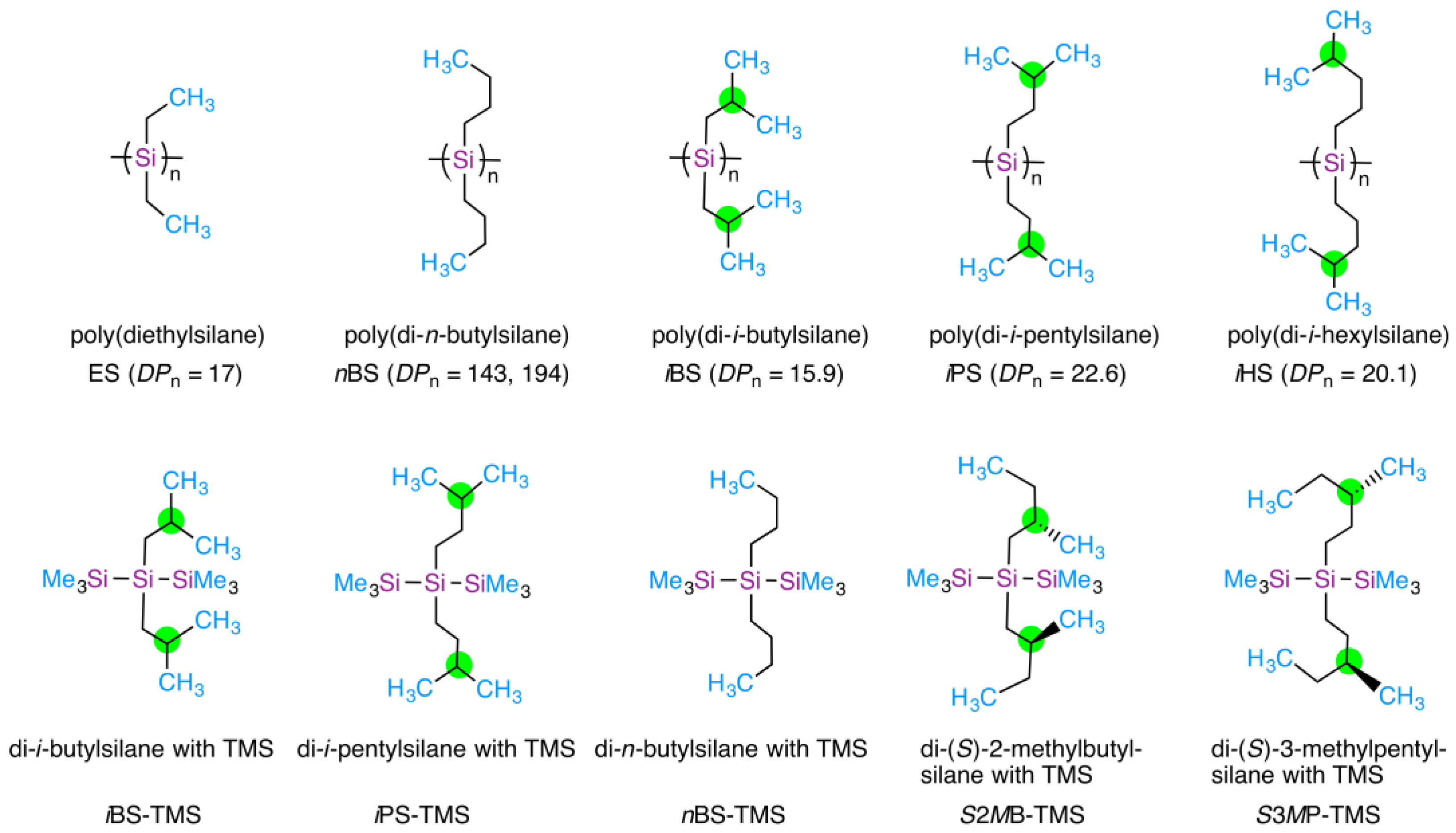
Figure 1. (Top) Chemical structures of poly(diethylsilane) (ES), poly(di-n-butylsilane) (nBS), poly(di-i-butylsilane) (iBS), poly(di-i-pentylsilane) (iPS), and poly(di-i-hexylsilane) (iHS) studied in the present and previous works [58]. The alkyl side chains carry two CH3 (ES and n-BS) and four CH3 (iBS, iPS, and iHS) directionally hindered rotors per repeating unit. DPn is the number-average degree of polymerization. (Bottom) Chemical structures of iBS-TMS, iPS-TMS, nBS-TMS, S2MBS-TMS, and S3MPS-TMS as trimeric models of iBS, iPS, nBS, and hypothetical poly((S)-2-methylbutylsilane) and poly((S)-3-methylpentylsilane for VCD, IR, and Mulliken charge calculations, as discussed later. The CH3 groups and silicon atoms are highlighted in blue and purple, respectively. Green circles indicate the branching points of the i-alkyl and (S)-alkyl side chains.
Symmetry 17 01544 g001
Figure 2. Chemical structures of oxygen-containing achiral alkanols and alkyl ethers as solvent molecules to test iBS, iPS, and iHS, and for comparison, i-octane. Use of tetrahydrofuran and i-octane as solvents did not generate optically active iBS, iPS, and iHS. CH3 groups and oxygen atoms are highlighted in blue and red, respectively.
Symmetry 17 01544 g002
Figure 3. Changes in CD and UV spectra (N = 3) of iBS in n-propanol at (a) 15 °C, (b) 40 °C, (c) 70 °C, (d) 75 °C, and (e) 90 °C. (f) Changes in the gabs values at 295 nm for the 295 nm CD band as a function of temperature. Here, the red, blue, and red lines and filled circles refer to the three independent results. The yellow bars in (ae) are due to handed helices at ~270 nm, ~295 nm, and ~315 nm, while those in (f) are due to three second-order phase transitions of the 295 nm CD band at TC1, TC2, and TC3, and the gray bar is due to the coexistence of two opposite helices at 295 nm and 315 nm. The dashed lines in (af) are the baselines of the CD spectra at the right-side ordinate and the gabs at the left-side ordinate, respectively. For further evidence, all CD and UV spectra (N = 3) in the range from –5 °C to 90 °C at 5 °C intervals are shown in Figure S8 in Supplementary Materials.
Symmetry 17 01544 g003
Figure 4. Changes in the CD and UV spectra (N = 3) of iBS in i-propanol at (a) –5 °C, (b) 40 °C, (c) 50 °C, (d) 55 °C, and (e) 70 °C. (f) Changes in the gabs values at 295 nm as a function of temperature. Red, blue, and green lines and their filled circles show the results. The yellow bars in (ae) are due to the three handed helices at ~270 nm, ~295 nm, and ~315 nm [58]. The yellow bars in (f) are due to the three second-order helix–helix transitions of the 295 nm CD band at TC1, TC2, and TC3, while the gray bars in (f) are due to the two opposite screw sense helices at 295 nm and 315 nm. The dashed lines in (af) are the baselines of the CD spectra on the right and left sides of the ordinate, respectively. For further evidence, all the CD and UV spectra (N = 3) of iBS in i-propanol in the range from −5 °C to 70 °C at 5 °C intervals are displayed in Figure S9 in Supplementary Materials.
Symmetry 17 01544 g004aSymmetry 17 01544 g004b
Figure 5. Changes in CD and UV spectra (N = 3) of iBS in i-butanol at (a) –5 °C, (b) 35 °C, (c) 50 °C, (d) 65 °C, and (e) 90 °C. (f) Changes in the gabs values at 295 nm for the 295 nm CD band as a function of temperature. The red, blue, and green lines and filled circles are due to the results. The yellow bars in (ae) are due to the three handed helices at ~270 nm, ~295 nm, and ~315 nm, respectively [58]. The yellow bars in (f) are due to three second-order helix–helix transitions of the 295 nm CD band at TC1, TC2, and TC3, while the gray bar in (f) is due to two opposite helices at 295 nm and 315 nm. The dashed lines in (af) are the baselines of the CD spectra on the right and left sides of the ordinate, respectively. For further evidence, all the CD and UV spectra (N = 3) of iBS in i-butanol in the range from –5 °C to 90 °C at 5 °C intervals are displayed in Figure S10 in Supplementary Materials.
Symmetry 17 01544 g005aSymmetry 17 01544 g005b
Figure 6. Changes in the CD and UV spectra (N = 3) of iBS in i-pentanol at (a) –5 °C, (b) 40 °C, (c) 55 °C, (d) 65 °C, and (e) 85 °C. (f) Changes in the gabs values at 295 nm for the 295 nm CD band as a function of temperature. Red, blue, and green lines and their filled circles are due to the results. The yellow bars in (ae) are due to the three handed helices at ~270 nm, ~295 nm, and ~315 nm [58]. The yellow bars in (f) are due to three second-order helix–helix transitions of the 295 nm CD band at TC1 (>100 °C, onset temperature of preferential screw sense helicity, expected), TC2 (55 °C, helix–helix transition), and TC3 (25 °C, helix–helix transition), and the gray bars in (f) are due to two opposite helices at 295 nm and 315 nm. The dashed lines in (af) are the baselines of the CD spectra on the right and left sides of the ordinate, respectively. For further evidence, all the CD and UV spectra (N = 3) of iBS in i-pentanol in the range from –5 °C to 90 °C at 5 °C intervals are shown in Figure S11 in Supplementary Materials.
Symmetry 17 01544 g006
Figure 7. Changes in the CD and UV spectra of iBS in (a) p-dioxane-h8 at 15, 25, 65, and 85 °C and (b) p-dioxane-d8 at 25, 30, 45, and 80 °C. Changes in the gabs values of (c) p-dioxane-h8 at 298 nm of the 295 nm CD band as a function of temperature and (d) p-dioxane-d8 at 298 nm of the 295 nm CD band as a function of temperature. The yellow bars in (a,b) are due to the three handed helices at ~270 nm, ~295 nm, and ~315 nm, respectively [58]. The yellow bars in (c) are due to three second-order helix–helix transitions in p-dioxane-h8 of the 295 nm CD band at TC1 (77 °C, helix generation), TC2 (52 °C, helix–helix transition), and TC3 (18 °C, helix–helix transition), while those in (d) are due to three transitions in p-dioxane-d8 at TC1 (77 °C, helix generation), TC2 (38 °C, helix–helix transition), and TC3 (28 °C, helix–helix transition). The dashed lines in (ad) are the baselines of the CD spectra on the right and left sides of the ordinate, respectively. For further evidence, all the CD and UV spectra (N = 1) of iBS in p-dioxane-h8 and -d8 in the range from 15 °C to 90 °C at 5 °C intervals, respectively, are displayed in Figures S13 and S14 in Supplementary Materials.
Symmetry 17 01544 g007
Figure 8. Changes in the CD and UV spectra (N = 1) of iPS in n-propanol at (a) –5 °C and 10 °C, (b) 25 °C and 30 °C, (c) 65 °C and 70 °C, (d) 75 °C and 85 °C, and (e) 90 °C. (f) Changes in the gabs of iPS at 298 nm of the 295 nm CD band in n-propanol as a function of temperature. The yellow bars in (ae) are due to three handed helices at ~280 nm, ~300 nm, and ~320 nm, respectively [58]. The yellow bars in (f) are due to four second-order helix–helix transitions of iPS in n-propanol at TC1 (> 97 °C, onset temperature of preferential screw sense helicity, expected), TC2 (77 °C, helix–helix transition), TC3 (67 °C, helix–helix transition), and TC4 (27 °C, helix–helix transition). The dashed lines in (af) are the baselines of the CD spectra at the right side of the ordinate and the gabs at the left side of the ordinate, respectively. For further evidence, all the CD and UV spectra of iPS in n-propanol in the range from –5 °C to 90 °C at 5 °C intervals are shown in Figure S15 in Supplementary Materials.
Symmetry 17 01544 g008
Figure 9. Solvent dependence of maximum gabs: (a) iBS at 290–300 nm below 25 °C and (b) iPS at 290–300 nm at 0 °C.
Symmetry 17 01544 g009
Figure 10. CD and CPL characteristics of iBS in n-propanol at –5 °C: (a) CD and UV spectra with the greatest gabs value at 295 nm (N = 3), (b) CPL and PL spectra excited at 290 nm, revealing the greatest glum with (+) and (–) signs at 340 nm, (c) histogram (N = 5), and (d) normalized CD, UV, CPL, and PL spectra as a function of wavenumber. CD and CPL characteristics of iPS in n-propanol at –5 °C: (e) CD and UV spectra with the greatest gabs value at 299 nm (N = 3) and (f) CPL and PL spectra showing the greatest glum value at 330 nm showing the (+)-sign with (g) its histogram (N = 4), and (h) normalized CD, UV, CPL, and PL spectra as a function of the wavenumber. The yellow bars in (a,b,d,e,f,h) are due to helices in the S0 and S1 states. The dash lines (red and blue) in (d,h) are taken from (a,c). For further evidence, all CPL and PL spectra of iBS and iPS in n-propanol at –5 °C are shown in Figure S19a–e,i–l in Supplementary Materials.
Symmetry 17 01544 g010
Figure 11. VCD and IR spectra with residual noise at 23 °C for (a) n-propanol, (b) n-butanol, (c) n-pentanol, (d) i-propanol, (e) i-butanol, (f) i-pentanol, (g) 2,4-dimethyl-3-pentanol, (h) geraniol, (i) p-dioxane-h8, and (j) p-dioxane-d8. Additionally, the VCD and IR spectra of other alkanols (methanol, ethanol, n-hexanol, n-octanol, cyclopentanol, and 2-ethyl-1-butanol), cyclic alkyl ethers (THP, THF, and 15CE5), and acyclic alkyl ethers (diglyme) are shown in Figure S20 in Supplementary Materials. The VCD signals in the gray area of (a,b,di) are not reliable because IR absorbance > 1.0, meaning IR transmittance < 10%.
Symmetry 17 01544 g011
Figure 12. Pseudo-cyclic structures of alkanols and twisted cyclic ethers. The three hydrogen atoms of the CH3 groups are highlighted in blue, black, and red, respectively. Two hydrogen atoms in CH2 groups are highlighted in blue and black, respectively. The oxygen atoms are highlighted in red. Directional rotation and twisting motions are indicated by green arrows. * in black and * in red denote pseudo-stereocenters at the carbon atom and O atom, respectively.
Symmetry 17 01544 g012
Figure 13. Mulliken charges obtained with MP2 (6-31G(d,p) basis set): (a) n-propanol (trans), (b) n-propanol (gauche), (c) n-butanol (trans), (d) n-butanol (gauche), (e) n-pentanol (trans), (f) n-pentanol (gauche), (g) i-propanol, (h) i-butanol, (i) p-dioxane (1,4-boat twisted by 21°), and (j) p-dioxane (2,5-boat twisted by 29°). Green and red indicate (+)- and (–)-Mulliken charges, respectively.
Symmetry 17 01544 g013aSymmetry 17 01544 g013b
Figure 14. Schematic illustration of intramolecular C(δ)–H(δ+)/O(δ) interactions with attractive forces. (a) Hypothetical pseudo-six-membered ring structure of n-propanol and (b) hypothetical pseudo-five-membered ring structure of i-propanol. The three H1, H2, and H3 atoms of the CH3 rotor are highlighted in blue, pink, and black, respectively. The H atom and the two lone pairs of O atoms are highlighted in blue, pink, and black, respectively. The unidirectional motion of the CH3 rotor is indicated by the green arrow. Note: * in black and * in red denote pseudo-stereocenters at the carbon atom and O atom, respectively. Note: the EW charge for 1H is −0.108 and for 16O is +6.216 [58]. 1H thus experiences an attractive force with 16O [58].
Symmetry 17 01544 g014
Figure 15. Intramolecular repulsive C)–H+)/Si+) interactions with same-sign Mulliken charges between branched alkyl groups and Si–Si bonds. Hypothetical pseudo-six-membered and pseudo-seven-membered ring structures of side chains with Si atoms: (a) i-butyl (β-branched achiral), (b) (S)-2-methylbutyl (β-branched chiral), (c) i-pentyl (γ-branched achiral), and (d) (S)-3-methylpentyl (γ-branched chiral). Three H1, H2, and H3 atoms of the CH3 terminus, responsible for the HINS-induced directional rotor [58], are highlighted in blue, pink, and black, respectively. Si atoms are highlighted in red. Unidirectional rotor motions of C–C bonds are indicated by thin green arrows for i-butyl and i-pentyl. Unidirectional rotor motions of the C–C bonds are shown as thick green arrows for the (S)-2-methylbutyl and (S)-3-methylpentyl groups. The EW charges of 1H and 28Si have opposite signs; for 1H: Qw = –0.108 and for 28Si: Qw = +12.488 [58], although the Mulliken charges of 1H and 28Si have the same sign; for 1H: ~+0.1 and for 28Si: +0.5 [58]. Note: * in black and * in red refer to pseudo-stereocenters and true stereocenters, respectively.
Symmetry 17 01544 g015
Figure 16. Intermolecular attractive Si(δ+)/O(δ) interactions between O (one of the two lone pairs) and Si (empty d-orbital), and intermolecular repulsive C–H(δ+)/H(δ+)–C and intermolecular attractive C–H(δ+)/O(δ) interactions. The three hydrogen atoms of the CH3 rotors are highlighted in blue, red, and black, respectively. The two lone pairs of the oxygen atoms are highlighted in black and red, respectively. The Mulliken charges of the 16O and 28Si atoms have opposite signs of –0.64 and +0.46, respectively. The Mulliken charges of the 1H atoms range from +0.10 to +0.13. The difference in the strength of the Si(δ+)/O(δ) interaction is illustrated for the less bulky n-propanol (left) and more bulky 2-ethyl-1-butanol (right) when both Si atoms bear an i-butyl group. The difference in the bulkiness induces opposite sense Si–Si main-chain helicity. Note: * in black and * in red indicate pseudo-stereocenters on the carbon and oxygen atoms, respectively.
Symmetry 17 01544 g016
Figure 17. Schematic illustrations of the directional motions of +NH3 and CH3 hindered rotors for zwitterion species of glycine, L- and D-alanine, L- and D-valine, and L- and D-leucine. The CH3 and +NH3 rotors are highlighted in green and blue, respectively. The same directional motion of the rotors is assumed to cause energy inequality between L- and D-amino acids, which are then no longer enantiomeric. Here, +NH3 and CH3 groups, and, presumably, +OH3 ion, which all possess three identical 1H atoms, belong to the HINS nuclear spin-1/2 system in TWP as well as CF3 group containing three identical 19F-1/2 atoms in TWP [58] (see Figure 18). The HINS nuclear-spin-1/2 system in TWP is responsible for the unidirectional motions [58].
Symmetry 17 01544 g017
Figure 18. Proposed unidirectional motions (red arcs) of CH3, +NH3, +OH3, and CF3 rotors which are HINS systems in TWP [58]. The three black arrows represent the handed directions of the three identical nuclear-spin-1/2 atoms.
Symmetry 17 01544 g018
Figure 19. (a) CD and UV spectra of zwitterionic glycine (IEP 5.97) in neutral water at 20 °C. [Conc]0 = 1.00 × 10⁻² M, path length 1.0 mm, with 0.5 nm data interval. (b) Simulated CD and UV spectra (6 singlet transitions with fwhm of 0.10 eV) of zwitterionic glycine in H2O obtained with TD-DFT (B3LYP/6-31G(d,p)) using IEF-PCM. The blue bars in (a,b) indicate two major CD extrema. The dashed lines in (a,b) are the baselines of the CD spectra. (c,d) Visualization of molecular orbitals (isovalue of 0.08): the first LUMO (#21) and the first HOMO (#20), respectively. (e) Unidirectional rotation (yellow arc) of +NH3 rotor drives unidirectional rotation (white arc) of CO2 rotor responsible for the observed CD spectrum. Based on their Mulliken charges (red: negative and green: positive), an attractive interaction between the +NH3 and CO2 rotors in H2O is expected.
Symmetry 17 01544 g019
Table 1. CD polarization and inversion temperatures (in °C) of iBS, iPS, iHS, nBS, and ES in achiral solvents.
Polymers
(Side Chain Type)
SolventsTC1/°CTC2/°CTC3/°CTC4/°CNumbers of CH3 Rotors
as Repeating Unitas Solvent
iBS
(β-branched)
n-propanol>97 *7229<–20 *41
i-propanol>83 *6032 42
i-butanol>100 *7228 42
i-pentanol>100 *5525 42
p-dioxane-h8775218 40
p-dioxane-d8773828<10 *40
iPS
(γ-branched)
n-propanol>97 *80672741
n-pentanol~10 *41
n-hexanol~10 *41
n-octanol~20 *41
i-propanol~20 *42
i-butanol~20 *42
iHS
(δ-branched)
n-octanol~20 *41
nBS **
(non-branched)
n-undecane40 22
n-dodecane10528 22
n-dodecane-d264225 22
n-tridecane10528 22
n-tetradecane95 *28 22
ES **
(non-branched)
n-propanol30<–10* 21
TC1 denotes the onset temperature of preferential screw sense helicity, whereas TC2, TC3, and TC4 are the helix–helix transition temperatures; [*] indicates expected values; [**] were taken from [58]. The number of CH3 rotors as repeating units determines the number of helix–helix transitions; four and two rotors lead to four and two transitions, respectively.
Table 2. Temperature- and solvent-dependent chiroptical codes—relations between chiroptical signs and clockwise and counterclockwise motions of CH3 rotors in polysilane side chains.
SystemsT > TC1 1–3TC1 > T > TC2 1–3TC2 > T > TC3 1–3TC3 > T > TC4 1–3T < TC4 1–3
iBS,
n-propanol
(⟲+⟳) + (⟳+⟲)
= 0
(⟲+⟳) + (⟲+⟲)
= –2
(⟲+⟳) + (⟳+⟳)
= +2
(⟲+⟲) + (⟲+⟲)
= –4
(⟳+⟳) + (⟳+⟳)
= +4
iBS,
i-propanol
(⟲+⟳) + (⟳+⟲)
= 0
(⟲+⟳) + (⟲+⟲)
= –2
(⟲+⟳) + (⟳+⟳)
= +2
(⟲+⟲) + (⟲+⟲)
= –4
Possibly
= +4
iBS,
p-dioxane-d8
(⟲+⟳) + (⟳+⟲)
= 0
(⟲+⟳) + (⟲+⟲)
= –2
(⟲+⟳) + (⟳+⟳)
= +2
(⟲+⟲) + (⟲+⟲)
= –4
Possibly
= +4
iPS,
n-propanol
(⟲+⟳) + (⟳+⟲)
= 0
(⟳+⟳) + (⟳+⟲)
= +2
(⟲+⟲) + (⟳+⟲)
= –2
(⟳+⟳) + ⟳+⟳)
= +4
(⟲+⟲) + (⟲+⟲)
= –4
SystemsT > TC1TC1 > T > TC2T < TC2
nBS,
n-dodecane-h26
⟲+⟳
= 0
⟲+⟲
= –2
⟳+⟳
= +2
nBS,
n-dodecane-h26
with THF
Possibly
= 0
⟳+⟳
= +2
⟲+⟲
= –2
nBS,
n-dodecane-d26
⟲+⟳
= 0
⟲+⟲
= –2
⟳+⟳
= +2
1 ⟳ and ⟲: Clockwise and counterclockwise motions of one CH3 rotor, respectively. 2 (⟲+⟳) and (⟳+⟲): disrotatory motions for a couple of two CH3 rotors, causing no effective twisting torque. 3 (⟳+⟳) and (⟲+⟲): conrotatory motions for a couple of two CH3 rotors, producing an effective left- or right-handed twisting torque.
Disclaimer/Publisher’s Note: The statements, opinions and data contained in all publications are solely those of the individual author(s) and contributor(s) and not of MDPI and/or the editor(s). MDPI and/or the editor(s) disclaim responsibility for any injury to people or property resulting from any ideas, methods, instructions or products referred to in the content.