Article

Characteristics of Foot Pressure Distribution During Standing and Walking with Anatomical Leg Length Discrepancy—A Comparative Analysis of Patients with and Without Low Back Pain

1
Department of Propaedeutic, Physical Diagnostics and Dental Physiotherapy, Faculty of Medicine and Dentistry, Pomeranian Medical University, 70-204 Szczecin, Poland
2
Konior, Maciąg Health Centre Sp.J., 72-200 Nowogard, Poland
3
PODOLOGIA.pl, 70-465 Szczecin, Poland
4
Department of Functional Diagnostics and Physical Medicine, Faculty of Health Sciences, Pomeranian Medical University, 71-210 Szczecin, Poland
5
Department of Human Nutrition and Metabolomics, Pomeranian Medical University in Szczecin, 71-460 Szczecin, Poland
6
FIKOU Physiotherapy, 44-100 Gliwice, Poland
7
Orthogenic Rehabilitation and Podology Center, 45-573 Wroclaw, Poland
*
Author to whom correspondence should be addressed.
Symmetry 2025, 17(7), 1059; https://doi.org/10.3390/sym17071059
Submission received: 21 February 2025 / Revised: 30 April 2025 / Accepted: 27 May 2025 / Published: 4 July 2025
(This article belongs to the Section Life Sciences)

Abstract

Body asymmetry is often analysed in the context of low back pain (LBP). To date, research has mainly focused on the general relationships between asymmetry and pain, with less attention paid to issues related to pressure distribution and its potential impact on the occurrence of LBP. The aim of this study was to compare biomechanical parameters in people with anatomical leg length discrepancy with and without LBP to identify overloads that may lead to pain. Early detection of common abnormalities in these parameters in both groups may influence the early prevention of 0LBP in the course of LLD. Materials and methods: This study included 60 patients with diagnosed LLD, of whom 30 had LBP (group 1, NP) and 30 were pain-free (group 2, NwP). Body weight distribution during standing and walking was analysed using pedobarography. The analysis was carried out in two stages, the first being the analysis of the biomechanical parameters for the whole study population, for group 1 with LBP and group 2 without LBP, while the second stage focused on the main issue, i.e., the comparison of the group with LBP with the group without LBP. The study included standing and walking tests. Left–right pressure distribution and ground contact time were analysed. In addition, the angle of foot abduction was analysed to indirectly assess compensatory mechanisms resulting from the asymmetry. Results: The standing test showed significantly greater pressure on the longer limb (p = 0.022) in the whole study population (N = 60). When divided into groups, it was found that in those with LBP (NP = 30), the difference was not statistically significant (p = 0.359), whereas in those without pain (NwP = 30), the pressure on the longer limb was significantly greater (p = 0.002). No differences were found between the groups in the comparative analysis. The angle of foot abduction was greater than normal across the study population (N = 60), with greater values in the shorter limb (12.83° vs. 11.04°), which was close to significance (p = 0.065). The group with LBP (NP = 30) showed a similar trend, also close to statistical significance (p = 0.054), with significantly higher values of abduction angle in both legs compared to the group without LBP (NwP = 30). In the walking test, the left–right load distributions were significantly dispersed. The mean pressure on the longer limb was significantly higher in group 1 (NP = 30) (p = 0.031), whereas this difference was not statistically significant in group 2 (NwP = 30). For mean peak pressure, there were no significant differences in any of the groups tested. In addition, the mean ground contact time during gait was longer for the longer limb in the whole study population (N = 60) (938.8 ms vs. 915 ms), but again, this difference did not reach statistical significance (p = 0.305). Comparative analysis showed no differences between the groups. Conclusions: This study showed that in people with anatomical LLD, both with and without LBP, most parameters reflected marked asymmetries in peak and mean pressures and abduction angles. A prolongation of ground contact time has also been shown, and even though some parameters were not statistically significant, it is important to note the high dispersion of left–right loading, which provides information on body load asymmetries in patients with anatomical LLD. Given that there were no differences between the groups for most of the parameters, it is important for both clinical practice and further research that the abnormalities observed in both groups (NP = 30, NwP = 30) may have been a significant predictor of the development of LBP, as the abnormalities preceded the onset of pain. This should be taken into account in diagnostic and preventive measures.

1. Introduction

Chronic and severe low back pain is a cause of disability, often long-term [1,2,3]. The prevalence of non-specific low back pain ranges from 4% to 20%, with a linear increase from 30 to 60 years of age [4]. On the other hand, pain is increasingly reported in young people [5,6,7,8]. The cause of pain may also be related to postural defects and dysfunction of the joints, ligaments and myofascial structures [2].
Both structural and functional leg length discrepancies (LLD) contribute to the uneven loading of the lower limbs, leading to the development of postural and gait abnormalities. They can also result in functional limitations [9,10,11,12,13,14]. It should be emphasised that the absence of pain in LLD does not mean that the posture is normal [15,16]. It has been shown that LLD directly contributes to the development of pelvic asymmetry [17] and scoliotic posture [14,18]. In most cases, LBP is associated with postural asymmetry in the pelvis [19,20,21,22,23]. A number of studies have shown that pelvic asymmetry alters body mechanics, placing asymmetric stress on different segments of the body, resulting in musculoskeletal pain [23,24]. Changes in trunk kinematics observed during standing, walking [25] and sitting [26] are associated with non-specific low back pain. LLD has been repeatedly shown to lead to a number of postural and functional compensations that have a long-term impact on the development of degenerative processes, including in the spine [27,28,29,30].
Any postural asymmetry will consequently alter the mechanical distribution of foot pressures and lead to postural imbalances [11,12,13,31]. This is because anatomical limb length inequality immediately alters the alignment of the entire posture, which directly affects postural patterns, both static and dynamic (including locomotion). A detailed analysis of the biomechanical parameters during standing and walking in people with LLD is highly warranted, mainly because of the link between postural asymmetry and the development of musculoskeletal pathologies, as discussed above. There have been research studies looking at plantar pressure in people with LLD and the effects of LLD on pelvic asymmetries and spinal alignment. Most of the available studies have used induced (simulated) conditions, which do not allow for a reliable assessment of the actual compensation processes [12,14,32,33,34,35]. Previous analyses of pressure distribution abnormalities in people with LLD and LLD with LBP have mainly focused on analysing centre of pressure (COP) abnormalities. Despite numerous studies on the relationship between limb asymmetry and pain, there is still a lack of analyses comparing pressure distribution in individuals with anatomical leg length discrepancy (LLD). Meanwhile, pressure distribution abnormalities may play an important role in the development of pain and musculoskeletal dysfunction, including degenerative changes. Particular attention should be paid to the growing problem of low back pain (LBP), which can result from limb length asymmetry, leading to pelvic misalignment, muscle strain and spinal overload.
Research into the biomechanics of standing and walking in people with LLD is important for both preventing tension pain and limiting progressive degenerative changes in the musculoskeletal system. To this end, pedobarographic examinations can be used to assess biomechanical parameters such as plantar pressure distribution, ground contact time and foot abduction angle [32,36,37,38]. The latter parameter may be an indicator of a compensatory mechanism whereby the plane of support is increased in order to maintain balance. The aim of this study was to analyse pressure distribution by comparing two groups of patients with anatomical leg length discrepancy (LDD)—with and without pain—to identify common biomechanical parameters. This allowed for the early detection of abnormal load distribution, which could lead to the faster implementation of prophylactic measures to prevent LBP.

2. Description of This Study

2.1. Aim

The aim of this study was a comparative analysis of pressure distribution and spatial–temporal parameters in people with LLD, divided into those with and without low back pain (LBP). In addition, a comparative analysis was performed between the groups with and without LBP to investigate the differences between them.

2.2. Material and Methods

The study included sixty (N = 60) adults, 23 women and 37 men, with LLD confirmed by radiography (long-film X-ray with limb length measurement) (nf = 23 women, nf% = 38.33%; nm = 37 men, nm%= 61.67%). Participants ranged in age from 18 to 60 years (with an average age of 42). The participants were divided into two equal groups, according to the following criteria:
  • Group 1—30 participants (NP = 30; 50%) with LLD and low back pain (LBP), including 9 women and 21 men (nf = 9 women, nf%= 30%; nm = 21 men, nm%= 70%);
  • Group 2—30 participants (NwP = 30; 50%) with LLD without LBP, including 14 women and 16 men (nf = 14 women, nf%= 46.66%; nm = 16 men, nm% = 53.33%).

2.3. Inclusion Criteria

-
Individuals with radiographically confirmed LLD, with and without low back pain (LBP);
-
Individuals aged over 18 and under 60 years.

2.4. Exclusion Criteria

-
Individuals with a history of spinal or limb surgery;
-
Individuals with a history of mechanical musculoskeletal and other injuries likely to affect the distribution of foot pressure, unrelated to LLD;
-
Individuals with a history of injuries to the limbs and axial skeleton with a significant effect on musculoskeletal biomechanics;
-
Those with diabetes and other endocrinopathies;
-
Those with rheumatic diseases;
-
Those with other diseases that affect fitness and locomotion;
-
Those who were pregnant.

2.5. Research Methodology

  • Limb length was measured by radiography (X-ray) in a standing position with equal weight on both legs (i.e., central alignment of the long axis of the patella). The study included both an absolute leg length measurement, which involved measuring the actual length of the bones from the femoral head to the lateral malleolus, as well as a relative measurement, which took into account the alignment of the pelvis and spine, measuring the length from the anterior superior iliac spine to the malleolus.
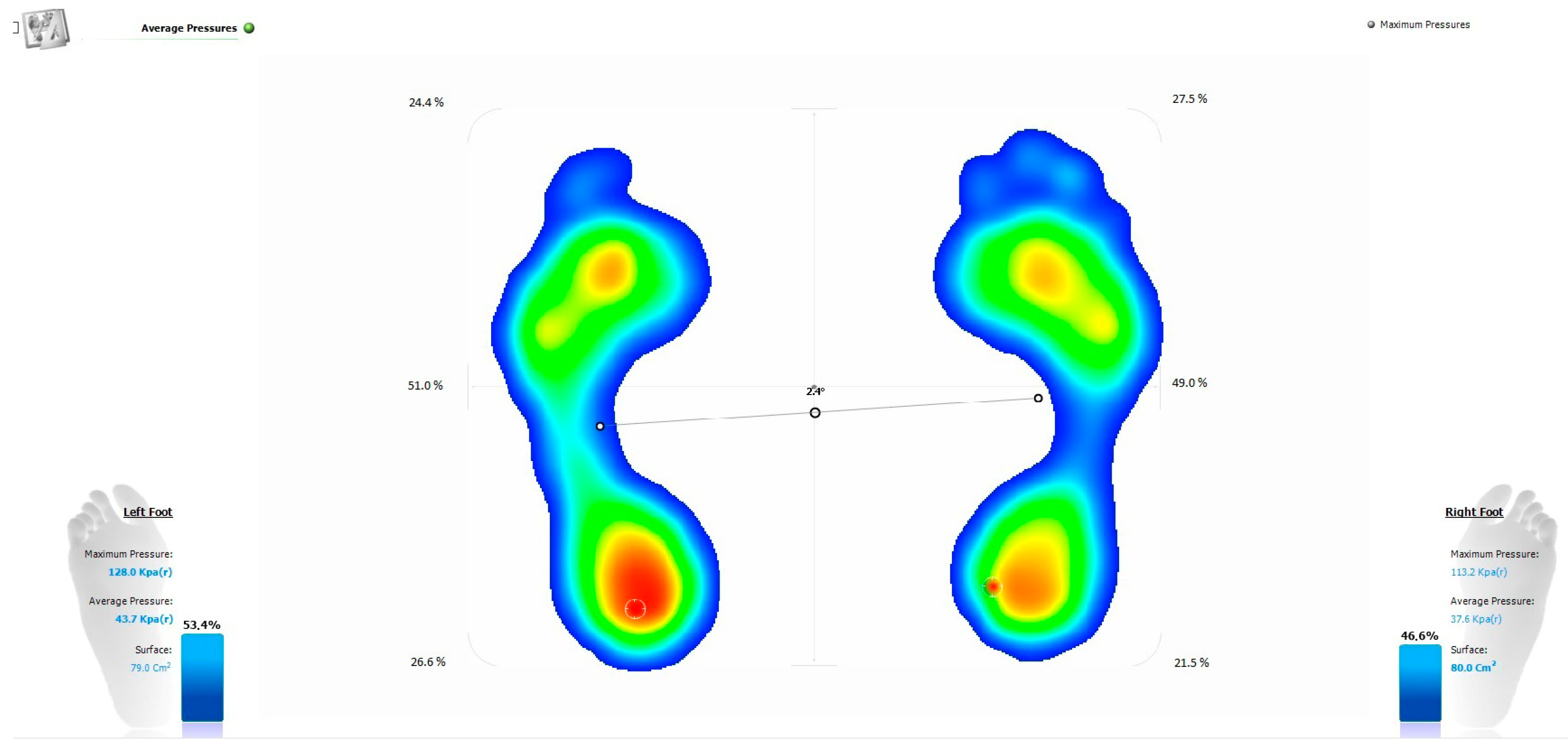
  • Pedobarography was conducted using the EPS R2 pedobarograph (LETSENSE GROUP LORAN ENGINEERING, Castel Maggione Bologna, Italy), paired with Biomech Studio software version 1.6.4.28272. The test was performed barefoot in casual clothing to eliminate potential confounding factors. Participants did not engage in any physical activity on the day of testing. Standing pedobarography was performed for 20 s with a sampling interval of one millisecond. In this way, the procedure eliminated the need for repeated measurements, as the recording of front–back and left–right oscillations at such a high frequency ensured the high sensitivity and reliability of the results obtained. In the dynamic test, the gait path was designed to allow free walking, without having to aim at the instrument. As a result, the footprints where the patient missed the mat could be erased, thus ensuring the greater accuracy of the results. Twenty footprints for each foot were included in the analysis, allowing a reliable assessment of gait parameters. The following parameters were assessed by pedobarography:
    a.
    Left–right pressure distribution during standing (right and left side of the body) (Figure 1);
    b.
    Placement of the feet on the ground (foot abduction angle) (Figure 2);
    c.
    Mean and peak pressure during walking (Figure 3);
    d.
    Ground contact time of the left and right foot (Figure 3).

2.6. Data Analysis

Descriptive statistics were calculated for all patients and for the groups with and without LBP. Differences in the mean values of the measured parameters were examined between the shorter and longer legs (for all patients and in the groups with and without LBP), and between the groups with and without LBP for the shorter and longer legs. The Shapiro–Wilk test showed a statistically significant deviation from the normal distribution for most parameters in the compared groups, so the statistical significance of the differences in these distributions was then tested using the Wilcoxon–Mann–Whitney test. For static loading and abduction, the number of patients within the normal range in both groups was compared, and the significance of the differences was tested using Pearson’s Chi-squared test with Yates’ continuity correction. The results were considered statistically significant at p < 0.05. Statistical calculations were performed using the R software, ver.4.3.0.

3. Results

Descriptive statistics for all 60 patients and the groups with and without LBP are presented in Table 1, Table 2 and Table 3.

3.1. Results Obtained During Standing Test

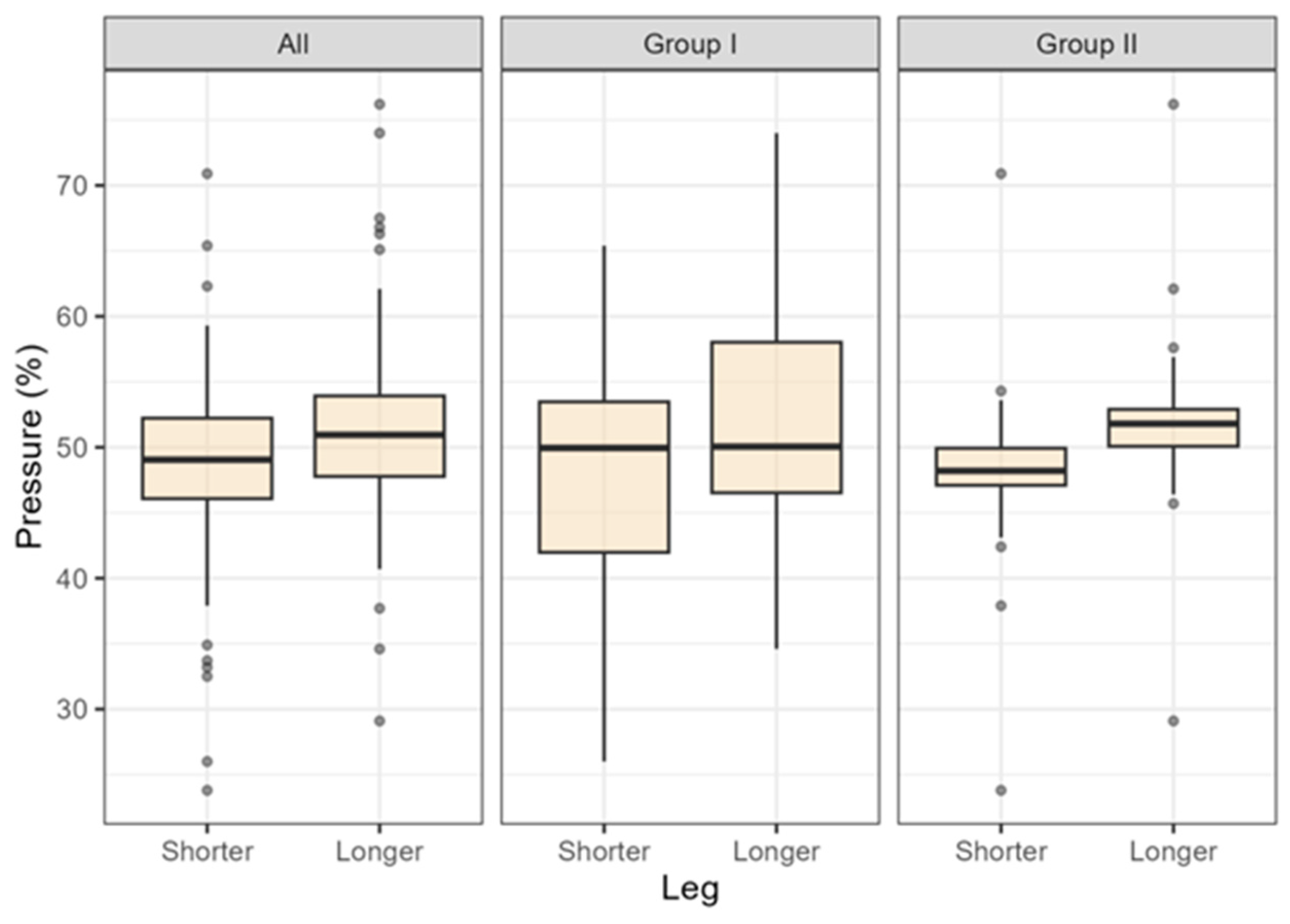
3.1.1. Left–Right Distribution of Foot Pressure During Standing (i.e., Pressure in the Shorter Limb vs. Pressure in the Longer Limb; The Range of 48–52% Was Adopted as the Normal Range of Left–Right Pressure Distribution for Both Limbs)

The percentage distribution of the pressure on the shorter and longer leg is shown in Figure 4.
A significantly higher percentage of pressure on the longer leg was observed in the study group as a whole (NA = 60; 100%) and in the group of patients without LBP (NwP = 30; 50%) (Table 4). Pressure was also higher in patients with LBP (NP = 30; 50%), but the difference was not significant. There was also no statistically significant difference between the groups with and without LBP in the distribution of pressure on the shorter and longer leg.
In the group with LBP, the pressure distribution on the left and right leg was normal in 10 out of 30 patients, and in the group without LBP, in 10 out of 30 patients. This difference was not statistically significant (χ2 [1] = 0.282, p = 0.595).

3.1.2. Foot Abduction Angle During Standing

The distribution of the angle of foot abduction on the shorter and longer leg is shown in Figure 5.
In the study group, a greater angle of abduction was observed in the foot of the shorter leg. Patients with LBP had, on average, a greater angle of abduction in both legs than patients without LBP. However, no significant differences in the mean values were observed in any of the tests performed for the angle of abduction (Table 5). The difference between the shorter and longer leg in the group of patients with LBP was very close to significance. A study with a larger sample might confirm this significance.
In the group with LBP, the foot abduction angle in the shorter leg was normal in 4 out of 30 patients, and in the group without LBP, in 7 out of 30 patients. The difference was not statistically significant (21 = 0.445, p = 0.505). In the group with LBP, the foot abduction angle in the longer leg was normal in 10 out of 30 patients, and in the group without LBP, in 9 out of 30 patients. The difference was not statistically significant (21 = 0.001, p > 0.99).

3.2. Results Obtained During Walking Test (Spatial–Temporal Parameters)

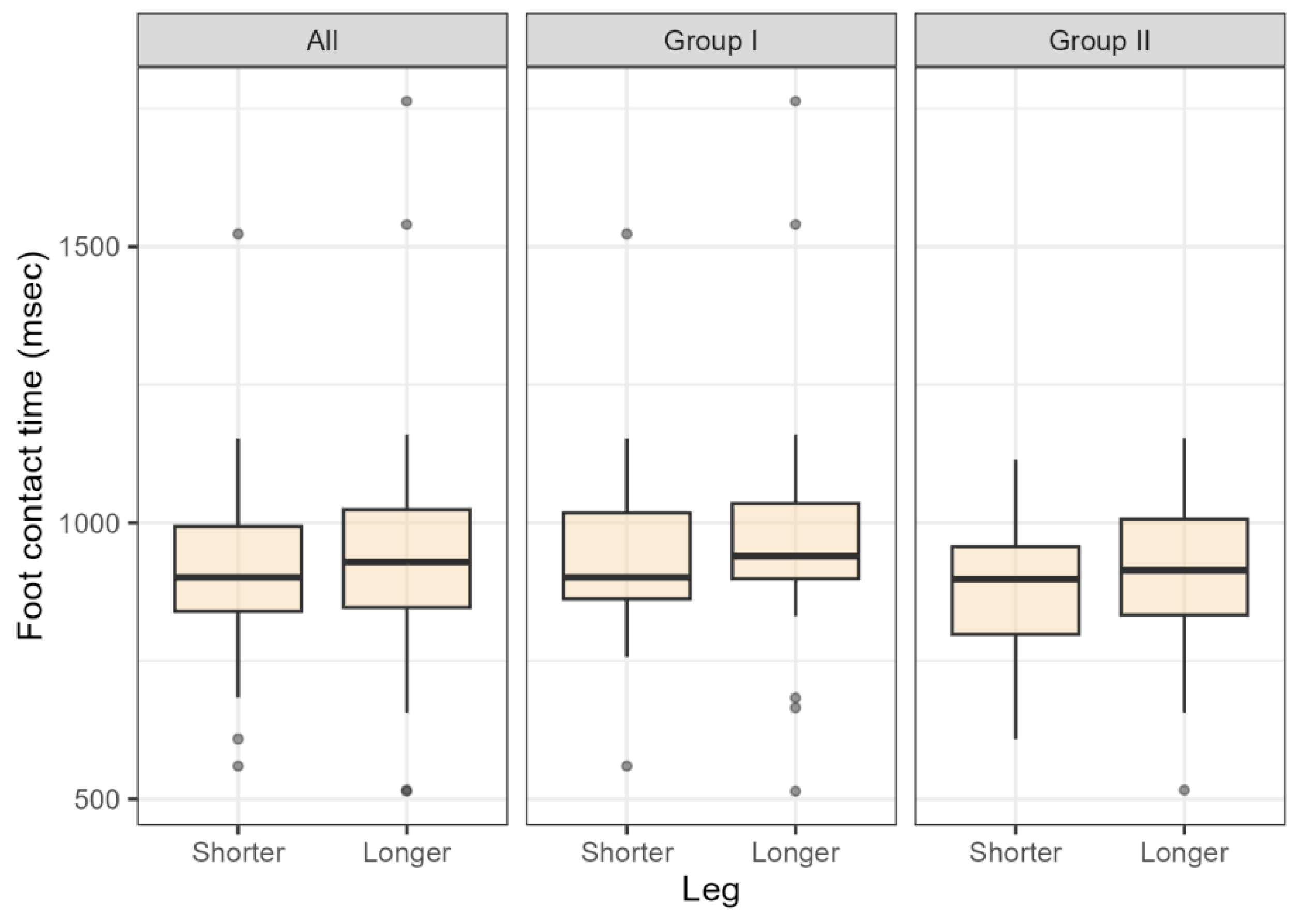
3.2.1. Foot Contact Time

The distribution of the foot contact time for the shorter and longer leg is shown in Figure 6.
In the study population, the longer leg had a longer ground contact time than the shorter leg. Both feet in the group with LBP also had a longer contact time. However, no statistically significant differences in the distributions were observed for ground contact time in any of the tests (Table 6).

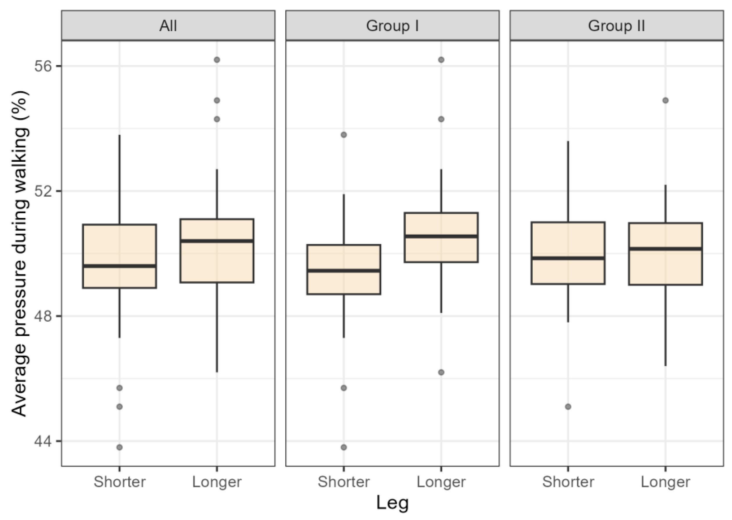
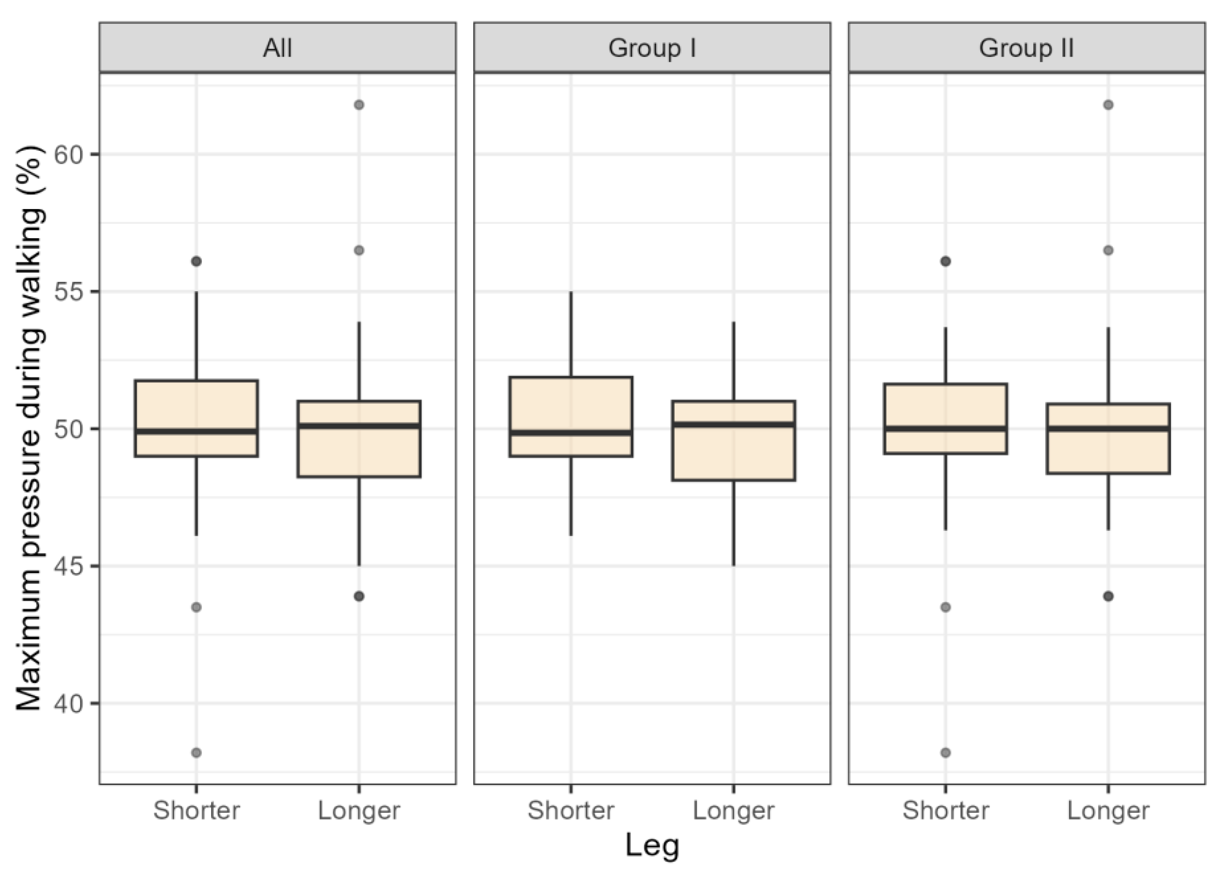
3.2.2. Foot Pressure Distribution (Mean Pressure and Peak Pressure)

The distribution of the mean pressure on the shorter and longer leg is shown in Figure 7.
The pressure on the longer leg was statistically significantly greater in group 1, and the difference in group 2 was not significant. There were no statistically significant differences in the distribution of mean pressure in the groups with or without LBP, either for the shorter or the longer leg (Table 7).
The distribution of the peak pressure on the shorter and longer leg is shown in Figure 8.
There was no significant difference in the distribution of peak pressure on the shorter or the longer leg. There were no statistically significant differences in the distribution of peak pressure in the groups with or without LBP, either for the shorter or the longer leg (Table 8).
There were no statistically significant differences in the mean values of mean and peak pressure (tested separately) between the shorter and longer leg. However, it should be noted that people with LBP had noticeably higher mean and peak pressure in both feet. (Table 7 and Table 8).

4. Discussion

Postural asymmetries contribute to the development of postural abnormalities, causing increased tension in myofascial structures, which can lead to degenerative changes in joints and bone structures [23,26,27,28,29,30]. Postural asymmetries affect the biomechanics of the whole body, both during locomotion and at rest (sitting, standing, lying down) [25,26], and are also manifested in balance problems [11,12,13,31]. This consequently affects the distribution of forces and pressures [39]. Importantly, most of these changes are asymptomatic prior to the onset of pain [9,10,13,14,15,16].
Leg length discrepancy (LLD) is directly responsible for the development of postural asymmetry and can trigger a chain of pathological processes [9,13,14,17,18,30,40,41,42,43]. Even the smallest difference in limb length can lead to pelvic misalignment, which is one of the causes of LBP [17,34,44,45]. The result is the impaired function and alignment of the lower spine. Structural (including degenerative) and functional changes in the spine have been repeatedly shown in the literature to be significantly correlated with LBP [27,28,46,47,48,49,50,51]. This includes scoliotic posture as a result of LLD compensation [30]. All of these factors may contribute to the development of LBP in the future. The authors believe that the early detection of abnormalities in the distribution of body pressures may be an important factor in the prevention of asymmetry-induced pain that occurs as a result of LLD. The contemporary literature does not provide sufficient data on the relationship between abnormal pressure distribution and postural asymmetry in low back pain (LBP) syndrome. For this reason, the authors undertook a comprehensive review of the available sources. However, there is a lack of recent research conducted directly on patients in real-life conditions, and most of the existing studies are based on simulated conditions.
Mannello [52] showed that the clinical significance of LLD was strongly dependent on both the degree of limb inequality and the ability of the pelvis and spine to compensate for this difference [52]. White et al. [18] noted that limb length differences of 1–3 cm resulted in significant differences in limb loading. In their analysis of ground reaction force parameters during walking, they showed that the shorter limb sustained greater loads during the landing phase, while the push-off force was greater in the longer limb with true LLD and in the shorter limb with simulated LLD [18]. Similar results were observed by Perttunen et al. in a study of people with anatomical LLD, where they found a longer stance phase and higher peak plantar pressures under the big toe for the longer leg. The researchers also observed that the ground reaction force in the push-off phase was greater in the longer limb [53]. Khamis and Carmeli found that even a 10 mm difference in limb length had an effect on gait pattern, and the greater the difference, the greater the deviation [13]. In one of the more recent studies conducted by the team of Pereiro-Buceta et al. [32] under simulated conditions, it was shown that a difference in limb length was associated with a decrease in mean and peak pressures on the longer limb and an overload on the shorter side. However, it should be emphasised that the study was conducted under artificially induced conditions, making it impossible to fully assess the compensatory mechanisms that may be adopted by patients with real LLD. In the latter case, patients walk with a particular gait pattern for many years, which may lead to adaptations that are not detectable in simulation studies. The studies cited above mainly focused on the analysis of dynamic loads, such as ground reaction force parameters during gait, but did not consider static loads, which can also have a significant impact on the biomechanics of the body. Extending the research to include static loads is therefore an important addition to the existing analyses. The study of static loading provides a better understanding of the compensatory mechanisms and overloading that may lead to the development of pain, including LBP, in patients with leg length discrepancy.
Taking these aspects into account, the analysis of pressure distribution during standing and walking plays an important role in the prevention of postural disorders. Of particular importance are static and dynamic tests that reflect actual day-to-day functional activities. In order to provide a more comprehensive assessment of biomechanical loading, our study also looked into the distribution of left–right plantar pressures during standing (on the left and right sides of the body) and the angle of foot abduction, which may reflect compensatory mechanisms associated with changes in the plane of support. In addition, mean and peak pressures and ground contact time were included in the analysis. The latter parameter is an important factor in overloading, as prolonged ground contact time can lead to fatigue- and overuse-related changes in musculoskeletal structures, as confirmed by findings from previous studies.
The use of pedobarography in the research methodology seems fully justified, as it allows for rapid and precise measurements of pressure distribution and ground contact parameters. This method also makes it possible to assess body imbalances and postural patterns relevant to biomechanics and gait patterns, including pathologies. It can also be used to analyse the effects of compensating for differing leg lengths [32]. Further studies of this type are warranted, especially in the context of analysing the relationship between abnormal load distribution and postural defects, which may be a secondary cause of overload and pain. Our study was conducted in real-life conditions in people with anatomical LLD and included a standing test. The participants were divided into two groups: with and without pain. The aim of this approach was to identify the common biomechanical symptoms present in both groups, as the early detection of abnormalities in pressure distribution may contribute to the prevention of low back pain (LBP) due to the asymmetry that arises in patients with limb length inequality. During the standing test, greater loading was observed in the longer leg, a result that was statistically significant for the whole study population (N = 60) and in the group without LBP, perhaps suggesting the presence of a compensatory loading pattern. Although this difference did not reach the level of statistical significance in patients with LBP (p = 0.359), it followed a similar trend, which may indicate the need for analysis in a larger sample. It is noteworthy that a comparison of the groups with and without LBP showed no significant differences in pressure during standing, further confirming the similarity of the load distribution pattern regardless of the presence of pain. In the walking test, significant differences were observed in left–right load distribution, suggesting dynamic changes in force distribution occurring in the study population. In group 1 (with LBP), the mean pressure on the longer leg was significantly higher (p = 0.031), which may have indicated the presence of a compensatory mechanism whereby patients with low back pain tried to reduce the load on the affected side. On the other hand, in group 2 (without LBP), the difference in pressure between the limbs was not statistically significant (p = 0.976), which may have indicated a more balanced load distribution in this population. However, when analysing mean peak pressure, no significant differences were found in either group, suggesting that although there were changes in load distribution, these did not translate into statistically significant changes in peak pressure on the limbs. A marked discrepancy in load distribution between the right and left leg, including chaotic overloading of one limb, may have indicated suboptimal compensatory mechanisms in patients with LLD. This suggests that the body attempts to balance loads in response to the limb length difference, which leads to uneven force distribution and may result in overloading one limb. Such changes may represent a compensatory mechanism whereby one limb takes on a greater load in response to leg length inequality, which can consequently affect the functioning of the whole musculoskeletal system. Further research is needed to better understand these mechanisms and their potential impact on the onset of pain and musculoskeletal dysfunction. The results of the present study are in contrast to the observations made by Pereiro-Buceta et al. [33] in a study conducted under simulated conditions. Their study showed that limb length inequality was associated with reduced mean and peak pressures, but it should be emphasised that the study was based on artificially induced conditions, which do not provide a full insight into the compensatory mechanisms that may be present in patients with real limb inequality. Patients with real LLD, who walk with a particular gait pattern for many years, may have adaptations that are not detectable in simulation studies. In contrast to these findings, our study, conducted under real-life conditions, revealed significant deviations from limb loading symmetry, which may have important implications for the prevention and rehabilitation of patients with LBP. When analysing biomechanical parameters related to load asymmetry, the time of foot contact with the ground is an important factor to consider. Prolonged ground contact time may be an important factor in the development of overuse injuries in the human body. The results of our study showed no statistically significant differences in the duration of ground contact time between the shorter and longer leg (p = 0.305). However, a significant scatter was observed between the loading times of the right and left limb, indicating anomalies in this area. This type of discrepancy can lead to biomechanical dysfunctions and permanent changes in the structures of the human body, which can have negative consequences for musculoskeletal health in the future. Therefore, although the lack of statistical significance prevented any firm conclusions about the role of the respective limbs, the inequality in the distribution of loading times pointed to potential problems that may require further analysis and wider research.
Including foot abduction in the analysis was based on the authors’ ongoing observations. When analysing patient behaviour, it was noted that people with LLD tended to rotate their legs outwards, increasing the distance between their feet and pointing their toes away from the midline of the body. The results of this study showed that the angle of foot abduction was significantly greater in the shorter limb (p = 0.022). In addition, patients in group 1 (with LBP) had a significantly greater angle of abduction in both limbs compared to those in group 2 (without LBP). These observations may suggest that people with back pain seek a larger plane of support, consistent with the hypothesis that these people reposition their lower limbs to achieve a more stable and comfortable posture in order to reduce pain. Such changes in biomechanics may be part of the compensatory mechanisms that can lead to the perpetuation of pathological postural and gait patterns and, in the longer term, to the development of biomechanical and overuse issues. However, further research and development is needed in this area, particularly studies that focus on compensation for leg length difference, which could confirm or refute the hypothesis that limb external rotation and foot abduction act as compensatory mechanisms for pressure distribution abnormalities resulting from limb length inequality.
The results of our study clearly show that LLD disrupts pressure distribution, ground contact time and foot alignment in all participants, both with and without pain. These abnormalities are important in the context of subsequent dysfunction and pain. Therefore, in order to eliminate these abnormalities and compensations that can potentially lead to health problems, it is essential to identify compensatory mechanisms and implement effective therapeutic interventions. A study by D’Amico and colleagues (2012) of 300 patients with low back pain showed that the use of underfoot wedges to compensate for differences in limb length led to a sustained improvement in posture, including in patients with pain in the lower back [39]. Limb length equalisation with the use of customised orthotics (insoles) not only rebalanced posture, but also reduced spinal deformities. Numerous studies have shown that custom orthotics significantly improve patients’ quality of life, enable them to return to work and reduce pain [29,54].
In the context of the above results, it can be concluded that it would be valuable for further research to repeat the analyses in patients after limb length equalisation and look at their biomechanical parameters. This type of study could provide additional information on the effect of limb length equalisation on improving pressure distribution and reducing the symptoms associated with low back pain, as well as the effectiveness of this intervention in the context of prevention and rehabilitation.

5. Conclusions

This study has shown that anatomical limb length discrepancy (LLD) has a significant impact on pressure distribution, ground contact time and foot alignment, resulting in postural and biomechanical abnormalities. Limb length asymmetries may contribute to the development of low back pain (LBP), as evidenced by changes in pressure distribution and foot alignment in patients with LBP. Participants with LBP had significantly higher mean and peak pressures on the longer limb, which may have indicated overloading of that limb as a result of the body’s efforts to compensate. The prolonged ground contact time and greater angle of foot abduction in the LBP group may have reflected compensatory mechanisms aimed at increasing the plane of support to reduce pain. These alterations in gait biomechanics can lead to pathological postural patterns, which, in the long term, can overload the musculoskeletal system and result in the development of pain. The observations related to the limb rotation and foot abduction indicated the presence of compensatory mechanisms designed to equalise foot loading, but which may have resulted in the perpetuation of pathological postural and gait patterns. Our findings suggest that the early detection of pressure distribution abnormalities using tools such as pedobarography may facilitate the more effective diagnosis and risk assessment of LBP. Early intervention, including the use of orthotic insoles, can help reduce the risk of overload and pain. It is also important that further research should focus on analysing the relationship between pressure distribution abnormalities and postural defects, which may be a secondary cause of overload and back pain. Our findings also point to the need for further research, in particular into the use of therapies that aim to equalise limb length. Post-intervention analyses may provide valuable information on the effectiveness of the relevant interventions in improving pressure distribution and reducing LBP-related symptoms. Such research could also contribute to the advancement of prevention and rehabilitation and a better understanding of the role of compensatory mechanisms in the context of LLD and LBP. In summary, the early identification of load distribution abnormalities and appropriate therapeutic intervention, including the correction of limb length asymmetry, can have a significant impact on the prevention and treatment of low back pain due to uneven load distribution in the musculoskeletal system.

Author Contributions

Conceptualization, K.K., A.B.-J. and D.L.-K.; Methodology, K.K., A.B.-J. and A.L.; Formal Analysis: K.K.; A.B.-J. and P.S.; Original Draft Preparation: K.K., E.S. and A.W.; Writing—Review and Editing: A.L. and K.G.; Supervision: D.L.-K. All authors have read and agreed to the published version of the manuscript.

Funding

This research received no external funding.

Institutional Review Board Statement

Resolution No. KB-006/46/2022.

Informed Consent Statement

Informed consent was obtained from all study participants. Written informed consent was obtained from the patient(s) to publish this paper.

Data Availability Statement

The original contributions presented in this study are included in the article. Further inquiries can be directed to the corresponding author.

Conflicts of Interest

Authors Aleksandra Bitenc-Jasiejko and Anna Walińska were employed by the company POD-OLOGIA.pl. The remaining authors declare that the research was conducted in the absence of any commercial or financial relationships that could be construed as a potential conflict of interest.

Abbreviations

LLDleg length discrepancy
LBPlow back pain
COMcentre of mass
COPcentre of pressure

References

  1. Vos, T.; Flaxman, A.D.; Naghavi, M.; Lozano, R.; Michaud, C.; Ezzat, M. Years lived with disability (YLDs) for 1160 sequelae of 289 diseases and injuries 1990–2010: A systematic analysis for the Global Burden of Disease Study 2010. Lancet 2012, 380, 2163–2196. [Google Scholar] [CrossRef] [PubMed]
  2. Patrick, N.; Emanski, E.; Knaub, M.A. Acute and chronic low back pain. Med. Clin. N. Am. 2014, 98, 777–778. [Google Scholar] [CrossRef] [PubMed]
  3. Golob, A.L.; Wipf, J.E. Low back pain. Med. Clin. N. Am. 2014, 98, 405–428. [Google Scholar] [CrossRef]
  4. Meucci, R.D.; Fassa, A.G.; Xavier Faria, N.M. Prevalence of chronic low back pain: Systematic review. Rev. Saude Publica 2015, 49, 1. [Google Scholar] [CrossRef]
  5. Pellisé, F.; Balagué, F.; Rajmil, L.; Cedraschi, C.; Aguirre, M.; Fontecha, C.G.; Pasarín, M.; Ferrer, M. Prevalence of low back pain and its effect on health-related quality of life in adolescents. Arch. Pediatr. Adolesc. Med. 2009, 163, 65–71. [Google Scholar] [CrossRef]
  6. Jeffries, L.J.; Milanese, S.F.; Grimmer-Somers, K.A. Epidemiology of adolescent spinal pain: A systematic overview of the research literature. Spine 2007, 32, 2630–2637. [Google Scholar] [CrossRef]
  7. Deyo, R.A.; Rainville, J.; Kent, D.L. What can the history and physical examination tell us about low back pain? JAMA 1992, 268, 760–765. [Google Scholar] [CrossRef]
  8. Dionne, C.E.; Dunn, K.M.; Croft, P.R. Does back pain prevalence really decrease with increasing age? A systematic review. Age Ageing 2006, 35, 229–234. [Google Scholar] [CrossRef]
  9. Queiros, A.F.; Costa, F.G. Leg length discrepancy: A brief review. Port. J. Orthop. Traumatol. 2018, 26, 14–19. [Google Scholar]
  10. Swaminathan, V.; Cartwright-Terry, M.; Moorehead, J.D.; Bowey, A.; Scott, S.J. The effect of leg length discrepancy upon Pressure distribution in the static phase (standing). Gait Posture 2014, 40, 561–563. [Google Scholar] [CrossRef]
  11. Bonnet, C.T.; Cherraf, S.; Szaffarczyk, S.; Rougier, P.R. The contribution of body weight distribution and center of pressure location in the control of mediolateral stance. J. Biomech. 2014, 47, 1603–1608. [Google Scholar] [CrossRef] [PubMed]
  12. Azizan, N.A.; Basaruddin, K.S.; Salleh, A.F.; Sulaiman, A.R.; Safar, M.J.; Rusli, W.M. Leg length discrepancy: Dynamic balance response during gait. J. Health Eng. 2018, 2018, 7815451. [Google Scholar] [CrossRef] [PubMed]
  13. Khamis, S.; Carmeli, E. Relationship and significance of gait deviations associated with limb length discrepancy: A systematic review. Gait Posture 2017, 57, 115–123. [Google Scholar] [CrossRef]
  14. Walsh, M.; Connolly, P.; Jenkinson, A.; O’Brien, T. Leg length discrepancy: An experimental study of compensatory changes in three dimensions using gait analysis. Gait Posture 2000, 12, 156–161. [Google Scholar] [CrossRef]
  15. D’Amico, M.; Kinel, E.; D’Amico, G.; Roncoletta, P. A 3D Spine and Full Skeleton Model for Opto-Electronic Stereo- Photogrammetric Multi-Sensor Biomechanical Analysis in Posture and Gait. In Innovations in Spinal Deformities and Postural Disorders; INTECH: Rijeka, Croatia, 2017. [Google Scholar]
  16. D’Amico, M.; Kinel, E.; Roncoletta, P. Normative 3D Opto-Electronic Stereo-Photogrammetric Posture and Spine Morphology Data in Young Healthy Adult Population. PLoS ONE 2017, 12, e0179619. [Google Scholar] [CrossRef]
  17. Yu, Q.; Huang, H.; Zhang, Z.; Hu, X.; Li, W.; Li, L.; Chen, M.; Liang, Z.; Leung Ambrose Lo, W.; Wang, C. The association between pelvic asymmetry and non-specific chronic low back pain as assessed by the global postural system. BMC Musculoskelet. Disord. 2020, 21, 596. [Google Scholar] [CrossRef]
  18. White, S.C.; Gilchrist, L.A.; Wilk, B.E. Asymmetric limb Pressureing with true or simulated leg-length differences. Clin. Orthop. Relat. Res. 2004, 421, 287–292. [Google Scholar] [CrossRef]
  19. Al-Eisa, E.; Egan, D.A.; Wassersug, R. Fluctuating asymmetry and low back pain. Evol. Hum. Behav. 2004, 25, 31–37. [Google Scholar] [CrossRef]
  20. Grundy, P.F.; Roberts, C.J. Does unequal leg length cause back pain? Lancet 1984, 2, 256–258. [Google Scholar] [CrossRef]
  21. Knutson, G.A. Incidence of foot rotation, pelvic crest unleveling, and supine leg length alignment asymmetry and their relationship to self-reported back pain. J. Manip. Physiol. Ther. 2002, 25, 110E. [Google Scholar] [CrossRef]
  22. Levangie, P.K. The association between static pelvic asymmetry and low back pain. Spine 1999, 24, 1234–1242. [Google Scholar] [CrossRef] [PubMed]
  23. Fann, A.V. The prevalence of postural asymmetry in people with and without chronic low back pain. Arch. Phys. Med. Rehabil. 2002, 83, 1736–1738. [Google Scholar] [CrossRef]
  24. Al-Eisa, E.; Egan, D.A.; Fenety, A. The association between lateral pelvic tilt and asymmetry in sitting pressure distribution. J. Man. Manip. Ther. 2004, 12, 133–142. [Google Scholar] [CrossRef]
  25. Lund, T.; Nydegger, T.; Schlenzka, D.; Oxland, T.R. Three-dimensional motion patterns during active bending in patients with chronic low back pain. Spine 2002, 27, 1865–1874. [Google Scholar] [CrossRef] [PubMed]
  26. Al-Eisa, E.; Egan, D.A.; Deluzio, K.; Wassersug, R. Effects of Pelvic Asymmetry and Low Back Pain on Trunk Kinematics During Sitting: A Comparison with Standing. Spine 2006, 31, E135–E143. [Google Scholar] [CrossRef] [PubMed]
  27. Friberg, O. Clinical symptoms and biomechanics of lumbar spine and hip joint in leg length inequality. Spine 1983, 8, 643–651. [Google Scholar] [CrossRef]
  28. Kendall, J.C.; Birdb, A.R.; Azari, M.F. Foot posture, leg length discrepancy and low back pain—Their relationship and clinical management using foot orthoses—An overview. Foot 2014, 24, 75–80. [Google Scholar] [CrossRef]
  29. Defrin, R.; Ben Benyamin, S.; Aldubi, R.D.; Pick, C.G. Conservative correction of LegLength discrepancies of 10mm or less for the relief of chronic low back Pain. Arch. Phys. Med. Rehabil. 2005, 86, 2075–2080. [Google Scholar] [CrossRef]
  30. Applebaum, A.; Nessim, A.; Cho, W. Overwiew and Spinal Implications of Leg Lenght Discrepancy: Narrative Review. Clin. Orthop. Surg. 2021, 13, 127–134. [Google Scholar] [CrossRef]
  31. King, A.C.; Wang, Z.; Newell, K.M. Asymmetry of recurrent dynamics as a function of postural stance. Exp. Brain Res. 2012, 220, 239–250. [Google Scholar] [CrossRef]
  32. Pereiro-Buceta, H.; Calvo-Lobo, C.; Becerro-de-Bengoa-Vallejo, R.; Losa-Iglesias, M.E.; Romero-Morales, C.; López-López, D.; Martínez-Jiménez, E.-M. Intra and intersession repeatability and reliability of dynamic parameters in pressure platform assessments on subjects with simulated leg length discrepancy. A cross-sectional research. Sao Paulo Med. J. 2021, 139, 424–434. [Google Scholar] [CrossRef] [PubMed]
  33. Pereiro-Buceta, H.; Becerro-de-Bengoa-Vallejo, R.; Losa-Iglesias, M.E.; López-López, D.; Navarro-Flores, E.; Martínez-Jiménez, E.M.; Martiniano, J.; Calvo-Lobo, C. The Effect of Simulated Leg-Length Discrepancy on the Dynamic Parameters of the Feet during Gait—Cross-Sectional Research. Healthcare 2021, 9, 932. [Google Scholar] [CrossRef] [PubMed]
  34. Betsch, M.; Wild, M.; Große, B.; Rapp, W.; Horstmann, T. The effect of simulating leg length inequality on spinal posture and pelvic position: A dynamic rasterstereographic analysis. Eur. Spine J. 2012, 21, 691–697. [Google Scholar] [CrossRef] [PubMed]
  35. Vella, S.P.; Swain, M.; Downie, A.; Howarth, S.J.; Funabashi, M.; Engel, R.M. Induced leg length inequality affects pelvis orientation during upright standing immediately following a sit-to-stand transfer: A pre-post measurement study. BMC Musculoskelet. Disord. 2023, 24, 203. [Google Scholar] [CrossRef]
  36. De Bengoa Vallejo, R.B.; Iglesias, M.E.L.; Zeni, J.; Thomas, S. Reliability and Repeatability of the Portable EPS-Platform Digital Pressure-Plate System. J. Am. Podiatr. Med. Assoc. 2004, 103, 197–203. [Google Scholar]
  37. Lorkowski, J.; Gawronska, K. Pedobarography in Physiotherapy: A Narrative Review on Current Knowledge. Adv. Exp. Med. Biol. 2022, 1375, 13–22. [Google Scholar]
  38. Klöpfer-Krämer, I.; Brand, A.; Wackerle, H.; Müßig, J.; Kröger, I.; Augat, P. Gait analysis—Available platforms for outcome assessment. Injury 2020, 51 (Suppl. 2), S90–S96. [Google Scholar] [CrossRef]
  39. D’Amico, M.; Roncoletta, P.; Di Felice, F.; Porto, D.; Bellomo, R.; Saggini, R. LBP and lower limb discrepancy: 3D evaluation of postural rebalancing via underfoot wedge correction. Stud. Health Technol. Inform. 2012, 176, 108–112. [Google Scholar]
  40. Steele, J. Handedness in past human populations: Skeletal markers. Laterality 2000, 5, 193–220. [Google Scholar] [CrossRef]
  41. Lazenby, R.A. Skeletal biology, functional asymmetry and the origins of handedness. J. Theor. Biol. 2002, 218, 129–138. [Google Scholar] [CrossRef]
  42. Plochocki, J.H. Bilateral variation in limb articular surface dimensions. Am. J. Hum. Biol. 2004, 16, 328–333. [Google Scholar] [CrossRef] [PubMed]
  43. Sládek, V.; Berner, M.; Sosna, D.; Sailer, R. Human manipulative behavior in the Central European Late Eneolithic and Early Bronze Age: Humeral bilateral asymmetry. Am. J. Phys. Anthropol. 2007, 133, 669–681. [Google Scholar] [CrossRef]
  44. Giles, L.; Singer, K. Clinical Anatomy and Management of Low Back Pain; Butterworth Heinemann: Oxford, UK, 1997; Volume 1, 411p, ISBN 0 7506 2305 0. [Google Scholar]
  45. Knutson, G.A. Anatomic and functional leg-length inequality: A review and recommendation for clinical decision-making. Part, I.; anatomic leg-length inequality: Prevalence, magnitude, effects and clinical significance. Chiropr. Osteopat. 2005, 13, 11. [Google Scholar] [CrossRef]
  46. Hamada, T.; Matsubara, H.; Kato, S.; Hikichi, T.; Shimokawa, K.; Demura, S.; Tsuchiya, H. Correlation Analysis between Leg-length Discrepancy and Lumbar Scoliosis Using Full-length Standing Radiographs. Strateg. Trauma Limb Reconstr. 2022, 17, 144–147. [Google Scholar]
  47. Golightly, Y.M.; Tate, J.J.; Burns, C.B.; Gross, M.T. Changes in pain and disability secondary to shoe lift intervention in subjects with limb length inequality and chronic low back pain: A preliminary report. J. Orthop. Sports Phys. Ther. 2007, 37, 380–388. [Google Scholar] [CrossRef]
  48. Gofton, J.P. Persistent low back pain and leg length disparity. J. Rheumatol. 1985, 12, 747–750. [Google Scholar]
  49. Helliwell, M. Leg length inequality and low back pain. Pract. Tioner. 1985, 229, 483–485. [Google Scholar]
  50. Menez, C.; L’Hermette, M.; Coquart, J. Orthotic Insoles Improve Gait Symmetry and Reduce Immediate Pain in Subjects with Mild Leg Length Discrepancy. Front. Sports Act. Living 2020, 2, 579152. [Google Scholar] [CrossRef] [PubMed]
  51. Menez, C.; Coquart, J.; Dodelin, D.; Tourny, C.; L’Hermette, M. Effects of Orthotic Insoles on Gait Kinematics and Low-Back Pain in Patients with Mild Leg Length Discrepancy. J. Am. Podiatr. Med. Assoc. 2021, 111, 1–10. [Google Scholar] [CrossRef]
  52. Mannello, D.M. Leg Length Inequality. J. Manip. Physiol. Ther. 1992, 15, 576–590. [Google Scholar]
  53. Perttunen, J.R.; Anttila, E.; Södergård, J.; Merikanto, J.; Komi, P.V. Gait asymmetry in patients with limb length discrepancy. Scand. J. Med. Sci. Sports 2004, 14, 49–56. [Google Scholar] [CrossRef] [PubMed]
  54. Rannisto, S.; Okuloff, A.; Uitti, J.; Paananen, M.; Rannisto, P.-H.; Malmivaara, A.; Karppinen, J. Leg-length Discrepancy Is Associated with Low Back Pain Among Those Who Must Stand while Working. BMC Musculoskelet. Disord. 2015, 16, 110. [Google Scholar] [CrossRef] [PubMed]
Figure 1. Example of left–right plantar pressure distribution.
Symmetry 17 01059 g001
Figure 2. Example of foot abduction angle during standing.
Symmetry 17 01059 g002
Figure 3. Example of test results for ground contact time, mean pressure and peak pressure.
Symmetry 17 01059 g003
Figure 4. Percentage distribution of the pressure on the shorter and longer leg.
Symmetry 17 01059 g004
Figure 5. Distribution of foot abduction angle in the shorter and longer leg.
Symmetry 17 01059 g005
Figure 6. The distribution of the foot contact time for the shorter and longer leg.
Symmetry 17 01059 g006
Figure 7. Distribution of mean pressure on the shorter and longer leg.
Symmetry 17 01059 g007
Figure 8. The distribution of the peak pressure on the shorter and longer leg.
Symmetry 17 01059 g008
Table 1. Descriptive statistics, all 60 patients (NA = 60; 100%).
TestMeasurementLeg/SideNAMinQ1MedianQ3MaxMeanSD
StandingPressureShorter6023.846.149.152.270.948.158.27
Longer6029.147.851.053.976.251.858.27
Angle of foot abductionShorter600.78.711.516.636.712.837.23
Longer600.06.18.914.330.111.047.47
WalkingFoot contact timeShorter60559.9839.8901.3993.51523.0914.99149.58
Longer60514.3847.0929.01024.21763.3938.32194.18
Mean pressureShorter6034.487.0108.0128.9175.6105.4034.64
Longer6037.687.8110.2131.7163.6106.1333.39
Peak pressureShorter6065.4204.9287.4343.5613.5279.50118.73
Longer6069.2201.5273.9341.3764.8278.57123.66
Table 2. Descriptive statistics, group 1 (patients with LBP, NP = 30; 50%).
TestMeasurementLeg/SideNPMinQ1MedianQ3MaxMeanSD
StandingPressureShorter302641.97549.9553.47565.448.229.43
Longer3034.646.52550.0558.0257451.789.43
Angle of foot abductionShorter300.910.313.3517.631.214.406.52
Longer3007.659.214.153011.126.29
WalkingFoot contact timeShorter30559.9862.35901.251018.31523947.95169.05
Longer30514.3898.63939.751034.81763.3978.17229.07
Average pressureShorter3040.4102.93116.2130.23175.6115.8526.57
Longer3041.6103.03118.6131.95163.3118.0725.73
Maximum pressureShorter3065.4272.45300.65356.98547.7307.4595.41
Longer3069.2269.93302.55345.53491303.0787.39
Table 3. Descriptive statistics, group 2 (patients without LBP, NwP = 30; 50%).
TestMeasurementLeg/SideNwPMinQ1MedianQ3MaxMeanSD
StandingPressureShorter3023.847.148.249.92570.948.087.09
Longer3029.150.07551.852.976.251.927.09
Angle of foot abductionShorter300.77.510.0513.1536.711.267.67
Longer301.14.1757.8514.52530.110.958.61
WalkingFoot contact timeShorter30608.8798.45898956.751114.5882.03121.22
Longer30516.2833.15913.951006.81153.3898.47144.74
Mean pressureShorter3034.475.92590.9126.05171.894.9538.84
Longer3037.674.8589.8129163.694.1936.22
Maximum pressureShorter3071.9165.35221.1324.48613.5251.55134.02
Longer3073.7164.43217.2304.45764.8254.07149.10
Table 4. Comparison of mean pressure percentage by Wilcoxon–Mann–Whitney test.
Loading,
Shorter Leg
Loading,
Longer Leg
p
Mean [%]SD [%]Mean [%]SD [%]
All participants NA = 60; 100%48.158.27451.858.2740.022
Group 1 (with LBP, NP = 30; 50%)48.229.43451.789.4340.359
Group 2 (without LBP, NwP = 30; 50%)48.087.08951.927.0890.002
Group 1 (with LBP, NP = 30)Group 2 (without LBP, NwP = 30)p
Mean [%]SD [%]Mean [%]SD [%]
Loading, shorter leg48.229.43448.087.0890.231
Loading, longer leg51.789.43451.927.0890.231
Table 5. Comparison of the mean angle of foot abduction by the Wilcoxon–Mann–Whitney test.
Abduction
Shorter Leg
Abduction
Longer Leg
p
Mean [°]SD [°]Mean [°]SD [°]
All participants NA = 60; 100%12.837.22911.047.4740.065
Group 1 (with LBP, NP = 30; 50%)14.46.51511.126.2910.054
Group 2 (without LBP, NwP = 30; 50%)11.267.66710.958.6060.451
Group 1 (with LBP, NP = 30)Group 2 (without LBP, NwP = 30)p
Mean [°]SD [°]Mean [°]SD [°]
Abduction, shorter leg14.46.51511.267.6670.022
Abduction, longer leg11.126.29110.958.6060.340
Table 6. Mean value of ground contact time using Wilcoxon–Mann–Whitney test.
Contact Time, Shorter LegContact Time, Longer Legp
Mean [ms]SD [ms]Mean [ms]SD [ms]
All participants NA = 60; 100%915149.6938.3194.20.305
Group 1 (with LBP, NP = 30; 50%)947.9169.1978.2229.10.390
Group 2 (without LBP, NwP = 30; 50%)882121.2898.5144.70.562
Group 1 (with LBP, NP = 30)Group 2 (without LBP, NwP = 30)p
Mean [ms]SD [ms]Mean [ms]SD [ms]
Contact time, shorter leg947.9169.1882121.20.150
Contact time, longer leg978.2229.1898.5144.70.208
Table 7. Comparison of mean pressure using Wilcoxon–Mann–Whitney test.
Mean Pressure, Shorter LegMean Pressure, Longer Legp
Mean [%]SD [%]Mean [%]SD [%]
All participants NA = 60; 100%49.71.74750.31.7470.101
Group 1 (with LBP, NP = 30; 50%)49.461.89050.541.8900.031
Group 2 (without LBP, NwP = 30; 50%)49.931.58950.071.5890.976
Group 1 (with LBP, NP = 30)Group 2 (without LBP, NwP = 30)p
Mean [%]SD [%]Mean [%]SD [%]
Mean pressure, shorter leg49.461.89049.931.5890.284
Mean pressure, longer leg50.541.89050.071.5890.284
Table 8. Comparison of mean peak pressure using Wilcoxon–Mann–Whitney test.
Peak Pressure, Shorter LegPeak Pressure, Longer Legp
Mean [%]SD [%]Mean [%]SD [%]
All participants NA = 60; 100%50.092.79749.912.7970.530
Group 1 (with LBP, NP = 30; 50%)50.192.04549.812.0450.888
Group 2 (without LBP, NwP = 30; 50%)49.993.42250.013.4220.530
Group 1 (with LBP, NP = 30)Group 2 (without LBP, NwP = 30)p
Mean [%]SD [%]Mean [%]SD [%]
Peak pressure, shorter leg50.192.04549.993.4220.734
Peak pressure, longer leg49.812.04550.013.4220.734
Disclaimer/Publisher’s Note: The statements, opinions and data contained in all publications are solely those of the individual author(s) and contributor(s) and not of MDPI and/or the editor(s). MDPI and/or the editor(s) disclaim responsibility for any injury to people or property resulting from any ideas, methods, instructions or products referred to in the content.

