Review

Individual Differences in Vertebrate Behavioural Lateralisation: The Role of Genes and Environment

by
Angelo Bisazza
1,2 and
Tyrone Lucon-Xiccato
3,*
1
Department of General Psychology, University of Padova, 35122 Padova, Italy
2
Padova Neuroscience Center—PNC, University of Padova, 35131 Padova, Italy
3
Department of Life Sciences and Biotechnology, University of Ferrara, 44121 Ferrara, Italy
*
Author to whom correspondence should be addressed.
Symmetry 2025, 17(4), 527; https://doi.org/10.3390/sym17040527
Submission received: 12 February 2025 / Revised: 16 March 2025 / Accepted: 19 March 2025 / Published: 31 March 2025

Abstract

:
In humans, certain functions such as language and spatial attention are lateralised, meaning they are predominantly or exclusively performed by one hemisphere. Intriguingly, a significant portion of individuals exhibit a mirrored pattern of asymmetry, which has been attributed to genes, environmental influences, or other factors. As lateralisation occurs across all major groups of vertebrates, literature in other species might provide valuable insights into these mechanisms. We reviewed current knowledge on the genetic and environmental factors underlying individual variability in lateralisation in non-human vertebrates. Evidence of a genetic basis for the direction, strength of asymmetries, or both has been reported in about a dozen species of fish and mammals. Nevertheless, a careful examination revealed that none of these studies can definitively exclude the influence of non-genetic factors. On the other hand, studies from the past decade have suggested that environmental factors can shape both the direction and strength of lateralisation in adaptive ways, aligning the individual’s phenotype with local environmental conditions. Overall, this review supports the potential role of both genetic differences and environmentally driven plasticity in shaping lateralisation variance while highlighting literature gaps that prevent a precise disentanglement of the roles of these factors.

1. Introduction

A characteristic that vertebrates share with most other animal groups is external bilateral symmetry. Generally, in bilateral animals, this symmetry is also reflected in the arrangement of internal structures. In vertebrates, many internal structures, such as the skeleton, muscular system, and excretory and reproductive systems, exhibit this symmetry. However, over the course of evolution, significant asymmetries have emerged in other internal organs, particularly those in the cardiovascular and digestive systems [1,2,3].
Regarding the arrangement of the nervous system, it is also highly symmetrical in both its central and peripheral components. The brain of vertebrates typically consists of two largely identical halves, each replicating the same neural structures on the right and left sides. Until the second half of the nineteenth century, the prevailing belief was that the brain was functionally symmetrical as well. However, toward the end of the century, discoveries by researchers such as Fritsch and Hitzig, who found that the left hemisphere controls the muscles on the right side of the body and vice versa, and Paul Broca, who identified language areas in specific regions of the brain, led to the understanding that some functions are asymmetrically localised in the cerebral cortex [4,5,6].
In humans, functions such as language and spatial attention are localised in one hemisphere, with language typically controlled by the left hemisphere and spatial attention by the right hemisphere. However, this typical asymmetry is observed in only a proportion of individuals, ranging from 85% to 95%, depending on the specific function and the method of investigation. The remaining individuals exhibit either bilateral control or control by the opposite hemisphere [7,8]. Behavioural asymmetries are also evident. In addition to easily observed traits like handedness and footedness, there are subtler behavioural asymmetries, including ear and eye preferences, asymmetries in facial expressions of emotion, and directional biases in turning, that often go unnoticed without specific methods of detection [9]. Each of these behaviours typically shows a directional bias that is consistent across most individuals [10]. Interestingly, the direction of handedness and other behavioural asymmetries are associated with hemispheric dominance in language processing and spatial processing [11,12,13,14,15]. Asymmetries also exist in the organisation of internal organs, although in this case, complete or partial reversed distribution of the viscera and heart (i.e., situs inversus) only occurs in approximately 1 in 10,000 individuals [16].
Lateralisation has long fascinated philosophers and scientists. Inquiries into the existence of handedness can be traced back to Greek and Roman philosophers, while those concerning functional brain asymmetries began soon after Paul Broca’s discovery of language areas (for a historical account: [17,18]). Various aspects of lateralisation have been deeply investigated, such as the mechanisms underpinning symmetry breaking at the level of species, the neural bases, and the effect of pharmacological manipulations (reviewed in [19,20]). However, despite significant research efforts, many questions remain unanswered, particularly regarding lateralisation variance and individual differences, which are the focus of this review: Why is there variation in handedness and lateralisation of cognitive functions among individuals? Why do some individuals exhibit atypical lateralisation of certain functions? Is this individual variability due to genetic factors, specific environmental influences during development, or the unique experiences of each individual? This knowledge gap might be due to the fact that, for the first century and a half, research focused exclusively on humans, where ethical and procedural limitations, coupled with the human nervous system’s unique complexity, present major obstacles to disentangling contributing factors.
In this review, we examine how studies on non-human vertebrates contribute to understanding the factors underlying individual variability in brain lateralisation. The study of non-human species with an evolutionary perspective has in many cases proven fruitful in unravelling complex biological problems and has only recently been applied to the study of human behaviour and cognition (e.g., [21,22,23,24]). Our review mainly focuses on behavioural studies aimed at understanding lateralisation in cognitive functioning. For readers interested in the aspects not addressed in this work, such as the morphological, physiological, and functional cerebral asymmetries and their emergence during development, we recommend two excellent earlier articles [25,26].
The first section of our review will briefly summarise the main theories and current understanding of the causes of individual differences in laterality in humans, discussing evidence for genetic factors and then environmental influences. In the subsequent section, we will identify the main obstacles that have limited progress in research in humans and outline the advantages of studying non-human species, including the potential for answering questions on the evolution and maintenance of lateralisation variance. The last two sections will examine the animal literature on individual differences in lateralisation in detail. Knowledge supporting a genetic explanation and evidence supporting mechanisms of phenotypic plasticity will be reviewed in separate sections since these two lines of research have traditionally developed independently. In the concluding section, we will integrate and summarise these two lines of research and suggest avenues for future research.

2. A Brief Summary of the Knowledge on the Basis of Individual Differences in Lateralisation in Humans

Although the aim of this work is to summarize the existing research on the determinants of intraspecific variability in lateralisation within animal models, it is essential to briefly acknowledge the relevant studies conducted on humans. Besides preceding studies on other animals, the literature in humans is also relatively large and provides critical context and foundational insights for understanding lateralisation. Attempts to understand the mechanisms of individual variability in human lateralisation date back to the turn of the nineteenth century [27,28,29], but it is only in the last three decades that systematic experimental research has been undertaken. For readers interested in this topic, we refer to several reviews published on this subject (e.g., [18,20,30,31,32,33,34]). Empirical studies in this field have traditionally been divided between those investigating the genetic underpinnings of individual differences and those examining potential environmental factors.

2.1. Human Studies on the Genetic Basis of Individual Differences

Research on the genetic basis of lateralisation has primarily focused on handedness, since it can be directly assessed with simple behavioural tests. Relatively fewer studies have explored lateralisation of sensory functions or hemispheric dominance for specific cognitive processes, such as language, whose measurement requires complex and often expensive procedures. However, handedness can provide indications of lateralisation of cognitive functions [11,13,14]. The extent to which asymmetries in different functions are governed by a common mechanism remains uncertain, as studies have produced conflicting results. Notably, for the two most extensively studied lateralised functions—language and spatial attention—the correlation between atypical localisation of these functions and atypical hand preferences appears weak in most studies. By comparison, the relationship between the asymmetries in language and spatial attention is more consistent, though not absolute [8,35]. A recent large-scale screening study has suggested that handedness and language processing asymmetries may share a common genetic and developmental origin [36]. The study highlights that heritable asymmetries in hand preference are associated with asymmetries in cortical thickness and surface area in language-related brain regions and that these asymmetries are established during early foetal development. These asymmetries are thought to derive from variations in 18 loci, most of which encode microtubule-related proteins [36].
The primary evidence supporting the notion that individual variation in handedness and other forms of lateralisation has a genetic basis comes from two key sources. First, research has shown that lateralisation emerges early in foetal development. By 10 weeks of gestation, 85% of foetuses move their right arm more frequently than their left. By 15 weeks, 90% of foetuses are observed sucking their right thumb, while the remaining 10% suck their left thumb. These early individual differences align with handedness measured later in life, typically at 10–12 years of age, using the Edinburgh Handedness Inventory (reviewed by [37]). The second line of evidence comes from numerous studies showing that offspring often display asymmetry patterns that are significantly correlated with those of their parents (e.g., [38,39,40,41]). These studies typically report medium to high heritability for the direction of asymmetries, although the degree of heritability varies depending on the specific function being studied and the methodological approaches used.
However, evidence from two other areas of research suggests a much smaller genetic contribution to the direction of functional asymmetries. First, twin studies have estimated much lower heritability of handedness, such as 0.25 [42,43,44]. It is commonly acknowledged that these figures should be considered as an upper limit, as twin studies tend to produce inflated estimates of heritability due to the failure of current models to account for the potentially large influence of some factors such as environmental effects, epistatic interactions, or epigenetic regulation [45,46,47]. However, a recent comprehensive meta-analysis on the heritability of human traits, based on over 2700 twin studies and data from more than 14 million trait measurements, concluded that for the majority of human complex traits, observed twin correlations can be explained by a simple model incorporating only additive variance. The data do not support a substantial influence of shared environment, dominance, or epistasis [48]. Twin studies have frequently observed monozygotic twins who are discordant for handedness, a finding that contradicts simple Mendelian models of inheritance for hand preference. Various theoretical models, both single-locus and multi-locus, have been proposed to explain the inheritance of handedness, but none fully account for the empirical data (for discussion of these models, see [17,49]). Regarding the other lateralised functions, the recent development of non-invasive functional imaging techniques has enabled large-scale studies. In a study by Bishop and Bates [50], 194 pairs of twin children—half monozygotic and half dizygotic—were examined using functional transcranial Doppler ultrasound to measure language lateralisation. While heritability estimates for handedness measures were consistent with previous research, there was no correlation in language lateralisation among both monozygotic and dizygotic twins, and the heritability estimate for this trait was 0.
The second line of evidence of small genetic effects on lateralisation comes from molecular studies. Over the past 20 years, the advent of human genome sequencing and genome-wide association studies has led to considerable efforts to identify the genes responsible for individual variability in lateralisation. Findings across these studies have often been inconsistent, and the identified candidate genes usually explain only a small fraction of the observed phenotypic variance. Heritability estimates obtained through these methods are much lower than those derived from classical family correlation studies [33,51].
The discrepancy between heritability estimates from family studies and results obtained from molecular genetic research is not unique to lateralisation but has been observed for many other human traits. This issue is often referred to as “the missing heritability” problem [52,53]. This discrepancy has been attributed to three potential causes: first, the molecular methods employed may not be sufficiently powerful to detect the numerous genetic variants with weak effects on a trait; second, the methods for calculating heritability in family studies may largely underestimate environmental effects; and finally, epigenetic mechanisms may influence the expression of the trait.

2.2. Plasticity as Possible Source of Phenotypic Variation in Humans

Various authors have suggested the possibility that genetic factors determine brain asymmetries, but individual variations in lateralisation are predominantly due to environmental factors. The failure of molecular genetics research to identify specific genes that account for the heritability observed in family studies has renewed interest in the potential role of phenotypic plasticity in determining these differences. Phenotypic plasticity is the ability of a genotype to produce different phenotypes in response to varying environmental conditions. It enables organisms to respond to environmental challenges on short temporal scales and typically evolves through natural selection. Excellent accounts have been published on the environmental mechanisms that may give rise to individual variation in lateralisation. We will mention here only the main mechanisms that have been proposed (for a comprehensive discussion, see [54]).
A set of hypotheses focuses on the influence of the prenatal environment on lateralisation. Maternal stress and exposure to maternal androgens during pregnancy are among the most studied factors. These hypotheses have the advantage of reconciling environmental influences with the observation that handedness, as measured at 10–15 weeks of gestation, largely predicts adult handedness. Some studies have found evidence of a relationship between maternal stress and handedness. For instance, the likelihood of a child being mixed-handed (using one hand for some tasks and the other for others) increases with the number of traumatic events experienced by the mother during late pregnancy [55]. Early studies suggested a link between left-handedness and birth stress, but findings have been mixed [56,57]. A comprehensive study involving over 10,000 children found no significant relationship between birth stress and handedness [58]. While some research suggests that maternal testosterone levels may influence the direction or strength of lateralisation, results are inconsistent, and a meta-analysis found little support for a connection between prenatal testosterone and handedness in humans [59].
Several other factors also seem to contribute modestly to individual differences in lateralisation. A large-scale study involving over 500,000 participants found that the probability of being left-handed was significantly influenced by variables such as the year, season, and location of birth, as well as birth weight, twinning, and breastfeeding [60]. However, the combined predictive power of these variables was low. Last, one frequently proposed hypothesis is that early social environments may also influence handedness and other functional asymmetries in humans. Among the candidate factors are right-left asymmetries in parent-child interactions and early asymmetric sensory experiences. Some empirical support exists for the idea that maternal cradling asymmetries may influence the development of motor and sensory asymmetries in infants at the tactile, visual, and auditory levels [61,62]. However, these factors appear to explain only a small portion of the phenotypic variance [60].

2.3. Limits of Research in Humans

As highlighted in the brief review above, no single hypothesis regarding genetic or environmental factors can fully account for the consistent occurrence of left-handedness in approximately 10% of the human population and for the persistent presence of polymorphisms in other lateralised functions. While both environmental variables and genetic differences do explain a portion of atypical asymmetries, even when considered collectively, these factors do not explain the observed phenotypic variance and novel approaches, and models are currently being proposed to fill this gap (e.g., [63]. Considering the long research history in this field, progress in understanding the origin of phenotypic diversity in human lateralisation has been relatively limited (reviewed in [33,64]).
The difficulty in understanding human lateralisation stems from several inherent challenges. The primary issue is that the only overt behavioural trait that can be measured in humans is handedness. Measuring the lateralisation of other functions requires complex, costly, and often invasive methods that are typically conducted in highly artificial settings. Furthermore, it remains uncertain whether hand preference is governed by the same mechanisms that control other functional asymmetries of the brain [8,11,13,36]. The second issue is the complexity of distinguishing genetic factors from environmental influences. Given the substantial impact of familial environment on human development, methods commonly used to assess heritability in other species are difficult to apply. The only methods yielding robust results are those based on twin studies, which seldom achieve sample sizes sufficient for reliable estimates. Additionally, the long human lifespan makes it challenging to study the impact of early developmental factors, such as maternal stress during pregnancy, except through retrospective data, which can be difficult to obtain. The difficulty in investigating environmental influences in our species is further compounded by the impossibility, for ethical reasons, of experimentally manipulating the conditions under which an individual develops. Finally, studies conducted in hunter-gatherer populations—those living under ecological conditions similar to those prevalent throughout much of our evolutionary history—are exceedingly rare, and none have addressed the topics under discussion. Thus, for humans, we lack the opportunity to link laboratory findings with their potential adaptive significance.

3. Animal Models to Study Lateralisation

3.1. Lateralisation in Non-Human Vertebrates

As with many other areas of research, the study of brain lateralisation could benefit greatly from the use of non-human animals. Indeed, since the mid-1990s, there has been a notable increase in studies examining lateralisation in model species. Most of these studies focus on the behavioural manifestations of lateralisation (also known as behavioural lateralisation or laterality), though there is also substantial research into the neural bases of lateralisation. Research has encompassed approximately 100 species across all main vertebrate groups, with the exception of jawless fishes. In nearly all these species, some form of behavioural or functional asymmetry has been reported (reviewed in [65,66,67,68]). Certain species, such as the mouse (Mus musculus), domestic chick (Gallus gallus), zebrafish (Danio rerio), and goldbelly topminnow (Girardinus falcatus), have emerged as key models for studying lateralisation from multiple perspectives, including behavioural, neurobiological, genetic, and ecological.
The prerequisite for studying the genetic and environmental factors influencing lateralisation is the presence of consistent individual variation within the species under investigation. Variance in both the direction and strength of lateralisation has been reported in virtually all species investigated. Critically, the lateralisation variation observed in non-human vertebrates is often larger and more continuous compared to the almost bimodal distribution observed in most measures of laterality in humans. Regarding lateralisation strength, the degree of intraspecific variability can vary greatly depending on the species and the task (Figure 1). In many species, such as most fishes, individuals exhibit continuous variation, from no left-right asymmetry to strong lateralisation. In some cases, like ocular preference in domestic chicks or paw preference in mice and parrots, the pattern resembles human handedness, with most individuals consistently favouring one eye or limb and only a small minority using both.
Regarding asymmetry direction, in some cases, similar proportions of right- and left-lateralised animals are observed. For instance, in mice, individuals preferring the right or left paw are roughly equal (Figure 1b). In this case, it is said that there is lateralisation at the individual level but not at the population level. However, most studied species show population-level left-right biases, with the majority of individuals aligning their lateralisation direction (Figure 1c). To account for its multifaceted nature, lateralisation is typically measured using two distinct indices: relative and absolute lateralisation (Figure 1). Relative lateralisation indicates the extent to which a group’s lateralisation is biased toward the right or left and is generally measured as the average proportion of right (or sometimes left) eye or limb use in a given task. The strength of lateralisation, independent of its direction, is expressed by the absolute lateralisation index, which is calculated as the average of the absolute values of laterality scores.
One critical assumption for using animal models is that the phenomenon under study is based on conserved mechanisms. Several lines of evidence suggest that brain lateralisation has a common origin and is based on highly conserved mechanisms across vertebrates and possibly all bilaterians [66,69]. For example, similar to humans, the production and perception of species-specific vocalizations appear to be controlled predominantly by the left side of the brain in various vertebrates, including rhesus monkeys, Japanese macaques, marmosets, mice, chaffinches, canaries, and leopard frogs. In humans, face recognition is primarily controlled by the right hemisphere [70]. Similarly, the right side of the brain is involved in social responses in chimpanzees, rhesus monkeys, sheep, domestic chicks, tadpoles of five anuran amphibians, and five distantly related species of teleost fish.

3.2. Practical Advantages of Animal Models

In the study of lateralisation, animal models offer traditional advantages such as shorter generation times, ease of genetic manipulation, and the ability to screen phenotypes and perform crosses. Moreover, a specific advantage unique to this field lies in the fact that many vertebrates, according to the taxon and ecological specialisation, possess well-separated and oppositely positioned paired sensory organs. This greatly facilitates the study of functional asymmetries, eliminating the need for complex techniques or invasive methods in these species. For instance, in vision studies—the most investigated sensory system in behavioural lateralisation—humans and other primates have forward-facing eyes with substantial overlap of visual fields, so each retina receives input from both the left and right hemifield. In contrast, many other vertebrates have their eyes positioned on the sides of the head with minimal overlap of the visual fields. Furthermore, in birds and ectothermic vertebrates, complete decussation of the visual pathways, coupled with the absence of large commissures like the corpus callosum of placental mammals, means that visual input from one side of the body is largely or exclusively processed by the contralateral side of the brain.
This arrangement has two important consequences. First, cerebral asymmetries in processing and responding to stimuli often lead to evident and measurable behavioural lateralisation. Second, experimental setups can be designed to be minimally invasive while clearly highlighting right-left asymmetries in information processing. A third advantage, particularly with some species, is the opportunity to study these phenomena directly in their natural environments, allowing researchers to correlate mechanisms determining cerebral and behavioural asymmetries with the species’ ecology (e.g., [71,72]).

3.3. Animal Models and the Study of Evolution of Lateralisation

Another key advantage of studies in non-human animals is the possibility to analyse intraspecific variability in lateralisation and its causes from an evolutionary perspective. Individual differences provide the raw material upon which natural selection acts. As seen above, a notable finding in animal studies is the presence of large interindividual variance in the strength and the direction of lateralisation. In several species, evidence suggests that the strength of lateralisation might be under selection. It has been proposed that having the two brain hemispheres specialized for different functions provides a selective advantage by enabling animals to process information in parallel across hemispheres, thereby performing multiple cognitive tasks simultaneously [73,74,75]. Empirical studies in the laboratory confirmed this idea: lateralised domestic chicks under predation risk are more efficient at collecting food than non-lateralised ones while also being more responsive to an approaching predator [76]; similarly, lateralised female goldbelly topminnows experience less interference in foraging efficiency from harassing males [77]. In other instances, superior performance by lateralised animals relates to single cognitive and motor functions—a scenario where it is more difficult to envision more efficient multitasking. Such examples have been observed in fish, amphibians, birds, and mammals across a wide range of abilities, including spatial orientation, problem-solving, executive functions, numerical abilities, and group coordination [78,79,80,81,82].
There is also evidence of ecological costs associated with extreme hemispheric specialization, primarily due to sensory and motor biases that may disadvantage the individual in interacting with their physical or social environment. In the laboratory, strongly lateralised goldbelly topminnow individuals are less efficient than non-lateralised ones when comparing information from the two sides of the body, for instance when negotiating complex obstacles or choosing the safest available social group [83]. In the common toad (Bufo bufo), hemispheric specialization may lead to missed feeding opportunities when the prey appears on the side opposite to the one specialized for prey capture [84]. Toads are also more reactive when predatory attacks come from one side compared to the other [85], and strongly footed pheasants (Phasianus colchicus) show reduced survival in natural populations [86].
The cognitive and ecological benefits, such as the dual-tasking advantage mentioned earlier, rely on hemisphere specialization and should, in principle, be independent of asymmetry direction. Therefore, we might expect that in a population, individuals would be lateralised, with roughly half oriented in one direction and the other half in the opposite. Contrary to this expectation, however, in both laboratory and natural populations, a significant proportion of individuals exhibit the same direction of lateralisation. There has been considerable discussion regarding the potential advantages for an individual of being aligned in the same direction as others. Although evidence is limited [87,88], the most widely accepted hypothesis is that population-level lateralisation offers advantages for social interactions and for coordinating collective activities such as schooling and migratory flight [73,74]. However, aligning with the majority of the population may also entail costs for the individual, as it could make its behaviour more predictable to predators, prey, or competitors [89,90,91]. Therefore, under certain conditions, polymorphism in the direction of asymmetries might be an evolutionarily stable strategy [92,93,94].
It should be also considered that in nature, the ecological factors known to influence the strength of lateralisation—such as predation risk, prey availability, social density, and intraspecific competition—and the benefits of directional alignment often exhibit fine-scaled spatial and temporal variation. As a consequence, the strength and direction of lateralisation often vary subtly across different environments [95,96,97,98]. Classically, this adaptation to specific environmental conditions is thought to occur through shifts in allele frequencies in response to selective regimes. This type of response requires several generations with persistent directional selection. However, rapid ecological changes relative to generation time or fine-scale habitat variation combined with a lack of geographic barriers often prevent local adaptation. In recent decades, it has become evident that in such cases, adaptation can occur through a second important mechanism: adaptive phenotypic plasticity [99,100,101,102]. Under this mechanism, genetic architectures may evolve to allow for the development of alternative phenotypes from the same genotype, depending on the ecological conditions in which the animal develops. This evolutionary strategy can better cope with environmental variation by allowing local adaptation. Therefore, studying the mechanisms behind lateralisation variation in vertebrates can provide insights into the evolution of this trait.

4. Genetic Contribution to Individual Differences in Non-Human Vertebrates

Similar to human studies, research on the determinants of lateralisation differences in vertebrates has traditionally focused on either genetic bases or environmental factors. Accordingly, these two aspects will be examined separately. Genetic investigations have mostly concentrated on laboratory animals, in which it is possible to control environmental effects by keeping individuals with different genotypes in standardised conditions. These studies have focussed on a relatively small number of species (Table 1). In mammals, research has predominantly centred on the mouse model, specifically examining paw preference. Genetic studies in other mammals are limited to a few primate species. To date, there are no specific studies on the genetic basis of individual differences in birds, reptiles, amphibians, and cartilaginous and jawless fish. An incidental observation related to birds is found in a study by Romano et al. [103], which investigated lateralisation in gull chicks performing two motor tasks. This study found evidence of lateralisation at both the population and individual levels. However, the research indicated that lateralisation was influenced by sex and birth order but showed little variation among families, suggesting minimal or no heritability of the trait. In fish, genetic studies on lateralisation have been conducted in four teleost species: the zebrafish (a member of the egg-laying family Cyprinidae) and three species within the livebearing family Poeciliidae. Other research in fish has explored the genetic determinants of morphological asymmetries, which may be linked to behavioural and brain asymmetries.

4.1. The Mouse Paw Preference Model

In pioneering work, Collins [45,46], using a food retrieval task, discovered that within inbred strains, only a minority of subjects exhibited ambidextrous behaviour. Across all strains, the majority of mice displayed pronounced lateralisation, with approximately half favouring their right paw and the other half favouring their left paw. Attempts to breed lines with distinct paw preference directions were unsuccessful, as each generation produced offspring with roughly equal proportions of dextral and sinistral individuals, regardless of the direction of artificial selection. These findings suggest that the direction of paw preference is not genetically determined in mice.
However, a genetic basis was found for the strength of lateralisation of paw preference. This was demonstrated by the successful selection of both strongly and weakly lateralised lines from a single heterogeneous stock [115]. Supporting this notion, a comprehensive examination of paw preference across various inbred strains revealed significant differences in the strength of lateralisation. Some strains exhibited weak lateralisation akin to the weakly lateralised lines of Collins, while others demonstrated pronounced lateralisation similar to the strongly lateralised lines. Reciprocal crosses indicated that the trait is additive, with no discernible maternal or X-linked effects [116,117].
Overall, Collins’s investigations suggest that in mice, the strength of laterality is heritable, while the direction of bias may be determined by different mechanisms, such as environmental factors [118]. Genetic contribution to the strength but not to the direction of laterality was confirmed by a genome-wide scan study [114]. In a quantitative trait locus (QTL) breeding analysis, they detected positive association with two marker genes (one for forepaw and the other for hind paw preference) both on chromosome 4, while no QTL for the direction of laterization was detected. Research on paw preference in rats (Rattus norvegicus) suggests a similar condition, with asymmetries in paw usage at the individual but not at the population level (reviewed in [119]).
Two studies examining paw preference in a large sample of laboratory mouse strains and crosses challenged this view. In the first study, Biddle et al. [116] reported small but significant deviations from equal paw usage in some strains (both leftward and rightward) and a significant difference among some strains, raising the possibility that the direction of paw preference in mice might be a heritable trait as well. A recent meta-analysis of studies on mice and rats found strong evidence for individual-level laterality but no support for the existence of population bias in the direction of paw preference [119].

4.2. Heritability of Lateralisation in Primates

In chimpanzees, Hopkins and colleagues [111] found that offspring exhibited hand preferences similar to those of their biological parents more frequently than would be expected by chance. This finding suggested a hereditary component to hand preference. However, a subsequent study involving cross-fostering experiments indicated that the resemblance between parent and offspring in chimpanzees may be attributed to environmental rather than genetic mechanisms [94]. The specific environmental factors responsible for transmitting laterality direction between mother and offspring remain unidentified but could include prenatal hormonal environments, asymmetric foetal positioning, or maternal cradling biases [94,120].
In another study, Westergaard and Suomi [110] examined lateral biases in looking, reaching, and turning behaviours in capuchin monkey families (Table 1). They identified both maternal and paternal influences on the direction of looking bias, but no significant parental contribution to reaching bias direction. Furthermore, they noted differing maternal and paternal effects on offspring turning bias direction. However, there was no evidence of parental influence on the strength of lateral bias for any of these behaviours. Similar to the findings in chimpanzees, it is possible that in capuchin monkeys, the concordance of lateral biases between parents and offspring is attributable to environmental factors rather than genetic mechanisms.

4.3. The Genetic of Laterality in Poecilid Fish

Poeciliids are a family of small, internally fertilizing fish that give birth to live young. They have proven to be valuable model organisms in various fields, including genetics, ecology, and cognitive sciences [121]. Known for their adaptability, Poeciliids can colonize a wide range of environments. They typically inhabit densely vegetated small lowland streams and lakes, but the same species can occur in diverse habitats, and these include warm springs, highly turbid ponds, mountain streams at high elevations, brackish water of costal lagoons, or salt marshes [122]. Research on the genetics of lateralisation has been conducted in three species within this family: Brachyraphis episcopi, Girardinus falcatus, and Gambusia hubbsi.

4.3.1. Brachyraphis episcopi

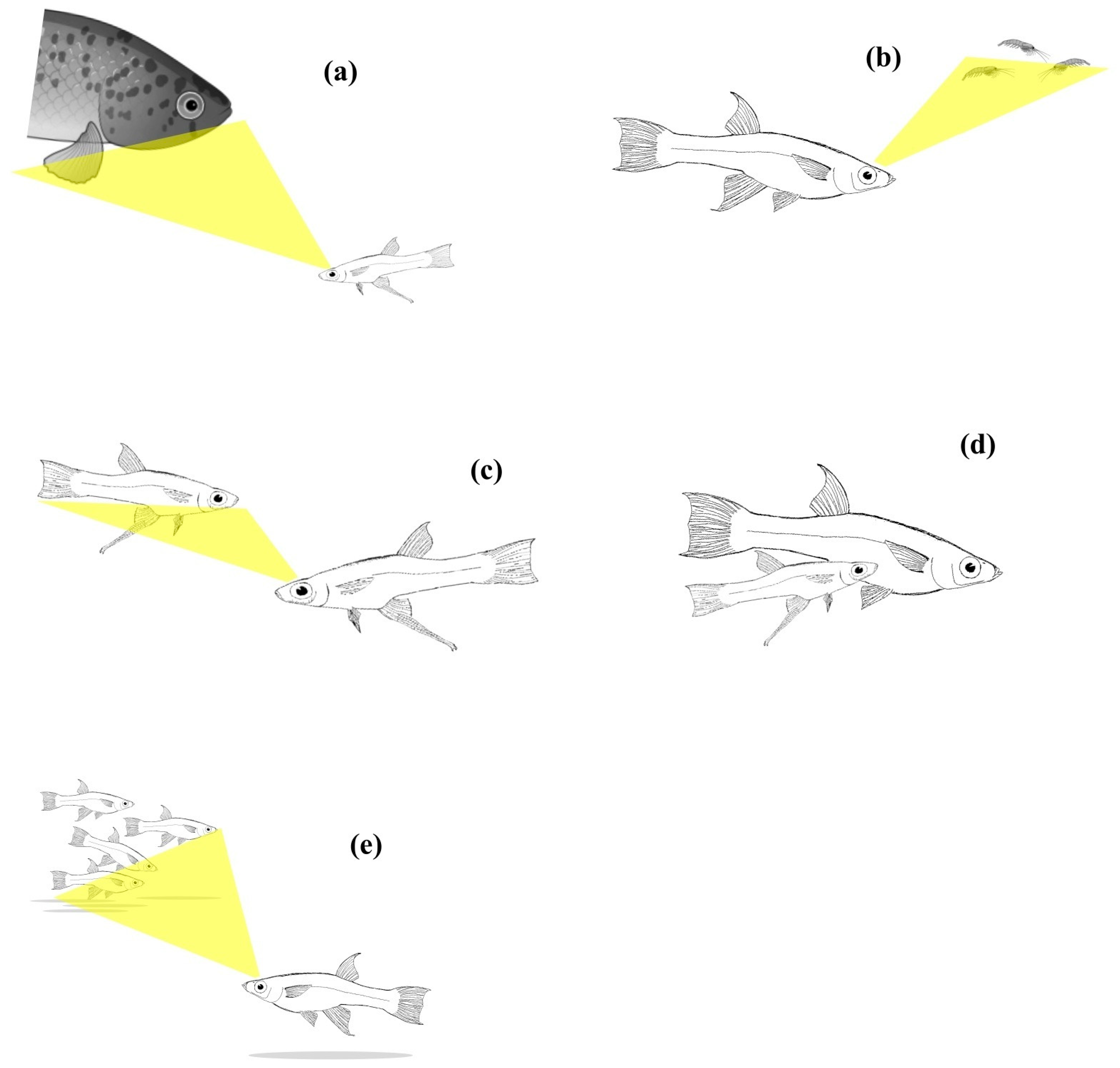
Brown et al. [96] investigated the behavioural lateralisation of wild-caught B. episcopi from various populations in Panama, distinguishing between those from areas with high and low predator presence. They found that fish from high predation sites exhibited a significant preference for using their right eye when viewing live predators, whereas fish from low predation sites showed no such eye preference. This difference in eye usage was a notable indicator of behavioural lateralisation linked to predation risk.
In a subsequent study, Brown and colleagues [104] explored the influence of genetic and environmental factors on the expression of lateralisation. They examined the lateralisation of wild-caught females from high and low predation areas, along with their offspring reared under controlled laboratory conditions. The study revealed that both the wild-caught females from high predation sites and their laboratory-reared offspring displayed stronger lateralised behaviours compared to those from low predation sites. While there were differences in the direction of lateralisation between wild-caught females and their offspring, the consistency in the strength of lateralisation across generations suggested a heritable component to this trait.

4.3.2. Girardinus falcatus

Bisazza and collaborators [105,106,123] investigated lateralisation and its genetic basis in the goldbelly topminnows, G. falcatus, a small livebearing fish native to Cuba. The fish were maintained in a large, outbred laboratory population under seminatural conditions, and various aspects of their behavioural lateralisation have been studied extensively over more than a decade. In the original population, fish exhibited consistent biases in eye preference: they preferred their right eye when observing potential predators or sexual partners and their left eye when interacting with social partners. Motor asymmetries were also observed, with a population-level preference for turning to the right when encountering an obstacle [124]. While these population-level biases were statistically significant, the deviation from chance levels was modest in all cases, with means ranging between 60% and 70%. Furthermore, there was considerable individual variation around the mean for all measured traits.
The asymmetry in eye use during predator inspection was selected for subsequent genetic analyses. Bisazza et al. [105] used a parent–offspring regression approach to calculate heritability, finding it to exceed 0.5 for this trait. A similar heritability estimate was obtained in a second experiment where progeny were separated at birth to minimize potential postnatal influences, such as social learning [105]. An artificial selection experiment was then conducted over six generations [106]. This experiment involved selecting two lines for right-eye dominance (Right lines), two for left-eye dominance (Left lines), one for no eye preference in predator inspection (NL line), and one unselected control line. An immediate response to selection was observed in all directionally selected lines, with similar responses in both Left and Right lines and no significant sex differences. The NL line also showed a response to selection, with the proportion of non-lateralised individuals increasing significantly from approximately 25% in the unselected population to around 50% by the end of the experiment. Unexpectedly, the response to selection ceased after the first two generations. This outcome does not appear to be due to the exhaustion of additive genetic variance, as there was still considerable phenotypic variability even after six generations of selection and a counter-selection experiment showed that the direction of selection could be reversed in just one generation.
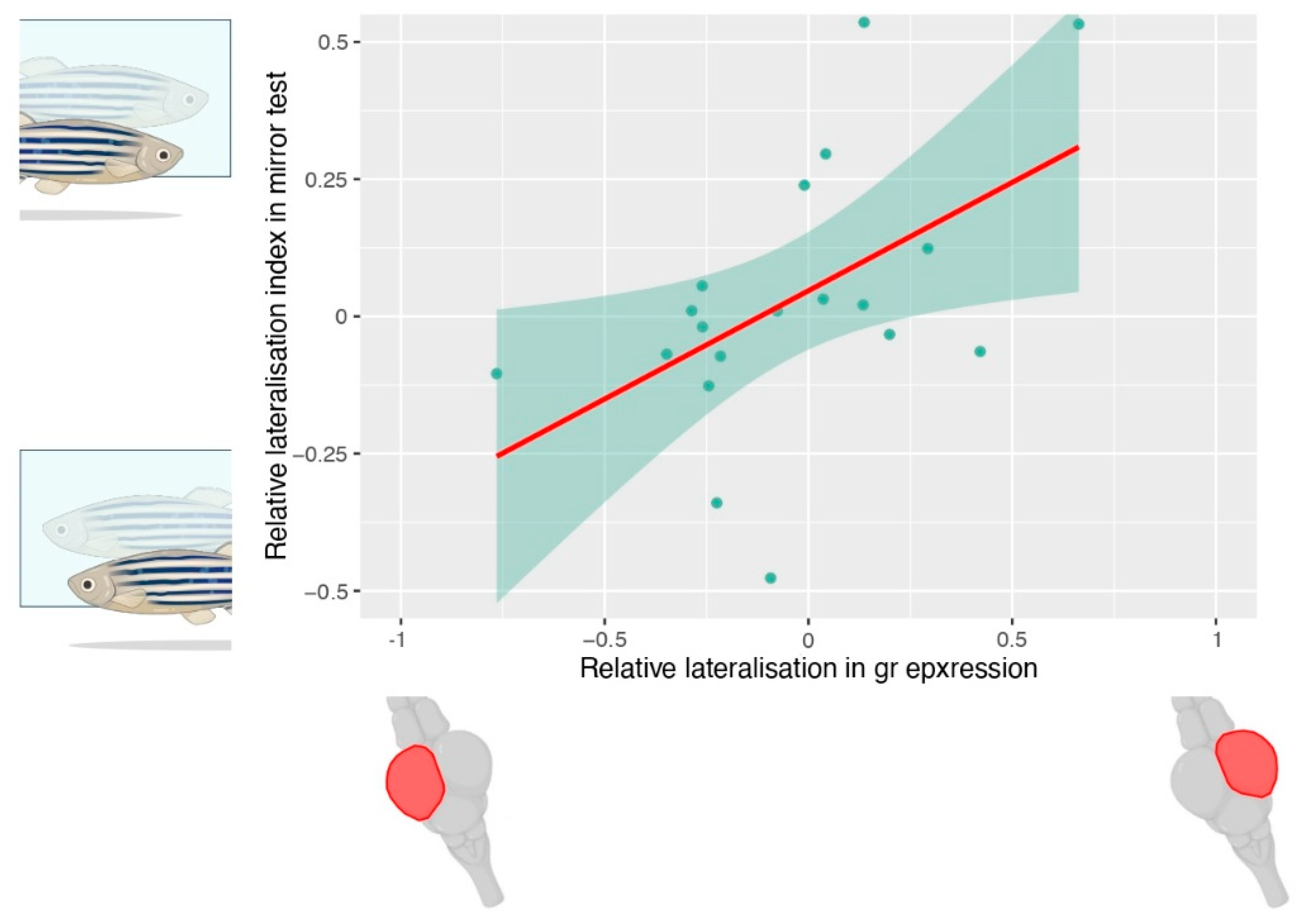
An intriguing continuation of the artificial selection experiment involved investigating whether other behavioural asymmetries were correlated with the selected trait. Strongly lateralised fish from the Right lines and the Left lines were compared with fish from the NL line in a battery of lateralisation tests [106,125,126,127,128]. Left or Right line obtained nearly opposite scores in all lateralisation tests, suggesting a mirror-reversed organisation of cerebral functions between the Left and Right fish (Figure 2). In contrast, NL fish exhibited reduced or no lateralisation across various tests, indicating a more bilateral representation of cognitive functions in their brains.

4.3.3. Gambusia hubbsi

Hulthén and colleagues [98] investigated behavioural lateralisation in Bahamas mosquitofish, G. hubbsi. This species is fragmented into small, isolated populations inhabiting water-filled caves in the Bahamas islands. These caves vary in predation risk due to the presence or absence of major fish predators, offering a unique opportunity to examine how natural variation in predation risk shapes behavioural lateralisation and the heritability of this trait. The researchers tested wild fish from high and low predation risk caves and found that those from high-predation sites were more strongly lateralised than those from low-predation sites, with this difference being especially pronounced in females. However, the populations from high-predation sites did not align in a common left-right direction and did not significantly differ in the direction of lateralisation from populations in low-predation sites. This finding suggests that the benefits of lateralisation may not necessarily rely on a specific directional bias but rather on increased strength under high predation risk.
To explore the genetic contribution to these differences, a lateralisation assay was conducted on adult second-generation laboratory-raised fish derived from a high-predation population. These fish were raised under common laboratory conditions. The experiment revealed fairly high heritability, with both sexes showing significant heritability in lateralisation direction, while only males displayed significant heritability in lateralisation strength. The magnitude of heritability in G. hubbsi was overall similar to that reported for G. falcatus [105].

4.3.4. Methodological Issue in Research on Poeciliid Fish

The studies conducted on the three species of poeciliids provide compelling evidence for a genetic basis of lateralisation within this fish family. However, these findings should be interpreted cautiously due to differences among the three species studied and potential methodological limitations. Specifically, while heritability of lateralisation traits was demonstrated in all three species, the heritability of strength and direction varied (Table 1). In B. episcopi, heritability was observed in the strength of lateralisation, though not in the direction. This finding contrasts with the results from G. falcatus, where an artificial selection experiment suggested a genetic basis for both direction and strength of lateralisation. Similarly, in G. hubbsi, heritability was found for both direction and strength of lateralisation, although the latter was significant only in males. These differences could reflect species-specific variations or might be attributable to the different experimental approaches used in each study.
One notable limitation across all studies is that environmental factors cannot be entirely ruled out. Given that all three species are viviparous, there is substantial potential for prenatal influences on the development of lateralisation. Similar to the maternal effects hypothesised to account for inconsistencies in human twin studies or the perplexing results of cross-fostering experiments in chimpanzees, maternal influences in poeciliids might affect the development of lateralisation traits. For example, the prenatal environment, including maternal stress and sex hormone levels or asymmetric foetal positioning, could contribute to the lateralisation observed in offspring, making it difficult to disentangle genetic factors from environmental ones [129,130]. Another significant methodological consideration is that the offspring in all three studies were allowed to remain with their parents for a period ranging from a few hours to several weeks. This introduces the possibility of shared environmental effects, particularly given the highly gregarious nature of poeciliids. Behavioural traits that exhibit left-right polarity, such as shoaling, mating, fighting, or predator evasion, might be transmitted culturally within a population, further complicating the assessment of genetic versus environmental contributions. Last, poeciliids are known for their pronounced cannibalistic behaviour, with adults often preying on newborn fry [131,132]. This raises the possibility that differential predation based on lateralisation traits could occur shortly after birth. For instance, parents with a specific lateralisation pattern in predatory behaviour might more easily capture offspring that exhibit a given lateralisation pattern in antipredator responses. Such selective predation could create an apparent parent-offspring resemblance in lateralisation traits that might be mistakenly attributed to genetic inheritance. The experiment conducted by Bisazza et al. [105] probably offers the most robust control for postnatal influences. In that study, heritability of lateralisation traits remained consistent whether offspring were kept with their parents or separated at birth. However, even in this case, the separation was not immediate, typically occurring a few hours after birth. This leaves open the possibility that some degree of selective predation or other postnatal influences could still have occurred during this brief period.
In summary, while the studies on these poeciliid species suggest a genetic basis for lateralisation, the potential influence of environmental factors, particularly prenatal effects, shared environments, and selective predation, cannot be completely excluded. Further research, with more rigorous controls for these variables, would be necessary to definitively establish the genetic underpinnings of lateralisation in these species.
Finally, modern research has demonstrated that the transmission of traits from one generation to the next can also occur through transgenerational epigenetic inheritance (reviewed in [133,134]). One of the most common mechanisms is DNA methylation, which can persist across multiple generations. It has been recently suggested that, in some cases, individual variation in lateralisation might also be attributable to such mechanisms [135]. Epigenetic inheritance can be challenging to distinguish from classical Mendelian inheritance, and we cannot entirely rule out the possibility that some of the findings described above might, at least in part, be explained by epigenetic mechanisms.

4.4. Genes, Brain Asymmetries, and Behavioural Lateralisation in the Zebrafish

The limitations encountered when studying the genetic basis of laterality in mammals and viviparous fish may be partially overcome by examining species like zebrafish (D. rerio), which are oviparous and do not exhibit parental care. In such species, postnatal environmental factors are more easily controlled, though prenatal factors, such as maternal effects on eggs, may still complicate interpretations of heritability.
Molecular research on zebrafish and other model organisms has revealed the involvement of the Nodal signalling pathway in the development of the left-right axis of the body, leading to the asymmetric positioning of visceral organs [136,137]. In zebrafish, where it has been studied in detail, this pathway plays a crucial role in the early development of asymmetric gene expression in the forebrain. The habenular complex is a paired structure found in the diencephalon of all vertebrates, which is part of an evolutionarily highly conserved pathway playing an important role in learning and emotional response and regulates social and sexual behaviours [138,139]. Nodal signalling determines in zebrafish the asymmetric positioning of the parapineal gland (on the left side in most fish), leading to differentiation of functions between the left and right habenula [140]. The habenular complex is the target of both visual and olfactory projections. By early manipulation of forebrain differentiation, Dreosti et al. [141] were able to suppress habenular asymmetry and generate either fish with a double-left- or a double-right-type habenula. Interestingly, these two types showed loss of responsiveness to odour and light stimuli, respectively. Barth et al. [107] studied the frequent-situs-inversus (fsi) mutation in zebrafish, which causes a reversal in the positioning of visceral organs and a high frequency of reversal in parapineal gland positioning. They observed differences in lateralisation assays between fsi zebrafish and controls, suggesting that asymmetries within the epithalamus could be the neural basis for behavioural asymmetries, even in wild-type zebrafish (Table 1).
Low frequencies of individuals with a right-sided parapineal gland are also found in wild-type zebrafish strains. Facchin et al. [108] explored this phenomenon by artificially selecting for right-eye or left-eye preference in zebrafish when viewing a conspecific (their own reflection in a mirror). Selection for right-eye dominance led to an increased prevalence of reversed asymmetry in the epithalamus, while selection for left-eye dominance reduced it. This provides further indirect evidence of a link between epithalamic asymmetry and the lateralisation of cognitive functions. Dadda et al. [142] further investigated this hypothesis by using a foxD3:GFP marker to assess parapineal gland positioning in live larval zebrafish. They compared individuals with left- or right-positioned parapineal glands across various laterality tests as they matured into adults. Although significant differences were observed between fish with opposite parapineal positions in all laterality tests, the study highlighted that early asymmetric parapineal gland positioning is not the sole determinant of behavioural lateralisation in zebrafish. This suggests that additional genetic or environmental factors contribute to shaping this trait.
Subsequent research has identified some maternally inherited genes that may play a significant role in determining behavioural and brain asymmetries in zebrafish [109]. However, the genetic basis of lateralisation in zebrafish remains incompletely understood. Continued research, particularly focusing on genetic and environmental interactions, is needed to fully elucidate the mechanisms behind lateralisation in this model organism.

4.5. Morphological Asymmetries and Behaviour

As noted above, asymmetric Nodal expression is broadly present across vertebrates and plays a role in the development of asymmetries in visceral organs, as well as in anatomical and potentially functional asymmetries of the nervous system. Numerous species also exhibit asymmetries in external morphology [20,143]. A comprehensive survey of morphological laterality in fishes found that this feature is quite widespread, being present in all orders of cartilaginous and bony fishes, as well as in several jawless fish. The trait is also shared by sarcopterygians, the closest living relatives of the ancestors of land vertebrates [144]. Indeed, directional asymmetries in morphology have been documented in many tetrapods [145,146]. In some cases, morphological asymmetries support fitness-related behaviours—such as locomotion, mating, or foraging—and are linked to behavioural asymmetries that mirror those associated with brain lateralisation [147,148]. In some cases, the exact mechanisms underlying these asymmetries are unknown [149], but in others, the developmental processes underlying morphological asymmetries were suggested to be the same as those producing brain asymmetries [150,151]. Therefore, elucidating the mechanisms behind the former could provide insights into the determination of the latter. For this purpose, we review a set of studies providing information on the genetic bases of morphological asymmetries.

4.5.1. Asymmetry in Mouth Opening

Scale-eating cichlids, such as Perissodus microlepis, exhibit a fascinating example of morphological asymmetry, with their mouths skewed to one side, which allows them to preferentially feed on the scales of their prey from the side toward which their mouth opens. Genetic studies on these cichlids have shown a distinctive hereditary pattern. When two individuals with the same mouth direction (either both right- or both left-mouthed) are crossed, their offspring also exhibit the same mouth direction. However, crosses between a right-mouth and a left-mouth individual result in a progeny with an approximately 2:1 ratio of left- to right-mouthed individuals. This hereditary pattern has been observed not only in P. microlepis but also in other scale-eating cichlid species and other fish species with asymmetric mouth opening, such as certain freshwater gobies and zebrafish [152]. Genome-wide analysis in P. microlepis has identified candidate genomic regions that may harbour the genes responsible for this asymmetric mouth opening, suggesting that the trait is influenced by multiple loci. However, the exact genetic mechanisms and their link with cerebral lateralisation remain to be fully elucidated.
While much attention has been given to the morphological asymmetry related to feeding, less is known about whether there is a correlation between mouth asymmetry and other aspects of behavioural lateralisation in these species. However, research on the poecilid fish Girardinus metallicus provides some insight. In this species, individuals exhibit either a leftward or rightward inclination of the head, and this morphological asymmetry is linked to specific behavioural lateralisation [147]. For example, when confronted with a predator, right-inclined individuals tend to turn leftward, using their right eye to monitor the predator, while left-inclined individuals turn rightward. Similarly, in a fast-escape test triggered by a vibratory stimulus, right-inclined individuals tend to escape to the right and left-inclined individuals to the left. These findings in G. metallicus suggest a potential link between functional lateralisation and morphological asymmetry. However, it remains to be seen whether this association holds true in species where the genetic mechanisms underlying mouth asymmetry have been clarified, such as in scale-eating cichlids and other species with similar feeding specializations. Further research is needed to explore these connections and determine whether lateralised behaviour and morphological asymmetry are coordinated traits driven by common genetic and environmental factors across these species.

4.5.2. Asymmetry in Male Intromittent Organs

In some fish species within the families Poeciliidae and Anablepidae, males exhibit an asymmetric gonopodium, the modified anal fin used for insemination. This fin bends either to the left or right, and typically, males approach females and attempt to mate from the side corresponding to the direction of the gonopodium bend [153,154].
Johnson et al. [155] explored this phenomenon in Xenophallus umbratilis, a species within the Poeciliidae family. Their study revealed that male X. umbratilis with a left-bending gonopodium exhibited a preference for using their right eye when viewing potential mates or predators. Conversely, males with a right-bending gonopodium showed a preference for using their left eye. This association between gonopodium orientation and behavioural lateralisation could be explained by either a common genetic mechanism driving both morphological and brain asymmetries or by phenotypic plasticity influencing behaviour based on morphological traits.
While the mechanisms underlying gonopodium direction in X. umbratilis remain unclear, research in the Anablepidae family offers some insights. In these species, both males and females exhibit asymmetric genitalia, meaning that successful mating requires compatible asymmetries between partners. Interestingly, Torres-Dowdall et al. [154] found that in most species, populations maintain similar proportions of left- and right-sided gonopodium orientation. However, in contrast to what might be expected, their breeding experiments revealed no correlation between parental and offspring asymmetry, suggesting that the direction of this trait is not genetically inherited. A whole-genome association analysis also failed to identify any genetic markers linked to gonopodium asymmetry. Despite this, certain species within Anablepidae consistently display a population-wide bias toward left-sided gonopodia. Torres-Dowdall et al. [156] found that this left-sided bias evolved independently in multiple lineages, indicating that natural selection may be acting on this trait. However, breeding experiments showed that an excess of left-sided offspring were produced regardless of the father’s gonopodial direction, highlighting a paradox between the apparent evolutionary selection for left-sidedness and the lack of clear genetic inheritance for this trait.
Interestingly, the correlation between gonopodial asymmetry and other behavioural asymmetries is not consistent across species. In X. umbratilis, some aspects of behavioural lateralisation are associated with gonopodial asymmetry, but this is not universally observed. In Anablepidae, studies have shown that gonopodial asymmetry does not predict behavioural lateralisation in contexts other than mating. For example, Bisazza et al. [153] found no correlation between gonopodium direction and escape behaviour in response to predators. Similarly, Torres-Dowdall et al. [157] found no link between gonopodial asymmetry and the asymmetry in eye use during predator inspection. The relationship between external morphological asymmetries, such as gonopodium orientation, and behavioural lateralisation remains unresolved.

4.5.3. Behavioural and Morphological Asymmetries in Flatfish

Flatfish, such as flounders and soles, undergo metamorphosis during their development, transforming an externally perfectly symmetrical hatchling into an asymmetrical, bottom-adapted adult. This transformation occurs through the migration of one eye to the opposite side, accompanied by modifications of the musculoskeletal system and the differentiation of pigmentation between the two sides of the body. Some species are left-sided, others right-sided, but in nearly all species, some individuals exhibit reversed asymmetry, with both morphology and behaviour being mirror images of the most common form. Schreiber [158] highlighted that larvae start showing lateralised swimming and feeding behaviours before morphological modifications are evident. Through a series of experiments, Schreiber demonstrated that in the southern flounder (Paralichthys lethostigma), both behavioural asymmetries and eye migration develop in response to thyroid hormones released during metamorphosis, but they are independent of each other. In this species, larval behavioural asymmetries can accurately predict post-metamorphic sidedness.
The study does not provide information on the mechanisms underlying these intraspecific differences. Indeed, early treatment of pre-metamorphic larvae with thyroid hormones induced early settling behaviour but had no effect on the direction of settlement. However, other studies suggest that genetic factors may play a role in determining sidedness variation. The first evidence came from a study on the Japanese flounder (Paralichthys olivaceus), where a clonal line was established that produced a relatively higher frequency of reversed phenotypes [159]. These reversely eyed individuals also exhibited a high frequency of visceral organ inversion. Yet, the reversal of eye migration direction was not always accompanied by a corresponding reversal in visceral organ orientation, suggesting that the genetic mechanisms underlying these two types of asymmetries are at least partially independent. Notably, the closely related southern flounder, Paralichthys lethostima, shows anatomical asymmetries in the habenular complex similar to those described in the zebrafish brain (see Section 4.4), and in this species, the direction of cerebral asymmetries appears to be independent of post-metamorphic sidedness [160].
Policansky [161] compared the progeny of crosses between left-sided parents and between right-sided parents in two populations of the starry flounder (Platichthys stellatus). Both crosses produced an admixture of left and right progeny, but the percentage of left progeny was significantly higher in crosses between left-sided parents. A significant heritability was found in both populations, but the study also suggests either a polygenic control or a large environmental component.
In recent years, considerable research has been conducted at the molecular level to identify the genes involved in the development of asymmetries during flatfish metamorphosis and those that may underlie intraspecific differences in the direction of eye migration (reviewed in [162]). Although significant progress has been made in understanding the molecular mechanisms underlying eye migration, the genetics of morphological and behavioural directional asymmetries in flatfish remain far from fully understood.
Overall, studies on flatfish suggest a genetic basis for the direction of settlement sidedness and the accompanying anatomical modifications. The strong correlation between the direction of lateralised swimming and feeding behaviours and settlement sidedness further suggests that behavioural asymmetries may also be heritable. However, the observation that, in at least one species, the direction of asymmetries in diencephalic structures—specifically those linked to behavioural lateralisation in zebrafish—is entirely independent of settlement sidedness raises the possibility that the mechanisms determining behavioural asymmetry in flatfish may differ from those in other teleost fish.

5. Phenotypic Plasticity of Lateralisation in Non-Human Vertebrates

Phenotypic plasticity, namely the capacity of a given genotype to produce different phenotypes if exposed to different environmental conditions [163,164], is a mechanism known to generate intraspecific variance in many traits. Some of the most known examples of phenotypic plasticity in animals regard variations in morphological defences observed in prey species from populations with or without predators. For instance, water fleas (Daphnia spp.) develop head spines when exposed to predator cues [165,166,167,168], and crucian carps (Carassius carassius) exposed to their predator northern pike (Esox lucius) develop an increased body depth [169,170]. Evidence has suggested that the phenotypes developing via plasticity in response to the perceived predation risk confer survival advantages for the prey, such as more efficient escape responses [171] and increased handling time for the predators [172]. These forms of plasticity conferring fitness advantages are considered adaptive and maintained by selection [173]. Adaptive plasticity is now recognised as a contributor to local adaptation [102,174] as much as genetic mechanisms of adaptation.
The evidence of phenotypic plasticity has been growing steadily in the last decades and now also encompasses virtually all animal traits, including life-history [175,176], physiological [177,178], behavioural [179,180], and cognitive traits [181,182]. The list of environmental factors determining plasticity is also remarkable. For instance, plasticity has been reported not only in response to the presence of predators, but also to resources availability [183], temperature [184], illumination [185,186], and social environment [187,188]. It is worth noting that not all forms of plasticity are considered adaptive (reviewed in [99]). Moreover, in many cases, testing the adaptive significance of plasticity is not possible or quite difficult [173], leading to uncertainty in the attribution.

5.1. Light-Induced Lateralisation Plasticity in Birds

In the late 1970s, L. Rogers and J. Anson proposed that also lateralisation might display phenotypic plasticity [189]. In their study species, the domestic chicken (Gallus gallus domesticus), a marked hemispheric specialisation has been reported (reviewed in [190]). For instance, Vallortigara and Andrew [191] experimentally forced chicks to respond to visual stimuli with a specific eye by masking the other eye. Their study showed that only left-eyed individuals could recognise stimulus chicks, suggesting right-hemispheric processing of conspecific recognition. Rogers and Anson [189] noted that visual stimulation begins to evoke a response in the tectum of the chicks only after day 17 of incubation, a developmental stage in which the embryo is oriented asymmetrically with respect to the eggshell, with the left eye exposed to light input passing through the shell and the right eye occluded by the wing. They proposed that light stimulation of the left eye, and therefore the right hemisphere, might induce the observed hemispheric specialisation in visually driven behaviours. In an elegant experiment, Rogers then demonstrated this hypothesis. Chicks hatching from eggs incubated in the presence of light displayed increased lateralised behaviour as compared to chicks from dark incubated eggs [192]. This finding has been confirmed in a set of studies in chicks conducted with different methodologies and different experimental goals (Table 2). A similar lateralisation plasticity has been also observed in experiments manipulating light exposure during development in pigeons (Columba livia), in quails (Coturnix coturnix japonica), and in European robins (Erithacus rubecula) [193,194,195,196,197,198,199]. However, the plasticity observed in the two most used models (e.g., chickens and pigeons) showed some differences in terms of the anatomical pathways involved [200].
Under laboratory conditions, the light-mediated plasticity demonstrated by the studies in domestic chicks is not expected to generate variation across individuals. All the individuals display the same postural asymmetry in the eggs and, therefore, a very similar eye exposure to light during the early development in the laboratory. It was rather suggested that this plasticity might reduce pre-existing individual variation (e.g., genetic variation) by aligning the lateralisation of the entire population [192]. However, in nature, the situation can be different from this perspective. A hen might alter the lateralisation of its offspring by selecting a particular nest site, with more or less light, and/or by varying the time spent incubating the eggs [74,208]. While even under this scenario variation within broods in light-induced lateralisation, plasticity is expected to be low; variation across broods might be significant. Notably, further variation might arise from the fact that factors such as the sex of the chick modulate light-induced plasticity (e.g., [190]). It is also possible to hypothesise that variation in lateralisation induced by light exposure has an adaptive significance in chicks. Dark-incubated chicks, which show weaker lateralisation, outcompete conspecifics for access to food and are more responsive to predators than light-incubated, strongly lateralised chicks [202,215]. Light-induced lateralisation might ultimately allow the hen to produce offspring with a phenotype adapted to predation and competition risk in their environment. This idea is also supported by the fact that the effects of the lateralisation phenotype described were not present in all the behaviours investigated [205]. However, a non-adaptive explanation cannot be ruled out. All these studies on chicks exposed the subjects to a somehow unnatural developmental situation (e.g., continuous/complete absence of light). Therefore, the observed effects on lateralisation might ultimately be due to the unnatural developmental condition rather than to an adaptive response. It remains possible that the light-induced plasticity observed in chicks is indicative of a generalised mechanism in the vertebrate brain, which in other species can be a more significant source of individual variation. The next section addresses evidence supporting this hypothesis.

5.2. Light-Induced Plasticity in Other Vertebrates

Outside birds, the effect of light during the development of lateralisation, has been investigated in teleost fish and in amphibians. In these groups, most species are oviparous and produce relatively transparent eggs. This directly exposes the embryo to different levels of light exposure according to environmental conditions. Moreover, to the best of our knowledge, no clear patterns of embryo postural asymmetries that might affect light exposure differently between the two eyes at the species level have been reported. The teleost fish with the most available data on light-induced lateralisation plasticity is the zebrafish (D. rerio) [212,216,217], a species already widely used in developmental and molecular biology research. Three studies in zebrafish concur in indicating that larvae whose eggs and early larvae were kept in the dark display weaker lateralisation compared to larvae exposed to light for a total of 6 days after fertilisation. Since the left habenula and the parapineal are photoreceptive, it is suggested that these asymmetric epithalamic regions might be the actual target of light-induced lateralisation plasticity [140]. The same pattern of results was also found in western rainbowfish (Melatotaenia australis): eggs incubated in darkness led to a reduction in lateralisation [218].
The aforementioned data in fish suggests that also in this group, the exposure to markedly different illumination conditions during early development (i.e., presence versus absence of light) affects lateralisation, as previously reported in birds. As discussed for the avian studies, however, the treatments used in the fish studies were rather unnatural, as it is unlikely that in nature, some eggs would be exposed to complete darkness. One may propose that a block or slowdown of certain developmental processes, rather than adaptive plasticity, might explain the reduced lateralisation of embryos developing under dark conditions. In support of this idea, Berlinghieri and colleagues [218] also reported that after being moved to a condition with light, the rainbowfish whose eggs were kept in the dark developed a normal lateralisation phenotype. This prevents researchers from fully investigating the adaptive significance of the observed plasticity and how this may determine variation within the population.
A more ecological approach has been adopted in a study of goldbelly topminnows (G. falcatus) [129]. This species is a livebearing fish, and the embryos develop inside the mother for approximately 1 month. During this stage, Dadda and Bisazza [129] maintained the females under a standard photoperiod (12 h:12 h, dark:light). However, one group of females was kept under either high or low light intensity. The treatment might resemble a natural situation in which different populations are exposed to different amounts of light due to differences in the density of canopies (e.g., [219,220]) and in water turbidity (e.g., [221]). Keeping the females under altered light conditions indirectly affected the amount of light received by the embryos through the maternal tissues. The study reported lateralisation loss due to the treatment with low light intensity. Another study with potential application to natural situations was performed by Sovrano and colleagues [222] in zebrafish. They first confirmed the lateralisation disruption in zebrafish larvae raised under dark conditions. Additionally, they raised groups of larvae under different monochromatic light (red, green, or violet), demonstrating again an alteration in lateralisation. As the canopies affect the light spectrum in the environment (reviewed in [223]), including the light reaching the surface of water bodies of underneath (e.g., [224]), this study further supports the potential of animals to show natural variation in lateralisation due to plasticity in response to variable light conditions in the environment.
An attempt to re-create an even more realistic setting has been performed by Lucon-Xiccato et al. [225]. They raised edible frog (Pelophylax esculentus) tadpoles in mesocosms with either high or low abundance of duckweed (Leman minor). In the condition with high vegetation abundance, the entire surface of the mesocosm tank was covered, determining a significant reduction in the light reaching the eggs compared to the condition with low vegetation abundance, in which the vegetation did not cover more than 20% of the water surface. The tadpoles exposed to high vegetation abundance, and thus to low light conditions, were less lateralised. A group of tadpoles was also switched from the two vegetation treatments after hatching. This treatment reduced both absolute lateralisation and relative lateralisation. This study in tadpoles has the advantage of re-creating a situation similar to that of natural habitats with different light intensities due to different cover abundance. However, it remains difficult to interpret because the amount of vegetation in the environment is also related to predation risk in tadpoles (e.g., [226,227], a factor discussed in the next section of this review.
A last study of interest for light plasticity involved the medaka fish (Oryzia latipes). In its natural area, the medaka is exposed to marked seasonal variations in environmental factors, including the photoperiod (e.g., [228,229]). Lucon-Xiccato and colleagues [230] manipulated the photoperiod in laboratory mesocosms with adult medakas to simulate two different seasonal conditions, the summer with long photoperiod (i.e., 16 h of light) and the winter with short photoperiods (i.e., 8 h of light). After one month of treatment, the subjects’ lateralisation was scored in a social context, revealing that individuals exposed to the summer photoperiod displayed a left-eye preference (e.g., right hemisphere processing) to observe the stimulus, whereas individuals of the winter photoperiod showed the opposite lateralisation pattern. Therefore, at least for seasonal variation, the duration of daily light exposure can be a source of lateralisation plasticity, even in adult individuals.
One noteworthy study by Dadda and Bisazza [231] further explores the influence of visual environmental factors on lateralisation, even though it does not directly relate to the effects of light. In this experiment, guppies (Poecilia reticulata) were exposed to an asymmetric visual experience during their development. The experimental setup involved placing the fish in an aquarium with “windows” that allowed them to observe other guppies in an adjacent compartment. Given that guppies are a social species, it was expected that the subjects would actively try to observe the other fish. However, due to the asymmetric design of the windows, each guppy could only observe the other fish using one specific eye, either the right or the left. This design created an asymmetric visual input from social stimuli during a critical period of development. When the guppies were later tested in a different apparatus without any asymmetric construction, the results revealed that the fish exposed to social companions with their right eye continued to prefer observing social stimuli with their right eye. Similarly, those that had previously used their left eye showed a preference for using their left eye. This study provides evidence that not only light exposure, but also specific environmental stimuli can influence the development of the visual system and contribute to the plasticity of lateralisation.
In conclusion, perhaps the most conservative, albeit comprehensive, interpretation of the many studies modulating the presence of light and the few studies modulating qualitatively the amount and type of light is that lateralisation plasticity is probably quite widespread and has the potential of being the source of individual variation in natural populations. However, more research on naturalistic settings is required to confirm this interpretation.

5.3. Predator-Induced Lateralisation Plasticity

A second line of evidence supporting the presence of environmentally driven lateralisation plasticity involves the response to predation risk. Besides being a major factor determining plasticity of typical morphological defences (e.g., [166,170]), the risk of predation experienced by an individual in the past can affect several aspects of its current cognitive system [232,233,234]. This widespread effect is probably due to the balance between the costs and the benefits of anti-predator defences, favouring plasticity-based adaptations triggered by individual experiences. In studies focused on plasticity, the risk of predation is usually manipulated over relatively long treatment periods before the testing, simulating the so-called background risk. The testing is then conducted in a similar situation (with no predator cues) for both subjects previously exposed to predation risk and control subjects not exposed to risk. This is done because prey usually also adopt contextual behavioural responses to the presence of a predator, which might be confused with strict-sense plasticity.
In 2000, De Santi and colleagues [235] collected data on guppies that might be indicative of phenotypic plasticity of lateralisation driven by predator exposure. Many fish species display an inspection behaviour, usually conducted by a group of individuals, when they encounter a new potential predator. To study predator inspection in guppies with different experiences, the authors exposed a group of subjects to a live predator placed behind a transparent partition before the inspection trials. The experience with the predator consisted of four separate exposures administered across 4 days, in which the predator was always observed attempting to capture a guppy. After the treatment with the predator, De Santi and colleagues assayed the lateralisation of guppies’ inspection behaviour. Guppies exposed to the predator approached it more closely in trials where a conspecific was present on the right side compared to a condition with the conspecific in the left side. A group of control subjects with no predator experience did not show this lateralisation behaviour. Although shorter than that adopted by studies on predation risk, it might resemble a case of background treatment. We might therefore conclude that guppies experienced with the predator were more lateralised compared to control guppies due to phenotypic plasticity. However, as the predator stimulus involved in the trial was of the same predator species used for the treatment, it is equally possible that the subjects from the two groups varied in how they perceived the predator stimulus in the inspection trial. Subjects from the control treatment might not have responded to the stimulus as to a real predator having not learned its danger. It is indeed common that individual fish have to learn to recognise dangerous species in order to perform appropriate anti-predator behaviour [236].
A subsequent study on guppies resolved the uncertainty in the interpretation [237]. The subjects were exposed to chemical cues of a predator before being tested with a detour test, using alternately a social companion and a predator as stimulus. The chemical cue from the predator consisted of water from the tank that housed the predator. As the predator was fed twice per day with a guppy, the water from its tank was supposed to contain alarm cues released by the tissues of injured guppies and diet cues deriving from the predator due to its specific diet. The study found that guppies were more lateralised when reared with predator cues. Interestingly, this study tested guppies from two populations to explore possible genetic differences. One population was naturally exposed to predators and one came from an environment with reduced predation risk. There were no differences in lateralisation between the two populations, suggesting that for this lateralisation aspect phenotypic plasticity is more important than genetic population differences.
Following studies have reported an effect of predation risk on lateralisation in at least five additional species of teleost fish and one amphibian species (Table 3). Most of these studies suggested that background predation risk increased lateralisation in individuals. Results were consistent in the studies on early developmental stages, with also two studies manipulating the risk in embryos of wood frogs and goldbelly topminnows, respectively. However, the opposite effect (i.e., a decrease in lateralisation) was found in the study on adult fathead minnows. In goldbelly topminnows and fathead minnows, evidence also suggests that the individuals exposed to predation tend to align the direction of lateralisation at the level of population and social group, respectively. The study by Ferrari et al. [238] additionally found that the strength of lateralisation can vary throughout the day according to daily variation in predation risk.
The observed plasticity in response to predation risk could be adaptive, considering various reports that suggest stronger anti-predator responses in strongly lateralised individuals (e.g., [243,244]). To the best of our knowledge, only two studies attempted to test this hypothesis. Ferrari and colleagues [241] found that lateralised individuals arising from the treatment with predator cues did survive better in mesocosm with real predators, albeit this survival advantage could be due to other effects of the background predation risk. Lucon-Xiccato and colleagues [242] found that some individuals with higher lateralisation from a population showing predator-mediated lateralisation plasticity displayed greater predator recognition learning performance; however, the lateralisation of these individuals was not the result of the background risk manipulation. Therefore, while the evidence points toward an adaptive plasticity, the situation is not completely clear.
Overall, the literature includes relatively robust evidence of lateralisation plasticity in response to predation risk across anamniotes, especially teleost fish. The various species investigated mostly showed a similar pattern of effects, suggesting that this plasticity might be driven by evolutionarily conserved forces. In part, these effects resemble those observed in case of genetic adaptation [104], discussed in the early sections of this review. It is also worth noting that more evidence is present in amphibians if we include studies that could be interpreted also in terms of contextual responses rather than strict plasticity [245,246]. With the same approach, one study could be suggestive of an effect on a reptile [247]. Lastly, one study reported lateralisation plasticity in an invertebrate species, suggesting that this phenomenon is also widespread beyond vertebrates [248]. Experience with predation risk can be a major contributor to variation in lateralisation within and between populations.

5.4. Hormonal Effects on Lateralisation

A third line of investigation suggesting plasticity in animal lateralisation involves hormonal effects. Two types of hormones, testosterone and glucocorticoids, have been studied for this purpose. Research on testosterone was initially stimulated by findings in human studies. In our species, cognitive functions such as hand preference, language, and spatial orientation are often lateralised with varying degrees of strength and direction in males and females. Accordingly, four hypotheses (reviewed in [59,249]) proposed a role for testosterone in shaping lateralisation of cognitive functions. Most of these hypotheses, and the resulting experimental studies, have focused on explaining sex differences in lateralisation. For instance, Denenberg and colleagues [250] found that male rats have a larger corpus callosum compared to females, and that treatment with testosterone during infancy increased corpus callosum size under certain circumstances, suggesting a role for this hormone in determining sex differences in lateralisation through organisational effects on brain structure and connectivity. However, one of these hypotheses, in particular, proposed that small variations in prenatal exposure to testosterone might determine individual variability in lateralisation [251]. This effect on individual differences is predicted to occur in both sexes. If confirmed, such an effect would be more similar to the typical examples of plasticity discussed above and could potentially be part of adaptive response mechanisms.
A few studies have attempted to confirm the effect of testosterone on lateralisation in non-human vertebrates. For example, Possenti and colleagues [252] increased testosterone concentration within physiological limits in the yolk of yellow-legged gull (Larus michahellis) eggs and then assessed the lateralisation of the offspring. While no sex-dependent effects were recorded, the testosterone treatment strengthened individual lateralisation. Similarly, Riedstra and colleagues [253] conducted experiments in which domestic chicken eggs were injected with testosterone. They found that prenatal exposure to various physiological levels of testosterone did not affect lateralisation. However, in adult chickens, they observed correlations between circulating testosterone levels and lateralisation, suggesting an activational effect of testosterone on lateralisation rather than the organisational effect proposed in most hypotheses. At least within males, this effect might be due to adaptive plasticity. A meta-analysis of studies in humans, other mammals, and birds [59] concluded that testosterone seems to influence lateralisation. However, the effects mediated by testosterone did not overlap those typically observed in males, suggesting that testosterone is not the main factor affecting sex differences in lateralisation. Overall, the possibility of testosterone-guided plasticity of lateralisation seems plausible, though it requires further experimental investigation.
Glucocorticoids are a class of steroid hormones with broad capacities to modulate individual phenotypes, allowing organisms to adapt to environmental conditions such as varying levels of stress. The role of glucocorticoids in lateralisation has been suggested by two studies. In chickens, corticosterone—the main glucocorticoid in tetrapods—was found to regulate lateralisation through a maternal effect. Henriksen and colleagues [254] manipulated plasma corticosterone levels in laying hens and found that the offspring of hens with higher corticosterone levels displayed reduced lateralisation in a visual task. Notably, this effect might also be mediated by testosterone, as they observed that elevated maternal corticosterone levels led to increased testosterone in the offspring. Ferrari and colleagues [238] instead focused on fish and the primary glucocorticoid hormone in this group, cortisol. They found that Ambon damselfish (Pomacentrus amboinensis) treated with cortisol exhibited stronger lateralisation. Additionally, Lucon-Xiccato provided further evidence supporting the role of glucocorticoids in lateralisation in fish. In a recent experiment, his team measured lateralisation in zebrafish and then analysed their brains to quantify the expression levels of the gene for one of the main glucocorticoid receptors, gr. The data shown in Figure 3 suggested a complex pattern of lateralisation control involving gr [255]. These findings suggest that glucocorticoids and their receptors, like testosterone, might play a role in adapting individual lateralisation to environmental conditions. However, much more research is needed to fully understand these effects.

6. Conclusions

Overall, the literature reviewed in this work suggests the presence of a significant body of research exploring the potential causes of intraspecific differences in lateralisation in non-human vertebrates. The two main factors investigated—genetic determinants and environmental influences—appear both involved at different levels.
Research on the genetic bases of lateralisation has found several supporting pieces of evidence across various vertebrate groups, ranging from mammals to fish. However, it is important to note that alternative explanations, such as environmental influences and epigenetic mechanisms, are often difficult to completely rule out. Part of the issue stems from the fact that most genetic studies on lateralisation have focused on a limited number of model species within each group, leading to low systematic coverage. Additionally, some of these species may not be ideal for excluding confounding factors. For example, in livebearing fish, the maternal environment during development can significantly impact the offspring, making it challenging to isolate genetic influences. Future studies should aim to identify species that allow for more controlled investigations into the genetic bases of lateralisation. The use of the zebrafish model is promising for this purpose, especially given the numerous neurobiological and genetic tools available in this species. To broaden phylogenetic coverage and investigate interspecific variation, it will also be important to study mammals other than primates and laboratory rodents, as well as birds like the chicken, which have been more extensively used in the study of plasticity. Finally, to better understand the role of genes in shaping lateralisation, it will be crucial to adopt other research approaches that are currently underutilised, including half-siblings and cross-fostering designs, as well as quantitative trait loci analysis.
Research on environmental effects leaves no doubt that these mechanisms are also involved in individual differences in lateralisation. It has highlighted two main factors triggering phenotypic plasticity: exposure to light and the presence of predators. Notably, in some cases, these factors appear to cause adaptive plasticity, but it is currently difficult to rule out that what is measured may be more of a generalised lateralisation response to stress situations. A third potentially important factor is related to hormones, although there is less evidence in this regard, and hormones may be, as in the case of stress, the control mechanism rather than the primary cause of lateralisation plasticity. We suggest that the factors identified in this review represent only a small fraction of those that can lead to plasticity in lateralisation. Many other factors may be involved, such as social environment, habitat complexity, temperature, and food availability. This is supported by a growing body of literature describing how global change and anthropogenic activities may impact lateralisation [256,257,258,259,260]. Although these cases are likely due to non-adaptive plasticity, as they are triggered by human-made chemicals and situations different from those in which the organism evolved, they nonetheless highlight the high plasticity potential of this trait. As with the genetic mechanisms, the species coverage for plasticity research is relatively low. In particular, most studies on plasticity have focused on anamniotes and birds. This makes it difficult to generalise the findings to other groups, especially mammals, suggesting the need to broaden the range of species used in this research.
In conclusion, the findings available so far, though fragmentary, suggest that intraspecific variability in animal lateralisation may result from both genetic and environmental factors. However, they do not provide an accurate estimate of the relative importance of these factors. The same conclusion has been reached by two earlier reviews with a goal similar to that of the present work [25,26]. Collecting more data across a larger number of species, using consistent and robust methodologies, will enable better inferences on this point, particularly through the application of meta-analytic approaches [261,262]. It also remains unexplored whether these factors interact, potentially leading to different plasticity responses across different genotypes (e.g., genotype by environment interaction [263]). In this context, a more naturalistic approach will be important for understanding the evolutionary pressures acting on individual differences in lateralisation. Other factors affecting lateralisation, such as epigenetic influences, should also be explored [33].
The findings from research in non-human species may provide relevant insights into humans, where both genetic and environmental sources of variation are likely to be determinants of intraspecific variability in lateralisation. The question is now whether the same environmental factors and genetic pathways discovered in other vertebrates apply to humans. In spite of a lack of direct studies, indirect evidence seems to support this view. For example, the position assumed by foetuses in utero has been proposed as a determinant of lateralisation in both humans and chimpanzees [121,264]. Stressors alter lateralisation in humans as well as in other vertebrates through the glucocorticoid pathway [255,265]. The Nodal pathway action on lateralisation has been reported in humans and other vertebrates [136,266]. All this evidence suggests the potential and the importance of enhancing the use of animals as models to understand individual differences in human lateralisation.

Author Contributions

Conceptualization, A.B. and T.L.-X.; writing—original draft preparation, A.B. and T.L.-X.; writing—review and editing, A.B. and T.L.-X.; visualization, A.B. and T.L.-X.; funding acquisition, T.L.-X. All authors have read and agreed to the published version of the manuscript.

Funding

Project funded under the National Recovery and Resilience Plan (NRRP), Mission 4 Component 2 Investment 1.3—Call for tender No. 341 of 15/03/2022 of Italian Ministry of University and Research funded by the European Union—NextGenerationEU Award Number: Project code PE0000006, Concession Decree No. 1553 of 11/10/2022 adopted by the Italian Ministry of University and Research, CUP D93C22000930002, “A multiscale integrated approach to the study of the nervous system in health and disease” (MNESYS).

Conflicts of Interest

The authors declare no conflicts of interest.

References

  1. Boorman, C.J.; Shimeld, S.M. The evolution of left–right asymmetry in chordates. Bioessays 2002, 24, 1004–1011. [Google Scholar] [CrossRef] [PubMed]
  2. Manuel, M. Early evolution of symmetry and polarity in metazoan body plans. Comptes Rendus Biol. 2009, 332, 184–209. [Google Scholar] [CrossRef] [PubMed]
  3. Namigai, E.K.; Kenny, N.J.; Shimeld, S.M. Right across the tree of life: The evolution of left–right asymmetry in the Bilateria. Genesis 2014, 52, 458–470. [Google Scholar] [PubMed]
  4. Fritsch, G.T.; Hitzig, E. Uber die elektrische Erregbarkeit des Grosshirns. Arch. Anat. Physiol. 1870, 37, 300–332. [Google Scholar]
  5. Harris, L.J. The discovery of cerebral specialization. A Hist. Neuropsychol. 2019, 44, 1–14. [Google Scholar]
  6. Wickens, A.P. A History of the Brain: From Stone Age Surgery to Modern Neuroscience; Psychology Press: Hove, UK, 2014. [Google Scholar]
  7. O’Regan, L.; Serrien, D.J. Individual differences and hemispheric asymmetries for language and spatial attention. Front. Hum. Neurosci. 2018, 12, 380. [Google Scholar]
  8. Packheiser, J.; Schmitz, J.; Arning, L.; Beste, C.; Güntürkün, O.; Ocklenburg, S. A large-scale estimate on the relationship between language and motor lateralization. Sci. Rep. 2020, 10, 13027. [Google Scholar]
  9. Mandal, M.K.; Bulman-Fleming, M.B.; Tiwari, G. (Eds.) Side Bias: A Neuropsychological Perspective; Kluwer Academic Publishers: Dordrecht, The Netherlands, 2000. [Google Scholar]
  10. Marzoli, D.; Tommasi, L. Side biases in humans (Homo sapiens): Three ecological studies on hemispheric asymmetries. Naturwissenschaften 2009, 96, 1099–1106. [Google Scholar]
  11. Bourne, V.J. Examining the relationship between degree of handedness and degree of cerebral lateralization for processing facial emotion. Neuropsychology 2008, 22, 350. [Google Scholar] [CrossRef]
  12. Costanzo, E.Y.; Villarreal, M.; Drucaroff, L.J.; Ortiz-Villafañe, M.; Castro, M.N.; Goldschmidt, M.; Wainsztein, A.E.; Ladrón-de-Guevara, M.L.; Romero, C.; Brusco, L.I.; et al. Hemispheric specialization in affective responses, cerebral dominance for language, and handedness: Lateralization of emotion, language, and dexterity. Behav. Brain Res. 2015, 288, 11–19. [Google Scholar]
  13. Groen, M.A.; Whitehouse, A.J.; Badcock, N.A.; Bishop, D.V. Associations between handedness and cerebral lateralisation for language: A comparison of three measures in children. PLoS ONE 2013, 8, e64876. [Google Scholar] [CrossRef] [PubMed]
  14. Khedr, E.M.; Hamed, E.; Said, A.; Basahi, J. Handedness and language cerebral lateralization. Eur. J. Appl. Physiol. 2002, 87, 469–473. [Google Scholar] [CrossRef] [PubMed]
  15. Papousek, I.; Schulter, G. Quantitative assessment of five behavioural laterality measures: Distributions of scores and intercorrelations among right-handers. Laterality Asymmetries Body Brain Cogn. 1999, 4, 345–362. [Google Scholar] [CrossRef] [PubMed]
  16. Eitler, K.; Bibok, A.; Telkes, G. Situs inversus totalis: A clinical review. Int. J. Gen. Med. 2022, 15, 2437–2449. [Google Scholar] [CrossRef]
  17. Corballis, M.C.; Badzakova-Trajkov, G.; Häberling, I.S. Right hand, left brain: Genetic and evolutionary bases of cerebral asymmetries for language and manual action. Wiley Interdiscip. Rev. Cogn. Sci. 2012, 3, 1–17. [Google Scholar] [CrossRef]
  18. McManus, I.C. Right Hand, Left Hand: The origins of Asymmetry in Brains, Bodies, Atoms, and Cultures; Harvard University Press: Cambridge, MA, USA, 2002. [Google Scholar]
  19. Güntürkün, O.; Ströckens, F.; Ocklenburg, S. Brain lateralization: A comparative perspective. Physiol. Rev. 2020, 100, 1019–1063. [Google Scholar] [CrossRef]
  20. Palmer, A.R. Symmetry breaking and the evolution of development. Science 2004, 306, 828–833. [Google Scholar] [CrossRef]
  21. Apostolou, M. Sexual selection under parental choice: The role of parents in the evolution of human mating. Evol. Hum. Behav. 2007, 28, 403–409. [Google Scholar] [CrossRef]
  22. Fehr, E.; Fischbacher, U. The nature of human altruism. Nature 2003, 425, 785–791. [Google Scholar] [CrossRef]
  23. Puts, D.A. Beauty and the beast: Mechanisms of sexual selection in humans. Evol. Hum. Behav. 2010, 31, 157–175. [Google Scholar] [CrossRef]
  24. Rosenberg, K.R. The evolution of human infancy: Why it helps to be helpless. Annu. Rev. Anthropol. 2021, 50, 423–440. [Google Scholar] [CrossRef]
  25. Güntürkün, O.; Ocklenburg, S. Ontogenesis of lateralization. Neuron 2017, 94, 249–263. [Google Scholar] [PubMed]
  26. Manns, M. It is not just in the genes. Symmetry 2021, 13, 1815. [Google Scholar] [CrossRef]
  27. Baldwin, J.M. Origin of right or left handedness. Science 1890, 404, 247–248. [Google Scholar] [CrossRef]
  28. Jordan, H.E. The inheritance of left-handedness. J. Hered. 1911, 2, 19–29. [Google Scholar]
  29. Kellogg, G.M. The physiology of right-and left-handedness. J. Am. Med. Assoc. 1898, 30, 356–358. [Google Scholar]
  30. Duboc, V.; Dufourcq, P.; Blader, P.; Roussigné, M. Asymmetry of the brain: Development and implications. Annu. Rev. Genet. 2015, 49, 647–672. [Google Scholar] [CrossRef]
  31. Francks, C. The genetic bases of brain lateralization. In Human Language: From Genes and Brain to Behavior; MIT Press: Cambridge, MA, USA, 2019; pp. 595–608. [Google Scholar]
  32. McManus, C. Cerebral polymorphisms for lateralisation: Modelling the genetic and phenotypic architectures of multiple functional modules. Symmetry 2022, 14, 814. [Google Scholar] [CrossRef]
  33. Schmitz, J.; Metz, G.A.; Güntürkün, O.; Ocklenburg, S. Beyond the genome—Towards an epigenetic understanding of handedness ontogenesis. Prog. Neurobiol. 2017, 159, 69–89. [Google Scholar]
  34. Zhang, H.T.; Hiiragi, T. Symmetry breaking in the mammalian embryo. Annu. Rev. Cell Dev. Biol. 2018, 34, 405–426. [Google Scholar]
  35. Flöel, A.; Buyx, A.; Breitenstein, C.; Lohmann, H.; Knecht, S. Hemispheric lateralization of spatial attention in right-and left-hemispheric language dominance. Behav. Brain Res. 2005, 158, 269–275. [Google Scholar] [PubMed]
  36. Sha, Z.; Pepe, A.; Schijven, D.; Carrión-Castillo, A.; Roe, J.M.; Westerhausen, R.; Joliot, M.; Fisher, S.E.; Crivello, F.; Francks, C. Handedness and its genetic influences are associated with structural asymmetries of the cerebral cortex in 31,864 individuals. Proc. Natl. Acad. Sci. USA 2021, 118, e2113095118. [Google Scholar] [CrossRef] [PubMed]
  37. Hepper, P.G. The developmental origins of laterality: Fetal handedness. Dev. Psychobiol. 2013, 55, 588–595. [Google Scholar] [PubMed]
  38. Bryden, M.P. Speech lateralization in families: A preliminary study using dichotic listening. Brain Lang. 1975, 2, 201–211. [Google Scholar] [CrossRef]
  39. Reiss, M.; Reiss, G. Ocular dominance: Some family data. Laterality Asymmetries Body Brain Cogn. 1997, 2, 7–16. [Google Scholar]
  40. Risch, N.; Pringle, G. Segregation analysis of human hand preference. Behav. Genet. 1985, 15, 385–400. [Google Scholar]
  41. Somers, M.; Ophoff, R.A.; Aukes, M.F.; Cantor, R.M.; Boks, M.P.; Dauwan, M.; de Visser, K.L.; Kahn, R.S.; Sommer, I.E. Linkage analysis in a Dutch population isolate shows no major gene for left-handedness or atypical language lateralization. J. Neurosci. 2015, 35, 8730–8736. [Google Scholar] [CrossRef]
  42. Medland, S.E.; Duffy, D.L.; Wright, M.J.; Geffen, G.M.; Martin, N.G. Handedness in twins: Joint analysis of data from 35 samples. Twin Res. Hum. Genet. 2006, 9, 46–53. [Google Scholar] [CrossRef]
  43. Sicotte, N.L.; Woods, R.P.; Mazziotta, J.C. Handedness in twins: A meta-analysis. Laterality Asymmetries Body Brain Cogn. 1999, 4, 265–286. [Google Scholar]
  44. Vuoksimaa, E.; Koskenvuo, M.; Rose, R.J.; Kaprio, J. Origins of handedness: A nationwide study of 30 161 adults. Neuropsychologia 2009, 47, 1294–1301. [Google Scholar]
  45. Bourrat, P.; Lu, Q.; Jablonka, E. Why the missing heritability might not be in the DNA. BioEssays 2017, 39, 1700067. [Google Scholar]
  46. Mayhew, A.; Meyre, D. Assessing the heritability of complex traits in humans: Methodological challenges and opportunities. Curr. Genom. 2017, 18, 332–340. [Google Scholar] [CrossRef] [PubMed]
  47. Matthews, L.J.; Turkheimer, E. Three legs of the missing heritability problem. Stud. Hist. Philos. Sci. 2022, 93, 183–191. [Google Scholar] [PubMed]
  48. Polderman, T.J.; Benyamin, B.; De Leeuw, C.A.; Sullivan, P.F.; Van Bochoven, A.; Visscher, P.M.; Posthuma, D. Meta-analysis of the heritability of human traits based on fifty years of twin studies. Nat. Genet. 2015, 47, 702–709. [Google Scholar] [CrossRef]
  49. Schaafsma, S.M.; Riedstra, B.J.; Pfannkuche, K.A.; Bouma, A.; Groothuis TG, G. Epigenesis of behavioural lateralization in humans and other animals. Philos. Trans. R. Soc. B Biol. Sci. 2009, 364, 915–927. [Google Scholar] [CrossRef]
  50. Bishop, D.V.; Bates, T.C. Heritability of language laterality assessed by functional transcranial Doppler ultrasound: A twin study. Wellcome Open Res. 2019, 4, 161. [Google Scholar]
  51. Paracchini, S.; Scerri, T. Genetics of human handedness and laterality. Lateralized Brain Functions: Methods in Human and Non-Human Species; Humana Press: Totowa, NJ, USA, 2017; pp. 523–552. [Google Scholar]
  52. Eichler, E.E.; Flint, J.; Gibson, G.; Kong, A.; Leal, S.M.; Moore, J.H.; Nadeau, J.H. Missing heritability and strategies for finding the underlying causes of complex disease. Nat. Rev. Genet. 2010, 11, 446–450. [Google Scholar]
  53. Manolio, T.A.; Collins, F.S.; Cox, N.J.; Goldstein, D.B.; Hindorff, L.A.; Hunter, D.J.; McCharty, M.I.; Ramos, E.M.; Cardon, L.R.; Chakravarti, A.; et al. Finding the missing heritability of complex diseases. Nature 2009, 461, 747–753. [Google Scholar]
  54. Provins, K.A. Handedness and speech: A critical reappraisal of the role of genetic and environmental factors in the cerebral lateralization of function. Psychol. Rev. 1997, 104, 554. [Google Scholar]
  55. Obel, C.; Hedegaard, M.; Brink, T.; Secher, N.J.; Olsen, J. Psychological factors in pregnancy and mixed-handedness in the offspring. Dev. Med. Child Neurol. 2003, 45, 557–561. [Google Scholar]
  56. Coren, S. Family patterns in handedness: Evidence for indirect inheritance mediated by birth stress. Behav. Genet. 1995, 25, 517–524. [Google Scholar] [CrossRef] [PubMed]
  57. Levander, M.; Schalling, D.; Levander, S.E. Birth stress, handedness and cognitive performance. Cortex 1989, 25, 673–681. [Google Scholar] [CrossRef] [PubMed]
  58. Nicholls, M.E.; Johnston, D.W.; Shields, M.A. Adverse birth factors predict cognitive ability, but not hand preference. Neuropsychology 2012, 26, 578. [Google Scholar] [CrossRef]
  59. Pfannkuche, K.A.; Bouma, A.; Groothuis, T.G. Does testosterone affect lateralization of brain and behaviour? A meta-analysis in humans and other animal species. Philos. Trans. R. Soc. B Biol. Sci. 2009, 364, 929–942. [Google Scholar] [CrossRef] [PubMed]
  60. De Kovel, C.G.; Carrión-Castillo, A.; Francks, C. A large-scale population study of early life factors influencing left-handedness. Sci. Rep. 2019, 9, 584. [Google Scholar] [CrossRef]
  61. Donnot, J.; Vauclair, J. Infant holding preferences in maternity hospitals: Testing the hypothesis of the lateralized perception of emotions. Dev. Neuropsychol. 2007, 32, 881–890. [Google Scholar] [CrossRef]
  62. Vervloed, M.P.; Hendriks, A.W.; van den Eijnde, E. The effects of mothers’ past infant-holding preferences on their adult children’s face processing lateralisation. Brain Cogn. 2011, 75, 248–254. [Google Scholar] [CrossRef]
  63. Gerrits, R. Variability in hemispheric functional segregation phenotypes: A review and general mechanistic model. Neuropsychol. Rev. 2024, 34, 27–40. [Google Scholar] [CrossRef]
  64. Young, G. Multi-factorial Causality in Laterality. In Causality and Development; Springer: Berlin, Germany, 2019. [Google Scholar]
  65. Bisazza, A.; Brown, C. Lateralization of cognitive functions in fish. In Fish Cognition and Behavior; Brown, C., Laland, K., Kraus, J., Eds.; Wiley: Hoboken, NJ, USA, 2011. [Google Scholar]
  66. Bisazza, A.; Rogers, L.J.; Vallortigara, G. The origins of cerebral asymmetry: A review of evidence of behavioural and brain lateralization in fishes, reptiles and amphibians. Neurosci. Biobehav. Rev. 1998, 22, 411–426. [Google Scholar] [CrossRef]
  67. Vallortigara, G.; Rogers, L.J.; Bisazza, A. Possible evolutionary origins of cognitive brain lateralization. Brain Res. Rev. 1999, 30, 164–175. [Google Scholar] [CrossRef]
  68. Wiper, M.L. Evolutionary and mechanistic drivers of laterality: A review and new synthesis. Laterality Asymmetries Body Brain Cogn. 2017, 22, 740–770. [Google Scholar] [CrossRef] [PubMed]
  69. Corballis, M.C. The evolution and genetics of cerebral asymmetry. Philos. Trans. R. Soc. B Biol. Sci. 2009, 364, 867–879. [Google Scholar] [CrossRef] [PubMed]
  70. Rossion, B.; Lochy, A. Is human face recognition lateralized to the right hemisphere due to neural competition with left-lateralized visual word recognition? A critical review. Brain Struct. Funct. 2022, 227, 599–629. [Google Scholar] [CrossRef] [PubMed]
  71. Franklin, W.E., III; Lima, S.L. Laterality in avian vigilance: Do sparrows have a favourite eye? Anim. Behav. 2001, 62, 879–885. [Google Scholar] [CrossRef]
  72. Krakauer, A.H.; Blundell, M.A.; Scanlan, T.N.; Wechsler, M.S.; McCloskey, E.A.; Jennifer, H.Y.; Patricelli, G.L. Successfully mating male sage-grouse show greater laterality in courtship and aggressive interactions. Anim. Behav. 2016, 111, 261–267. [Google Scholar] [CrossRef]
  73. Rogers, L.J. Evolution of hemispheric specialization: Advantages and disadvantages. Brain Lang. 2000, 73, 236–253. [Google Scholar] [CrossRef]
  74. Rogers, L.J. Advantages and disadvantages of lateralization. In Comparative Vertebrate Lateralization; Cambridge University Press: Cambridge, UK, 2002; pp. 126–153. [Google Scholar]
  75. Vallortigara, G.; Rogers, L. Survival with an asymmetrical brain: Advantages and disadvantages of cerebral lateralization. Behav. Brain Sci. 2005, 28, 575–589. [Google Scholar] [CrossRef]
  76. Rogers, L.J.; Zucca, P.; Vallortigara, G. Advantages of having a lateralized brain. Proc. R. Soc. Lond. Ser. B Biol. Sci. 2004, 271, S420–S422. [Google Scholar] [CrossRef]
  77. Dadda, M.; Bisazza, A. Lateralized female topminnows can forage and attend to a harassing male simultaneously. Behav. Ecol. 2006, 17, 358–363. [Google Scholar] [CrossRef]
  78. Bisazza, A.; Dadda, M. Enhanced schooling performance in lateralized fishes. Proc. R. Soc. B Biol. Sci. 2005, 272, 1677–1681. [Google Scholar] [CrossRef]
  79. Gatto, E.; Agrillo, C.; Brown, C.; Dadda, M. Individual differences in numerical skills are influenced by brain lateralization in guppies (Poecilia reticulata). Intelligence 2019, 74, 12–17. [Google Scholar] [CrossRef]
  80. Lucon-Xiccato, T.; Montalbano, G.; Dadda, M.; Bertolucci, C. Lateralization correlates with individual differences in inhibitory control in zebrafish. Biol. Lett. 2020, 16, 20200296. [Google Scholar] [CrossRef] [PubMed]
  81. Magat, M.; Brown, C. Laterality enhances cognition in Australian parrots. Proc. R. Soc. B Biol. Sci. 2009, 276, 4155–4162. [Google Scholar] [CrossRef] [PubMed]
  82. McGrew, W.C.; Marchant, L.F. Laterality of hand use pays off in foraging success for wild chimpanzees. Primates 1999, 40, 509–513. [Google Scholar] [CrossRef]
  83. Dadda, M.; Zandona, E.; Agrillo, C.; Bisazza, A. The costs of hemispheric specialization in a fish. Proc. R. Soc. B Biol. Sci. 2009, 276, 4399–4407. [Google Scholar] [CrossRef]
  84. Vallortigara, G.; Rogers, L.J.; Bisazza, A.; Lippolis, G.; Robins, A. Complementary right and left hemifield use for predatory and agonistic behaviour in toads. NeuroReport 1998, 9, 3341–3344. [Google Scholar] [CrossRef]
  85. Lippolis, G.; Bisazza, A.; Rogers, L.J.; Vallortigara, G. Lateralisation of predator avoidance responses in three species of toads. Laterality Asymmetries Body Brain Cogn. 2002, 7, 163–183. [Google Scholar] [CrossRef]
  86. Whiteside, M.A.; Bess, M.M.; Frasnelli, E.; Beardsworth, C.E.; Langley, E.J.; van Horik, J.O.; Madden, J.R. Low survival of strongly footed pheasants may explain constraints on lateralization. Sci. Rep. 2018, 8, 13791. [Google Scholar] [CrossRef]
  87. Bisazza, A.; Cantalupo, C.; Capocchiano, M.; Vallortigara, G. Population lateralisation and social behaviour: A study with 16 species of fish. Laterality Asymmetries Body Brain Cogn. 2000, 5, 269–284. [Google Scholar] [CrossRef]
  88. Mitchell, A.; Booth, D.J.; Nagelkerken, I. Ocean warming and acidification degrade shoaling performance and lateralization of novel tropical–temperate fish shoals. Glob. Change Biol. 2022, 28, 1388–1401. [Google Scholar] [CrossRef]
  89. Billiard, S.; Faurie, C.; Raymond, M. Maintenance of handedness polymorphism in humans: A frequency-dependent selection model. J. Theor. Biol. 2005, 235, 85–93. [Google Scholar] [CrossRef] [PubMed]
  90. Garcia-Munoz, E.; Rato, C.; Jorge, F.; Carretero, M.A. Lateralization in escape behaviour at different hierarchical levels in a Gecko: Tarentola angustimentalis from Eastern Canary Islands. PLoS ONE 2013, 8, e78329. [Google Scholar] [PubMed]
  91. Ghirlanda, S.; Frasnelli, E.; Vallortigara, G. Intraspecific competition and coordination in the evolution of lateralization. Philos. Trans. R. Soc. B Biol. Sci. 2009, 364, 861–866. [Google Scholar]
  92. Faurie, C.; Raymond, M.; Uomini, N. Origins, development, and persistence of laterality in humans. In Laterality in Sports; Academic Press: Cambridge, MA, USA, 2016; pp. 11–30. [Google Scholar]
  93. Ghirlanda, S.; Vallortigara, G. The evolution of brain lateralization: A game-theoretical analysis of population structure. Proc. R. Soc. London. Ser. B Biol. Sci. 2004, 271, 853–857. [Google Scholar]
  94. Hopkins, W.D. Heritability of hand preference in chimpanzees (Pan troglodytes): Evidence from a partial interspecies cross-fostering study. J. Comp. Psychol. 1999, 113, 307. [Google Scholar] [CrossRef]
  95. Berg, M.L.; Micallef, S.A.; Eastwood, J.R.; Ribot, R.F.; Bennett, A.T. Spatial and temporal patterns of lateralization in a parrot species complex. Evol. Ecol. 2020, 34, 789–802. [Google Scholar]
  96. Brown, C.; Gardner, C.; Braithwaite, V.A. Population variation in lateralized eye use in the poeciliid Brachyraphis episcopi. Proc. R. Soc. Lond. Ser. B Biol. Sci. 2004, 271, S455–S457. [Google Scholar] [CrossRef]
  97. García-Muñoz, E.; Gomes, V.; Carretero, M.A. Lateralization in refuge selection in Podarcis hispanica at different hierarchical levels. Behav. Ecol. 2012, 23, 955–959. [Google Scholar]
  98. Hulthén, K.; Heinen-Kay, J.L.; Schmidt, D.A.; Langerhans, R.B. Predation shapes behavioral lateralization: Insights from an adaptive radiation of livebearing fish. Behav. Ecol. 2021, 32, 1321–1329. [Google Scholar]
  99. Ghalambor, C.K.; McKay, J.K.; Carroll, S.P.; Reznick, D.N. Adaptive versus non-adaptive phenotypic plasticity and the potential for contemporary adaptation in new environments. Funct. Ecol. 2007, 21, 394–407. [Google Scholar]
  100. Pigliucci, M. Phenotypic Plasticity: Beyond Nature and Nurture; JHU Press: Baltimore, MD, USA, 2001. [Google Scholar]
  101. Sultan, S.E. Evolutionary implications of phenotypic plasticity in plants. In Evolutionary Biology; Springer: Boston, MA, USA, 1987; Volume 21, pp. 127–178. [Google Scholar]
  102. West-Eberhard, M.J. Phenotypic plasticity and the origins of diversity. Annu. Rev. Ecol. Syst. 1989, 20, 249–278. [Google Scholar]
  103. Romano, M.; Parolini, M.; Caprioli, M.; Spiezio, C.; Rubolini, D.; Saino, N. Individual and population-level sex-dependent lateralization in yellow-legged gull (Larus michahellis) chicks. Behav. Process. 2015, 115, 109–116. [Google Scholar] [CrossRef] [PubMed]
  104. Brown, C.; Western, J.; Braithwaite, V.A. The influence of early experience on, and inheritance of, cerebral lateralization. Anim. Behav. 2007, 74, 231–238. [Google Scholar] [CrossRef]
  105. Bisazza, A.; Facchin, L.; Vallortigara, G. Heritability of lateralization in fish: Concordance of right–left asymmetry between parents and offspring. Neuropsychologia 2000, 38, 907–912. [Google Scholar] [CrossRef]
  106. Bisazza, A.; Dadda, M.; Facchin, L.; Vigo, F. Artificial selection on laterality in the teleost fish Girardinus falcatus. Behav. Brain Res. 2007, 178, 29–38. [Google Scholar] [CrossRef]
  107. Barth, K.A.; Miklosi, A.; Watkins, J.; Bianco, I.H.; Wilson, S.W.; Andrew, R.J. Fsi zebrafish show concordant reversal of laterality of viscera, neuroanatomy, and a subset of behavioral responses. Curr. Biol. 2005, 15, 844–850. [Google Scholar]
  108. Facchin, L.; Argenton, F.; Bisazza, A. Lines of Danio rerio selected for opposite behavioural lateralization show differences in anatomical left–right asymmetries. Behav. Brain Res. 2009, 197, 157–165. [Google Scholar]
  109. Domenichini, A.; Dadda, M.; Facchin, L.; Bisazza, A.; Argenton, F. Isolation and genetic characterization of mother-of-snow-white, a maternal effect allele affecting laterality and lateralized behaviors in zebrafish. PLoS ONE 2011, 6, e25972. [Google Scholar]
  110. Westergaard, G.C.; Suomi, S.J. Lateral bias in capuchin monkeys (Cebus apella): Concordance between parents and offspring. Dev. Psychobiol. J. Int. Soc. Dev. Psychobiol. 1997, 31, 143–147. [Google Scholar] [CrossRef]
  111. Hopkins, W.D.; Bales, S.A.; Bennett, A.J. Heritability of hand preference in chimpanzees (Pan). Int. J. Neurosci. 1994, 74, 17–26. [Google Scholar]
  112. Collins, R.L. On the inheritance of handedness. I. Laterality in inbred mice. J. Hered. 1968, 59, 9–12. [Google Scholar] [CrossRef] [PubMed]
  113. Collins, R.L. On the inheritance of handedness. II. Selection for sinistrality in mice. J. Hered. 1969, 60, 117–119. [Google Scholar] [CrossRef] [PubMed]
  114. Roubertoux, P.L.; Le Roy, I.; Tordjman, S.; Cherfou, A.; Migliore-Samour, D. Analysis of quantitative trait loci for behavioral laterality in mice. Genetics 2003, 163, 1023–1030. [Google Scholar] [CrossRef]
  115. Collins, R.L. On the inheritance of direction and degree of asymmetry. In Cerebral Lateralization in Nonhuman Species; Glick, S.D., Ed.; Academic Press: New York, NY, USA, 1985; pp. 41–71. [Google Scholar]
  116. Biddle, F.G.; Coffaro, C.M.; Ziehr, J.E.; Eales, B.A. Genetic variation in paw preference (handedness) in the mouse. Genome 1993, 36, 935–943. [Google Scholar] [CrossRef] [PubMed]
  117. Gerritsen, J.A.; Biddle, F.G. The SWV and C57BL/6 inbred strains exhibit the same difference in lateralization of handedness as the selected LO and HI lines. Mouse Genome 1992, 90, 90–92. [Google Scholar]
  118. Collins, R.L. When left-handed mice live in right-handed worlds. Science 1975, 187, 181–184. [Google Scholar] [CrossRef]
  119. Manns, M.; El Basbasse, Y.; Freund, N.; Ocklenburg, S. Paw preferences in mice and rats: Meta-analysis. Neurosci. Biobehav. Rev. 2021, 127, 593–606. [Google Scholar] [CrossRef]
  120. Hopkins, W.D. Laterality in maternal cradling and infant positional biases: Implications for the development and evolution of hand preferences in nonhuman primates. Int. J. Primatol. 2004, 25, 1243–1265. [Google Scholar] [CrossRef]
  121. Evans, J.P.; Pilastro, A.; Schlupp, I. (Eds.) Ecology and Evolution of Poeciliid Fishes; University of Chicago Press: Chicago, IL, USA, 2011. [Google Scholar]
  122. Meffe, G.K.; Snelson, F.F. An ecological overview of poeciliid fishes. In Ecology and Evolution of Livebearing Fishes (Poeciliidae). Prentice Hall: Englewood Cliffs, NJ, USA, 1989; pp. 13–31. [Google Scholar]
  123. Bisazza, A.; Sovrano, V.A.; Vallortigara, G. Consistency among different tasks of left–right asymmetries in lines of fish originally selected for opposite direction of lateralization in a detour task. Neuropsychologia 2001, 39, 1077–1085. [Google Scholar] [CrossRef]
  124. Bisazza, A.; Facchin, L.; Pignatti, R.; Vallortigara, G. Lateralization of detour behaviour in poeciliid fish: The effect of species, gender and sexual motivation. Behav. Brain Res. 1998, 91, 157–164. [Google Scholar] [CrossRef]
  125. Agrillo, C.; Dadda, M.; Bisazza, A. Escape behaviour elicited by a visual stimulus. A comparison between lateralised and non-lateralised female topminnows. Lateralit 2009, 14, 300–314. [Google Scholar]
  126. Bisazza, A.; Dadda, M.; Cantalupo, C. Further evidence for mirror-reversed laterality in lines of fish selected for leftward or rightward turning when facing a predator model. Behav. Brain Res. 2005, 156, 165–171. [Google Scholar] [PubMed]
  127. Dadda, M. Vantaggi Selettivi Della Lateralizzazione Cerebrale. Master Degree Thesis, University of Padova, Padova, Italy, 2001. [Google Scholar]
  128. Dadda, M.; Zandonà, E.; Bisazza, A. Emotional responsiveness in fish from lines artificially selected for a high or low degree of laterality. Physiol. Behav. 2007, 92, 764–772. [Google Scholar] [PubMed]
  129. Dadda, M.; Bisazza, A. Prenatal light exposure affects development of behavioural lateralization in a livebearing fish. Behav. Process. 2012, 91, 115–118. [Google Scholar]
  130. Dadda, M.; Vendramin, V.; Agrillo, C. Prenatal visual exposure to a predator influences lateralization in goldbelly topminnows. Symmetry 2020, 12, 1257. [Google Scholar] [CrossRef]
  131. Dionne, M. Cannibalism, food availability, and reproduction in the mosquito fish (Gambusia affinis): A laboratory experiment. Am. Nat. 1985, 126, 16–23. [Google Scholar]
  132. Meffe, G.K. Density-dependent cannibalism in the endangered Sonoran topminnow (Poeciliopsis occidentalis). Southwest. Nat. 1984, 29, 500–503. [Google Scholar]
  133. Bošković, A.; Rando, O.J. Transgenerational epigenetic inheritance. Annu. Rev. Genet. 2018, 52, 21–41. [Google Scholar]
  134. Fitz-James, M.H.; Cavalli, G. Molecular mechanisms of transgenerational epigenetic inheritance. Nat. Rev. Genet. 2022, 23, 325–341. [Google Scholar]
  135. Wang, L.; Liu, Z.; Lin, H.; Ma, D.; Tao, Q.; Liu, F. Epigenetic regulation of left–right asymmetry by DNA methylation. EMBO J. 2017, 36, 2987–2997. [Google Scholar]
  136. Concha, M.L.; Burdine, R.D.; Russell, C.; Schier, A.F.; Wilson, S.W. A nodal signaling pathway regulates the laterality of neuroanatomical asymmetries in the zebrafish forebrain. Neuron 2000, 28, 399–409. [Google Scholar] [CrossRef] [PubMed]
  137. Halpern, M.E.; Liang, J.O.; Gamse, J.T. Leaning to the left: Laterality in the zebrafish forebrain. Trends Neurosci. 2003, 26, 308–313. [Google Scholar] [CrossRef] [PubMed]
  138. Jesuthasan, S. Fear, anxiety, and control in the zebrafish. Dev. Neurobiol. 2012, 72, 395–403. [Google Scholar] [CrossRef] [PubMed]
  139. Ogawa, S.; Parhar, I.S. Role of habenula in social and reproductive behaviors in fish: Comparison with mammals. Front. Behav. Neurosci. 2022, 15, 818782. [Google Scholar] [CrossRef]
  140. Signore, I.A.; Palma, K.; Concha, M.L. Nodal signalling and asymmetry of the nervous system. Philos. Trans. R. Soc. B Biol. Sci. 2016, 371, 20150401. [Google Scholar] [CrossRef]
  141. Dreosti, E.; Llopis, N.V.; Carl, M.; Yaksi, E.; Wilson, S.W. Left-right asymmetry is required for the habenulae to respond to both visual and olfactory stimuli. Curr. Biol. 2014, 24, 440–445. [Google Scholar] [CrossRef]
  142. Dadda, M.; Domenichini, A.; Piffer, L.; Argenton, F.; Bisazza, A. Early differences in epithalamic left–right asymmetry influence lateralization and personality of adult zebrafish. Behav. Brain Res. 2010, 206, 208–215. [Google Scholar] [CrossRef]
  143. Palmer, A.R. Animal asymmetry. Curr. Biol. 2009, 19, R473–R477. [Google Scholar] [CrossRef]
  144. Hori, M.; Nakajima, M.; Hata, H.; Yasugi, M.; Takahashi, S.; Nakae, M.; Yamaoka, K.; Kohda, M.; Kitamura, J.; Maehata, M.; et al. Laterality is universal among fishes but increasingly cryptic among derived groups. Zool. Sci. 2017, 34, 267–274. [Google Scholar] [CrossRef]
  145. Malashichev, Y.B. Asymmetries in amphibians: A review of morphology and behaviour. Laterality Asymmetries Body Brain Cogn. 2002, 7, 197–217. [Google Scholar] [CrossRef]
  146. Laia, R.C.; Pinto, M.P.; Menezes, V.A.; Rocha, C.F.D. Asymmetry in reptiles: What do we know so far? Springer Sci. Rev. 2015, 3, 13–26. [Google Scholar] [CrossRef]
  147. Matsui, S.; Takeuchi, Y.; Hori, M. Relation between morphological antisymmetry and behavioral laterality in a poeciliid fish. Zool. Sci. 2013, 30, 613–618. [Google Scholar] [CrossRef] [PubMed]
  148. Takeuchi, Y.; Hori, M.; Myint, O.; Kohda, M. Lateral bias of agonistic responses to mirror images and morphological asymmetry in the Siamese fighting fish (Betta splendens). Behav. Brain Res. 2010, 208, 106–111. [Google Scholar] [CrossRef]
  149. Palmer, A.R. What determines direction of asymmetry: Genes, environment or chance? Philos. Trans. R. Soc. B Biol. Sci. 2016, 371, 20150417. [Google Scholar]
  150. Schreiber, A.M. Flatfish: An asymmetric perspective on metamorphosis. Curr. Top. Dev. Biol. 2013, 103, 167–194. [Google Scholar]
  151. Suzuki, T.; Washio, Y.; Aritaki, M.; Fujinami, Y.; Shimizu, D.; Uji, S.; Hashimoto, H. Metamorphic pitx2 expression in the left habenula correlated with lateralization of eye-sidedness in flounder. Dev. Growth Differ. 2009, 51, 797–808. [Google Scholar]
  152. Seki, S.; Kohda, M.; Hori, M. Asymmetry of mouth morph of a freshwater goby, Rhinogobius flumineus. Zool. Sci. 2000, 17, 1321–1325. [Google Scholar]
  153. Bisazza, A.; Cantalupo, C.; Vallortigara, G. Lateral asymmetries during escape behavior in a species of teleost fish (Jenynsia lineata). Physiol. Behav. 1997, 61, 31–35. [Google Scholar]
  154. Torres-Dowdall, J.; Rometsch, S.J.; Aguilera, G.; Goyenola, G.; Meyer, A. Asymmetry in genitalia is in sync with lateralized mating behavior but not with the lateralization of other behaviors. Curr. Zool. 2020, 66, 71–81. [Google Scholar]
  155. Johnson, E.S.; Nielsen, M.E.; Johnson, J.B. Does asymmetrical gonopodium morphology predict lateralized behavior in the fish Xenophallus umbratilis? Front. Ecol. Evol. 2020, 8, 606856. [Google Scholar]
  156. Torres-Dowdall, J.; Rometsch, S.J.; Velasco, J.R.; Aguilera, G.; Kautt, A.F.; Goyenola, G.; Petry, A.C.; Deprá, G.C.; da Graça, W.J.; Meyer, A. Genetic assimilation and the evolution of direction of genital asymmetry in anablepid fishes. Proc. R. Soc. B 2022, 289, 20220266. [Google Scholar] [CrossRef] [PubMed]
  157. Torres-Dowdall, J.; Rometsch, S.J.; Kautt, A.F.; Aguilera, G.; Meyer, A. The direction of genital asymmetry is expressed stochastically in internally fertilizing anablepid fishes. Proc. R. Soc. B 2020, 287, 20200969. [Google Scholar] [CrossRef] [PubMed]
  158. Schreiber, A.M. Asymmetric craniofacial remodeling and lateralized behavior in larval flatfish. J. Exp. Biol. 2006, 209, 610–621. [Google Scholar] [CrossRef] [PubMed]
  159. Hashimoto, H.; Mizuta, A.; Okada, N.; Suzuki, T.; Tagawa, M.; Tabata, K.; Yokoyama, Y.; Sakaguchi, M.; Tanaka, M.; Toyohara, H. Isolation and characterization of a Japanese flounder clonal line, reversed, which exhibits reversal of metamorphic left-right asymmetry. Mech. Dev. 2002, 111, 17–24. [Google Scholar] [CrossRef]
  160. Kuan, Y.S.; Gamse, J.T.; Schreiber, A.M.; Halpern, M.E. Selective asymmetry in a conserved forebrain to midbrain projection. J. Exp. Zool. Part B Mol. Dev. Evol. 2007, 308, 669–678. [Google Scholar] [CrossRef]
  161. Policansky, D. Flatfishes and the inheritance of asymmetries. Behav. Brain Sci. 1982, 5, 262–265. [Google Scholar] [CrossRef]
  162. Bao, B. Genetic basis for eye migration in flatfish. In Flatfish Metamorphosis; Springer Nature: Singapore, 2023; pp. 249–267. [Google Scholar]
  163. Miner, B.G.; Sultan, S.E.; Morgan, S.G.; Padilla, D.K.; Relyea, R.A. Ecological consequences of phenotypic plasticity. Trends Ecol. Evol. 2005, 20, 685–692. [Google Scholar] [CrossRef]
  164. Whitman, D.W.; Agrawal, A.A. What is phenotypic plasticity and why is it important. In Phenotypic Plasticity of Insects: Mechanisms and Consequences; Science Publisher: Berlin/Heidelberg, Germany, 2009; pp. 1–63. [Google Scholar]
  165. Black, A.R. Predator-induced phenotypic plasticity in Daphnia pulex: Life history and morphological responses to Notonecta and Chaoborus. Limnol. Oceanogr. 1993, 38, 986–996. [Google Scholar] [CrossRef]
  166. Lüning, J. Phenotypic plasticity of Daphnia pulex in the presence of invertebrate predators: Morphological and life history responses. Oecologia 1992, 92, 383–390. [Google Scholar] [CrossRef]
  167. Oda, S.; Hanazato, T.; Fujii, K. Change in phenotypic plasticity of a morphological defence in Daphnia galeata (Crustacea: Cladocera) in a selection experiment. J. Limnol. 2007, 66, 142–152. [Google Scholar] [CrossRef]
  168. Spitze, K. Predator-mediated plasticity of prey life history and morphology: Chaoborus americanus predation on Daphnia pulex. Am. Nat. 1992, 139, 229–247. [Google Scholar]
  169. Brönmark, C.; Miner, J.G. Predator-induced phenotypical change in body morphology in crucian carp. Science 1992, 258, 1348–1350. [Google Scholar]
  170. Brönmark, C.; Pettersson, L.B. Chemical cues from piscivores induce a change in morphology in crucian carp. Oikos 1994, 70, 396–402. [Google Scholar]
  171. Domenici, P.; Turesson, H.; Brodersen, J.; Brönmark, C. Predator-induced morphology enhances escape locomotion in crucian carp. Proc. R. Soc. B Biol. Sci. 2008, 275, 195–201. [Google Scholar]
  172. Nilsson, P.A.; Brönmark, C.; Pettersson, L.B. Benefits of a predtor-induced morphology in crucian carp. Oecologia 1995, 104, 291–296. [Google Scholar] [CrossRef]
  173. Gotthard, K.; Nylin, S. Adaptive plasticity and plasticity as an adaptation: A selective review of plasticity in animal morphology and life history. Oikos 1995, 74, 3–17. [Google Scholar]
  174. Price, T.D.; Qvarnström, A.; Irwin, D.E. The role of phenotypic plasticity in driving genetic evolution. Proc. R. Soc. Lond. Ser. B Biol. Sci. 2003, 270, 1433–1440. [Google Scholar]
  175. Handelsman, C.A.; Broder, E.D.; Dalton, C.M.; Ruell, E.W.; Myrick, C.A.; Reznick, D.N.; Ghalambor, C.K. Predator-induced phenotypic plasticity in metabolism and rate of growth: Rapid adaptation to a novel environment. Integr. Comp. Biol. 2013, 53, 975–988. [Google Scholar]
  176. Nylin, S.; Gotthard, K. Plasticity in life-history traits. Annu. Rev. Entomol. 1998, 43, 63–83. [Google Scholar]
  177. Ekström, A.; Sandblom, E.; Blier, P.U.; Dupont Cyr, B.A.; Brijs, J.; Pichaud, N. Thermal sensitivity and phenotypic plasticity of cardiac mitochondrial metabolism in European perch, Perca fluviatilis. J. Exp. Biol. 2017, 220, 386–396. [Google Scholar]
  178. Menéndez, J.; Ruperto, E.F.; Taraborelli, P.A.; Sassi, P.L. Phenotypic plasticity in the energy metabolism of a small Andean rodent: Effect of short-term thermal acclimation and developmental conditions. J. Exp. Zool. Part A Ecol. Integr. Physiol. 2022, 337, 303–315. [Google Scholar]
  179. Quinn, J.L.; Cresswell, W. Personality, anti-predation behaviour and behavioural plasticity in the chaffinch Fringilla coelebs. Behav. 2005, 142, 1377–1402. [Google Scholar]
  180. Tüzün, N.; Savaşçı, B.B.; Stoks, R. Seasonal time constraints shape life history, physiology and behaviour independently, and decouple a behavioural syndrome in a damselfly. Oikos 2021, 130, 274–286. [Google Scholar]
  181. Cauchoix, M.; Chaine, A.S.; Barragan-Jason, G. Cognition in context: Plasticity in cognitive performance in response to ongoing environmental variables. Front. Ecol. Evol. 2020, 8, 106. [Google Scholar]
  182. Lucon-Xiccato, T.; Montalbano, G.; Bertolucci, C. Adaptive phenotypic plasticity induces individual variability along a cognitive trade-off. Proc. R. Soc. B 2023, 290, 20230350. [Google Scholar]
  183. Du, W.G. Phenotypic plasticity in reproductive traits induced by food availability in a lacertid lizard, Takydromus septentrionalis. Oikos 2006, 112, 363–369. [Google Scholar]
  184. Noble, D.W.; Stenhouse, V.; Schwanz, L.E. Developmental temperatures and phenotypic plasticity in reptiles: A systematic review and meta-analysis. Biol. Rev. 2018, 93, 72–97. [Google Scholar]
  185. Härer, A.; Karagic, N.; Meyer, A.; Torres-Dowdall, J. Reverting ontogeny: Rapid phenotypic plasticity of colour vision in cichlid fish. R. Soc. Open Sci. 2019, 6, 190841. [Google Scholar]
  186. Wright, D.S.; Rietveld, E.; Maan, M.E. Developmental effects of environmental light on male nuptial coloration in Lake Victoria cichlid fish. PeerJ 2018, 6, e4209. [Google Scholar]
  187. Karlsson, K.; Eroukhmanoff, F.; Svensson, E.I. Phenotypic plasticity in response to the social environment: Effects of density and sex ratio on mating behaviour following ecotype divergence. PLoS ONE 2010, 5, e12755. [Google Scholar]
  188. Rodd, F.H.; Reznick, D.N.; Sokolowski, M.B. Phenotypic plasticity in the life history traits of guppies: Responses to social environment. Ecology 1997, 78, 419–433. [Google Scholar]
  189. Rogers, L.J.; Anson, J.M. Lateralisation of function in the chicken fore-brain. Pharmacol. Biochem. Behav. 1979, 10, 679–686. [Google Scholar] [PubMed]
  190. Rogers, L.J. Early experiential effects on laterality: Research on chicks has relevance to other species. Laterality Asymmetries Body Brain Cogn. 1997, 2, 199–219. [Google Scholar]
  191. Vallortigara, G.; Andrew, R.J. Lateralization of response by chicks to change in a model partner. Anim. Behav. 1991, 41, 187–194. [Google Scholar]
  192. Rogers, L.J. Light experience and asymmetry of brain function in chickens. Nature 1982, 297, 223–225. [Google Scholar]
  193. Casey, M.B.; Sleigh, M.J. Prenatal visual experience induces postnatal motor laterality in Japanese quail chicks (Coturnix coturnix japonica). Dev. Psychobiol. 2014, 56, 489–497. [Google Scholar]
  194. George, I.; Lerch, N.; Jozet-Alves, C.; Lumineau, S. Effect of embryonic light exposure on laterality and sociality in quail chicks (Coturnix coturnix japonica). Appl. Anim. Behav. Sci. 2021, 236, 105270. [Google Scholar]
  195. Gülbetekin, E.; Güntürkün, O.; Dural, S.; Cetinkaya, H. Visual asymmetries in Japanese quail (Coturnix japonica) retain a lifelong potential for plasticity. Behav. Neurosci. 2009, 123, 815. [Google Scholar]
  196. Letzner, S.; Manns, M.; Güntürkün, O. Light-dependent development of the tectorotundal projection in pigeons. Eur. J. Neurosci. 2020, 52, 3561–3571. [Google Scholar]
  197. Letzner, S.; Patzke, N.; Verhaal, J.; Manns, M. Shaping a lateralized brain: Asymmetrical light experience modulates access to visual interhemispheric information in pigeons. Sci. Rep. 2014, 4, 4253. [Google Scholar]
  198. Manns, M.; Güntürkün, O. Monocular deprivation alters the direction of functional and morphological asymmetries in the pigeon’s (Columba livia) visual system. Behav. Neurosci. 1999, 113, 1257. [Google Scholar] [CrossRef] [PubMed]
  199. Skiba, M.; Diekamp, B.; Güntürkün, O. Embryonic light stimulation induces different asymmetries in visuoperceptual and visuomotor pathways of pigeons. Behav. Brain Res. 2002, 134, 149–156. [Google Scholar] [CrossRef] [PubMed]
  200. Manns, M.; Ströckens, F. Functional and structural comparison of visual lateralization in birds–similar but still different. Front. Psychol. 2014, 5, 206. [Google Scholar] [CrossRef] [PubMed]
  201. Archer, G.S.; Mench, J.A. Exposing avian embryos to light affects post-hatch anti-predator fear responses. Appl. Anim. Behav. Sci. 2017, 186, 80–84. [Google Scholar] [CrossRef]
  202. Wichman, A.; Freire, R.; Rogers, L.J. Light exposure during incubation and social and vigilance behaviour of domestic chicks. Laterality 2009, 14, 381–394. [Google Scholar] [CrossRef]
  203. Zappia, J.V.; Rogers, L.J. Light experience during development affects asymmetry of forebrain function in chickens. Dev. Brain Res. 1983, 11, 93–106. [Google Scholar] [CrossRef]
  204. Lorenzi, E.; Mayer, U.; Rosa-Salva, O.; Morandi-Raikova, A.; Vallortigara, G. Spontaneous and light-induced lateralization of immediate early genes expression in domestic chicks. Behav. Brain Res. 2019, 368, 111905. [Google Scholar] [CrossRef]
  205. Wichman, A.; Rogers, L.J.; Freire, R. Visual lateralization and development of spatial and social spacing behaviour of chicks (Gallus gallus domesticus). Behav. Process. 2009, 81, 14–19. [Google Scholar] [CrossRef]
  206. Andrew, R.J.; Johnston, A.N.; Robins, A.; Rogers, L.J. Light experience and the development of behavioural lateralisation in chicks: II. Choice of familiar versus unfamiliar model social partner. Behav. Brain Res. 2004, 155, 67–76. [Google Scholar] [CrossRef]
  207. Chiandetti, C.; Vallortigara, G. Distinct effect of early and late embryonic light-stimulation on chicks’ lateralization. Neuroscience 2019, 414, 1–7. [Google Scholar] [CrossRef]
  208. Rogers, L.J.; Andrew, R.J.; Johnston AN, B. Light experience and the development of behavioural lateralization in chicks: III. Learning to distinguish pebbles from grains. Behav. Brain Res. 2007, 177, 61–69. [Google Scholar] [CrossRef] [PubMed]
  209. Chiandetti, C.; Regolin, L.; Rogers, L.J.; Vallortigara, G. Effects of light stimulation of embryos on the use of position-specific and object-specific cues in binocular and monocular domestic chicks (Gallus gallus). Behav. Brain Res. 2005, 163, 10–17. [Google Scholar] [CrossRef] [PubMed]
  210. Costalunga, G.; Kobylkov, D.; Rosa-Salva, O.; Morandi-Raikova, A.; Vallortigara, G.; Mayer, U. Responses in the left and right entopallium are differently affected by light stimulation in embryo. iScience 2024, 27, 109268. [Google Scholar] [PubMed]
  211. Costalunga, G.; Kobylkov, D.; Rosa-Salva, O.; Vallortigara, G.; Mayer, U. Light-incubation effects on lateralisation of single unit responses in the visual Wulst of domestic chicks. Brain Struct. Funct. 2021, 227, 1–17. [Google Scholar]
  212. Andrew, R.J.; Osorio, D.; Budaev, S. Light during embryonic development modulates patterns of lateralization strongly and similarly in both zebrafish and chick. Philos. Trans. R. Soc. B Biol. Sci. 2009, 364, 983–989. [Google Scholar]
  213. Rogers, L.J.; Deng, C. Light experience and lateralization of the two visual pathways in the chick. Behav. Brain Res. 1999, 98, 277–287. [Google Scholar]
  214. Rogers, L.J.; Rajendra, S. Modulation of the development of light-initiated asymmetry in chick thalamofugal visual projections by oestradiol. Exp. Brain Res. 1993, 93, 89–94. [Google Scholar] [CrossRef]
  215. Rogers, L.J.; Workman, L. Light exposure during incubation affects competitive behaviour in domestic chicks. Appl. Anim. Behav. Sci. 1989, 23, 187–198. [Google Scholar]
  216. Budaev, S.; Andrew, R.J. Patterns of early embryonic light exposure determine behavioural asymmetries in zebrafish: A habenular hypothesis. Behav. Brain Res. 2009, 200, 91–94. [Google Scholar] [CrossRef]
  217. Budaev, S.V.; Andrew, R.J. Shyness and behavioural asymmetries in larval zebrafish (Brachydanio rerio) developed in light and dark. Behaviour 2009, 146, 1037–1052. [Google Scholar]
  218. Berlinghieri, F.; Jansen, N.; Riedstra, B.; Brown, C.; Groothuis, T.G. The effect of light during embryonic development on laterality and exploration in Western Rainbowfish. Laterality 2024, 29, 1–18. [Google Scholar] [CrossRef] [PubMed]
  219. Davies-Colley, R.J.; Quinn, J.M. Stream lighting in five regions of North Island, New Zealand: Control by channel size and riparian vegetation. N. Z. J. Mar. Freshw. Res. 1998, 32, 591–605. [Google Scholar] [CrossRef]
  220. Reznick, D.; Butler, M.J., IV; Rodd, H. Life-history evolution in guppies. VII. The comparative ecology of high-and low-predation environments. Am. Nat. 2001, 157, 126–140. [Google Scholar] [CrossRef]
  221. Cobcroft, J.M.; Pankhurst, P.M.; Hart, P.R.; Battalene, S.C. The effects of light intensity and algae-induced turbidity on feeding behaviour of larval striped trumpeter. J. Fish Biol. 2001, 59, 1181–1197. [Google Scholar]
  222. Sovrano, V.A.; Bertolucci, C.; Frigato, E.; Foà, A.; Rogers, L.J. Influence of exposure in ovo to different light wavelengths on the lateralization of social response in zebrafish larvae. Physiol. Behav. 2016, 157, 258–264. [Google Scholar] [CrossRef]
  223. Endler, J.A. The color of light in forests and its implications. Ecol. Monogr. 1993, 63, 1–27. [Google Scholar] [CrossRef]
  224. DeNicola, M.; Hoagland, K.D.; Roemer, S.C. Influences of canopy cover on spectral irradiance and periphyton assemblages in a prairie stream. J. N. Am. Benthol. Soc. 1992, 11, 391–404. [Google Scholar] [CrossRef]
  225. Lucon-Xiccato, T.; Dadda, M.; Bisazza, A. Vegetation cover induces developmental plasticity of lateralization in tadpoles. Curr. Zool. 2020, 66, 393–399. [Google Scholar] [CrossRef]
  226. Babbitt, K.J.; Tanner, G.W. Effects of cover and predator identity on predation of Hyla squirella tadpoles. J. Herpetol. 1997, 31, 128–130. [Google Scholar] [CrossRef]
  227. Kopp, K.; Wachlevski, M.; Eterovick, P.C. Environmental complexity reduces tadpole predation by water bugs. Can. J. Zool. 2006, 84, 136–140. [Google Scholar] [CrossRef]
  228. Fujisawa, K.; Takami, T.; Shintani, H.; Sasai, N.; Matsumoto, T.; Yamamoto, N.; Sakaida, I. Seasonal variations in photoperiod affect hepatic metabolism of medaka (Oryzias latipes). FEBS Open Bio 2021, 11, 1029–1040. [Google Scholar] [PubMed]
  229. Masahiko, A.; Isao, H. Seasonal changes in ovarian response to photoperiods in orangered type medaka. Zool. Sci. 1989, 6, 943–950. [Google Scholar]
  230. Lucon-Xiccato, T.; Montalbano, G.; Frigato, E.; Loosli, F.; Foulkes, N.S.; Bertolucci, C. Medaka as a model for seasonal plasticity: Photoperiod-mediated changes in behaviour, cognition, and hormones. Horm. Behav. 2022, 145, 105244. [Google Scholar] [PubMed]
  231. Dadda, M.; Bisazza, A. Early visual experience influences behavioral lateralization in the guppy. Anim. Cogn. 2016, 19, 949–958. [Google Scholar] [CrossRef]
  232. El Hage, W.; Griebel, G.; Belzung, C. Long-term impaired memory following predatory stress in mice. Physiol. Behav. 2006, 87, 45–50. [Google Scholar]
  233. Ferrari, M.C. Short-term environmental variation in predation risk leads to differential performance in predation-related cognitive function. Anim. Behav. 2014, 95, 9–14. [Google Scholar]
  234. Ramalingam, L.; Madhaiyan, V. Effect of acute predator stress on learning and memory in a zebrafish model. Natl. J. Physiol. Pharm. Pharmacol. 2023, 13, 2304–2308. [Google Scholar]
  235. De Santi, A.; Bisazza, A.; Cappelletti, M.; Vallortigara, G. Prior exposure to a predator influences lateralization of cooperative predator inspection in the guppy, Poecilia reticulata. Ital. J. Zool. 2000, 67, 175–178. [Google Scholar]
  236. Kelley, J.L.; Magurran, A.E. Learned predator recognition and antipredator responses in fishes. Fish Fish. 2003, 4, 216–226. [Google Scholar]
  237. Broder, E.D.; Angeloni, L.M. Predator-induced phenotypic plasticity of laterality. Anim. Behav. 2014, 98, 125–130. [Google Scholar] [CrossRef]
  238. Ferrari, M.C.; McCormick, M.I.; Mitchell, M.D.; Allan, B.J.; Gonçalves, E.J.; Chivers, D.P. Daily variation in behavioural lateralization is linked to predation stress in a coral reef fish. Anim. Behav. 2017, 133, 189–193. [Google Scholar]
  239. Lucon-Xiccato, T.; Crane, A.L.; Ferrari, M.C.; Chivers, D.P. Exposure to predation risk reduces lateralization in fathead minnows. Can. J. Exp. Psychol. 2020, 74, 260. [Google Scholar] [PubMed]
  240. Panizzon, P.; Gismann, J.; Riedstra, B.; Nicolaus, M.; Brown, C.; Groothuis, T. Effects of early predation and social cues on the relationship between laterality and personality. Behav. Ecol. 2024, 35, arae012. [Google Scholar] [CrossRef] [PubMed]
  241. Ferrari, M.C.; McCormick, M.I.; Allan, B.J.; Choi, R.B.; Ramasamy, R.A.; Chivers, D.P. The effects of background risk on behavioural lateralization in a coral reef fish. Funct. Ecol. 2015, 29, 1553–1559. [Google Scholar]
  242. Lucon-Xiccato, T.; Chivers, D.P.; Mitchell, M.D.; Ferrari, M.C. Prenatal exposure to predation affects predator recognition learning via lateralization plasticity. Behav. Ecol. 2016, 28, 253–259. [Google Scholar]
  243. Dadda, M.; Bisazza, A. Does brain asymmetry allow efficient performance of simultaneous tasks? Anim. Behav. 2006, 72, 523–529. [Google Scholar]
  244. Dadda, M.; Koolhaas, W.H.; Domenici, P. Behavioural asymmetry affects escape performance in a teleost fish. Biol. Lett. 2010, 6, 414–417. [Google Scholar]
  245. Gazzola, A.; Balestrieri, A.; Scribano, G.; Fontana, A.; Pellitteri-Rosa, D. Contextual behavioural plasticity in Italian agile frog (Rana latastei) tadpoles exposed to native and alien predator cues. J. Exp. Biol. 2021, 224, jeb240465. [Google Scholar] [CrossRef]
  246. Gazzola, A.; Guadin, B.; Balestrieri, A.; Pellitteri-Rosa, D. Effects of predation risk on the sensory asymmetries and defensive strategies of Bufotes balearicus tadpoles. Anim. Cogn. 2023, 26, 491–501. [Google Scholar]
  247. Pellitteri-Rosa, D.; Gazzola, A. Context-dependent behavioural lateralization in the European pond turtle, Emys orbicularis (Testudines, Emydidae). J. Exp. Biol. 2018, 221, jeb186775. [Google Scholar]
  248. Jozet-Alves, C.; Hébert, M. Embryonic exposure to predator odour modulates visual lateralization in cuttlefish. Proc. R. Soc. B Biol. Sci. 2013, 280, 20122575. [Google Scholar] [CrossRef] [PubMed]
  249. Beking, T.; Geuze, R.H.; Groothuis, T.G. Investigating effects of steroid hormones on lateralization of brain and behavior. In Lateralized Brain Functions: Methods in Human and Non-Human Species; Humana: New York, NY, USA, 2017; pp. 633–666. [Google Scholar]
  250. Denenberg, V.H.; Fitch, R.H.; Schrott, L.M.; Cowell, P.E.; Waters, N.S. Corpus callosum: Interactive effects of infantile handling and testosterone in the rat. Behav. Neurosci. 1991, 105, 562. [Google Scholar] [CrossRef] [PubMed]
  251. Lauter, J.L. The EPIC model of functional asymmetries: Implications for research on laterality in the auditory and other systems. Front. Biosci. 2007, 12, 3734–3756. [Google Scholar] [CrossRef] [PubMed]
  252. Possenti, C.D.; Romano, A.; Caprioli, M.; Rubolini, D.; Spiezio, C.; Saino, N.; Parolini, M. Yolk testosterone affects growth and promotes individual-level consistency in behavioral lateralization of yellow-legged gull chicks. Horm. Behav. 2016, 80, 58–67. [Google Scholar] [CrossRef]
  253. Riedstra, B.J.; Pfannkuche, K.A.; Groothuis, A.G. Organisational and Activational Effects of Prenatal Exposure to Testosterone on Lateralisation in the Domestic Chicken (Gallus gallus domesticus). In Behavioral Lateralization in Vertebrates: Two Sides of the Same Coin; Springer: Berlin/Heidelberg, Germany, 2012; pp. 87–105. [Google Scholar]
  254. Henriksen, R.; Rettenbacher, S.; Groothuis, T.G. Maternal corticosterone elevation during egg formation in chickens (Gallus gallus domesticus) influences offspring traits, partly via prenatal undernutrition. Gen. Comp. Endocrinol. 2013, 191, 83–91. [Google Scholar] [CrossRef]
  255. Rovegno, E.; Frigato, E.; Dalla Valle, L.; Bertolucci, C.; Lucon-Xiccato, T. Linking stress and cognitive lateralisation: Expression of glucocorticoid-receptor affects individual differences in lateralisation in zebrafish. Anim. Cogn. 2024, 28, 21. [Google Scholar] [CrossRef]
  256. Besson, M.; Gache, C.; Bertucci, F.; Brooker, R.M.; Roux, N.; Jacob, H.; Cécile, B.; Anna Sovrano, V.A.; Dixson, D.L.; Lecchini, D. Exposure to agricultural pesticide impairs visual lateralization in a larval coral reef fish. Sci. Rep. 2017, 7, 9165. [Google Scholar] [CrossRef]
  257. Domenici, P.; Allan, B.; McCormick, M.I.; Munday, P.L. Elevated carbon dioxide affects behavioural lateralization in a coral reef fish. Biol. Lett. 2012, 8, 78–81. [Google Scholar] [CrossRef]
  258. Domenici, P.; Allan, B.J.; Watson, S.A.; McCormick, M.I.; Munday, P.L. Shifting from right to left: The combined effect of elevated CO2 and temperature on behavioural lateralization in a coral reef fish. PLoS ONE 2014, 9, e87969. [Google Scholar] [CrossRef]
  259. Suriyampola, P.S.; Huang, A.J.; Lopez, M.; Conroy-Ben, O.; Martins, E.P. Exposure to environmentally relevant concentrations of Bisphenol-A linked to loss of visual lateralization in adult zebrafish (Danio rerio). Aquat. Toxicol. 2024, 268, 106862. [Google Scholar] [CrossRef]
  260. De Russi, G.; Bertolucci, C.; Lucon-Xiccato, T. Artificial light at night impairs visual lateralisation in a fish. J. Exp. Biol. 2025, 228, JEB249272. [Google Scholar] [CrossRef] [PubMed]
  261. Paul, J.; Barari, M. Meta-analysis and traditional systematic literature reviews—What, why, when, where, and how? Psychol. Mark. 2022, 39, 1099–1115. [Google Scholar]
  262. Gregory, A.T.; Denniss, A.R. An introduction to writing narrative and systematic reviews—Tasks, tips and traps for aspiring authors. Heart Lung Circ. 2018, 27, 893–898. [Google Scholar] [PubMed]
  263. Pigliucci, M. Evolution of phenotypic plasticity: Where are we going now? Trends Ecol. Evol. 2005, 20, 481–486. [Google Scholar]
  264. Previc, F.H. A general theory concerning the prenatal origins of cerebral lateralization in humans. Psychol. Rev. 1991, 98, 299. [Google Scholar]
  265. Ocklenburg, S.; Korte, S.M.; Peterburs, J.; Wolf, O.T.; Güntürkün, O. Stress and laterality–The comparative perspective. Physiol. Behav. 2016, 164, 321–329. [Google Scholar] [CrossRef]
  266. Brandler, W.M.; Morris, A.P.; Evans, D.M.; Scerri, T.S.; Kemp, J.P.; Timpson, N.J.; Pourcain, B.; Smith, G.D.; Ring, S.M.; Stein, J.; et al. Common variants in left/right asymmetry genes and pathways are associated with relative hand skill. PLoS Genet. 2013, 9, e1003751. [Google Scholar] [CrossRef]
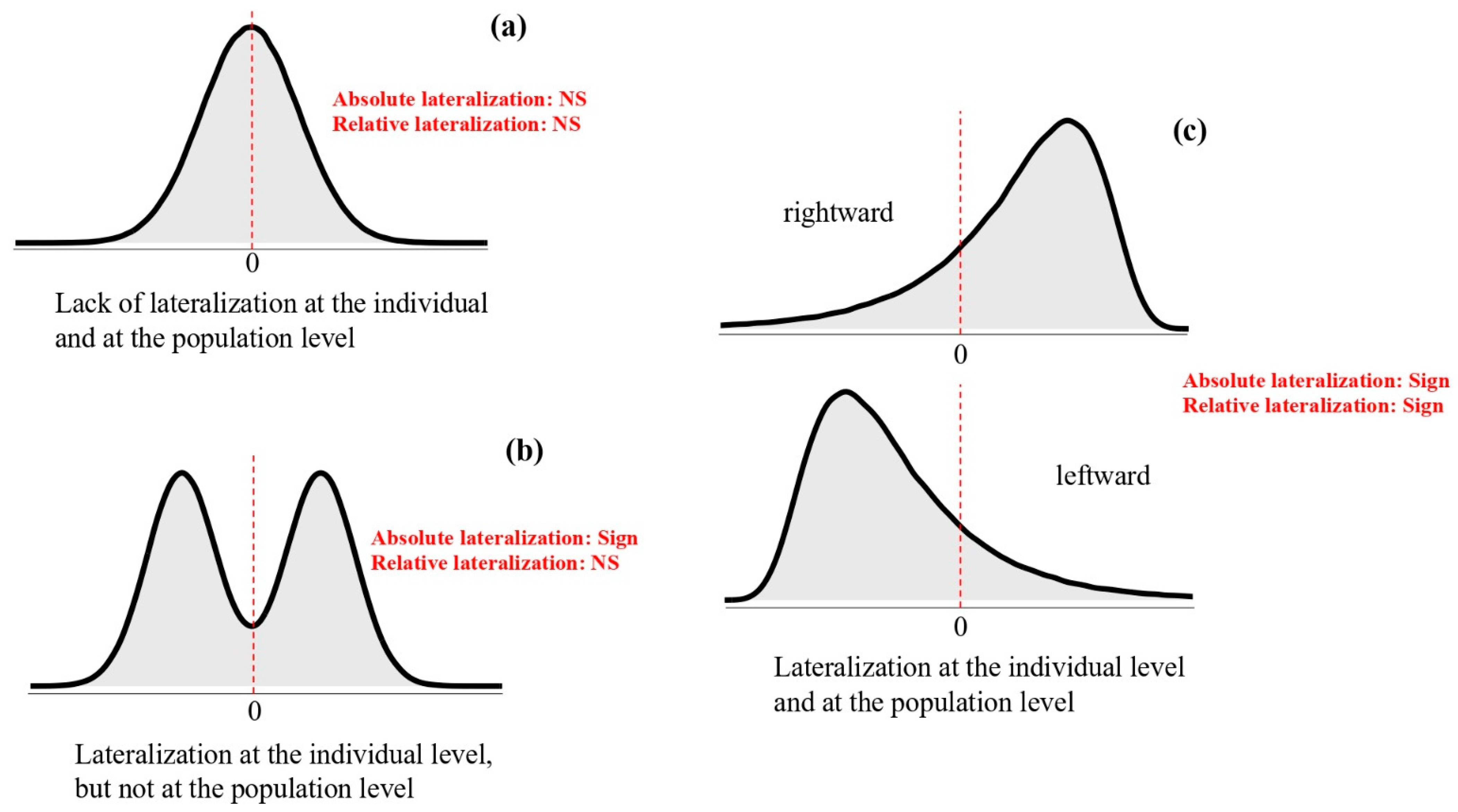
Figure 1. Different patterns of frequency distribution of lateralisation in a population and corresponding significance of the two most used lateralisation indexes against zero (in red): (a) the relative ad absolute lateralisation indexes are not significantly different from 0 (NS), suggesting no or weak lateralisation at the individual and no lateralisation bias at the population level; (b) the absolute lateralisation indexes is significant (Sign), while the relative lateralisation index is non-significant; (c) both relative and absolute lateralisation indexes are significant, indicating lateralisation at the individual level and a rightward (top) or a leftward (bottom) lateralisation bias at the population level.
Symmetry 17 00527 g001
Figure 2. Goldbelly topminnow were artificially selected for right-eye dominance (Right lines) and for left-eye dominance (Left lines) when inspecting a dummy predator. Fish from Right and Left lines were then compared in several behavioural lateralisation tests [123,125,126,127,128] and were found to exhibit strong and opposing biases in all these measures of lateralisation. For example, fish artificially selected for right-eye dominance (a) in predator inspection (Right lines) captured more often prey located on their left side; (b) males engaged in intrasexual attacks more often on their right side (c) and preferred to initiate mating attempts on their left side; (d) females preferred to keep shoal mates on their right side (e). Left lines showed a mirror-reversed pattern of lateralisation.
Symmetry 17 00527 g002
Figure 3. The asymmetry in expression of the gene for the glucocorticoid receptor gr in zebrafish predicted behavioural lateralisation. Individuals with higher levels of mesencephalic gr expression in the right hemisphere processed social stimuli using the left eye, and therefore the right hemisphere, in a mirror test. Dots represent individual data points; the red line and the shaded area were predicted from linear regression. Data from Rovegno et al. [255].
Symmetry 17 00527 g003
Table 1. Studies indicating evidence (+) or lack of evidence (−) of genetic contribution to individual differences in lateralisation in non-human vertebrates.
Vertebrate GroupSpecies MethodTrait InvestigatedGenetic Contribution: Direction/StrengthReference
Teleost fishBishop toothcarp Brachyraphis episcopi Common garden designEye preference for social or unfamiliar stimuli−/+[104]
Bahamas mosquitofish Gambusia hubbsiSibling concordanceTurning direction+/+[98]
Goldbelly topminnows Girardinus falcatus Parent-offspring concordanceEye preference for predator+/+[105]
Goldbelly topminnows Girardinus falcatus Artificial selectionEye preference for predator+/+[106]
Zebrafish
Danio rerio
Study of mutants (fsi mutation)Forebrain asymmetry,
eye preference for social stimuli and food
+/+[107]
Zebrafish
Danio rerio
Motor asymmetries−/−[107]
Zebrafish
Danio rerio
Artificial selectionEye preference for social stimuli+/+[108]
Zebrafish
Danio rerio
Candidate gene analysisForebrain asymmetry, eye preference for predator and social stimuli+/+[109]
BirdYellow-legged gull
Larus michahellis
Sibling concordanceReverting from supine posture−/−[103]
Yellow-legged gull
Larus michahellis
Begging−/−[103]
MammalCapuchin monkey
Cebus apella
Parent-offspring concordanceLooking bias +/−[110]
Capuchin monkey
Cebus apella
Hand preference−/−[110]
Capuchin monkey
Cebus apella
Turning bias+/−[110]
Chimpanzee
Pan troglodytes
Parent-offspring concordanceHand preference+/−[111]
Chimpanzee
Pan troglodytes
Parent-offspring concordance (Cross-fostering design)Hand preference (coordinated bimanual tasks)−/−[111]
Mouse
Mus musculus
Artificial selection,Paw preference−/+ [112,113]
Mouse
Mus musculus
QTLPaw preference−/+ [114]
Table 2. Studies suggesting plasticity of lateralisation in various traits induced by light exposure during early development in chicks.
Trait Investigated References
Antipredator response[201,202]
Attack behaviour [192,203]
Brain gene expression [204]
Competition[205]
Copulation [192,203]
Fear [201]
Flexibility/perseveration [206,207,208]
Monocular visual learning [209]
Neuronal activity [210,211]
Novelty exploration[206]
Refuge seeking [206]
Social behaviour [206]
Stimulus recognition [206,212]
Tectofugal visual projections [213]
Thalamofugal visual projections [213,214]
Vigilance behaviour [202]
Table 3. Studies reporting predation-induced plasticity of behavioural lateralisation in vertebrates.
Vertebrate Group Species (Age) Manipulation Effect of Predation Risk Reference
Teleost fish Ambon damselfish Pomacentrus amboinensis (juveniles) Exposure to alarm cues (9 days) Increased lateralisation [238]
Fathead minnows
Pimephales promelas (adults)
Exposure to alarm cues (for either 2, 4, or 8 days) Decreased lateralisation [239]
Goldbelly topminnows Girardinus falcatus (juveniles) Maternal visual exposure to predator (18 times across 6 weeks) Increased lateralisation [130]
Guppies, Poecilia reticultata (adults) Exposure to alarm and predator cues (from the past generation) Increased lateralisation[237]
Three-spined stickle-backs Gasterosteus aculeatus (juveniles) Olfactory exposure to predator Increased lateralisation [240]
Whitetail damselfish Pomacentrus chrysurus (juveniles) Exposure to alarm cues (4 days) Increased lateralisation [241]
Amphibian Wood frog
Lithobates sylvaticus (tadpoles)
Exposure to alarm cues as embryos (between eggs laying and hatching) Increased lateralisation [242]
Disclaimer/Publisher’s Note: The statements, opinions and data contained in all publications are solely those of the individual author(s) and contributor(s) and not of MDPI and/or the editor(s). MDPI and/or the editor(s) disclaim responsibility for any injury to people or property resulting from any ideas, methods, instructions or products referred to in the content.