Abstract
Hip dysplasia (HD) is a typical developmental abnormality of the hip joint, and discomfort is often found in adulthood. This study compared patients with symptomatic HD in muscle strength, dynamic balance, and range of motion (ROM) with healthy individuals. Patients included those who complained of unilateral pain although the lateral center edge angle (LCEA) exhibited bilateral abnormality. Participants (n = 95; men: 46, women: 49) were divided into symptomatic and asymptomatic sides, and a healthy group without a history of hip joint disease (n = 70; men: 30, women: 40) was compared. Hip flexion, extension, abduction, and adduction were performed at an angular velocity of 30°/s using an isokinetic strength test device. The Y-balance test was conducted to measure dynamic balance, and ROM was measured using an electronic goniometer to evaluate flexion, extension, adduction, abduction, and internal and external rotations. In addition, the pain visual analog scale (VAS) and hip and groin outcome scale (HAGOS), a subjective evaluation of the hip joint, were evaluated. ROM (flexion, abduction, internal rotation, and external rotation) was significantly decreased in the HD symptomatic sides of men and women compared to those of the healthy group and the asymptomatic side, and the dynamic balance, flexion, and abduction muscle strength were also lower on the symptomatic sides. Although the LCEA of the HD asymptomatic side was lower than that of the healthy group, there were no significant differences in VAS, flexion, extension, abduction ROM, and extension strength compared to those of healthy individuals. In conclusion, patients with HD were mostly bilateral, and on the symptomatic side, there was a decrease in ROM, dynamic balance, and muscle strength; however, on the asymptomatic side, the function was relatively close to normal.
Keywords:
hip dysplasia; strength; range of motion; dynamic balance; asymptomatic; symptomatic; asymmetry 1. Introduction
Hip dysplasia (HD) is a condition in which the ball-shaped head of the femur is uncovered in the hip socket [1,2]. HD causes instability owing to the shallow depth of the hip joint and sometimes causes dislocation and subluxation. In addition, the deformed hip joint causes friction between the femoral head and acetabulum, damaging surrounding tissues such as the labrum. This results in pain and functional limitations of the hip joint [3]. HD is considered a leading precursor of osteoarthritis, as development of hip osteoarthritis in 20–40% of patients is due to HD [4].
Because HD is a disease that originally occurs during growth, it is referred to as developmental dysplasia of the hip (DDH), with a prevalence of 0.24%; however, most cases are not recognized and only present with pain in adulthood [5]. Although there is no clear etiology, it is known that the incidence rate is higher in women than in men, and the probability of occurrence is slightly higher due to family history and occurs more frequently in babies that presented in breech position [6,7,8]. Treatment includes surgery and conservative therapy, and the decision is made by a specialist based on the severity, radiological findings, physical examination, and the patient’s symptoms. Typically, surgical treatments include arthroplasty, osteotomy, and debridement [9]. Conservative therapy should be considered if immediate surgery is not required [4]. Because HD involves the morphological structure, correcting the biomechanical structure through surgery is a pivotal treatment method. However, because HD does not necessarily require surgery, other modalities such as physical therapy, injections, and exercise rehabilitation may be beneficial. In a previous study, 56% of the participants opted for surgery and 44% preferred conservative treatment; the results were satisfactory in both groups [10]. In addition, it was reported that flexion strength, pain scale, and subjective hip joint score in patients with HD significantly improved after 8 weeks of strength training [11].
The symptoms of HD vary according to severity and location, and 97% of patients complain of intra-articular discomfort [12]. In addition, gait changes and abnormalities in patients with HD and reduced range of motion (ROM), resulting in anxiety and depression, negatively affect the patient’s daily life [13,14,15]. Despite the fact that hip muscle strength and balance assessment are required to complete the objective physical examination of patients with HD, there is limited information available to understand the condition of patients with HD [16,17]. Some muscle strength studies on HD have been performed before and after surgery [18,19]. However, these studies did not distinguish between the sexes, and studies measuring balance are uncommon. Very few studies have used an isokinetic dynamometer, which is considered the gold standard for measuring muscle strength [20].
Muscle strength, ROM, and balance are objective, representative physical examinations to evaluate whether a patient has improved along with the pain, and ultimately, it is very important for orthopedic surgeons, physical therapists, and sports medicine specialists to recognize the specificity of HD [17,21,22]. Therefore, the purpose of this study was to include men and women with unilateral symptoms. The isokinetic muscle strength, dynamic balance, and ROM of the symptomatic and asymptomatic sides were compared with those of healthy individuals. The results of this study are intended to help elucidate the functional characteristics and asymmetry in patients with HD and contribute to the creation of rehabilitation and improvement programs. In this study, the following hypothesis is proposed: the strength and balance would be lower than those in the healthy group, and muscle strength and balance will have different trends on the symptomatic and asymptomatic sides compared to those of the healthy group.
2. Materials and Methods
2.1. Experimental Design
In this study, the healthy group and HD patients were recruited through the bulletin board. Patients with only unilateral HD symptoms were included, and bilaterally severe patients were excluded. The participants voluntarily participated, listened to the explanation of the study from the researcher, and filled out a written consent form. This study complied with the tenets of the Declaration of Helsinki and was approved by the Institutional Review Board of Gangneung-Wonju National University (approval number: GWNU IRB 2021-13).
In the order of the test, subjective score, ROM, dynamic balance, and isokinetic muscle strength were measured using a questionnaire. For measurement, the dominance side of a healthy person was measured, and both sides of the HD patient were examined. The analysis compared the healthy group and the asymptomatic HD side (HDast), and the healthy group and the symptomatic HD side (HDst).
2.2. Participants
Participants were adults (age: 20–45 years) who visited our hospital for hip pain and voluntarily participated in the study after obtaining information via a bulletin board at the center. The specialist diagnosed HD based on measurements using X-rays (lateral center edge angle, LCEA < 25°) (Figure 1A) [23]. Physical examination and pain scale evaluation were performed for diagnosis. The LCEA was measured in two ways: (1) a line perpendicular to the transverse axis of the pelvis from the center of the femoral head and (2) a line from the center of the femoral head to the lateral point of the acetabulum [24].
Figure 1.
(A) Center edge of angle of hip dysplasia; (B), hip strength test; (C), Y-balance test.
The initial number of patients was 150; however, those who complained of severe bilateral hip pain (n = 8), who required surgery (n = 16), and those with a history of treatment in the lower extremities (n = 7) were excluded. In addition, to exclude the possibility of pain due to degeneration, patients over 45 years of age (n = 24) were excluded. The final analysis included 46 men and 49 women patients with HD; healthy individuals (men: 30, women: 40) were recruited through the bulletin board as the control group (age: 20–45 years, without past orthopedic diseases or pain in the hip, knee, or lower back).
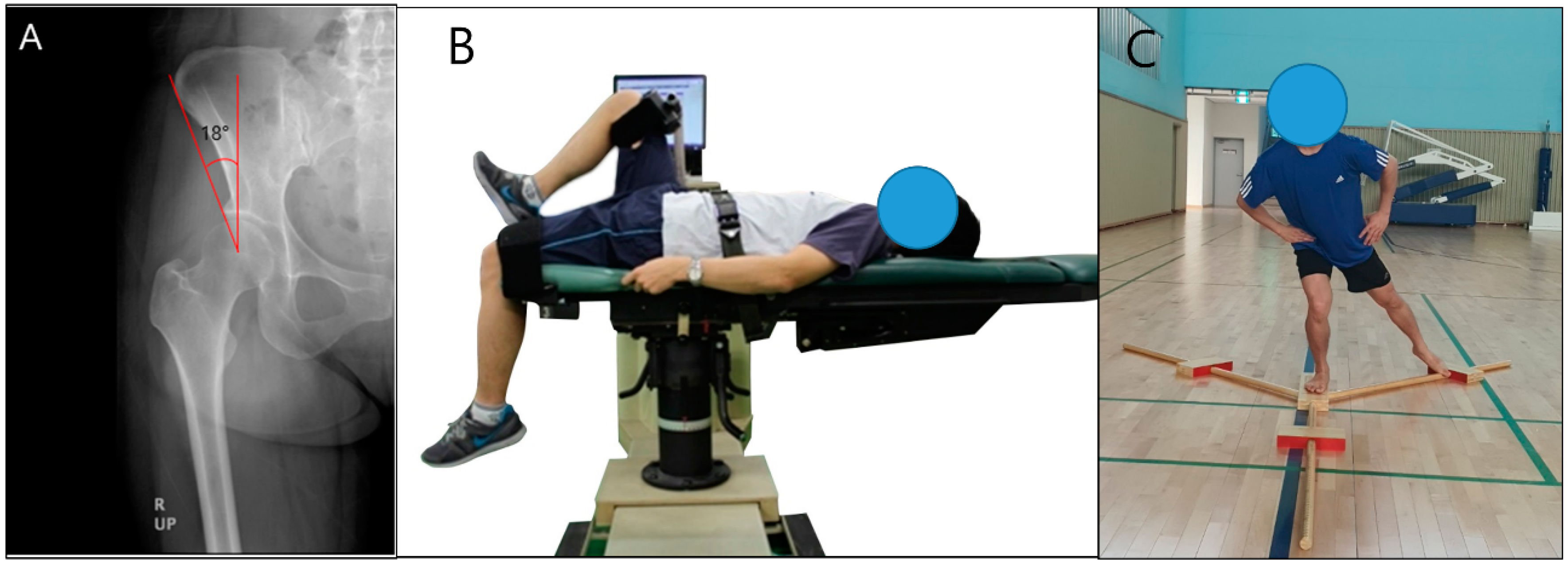
2.3. Isokinetic Strength Test
The isokinetic strength test was performed using a CSMi isokinetic dynamometer (CSMi HUMAC NORM; Stoughton, MA, USA) (Figure 1B). Hip abduction, adduction, flexion, and extension were performed in concentric contraction mode at angular velocity of 30°/s [25,26]. Before the test, the patient performed warm-up exercises for 15–20 min using a stationary bicycle, and the active ROM and pain status for the test were determined. The examiner provided explanations and demonstrations, and then exercises were performed to familiarize the patient with the examination. The asymptomatic joint was measured first, followed by the symptomatic side. After practicing three times, the test was conducted four times. Muscle strength was expressed as far as possible within the range at which the pain could be tolerated. Abduction and adduction were measured with participants in the side-lying position. The axis of the hip joint was aligned with the machine. The axis of the hip was superior and medial to the greater trochanter. The angle of the examination was measured from 0° in the neutral position to 45° degrees of abduction. The flexion and extension tests were performed with participants in the supine position. The angle of examination was measured from 0° of neutral forge to 100° of flexion. The handrail was held, and the chest, pelvis, and opposite leg were fixed with straps to prevent body movement during the examination. Gravity adjustment was performed to eliminate the effects of the weight of the lower extremity segment. After the values were recorded as Nm values, the Nm values per body weight (Nm/kg) were calculated for the analysis.
2.4. Dynamic Balance Test
Dynamic balance was measured using the YBT (Y Balance Test™, Cerder Park, TX, USA). The YBT measures balance ability in three directions using a Y-shaped device: anterior, posteromedial, and posterolateral (Figure 1C) [27]. The participant stood in the center of the equipment on one foot and measured the furthest possible distance that can be reached with the opposite leg in the three directions. Before the test, three or more exercises were attempted to enable the participants to fully understand, and the measurer explained and demonstrated to familiarize the participants with the test. The asymptomatic side of the participant was examined first, and then the symptomatic side was measured. The anterior direction was measured first, followed by the posteromedial and posterolateral directions. If during the test balance was lost and both feet touched the ground, it was considered a failure, and the test was redone after 5 min of rest. The test was conducted three times, and the average value was used. The formula is as follows:
where the length of the lower extremity is the distance from the anterior superior iliac spine to the center of the medial malleolus. A safe test environment was established in preparation for the risk of falls that could occur during the measurement. In the event of a fall, a collision hazard prevention, anti-skid, and assistive monitoring environment was created.
Score = [(sum of distance in three directions)/(length of lower extremity × 3)] × 100
2.5. Range of Motion Measurement
The ROM test measured abduction, adduction, flexion, and extension by using an electronic goniometer (CYBEX EDI 320, CYBEX Inc., Stoughton, MA, USA). Adduction, abduction, extension, and flexion were measured with the participants lying supine. Internal and external rotations were measured with participants sitting down. The maximum active ROM was measured by an experienced examiner, and the higher value was recorded twice. If the error exceeded 3°, it was re-measured.
2.6. Subjective Hip Scoring Questionnaire
The Copenhagen Hip and Inguinal Outcome Score (HAGOS) is a frequently used clinically based questionnaire for subjective self-assessment of hip patients and has demonstrated reliability and validity [28,29]. The questionnaire consisted of six parts (37 questions): pain (10 questions), symptoms (7 questions), function in daily living (5 questions), sport and recreation (8 questions), participation in physical activities (2 questions), and quality of life (5 questions). In the score calculation, 100 points were calculated for each of the six parts, and the final average of the last six parts was calculated. Lower scores signified worse conditions.
2.7. Data Analysis
The sample size was calculated using G*power software (G*power 3.1, University of Düsseldorf, Düsseldorf, Germany): effect size d = 0.5; α error = 0.05; power, (1 − β err prob) = 0.80; and difference between two independent means (two groups). Statistical analysis was performed using SPSS 25.0 (SPSS Inc., Chicago, IL, USA). The Kolmogorov–Smirnov test revealed that the main variables being analyzed did not display normal distribution. Therefore, a non-parametric analysis was performed. Comparisons between the healthy and patient groups were performed using the Mann–Whitney U test. To compare the three groups (healthy group, HD asymptomatic side, and HD symptomatic side), the Kruskal–Wallis test was performed. The post hoc test was conducted using the Bonferroni correction. For categorical variables, the chi-squared test was used. Statistical significance was set at p < 0.05.
3. Results
3.1. General Characteristics of Participants
Table 1 presents the general characteristics of the healthy and HD groups. Age, height, weight, and BMI were not significantly different between the groups. Patients exhibited a higher incidence of bilateral HD. In the HAGOS, which subjectively evaluates hip discomfort in various situations, all components were significantly lower in the HD group than in the healthy group.
Table 1.
General characteristics of participants.
3.2. Hip Morphology Angle, Pain, and Range of Motion
The LCEA, VAS, and ROM values of the asymptomatic (HDast), symptomatic (HDst), and healthy groups were compared (Table 2). Both men and women had asymptomatic and symptomatic LCEAs, which were significantly lower than those in the healthy group. Pain on the symptomatic side of the patients was greater than that in the healthy group and the asymptomatic side. There was no significant difference in pain on the asymptomatic side in both sexes compared to that in healthy individuals, although the LCEA exhibited a significant difference.
Table 2.
Participant’s hip morphology angle, pain, and range of motion.
Regarding the symptomatic side, ROM in men and women, flexion, abduction, internal rotation, and external rotation were significantly lower than in the healthy and asymptomatic groups. Abduction of the asymptomatic side in men was significantly reduced compared with that in the healthy group.
3.3. Y-Balance Test of Healthy Participants and Patients with Hip Dysplasia
The posterolateral scores of the asymptomatic side of men and women with HD were significantly lower than those of the healthy group. Additionally in the symptomatic side, the anterior, posteromedial, posterolateral and total scores were significantly reduced compared to those of the healthy group and the asymptomatic side (Table 3).
Table 3.
Y-balance test of healthy participants and patients with hip dysplasia.
3.4. Isokinetic Hip Strength of Healthy Participants and Patients with Hip Dysplasia
The muscular strength of symptomatic men and women was significantly weaker in flexion and abduction than those in the healthy group and those of the asymptomatic side. Similarly, the asymptomatic group had significantly lower flexion and abduction than the healthy group. There was no significant difference in men’s adduction, but women’s adduction was significantly lower compared to that in the healthy group and of the asymptomatic side (Table 4).
Table 4.
Isokinetic hip strength of healthy and hip dysplasia patients.
4. Discussion
HD is sometimes referred to as acetabular dysplasia and is a secondary hip joint problem that occurs because the acetabulum does not sufficiently cover the femoral head. A general diagnosis is to measure LCEA using X-rays, and >25° is considered normal, pathological disorder is <20°, and borderline dysplasia is 20–25° [24]. HD is a classic developmental disorder; with an incidence of 0.24%, but because it does not necessarily cause pain, it is often undiagnosed or is only discovered in adulthood [5]. Therefore, there is little information about the muscle strength that accurately reflects the condition of patients with HD. The purpose of this study was to analyze the characteristics of hip adduction, abduction, flexion, and extension muscle strength using isokinetic equipment in patients with HD.
The study by Jacobsen et al. [30] was a very rare study that investigated the muscle strength of 100 patients (men: 17; women: 83) with HD, although there was no sex distinction or control group. The muscle strength of the patients was 1.2 Nm/kg in flexion, 1.2 Nm/kg in extension, 1.8 Nm/kg in adduction, and 1.1 Nm/kg in abduction; those results were in agreement with the findings of this study. The representative result of this study was that both men and women with HD had significantly lower muscle strength in abduction and flexion than those of the healthy group. These results were identical to those of Sørensen et al., who compared patients with HD and controls [31].
Some studies have evaluated the muscle strength before and after HD surgery. In a study comparing them with the control group, preoperative muscle strength of 82 HD patients was flexion 1.2 Nm/kg, extension 1.8 Nm/kg, abduction 1.2 Nm/kg, and adduction 1.1 Nm/kg, and all movements were weaker than those in the healthy group [19]. Sucato et al. [18] measured abduction and flexion using isokinetic equipment before and after Ganz periacetabular osteotomy surgery in 24 adolescent patients with HD. Before surgery, abduction was 0.618 Nm/kg and flexion was 0.824 Nm/kg. Because these studies included patients requiring surgery, the conditions were different from those of the participants in this study; therefore, the results would have been slightly different.
Although not studied in HD, weak muscle strength has often been reported in other hip disorders. In a muscle strength study comparing healthy individuals and patients with acetabulum impingement, there was a difference of 28% in adduction, 11% in abduction, and 18% in external rotation, with no significant difference in extension [32]. When comparing the muscle strength according to hip osteoarthritis, adduction 25%, abduction 31%, and extension 18% were significantly lower than those in the healthy group, but no significant difference was found in extension. The results of this study were similar to those of the present study [33]. In this study, extension and adduction were less affected by HD. These results have also been reported in previous studies. In one study, the muscle strengths that exhibited the least change before and after surgery were extension and adduction [19]. Another study analyzed only abduction and flexion, whereas adduction and extension were excluded [18,34]. One study measured abduction only in patients with HD [35].
In the current study, the YBT was measured to determine the dynamic balance of HD. YBT has long been used as a tool to evaluate the coordination ability of athletes, patients, and older individuals [27,36]. Although studies on balance in HD patients are very rare, it is instructive to measure dynamic balance because HD causes instability due to the shallow ball-and-sock joint depth.
The YBT results displayed a different trend from that of muscle strength. There was no significant difference between the asymptomatic HD side and the healthy group, but the symptomatic side was significantly lower. This indicates that the association between muscle strength and dynamic balance is not strong, and similar results have been reported in a previous study. Wilson et al. [37] analyzed the correlation between the hip muscle strength test and YBT in a healthy group and determined that it was ‘moderate’ or ‘weak’.
Furthermore, Kumar et al. demonstrated no significant difference between YBT and hip joint lesions. Radiological results and YBT results were analyzed for the healthy group and the group with hip disorders. The group with acetabulum and femur cartilage lesions and osteoarthritis did not show significant YBT values compared with those of the healthy group [38]. However, it is controversial that this study was conducted without sex distinction.
The inclusion criterion for this study was unilateral pain. However, the radiographic results revealed that there were bilateral patients. This means that bilateral HD could present with unilateral pain; this finding was mentioned in a previous study [39]. This further confirms that HD does not necessarily cause pain and suggests the possibility of conservative therapy. In this study, unilaterality was found in 65.6% of men and 62.5% of women. Previous studies also reported that HD was unilateral in 63.4% of men with a higher incidence in women (75.5%) [40].
The results of this study can be applied in clinical practice as follows: Regarding rehabilitation and sports medicine, the primary goal is to improve the functions of soft tissues such as muscles, ligaments, tendons, and nerves; however, HD is a morphological problem involving the bones. Therefore, it is necessary to improve the tolerance to pain and the function of daily life by improving the flexibility and muscle strength of the surrounding tissues through stretching. Symptomatic HD includes abduction, flexion, and internal rotation. Even if symptoms appear unilateral, it is possible that the patient has bilateral HD, and muscle strength improvement rather than ROM improvement is recommended.
The limitations of this study are that the age range was relatively large and it was a single-center study. Many patients with DDH are asymptomatic, whereas the number of symptomatic patients is very small. Therefore, further studies involving larger cohorts from multiple institutions should be recruited. There are eccentric and concentric contractions in muscle contraction modes, and concentric contractions were measured in this study. Since studies on muscle strength tests in HD patients are rare, we conducted a concentric contraction mode with reference to previous study for safety [18]. In future studies, rehabilitation interventions are required. Several studies have evaluated muscle strength and subjective scores before and after surgery [18,19]; however, rehabilitation exercise intervention studies are rare [11]. Therefore, studies using various interventions, such as walking, cycling, water exercise, stretching, strength training, and balance training, are required to develop comprehensive rehabilitation programs to relieve pain and enhance the quality of life and activities of daily living of patients with HD.
5. Conclusions
This study measured ROM, muscle strength, and dynamic balance in symptomatic patients with HD. The symptomatic side of the patient exhibited weak abduction and flexion muscle strength and decreased dynamic balance compared with the healthy group and the asymptomatic side. Although the LCEA of the asymptomatic group was small, the pain scale was mild. In addition, flexion, extension, and adduction ROM were normal in both men and women.
Author Contributions
Conceptualization, H.W. and T.C.; methodology, H.Y. and Y.K.; formal analysis, H.W. and Y.K.; investigation, H.Y. and Y.K.; writing—original draft preparation, H.W. and H.Y.; writing—review and editing, Y.K. and T.C.; software, Y.K.; supervision, Y.K. and TC. All authors have read and agreed to the published version of the manuscript.
Funding
This study received no external funding.
Institutional Review Board Statement
The patient submitted written informed consent. The study was approved by the researcher’s institutional review board center (approved number: GWNU IRB 2020-16) and conducted in accordance with the Helsinki Declaration.
Informed Consent Statement
Informed consent was obtained from subject involved in the study.
Data Availability Statement
The data are not publicly available due to privacy or ethical.
Conflicts of Interest
The authors declare no conflict of interest.
References
- Schmitz, M.R.; Murtha, A.S.; Clohisy, J.C.; Group, A.S. Developmental dysplasia of the hip in adolescents and young adults. JAAOS J. Am. Acad. Orthop. Surg. 2020, 28, 91–101. [Google Scholar] [CrossRef] [PubMed]
- Dezateux, C.; Rosendahl, K. Developmental dysplasia of the hip. Lancet 2007, 369, 1541–1552. [Google Scholar] [CrossRef] [Green Version]
- Storer, S.K.; Skaggs, D.L. Developmental dysplasia of the hip. Am. Fam. Physician 2006, 74, 1310–1316. [Google Scholar] [PubMed]
- Gala, L.; Clohisy, J.C.; Beaule, P.E. Hip Dysplasia in the Young Adult. J. Bone Jt. Surg. Am. Vol. 2016, 98, 63–73. [Google Scholar] [CrossRef] [PubMed]
- Kolb, A.; Schweiger, N.; Mailath-Pokorny, M.; Kaider, A.; Hobusch, G.; Chiari, C.; Windhager, R. Low incidence of early developmental dysplasia of the hip in universal ultrasonographic screening of newborns: Analysis and evaluation of risk factors. Int. Orthop. 2016, 40, 123–127. [Google Scholar] [CrossRef] [PubMed]
- Chan, A.; McCaul, K.A.; Cundy, P.J.; Haan, E.A.; Byron-Scott, R. Perinatal risk factors for developmental dysplasia of the hip. Arch. Dis. Child. -Fetal Neonatal Ed. 1997, 76, F94–F100. [Google Scholar] [CrossRef] [PubMed]
- Cady, R.B. Developmental dysplasia of the hip: Definition, recognition, and prevention of late sequelae. Pediatric Ann. 2006, 35, 92–99. [Google Scholar] [CrossRef] [PubMed]
- Bache, C.E.; Clegg, J.; Herron, M. Risk factors for developmental dysplasia of the hip: Ultrasonographic findings in the neonatal period. J. Pediatric Orthop. B 2002, 11, 212–218. [Google Scholar]
- Sanchez-Sotelo, J.; Berry, D.J.; Trousdale, R.T.; Cabanela, M.E. Surgical treatment of developmental dysplasia of the hip in adults: II. Arthroplasty options. JAAOS J. Am. Acad. Orthop. Surg. 2002, 10, 334–344. [Google Scholar] [CrossRef] [PubMed]
- Hunt, D.; Prather, H.; Harris Hayes, M.; Clohisy, J.C. Clinical outcomes analysis of conservative and surgical treatment of patients with clinical indications of prearthritic, intra-articular hip disorders. PM&R J. Inj. Funct. Rehabil. 2012, 4, 479–487. [Google Scholar] [CrossRef] [Green Version]
- Mortensen, L.; Schultz, J.; Elsner, A.; Jakobsen, S.S.; Søballe, K.; Jacobsen, J.S.; Kierkegaard, S.; Dalgas, U.; Mechlenburg, I. Progressive resistance training in patients with hip dysplasia: A feasibility study. J. Rehabil. Med. 2018, 50, 751–758. [Google Scholar] [CrossRef] [Green Version]
- Nunley, R.M.; Prather, H.; Hunt, D.; Schoenecker, P.L.; Clohisy, J.C. Clinical presentation of symptomatic acetabular dysplasia in skeletally mature patients. JBJS 2011, 93, 17–21. [Google Scholar] [CrossRef] [Green Version]
- Marangoz, S.; Atilla, B.; Gök, H.; Yavuzer, G.; Ergin, S.; Tokgözoğlu, A.M.; Alpaslan, M. Gait analysis in adults with severe hip dysplasia before and after total hip arthroplasty. Hip Int. 2010, 20, 466–472. [Google Scholar] [CrossRef] [PubMed] [Green Version]
- Hampton, S.; Nakonezny, P.; Richard, H.; Wells, J. Pain catastrophizing, anxiety, and depression in hip pathology. Bone Jt. J. 2019, 101, 800–807. [Google Scholar] [CrossRef]
- Matsuda, D.K.; Wolff, A.B.; Nho, S.J.; Salvo, J.P., Jr.; Christoforetti, J.J.; Kivlan, B.R.; Ellis, T.J.; Carreira, D.S.; Multicenter Arthroscopic Study of the Hip (MASH) Study Group. Hip dysplasia: Prevalence, associated findings, and procedures from large multicenter arthroscopy study group. Arthrosc. J. Arthrosc. Relat. Surg. 2018, 34, 444–453. [Google Scholar] [CrossRef] [PubMed]
- Scott, E.J.; Willey, M.C.; Mercado, A.; Davison, J.; Wilken, J.M. Assessment of disability related to hip dysplasia using objective measures of physical performance. Orthop. J. Sports Med. 2020, 8, 2325967120903290–2325967120903298. [Google Scholar] [CrossRef] [PubMed]
- Martin, H.D.; Kelly, B.T.; Leunig, M.; Philippon, M.J.; Clohisy, J.C.; Martin, R.L.; Sekiya, J.K.; Pietrobon, R.; Mohtadi, N.G.; Sampson, T.G. The pattern and technique in the clinical evaluation of the adult hip: The common physical examination tests of hip specialists. Arthrosc. J. Arthrosc. Relat. Surg. 2010, 26, 161–172. [Google Scholar] [CrossRef] [PubMed]
- Sucato, D.J.; Tulchin, K.; Shrader, M.W.; DeLaRocha, A.; Gist, T.; Sheu, G. Gait, hip strength and functional outcomes after a Ganz periacetabular osteotomy for adolescent hip dysplasia. J. Pediatric Orthop. 2010, 30, 344–350. [Google Scholar] [CrossRef]
- Jacobsen, J.S.; Jakobsen, S.S.; Søballe, K.; Hölmich, P.; Thorborg, K. Isometric hip strength impairments in patients with hip dysplasia are improved but not normalized 1 year after periacetabular osteotomy: A cohort study of 82 patients. Acta Orthop. 2021, 92, 285–291. [Google Scholar] [CrossRef] [PubMed]
- Zapparoli, F.Y.; Riberto, M. Isokinetic evaluation of the hip flexor and extensor muscles: A systematic review. J. Sport Rehabil. 2017, 26, 556–566. [Google Scholar] [CrossRef]
- Martin, H.D.; Palmer, I.J. History and physical examination of the hip: The basics. Curr. Rev. Musculoskelet. Med. 2013, 6, 219–225. [Google Scholar] [CrossRef] [PubMed] [Green Version]
- Hunt, D.; Clohisy, J.; Prather, H. Acetabular labral tears of the hip in women. Phys. Med. Rehabil. Clin. North Am. 2007, 18, 497–520. [Google Scholar] [CrossRef] [PubMed]
- LaPrade, M.D.; Melugin, H.P.; Hale, R.F.; Leland, D.P.; Bernard, C.D.; Sierra, R.J.; Trousdale, R.T.; Levy, B.A.; Krych, A.J. Incidence of hip dysplasia diagnosis in young patients with hip pain: A geographic population cohort analysis. Orthop. J. Sports Med. 2021, 9, 2325967121989087–2325967121989093. [Google Scholar] [CrossRef] [PubMed]
- Vaudreuil, N.J.; McClincy, M.P. Evaluation and treatment of borderline dysplasia: Moving beyond the lateral center edge angle. Curr. Rev. Musculoskelet. Med. 2020, 13, 28–37. [Google Scholar] [CrossRef] [PubMed]
- CSMi. Humac Norm Users Guide; Computer Sports Medicine, Inc.: Stoughton, MA, USA, 2019. [Google Scholar]
- Dvir, Z. Isokinetics: Muscle Testing, Interpretation, and Clinical Applications, 2nd ed.; Churchill Livingstone: London, UK, 2004. [Google Scholar]
- Powden, C.J.; Dodds, T.K.; Gabriel, E.H. The reliability of the star excursion balance test and lower quarter Y-balance test in healthy adults: A systematic review. Int. J. Sports Phys. Ther. 2019, 14, 683–694. [Google Scholar] [CrossRef]
- Thorborg, K.; Hölmich, P.; Christensen, R.; Petersen, J.; Roos, E.M. The Copenhagen Hip and Groin Outcome Score (HAGOS): Development and validation according to the COSMIN checklist. Br. J. Sports Med. 2011, 45, 478–491. [Google Scholar] [CrossRef] [Green Version]
- Thomeé, R.; Jónasson, P.; Thorborg, K.; Sansone, M.; Ahldén, M.; Thomeé, C.; Karlsson, J.; Baranto, A. Cross-cultural adaptation to Swedish and validation of the Copenhagen Hip and Groin Outcome Score (HAGOS) for pain, symptoms and physical function in patients with hip and groin disability due to femoro-acetabular impingement. Knee Surg. Sports Traumatol. Arthrosc. 2014, 22, 835–842. [Google Scholar] [CrossRef] [Green Version]
- Jacobsen, J.S.; Hölmich, P.; Thorborg, K.; Bolvig, L.; Jakobsen, S.S.; Søballe, K.; Mechlenburg, I. Muscle-tendon-related pain in 100 patients with hip dysplasia: Prevalence and associations with self-reported hip disability and muscle strength. J. Hip Preserv. Surg. 2018, 5, 39–46. [Google Scholar] [CrossRef] [Green Version]
- Sørensen, H.; Nielsen, D.B.; Jacobsen, J.S.; Søballe, K.; Mechlenburg, I. Isokinetic dynamometry and gait analysis reveal different hip joint status in patients with hip dysplasia. Hip Int. 2019, 29, 215–221. [Google Scholar] [CrossRef]
- Casartelli, N.; Maffiuletti, N.; Item-Glatthorn, J.; Staehli, S.; Bizzini, M.; Impellizzeri, F.; Leunig, M. Hip muscle weakness in patients with symptomatic femoroacetabular impingement. Osteoarthr. Cartil. 2011, 19, 816–821. [Google Scholar] [CrossRef] [Green Version]
- Arokoski, M.H.; Arokoski, J.P.; Haara, M.; Kankaanpää, M.; Vesterinen, M.; Niemitukia, L.H.; Helminen, H.J. Hip muscle strength and muscle cross sectional area in men with and without hip osteoarthritis. J. Rheumatol. 2002, 29, 2185–2195. [Google Scholar] [PubMed]
- Sucato, D.J.; Tulchin-Francis, K.; de La Rocha, A.; Kulkarni, V.; Podeszwa, D.A. Improved functional outcome with no decrease in hip strength 2 years following Ganz periacetabular osteotomies for adolescent hip dysplasia. J. Pediatric Orthop. B 2015, 24, 99–105. [Google Scholar] [CrossRef] [PubMed]
- Kuroda, D.; Maeyama, A.; Naito, M.; Moriyama, S.; Yoshimura, I.; Nakamura, Y.; Kiyama, T. Dynamic hip stability, strength and pain before and after hip abductor strengthening exercises for patients with dysplastic hips. Isokinet. Exerc. Sci. 2013, 21, 95–100. [Google Scholar] [CrossRef]
- Sipe, C.L.; Ramey, K.D.; Plisky, P.P.; Taylor, J.D. Y-balance test: A valid and reliable assessment in older adults. J. Aging Phys. Act. 2019, 27, 663–669. [Google Scholar] [CrossRef]
- Wilson, B.R.; Robertson, K.E.; Burnham, J.M.; Yonz, M.C.; Ireland, M.L.; Noehren, B. The relationship between hip strength and the Y balance test. J. Sport Rehabil. 2018, 27, 445–450. [Google Scholar] [CrossRef] [Green Version]
- Kumar, D.; Wyatt, C.R.; Lee, S.; Nardo, L.; Link, T.M.; Majumdar, S.; Souza, R.B. Association of cartilage defects, and other MRI findings with pain and function in individuals with mild–moderate radiographic hip osteoarthritis and controls. Osteoarthr. Cartil. 2013, 21, 1685–1692. [Google Scholar] [CrossRef] [Green Version]
- Jacobsen, S.; Rømer, L.; Søballe, K. The other hip in unilateral hip dysplasia. Clin. Orthop. Relat. Res. 2006, 446, 239–246. [Google Scholar] [CrossRef]
- Loder, R.T.; Skopelja, E.N. The epidemiology and demographics of hip dysplasia. Int. Sch. Res. Not. 2011, 2011, 1–46. [Google Scholar] [CrossRef] [Green Version]
Publisher’s Note: MDPI stays neutral with regard to jurisdictional claims in published maps and institutional affiliations. |
© 2022 by the authors. Licensee MDPI, Basel, Switzerland. This article is an open access article distributed under the terms and conditions of the Creative Commons Attribution (CC BY) license (https://creativecommons.org/licenses/by/4.0/).