Article

Contributions of the Right Prefrontal and Parietal Cortices to the Attentional Blink: A tDCS Study

1
Department of Psychology, Sapienza University of Rome, Via Dei Marsi, 78, 00185 Rome, Italy
2
IRCCS Fondazione Santa Lucia, Via Ardeatina, 306, 00179 Rome, Italy
3
Department of Psychology, Bar Ilan University, Ramat Gan 52900, Israel
*
Author to whom correspondence should be addressed.
Symmetry 2021, 13(7), 1208; https://doi.org/10.3390/sym13071208
Submission received: 2 June 2021 / Revised: 19 June 2021 / Accepted: 26 June 2021 / Published: 6 July 2021
(This article belongs to the Special Issue Symmetry in Cognition and Emotion)

Abstract

:
The AB refers to the performance impairment that occurs when visual selective attention is overloaded through the very rapid succession of two targets (T1 and T2) among distractors by using the rapid serial visual presentation task (RSVP). Under these conditions, performance is typically impaired when T2 is presented within 200–500 ms from T1 (AB). Based on neuroimaging studies suggesting a role of top-down attention and working memory brain hubs in the AB, here we potentiated via anodal or sham tDCS the activity of the right DLPFC (F4) and of the right PPC (P4) during an AB task. The findings showed that anodal tDCS over the F4 and over P4 had similar effects on the AB. Importantly, potentiating the activity of the right frontoparietal network via anodal tDCS only benefitted poor performers, reducing the AB, whereas in good performers it accentuated the AB. The contribution of the present findings is twofold: it shows both top-down and bottom-up contributions of the right frontoparietal network in the AB, and it indicates that there is an optimal level of excitability of this network, resulting from the individual level of activation and the intensity of current stimulation.

1. Introduction

Our visual attention is limited in the amount of information that can be processed at any given moment; therefore, selection is essential to guarantee that relevant stimuli are not missed. The rapid serial visual presentation procedure (RSVP) is a well-consolidated strategy for studying the limitations of selective attention by overloading the attentional system through the very rapid presentation of multiple visual events. In the RSVP, stimuli are presented in foveal vision, at a rate of about ten items per second, and participants’ task is to detect and report two targets appearing among distractors. The temporal distance (lag) between the first (T1) and the second target (T2) is manipulated. Under these conditions, our ability to report T2 is severely compromised when it follows T1 within a time window of 200–500 milliseconds: a phenomenon known as the attentional blink (AB); [1,2]. In contrast, performance for T2 is spared when the two targets are presented in immediate succession (lag 1 sparing) or when T2 appears at later lags.
Accounts for the AB call upon the limitation of resources for stimulus encoding and for consolidation in working memory [3,4]. Accordingly, the AB would result from the failure to adequately consolidate T2 into a stable working memory representation while T1 is undergoing consolidation [5]. In contrast, the lag 1 sparing has been attributed to T1 and T2 sharing the same attentional episode when they appear in close temporal proximity [3,6]. In this case, T2 receives attentional enhancement along with T1, and it is encoded in working memory. Alternative accounts for the AB and sparing call upon the role of top-down attentional control mechanisms, involving target enhancement and/or distractor inhibition [7,8,9]. More specifically, it has been proposed that the AB reflects a temporal limit for reorienting attention between targets, see [10]. In this account, T1 onset would elicit a transient inhibition of bottom-up attention to protect processing against interference from distractors in the RSVP stream. If T2 appears during the inhibitory period, it cannot receive attentional processing, leading to the AB [2,8,9,11,12,13]. Importantly, as the temporal contiguity and attentional template matching between targets are crucial factors in determining both lag 1 sparing and the AB, working memory plays a central role in almost all accounts.
Evidence in support of the role of top-down attention and working memory in the AB comes from neuroimaging studies showing that the successful detection of T1 and T2 is associated to greater activity in the dorsolateral prefrontal cortex (DLPFC) responsible for the maintenance of task-set [14,15]. In addition, participants that do not show an AB have greater activation of the DLPFC than those who show an AB [16]. In addition, there is evidence that the activity of the right posterior parietal cortex (rPPC) is associated to the successful report of T1 and T2 [17], and in a split-brain patient, the AB was much stronger when T2 was presented to the right hemisphere compared to the left, [18]. Moreover, Dell’Acqua et al. [19], showed that the successful detection of T1 and T2 is associated to larger frontal P3a component, peaking at about 300 ms post target, and to delayed parietal P3b component peaking at about 450 ms. This evidence has been interpreted as reflecting inhibitory processes as the frontal P3a is smaller during the AB (i.e., when T2 is missed), whereas the parietal P3b reflects target consolidation in visual working memory. Finally, a MEG study by Gross et al. [20], has also provided some evidence on the involvement of the rPPC in the detection of T1 and T2 by showing an association between beta synchronization in frontal and parietal areas and the AB.
In summary, neuroimaging evidence links the activity of two key brain areas to the AB: the DLPFC involved in working memory and top-down control of attention and the posterior parietal cortex (PPC), involved in directing attention to salient stimuli and contributing to bottom-up actions, e.g., [21]. Importantly, these neuroimaging studies are correlational in nature and using transcranial direct current stimulation (tDCS) can provide more direct evidence. tDCS studies apply a constant, weak electric current using two scalp electrodes. The direction of the current-flow is from the positive (anodal) to the negative (cathodal) electrode. Anodal tDCS increases whereas cathodal tDCS decreases the excitability of cortical areas under the electrode [22], although these polarity-dependent stimulation effects might be more easily found in motor than in cognitive tasks [23]. To date, only two studies have used tDCS to assess the contribution of top-down attention to the AB by stimulating the left DLPFC. London and Slagter [24,25] used a RSVP, in which the two targets and distractors were all letters, but they differed based on color (T1 was red, T2 was green, and distractors were white). T2 could be presented at lag 2, lag 4 or lag 10. In addition, to assess the effects of tDCS on distractors inhibition, T2 could be primed (identity priming) or not by the preceding distractor (i.e., the distractor could be the same letter as T2). Participants performed the RSVP task while receiving sham, 1.0 mA anodal, or cathodal tDCS over the left DLPFC (F3), with the reference electrode placed over the right supraorbital area. Findings showed no differences between the effects of anodal and cathodal tDCS over the AB and on distractors’ inhibition as indexed by priming effects. However, there was some evidence that anodal (but not cathodal) tDCS effects depended on individuals’ performance levels, albeit London and Slagter [25] clarified that a post hoc correlational analysis revealed that individuals who benefited from anodal stimulation showed worse performance under cathodal tDCS, and vice versa. The second study, conducted by Sdoia et al. [26], showed that anodal tDCS over F3 reduces the AB at lag 3 whereas cathodal tDCS accentuates the AB at lag 5. Importantly, these effects remained even when controlling for initial individual differences in the AB size. There are clear methodological differences between these two studies as Sdoia et al. [26] used a RSVP, in which T1 and T2 were digits presented among distractor-letters. T2 followed T1 at lag 1, lag 3 or lag 5. One group performed the task while receiving sham or 1.5 mA anodal tDCS over the left DLPFC (F3), the other group performed the task while receiving sham or cathodal stimulation over F3. The reference electrode was placed over the right orbitofrontal cortex (FP2). However, regardless of these methodological differences, both tDCS studies have assessed the contribution of the left DLPFC (F3) to the AB, based on evidence that anodal stimulation over the left DLPFC improves working memory [27] and cognitive control [28,29,30]. However, as pointed out by Hommel et al. [31], it is unlikely that one single cortical structure or system underlies the AB and most likely both parietal and frontal regions play an important role in the AB. In fact, the right-lateralized frontoparietal network is involved in executive control and working memory [32,33,34,35] and it has been strongly implicated in processing during the AB and inattention blindness [36,37,38]. To our knowledge, the direct contribution of the rDLPFC and of the rPPC in modulating the AB has not been assessed using tDCS.
The activity of the rDLPFC has been extensively linked to top-down control (i.e., filtering) over access to working memory, whereas although the activity of the rPPC has been traditionally linked to spatial attention [21], it also contributes to visual processing [39] and to the amount of information being stored in working memory [40]. For instance, Li et al. [41] showed that during a change detection task, anodal tDCS over the rPPC increases the visual working memory capacity under no-distractor conditions, whereas anodal tDCS over the rDLPFC specifically increased the visual working memory capacity under distractor conditions. In addition, anodal tDCS over rPPC enhances visual short-term memory capacity [42,43] and unilateral (but not bilateral) tDCS over the right frontoparietal network improves working memory, albeit this improvement was especially robust for individuals with low working memory capacity [44]. In contrast, Lo et al. [45], found that anodal tDCS over the rPPC affects attentional orienting but not alerting and executive control. Finally, it has been suggested that the GABAergic system in the rPPC mediates the transient inhibition of attentional reorienting, leading to the AB and that the GABAergic system in the prefrontal cortex contribute to the reduction of attentional resources to T1 and the control of the attentional set for target identification [46]. Therefore, tDCS evidence points to the potential contribution of the rDLPFC and of the rPPC to the AB, although their relative contribution is unclear. This raises the question of whether the AB can be reduced by potentiating the activity of the rDLPFC and improving top-down attention compared to the opposite montage (i.e., enhancing the activity of the rPPC) and compared to sham tDCS.
The present study used anodal stimulation over the rDLPFC versus the rPPC to assess modulation of the AB. We argue that boosting, via anodal tDCS, the neural activity of the rDLPFC may improve temporal selective attention and reduce the AB (i.e., improve target detection when T1 and T2 are separated by distractors) as the rDLPFC is the area of converging afferences from the dorsal attention stream involved in top-down modulation of attention (i.e., filtering) as well as from the ventral attention stream, involved in distractor inhibition and visual short-term memory. In contrast, boosting, via anodal tDCS, the neural activity of the rPPC may improve bottom-up attention and visual short-term memory and it would serve as a more stringent control of the role of the rDLPFC and rPPC in the AB than the sham condition.

2. Method

2.1. Participants

Forty psychology students (32 female, 8 male) took part in the study in partial fulfilment of course credits. All participants were naive to the aims of the study and reported normal or corrected-to-normal vision. In addition, participants reported no use of medications or psychoactive drugs, and no history of neurological or psychiatric disorders. Exclusion criteria were having a history of epileptic seizure or a first-degree relative with a history of epileptic seizure, presence of head injuries or trauma, metal fragments or metallic implants, neurological or psychiatric disorders, having a sensitive skin, being pregnant. Twenty participants (18 females, 2 males; M age = 20.6, SD = 1.8) were randomly assigned to the group who received stimulation over the rDLPFC and 20 participants (14 females, 6 males; M age = 22.5, SD = 2.4) were randomly assigned to the group who received stimulation over the rPPC. The sample size was defined through power analysis G*Power software [47] using a medium-to-large partial η2 = 0.2 for the highest order interaction, and a power of 0.9 to increase the chance of replicability. This partial η2 was estimated from the group-by-lag interactions from previous behavioral studies (range: 0.18–0.23), and from Sdoia et al., [26] with η2 = 0.13 for the group-by-lag interaction, since London et al. [24,25] did not find any group-by-lag effect.

2.2. Materials and Apparatus

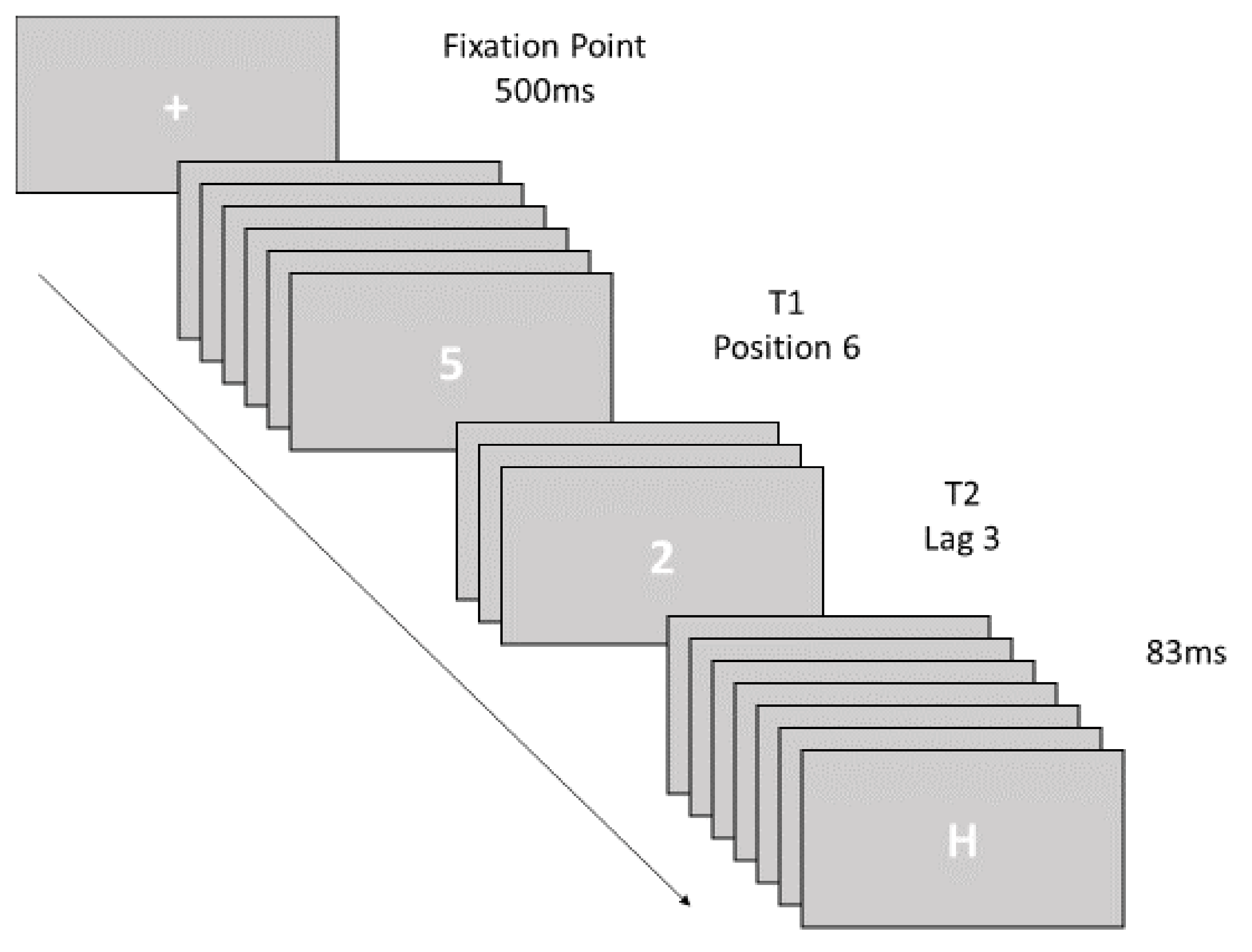
2.2.1. Attentional Blink Task

Participants performed a standard AB task, in which eight digits (i.e., from 2 to 9) served as T1 and T2 and 20 uppercase letters (i.e., A, B, C, D, E, F, G, H, K, L, M, P, Q, R, S, T, U, X, Y, Z) served as distractors. Both digits and letters were presented in white on a grey background approximately 55 cd/m2 and in 56-point Courier New font. Stimuli were presented at a viewing distance of approximately 50 cm from a CoreTM i5 computer via a 21.5” Dell P2210H (Analog) monitor (1600 × 900 pixels, 60 Hz). RSVP streams were presented using E-Prime Version 2.0 Professional software for Windows 7 Professional, which also recorded participants’ responses entered using a standard USB-keyboard.

2.2.2. Online Transcranial Direct Current Stimulation

Following scalp measurements, participants were randomly assigned to one of the two groups differing for the specific cortical site stimulated. Depending on group assignment, the anode was placed over the rDLPFC (F4) or over the rPPC (P4) according to 10–20 EEG International System, and the reference electrode was placed over the left supraorbital area. For the anodal stimulation, a 1.0 mA direct current (impedance was kept below 5 kΩ) was delivered by a battery-driven constant current stimulator (BrainStim E.M.S., srl Bologna, Italy) for 20 min during the AB task, with a 30-s fade in/fade out ramp. For the sham stimulation, the same fade in/fade out ramp was applied followed by the stimulation duration of 2 s. The size of the reference electrode was 4.7 × 4.7 cm2 (density 0.045 mA/cm2) and the anodal electrode had a diameter of 3.5 cm (density 0.10 mA/cm2). Using a larger reference electrode increases the diffusion of the received current therefore maximizing the potential anodal electrode effects [48]. We chose 1.0 mA based on evidence that this current intensity increases cortical excitation with unilateral stimulation of the motor cortex (M1), and that this effect does not hold for the 2 mA intensity or for the bilateral stimulation used in cognitive enhancement experiments [49].

2.3. Procedure

The experiment used a double-blind design: one experimenter was blind to the stimulation type (active or sham) participants were receiving in each session and oversaw mounting the electrodes and interacting with participants. A second experimenter assigned participants to conditions, selected the tDCS protocol and connected the electrodes. Participants completed two experimental sessions one week apart: one session with sham stimulation and the other with anodal stimulation (order counterbalanced between participants). Therefore, for each stimulation group (F4 and P4), half of the participants (N = 10) received anodal stimulation in the first session and sham stimulation in the second session, whereas the remaining participants (N = 10) underwent the opposite stimulation regime. All participants were told they were receiving active stimulation.
The stimulation started while participants read the task instructions presented on screen and completed nine practice trials, followed by 168 experimental trials divided in three blocks of 56 trials each. Between blocks there was a mandatory break of 30 s to maintain the duration of the AB task and that of the tDCS stimulation within the same range. All participants completed the AB task within the duration of the stimulation. Each trial started by pressing the spacebar. A central fixation point appeared for 500 ms, and it was followed by a stream of 18 stimuli: there were 16 distractor-letters and two target-digits (T1 and T2). Each stimulus was presented for 83ms with no interstimulus interval. The identity of T1 and T2 varied randomly (without replacement), and it could be a digit from 2 to 9 with the constraint that T1 and T2 identity was not the same in a stream. In each stream, T1 could appear in position 4, 6, or 8, whereas T2 was presented either at lag 1 (i.e., 83 ms, with no distractor between T1 and T2), at lag 3 (i.e., 166 ms, with two distractors between T1 and T2), and at lag 8 (i.e., 664 ms, seven distractors between T1 and T2), relative to T1. T1 serial position and the number of streams for each lag (1, 3, and 8) were counterbalanced across blocks. Participants were instructed to monitor the stream for two target-digits (T1 and T2) presented among distractor-letters and report at the end of the stream the two digits by pressing the corresponding key on the keyboard. Participants were informed that response speed and order of targets presentation were not relevant, and they were encouraged to guess if uncertain (see Figure 1).
Upon completion of the second session, participants were asked whether they noticed any differences between the two sessions, they were informed that they had received one active and one sham stimulation and asked whether they could tell which session they had received in each session. Participants were not able to report with certainty the session with active and that with sham stimulation. Participants were debriefed on the theoretical aspects of the study.

3. Experimental Design and Data Analyses
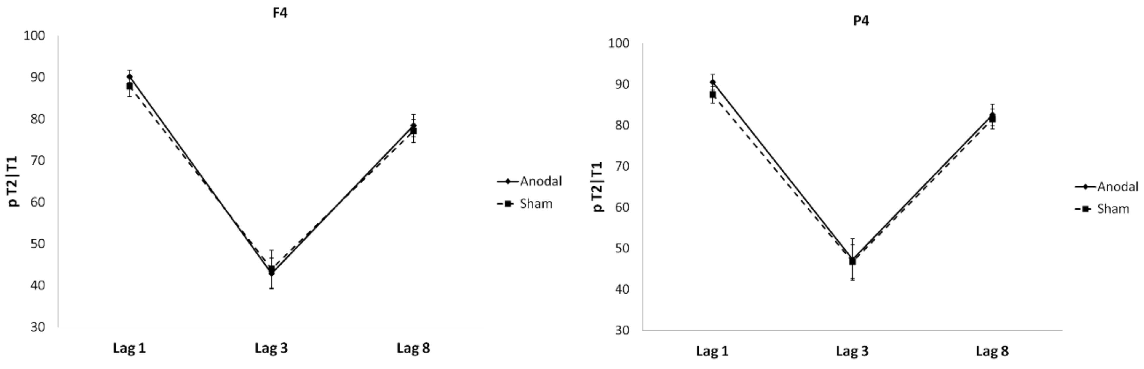
The experimental design is a 2 × 2 × 3 mixed-factorial design, with group (2: P4, F4) as the between- subjects factor and tDCS Session (2: Anodal, Sham) and lag (3: lag 1, lag 3, lag 8) as within-subject factors.
Performance accuracy was computed as the percentage of correct T1 reports (pT1) and as the percentage of correct T2 reports conditional on correctly reporting T1 (p T2|T1). Responses with inversion errors between T1 and T2 (e.g., 3 and 4 reported as 4 and 3), were accepted as correct. pT1 was analyzed using mixed repeated-measures ANOVA with tDCS session type (2: anodal, sham) as the repeated measure factor and stimulation-site (2: P4, F4) as the between-subject factor. pT2|T1 was analyzed using mixed repeated-measures ANOVA with tDCS session type (2: anodal, sham) and lag (3: lag 1, lag 3, lag 8) as within-subject factors and stimulation-site (2: P4, F4) as between-subject factor. Pairwise comparisons were Bonferroni-corrected.

4. Results

pT1: Overall performance accuracy for T1 was high 85.75% (SE = 1.15). ANOVA results with tDCS session (anodal, sham) and stimulation site (P4, F4) as between-subject factors showed no significant differences between the two tDCS sessions, F (1, 38) = 2.56, p = 0.118 and between stimulation site, F (1, 38) = 0.247, p = 0.622, (see Table 1). The two-way interaction was also not significant, F (1, 38) = 0.63, p = 0.43. Having ascertained that performance on T1 did not differ between sessions and stimulation site, performance on T2|T1 was analyzed.
pT2|T1: Overall performance accuracy for T2 conditional upon correctly reporting T1 (p T2|T1) was 71.39% (SE = 1.73). ANOVA results showed that the main effect of stimulation site (P4, F4) was not statistically significant, F (1, 38) = 0.587, p = 0.448, nor it was in interaction with any of the other factors. The main effect of lag was significant, F (2, 76) = 173.42, p < 0.001, partial η2 = 0.82. Pairwise comparisons revealed lag 1 sparing as p T2|T1 at lag 1 (M = 89.05, SE = 1.29) was greater than at lag 8 (M = 79.90, SE = 1.81), p < 0.001, followed by an AB as p T2|T1 at lag 3 (M = 45.23, SE = 3.2) was smaller than at lag 8, p < 0.001. Finally, that p T2|T1 at lag 1 was greater than at lag 8 indicates that the AB had not yet fully recovered by lag 8. The main effect of tDCS session (anodal vs. sham) was not statistically significant, F (1, 38) = 0.595, p = 0.445, nor it was in interaction with any of the other factors. Finally, the three-way interaction was not significant, F (2, 72) = 0.856, p = 0.429 (see Figure 2).
Therefore, findings show that participants’ performance was spared at lag 1 and it was followed by an AB at lag 3 and this pattern was present independently of stimulation.

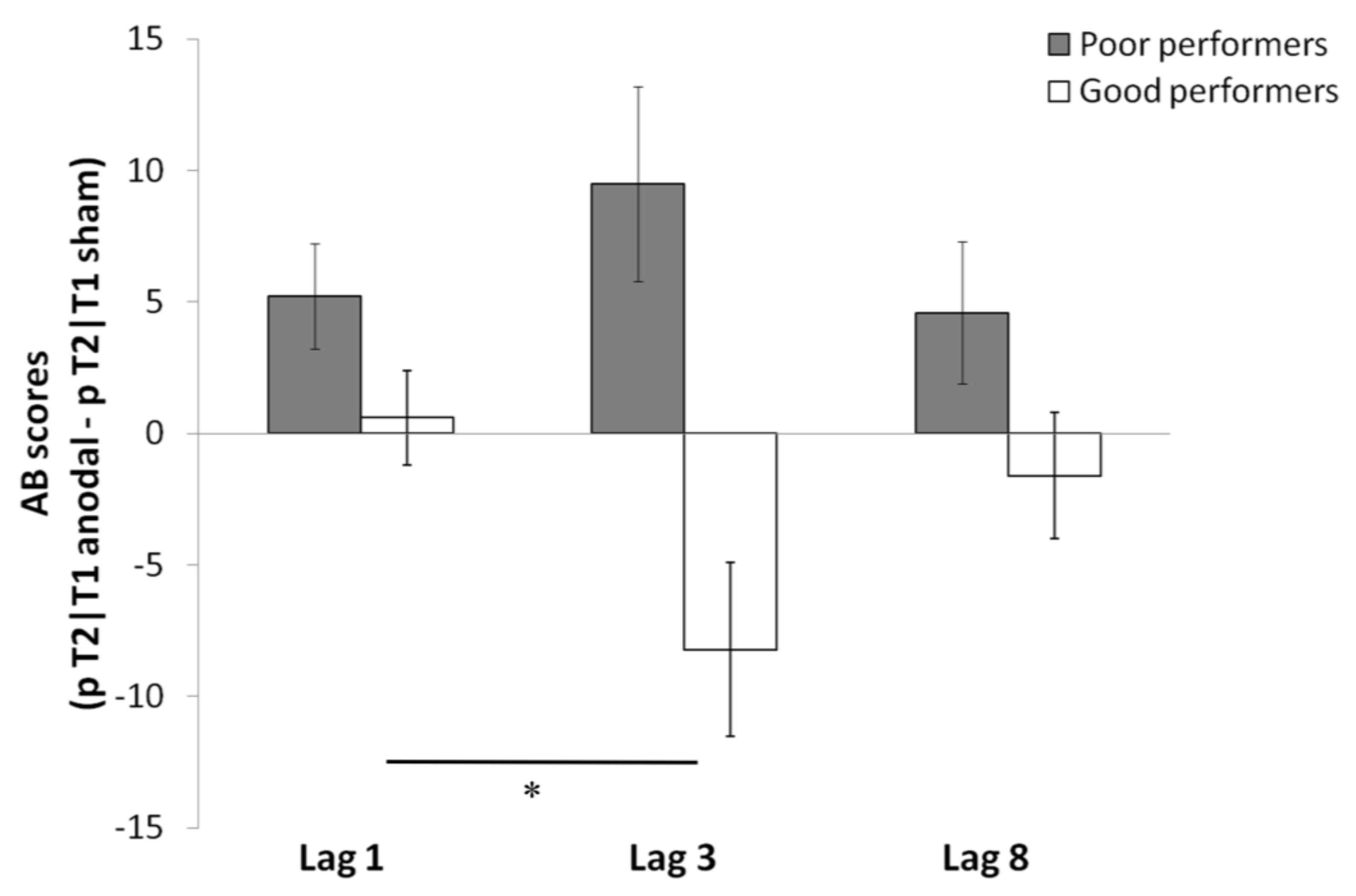
Individual Differences and tDCS Effects on AB and Sparing

As there is some evidence that the effect of anodal tDCS may depend on individual performance levels (Arciniega et al., 2018), to explore whether this was the case also for the AB, we split the sample based on the median of the overall performance during sham tDCS (Median = 70.84; see Figures S1 and S2 in Supplementary file. Therefore, participants were divided into “good performers” (N = 20) and “poor performers” (N = 20). We conducted a mixed factorial ANOVA on the AB scores (computed as the difference between anodal and sham) for each lag with stimulation site (2: P4, F4), performance group (2: poor, good Performers), and lag (3: lag 1, lag 3, lag 8) with the first two factors between subjects. A negative score indicates worse performance during anodal tDCS, and a positive score indicates better performance during anodal tDCS.
Results showed a significant main effect of performance group (good, poor), F (1, 36) = 0.493, p = 0.002 partial η2 = 0.244, which was due to better performance during active tDCS (i.e., positive scores) for poor performers (M = 6.42; SE = 2.06) than for good performers (M = −3.08, SE = 1.87). This main effect was qualified by a significant lag by performance group interaction, F (2, 72) = 4.72, p = 0.012 partial η2 = 0.116 (see Figure 3). Results of individual ANOVA conducted on each group showed that, for poor performers the main effect of lag was not significant, F (2, 34) = 1.07, p = 0.352, due to similar effects of anodal tDCS on performance across the three lags. In contrast, for good performers the main effect of lag was significant, F (2, 42) = 5.03, p = 0.011 partial η2 = 0.193. Pairwise comparisons showed no significant difference between lag 1 (M= 0.60, SE= 1.53) and lag 8 (M = −1.62, SE = 2.16), p =0.99, (i.e., lag 1 sparing). In contrast, there was a significant difference between lag 3 (M = −8.217, SE = 3.23) and lag 1, p = 0.019, indicating an AB. This pattern shows that anodal tDCS benefitted poor performers (i.e., all scores are positive and there is no substantial performance impairment at lag 3), but hindered good performers especially at lag 3 (i.e., enhancing the AB).
The lag F (2, 72) = 0.493, p = 0.613 and stimulation site (P4, F4), F (1, 36) = 0.049, p = 0.827 main effects and the lag by stimulation site interaction were not statistically significant, F (1, 36) = 0.493, p = 0.002 partial η2 = 0.244.
In summary, anodal tDCS improved performance for poor performers across all lags, whereas it increased the AB at lag 3 for good performers. Interestingly, these effects occurred for both P4 and F4 stimulation sites.

5. Discussion

The present study aimed at investigating the relative contribution of the rDLPFC and of the rPPC to the AB. We hypothesized that enhancing via tDCS the neural activity of the rDLPFC over that of the rPPC would potentiate top-down control of attention and improve target detection during the AB, that is, when T1 and T2 are separated by distractors, reducing the AB. The prediction was based on the argument that performance at the RSVP may rely on the activity of the rDLPFC involved in enhancing targets and inhibiting distractors and on evidence that the role of rPPC in bottom-up attention and visual short-term memory capacity. Participants performed the RSVP task while receiving anodal or sham tDCS. One group of participants received tDCS over the rDLPFC whereas the other group of participants received tDCS over the rPPC. We expected that anodal stimulation over the rDLPFC would reduce the AB compared to sham but also compared to anodal tDCS over the rPPC. This latter comparison would serve as a more stringent control of the role of the rDLPFC versus that of the rPPC in the AB.
Overall, findings show the typical pattern of temporal selective attention with performance sparing at lag 1—that is, performance for T2 conditional upon reporting T1 was unimpaired when the two targets occurred in immediate succession (i.e., lag 1 sparing). The lag 1 sparing was followed by an AB at lag 3—that is, performance for T2 was impaired—and recovered only at the later lag. Importantly, whereas this pattern was not affected by tDCS montage, when looking at the effects of anodal tDCS based on participants’ performance levels during sham, anodal tDCS over F4 and P4 improved performance across the three lags for bad performers but accentuated the AB for good performers.
The implications of the present findings are threefold. Firstly, potentiating the activity of the rDLPFC (F4) and of the rPPC (P4) via anodal tDCS had a similar effect on performance, suggesting the contribution of the right frontoparietal network in the AB rather than of a single brain area. This interpretation entails that neural excitation propagated along the right frontoparietal network, regardless of the site where the stimulation originated and it is consistent with evidence showing that anodal tDCS may synchronize several brain regions connected to the stimulation site that comprise a functional network [50,51,52]. The role of the rPPC is still not fully understood and some evidence points to the more ventral areas of rPPC being involved in top-down control of stimulus processing and targets identification [53,54,55,56]. Therefore, the rPPC together with the rDLPFC may be conceived as biasing the signal between competing stimulus representations. Because the mechanisms underlying the spread of sparing and the AB are still debated, past studies have typically manipulated target salience (exogenously or endogenously) to assess the contribution of resource depletion and active attentional enhancement/inhibition [57,58]. This is because for the resource depletion account, if exogenous attention for one target increases, performance on the other target suffers. In contrast, for the top-down attentional control account, attention can be allocated flexibly between the two targets (i.e., prioritizing T1 may not impair processing T2). That potentiating the activity of the rDLPFC and the rPPC via tDCS had similar effects on performance points to the contribution of both resource depletion and top-down attentional control to the AB.
Secondly, the findings of interactive effects of anodal tDCS and individuals’ performance when using low tDCS intensity are in line with evidence that the modulation of cognitive functions by tDCS depends on task difficulty and/or individual performance levels. For instance, improvements of visual working memory by anodal tDCS over the rPPC have been observed only when comparing low with high performers [59] and enhancement of visual short-term memory capacity by anodal tDCS over rPPC have emerged only when task difficulty is high [43]. Similarly, it has been shown that anodal tDCS over the rDLPFC benefits less experienced musicians but hinders the more experienced ones [60]. Furthermore, Jospe et al. [48] showed that tDCS over the motor cortex improved performance at a theory of mind task only for participants with low empathy, who typically perform less well at this task. Although the mechanisms underlying this relationship are still unclear, a recent work by Filmer et al. [61] has shown that the neurochemical excitability of the prefrontal cortex at baseline, as indexed by the relative concentrations of GABA and glutamate, is associated with the degree to which active tDCS impairs individuals’ performance. This would suggest that there is an optimal brain activation level for unimpaired cognitive performance and that this optimal level results from internal and external factors.
That in the present study tDCS effects depended on individuals’ performance levels whereas using a stronger current intensity (1.5 mA) in Sdoia et al. [26] they did not, points to a combined contribution of current intensity and individuals’ performance level to tDCS modulatory effects. This interpretation is in keep with Benwell et al. [62] who found that the effects of active tDCS over the rPPC depended on individual performance levels as well as on the stimulation intensity. Therefore, the present finding contributes to the growing body of evidence showing that, the relation between excitability of a brain area and its efficiency is not linear and it may follow a U-shape function, with tDCS effects partially depending on the baseline balance between excitation and inhibition of a cortical area [63]. Interestingly, this implies that for poor performers the beneficial effect of active tDCS might be offset by that of training.
In summary, the extra activation/excitability of the right frontoparietal network induced via tDCS befitted poor performers who did better at the RSVP task. By the same token, that good performers did much worse while receiving anodal tDCS, which especially enhanced the AB, suggests that their right-lateralized frontoparietal network was already optimally activated. Therefore, the present findings provide new insights on neural underpinnings of the AB by showing the contribution of both the rDLPFC and rPPC and by showing that low intensity, anodal tDCS over these brain areas affects the AB depending on the individuals’ performance levels.

Supplementary Materials

The following are available online at https://www.mdpi.com/article/10.3390/sym13071208/s1. Figure S1. Overall Performance on T2 (Total pT2) during sham tDCS. Figure S2. Overall Performance on T2 (Total pT2) during sham tDCS for the two groups (poor and good performers) based on median split.

Author Contributions

Conceptualization, A.P. and M.L.; methodology, A.P. and M.L.; software, F.D.L., B.M. and M.P. (Manuel Petrucci); validation, A.P., F.D.L., B.M. and M.P. (Manuel Petrucci); M.L. formal analysis, A.P. and M.L.; investigation, F.D.L., B.M. and M.P. (Manuel Petrucci). resources, A.P.; data curation, F.D.L., B.M. and M.P. (Manuel Petrucci); writing—original draft preparation, A.P. and M.L.; writing—review and editing, A.P., M.L. and M.P. (Mariella Pazzaglia); F.D.; supervision, A.P. and F.D.L.; project administration, A.P.; funding acquisition, A.P. All authors have read and agreed to the published version of the manuscript.

Funding

Italian Ministry of University and Research: RG11615502CCDE74, Italian Ministry of University and Research: RM11715C7DF6225C.

Institutional Review Board Statement

The study had received approval by the Ethics Committee of the Department of Psychology, Sapienza University of Rome (Ref. 21 May 2012).

Informed Consent Statement

Inform consent was obtained from all subjects.

Data Availability Statement

Not applicable.

Acknowledgments

A.P. is funded by Grant # RG11615502CCDE74 and Grant # RM11715C7DF6225C from the Italian Ministry of University and Research (MIUR).

Conflicts of Interest

The authors declare no conflict of interest.

Ethic Statement

The experimental procedure was in accordance with the ethical standards of the Ethics Committee of the Department of Psychology, Sapienza University of Rome (Ref. 21 May 2012) and with the 1964 Helsinki declaration and its later amendments or comparable ethical standards.

References

  1. Broadbent, D.E.; Broadbent, M.H.P. From detection to identification: Response to multiple targets in rapid serial visual presentation. Percept. Psychophys. 1987, 42, 105–113. [Google Scholar] [CrossRef] [PubMed] [Green Version]
  2. Raymond, J.E.; Shapiro, K.L.; Arnell, K.M. Temporary suppression of visual processing in an RSVP task: An attentional blink? J. Exp. Psychol. Hum. Percept. Perform. 1992, 18, 849–860. [Google Scholar] [CrossRef] [PubMed]
  3. Chun, M.M.; Potter, M.C. A two-stage model for multiple target detection in rapid serial visual presentation. J. Exp. Psychol. Hum. Percept. Perform. 1995, 21, 109–127. [Google Scholar] [CrossRef] [PubMed]
  4. Shapiro, K.; Raymond, J.; Arnell, K. The attentional blink. Trends Cogn. Sci. 1997, 1, 291–296. [Google Scholar] [CrossRef]
  5. Lasaponara, S.; Dragone, A.; Lecce, F.; Di Russo, F.; Doricchi, F. The “serendipitous brain”: Low expectancy and timing uncertainty of conscious events improve awareness of unconscious ones (evidence from the attentional blink). Cortex 2015, 71, 15–33. [Google Scholar] [CrossRef]
  6. Jolicœur, P.; Dell’Acqua, R. The Demonstration of Short-Term Consolidation. Cogn. Psychol. 1998, 36, 138–202. [Google Scholar] [CrossRef] [Green Version]
  7. Di Lollo, V.; Kawahara, J.; Shahab Ghorashi, S.M.; Enns, J.T. The attentional blink: Resource depletion or temporary loss of control? Psychol. Res. 2005, 69, 191–200. [Google Scholar] [CrossRef]
  8. Olivers, C.N.L.; Meeter, M. A boost and bounce theory of temporal attention. Psychol. Rev. 2008, 115, 836–863. [Google Scholar] [CrossRef]
  9. Wyble, B.; Bowman, H.; Nieuwenstein, M. The attentional blink provides episodic distinctiveness: Sparing at a cost. J. Exp. Psychol. Hum. Percept. Perform. 2009, 35, 787–807. [Google Scholar] [CrossRef] [Green Version]
  10. Martens, S.; Wyble, B. The attentional blink: Past, present, and future of a blind spot in perceptual awareness. Neurosci. Biobehav. Rev. 2010, 34, 947–957. [Google Scholar] [CrossRef] [Green Version]
  11. Fragopanagos, N.; Kockelkoren, S.; Taylor, J.G. A neurodynamic model of the attentional blink. Cogn. Brain Res. 2005, 24, 568–586. [Google Scholar] [CrossRef]
  12. Taatgen, N.A.; Juvina, I.; Schipper, M.; Borst, J.P.; Martens, S. Too much control can hurt: A threaded cognition model of the attentional blink. Cogn. Psychol. 2009, 59, 1–29. [Google Scholar] [CrossRef]
  13. Kihara, K.; Ikeda, T.; Matsuyoshi, D.; Hirose, N.; Mima, T.; Fukuyama, H.; Osaka, N. Differential Contributions of the Intraparietal Sulcus and the Inferior Parietal Lobe to Attentional Blink: Evidence from Transcranial Magnetic Stimulation. J. Cogn. Neurosci. 2011, 23, 247–256. [Google Scholar] [CrossRef]
  14. Mayr, U.; Diedrichsen, J.; Ivry, R.; Keele, S.W. Dissociating Task-set Selection from Task-set Inhibition in the Prefrontal Cortex. J. Cogn. Neurosci. 2006, 18, 14–21. [Google Scholar] [CrossRef]
  15. Banich, M.T. Executive Function. Curr. Dir. Psychol. Sci. 2009, 18, 89–94. [Google Scholar] [CrossRef]
  16. Feinstein, J.S.; Stein, M.B.; Castillo, G.N.; Paulus, M.P. From sensory processes to conscious perception. Conscious. Cogn. 2004, 13, 323–335. [Google Scholar] [CrossRef]
  17. Marois, R.; Yi, D.-J.; Chun, M.M. The Neural Fate of Consciously Perceived and Missed Events in the Attentional Blink. Neuron 2004, 41, 465–472. [Google Scholar] [CrossRef] [Green Version]
  18. Giesbrecht, B.; Kingstone, A. Right hemisphere involvement in the attentional blink: Evidence from a split-brain patient. Brain Cogn. 2004, 55, 303–306. [Google Scholar] [CrossRef]
  19. Dell’Acqua, R.; Doro, M.; Dux, P.; Losier, T.; Jolicœur, P. Enhanced frontal activation underlies sparing from the attentional blink: Evidence from human electrophysiology. Psychophysiol. 2016, 53, 623–633. [Google Scholar] [CrossRef]
  20. Gross, J.; Schmitz, F.; Schnitzler, I.; Kessler, K.; Shapiro, K.; Hommel, B. Modulation of long-range neural synchrony reflects temporal limitations of visual attention in humans. Proc. Natl. Acad. Sci. USA 2004, 101, 13050–13055. [Google Scholar] [CrossRef] [Green Version]
  21. Corbetta, M.; Shulman, G.L. Control of goal-directed and stimulus-driven attention in the brain. Nat. Rev. Neurosci. 2002, 3, 201–215. [Google Scholar] [CrossRef] [PubMed]
  22. Nitsche, M.A.; Paulus, W. Excitability changes induced in the human motor cortex by weak transcranial direct current stimulation. J. Physiol. 2000, 527 Pt 3, 633–639. [Google Scholar] [CrossRef]
  23. Jacobson, L.; Koslowsky, M.; Lavidor, M. tDCS polarity effects in motor and cognitive domains: A meta-analytical review. Exp. Brain Res. 2012, 216, 1–10. [Google Scholar] [CrossRef] [PubMed]
  24. London, R.E.; Slagter, H. Effects of Transcranial Direct Current Stimulation over Left Dorsolateral pFC on the Attentional Blink Depend on Individual Baseline Performance. J. Cogn. Neurosci. 2015, 27, 2382–2393. [Google Scholar] [CrossRef] [PubMed] [Green Version]
  25. London, R.E.; Slagter, H.A. No Effect of Transcranial Direct Current Stimulation over Left Dorsolateral Prefrontal Cortex on Temporal Attention. J. Cogn. Neurosci. 2021, 33, 756–768. [Google Scholar] [CrossRef] [PubMed]
  26. Sdoia, S.; Conversi, D.; Pecchinenda, A.; Ferlazzo, F. Access to consciousness of briefly presented visual events is modulated by transcranial direct current stimulation of left dorsolateral prefrontal cortex. Sci. Rep. 2019, 9, 10950. [Google Scholar] [CrossRef] [PubMed]
  27. Fregni, F.; Boggio, P.; Nitsche, M.; Bermpohl, F.; Antal, A.; Feredoes, E.; Marcolin, M.A.; Rigonatti, S.P.; Silva, M.T.; Paulus, W.; et al. Anodal transcranial direct current stimulation of prefrontal cortex enhances working memory. Exp. Brain Res. 2005, 166, 23–30. [Google Scholar] [CrossRef]
  28. Gladwin, T.E.; Uyl, T.D.; Fregni, F.F.; Wiers, R.W. Enhancement of selective attention by tDCS: Interaction with interference in a Sternberg task. Neurosci. Lett. 2012, 512, 33–37. [Google Scholar] [CrossRef]
  29. MacDonald, A. Dissociating the Role of the Dorsolateral Prefrontal and Anterior Cingulate Cortex in Cognitive Control. Science 2000, 288, 1835–1838. [Google Scholar] [CrossRef] [Green Version]
  30. Pecchinenda, A.; Ferlazzo, F.; Lavidor, M. Modulation of selective attention by polarity-specific tDCS effects. Neuropsychologia 2015, 68, 1–7. [Google Scholar] [CrossRef]
  31. Hommel, B.; Kessler, K.; Schmitz, F.; Gross, J.; Akyürek, E.; Shapiro, K.; Schnitzler, A. How the brain blinks: Towards a neurocognitive model of the attentional blink. Psychol. Res. 2006, 70, 425–435. [Google Scholar] [CrossRef] [Green Version]
  32. Egner, T.; Monti, J.M.P.; Trittschuh, E.H.; Wieneke, C.A.; Hirsch, J.; Mesulam, M.-M. Neural Integration of Top-Down Spatial and Feature-Based Information in Visual Search. J. Neurosci. 2008, 28, 6141–6151. [Google Scholar] [CrossRef] [Green Version]
  33. Bressler, S.L.; Menon, V. Large-scale brain networks in cognition: Emerging methods and principles. Trends Cogn. Sci. 2010, 14, 277–290. [Google Scholar] [CrossRef]
  34. Rottschy, C.; Langner, R.; Dogan, I.; Reetz, K.; Laird, A.; Schulz, J.B.; Fox, P.; Eickhoff, S. Modelling neural correlates of working memory: A coordinate-based meta-analysis. NeuroImage 2012, 60, 830–846. [Google Scholar] [CrossRef] [Green Version]
  35. Rottschy, C.; Caspers, S.; Roski, C.; Reetz, K.; Dogan, I.; Schulz, J.B.; Zilles, K.; Laird, A.R.; Fox, P.; Eickhoff, S.B. Differentiated parietal connectivity of frontal regions for “what” and “where” memory. Brain Struct. Funct. 2012, 218, 1551–1567. [Google Scholar] [CrossRef] [Green Version]
  36. Dehaene, S.; Changeux, J.-P. Experimental and Theoretical Approaches to Conscious Processing. Neuron 2011, 70, 200–227. [Google Scholar] [CrossRef] [Green Version]
  37. Driver, J. Perceptual awareness and its loss in unilateral neglect and extinction. Cognition 2001, 79, 39–88. [Google Scholar] [CrossRef]
  38. Rees, G. Neural correlates of consciousness. Ann. N. Y. Acad. Sci. 2013, 1296, 4–10. [Google Scholar] [CrossRef] [Green Version]
  39. Corbetta, M.; Patel, G.; Shulman, G.L. The Reorienting System of the Human Brain: From Environment to Theory of Mind. Neuron 2008, 58, 306–324. [Google Scholar] [CrossRef] [Green Version]
  40. Bettencourt, K.C.; Xu, Y. Decoding the content of visual short-term memory under distraction in occipital and parietal areas. Nat. Neurosci. 2016, 19, 150–157. [Google Scholar] [CrossRef]
  41. Li, S.; Cai, Y.; Liu, J.; Li, D.; Feng, Z.; Chen, C.; Xue, G. Dissociated roles of the parietal and frontal cortices in the scope and control of attention during visual working memory. NeuroImage 2017, 149, 210–219. [Google Scholar] [CrossRef] [PubMed]
  42. Tseng, P.; Hsu, T.-Y.; Chang, C.-F.; Tzeng, O.J.; Hung, D.L.; Muggleton, N.G.; Walsh, V.; Liang, W.-K.; Cheng, S.-K.; Juan, C.-H. Unleashing Potential: Transcranial Direct Current Stimulation over the Right Posterior Parietal Cortex Improves Change Detection in Low-Performing Individuals. J. Neurosci. 2012, 32, 10554–10561. [Google Scholar] [CrossRef] [PubMed] [Green Version]
  43. Wang, S.; Itthipuripat, S.; Ku, Y. Electrical Stimulation Over Human Posterior Parietal Cortex Selectively Enhances the Capacity of Visual Short-Term Memory. J. Neurosci. 2018, 39, 528–536. [Google Scholar] [CrossRef] [PubMed] [Green Version]
  44. Arciniega, H.; Gözenman, F.; Jones, K.T.; Stephens, J.A.; Berryhill, M.E. Frontoparietal tDCS Benefits Visual Working Memory in Older Adults with Low Working Memory Capacity. Front. Aging Neurosci. 2018, 10, 57. [Google Scholar] [CrossRef] [Green Version]
  45. Lo, O.; Van Donkelaar, P.; Chou, L. Effects of transcranial direct current stimulation over right posterior parietal cortex on attention function in healthy young adults. Eur. J. Neurosci. 2019, 49, 1623–1631. [Google Scholar] [CrossRef]
  46. Kihara, K.; Kondo, H.M.; Kawahara, J.I. Differential Contributions of GABA Concentration in Frontal and Parietal Regions to Individual Differences in Attentional Blink. J. Neurosci. 2016, 36, 8895–8901. [Google Scholar] [CrossRef] [Green Version]
  47. Faul, F.; Erdfelder, E.; Buchner, E.; Lang, A.-J. Statistical power analyses using G*Power 3.1: Tests for correlation and regression analyses. Behav. Res. Methods 2009, 41, 1149–1160. [Google Scholar] [CrossRef] [Green Version]
  48. Jospe, K.; Flöel, A.; Lavidor, M. The interaction between embodiment and empathy in facial expression recognition. Soc. Cogn. Affect. Neurosci. 2018, 13, 203–215. [Google Scholar] [CrossRef] [Green Version]
  49. Parkin, B.L.; Bhandari, M.; Glen, J.C.; Walsh, V. The physiological effects of transcranial electrical stimulation do not apply to parameters commonly used in studies of cognitive neuromodulation. Neuropsychologia 2019, 128, 332–339. [Google Scholar] [CrossRef] [Green Version]
  50. Polanía, R.; Nitsche, M.A.; Paulus, W. Modulating functional connectivity patterns and topological functional organization of the human brain with transcranial direct current stimulation. Hum. Brain Mapp. 2011, 32, 1236–1249. [Google Scholar] [CrossRef]
  51. Notturno, F.; Marzetti, L.; Pizzella, V.; Uncini, A.; Zappasodi, F. Local and remote effects of transcranial direct current stimulation on the electrical activity of the motor cortical network. Hum. Brain Mapp. 2014, 35, 2220–2232. [Google Scholar] [CrossRef]
  52. Kunze, T.; Hunold, A.; Haueisen, J.; Jirsa, V.; Spiegler, A. Transcranial direct current stimulation changes resting state functional connectivity: A large-scale brain network modeling study. Neuroimage 2016, 140, 174–187. [Google Scholar] [CrossRef]
  53. Behrmann, M.; Geng, J.J.; Shomstein, S. Parietal cortex and attention. Curr. Opin. Neurobiol. 2004, 14, 212–217. [Google Scholar] [CrossRef]
  54. Friedman-Hill, S.R.; Robertson, L.C.; Desimone, R.; Ungerleider, L.G. Posterior parietal cortex and the filtering of distractors. Proc. Natl. Acad. Sci. USA 2003, 100, 4263–4268. [Google Scholar] [CrossRef] [Green Version]
  55. Menon, V.; Ford, J.M.; Lim, K.; Glover, G.H.; Pfefferbaum, A. Combined event-related fMRI and EEG evidence for temporal—Parietal cortex activation during target detection. NeuroReport 1997, 8, 3029–3037. [Google Scholar] [CrossRef] [Green Version]
  56. Wojciulik, E.; Kanwisher, N. The Generality of Parietal Involvement in Visual Attention. Neuron 1999, 23, 747–764. [Google Scholar] [CrossRef] [Green Version]
  57. Petrucci, M.; Pecchinenda, A. Sparing and impairing: Emotion modulation of the attentional blink and the spread of sparing in a 3-target RSVP task. Atten. Percept. Psychophys. 2017, 80, 439–452. [Google Scholar] [CrossRef]
  58. Pecchinenda, A.; Monachesi, B.; Laeng, B. Fearful expressions of rapidly presented hybrid-faces modulate the lag 1 sparing in the attentional blink. Acta Psychol. 2020, 209, 103124. [Google Scholar] [CrossRef]
  59. Hsu, T.Y.; Juan, C.H.; Tseng, P. Individual Differences and State-Dependent Responses in Transcranial Direct Current Stimulation. Front. Hum. Neurosci. 2016, 10, 643. [Google Scholar] [CrossRef]
  60. Rosen, D.S.; Erickson, B.; Kim, Y.E.; Mirman, D.; Hamilton, R.H.; Kounios, J. Anodal tDCS to Right Dorsolateral Prefrontal Cortex Facilitates Performance for Novice Jazz Improvisers but Hinders Experts. Front. Hum. Neurosci. 2016, 10, 579. [Google Scholar] [CrossRef] [Green Version]
  61. Filmer, H.L.; Ehrhardt, S.E.; Bollmann, S.; Mattingley, J.B.; Dux, P. Accounting for individual differences in the response to tDCS with baseline levels of neurochemical excitability. Cortex 2019, 115, 324–334. [Google Scholar] [CrossRef] [PubMed]
  62. Benwell, C.S.; Learmonth, G.; Miniussi, C.; Harvey, M.; Thut, G. Non-linear effects of transcranial direct current stimulation as a function of individual baseline performance: Evidence from biparietal tDCS influence on lateralized attention bias. Cortex 2015, 69, 152–165. [Google Scholar] [CrossRef] [PubMed] [Green Version]
  63. Krause, B.; Kadosh, R.C. Not all brains are created equal: The relevance of individual differences in responsiveness to transcranial electrical stimulation. Front. Syst. Neurosci. 2014, 8, 25. [Google Scholar] [CrossRef] [PubMed]
Figure 1. Experimental timeline: In the example, T1 (digit 5) appears in position 6 of the stream (i.e., after five distractors), and T2 (digit 2) is presented at lag 3, relative to T1 (i.e., after two distractors).
Symmetry 13 01208 g001
Figure 2. Mean percentage of correct T2 identifications conditional upon correct identification of T1 (pT2|T1) as a function of lag (1, 3, 8) for the two stimulation groups (F4 and P4). Dashed lines denote sham tDCS, solid lines denote anodal tDCS. Error bars represent standard errors of the mean.
Symmetry 13 01208 g002
Figure 3. AB scores (computed as the difference between anodal and sham on pT2|T1) for poor and good performers as a function of lags. Positive scores indicate better performance during anodal tDCS. Error bars represent standard errors of the mean. * denotes p < 0.05.
Symmetry 13 01208 g003
Table 1. Means (SEs) for pT1 as function of session (anodal, sham tDCS), and stimulation site (F4, P4).
Group: F4Group: P4Overall
Sham tDCS84.76 (2.09)85.09 (2.09)84.93 (1.48)
Active tDCS85.60 (1.41)87.56 (1.41)86.58 (1.00)
Overall85.18 (1.63)86.33 (1.63)
Publisher’s Note: MDPI stays neutral with regard to jurisdictional claims in published maps and institutional affiliations.

