Article

Individuals with Unilateral Mild-to-Moderate Hip Osteoarthritis Exhibit Lower Limb Kinematic Asymmetry during Walking But Not Sit-to-Stand

1
Griffith Centre of Biomedical and Rehabilitation Engineering (GCORE), Menzies Health Institute Queensland, Griffith University, Gold Coast, QLD 4222, Australia
2
School of Allied Health Sciences, Griffith University, Gold Coast, QLD 4222, Australia
3
School of Physiotherapy, Faculty of Health Sciences, Australian Catholic University, Brisbane, QLD 4014, Australia
*
Author to whom correspondence should be addressed.
Symmetry 2021, 13(5), 768; https://doi.org/10.3390/sym13050768
Submission received: 19 March 2021 / Revised: 20 April 2021 / Accepted: 22 April 2021 / Published: 28 April 2021
(This article belongs to the Special Issue Symmetry and Biomechanics)

Abstract

:
Asymmetry during gait is associated with the evolution of secondary osteoarthritis. Kinematic asymmetry has been reported in advanced stages of hip osteoarthritis but has not been evaluated in earlier stages of the disease or has it been directly compared with unilateral and bilateral hip osteoarthritis. Our objective was to evaluate within-group symmetry and compare between-group asymmetry for three-dimensional pelvis, hip, knee, and ankle kinematics during walking and sit-to-stand in individuals with unilateral mild-to-moderate hip OA, bilateral mild-to-moderate hip osteoarthritis, and healthy controls. Twelve individuals with unilateral mild-to-moderate hip OA, nine individuals with bilateral mild-to-moderate symptomatic and radiographic hip OA, and 21 age-comparable healthy controls underwent three-dimensional motion analysis during walking and sit-to-stand. Pelvis and lower limb joint angles were calculated using inverse kinematics and between-limb symmetry was assessed for each group. Any resulting asymmetries (most affected minus contralateral limb) were compared between groups. Participants with unilateral hip osteoarthritis exhibited significantly less hip extension (7.90°), knee flexion (4.72°), and anterior pelvic tilt (3.38°) on their affected limb compared with the contralateral limb during the stance phase of walking. Those with unilateral hip osteoarthritis were significantly more asymmetrical than controls for sagittal plane hip and pelvis angles. No significant asymmetries were detected within- or between-groups for sit-to-stand. Individuals with unilateral hip osteoarthritis exhibited lower limb asymmetries consistent with those reported in advanced stages of disease during walking, but not sit-to-stand. Consideration of the possible negative effects of gait asymmetry on the health of the affected and other compensating joints appears warranted in the management of hip OA.

1. Introduction

Hip osteoarthritis (OA) is a prevalent degenerative joint disease causing pain, disability, and reduced quality of life [1]. The aetiology of hip OA is complex and varied, and can affect one or both limbs, depending on the determinants of the local joint mechanobiology [2,3]. Lower limb biomechanics are altered during activities of daily living in people with advanced hip OA compared with healthy controls [4] and remain altered following total hip replacement (THR) [5,6]. Asymmetry occurs during the performance of functional tasks such as walking and sit-to-stand when left and right lower limbs exhibit different biomechanical patterns. Asymmetrical lower limb biomechanics have been routinely reported in a wide range of gait conditions including stroke, cerebral palsy, ACL deficiency, and lower limb OA [7,8,9,10]. In the case of lower limb OA, altered biomechanical patterns in the lower extremity appear to reflect a motor control strategy intended to reduce symptoms and compensate for neuromuscular deficits including muscle weakness on the affected limb, and have the potential to contribute to ongoing disease progression by altering biomechanical patterns in the affected joint. Furthermore, compensatory changes in motion and loading patterns in adjacent joints or joints on the contralateral limb can also arise [11], which may be similarly incompatible with the joint tissue state and lead to degeneration of joint tissues [12]. While there is evidence for kinematic asymmetry during walking and sit-to-stand in individuals with advanced unilateral hip OA [11,13,14] and following THR [6,14,15,16,17,18], the presence of kinematic asymmetry in mild-to-moderate hip OA, where there is potential to prevent or slow disease progression, has not been established.
Biomechanical studies of asymmetry in people with hip OA to date have tended to focus on either walking or sit-to-stand. A systematic review of spatiotemporal characteristics during gait in people with hip OA [19] identified that individuals with unilateral and bilateral advanced hip OA take shorter steps [20,21,22] and spend more time in stance [20,23,24] with their most affected limb compared with the contralateral limb. People with unilateral advanced hip OA have also been reported to walk with less peak sagittal hip flexion and extension [11,13,14] and lower peak sagittal hip moments [25] on their affected limb compared with the contralateral limb. Asymmetries at the knee have also been reported in advanced unilateral hip OA, with greater knee extension and adduction moments on the contralateral limb compared with the affected limb during walking [11]. No studies to date have assessed kinematic symmetry during walking in bilateral mild-to-moderate hip OA or compared kinematic asymmetry between unilateral hip OA and bilateral hip OA groups. To the best of our knowledge, only one study investigated lower limb symmetry during sit-to-stand in mild-to-moderate hip OA and reported no lower limb kinematic asymmetry in a mixed cohort consisting of individuals with unilateral and bilateral hip OA [26]. However, following unilateral THR, patients performed sit-to-stand using less frontal plane range of motion on their affected hip compared with the contralateral hip [27]. Likewise, reduced extension moments on the affected hip compared with the contralateral hip were reported both before and after unilateral THR [17,27,28].
Taken together, the abovementioned studies suggest that lower limb kinematic and kinetic asymmetry is a feature of advanced hip OA during both walking and sit-to-stand and is not corrected by surgery. It is unclear whether lower limb asymmetry is also a feature of those with less severe disease, and whether any asymmetry is specific to those with unilateral hip OA or is also apparent in people with bilateral hip OA. Our group has previously reported that people with mild-to-moderate hip OA exhibit muscle weakness [29,30], less hip joint loading over a reduced range of motion in walking [31], and reduced hip motion over sit-to-stand [32] in the affected limb in people with mild-to-moderate hip OA compared with healthy controls. However, the extent of any gait asymmetry in unilateral and bilateral hip OA remains unknown. The aims of this study were therefore to: (1) evaluate kinematic symmetry at the pelvis, hip, knee, and ankle during walking and sit-to-stand in individuals within unilateral mild-to-moderate hip OA, bilateral mild-to-moderate hip OA, and healthy controls; and (2) compare measures of kinematic asymmetry between groups. We hypothesised that individuals with unilateral mild-to-moderate hip OA would exhibit kinematic asymmetry at the hip, as reported in advanced unilateral hip OA [11,13,14], and the unilateral hip OA group would exhibit greater asymmetry than both the bilateral hip OA and control groups for both tasks.

2. Materials and Methods

2.1. Participants

This study was a sub-group analysis, comprising a convenience sample of 42 participants (13 men, 29 women) enrolled in a broader study [31]. Individuals between 45 and 80 years of age were recruited from hospital orthopaedic waiting lists and word-of-mouth. Pelvis and hip joints were screened bilaterally with weight-bearing radiographs, supine magnetic resonance imaging (MRI), and the modified Harris Hip Score (HHS) [27]. Superomedial, apical, and superolateral joint space width (JSW) [33], and Kellgren–Lawrence (KL) grades [34] were assessed by one experienced radiologist. Included in this study were individuals graded mild-to-moderate for hip OA; HHS ≤ 95 [35] and a KL grade of 2 or 3 (2 = possible narrowing of JSW with osteophyte formation, 3 = narrowing of JSW, moderate osteophyte formation, sclerosis, and possible deformity of bony ends [36]) for their affected hip(s); and JSW ≤ 3 mm [33]. The bilateral hip OA group met the inclusion criteria for hip OA classification for both hip joints, whereas the unilateral hip OA group met the inclusion criteria for one hip joint and KL scores of ≤1 on their contralateral hip joint. The control group had minimal hip pain and dysfunction (HHS > 95) and KL grades ≤1 and JSW > 3 mm for both hip joints. Exclusion criteria included any neurological or lower limb musculoskeletal conditions, other than hip OA, that negatively impact their ability to walk. Ethics approval for the study was obtained from both the Queensland Health Human Research Ethics Committee (HREC/13/QHC/1614 (GU Ref No: PES/46/13/HREC), 2 April 2013) and the Griffith University Human Research Ethics Committee (HREC 2013/713, 25 September 2013). All participants provided written informed consent prior to participation. The index and non-index limbs for each group were defined as follows: OA-affected limb for unilateral hip OA group, most OA-affected hip for bilateral hip OA group, and a randomly assigned limb for healthy controls.

2.2. Experimental Protocol

Participants performed five trials of barefoot walking at a self-selected speed over a 10 m walkway [31]. Participants subsequently performed a standardised sit-to-stand task from a fixed height chair (45 cm), with 30 cm distance between the feet and arms crossed over their chest. The sit-to-stand was performed at a self-selected pace [32]. All individuals were able to perform walking and sit-to-stand tasks without assistance.

2.3. Data Collection and Analysis Procedures

Three-dimensional positional data from 60 retro-reflective markers (15 mm diameter) [37] were captured with a 12-camera VICON system (Oxford Metrics, Oxford, U.K.) at 200 Hz. A full-body marker model was achieved by attaching marker clusters to the thighs and shanks, while individual markers were placed on bony landmarks of the lower limbs (epicondyles, malleoli, and feet). Upper body markers were placed on the vertebrae, sternum, and clavicular notch as described previously [31,32].
MOtoNMS [38] was used to format motion capture data for musculoskeletal modelling. Three-dimensional marker positions were filtered using a low-pass, second-order, zero-lag, Butterworth filter with a 10 Hz cut-off frequency. Generic model bodies were linearly scaled [39] in open-source modelling software, OpenSim (version 3.3), using marker positions from an upright static trial. Hip joint centres were located from MRI as previously described [32]. Inverse kinematic analysis was used to determine sagittal and frontal plane angles for the pelvis and hip joints, and sagittal plane angles for the knee and ankle joints. The stance phase of the walking task was analysed from foot strike to foot off for the index and non-index limb. The stance phase was time normalized to 101 points. Calculated marker position errors during scaling and inverse kinematics were within recommended tolerances, 0.02 m and 0.04 m, respectively [39]. Data analyses were performed using MATLAB (release 2019B, The Mathworks Inc., Natick, MA, USA).

2.4. Statistical Analysis

Statistical analysis was completed using MATLAB (release 2019B, The Mathworks Inc., Natick, MA, USA). The significance level was set at p < 0.05. Data were assessed for normality using the Shapiro–Wilk test and homogeneity of variance was assessed using Levene’s test. Demographic, clinical, and spatiotemporal values were compared between groups using a one-way analysis of variance (ANOVA) or Kruskal–Wallis, with Bonferroni or Mann–Whitney U post hoc comparisons. Asymmetry was defined as within-group differences in continuous sagittal and frontal plane angles for the pelvis, hip, knee, and ankle and was evaluated using paired sample t-tests with statistical parametric or non-parametric mapping methods [40]. Between-group differences in asymmetry (calculated as the index minus non-index limb) were assessed using an ANOVA with statistical parametric or non-parametric mapping methods [40]. T-values that exceeded the t* threshold were interpreted to indicate within-group statistically significant differences.

3. Results

3.1. Participant Characteristics

The unilateral hip OA and bilateral hip OA groups had significantly higher body mass and body mass index (BMI) than the control group (Table 1). The unilateral hip OA and bilateral hip OA groups had significantly smaller JSW on the index limb compared with controls; the bilateral hip OA group also had significantly smaller JSW on the non-index limb compared with controls. The JSW difference (non-index minus index) of the unilateral hip OA group was significantly greater than both the bilateral hip OA and control groups. The overall mean HHS and subscale scores for pain and function were 71.96 (SD 13.02), 26.67 (SD 7.70), and 38.75 (SD 2.24), respectively, for the unilateral hip OA group, and 65.51 (SD 12.00), 21.11 (SD 6.01), and 38.44 (SD 5.60), respectively, for the bilateral hip OA group. The bilateral hip OA group had significantly lower pain scores than the unilateral hip OA and control groups (p < 0.05), though there were no other between-group differences. All individuals in the control group had an overall HHS greater than or equal to 99.

3.2. Spatiotemporal Asymmetry during Walking

There were no significant spatiotemporal asymmetries in walking velocity, step length, or step duration within the unilateral hip OA, bilateral hip OA, or control groups (Table 2). A significant between-group difference was detected in stance phase duration asymmetry between the bilateral hip OA versus both unilateral hip OA groups, and control groups, respectively (p < 0.05). No other significant between-group differences in spatiotemporal symmetry were detected.

3.3. Within-Group Kinematic Symmetry during Walking

Compared with their non-index limb, the unilateral hip OA group exhibited less hip extension with their index limb from 45–100% of stance (Figure 1A, average mean difference = 7.90° (SD 3.29)), less knee extension from 50–60% of stance (Figure 1C, average mean difference = 4.72° (SD 0.97)), and less anterior pelvic tilt from 5–20% of stance (Figure 1E, average mean difference = 3.38° (SD 0.67)). No significant asymmetries were detected within the bilateral hip OA or control group.

3.4. Between-Group Kinematic Asymmetry during Walking

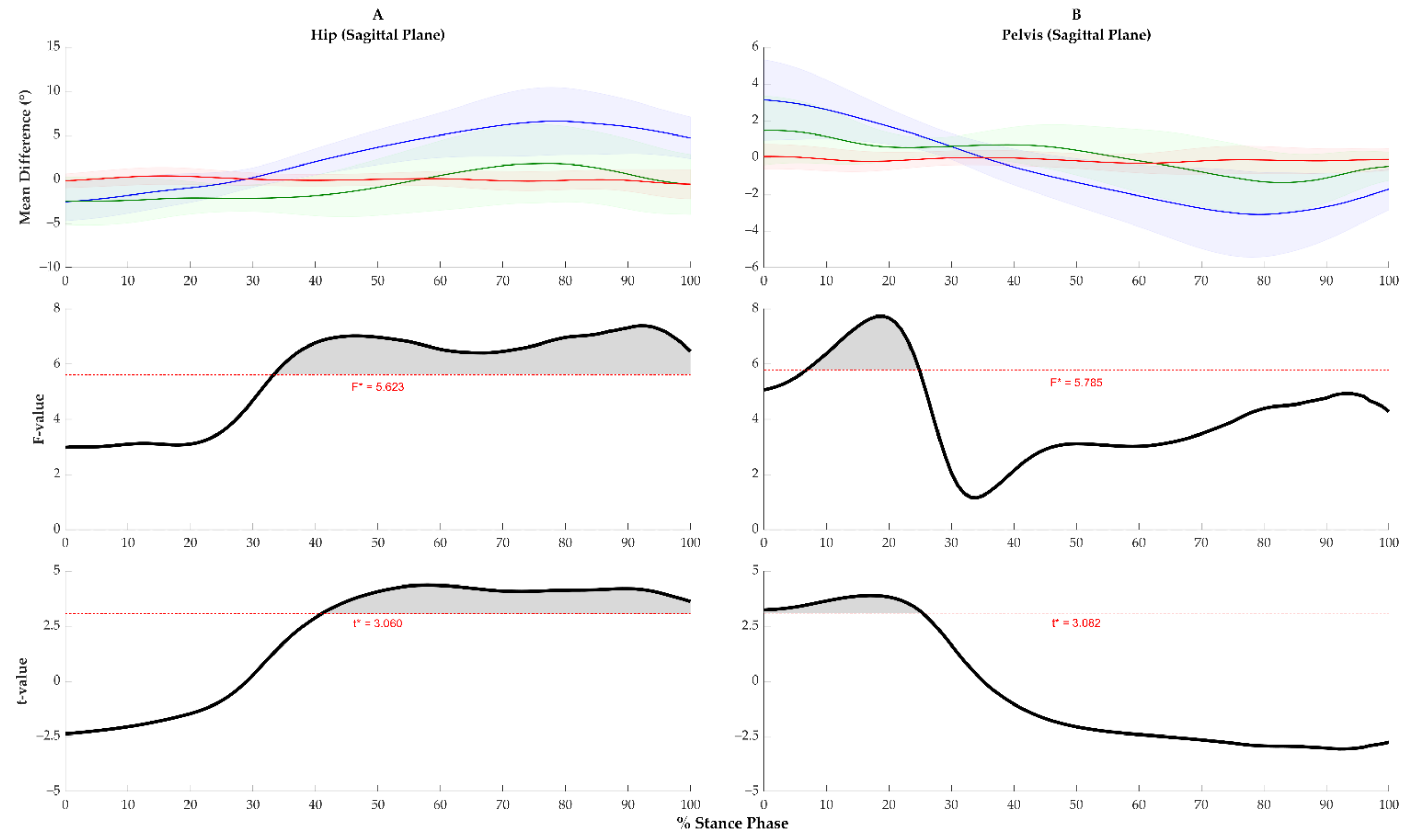
A significant main effect of the unilateral hip OA group was the detection of sagittal plane hip extension and anterior pelvic tilt kinematic asymmetries. Post hoc analyses revealed that, compared with the control group, the unilateral hip OA group exhibited greater hip flexion asymmetry for 50–100% of stance (Figure 2A, average mean difference = 4.8° (SD 2.67)) and greater anterior pelvic tilt asymmetry for 0–25% of stance (Figure 2B, average mean difference = 1.8° (SD 1.46)).

3.5. Kinematic Asymmetry during Sit-to-Stand

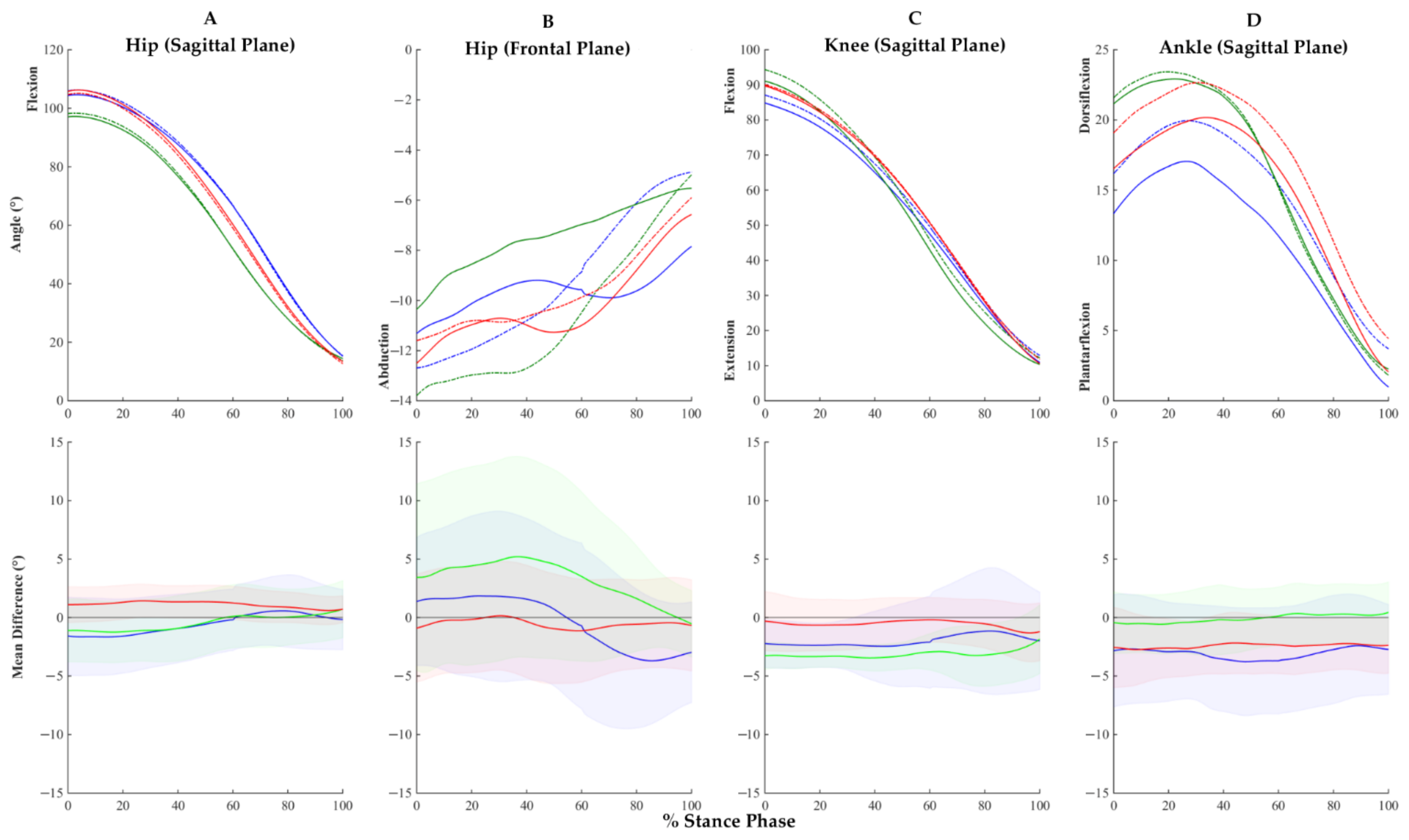
No significant within- or between-group differences were found for measures of kinematic asymmetry during sit-to-stand (Figure 3).

4. Discussion

This study investigated pelvis, hip, knee, and ankle kinematic asymmetry during walking and sit-to-stand in individuals with symptomatic and radiographic unilateral mild-to-moderate hip OA, bilateral mild-to-moderate hip OA, and healthy controls. The main finding was the within-group lower limb kinematic asymmetries were plane-, joint-, group-, and task-dependent. The observed asymmetries were restricted to the sagittal plane of the pelvis, hip, and knee in individuals with unilateral hip OA during walking. Between-group analysis further demonstrated that sagittal plane pelvis and hip asymmetries during walking were greater in unilateral hip OA than both bilateral hip OA and healthy control groups. Lower limb gait asymmetry during walking was previously reported in advanced unilateral hip OA [6,14,25]. Therefore, our study of people with mild-to-moderate hip OA confirms that lower limb kinematic asymmetry during walking is present at an earlier stage of the disease process than previously reported. While there is some limited evidence for an association between gait asymmetry and increased risk of developing secondary OA [5,11], a comprehensive understanding of the effect of gait asymmetry on hip OA disease progression in the affected and compensating joints is lacking. Consideration by clinicians of the potential effects of asymmetry on the long-term health of the affected joints and other compensating joints in hip OA appears warranted.
The unilateral hip OA group flexed their index hip ~8° less for >50% of the stance phase of walking and extended their index knee ~5° less around their peak knee extension (50–60% of stance phase) compared with their non-index limb. Consequently, the index limb moved through a more limited range during the walking cycle, which was previously shown to be an important determinant of disability in those with hip OA [41]. The unilateral hip OA group anteriorly tilted their pelvis ~3° less at heel strike on the index limb compared with the non-index limb, which limits hip flexion and may serve to reduce the range through which the hip joint is loaded. Although relatively small, and appearing in the absence of spatiotemporal asymmetries, these kinematic asymmetries are consistent with findings from advanced unilateral hip OA studies that showed reductions in maximum hip flexion, maximum hip extension, and maximum knee flexion on the index compared with the non-index limb, as well as differences in pelvic obliquity, pelvic tilt, and pelvic rotation [14,42]. Between-group analyses confirmed the unilateral hip OA group was significantly more asymmetrical than controls for pelvis and hip sagittal plane kinematics. Overall, the kinematic asymmetries present in our unilateral hip OA group are consistent with reported asymmetries in muscle strength and size in hip OA [43]. Due to the heterogeneous nature of hip OA, it is unknown what drives these asymmetries, which are potentially caused by some combination of pain-avoidance [44], muscle atrophy [30], and dysfunction [45]; however, this requires further investigation.
No lower limb kinematic asymmetries were detected during walking within the bilateral hip OA group. However, the bilateral hip OA group had specific similarities to the unilateral hip OA group in their walking kinematics, such as a tendency toward lesser hip flexion and pelvic tilt with their index compared with non-index limb. Our results are similar to those of Gallager et al. [46] who reported no kinematic asymmetries during walking 10 years post-bilateral-THR, while still presenting with reduced hip extension and flexion compared with a control group. The absence of kinematic asymmetry in the bilateral hip OA group may be related to the similarity in KL scores between their hips, which was, on average, 0.1 compared with 1.8 for the unilateral hip OA group. Additionally, between-limb differences in perceived pain and function may drive kinematic asymmetry as those that are affected with hip OA were shown to avoid or alter their movements due to pain [44]; however, this information was not recorded in our study. It follows that it may be important to distinguish between individuals with unilateral versus bilateral mild-to-moderate hip OA, in terms of both symptomatic and radiographic diagnoses, when assessing kinematic asymmetry in a clinical setting. Measures of kinematic asymmetry alone are unlikely to adequately identify the causes of the observed movement patterns in individuals with bilateral hip OA [47].
No within- or between-group kinematic asymmetries were detected in sit-to-stand, consistent with the findings from Eitzen et al. [26] who examined sit-to-stand in a mixed cohort consisting of individuals with unilateral hip OA and bilateral hip OA. While kinetic asymmetry was reported in several studies on advanced hip OA [17,28], kinematic symmetry was not assessed in advanced hip OA during STS; however, given that kinematic asymmetry has been reported after THR, it appears kinematic asymmetry during sit-to-stand is primarily a feature of advanced hip OA. Despite sit-to-stand requiring more lower-extremity muscle strength and hip range of motion compared with walking [48], the lack of kinematic asymmetry is perhaps related to task complexity. Contrary to the findings of Talis et al. [17], who indicated greater asymmetry in STS than walking post-unilateral-THR, we think that at the mild-to-moderate stages of hip OA, the in-phase limb movement and double support phase may impose tighter task constraints in sit-to-stand compared with the out-phase limb movement and single support phase during walking.
The clinical implications of our finding of lower limb gait asymmetry in people with unilateral mild-to-moderate hip OA are open to interpretation. In the short term, gait asymmetry could be interpreted as a highly functional adaptation that facilitates gait function in the presence of symptoms (e.g., pain [7,25]) and neuromuscular deficits (e.g., muscle weakness [30] and activation [45]). The long-term implications of these adaptations are less clear but have the potential to be detrimental for both the affected and compensating joints. For example, an elevated risk of secondary OA in the contralateral knee of people with advanced hip OA was reported [5,11]. However, it may not be asymmetry per se that is the problem, but rather that asymmetry has the potential to alter the mechanical environment of the joint in a manner that is detrimental to the tissue state [12]. As all biological tissues are sensitive to their mechanical environment, subtle changes in the magnitude, direction, timing, and region of loading can lead to tissue degeneration via mechanobiological pathways [49]. Our main practical recommendation based on the findings presented is therefore that altered loading (i.e., over- or under-loading, focused or distributed loading), which may be driven by asymmetry, has the potential to adversely affect joint health, and should be carefully monitored in gait pathologies such as hip OA.
An important strength of this study is the inclusion of both unilateral hip OA and bilateral hip OA groups. Our results show that differences are present between these groups and tailoring personalised management techniques may be appropriate. Furthermore, eligibility was based on radiographic and symptomatic criteria, which minimised the risk of participant misclassification [50]. The primary limitation of this study is neglecting to include kinetic measures in our analysis, including joint moments and articular loading, which can occur in the presence or absence of kinematic asymmetry. We accounted for possible differences in kinematics due to sex by balancing the distribution in our groups; however, we were unable to fully explore this variable due to our limited sample size, and larger sample sizes should be considered in the design of future studies. Finally, we assessed asymmetry as the absolute difference in kinematic measures between limbs. Relative measures of asymmetry, which consider the size of the difference relative to motion amplitude, were also explored but did not alter findings or interpretation. Therefore, we chose to report only absolute measures of asymmetry for simplicity. The implications of biomechanical asymmetry during activities of daily living for disease progression requires further investigation.

5. Conclusions

Individuals with unilateral hip OA exhibit lower limb asymmetries consistent with those reported in advanced stages of disease during walking, but not sit-to-stand. Consideration of the possible negative effects of gait asymmetry on the health of the affected and compensating joints appears warranted in the management of hip OA.

Author Contributions

Conceptualization, J.P.H. and R.S.B.; methodology, J.P.H.; software, J.P.H.; validation, J.P.H.; formal analysis, J.P.H.; investigation, J.P.H. and M.C.; resources, R.S.B.; data curation, J.P.H. and M.C.; writing—original draft preparation, J.P.H., L.E.D., D.J.S., and R.S.B.; writing—review and editing, J.P.H., L.E.D., D.J.S., and R.S.B.; visualization, J.P.H.; supervision, L.E.D., D.J.S., and R.S.B.; project administration, M.C., J.P.H., and R.S.B.; funding acquisition, M.C. and R.S.B. All authors have read and agreed to the published version of the manuscript.

Funding

Funding for this project was provided by a Griffith University Area of Strategic Investment Grant in Chronic Disease Prevention.

Institutional Review Board Statement

The study was conducted according to the guidelines of the Declaration of Helsinki, and approved by the Queensland Health Human Research Ethics Committee (HREC/13/QHC/1614 (GU Ref No: PES/46/13/HREC), 2 April 2013) and the Griffith University Human Research Ethics Committee (HREC 2013/713, 25 September 2013).

Informed Consent Statement

Informed consent was obtained from all subjects involved in the study.

Data Availability Statement

The data presented in this study is not publicly available due to privacy requirements. Data available on request from the corresponding author.

Conflicts of Interest

The authors declare no conflict of interest.

References

  1. Access Economics Arthritis Australia. Painful Realities: The Economic Impact of Arthritis in Australia in 2007. Available online: http://www.arthritisaustralia.com.au/ (accessed on 13 April 2020).
  2. Sun, H.B. Mechanical loading, cartilage degradation, and arthritis. Ann. N. Y. Acad. Sci. 2010, 1211, 37–50. [Google Scholar] [CrossRef] [PubMed]
  3. Hoaglund, F.T.; Steinbach, L.S. Primary osteoarthritis of the hip: Etiology and epidemiology. J. Am. Acad. Orthop. Surg. 2001, 9, 320–327. [Google Scholar] [CrossRef] [PubMed]
  4. Sadeghi, H.; Allard, P.; Prince, F.; Labelle, H. Symmetry and limb dominance in able-bodied gait: A review. Gait Posture 2000, 12, 34–45. [Google Scholar] [CrossRef]
  5. Shakoor, N.; Block, J.A.; Shott, S.; Case, J.P. Nonrandom evolution of end-stage osteoarthritis of the lower limbs. Arthritis Rheum. 2002, 46, 3185–3189. [Google Scholar] [CrossRef] [PubMed]
  6. Foucher, K.C.; Wimmer, M.A. Contralateral hip and knee gait biomechanics are unchanged by total hip replacement for unilateral hip osteoarthritis. Gait Posture 2012, 35, 61–65. [Google Scholar] [CrossRef] [PubMed]
  7. Creaby, M.W.; Bennell, K.L.; Hunt, M.A. Gait differs between unilateral and bilateral knee osteoarthritis. Arch. Phys. Med. Rehabil. 2012, 93, 822–827. [Google Scholar] [CrossRef] [PubMed]
  8. Dos Santos, A.N.; Pena, G.M.; Guilherme, E.M.; Rocha, N. Asymmetry in children with unilateral cerebral palsy during sit-to-stand movement: Cross-sectional, repeated-measures and comparative study. Clin. Biomech. 2020, 71, 152–159. [Google Scholar] [CrossRef] [PubMed]
  9. King, E.; Richter, C.; Franklyn-Miller, A.; Daniels, K.; Wadey, R.; Jackson, M.; Moran, R.; Strike, S. Biomechanical but not timed performance asymmetries persist between limbs 9months after acl reconstruction during planned and unplanned change of direction. J. Biomech. 2018, 81, 93–103. [Google Scholar] [CrossRef] [Green Version]
  10. Little, V.L.; Perry, L.A.; Mercado, M.W.V.; Kautz, S.A.; Patten, C. Gait asymmetry pattern following stroke determines acute response to locomotor task. Gait Posture 2020, 77, 300–307. [Google Scholar] [CrossRef]
  11. Shakoor, N.; Hurwitz, D.E.; Block, J.A.; Shott, S.; Case, J.P. Asymmetric knee loading in advanced unilateral hip osteoarthritis. Arthritis Rheum. 2003, 48, 1556–1561. [Google Scholar] [CrossRef]
  12. Saxby, D.J.; Lloyd, D.G. Osteoarthritis year in review 2016: Mechanics. Osteoarthr. Cartil. 2017, 25, 190–198. [Google Scholar] [CrossRef] [Green Version]
  13. Zeni, J., Jr.; Pozzi, F.; Abujaber, S.; Miller, L. Relationship between physical impairments and movement patterns during gait in patients with end-stage hip osteoarthritis. J. Orthop. Res. 2015, 33, 382–389. [Google Scholar] [CrossRef] [PubMed]
  14. Miki, H.; Sugano, N.; Hagio, K.; Nishii, T.; Kawakami, H.; Kakimoto, A.; Nakamura, N.; Yoshikawa, H. Recovery of walking speed and symmetrical movement of the pelvis and lower extremity joints after unilateral tha. J. Biomech. 2004, 37, 443–455. [Google Scholar] [CrossRef]
  15. Oken, O.F.; Yildirim, A.O.; Yavuzer, G.; Gulcek, M.; Unal, V.S.; Ucaner, A.; Oken, O. Gait symmetry in patients with unilateral partial hip arthroplasty. Eklem Hastalik. Cerrahisi 2010, 21, 86–90. [Google Scholar] [PubMed]
  16. Queen, R.M.; Watters, T.S.; Abbey, A.N.; Sabesan, V.J.; Vail, T.P.; Bolognesi, M.P. Gait symmetry: A comparison of hip resurfacing and jumbo head total hip arthroplasty patients. J. Arthroplast. 2011, 26, 680–685. [Google Scholar] [CrossRef] [PubMed]
  17. Talis, V.L.; Grishin, A.A.; Solopova, I.A.; Oskanyan, T.L.; Belenky, V.E.; Ivanenko, Y.P. Asymmetric leg loading during sit-to-stand, walking and quiet standing in patients after unilateral total hip replacement surgery. Clin. Biomech. 2008, 23, 424–433. [Google Scholar] [CrossRef] [PubMed]
  18. Hodt-Billington, C.; Helbostad, J.L.; Vervaat, W.; Rognsvag, T.; Moe-Nilssen, R. Changes in gait symmetry, gait velocity and self-reported function following total hip replacement. J. Rehabil. Med. 2011, 43, 787–793. [Google Scholar] [CrossRef] [Green Version]
  19. Constantinou, M.; Barrett, R.; Brown, M.; Mills, P. Spatial-temporal gait characteristics in individuals with hip osteoarthritis: A systematic literature review and meta-analysis. J. Orthop. Sports Phys. Ther. 2014, 44, 291-B7. [Google Scholar] [CrossRef] [Green Version]
  20. Cichy, B.; Wilk, M.; Sliwinski, Z. Changes in gait parameters in total hip arthroplasty patients before and after surgery. Med. Sci. Monit. 2008, 14, Cr159–Cr169. [Google Scholar]
  21. Kyriazis, V.; Rigas, C. Temporal gait analysis of hip osteoarthritic patients operated with cementless hip replacement. Clin. Biomech. 2002, 17, 318–321. [Google Scholar] [CrossRef]
  22. Lugade, V.; Wu, A.; Jewett, B.; Collis, D.; Chou, L.S. Gait asymmetry following an anterior and anterolateral approach to total hip arthroplasty. Clin. Biomech. 2010, 25, 675–680. [Google Scholar] [CrossRef]
  23. Isobe, Y.; Okuno, M.; Otsuki, T.; Yamamoto, K. Clinical study on arthroplasties for osteoarthritic hip by quantitative gait analysis. Comparison between total hip arthroplasty and bipolar endoprosthetic arthroplasty. Biomed. Mater. Eng. 1998, 8, 167–175. [Google Scholar] [PubMed]
  24. Vogt, L.; Banzer, W.; Bayer, I.; Schmidtbleicher, D.; Kerschbaumer, F. Overground and walkway ambulation with unilateral hip osteoarthritis: Comparison of step length asymmetries and reproducibility of treadmill mounted force plate readings. Physiother. Theory Pract. 2006, 22, 73–82. [Google Scholar] [CrossRef] [PubMed]
  25. Shakoor, N.; Foucher, K.C.; Wimmer, M.A.; Mikolains, R.A.; Khandelwal, S.; Block, J.A. Gait alterations and their association with radiographic severity and pain in unilateral hip osteoarthritis. Osteoarthr. Cartil. 2010, 18, S67–S68. [Google Scholar] [CrossRef] [Green Version]
  26. Eitzen, I.; Fernandes, L.; Nordsletten, L.; Snyder-Mackler, L.; Risberg, M.A. Weight-bearing asymmetries during sit-to-stand in patients with mild-to-moderate hip osteoarthritis. Gait Posture 2014, 39, 683–688. [Google Scholar] [CrossRef] [PubMed]
  27. Lamontagne, M.; Beaulieu, M.L.; Varin, D.; Beaule, P.E. Lower-limb joint mechanics after total hip arthroplasty during sitting and standing tasks. J. Orthop. Res. 2012, 30, 1611–1617. [Google Scholar] [CrossRef] [PubMed]
  28. Abujaber, S.B.; Marmon, A.R.; Pozzi, F.; Rubano, J.J.; Zeni, J.A. Sit-to-stand biomechanics before and after total hip arthroplasty. J. Arthroplast. 2015, 30, 2027–2033. [Google Scholar] [CrossRef] [PubMed] [Green Version]
  29. Loureiro, A.; Constantinou, M.; Beck, B.; Barrett, R.S.; Diamond, L.E. A 12-month prospective exploratory study of muscle and fat characteristics in individuals with mild-to-moderate hip osteoarthritis. BMC Musculoskelet. Disord. 2019, 20, 283. [Google Scholar] [CrossRef]
  30. Loureiro, A.; Constantinou, M.; Diamond, L.E.; Beck, B.; Barrett, R. Individuals with mild-to-moderate hip osteoarthritis have lower limb muscle strength and volume deficits. BMC Musculoskelet. Disord. 2018, 19, 303. [Google Scholar] [CrossRef]
  31. Constantinou, M.; Loureiro, A.; Carty, C.; Mills, P.; Barrett, R. Hip joint mechanics during walking in individuals with mild-to-moderate hip osteoarthritis. Gait Posture 2017, 53, 162–167. [Google Scholar] [CrossRef]
  32. Higgs, J.P.; Saxby, D.J.; Constantinou, M.; Loureiro, A.; Hoang, H.; Diamond, L.E.; Barrett, R.S. Individuals with mild-to-moderate hip osteoarthritis exhibit altered pelvis and hip kinematics during sit-to-stand. Gait Posture 2019, 71, 267–272. [Google Scholar] [CrossRef] [PubMed]
  33. Boniatis, I.; Costaridou, L.; Cavouras, D.; Kalatzis, I.; Panagiotopoulos, E.; Panayiotakis, G. A morphological index for assessing hip osteoarthritis severity from radiographic images. Brit. J. Radiol. 2008, 81, 129–136. [Google Scholar] [CrossRef]
  34. Kellgren, J.H.; Lawrence, J.S. Radiological assessment of osteo-arthrosis. Ann. Rheum. Dis. 1957, 16, 494–502. [Google Scholar] [CrossRef] [PubMed] [Green Version]
  35. Mahomed, N.N.; Arndt, D.C.; McGrory, B.J.; Harris, W.H. The Harris hip score: Comparison of patient self-report with surgeon assessment. J. Arthroplast. 2001, 16, 575–580. [Google Scholar] [CrossRef] [PubMed]
  36. Kohn, M.D.; Sassoon, A.A.; Fernando, N.D. Classifications in Brief: Kellgren-Lawrence Classification of Osteoarthritis. Clin. Orthop. Relat. Res. 2016, 474, 1886–1893. [Google Scholar] [CrossRef] [PubMed] [Green Version]
  37. Besier, T.F.; Sturnieks, D.L.; Alderson, J.A.; Lloyd, D.G. Repeatability of gait data using a functional hip joint centre and a mean helical knee axis. J. Biomech. 2003, 36, 1159–1168. [Google Scholar] [CrossRef]
  38. Mantoan, A.; Pizzolato, C.; Sartori, M.; Sawacha, Z.; Cobelli, C.; Reggiani, M. MOtoNMS: A MATLAB toolbox to process motion data for neuromusculoskeletal modeling and simulation. Source Code Biol. Med. 2015, 10, 12. [Google Scholar] [CrossRef] [PubMed] [Green Version]
  39. Delp, S.L.; Anderson, F.C.; Arnold, A.S.; Loan, P.; Habib, A.; John, C.T.; Guendelman, E.; Thelen, D.G. OpenSim: Open-source software to create and analyze dynamic simulations of movement. IEEE Trans. Biomed. Eng. 2007, 54, 1940–1950. [Google Scholar] [CrossRef] [PubMed] [Green Version]
  40. Pataky, T.C. Generalized n-dimensional biomechanical field analysis using statistical parametric mapping. J. Biomech. 2010, 43, 1976–1982. [Google Scholar] [CrossRef]
  41. Steultjens, M.P.; Dekker, J.; van Baar, M.E.; Oostendorp, R.A.; Bijlsma, J.W. Range of joint motion and disability in patients with osteoarthritis of the knee or hip. Rheumatology 2000, 39, 955–961. [Google Scholar] [CrossRef] [PubMed] [Green Version]
  42. Loizeau, J.; Allard, P.; Duhaime, M.; Landjerit, B. Bilateral gait patterns in subjects fitted with a total hip prosthesis. Arch. Phys. Med. Rehabil. 1995, 76, 552–557. [Google Scholar] [CrossRef]
  43. Loureiro, A.; Mills, P.M.; Barrett, R.S. Muscle weakness in hip osteoarthritis: A systematic review. Arthritis. Care Res. 2013, 65, 340–352. [Google Scholar] [CrossRef]
  44. Heuts, P.H.; Vlaeyen, J.W.; Roelofs, J.; de Bie, R.A.; Aretz, K.; van Weel, C.; van Schayck, O.C. Pain-related fear and daily functioning in patients with osteoarthritis. Pain 2004, 110, 228–235. [Google Scholar] [CrossRef] [PubMed]
  45. Sims, K.J.; Richardson, C.A.; Brauer, S.G. Investigation of hip abductor activation in subjects with clinical unilateral hip osteoarthritis. Ann. Rheum. Dis. 2002, 61, 687–692. [Google Scholar] [CrossRef] [PubMed] [Green Version]
  46. Gallagher, N.E.; Bruce-Brand, R.; Bennett, D.; O’Brien, S.; Beverland, D.E. No difference in gait kinematics or kinetics between limbs in bilateral total hip replacement patients at long-term follow-up. Clin. Biomech. 2019, 67, 166–170. [Google Scholar] [CrossRef]
  47. Viteckova, S.; Kutilek, P.; Svoboda, Z.; Krupicka, R.; Kauler, J.; Szabo, Z. Gait symmetry measures: A review of current and prospective methods. Biomed. Signal Process. Control. 2018, 42, 89–100. [Google Scholar] [CrossRef]
  48. Yoshioka, S.; Nagano, A.; Himeno, R.; Fukashiro, S. Computation of the kinematics and the minimum peak joint moments of sit-to-stand movements. Biomed. Eng. Online 2007, 6, 26. [Google Scholar] [CrossRef] [PubMed] [Green Version]
  49. Sanchez-Adams, J.; Leddy, H.A.; McNulty, A.L.; O’Conor, C.J.; Guilak, F. The mechanobiology of articular cartilage: Bearing the burden of osteoarthritis. Curr. Rheumatol. Rep. 2014, 16. [Google Scholar] [CrossRef] [PubMed] [Green Version]
  50. Arden, N.; Nevitt, M.C. Osteoarthritis: Epidemiology. Best Pract. Res. Clin. Rheumatol. 2006, 20, 3–25. [Google Scholar] [CrossRef]
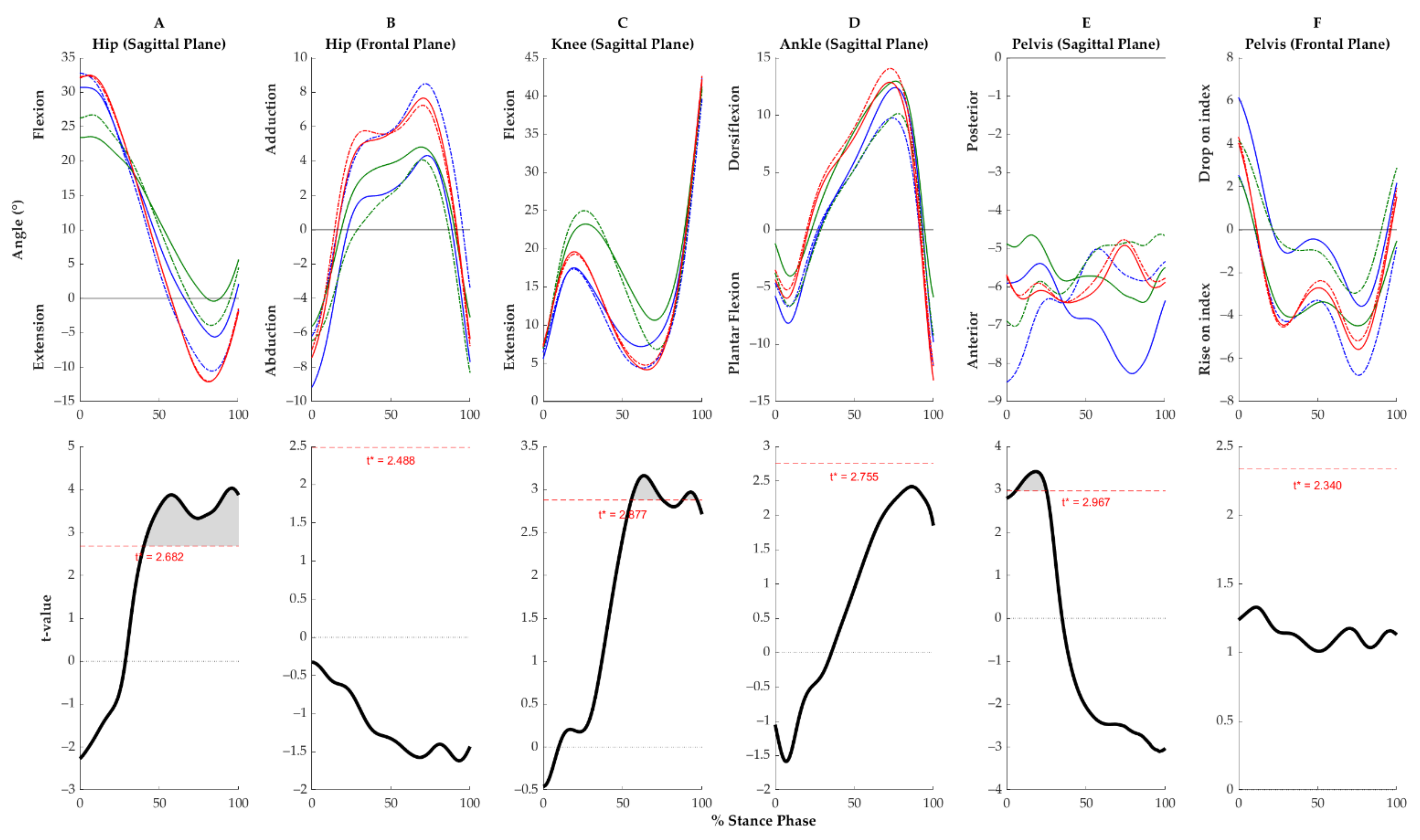
Figure 1. Top row: Ensemble average joint kinematics during the stance phase of the gait cycle for the unilateral hip osteoarthritis (blue), bilateral hip osteoarthritis (green), and control (red) groups. The index limb is displayed as a solid line and the non-index limb as a dashed line. Bottom row: Corresponding t-values (black) and t-threshold (red) from paired t-test with statistical parametric mapping for unilateral hip OA group only. T-values that exceeded the threshold were interpreted to indicate significant between-limb differences (p < 0.05).
Symmetry 13 00768 g001
Figure 2. Top row: Between-limb difference (±95% confidence interval) in sagittal plane pelvis and hip kinematics over the stance phase of the gait cycle for unilateral hip osteoarthritis (blue), bilateral hip osteoarthritis (green), and control (red) groups. Middle row: Corresponding F-values (black) and F-threshold (red) from analysis of variance with statistical parametric and non-parametric mapping. F-values that exceeded the threshold were interpreted to indicate between-group differences (p < 0.05). Bottom row: Corresponding t-values (black) and t-threshold (red) from post hoc t-test with statistical parametric and non-parametric mapping for unilateral vs. control groups. T-values that exceeded the threshold were interpreted to indicate significant between-limb differences (p < 0.05).
Symmetry 13 00768 g002
Figure 3. Top row: Ensemble average joint kinematics over the stance phase of sit-to-stand for the unilateral hip osteoarthritis (blue), bilateral hip OA (green), and control (red) groups. The index limb is displayed as a solid line and the non-index limb as a dashed line. Bottom row: Between-limb difference (±95% confidence interval) in joint kinematics during the stance phase of sit-to-stand for the unilateral hip osteoarthritis (blue), bilateral hip OA (green), and control (red) groups.
Symmetry 13 00768 g003
Table 1. Participant characteristics by group. Data are presented as the mean (one standard deviation).
Hip OAControl
UnilateralBilateral
n = 4212 (4 M, 8 F)9 (4 M, 5 F)21 (5 M, 16 F)
Age (years)62.5 (SD 10.0)63.2 (SD 6.0)58.7 (SD 8.2)
Age (range)58–7856–7149–75
Height (m)1.67 (SD 0.05)1.69 (SD 0.12)1.67 (SD 0.07)
Mass (kg)78.5 (SD 13.4) *81.6 (SD 16.9) *69.5 (SD 9.2)
BMI (kg/m2)27.2 (SD 3.2) *27.6 (SD 4.6) *25.3 (SD 3.4)
HHS73.04 (SD 12.06)65.12 (11.9)99.88 (SD 1.04)
IndexNon-indexIndexNon-index IndexNon-index
JSW (mm)2.5 (SD 0.9) *3.5 (SD 0.7)2.7 (SD 0.7) *3.1 (SD 0.7) *4.1 (SD 0.6)3.9 (SD 0.5)
JSW difference1 (SD 0.8) *, +−0.37 (SD 0.3)−0.21 (SD 0.4)
KL 0 5 1412
KL 1 7 79
KL 25 56
KL 37 43
KL difference1.8 (SD 1.1) *, +0.1 (SD 0.3)−0.1 (SD 0.6)
OA, osteoarthritis; n, number of participants; M, Male; F, Female; BMI, Body Mass Index; HHS, Harris Hip Score; JSW, Joint Space Width; KL, Kellgren–Lawrence score. * Indicates significant difference compared with control group (p < 0.05). + Indicates significant difference compared with bilateral hip OA group (p < 0.05).
Table 2. Spatiotemporal gait parameters by group. Data are presented as the mean (one standard deviation).
Hip OAControl
UnilateralBilateral
Gait Velocity (m/s)
Index1.19 (SD 0.21)1.11 (SD 0.11)1.31 (SD 0.11)
Non-index1.20 (SD 0.21)1.11 (SD 0.11)1.30 (SD 0.11)
Difference−0.01 (SD 0.03)0.001 (SD 0.03)0.008 (SD 0.03)
Step Length (m)
Index0.70 (SD 0.11)0.70 (SD 0.09)0.77 (SD 0.10)
Non-index0.70 (SD 0.11)0.69 (SD 0.05)0.74 (SD 0.05)
Difference 0.00 (SD 0.12)0.01 (SD 0.13)0.03 (SD 0.13)
Stance Duration (ms)
Index662 (SD 60)700 (SD 59)613 (SD 39)
Non-index660 (SD 62)714 (SD 58)612 (SD 40)
Difference0.6 (SD 9) +−14 (SD 8) *0.9 (SD 9)
OA, osteoarthritis. Mean difference calculated as index minus non-index limb. * indicates significant difference compared with control group (p < 0.05). + indicates significant difference compared with bilateral hip OA group (p < 0.05).
Publisher’s Note: MDPI stays neutral with regard to jurisdictional claims in published maps and institutional affiliations.