
Compensatory Ground Reaction Forces during Scoliotic Gait in Subjects with and without Right Adolescent Idiopathic Scoliosis


Abstract
:1. Introduction
2. Materials and Methods
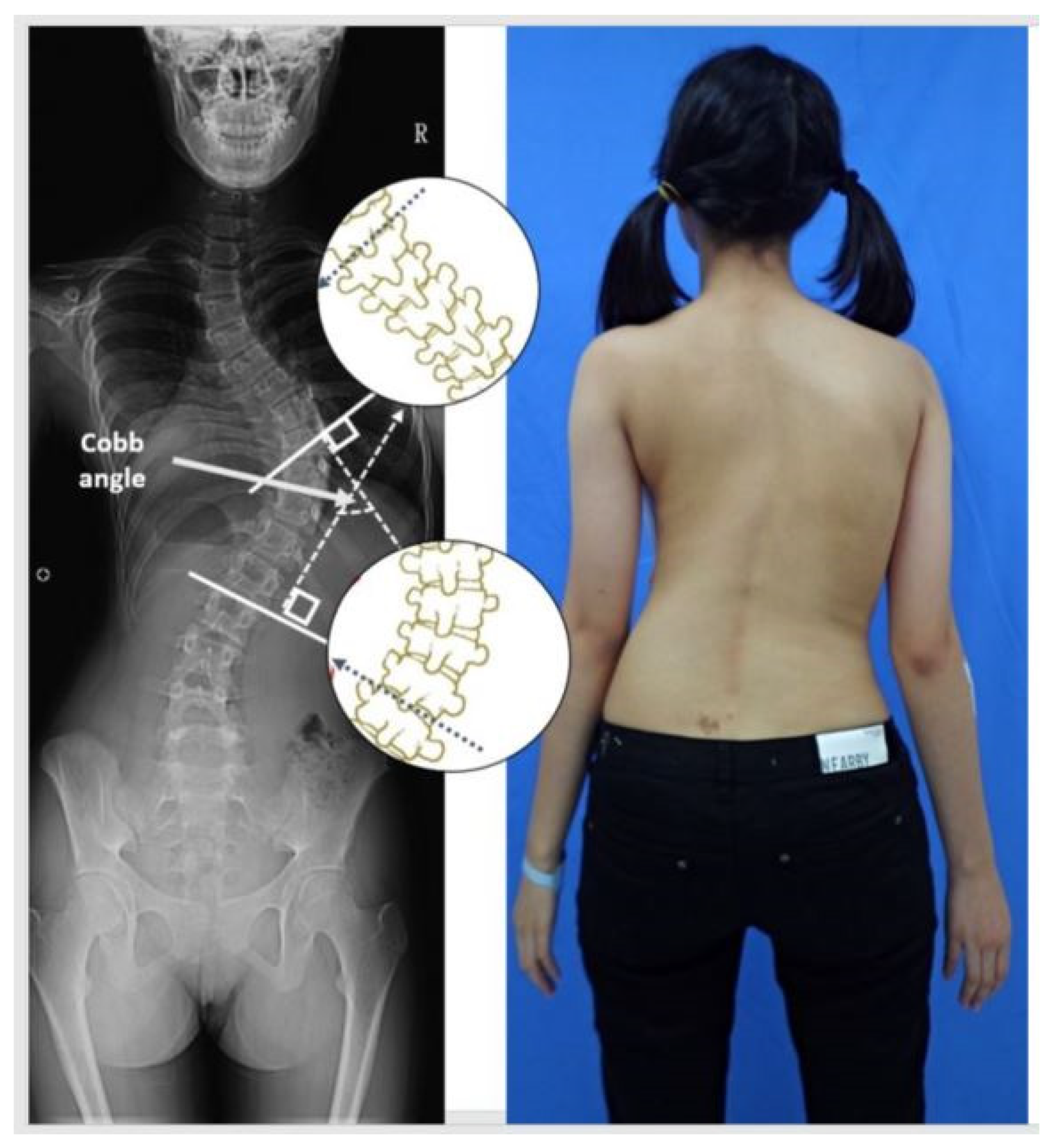
2.1. Subjects
2.2. Procedure
2.3. Statistical Analysis
3. Results
4. Discussion
5. Conclusions
Author Contributions
Funding
Institutional Review Board Statement
Informed Consent Statement
Data Availability Statement
Conflicts of Interest
References
- Nault, M.L.; Allard, P.; Hinse, S.; Le Blanc, R.; Caron, O.; Labelle, H.; Sadeghi, H. Relations between standing stability and body posture parameters in adolescent idiopathic scoliosis. Spine 2002, 27, 1911–1917. [Google Scholar] [CrossRef]
- Yang, J.H.; Suh, S.W.; Sung, P.S.; Park, W.H. Asymmetrical gait in adolescents with idiopathic scoliosis. Eur. Spine J. 2013, 22, 2407–2413. [Google Scholar] [CrossRef] [Green Version]
- Yang, F.; Kim, J.; Munoz, J. Adaptive gait responses to awareness of an impending slip during treadmill walking. Gait Posture 2016, 50, 175–179. [Google Scholar] [CrossRef] [PubMed]
- Park, H.J.; Sim, T.; Suh, S.W.; Yang, J.H.; Koo, H.; Mun, J.H. Analysis of coordination between thoracic and pelvic kinematic movements during gait in adolescents with idiopathic scoliosis. Eur. Spine J. 2016, 25, 385–393. [Google Scholar] [CrossRef]
- Sahin, F.; Urak, O.; Akkaya, N. Evaluation of balance in young adults with idiopathic scoliosis. Turk. J. Phys. Med. Rehabil. 2019, 65, 236–243. [Google Scholar] [CrossRef] [PubMed]
- Anari, J.B.; Tatad, A.; Cahill, P.J.; Flynn, J.M.; Harms Study, G. The Impact of Posterior Spinal Fusion (PSF) on Coronal Balance in Adolescent Idiopathic Scoliosis (AIS): A New Classification and Trends in the Postoperative Period. J. Pediatr. Orthop. 2020, 40, e788–e793. [Google Scholar] [CrossRef] [PubMed]
- Ozkunt, O.; Karademir, G.; Sariyilmaz, K.; Gemalmaz, H.C.; Dikici, F.; Domanic, U. Analysing the change of sagittal balance in patients with Lenke 5 idiopathic scoliosis. Acta Orthop. Traumatol. Turc. 2017, 51, 377–380. [Google Scholar] [CrossRef]
- Yusof, M.I.; Shaharudin, S.; Sivalingarajah, P. Does Vertical Ground Reaction Force of the Hip, Knee, and Ankle Joints Change in Patients with Adolescent Idiopathic Scoliosis after Spinal Fusion? Asian Spine J. 2018, 12, 349–355. [Google Scholar] [CrossRef] [PubMed] [Green Version]
- Chockalingam, N.; Dangerfield, P.H.; Rahmatalla, A.; Ahmed, E.N.; Cochrane, T. Assessment of ground reaction force during scoliotic gait. Eur. Spine J. 2004, 13, 750–754. [Google Scholar] [CrossRef] [PubMed] [Green Version]
- Park, Y.S.; Lim, Y.T.; Koh, K.; Kim, J.M.; Kwon, H.J.; Yang, J.S.; Shim, J.K. Association of spinal deformity and pelvic tilt with gait asymmetry in adolescent idiopathic scoliosis patients: Investigation of ground reaction force. Clin. Biomech. 2016, 36, 52–57. [Google Scholar] [CrossRef] [PubMed]
- Allard, P.; Chavet, P.; Barbier, F.; Gatto, L.; Labelle, H.; Sadeghi, H. Effect of body morphology on standing balance in adolescent idiopathic scoliosis. Am. J. Phys. Med. Rehabil. 2004, 83, 689–697. [Google Scholar] [CrossRef] [PubMed]
- Kramers-de Quervain, I.A.; Muller, R.; Stacoff, A.; Grob, D.; Stussi, E. Gait analysis in patients with idiopathic scoliosis. Eur. Spine J. 2004, 13, 449–456. [Google Scholar] [CrossRef] [PubMed] [Green Version]
- Schizas, C.G.; Kramers-de Quervain, I.A.; Stussi, E.; Grob, D. Gait asymmetries in patients with idiopathic scoliosis using vertical forces measurement only. Eur. Spine J. 1998, 7, 95–98. [Google Scholar] [CrossRef] [PubMed] [Green Version]
- Syczewska, M.; Graff, K.; Kalinowska, M.; Szczerbik, E.; Domaniecki, J. Influence of the structural deformity of the spine on the gait pathology in scoliotic patients. Gait Posture 2012, 35, 209–213. [Google Scholar] [CrossRef] [PubMed]
- Guo, J.; Liu, Z.; Lv, F.; Zhu, Z.; Qian, B.; Zhang, X.; Lin, X.; Sun, X.; Qiu, Y. Pelvic tilt and trunk inclination: New predictive factors in curve progression during the Milwaukee bracing for adolescent idiopathic scoliosis. Eur. Spine J. 2012, 21, 2050–2058. [Google Scholar] [CrossRef] [Green Version]
- Cunin, V. Early-onset scoliosis: Current treatment. Orthop. Traumatol. Surg. Res. 2015, 101, S109–S118. [Google Scholar] [CrossRef] [Green Version]
- Obeid, I.; Berjano, P.; Lamartina, C.; Chopin, D.; Boissiere, L.; Bourghli, A. Classification of coronal imbalance in adult scoliosis and spine deformity: A treatment-oriented guideline. Eur. Spine J. 2019, 28, 94–113. [Google Scholar] [CrossRef] [Green Version]
- Sahlstrand, T. An analysis of lateral predominance in adolescent idiopathic scoliosis with special reference to convexity of the curve. Spine 1980, 5, 512–518. [Google Scholar] [CrossRef] [PubMed]
- Sung, P.S.; Park, M.S. Lumbar spine coordination during axial trunk rotation in adolescents with and without right thoracic idiopathic scoliosis. Hum. Mov. Sci. 2020, 73, 102680. [Google Scholar] [CrossRef] [PubMed]
- Bruyneel, A.V.; Chavet, P.; Bollini, G.; Allard, P.; Mesure, S. The influence of adolescent idiopathic scoliosis on the dynamic adaptive behaviour. Neurosci. Lett. 2008, 447, 158–163. [Google Scholar] [CrossRef]
- Schlosser, T.P.; van Stralen, M.; Brink, R.C.; Chu, W.C.; Lam, T.P.; Vincken, K.L.; Castelein, R.M.; Cheng, J.C. Three-dimensional characterization of torsion and asymmetry of the intervertebral discs versus vertebral bodies in adolescent idiopathic scoliosis. Spine 2014, 39, E1159–E1166. [Google Scholar] [CrossRef]
- Kotwicki, T.; Walczak, A.; Szulc, A. Trunk rotation and hip joint range of rotation in adolescent girls with idiopathic scoliosis: Does the “dinner plate” turn asymmetrically? Scoliosis 2008, 3, 1. [Google Scholar] [CrossRef] [Green Version]
- Pal, G.P. Mechanism of production of scoliosis. A hypothesis. Spine 1991, 16, 288–292. [Google Scholar] [CrossRef] [PubMed]
- Sadeghi, H.; Allard, P.; Prince, F.; Labelle, H. Symmetry and limb dominance in able-bodied gait: A review. Gait Posture 2000, 12, 34–45. [Google Scholar] [CrossRef]
- Schlager, B.; Krump, F.; Boettinger, J.; Niemeyer, F.; Ruf, M.; Kleiner, S.; Beer, M.; Wilke, H.J. Characteristic morphological patterns within adolescent idiopathic scoliosis may be explained by mechanical loading. Eur. Spine J. 2018, 586, 5622. [Google Scholar] [CrossRef]
- Schlosser, T.P.; van der Heijden, G.J.; Versteeg, A.L.; Castelein, R.M. How ‘idiopathic’ is adolescent idiopathic scoliosis? A systematic review on associated abnormalities. PLoS ONE 2014, 9, e97461. [Google Scholar] [CrossRef] [Green Version]
- Oldfield, R.C. The assessment and analysis of handedness: The Edinburgh inventory. Neuropsychologia 1971, 9, 97–113. [Google Scholar] [CrossRef]
- Brophy, R.; Silvers, H.J.; Gonzales, T.; Mandelbaum, B.R. Gender influences: The role of leg dominance in ACL injury among soccer players. Br. J. Sports Med. 2010, 44, 694–697. [Google Scholar] [CrossRef]
- Andersen, T.E.; Floerenes, T.W.; Arnason, A.; Bahr, R. Video analysis of the mechanisms for ankle injuries in football. Am. J. Sports Med. 2004, 32, 69–79. [Google Scholar] [CrossRef] [PubMed]
- Sung, P.S. Increased double limb support times during walking in right limb dominant healthy older adults with low bone density. Gait Posture 2018, 63, 145–149. [Google Scholar] [CrossRef]
- Buczek, F.L.; Rainbow, M.J.; Cooney, K.M.; Walker, M.R.; Sanders, J.O. Implications of using hierarchical and six degree-of-freedom models for normal gait analyses. Gait Posture 2010, 31, 57–63. [Google Scholar] [CrossRef] [PubMed]
- Herzog, W.; Nigg, B.M.; Read, L.J.; Olsson, E. Asymmetries in ground reaction force patterns in normal human gait. Med. Sci. Sports Exerc. 1989, 21, 110–114. [Google Scholar] [CrossRef] [PubMed]
- Faul, F.; Erdfelder, E.; Lang, A.G.; Buchner, A. G*Power 3: A flexible statistical power analysis program for the social, behavioral, and biomedical sciences. Behav. Res. Methods 2007, 39, 175–191. [Google Scholar] [CrossRef]
- Portney, L.G.; Watkins, M.P. Foundations of Clinical Research Applications to Practice, 3rd ed.; Pearson Education Inc.: Upper Saddle River, NJ, USA, 2009. [Google Scholar]
- Bruyneel, A.V.; Chavet, P.; Bollini, G.; Allard, P.; Berton, E.; Mesure, S. Dynamical asymmetries in idiopathic scoliosis during forward and lateral initiation step. Eur. Spine J. 2009, 18, 188–195. [Google Scholar] [CrossRef] [Green Version]
- Crijns, T.J.; Stadhouder, A.; Smit, T.H. Restrained Differential Growth: The Initiating Event of Adolescent Idiopathic Scoliosis? Spine 2017, 42, E726–E732. [Google Scholar] [CrossRef]
- Lee, D.; Sung, P.S. Comparison of kinematic similarity index during gait between adults with and without nonspecific chronic neck pain. Gait Posture 2021, 91, 99–104. [Google Scholar] [CrossRef] [PubMed]
- Sung, P.S.; Schalk, B.M.; Latuszek, N.; Hosmer, E. Compensatory spinopelvic motions with and without a handheld task following a perturbation in standing between subjects with and without chronic low back pain. Gait Posture 2020, 76, 7–13. [Google Scholar] [CrossRef]
- Perry, J. Gait Analysis: Normal and Pathological Function; SLACK Inc.: Thorofare, NJ, USA, 1992. [Google Scholar]
- Herssens, N.; Verbecque, E.; Hallemans, A.; Vereeck, L.; Van Rompaey, V.; Saeys, W. Do spatiotemporal parameters and gait variability differ across the lifespan of healthy adults? A systematic review. Gait Posture 2018, 64, 181–190. [Google Scholar] [CrossRef]
- Gilbert, S.R.; Savage, A.J.; Whitesell, R.; Conklin, M.J.; Fineberg, N.S. BMI and magnitude of scoliosis at presentation to a specialty clinic. Pediatrics 2015, 135, e1417–e1424. [Google Scholar] [CrossRef] [Green Version]
- Tarrant, R.C.; Queally, J.M.; Moore, D.P.; Kiely, P.J. Prevalence and impact of low body mass index on outcomes in patients with adolescent idiopathic scoliosis: A systematic review. Eur. J. Clin. Nutr. 2018, 72, 1463–1484. [Google Scholar] [CrossRef]
- Sung, P.S.; Spratt, K.F.; Wilder, D.G. A possible methodological flaw in comparing dominant and nondominant sided lumbar spine muscle responses without simultaneously considering hand dominance. Spine 2004, 29, 1914–1922. [Google Scholar] [CrossRef] [PubMed]
- Mahaudens, P.; Detrembleur, C.; Mousny, M.; Banse, X. Gait in adolescent idiopathic scoliosis: Energy cost analysis. Eur. Spine J. 2009, 18, 1160–1168. [Google Scholar] [CrossRef] [PubMed] [Green Version]
- Wilczynski, J.; Bieniek, K. Correlations of somatic traits and postural defects in girls and boys aged 10–12. Acta Bioeng. Biomech. 2019, 21, 79–86. [Google Scholar] [PubMed]

| AIS Group (23) | Control Group (22) | Statistics | p | |
|---|---|---|---|---|
| Gender (male/female) | 3/20 | 2/20 | Chi-square = 0.18 | 0.52 |
| Age (years) | 14.96 ± 1.02 | 14.77 ± 1.10 | t = −0.57 | 0.56 |
| BMI | 13.07 ± 6.00 | 14.21 ± 7.49 | t = 0.56 | 0.57 |
| Cobb angle (degrees) | 23.14 ± 4.50 | NA | NA | NA |
| AIS Group | Control Group | 95% CI Upper/Lower | t | p | |
|---|---|---|---|---|---|
| Dominant limb | |||||
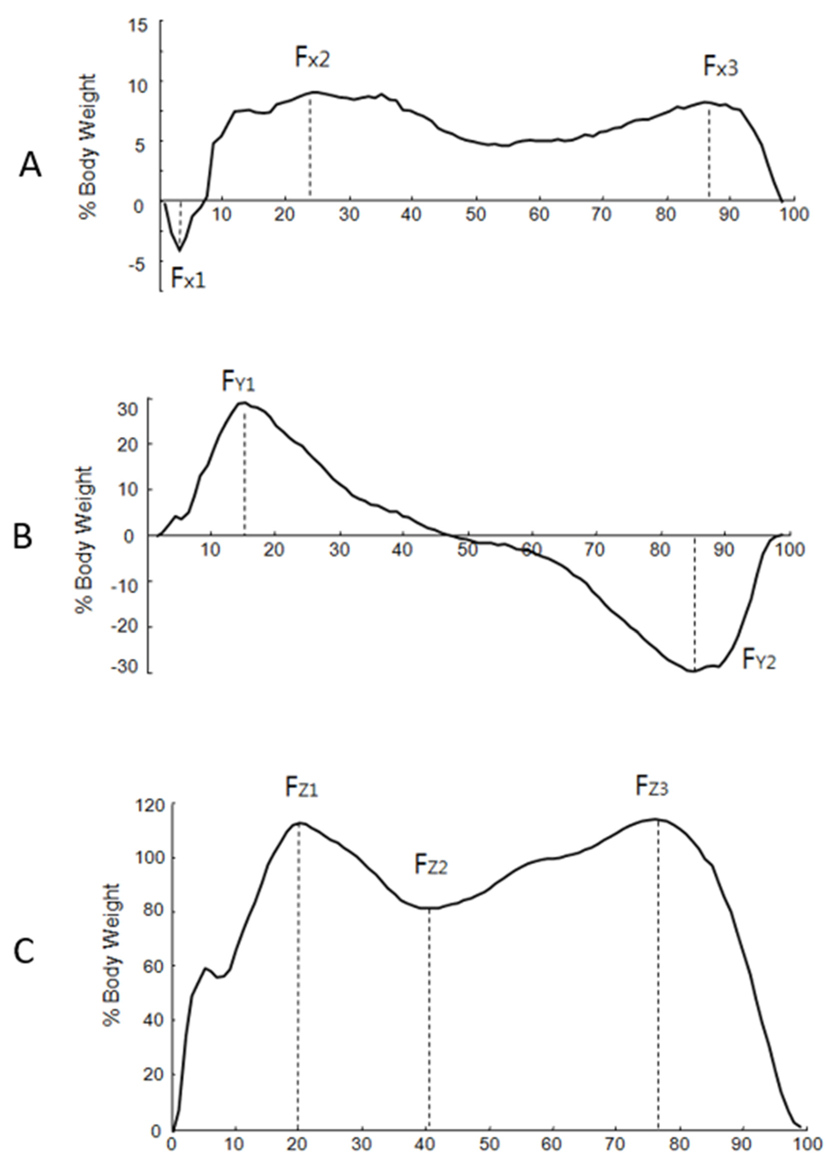
| Fx1 | −8.85 ± 3.91 | −7.75 ± 4.61 | −1.46/3.67 | 0.86 | 0.39 |
| Fx2 | 8.44 ± 4.10 | 10.12 ± 2.94 | −0.47/3.83 | 1.57 | 0.12 |
| Fx3 | 7.40 ± 3.44 | 9.68 ± 2.35 | 0.49/4.06 | 2.58 | 0.01 * |
| Fy1 | 38.24 ± 9.71 | 40.65 ± 6.81 | −2.65/7.48 | 0.96 | 0.34 |
| Fy2 | −45.56 ± 9.90 | −44.99 ± 7.42 | −4.74/5.87 | 0.21 | 0.83 |
| Fz1 | 115.22 ± 9.15 | 119.28 ± 8.36 | −1.22/9.33 | 1.55 | 0.19 |
| Fz2 | 80.90 ± 20.44 | 83.98 ± 6.72 | −6.16/12.31 | 0.67 | 0.51 |
| Fz3 | 121.26 ± 7.77 | 121.22 ± 6.55 | −4.36/4.30 | −0.02 | 0.98 |
| Non-dominant limb | |||||
| Fx1 | −4.93 ± 2.72 | −4.63 ± 2.84 | −1.37/1.97 | 0.36 | 0.71 |
| Fx2 | 13.85 ± 3.01 | 14.43 ± 3.19 | −1.28/2.43 | 0.62 | 0.54 |
| Fx3 | 11.14 ± 2.50 | 11.87 ± 2.46 | −0.77/2.22 | 0.97 | 0.33 |
| Fy1 | 37.59 ± 8.87 | 40.27 ± 8.71 | −2.61/7.97 | 1.01 | 0.31 |
| Fy2 | −42.44 ± 7.63 | −43.90 ± 7.75 | −6.09/3.16 | −0.64 | 0.52 |
| Fz1 | 114.01 ± 9.16 | 115.55 ± 9.11 | −3.95/7.03 | 0.56 | 0.57 |
| Fz2 | 84.19 ± 7.46 | 85.42 ± 6.46 | −2.98/5.43 | 0.59 | 0.56 |
| Fz3 | 117.46 ± 8.35 | 118.68 ± 5.27 | −3.01/5.44 | 0.58 | 0.56 |
| AIS Group | Control Group | |||||
|---|---|---|---|---|---|---|
| Cobb Angle | Age | BMI | Cobb Angle | Age | BMI | |
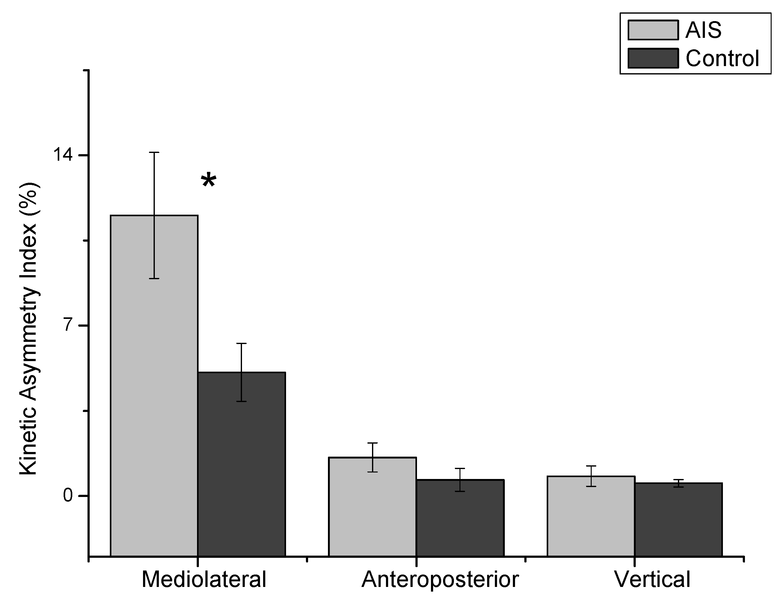
| KAI_x | −0.15 (0.51) | 0.06 (0.79) | 0.21 0.33 | NA | 0.15 0.49 | 0.40 0.06 |
| KAI_y | −0.13 (0.55) | 0.12 (0.57) | −0.02 (0.94) | NA | −0.02 (0.93) | −0.03 (0.90) |
| KAI_z | −0.45 (0.03) * | 0.02 (0.92) | −0.08 (0.72) | NA | −0.35 (0.11) | −0.04 (0.83) |
Publisher’s Note: MDPI stays neutral with regard to jurisdictional claims in published maps and institutional affiliations. |
© 2021 by the authors. Licensee MDPI, Basel, Switzerland. This article is an open access article distributed under the terms and conditions of the Creative Commons Attribution (CC BY) license (https://creativecommons.org/licenses/by/4.0/).
Share and Cite
Sung, P.S.; Park, M.S. Compensatory Ground Reaction Forces during Scoliotic Gait in Subjects with and without Right Adolescent Idiopathic Scoliosis. Symmetry 2021, 13, 2372. https://doi.org/10.3390/sym13122372
Sung PS, Park MS. Compensatory Ground Reaction Forces during Scoliotic Gait in Subjects with and without Right Adolescent Idiopathic Scoliosis. Symmetry. 2021; 13(12):2372. https://doi.org/10.3390/sym13122372
Chicago/Turabian StyleSung, Paul S., and Moon Soo Park. 2021. "Compensatory Ground Reaction Forces during Scoliotic Gait in Subjects with and without Right Adolescent Idiopathic Scoliosis" Symmetry 13, no. 12: 2372. https://doi.org/10.3390/sym13122372
APA StyleSung, P. S., & Park, M. S. (2021). Compensatory Ground Reaction Forces during Scoliotic Gait in Subjects with and without Right Adolescent Idiopathic Scoliosis. Symmetry, 13(12), 2372. https://doi.org/10.3390/sym13122372