Article

Interactions of Aβ1-42 Peptide and Its Three Fragments (Aβ8-12, Aβ8-13, and Aβ5-16) with Selected Nonsteroidal Drugs and Compounds of Natural Origin

by
Krzysztof Żamojć
,
Karolina Streńska
,
Dariusz Wyrzykowski
,
Lech Chmurzyński
and
Joanna Makowska
*
Faculty of Chemistry, University of Gdańsk, Wita Stwosza 63, 80-308 Gdańsk, Poland
*
Author to whom correspondence should be addressed.
Symmetry 2020, 12(10), 1579; https://doi.org/10.3390/sym12101579
Submission received: 2 September 2020 / Revised: 15 September 2020 / Accepted: 21 September 2020 / Published: 23 September 2020
(This article belongs to the Special Issue Functional Materials Engineering and Biomolecular Structure)

Abstract

:
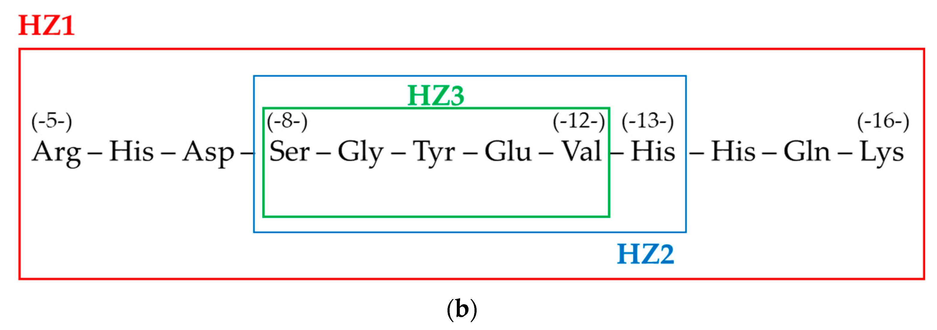
In the following paper, we present the results of our studies on the interactions of the Aβ1-42 peptide and its three short fragments, namely Aβ5-16 (RHDSGYEVHHQK; HZ1), Aβ8-13 (SGYEVH; HZ2), and Aβ8-12 (SGYEV; HZ3) with selected painkillers (ibuprofen and aspirin) and compounds of natural origin (anabasine and epinephrine). Steady-state fluorescence spectroscopy was used to study the binding properties of the selected systems. Additionally, based on molecular dynamics (MD) calculations supported by NMR-derived restrains, we have proposed the most likely area of the interactions of Aβ1-42 and Aβ5-16 peptides with the investigated compounds. The influence of symmetrically oriented side chains of amino acid residues present in the first part of the Aβ1-42 sequence on the stability of the resulting complexes has been discussed. Finally, the changes in the peptide structures on account of complex formation were analyzed.

Graphical Abstract

1. Introduction

Pharmacotherapeutic procedure in Alzheimer’s disease (AD) involves the use of pain relievers (e.g., drugs that enlarge the metabolic activity of the brain tissue), symptomatic relievers (e.g., antidepressant drugs, neuroleptics), substitution relievers (e.g., drugs that increase the concentration of acetylcholine in the brain), combined agents (drugs with different mechanisms of action), pleiotropic agents (estrogens, aluminum chelating agents), as well as agents reducing tau protein synthesis and deposition of amyloid β deposits with the use of agents inhibiting neurofibrillary degeneration [1,2]. On the other hand, it allows only to alleviate symptoms, but is not effective in stopping a development of the disease and reversing the resulting neurodegenerative changes [3,4] induced among others by chronic ethanol administration [5], chronic unpredictable mild stress [6], imbalance in various metal ions intake [7], or excessive amounts of reactive oxygen [8] and nitrogen species [9]. Thus, new drugs (agents currently used in the therapies of other diseases, as well as compounds found in raw materials of natural origin) and alternative treatment methods are sought and—along with an appropriate diet and reduction of deleterious environmental factors—could be very helpful in preventing Alzheimer’s disease [10].
Studies on potential drugs for Alzheimer’s disease include, among others, nonsteroidal anti-inflammatory drugs (NSAIDs) which exhibit anti-inflammatory, analgesic and antipyretic properties [11]. NSAIDs inhibit the activity of cyclooxygenase (COX) responsible for the altering of arachidonic acid to prostaglandins [12]—the mediators of inflammatory processes, resulting in the expansion of blood vessels, increased body temperature, increased capillary permeability, and inflammatory cells [13]. NSAIDs also probably affect amyloid precursor protein (APP) metabolism, independent of COX, thereby decreasing the amount of amyloid Aβ1-42 in the brain. Finally, in vitro studies have shown that NSAIDs modulate neuronal responses by, for example, inhibiting glial cell responses to amyloid β [14,15,16]. Despite some speculations about differences in the reliability of action between aspirin and various NSAIDs and a fact that additional studies in that field are required [17], aspirin has been found to have a protective effect in AD [18]. Ibuprofen can be used in chronic inflammatory Alzheimer’s disease as well—it probably contributes to the inhibition of γ secretase activity, which is responsible for the formation of amyloid β [19]. However, similarly to aspirin, the data on the beneficial effects of that drug on AD are not entirely clear and require further research [20]. Apart from NSAIDs, there are also reports in the literature on substances of natural origin as potentially active substances in Alzheimer’s disease [21], for example pyridine and piperidine alkaloids [22,23,24]. Anabasine is an example of nicotinic alkaloids, a component of a tobacco smoke [25,26]. Next to nicotine, it is an example of a ligand for neuronal nicotinic acetylcholine receptors (nAChRs) [27]. Since the most available drugs for the treatment of Alzheimer’s disease are indeed acetylcholinesterase inhibitors and muscarinic M1 receptors agonists [28], the studies on anabasine in AD development seem to be justified. In the same context, many neuroanatomic experiments revealed the influence of epinephrine (adrenaline) on the modulation of behavioral and cardiovascular function [29,30,31].
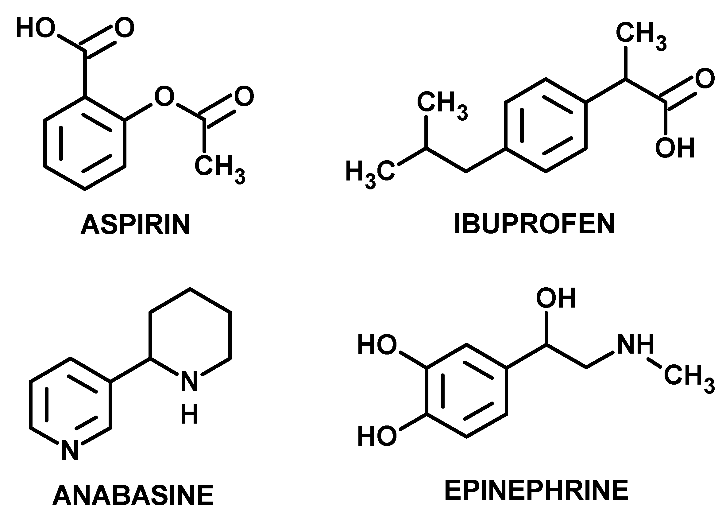
In this manuscript, we present the results of our studies on the interactions of human amyloid β protein fragment 1-42 (Aβ1-42; the structure is shown in Figure 1a) and its three chosen fragments, namely Aβ5-16 (RHDSGYEVHHQK; HZ1), Aβ8-13 (SGYEVH; HZ2), and Aβ8-12 (SGYEV; HZ3) (the sequences are presented in Figure 1b) with selected painkillers: ibuprofen and aspirin as well as two compounds of natural origin: anabasine and epinephrine (the structures are presented in Figure 2). The principal aim of this work was to study the binding properties of Aβ1-42 polypeptide to the chosen biologically active compounds. It should be noted that the conformation of the dominant family of HZ1 aligns appropriately on the corresponding section of the native Aβ1-42 [32], particularly when compared orientation of residues from the central part of the sequence with the most expanded side chains (i.e., tyrosine or histidine). Therefore, in particular, the attention has been focused on the influence of symmetrically oriented side chains of the amino acid residues in peptides under study on the stability of the resulting complexes. Furthermore, the studies were focused on the fragments from the first part of the sequence because previously it has been proven that probably three residues in the original sequence Aβ1-42 (namely, His6, His14 and His16) serve very often as binding sites for other ligands [33,34].

2. Experimental

2.1. Materials and Methods

Human amyloid β protein fragment 1-42 (95%, by HPLC) was obtained from AnaSpec Inc. (CA, USA) and used as received. Its shorter fragments chosen for the experiments (Aβ8-12, Aβ8-13, and Aβ5-16) were synthesized [36] and chromatographically purified [37] using procedures described previously. The peptides were then characterized using mass spectrometry. The concentration of the probes was confirmed spectrophotometrically using Perkin Elmer Lambda 650 UV/Vis spectrophotometer on the basis of the measurement of absorbance of tyrosine at 280 nm (ε280 = 1280 M−1cm−1) [37]. Aspirin (acetylsalicylic acid, >95%), ibuprofen (α-methyl-4-(isobutyl)phenylacetic acid, 99%), anabasine (neonicotine, >97%), and epinephrine (adrenaline, >95%) were obtained from Sigma Aldrich (Poland) and used as purchased.

2.2. Fluorescence Spectroscopy

Fluorometric experiments were carried out at 20 °C on a Cary Eclipse Varian spectrofluorometer equipped with a temperature controller and a multicell holder. The fluorescence emission spectra (λex = 275 nm) of all studied peptides were recorded from 280 to 400 nm. During titration experiments, 2 mL of each peptide solutions in 5 mM MES buffer pH 6.0 (cHZ1 = 38.8 µM, cHZ2 = 39.2 µM, cHZ3 = 34.5 µM) were titrated with six 5 μL aliquots of aspirin, ibuprofen, anabasine, and epinephrine solutions in DMSO (caspirin = 0.1 M, cibuprofen = 0.1 M, canabasine = 0.1 M, and cepinephrine = 0.01 M). The fluorescence intensity values determined at 305 nm (the maximum of emission of tyrosine and thus all peptides under study) were measured in the presence of increasing concentrations of selected drugs (pure DMSO was used as a control probe) and were further used to estimate the strength of interactions and determine association constants (where possible). The absorption of light by ibuprofen, aspirin, anabasine, and epinephrine at the excitation and emission wavelength of tyrosine (275 and 305 nm, respectively) has been considered and consequently fluorescence intensity values have been corrected for inner filter effect based on the Equation (1):
F corr = F obs × 10 A 275 + A 305 2 ,
where Fcorr and Fobs correspond to the corrected and observed fluorescence intensity values, respectively; while A275 and A305 correspond to the absorbance values measured at the excitation and emission wavelength, respectively [38,39].

2.3. Molecular Dynamics (MD)

Theoretical studies were performed using the AMBER 16 program [40] at constant temperature and volume (NVT scheme) and with the AMBER force field, the version ff14SB. All calculations were carried out in the periodic box, composed of water molecules, type TIP3P. During calculations, the Ewald procedure was implemented with a mesh of particles for electrostatic long-range interactions at a temperature of 10 °C. Molecular dynamics simulations were carried out with aspirin, ibuprofen, anabasine, and epinephrine added to Aβ1-42 and HZ1 (for shorter peptides, namely HZ2 and HZ3, the MD calculations were not performed due to the excessive mobility of the systems). In the calculations involving Aβ1-42, the peptide structure was defined on the basis of data from the PDB bank. In theoretical considerations for HZ1 peptide, limitations resulting from the NMR experiment, in the distance between the selected atoms and angles were taken into account (175 distance restraints and 32 dihedral angles restraints). In addition, during simulations for protons for which no NOE signals were observed, so-called “anti-NOE” limitations also were performed. This approach usually minimizes any deviation from the AMBER ff16SB force field that aims to favor the a-helical conformations. For each trajectory, the time of simulation was t = 10 ns, and the integration time step was 2 fs. In each simulation, counter ions have been added to neutralize systems.

3. Results

The fluorescence intensity of all selected peptides increases systematically with the increase of the amount of the added drug (painkiller and/or compound of natural origin), which may be a result of a variety of processes, among others excited state reactions, ground-state complex formations or collisional interactions (Figure 3). The dissociation constants (KD) of the resulting complexes can be determined by Lineweaver-Burk Equation (2):
1 F 0 F = 1 F 0 + K D F 0 [ Q ] ,
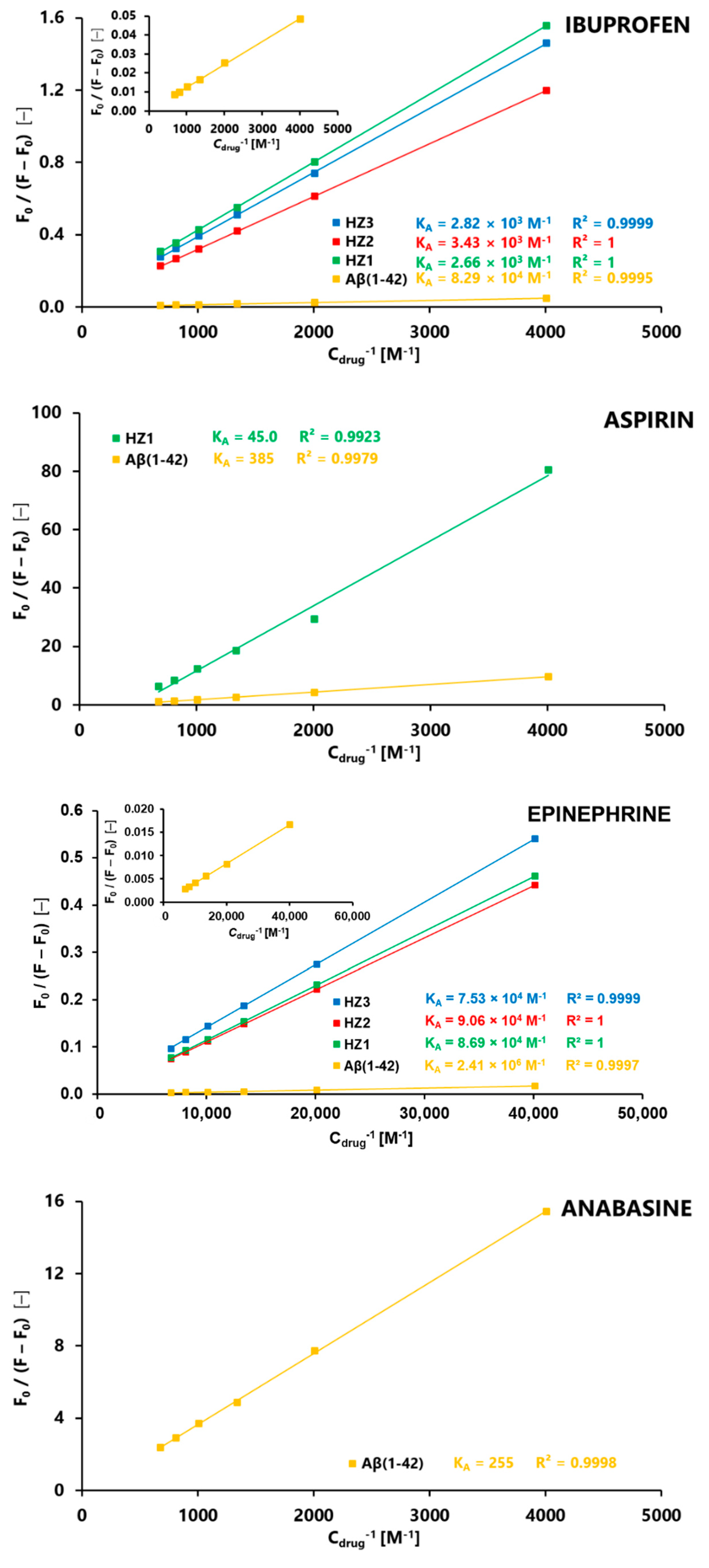
In the case of systems where a significant and linear relationship between F F 0 F 0 and   c drug was observed, the Lineweaver-Burk plots were constructed as the relationship of F 0 F 0 F vs. c drug 1 (Figure 4). From the regression equations of these curves, association constants ( K A =   K D 1 ) of the resulting complexes have been determined as the averages of two independent experiments. The values of these association constants are presented as insets in Figure 4.
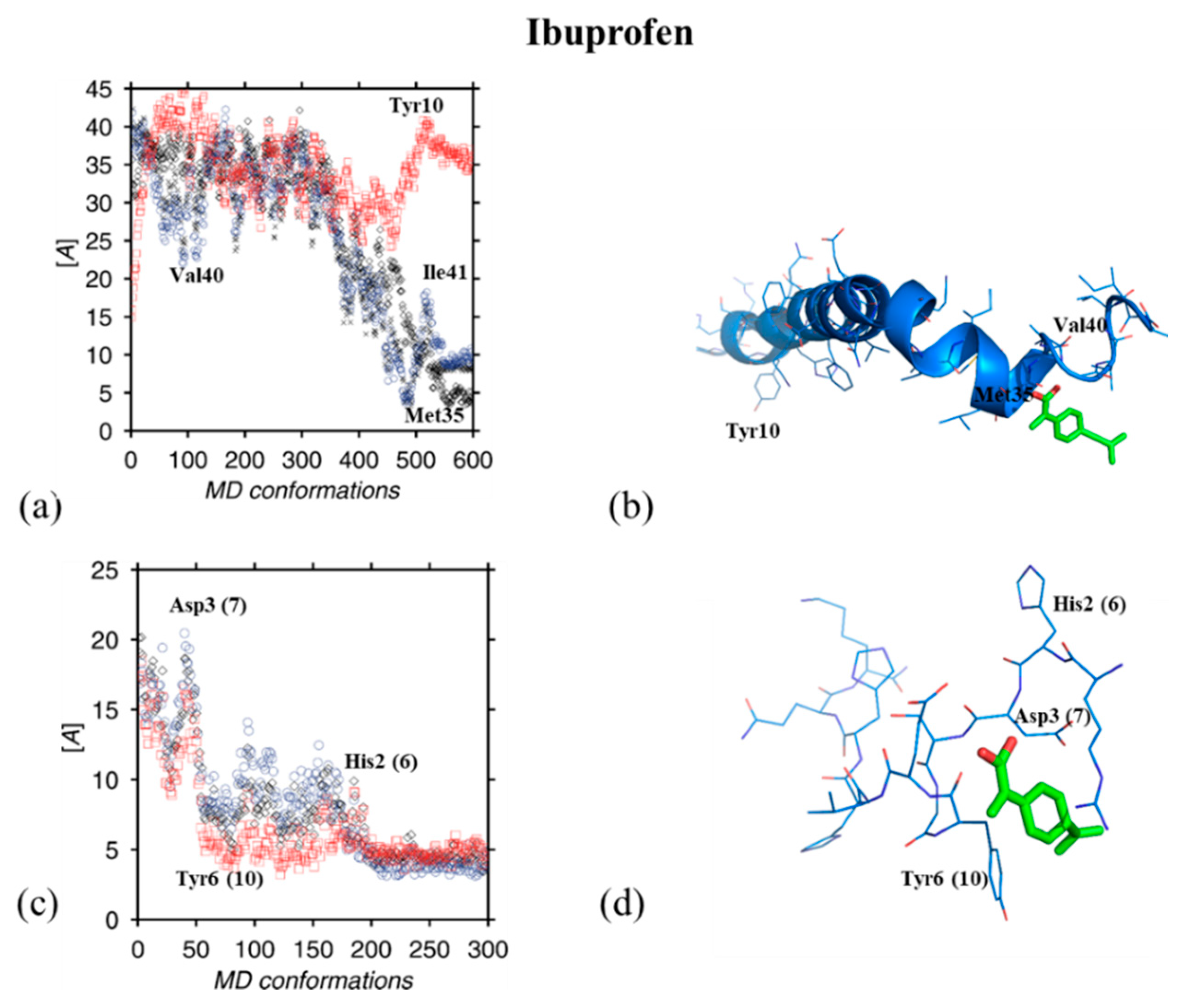
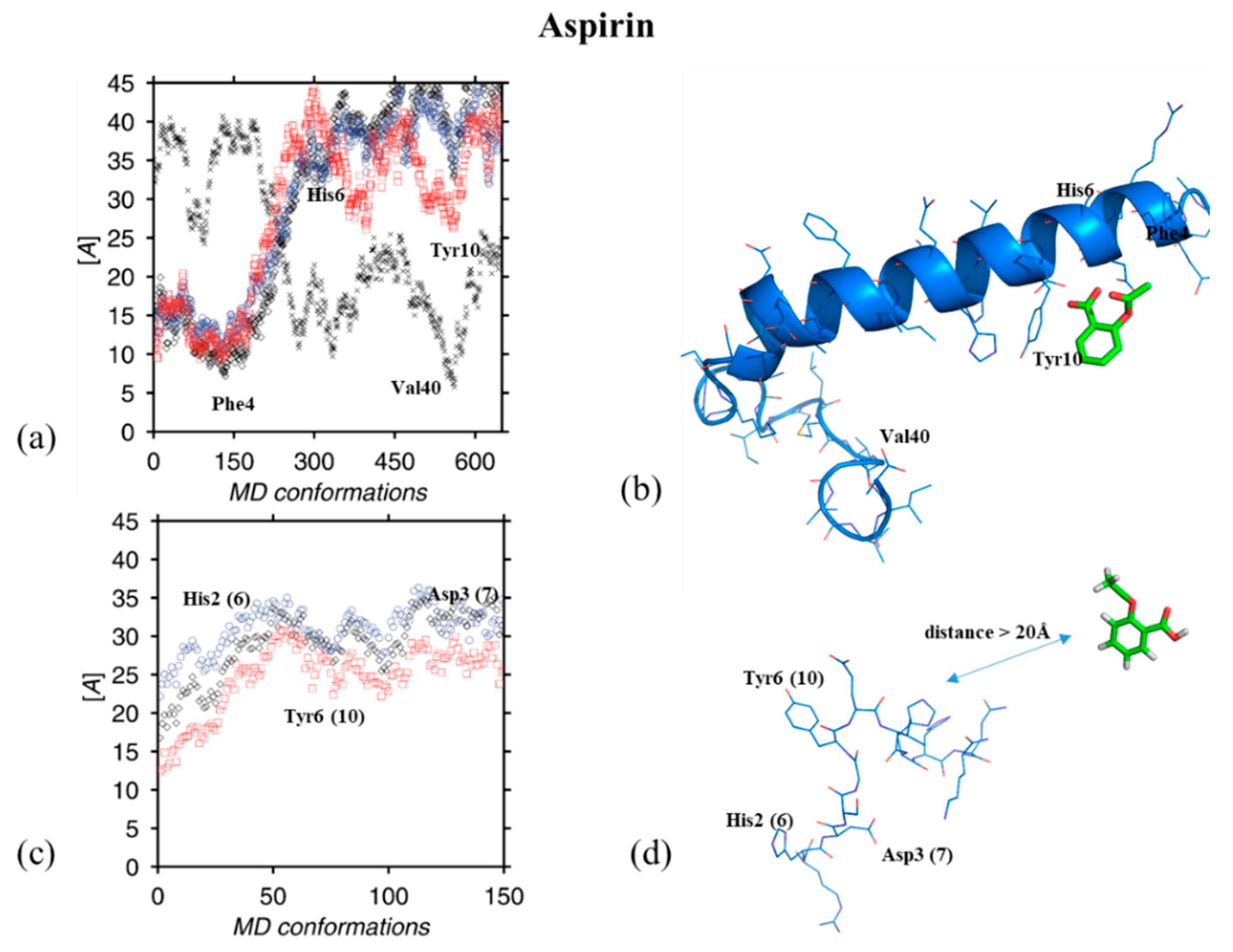
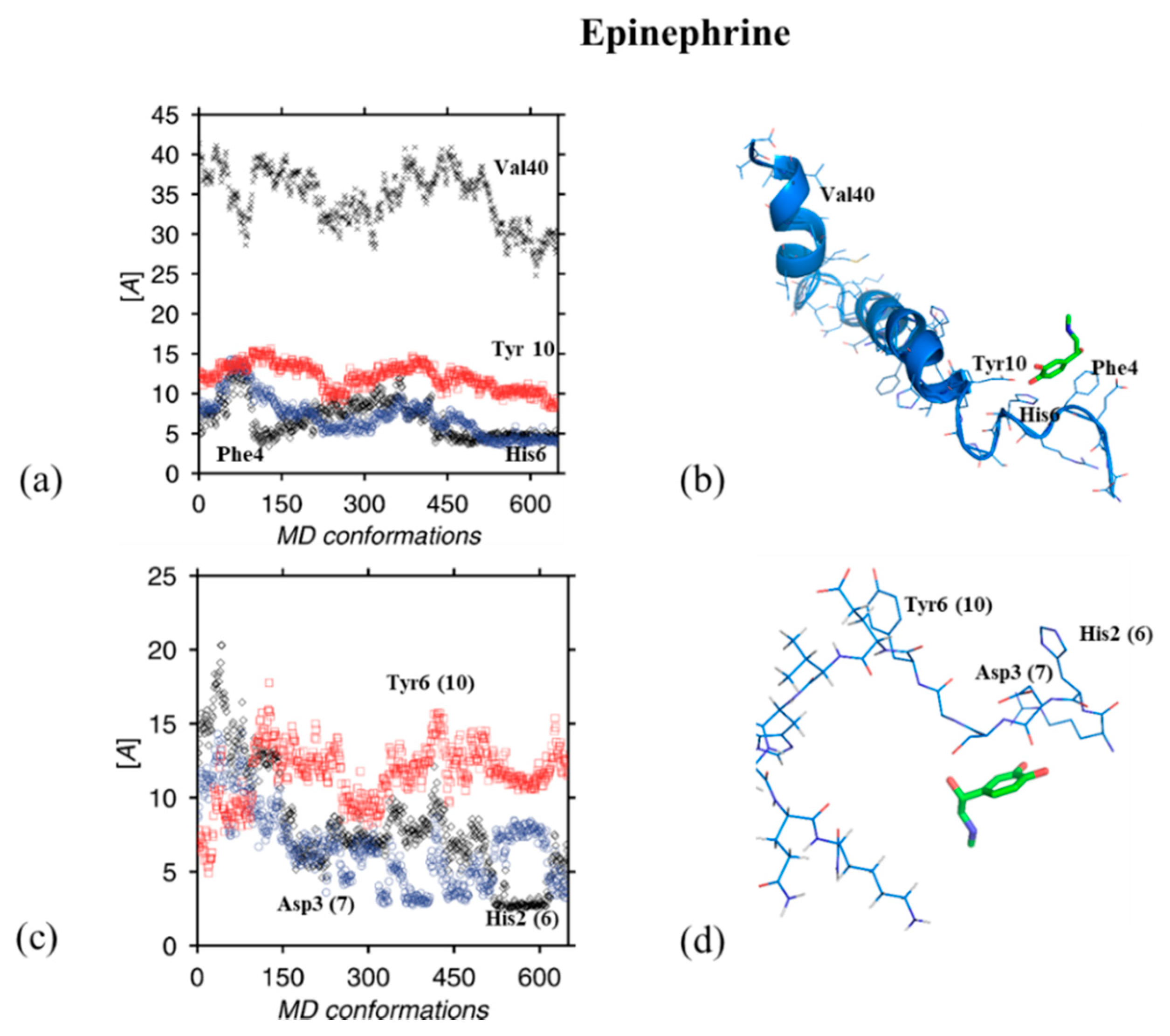
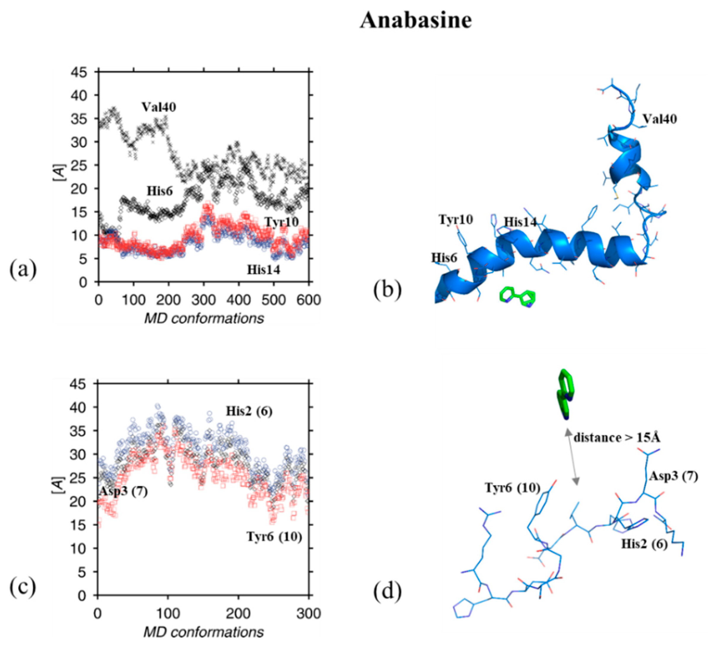
The molecular dynamics simulations (MD) were used to investigate the interactions of ibuprofen, aspirin, epinephrine, and anabasine with Aβ1-42 and HZ1 polypeptides. It should be noted that for HZ1 peptide the NMR-derived restrains were used for calculations. It has been previously confirmed, that the main conformation of HZ1 forms a well-defined bent structure in its central part (Ser4–His10) with quite flexible ends [32]. Figure 5, Figure 6, Figure 7 and Figure 8 present the results of theoretical calculations obtained for both peptides with ibuprofen, aspirin, epinephrine, and anabasine, respectively.

4. Discussion

From the inspection of Figure 3 and Figure 4, it can be observed that all studied peptides exhibit the strongest affinity to epinephrine. It is demonstrated by significantly higher values of association constants in case of peptide-epinephrine complexes (when compared to complexes with ibuprofen, aspirin, and anabasine), and is in a great agreement with MD simulations. It can be supposed that the presence of hydroxyl groups capable to form hydrogen bonds (under experimental conditions; pH 6.0) with the side chains of the peptides is probably the most important factor responsible for the strong epinephrine–peptide interaction. In case of interactions with Aβ1-42, epinephrine maintains a distinct distance in relation to the N-terminus and fluctuates steadily around the C-terminus (Phe4, His6 residues). The dominant spatial arrangement is probably stabilized by п-п interactions of two aromatic rings, Phe4 and epinephrine. Such a stabilization may be responsible for the obtained KA value which is approximately 30 times higher in case of complex of epinephrine with Aβ1-42 rather than with HZ1, HZ2 or HZ3. According to the HZ1 peptide it can be seen that epinephrine molecules—similarly to Aβ1-42—still locate around the beginning of the peptide chain causing its deeper bend. Epinephrine is located in the space defined by the electrostatic fields of Asp3 and His2. It is worth emphasizing that Tyr10 is also located within the space occupied by epinephrine, which probably stabilizes the position of the molecule. The observed possible interaction with Tyr10 explains similar (within the experimental error) KA values of complexes of epinephrine with HZ1, HZ2 and HZ3.
Both fluorescence spectroscopy and theoretical simulations revealed a significant affinity of the studied peptides towards ibuprofen, as well. However, in the case of Aβ1-42, the ibuprofen molecule shifted significantly towards the C-terminus during the simulation. The ligand found itself in a space defined by the electrostatic field of Met35, Val40 and Ile41 and clearly shifted from the N-terminus. In the case of HZ1, we clearly observed the sliding of the ibuprofen, especially its ring moiety, into the space defined by the side chains of tyrosine and histidine. Here, we observed clearly that the system is stabilized by п-п interaction of the side chain of tyrosine with ibuprofen. After the ibuprofen molecule was arranged parallel to the tyrosine side chain, the system showed stability during further calculations and did not change its position. Similarly to the results observed in case of epinephrine, the participation of Tyr10 in a stabilization of the system may be responsible for the comparable values of association constants of complexes of ibuprofen with HZ1, HZ2 and HZ3.
The results obtained from calculations for anabasine revealed the lack of clear affinity of that compound for both Aβ1-42 and HZ1. In case of the latter, the observed minimum distance to anabasine was above 15 Å. In case of Aβ1-42, during the simulations, it was possible to observe the beginning of the unfolding of the helix from the C-terminus part of the sequence, even though anabasine was oscillating around the middle of the sequence. Unfortunately, the dominant conformations of the two-component system clearly indicate the stability of the system when these two molecules are spaced, and are far away from each other. These observations are in an agreement with the results of spectrofluorometric titrations, since Aβ1-42 was the sole peptide in case of which the use of Lineweaver–Burk equation enabled estimation of association constant—its very low value confirms extremely slight interactions with anabasine.
The results obtained for aspirin are similar to those for anabasine. Molecular dynamics simulations revealed that aspirin destabilizes the Aβ1-42 structure, causing unfolding of the helix in the C-terminal part of the sequence, apart from the fact that it does not clearly interact with the system during the simulation. According to the HZ1 peptide, the dominant conformations of the two-component system clearly indicate the stability of the system when these two molecules are spaced, and are far away from each other. The results of spectrofluorometric titrations clearly confirm that, although the use of Lineweaver-Burk formula made it possible to determine appropriate KA values, they are very low and demonstrate very low interactions.
Furthermore, it has been proven that the sequence of the amino acid residues and the orientation of the side chains has the impact on the strength of the interaction on account of a steric effects which hinder the interactions between the tested compounds.

5. Conclusions

The fluorescence spectroscopy supported by molecular dynamics simulations (MD) with NMR-derived restrains was used to study the interactions of Aβ1-42 polypeptide and its derivatives with selected low-molecular weight organic compounds, namely ibuprofen and aspirin (the painkillers) as well as anabasine and epinephrine (naturally occurred compounds). In all cases, the shortening of the Aβ1-42 polypeptide chain caused changes in peptides conformation that were significant enough to affect their ability to bind the investigated compounds. Experimental results and MD calculations showed that epinephrine (adrenaline) reveals the highest affinity to the investigated peptides. It can be supposed that the presence of hydroxyl groups capable to form hydrogen bonds (under experimental conditions; pH 6.0) with the side chains of the peptides is probably the most important factor responsible for the strong epinephrine–peptide interaction. Furthermore, it has been proven that the sequence of the amino acid residues and the orientation of the side chains have the impact on the strength of the interaction on account of a steric effects which hinder the interactions between the tested compounds. Finally, it is worth emphasizing that only in the case of aspirin a strong effect on the disturbance of the structure of Aβ1-42 was observed. This phenomenon manifested itself in the unfolding of the C-terminal strand of the protein and can be explained by the specific arrangement of the peptide chain which folded so uniquely.

Author Contributions

Conceptualization, K.Ż., D.W. and J.M.; methodology, K.Ż., D.W. and J.M.; investigation, K.Ż., K.S., D.W. and J.M.; writing—original draft preparation, K.Ż., D.W., L.C. and J.M.; funding acquisition, K.Ż. All authors have read and agreed to the published version of the manuscript.

Funding

This research was funded by the Polish National Science Centre (NCN) under Grant No. 2016/23/D/ST4/01576.

Conflicts of Interest

The authors declare no conflict of interest.

References

  1. Minati, L.; Edginton, T.; Grazia Bruzzone, M.; Giaccone, G. Reviews: Current concepts in Alzheimer’s disease: A multidisciplinary review. Am. J. Alzheimer’s Dis. Other Dement. 2009, 24, 95–121. [Google Scholar] [CrossRef]
  2. Wärmländer, S.; Tiiman, A.; Abelein, A.; Luo, J.; Jarvet, J.; Söderberg, K.L.; Danielsson, J.; Gräslund, A. Biophysical studies of the amyloid β-peptide: Interactions with metal ions and small molecules. ChemBioChem 2013, 14, 1692–1704. [Google Scholar] [CrossRef] [PubMed]
  3. Hiremathad, A. A review: Natural compounds as anti-Alzheimer’s disease agents. Curr. Nutr. Food Sci. 2017, 13, 247–254. [Google Scholar] [CrossRef]
  4. Perez, A.; Li, T.; Hernandez, S.; Zhang, R.; Cao, C. The rationale of using coffee and melatonin as an alternative treatment for Alzheimer’s disease. J. Alzheimer’s Dis. Parkinsonism 2016, 6, 2161-0460. [Google Scholar] [CrossRef]
  5. Zhao, D.; Simon, J.E.; Wu, Q. A critical review on grape polyphenols for neuroprotection: Strategies to enhance bioefficacy. Crit. Rev. Food Sci. Nutr. 2020, 60, 597–625. [Google Scholar] [CrossRef]
  6. Abd El Wahab, M.G.; Ali, S.S.; Ayuob, N.N. The role of musk in relieving the neurodegenerative changes induced after exposure to chronic stress. Am. J. Alzheimer’s Dis. Other Dement. 2018, 33, 221–231. [Google Scholar] [CrossRef] [PubMed]
  7. Kabir, M.T.; Uddin, M.S.; Zaman, S.; Begum, Y.; Ashraf, G.M.; Bin-Jumah, M.N.; Bungau, S.G.; Mousa, S.A.; Abdel-Daim, M.M. Molecular mechanisms of metal toxicity in the pathogenesis of Alzheimer’s disease. Mol. Neurobiol. 2020. [Google Scholar] [CrossRef] [PubMed]
  8. Żamojć, K.; Zdrowowicz, M.; Rudnicki-Velasquez, P.B.; Krzymiński, K.; Zaborowski, B.; Niedziałkowski, P.; Jacewicz, D.; Chmurzyński, L. The development of 1,3-diphenylisobenzofuran as a highly selective probe for the detection and quantitative determination of hydrogen peroxide. Free Radic. Res. 2017, 51, 38–46. [Google Scholar] [CrossRef] [PubMed]
  9. Żamojć, K.; Jacewicz, D.; Zdrowowicz, M.; Chmurzyński, L. Kinetics of the reaction between 1,3-diphenylisobenzofuran and nitrogen dioxide studied by steady-state fluorescence. Res. Chem. Intermed. 2013, 39, 3023–3031. [Google Scholar] [CrossRef]
  10. Hong, Y.; Zhi, S.; Sheng, C. Current advances in the treatment of Alzheimer’s disease: Focused on considerations targeting Aβ and tau. Transl. Neurodegener. 2012, 1, 21. [Google Scholar] [CrossRef] [Green Version]
  11. Ali, M.M.; Ghouri, R.G.; Ans, A.H.; Akbar, A.; Toheed, A. Recommendations for anti-inflammatory treatments in Alzheimer’s disease: A comprehensive review of the literature. Cureus 2019, 11, e4620. [Google Scholar] [CrossRef] [PubMed] [Green Version]
  12. Fendrick, A.M.; Greenberg, B.P. A review of the benefits and risks of nonsteroidal anti-inflammatory drugs in the management of mild-to-moderate osteoarthritis. Osteopath. Med. Prim. Care 2009, 3, 1. [Google Scholar] [CrossRef] [PubMed] [Green Version]
  13. Suleyman, H.; Demircan, B.; Karagoz, Y. Anti-inflammatory and side effects of cyclo-oxygenase inhibitors. Pharmacol. Rep. 2007, 59, 247–258. [Google Scholar]
  14. Akiyama, H.; Barger, S.; Barnum, S.; Bradt, B.; Bauer, J.; Cole, G.M.; Cooper, N.R.; Eikelenboom, P.; Emmerling, M.; Fiebich, B.L.; et al. Inflammation and Alzheimer’s disease. Neurobiol. Aging 2000, 21, 383–421. [Google Scholar] [CrossRef]
  15. Jaturapatporn, D.; Isaac, M.G.E.K.N.; McCleery, J.; Tabet, N. Aspirin, steroidal and non-steroidal anti-inflammatory drugs for the treatment of Alzheimer’s disease. Cochrane Database Syst. Rev. 2012, 2. [Google Scholar] [CrossRef] [PubMed]
  16. Etminan, M.; Gill, S.; Samii, A. Effect of non-steroidal anti-inflammatory drugs on risk of Alzheimer’s disease: Systematic review and meta-analysis of observational studies. BMJ 2003, 327, 128. [Google Scholar] [CrossRef] [Green Version]
  17. Budimir, A. Metal ions, Alzheimer’s disease and chelation therapy. Acta Pharm. 2011, 61, 1–14. [Google Scholar] [CrossRef]
  18. Chandra, S.; Jana, M.; Pahan, K. Aspirin induces lysosomal biogenesis and attenuates amyloid plaque pathology in a mouse model of Alzheimer’s disease via PPARα. J. Neurosci. 2018, 38, 6682–6699. [Google Scholar] [CrossRef]
  19. Pasqualetti, P.; Bonomini, C.; Dal Forno, G.; Paulon, L.; Sinforiani, E.; Marra, C.; Zanetti, O.; Rossini, P.M. A randomized controlled study on effects of ibuprofen on cognitive progression of Alzheimer’s disease. Aging Clin. Exp. Res. 2009, 21, 102–110. [Google Scholar] [CrossRef]
  20. Sardiello, M.; Palmieri, M.; di Ronza, A.; Medina, D.L.; Valenza, M.; Gennarino, V.A.; Di Malta, C.; Donaudy, F.; Embrione, V.; Polishchuk, R.S.; et al. A gene network regulating lysosomal biogenesis and function. Science 2009, 325, 473–477. [Google Scholar] [CrossRef] [Green Version]
  21. Andrade, S.; Ramalho, M.J.; Loureiro, J.A.; Pereira, M.D.C. Natural compounds for Alzheimer’s disease therapy: A systematic review of preclinical and clinical studies. Int. J. Mol. Sci. 2019, 20, 2313. [Google Scholar] [CrossRef] [PubMed] [Green Version]
  22. Swan, G.E.; Lessov-Schlaggar, C.N. The effects of tobacco smoke and nicotine on cognition and the brain. Neuropsychol. Rev. 2007, 17, 259–273. [Google Scholar] [CrossRef] [PubMed]
  23. Moore, S.A.; Huckerby, T.N.; Gibson, G.L.; Fullwood, N.J.; Turnbull, S.; Tabner, B.J.; El-Agnaf, O.M.A.; Allsop, D. Both the d-(+) and l-(−) enantiomers of nicotine inhibit Aβ aggregation and cytotoxicity. Biochemistry 2004, 43, 819–826. [Google Scholar] [CrossRef] [PubMed]
  24. Grabowska, I.; Radecka, H.; Burza, A.; Radecki, J.; Kaliszan, M.; Kaliszan, R. Association constants of pyridine and piperidine alkaloids to amyloid ß peptide determined by electrochemical impedance spectroscopy. Curr. Alzheimer Res. 2010, 7, 165–172. [Google Scholar] [CrossRef] [PubMed]
  25. Levin, E.D.; Hao, I.; Burke, D.A.; Cauley, M.; Hall, B.J.; Rezvani, A.H. Effects of tobacco smoke constituents, anabasine and anatabine, on memory and attention in female rats. J. Psychopharmacol. 2014, 28, 915–922. [Google Scholar] [CrossRef]
  26. Rodgman, A.; Perfetti, T.A. The Chemical Components of Tobacco and Tobacco Smoke; CRC Press: Boca Raton, FL, USA, 2013. [Google Scholar]
  27. Buckingham, S.D.; Jones, A.K.; Brown, L.A.; Sattelle, D.B. Nicotinic acetylcholine receptor signaling: Roles in Alzheimer’s disease and amyloid neuroprotection. Pharmacol. Rev. 2009, 61, 39–61. [Google Scholar] [CrossRef] [Green Version]
  28. Daly, J.W. Nicotinic agonists, antagonists, and modulators from natural sources. Cell. Mol. Neurobiol. 2005, 25, 513–552. [Google Scholar] [CrossRef]
  29. Pradel, K.; Blasiak, T.; Solecki, W.B. Adrenergic receptor agonists’ modulation of dopaminergic and non-dopaminergic neurons in the ventral tegmental area. Neuroscience 2018, 375, 119–134. [Google Scholar] [CrossRef]
  30. Raskind, M.A.; Wilkinson, C.W.; Peskind, E.R. Aging and Alzheimer’s disease. Horm. Brain Behav. 2002, 5, 637–664. [Google Scholar]
  31. Oliveira, A.; Martinho, R.; Serrão, P.; Moreira-Rodrigues, M. Epinephrine released during traumatic events may strengthen contextual fear memory through increased hippocampus mRNA expression of Nr4a transcription factors. Front. Mol. Neurosci. 2018, 11, 334. [Google Scholar] [CrossRef] [Green Version]
  32. Makowska, J.; Żamojć, K.; Wyrzykowski, D.; Żmudzińska, W.; Uber, D.; Wierzbicka, M.; Wiczk, W.; Chmurzyński, L. Probing the binding of Cu2+ ions to a fragment of the Aβ(1–42) polypeptide using fluorescence spectroscopy, isothermal titration calorimetry and molecular dynamics simulations. Biophys. Chem. 2016, 216, 44–50. [Google Scholar] [CrossRef] [PubMed]
  33. Hureau, C.; Dorlet, P. Coordination of redox active metal ions to the amyloid precursor protein and to amyloid-β peptides involved in Alzheimer disease. Part 2: Dependence of Cu(II) binding sites with Aβ sequences. Coord. Chem. Rev. 2012, 256, 2175–2187. [Google Scholar] [CrossRef]
  34. Kim, D.; Kim, N.H.; Kim, S.H. 34 GHz pulsed ENDOR characterization of the copper coordination of an amyloid β peptide relevant to Alzheimer’s disease. Angew. Chem. Int. Ed. 2013, 52, 1139–1142. [Google Scholar] [CrossRef] [PubMed]
  35. Crescenzi, O.; Tomaselli, S.; Guerrini, R.; Salvadori, S.; D’Ursi, A.M.; Temussi, P.A.; Picone, D. Solution structure of the Alzheimer amyloid β-peptide (1–42) in an apolar microenvironment: Similarity with a virus fusion domain. Eur. J. Biochem. 2002, 269, 5642–5648. [Google Scholar] [CrossRef]
  36. Uber, D.; Wyrzykowski, D.; Tiberi, C.; Sabatino, G.; Żmudzińska, W.; Chmurzyński, L.; Papini, A.M.; Makowska, J. Conformation-dependent affinity of Cu (II) ions peptide complexes derived from the human Pin1 protein. J. Therm. Anal. Calorim. 2017, 127, 1431–1443. [Google Scholar] [CrossRef]
  37. Żamojć, K.; Kamrowski, D.; Zdrowowicz, M.; Wyrzykowski, D.; Wiczk, W.; Chmurzyński, L.; Makowska, J. A pentapeptide with tyrosine moiety as fluorescent chemosensor for selective nanomolar-level detection of copper(II) ions. Int. J. Mol. Sci. 2020, 21, 743. [Google Scholar] [CrossRef] [Green Version]
  38. Żamojć, K.; Zdrowowicz, M.; Hać, A.; Witwicki, M.; Rudnicki-Velasquez, P.B.; Wyrzykowski, D.; Wiczk, W.; Chmurzyński, L. Dihydroxy-substituted coumarins as fluorescent probes for nanomolar-level detection of the 4-amino-TEMPO spin label. Int. J. Mol. Sci. 2019, 20, 3802. [Google Scholar] [CrossRef] [Green Version]
  39. Makowska, J.; Żamojć, K.; Wyrzykowski, D.; Wiczk, W.; Chmurzyński, L. Copper (II) complexation by fragment of central part of FBP28 protein from Mus musculus. Biophys. Chem. 2018, 241, 55–60. [Google Scholar] [CrossRef]
  40. Case, D.A.; Berryman, J.; Betz, R.M.; Cerutti, D.S.; Cheatham, T.E., III; Darden, T.A.; Walker, R.C.; Onufriev, A.; Izadi, S.; Wu, X.; et al. Amber 2015 Reference Manual; University of California: San Francisco, CA, USA, 2015. [Google Scholar]
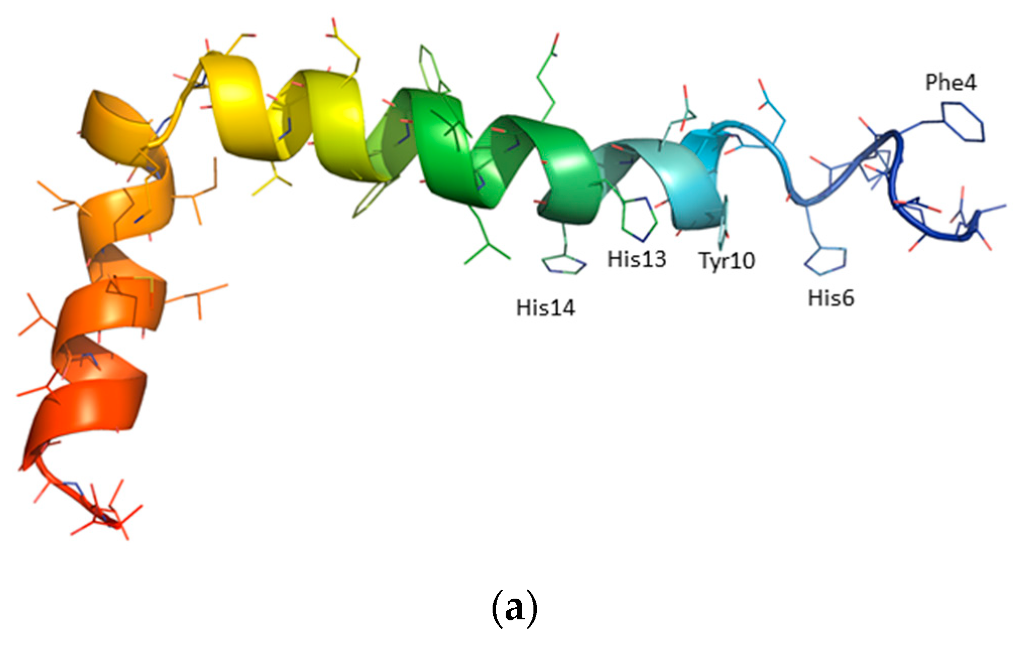
Figure 1. (a) The structure of Aβ1-42 polypeptide (PDB id code: 1IYT) [35]; (b) The sequences of three selected for the studies fragments of Aβ1-42 polypeptide: HZ1 (RHDSGYEVHHQK), HZ2 (SGYEVH), and HZ3 (SGYEV).
Symmetry 12 01579 g001aSymmetry 12 01579 g001b
Figure 2. Molecular structures of the investigated ligands: painkillers (aspirin and ibuprofen) and compounds of natural origin (anabasine and epinephrine).
Symmetry 12 01579 g002
Figure 3. The plots of F F 0 F 0 vs. c drug for all studied peptide-drug systems in 5 mM MES buffer (pH 6.0) at 20 °C.
Symmetry 12 01579 g003
Figure 4. The Lineweaver-Burk double-reciprocal curves of F 0 F 0 F vs. c drug 1 for all studied peptide-drug systems in 5 mM MES buffer (pH 6.0) at 20 °C.
Symmetry 12 01579 g004
Figure 5. (a) The distance between ibuprofen and the center of mass of side chains present in Aβ1-42 polypeptide: Tyr10 (red box), Met35 (black diamond), Val40 (black times) and Ile41 (blue circle) (b) the probable location of the ibuprofen molecule in Aβ1-42 polypeptide region; (c) distance between ibuprofen and the center of mass of side chains present in HZ1 peptide: His2 (6) (blue circle), Asp3 (7) (black diamond), Tyr6 (10) (red box) (the numbers in parentheses correspond to those in Aβ1-42); and (d) the probable location of the ibuprofen molecule in the HZ1 peptide region.
Symmetry 12 01579 g005
Figure 6. (a) The distance between aspirin and the center of mass of side chains present in Aβ1-42 polypeptide: Phe4 (black diamond), His6 (blue circle), Tyr10 (red box), Val40 (black times) (b) the probable location of the aspirin molecule in Aβ1-42 polypeptide region; (c) distance between aspirin and the center of mass of side chains present in HZ1 peptide: His2 (6) (blue circle), Asp3 (7) (black diamond), Tyr6 (10) (red box) (the numbers in parentheses correspond to those in Aβ1-42); and (d) the probable location of the aspirin molecule in the HZ1 peptide region.
Symmetry 12 01579 g006
Figure 7. (a) The distance between epinephrine and the center of mass of side chains present in Aβ1-42 polypeptide: Phe4 (black diamond), His6 (blue circle), Tyr10 (red box), and Val40 (black times) and (b) the probable location of the epinephrine molecule in Aβ1-42 polypeptide region; (c) distance between epinephrine and the center of mass of side chains present in HZ1 peptide: His2 (6) (blue circle), Asp3 (7) (black diamonds), Tyr6 (10) (red box) (the numbers in parentheses correspond to those in Aβ1-42); and (d) the probable location of the epinephrine molecule in the HZ1 peptide region.
Symmetry 12 01579 g007
Figure 8. (a) The distance between anabasine and the center of mass of side chains present in Aβ1-42 polypeptide: His6 (black diamond), Tyr10 (red box), His14 (blue circle), Val40 (black times) (b) the probable location of the anabasine molecule in Aβ1-42 polypeptide region; (c) distance between anabasine and the center of mass of side chains present in HZ1 peptide: His2 (6) (blue circle), Asp3 (7) (black diamond), Tyr6 (10) (red box) (the numbers in parentheses correspond to those in Aβ1-42); and (d) the probable location of the anabasine molecule in the HZ1 peptide region.
Symmetry 12 01579 g008