Article

A Novel Anti-Cadherin-17 Monoclonal Antibody, Ca17Mab-5, for Multiple Applications

1
Department of Antibody Drug Development, Tohoku University Graduate School of Medicine, 2-1 Seiryo-machi, Aoba-ku, Sendai 980-8575, Japan
2
Department of Otolaryngology, Head and Neck Surgery, Tohoku University Graduate School of Medicine, 1-1 Seiryo-machi, Aoba-ku, Sendai 980-8575, Japan
*
Authors to whom correspondence should be addressed.
These authors contributed equally to this work.
Antibodies 2026, 15(4), 59; https://doi.org/10.3390/antib15040059
Submission received: 21 May 2026 / Revised: 3 July 2026 / Accepted: 8 July 2026 / Published: 10 July 2026

Abstract

Background/Objectives: Cadherin-17 (CDH17, LI-cadherin) is a non-classical cadherin with an atypical structure and unique functions. CDH17 expression is restricted to normal intestinal epithelium. Furthermore, CDH17 functions as an oncoprotein that promotes tumor migration and invasion in colorectal, gastric, and pancreatic cancers. Therefore, CDH17 is an important diagnostic marker and therapeutic target. The CDH17-directed strategies, including monoclonal antibodies (mAbs), bispecific Abs, antibody–drug conjugates (ADCs), and chimeric antigen receptor (CAR) T cells, have been evaluated in preclinical and clinical studies. Therefore, developing mAbs that specifically recognize cell surface-expressing CDH17 is essential for advancing both tumor diagnosis and therapy. Methods: Anti-human CDH17 mAbs (named Ca17Mabs) were developed by immunizing a mouse with CDH17-overexpressed cells and a high-throughput screening using flow cytometry. Results: Among Ca17Mabs, a clone, Ca17Mab-5 (IgG1, κ) specifically recognized CDH17-overexpressed Chinese hamster ovary-K1 (CHO/CDH17) cells with no detectable cross-reactivity to 21 other CDHs by flow cytometry. Ca17Mab-5 also detected endogenous CDH17 in human colorectal cancer cell lines, COLO201 and COLO205. The apparent dissociation constant (KD) values of Ca17Mab-5 for CHO/CDH17 and COLO205 were estimated as 1.5 × 10⁻⁸ M and 1.3 × 10⁻⁸ M, respectively. Furthermore, Ca17Mab-5 detected endogenous CDH17 by Western blotting. In immunohistochemistry, Ca17Mab-5 exhibited clear membranous staining in normal colon epithelium, colorectal, gastric, and pancreatic cancers. Conclusions: Ca17Mab-5 is a versatile tool for detecting CDH17 and has potential for tumor diagnosis.

1. Introduction

Cadherins (CDHs) play crucial roles in cell–cell adhesion and the maintenance of tissue architecture [1]. Classical cadherins have five extracellular cadherin (EC) repeats, a single transmembrane domain, and a cytoplasmic domain with highly conserved binding regions for the armadillo proteins, including β-catenin and p120-catenin [1]. The EC repeats, originally identified in the extracellular region of the classical type I cadherin CDH1/E-cadherin [2], bind to Ca2+ and mediate homophilic interactions that are important for cell sorting into different compartments during organogenesis [3]. In epithelia, CDH1 forms adherens junctions via homophilic interactions, which define the features of epithelial sheets and apical adhesive structures [4]. The ability to interact with cytoplasmic armadillo proteins is a key function of classical CDHs. p120 catenin directly associates with the membrane-proximal region of the CDH cytoplasmic tail, and β-catenin serves as a scaffold to anchor α-catenin [5]. The cadherin–catenin complex associates directly or indirectly with signaling molecules, scaffolding proteins, and cytoskeletal regulators such as filamentous actin [5].
Cadherin-17 (CDH17), also known as liver-intestine CDH (LI-CDH) or seven-domain (7D) CDH, is a non-classical CDH with seven EC repeats, a single transmembrane domain, and a short cytoplasmic domain that lacks armadillo protein-binding motifs [6,7,8]. The tissue distribution of CDH17 varies across species. CDH17 is expressed in the liver and intestine in rats. In mice and humans, expression is restricted to small intestinal and colonic epithelial cells, with no detectable expression in the liver or stomach [9].
CDH17 plays a crucial role in intestinal barrier function [6]. CDH17-knockout (KO) mice displayed enhanced epithelial permeability in the small intestine and colon in vitro and in vivo models. Moreover, CDH17-KO mice exhibited an increased susceptibility to colitis induced by dextran sulfate sodium (DSS) [10]. In the DSS and azoxymethane-induced colorectal cancer model, CDH17-KO mice showed increased tumor formation and progression in the intestine [10]. Therefore, CDH17 is essential for intestinal homeostasis by limiting intestinal epithelial permeability and acts as a tumor suppressor in colitis-associated tumorigenesis.
Meanwhile, CDH17 is upregulated in colorectal cancer [11] and has been identified as a key mediator of metastatic progression and poor patient survival [12] through interactions with unique binding partners [13,14]. CDH17 binds to the desmosomal cadherin desmocollin-1 (DSC1) and indirectly interacts with actin filaments through the DSC1/p120–catenin complex, thereby promoting migration and invasiveness in CDH17-positive colorectal cancer [15]. CDH17 also interacts with α2β1 integrin via the RGD motif in its ectodomain, thereby promoting colorectal cancer liver metastasis [12,16]. Furthermore, a global transcriptome analysis revealed that silencing CDH17 in metastatic colorectal cancer cell lines markedly reduced the intestinal cancer stem cell marker LGR5, which led to attenuation of Wnt/β-catenin signaling, suppression of stemness-related genes, and consequent impairment of stem cell phenotypes [17].
CDH17 has been considered a promising therapeutic target for preventing metastatic progression in colorectal cancer, with approaches, including monoclonal antibodies (mAbs), bispecific Abs, antibody–drug conjugates (ADCs), and chimeric antigen receptor (CAR)-T [18,19]. CDH17-targeting ADCs have been developed and evaluated in clinical studies. TORL-3-600 is a humanized ADC with an MMAE payload that entered a phase I clinical trial for the treatment of advanced colorectal cancer (NCT05948826) [20]. AMT-676 [21] and YL217 [22] are humanized ADCs with topoisomerase I inhibitor payloads that have entered phase I clinical trials for the treatment of advanced solid tumors (NCT06400485 and NCT06859762, respectively). Furthermore, a T-cell engager cabotamig (CD3 × CDH17 bispecific Ab, NCT05411133) and anti-CDH17 CAR-T therapies (NCT06055439, NCT06501183, and NCT07216560) have been evaluated in clinical studies.
MAbs that detect CDH17 have been developed for Western blotting or immunohistochemistry (IHC). However, suitable anti-CDH17 mAbs for flow cytometry and IHC have been limited. We have developed the Cell-Based Immunization and Screening (CBIS), which includes immunizing antigen-overexpressed cells and high-throughput flow cytometry-based screening to generate highly versatile and specific mAbs. Using the CBIS method, we have developed anti-CDH1 [23], anti-CDH5/VE-cadherin [24], anti-CDH13 [25], and anti-CDH15/M-cadherin [26] mAbs for flow cytometry, Western blotting, and IHC. MAbs obtained from the CBIS method typically recognize conformational epitopes and are applicable in flow cytometry. Importantly, some of them are also applicable in Western blotting and IHC. In this study, we developed highly versatile anti-CDH17 mAbs by the CBIS method.

2. Materials and Methods

2.1. Cell Lines

Human glioblastoma (GBM) LN229, human colorectal cancer COLO205 and HCT116, Chinese hamster ovary (CHO)-K1, and mouse myeloma P3X63Ag8U.1 (P3U1) cell lines were obtained from the American Type Culture Collection (ATCC, Manassas, VA, USA). Another human colorectal cancer cell line, COLO201, was obtained from the Cell Resource Center for Biomedical Research, Institute of Development, Aging, and Cancer, Tohoku University (Miyagi, Japan). CHO-K1, P3U1, and CDH-overexpressed CHO-K1 (e.g., CHO/CDH17), COLO201, COLO205, HCT116, LN229, and CDH17-overexpressed LN229 (LN229/CDH17) were cultured as described previously [23].
CDH17-KO COLO205 (BINDS-86) was generated using the CRISPR/Cas9 system. A CDH17-specific sgRNA targeting the sequence 5′- CAACTGGATATGGCCAAGAG -3′ (TrueGuide™ Synthetic sgRNA, Thermo Fisher Scientific, Inc., Waltham, MA, USA) was synthesized and cloned into the GeneArt™ CRISPR Nuclease OFP Vector (Thermo Fisher Scientific, Inc.).

2.2. Establishment of Cadherin–Overexpressed Stable Transfectants

Full-length CDH17 cDNA (NM_004063.4) was obtained from OriGene Technologies, Inc. (Rockville, MD, USA). The CDH17 cDNA was cloned into the pCMV6 vector. The vector was transfected into LN229 or CHO-K1 cells using the Neon transfection system (Thermo Fisher Scientific, Inc.). Stable transfectants were sorted using an anti-CDH17 mAb (clone CDH17/2618, Novus Biologicals, Centennial, CO, USA). Finally, LN229/CDH17 and CHO/CDH17 were established. Type I cadherin-overexpressed CHO-K1: CHO/CDH1, CHO/CDH2 (CHO/PA16-CDH2), CHO/CDH3, CHO/CDH4 (CHO/PA16-CDH4), and CHO/CDH15 (CHO/PA16-CDH15) were previously established [23]. Type II cadherin-overexpressed CHO-K1: CHO/CDH5 (CHO/PA16-CDH5), CHO/CDH6, CHO/CDH7 (CHO/PA16-CDH7), CHO/CDH8 (CHO/PA16-CDH8), CHO/CDH9 (CHO/PA16-CDH9), CHO/CDH10 (CHO/PA16-CDH10), CHO/CDH11 (CHO/PA16-CDH11), CHO/CDH12 (CHO/PA16-CDH12), CHO/CDH18 (CHO/PA16-CDH18), CHO/CDH19 (CHO/PA16-CDH19), CHO/CDH20 (CHO/PA16-CDH20), CHO/CDH22 (CHO/PA16-CDH22), and CHO/CDH24 (CHO/PA16-CDH24) were established previously [27]. A truncated cadherin-overexpressed CHO-K1: CHO/CDH13 (CHO/PA16-CDH13), another 7D cadherin-overexpressed CHO-K1: CHO/CDH16 (CHO/PA16-CDH16), and an atypical cadherin-overexpressed CHO-K1: CHO/CDH26 (CHO/PA16-CDH26) were previously established [27].
Each cadherin expression was confirmed using an anti-CDH1 mAb (clone 67A4, BD Biosciences, Franklin Lakes, NJ, USA), an anti-CDH3 mAb (clone MM0508-9V11, Abcam, Cambridge, UK), an anti-CDH6 mAb (clone 427909, R&D Systems Inc., Minneapolis, MN, USA), an anti-CDH13 mAb (clone 392411, R&D Systems Inc.), an anti-CDH17 mAb (clone CDH17/2618), and an anti-PA16-tag mAb (clone NZ-33) to detect other cadherins.

2.3. Hybridoma Production

The animal experiment was approved by the Animal Care and Use Committee of Tohoku University (Permit No. 2022MdA-001) and conducted according to relevant guidelines and regulations to minimize animal suffering and distress in the laboratory. The animal was monitored daily for health during the full duration of the experiment. A reduction in more than 25% of the total body weight was defined as a humane endpoint. A 6-week-old female BALB/cAJcl mouse (CLEA Japan, Tokyo, Japan) was immunized intraperitoneally with LN229/CDH17 (1 × 10⁸ cells). For the first immunization, 2% Alhydrogel adjuvant (InvivoGen, San Diego, CA, USA) was included. This was followed by three weekly intraperitoneal injections of LN229/CDH17 (1 × 10⁸ cells) without adjuvant. Two days before splenocyte collection, a final intraperitoneal booster injection of LN229/CDH17 (1 × 10⁸ cells) was performed. Hybridomas were produced as described previously [23]. The hybridoma supernatants were screened by flow cytometry using CHO/CDH17 and parental CHO-K1 cells. The hybridoma culture supernatant containing Ca17Mab-5 in serum-free Hybridoma-SFM medium (Thermo Fisher Scientific, Inc.) was purified using Ab-Catcher Extra (ProteNova, Kagawa, Japan).

2.4. Flow Cytometry

Cells were harvested with 1 mM EDTA and washed with blocking buffer [0.1% bovine serum albumin in phosphate-buffered saline (PBS)]. The cells were incubated with primary mAbs for 30 min at 4 °C. The cells were then stained with Alexa Fluor 488-conjugated anti-mouse IgG (1:2000; Cell Signaling Technology, Inc., Danvers, MA, USA). Flow cytometric data were acquired on an SA3800 Cell Analyzer (Sony Corp., Tokyo, Japan). Cells were gated on forward scatter (FSC) and side scatter (SSC), and fluorescence intensity was analyzed using FlowJo software (ver. 10.8.1, BD Biosciences). At least two independent experiments were conducted.

2.5. Calculation of the Binding Affinity by Flow Cytometry

Cells were treated with serial dilutions of Ca17Mab-5 or CDH17/2618. The cells were stained with Alexa Fluor 488-conjugated anti-mouse IgG (1:200 dilution). The data were collected, and the geometric mean (GeoMean) was calculated in FlowJo. Three independent measurements were performed, and the fitting binding isotherms (horizontal axis, mAb concentration; vertical axis, GeoMean) determined the apparent dissociation constant (KD) values using built-in one-site binding models of GraphPad Prism 6 (GraphPad Software, Inc., La Jolla, CA, USA).

2.6. Western Blotting

Western blotting was performed as described previously [23]. Ca17Mab-5 (1 μg/mL), CDH17/2618 (1 μg/mL), and an anti-isocitrate dehydrogenase 1 (IDH1) mAb (clone RcMab-1-mG1, 2 μg/mL) were used as primary mAbs. Horseradish peroxidase-conjugated anti-mouse IgG (1:1000; Agilent Technologies Inc., Santa Clara, CA, USA) was used as secondary mAb. Chemiluminescence signals were developed using Pierce™ ECL Plus (Thermo Fisher Scientific, Inc.) or ImmunoStar LD (FUJIFILM Wako Pure Chemical Corporation, Osaka, Japan). The signals were imaged with ChemiDoc Touch MP (Bio-Rad Laboratories, Inc., Berkeley, CA, USA). Two independent experiments were conducted.

2.7. Immunohistochemistry Using Cell Blocks and Tissue Microarrays

IHC was performed using the VENTANA BenchMark ULTRA PLUS (Roche Diagnostics, Indianapolis, IN, USA). CHO-K1, CHO/CDH17, and COLO205 were fixed with 4% paraformaldehyde, and cell blocks were prepared using iPGell (Genostaff Co., Ltd., Tokyo, Japan). Formalin-fixed, paraffin-embedded (FFPE) cell sections were stained with Ca17Mab-5 (0.2 or 2 μg/mL) or CvMab-62 (2 μg/mL, IgG1 isotype control).
Colorectal cancer microarrays (CO243b, CO352a, CO353a, CO483b, and CO484b, US Biomax Inc., Rockville, MD, USA), a gastric cancer microarray (BS01012e, US Biomax Inc.), another gastric cancer microarray (PA484, US Biomax Inc.), and a normal human organ tissue microarray (FDA999x, US Biomax Inc.) were stained with Ca17Mab-5 (5 μg/mL or 10 μg/mL in CO243b). Staining was performed using the ultraView Universal DAB Detection Kit (Roche Diagnostics).

3. Results

3.1. Development of Anti-CDH17 mAbs by the CBIS Method

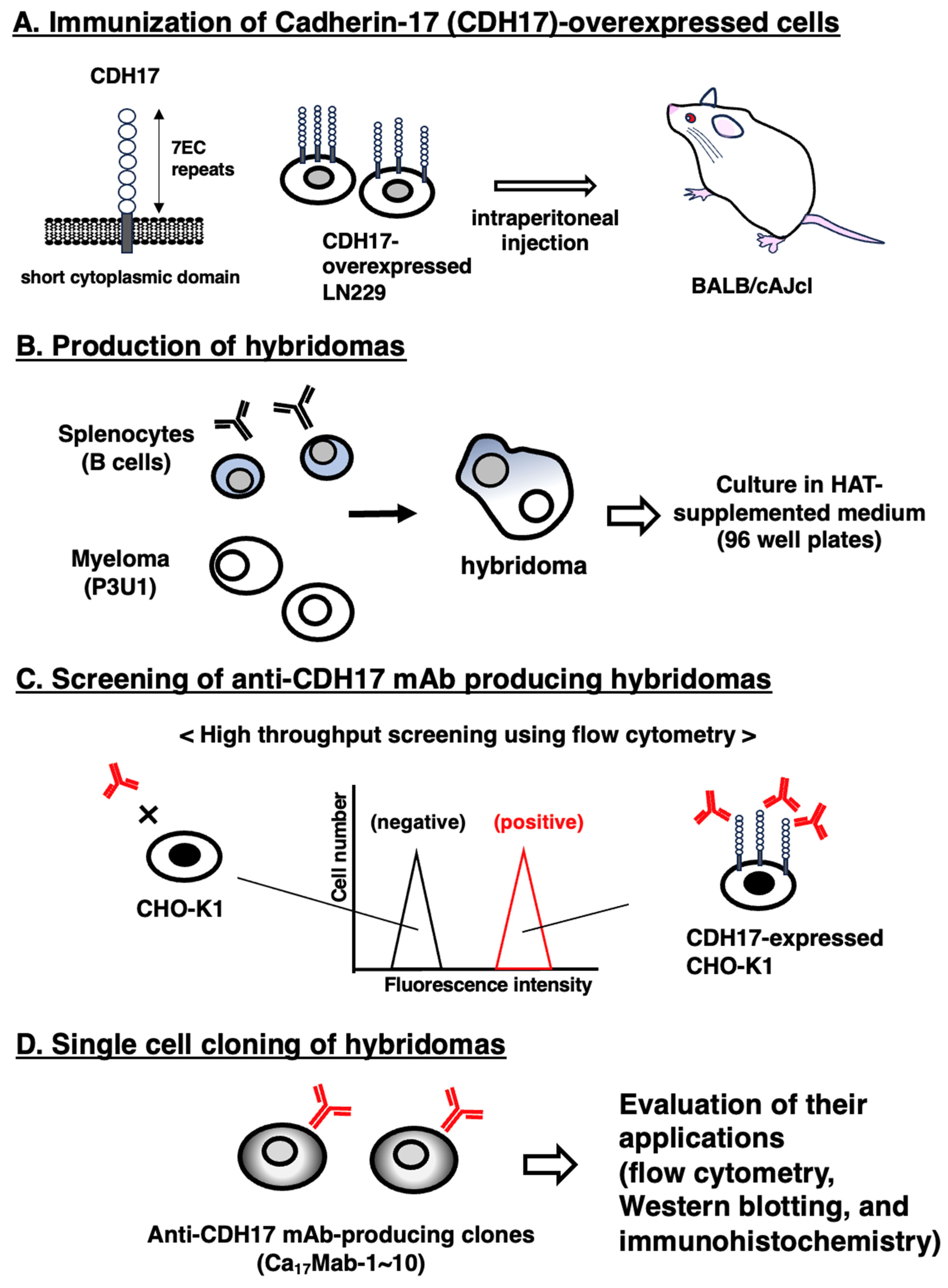
We first prepared the immunogen LN229/CDH17 as described in Section 2.2 and Figure 1A. LN229/CDH17 (1 × 10⁸ cells) were injected intraperitoneally five times into a BALB/cAJcl mouse (Figure 1A). Splenocytes were fused with myeloma P3U1 cells to generate hybridomas, which were plated in 96-well plates (Figure 1B). Hybridoma supernatants were screened to identify those positive for CHO/CDH17 and negative for CHO-K1 (Figure 1C). Flow cytometric screening identified 129 of 474 wells (27%) that showed strong signals with CHO/CDH17 compared to CHO-K1. Limiting dilution was then performed, and a total of 10 clones producing anti-CDH17 mAbs were established. Supernatants were further evaluated for use in flow cytometry, Western blotting, and immunohistochemistry (Figure 1D). Finally, clone Ca17Mab-5 (IgG1, κ) was selected because it can be used in all applications (https://www.med-tohoku-antibody.com/topics/antibody_bank.htm, accessed on 25 June 2026).

3.2. Flow Cytometric Analysis of Ca17Mab-5 Against CHO/CDH17 and CHO-K1

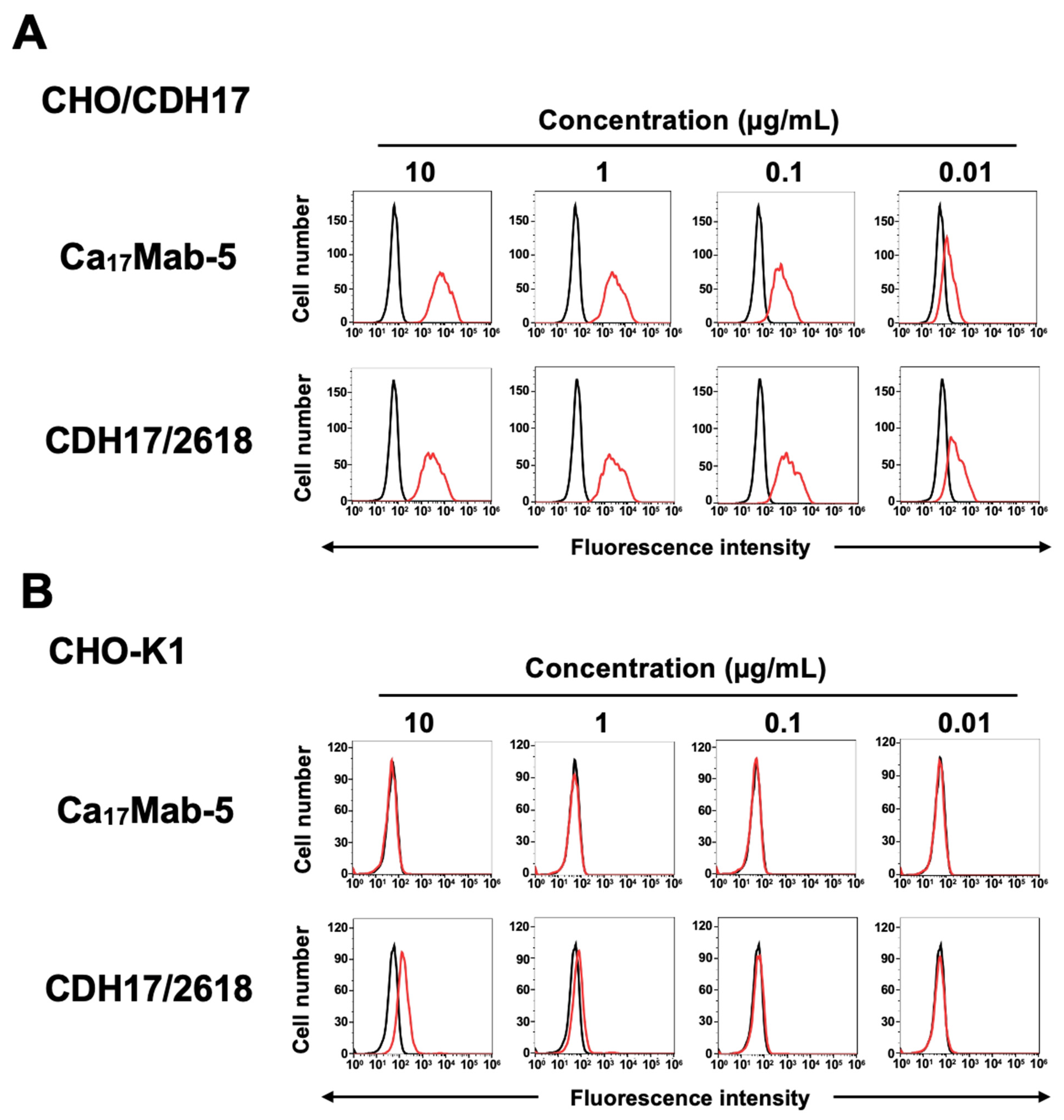
Purified Ca17Mab-5 was prepared, and its reactivity was assessed by flow cytometry. As shown in Figure 2A, Ca17Mab-5 reacted with CHO/CDH17 in a dose-dependent manner from 10 to 0.01 μg/mL. In contrast, Ca17Mab-5 did not recognize parental CHO-K1 even at 10 μg/mL (Figure 2B). Although a commercially available anti-CDH17 mAb (clone CDH17/2618) reacted with CHO/CDH17 in a dose-dependent manner from 10 to 0.01 μg/mL (Figure 2A), it also non-specifically recognized CHO-K1 at 10 μg/mL (Figure 2B).

3.3. Determination of the Specificity of Ca17Mab-5 Using CDHs-Overexpressed CHO-K1

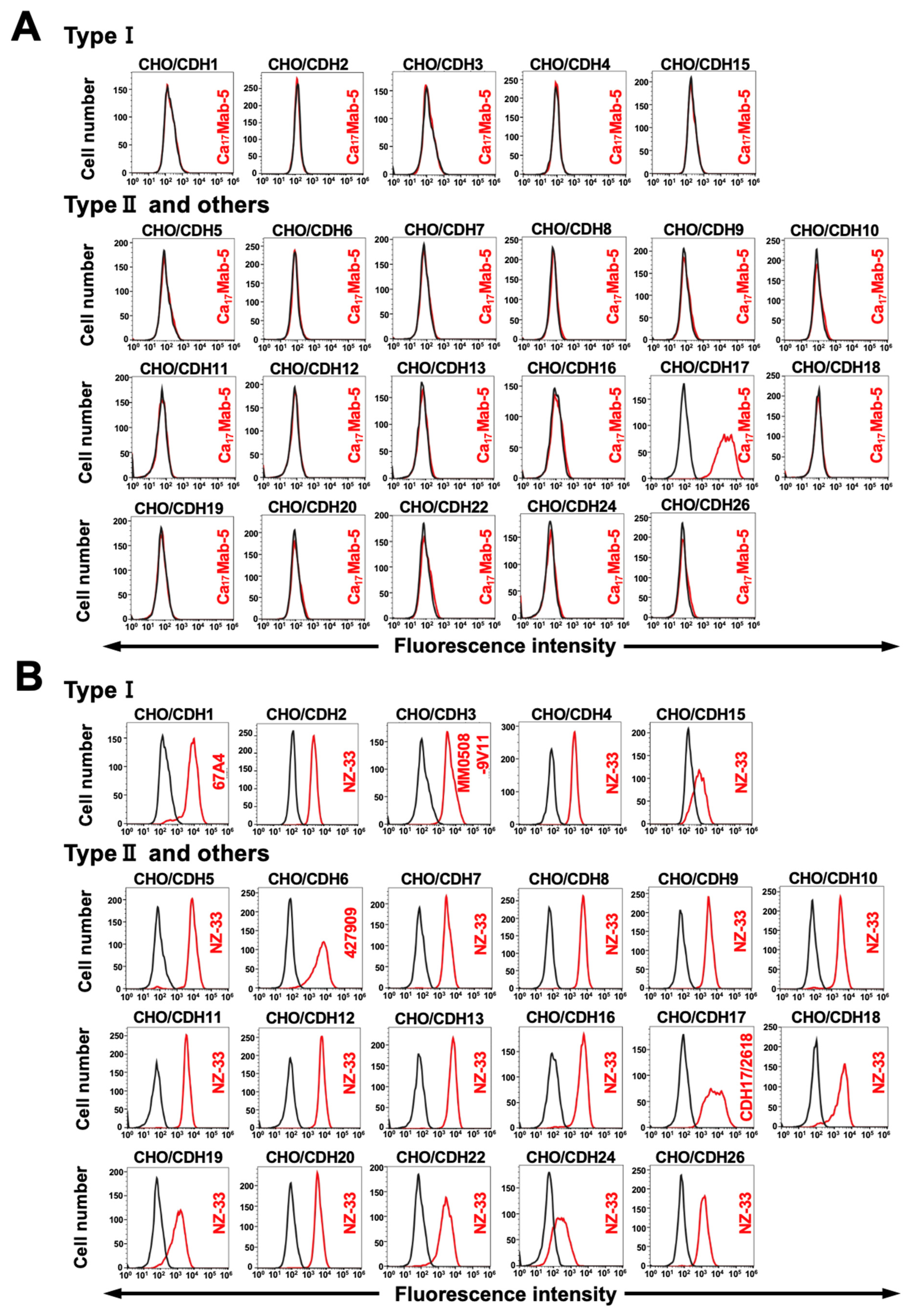
We previously generated CHO-K1 cells that overexpressed type I CDHs [23,26], type II CDHs, another 7D CDH, and an atypical CDH [27]. Therefore, the specificity of Ca17Mab-5 to those CDHs was investigated. As shown in Figure 3A, Ca17Mab-5 recognized CHO/CDH17 but did not cross-react with other CDHs-overexpressed in CHO-K1. The cell surface expression of each CDH was confirmed in Figure 3B. These results indicated that Ca17Mab-5 is a specific mAb to CDH17 among those CDHs.

3.4. Flow Cytometric Analysis of Ca17Mab-5 and CDH17/2618 Against Endogenous CDH17-Positive and Negative Cells

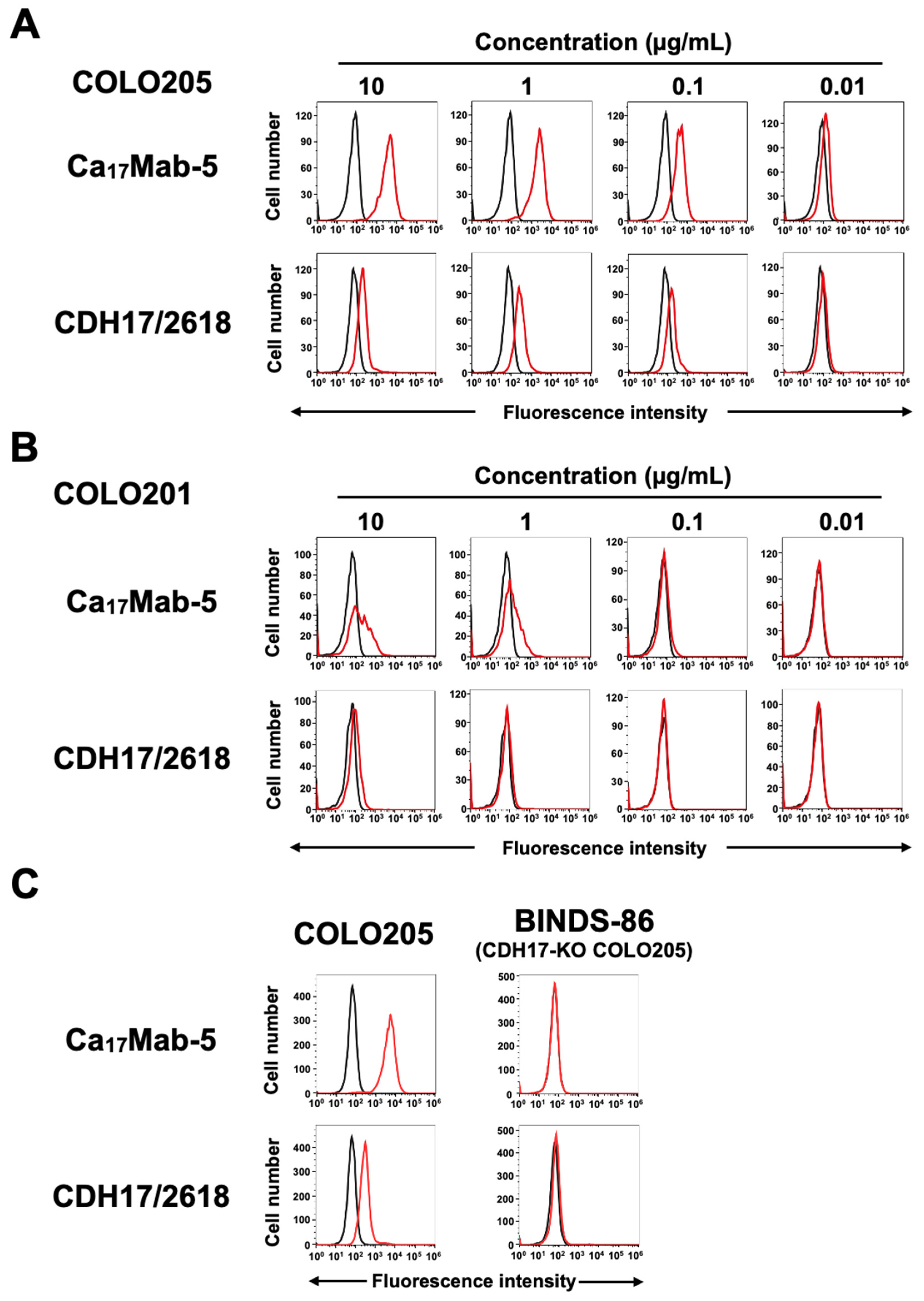
We then searched the expression of CDH17 in various colorectal cancer cell lines using Ca17Mab-5 and CDH17/2618. As shown in Figure 4, Ca17Mab-5 dose-dependently recognized endogenous CDH17 in COLO205. In contrast, the reactivity of CDH17/2618 was lower than that of Ca17Mab-5 (Figure 4A). Ca17Mab-5 exhibited a weak reactivity to COLO201, whereas CDH17/2618 barely reacted with COLO201 (Figure 4B). Additionally, Ca17Mab-5 and CDH17/2618 hardly recognized HCT116 (Supplementary Figure S1). Furthermore, we generated CDH17-KO COLO205 (BINDS-86). As shown in Figure 4C, the reactivity of Ca17Mab-5 completely disappeared in BINDS-86.
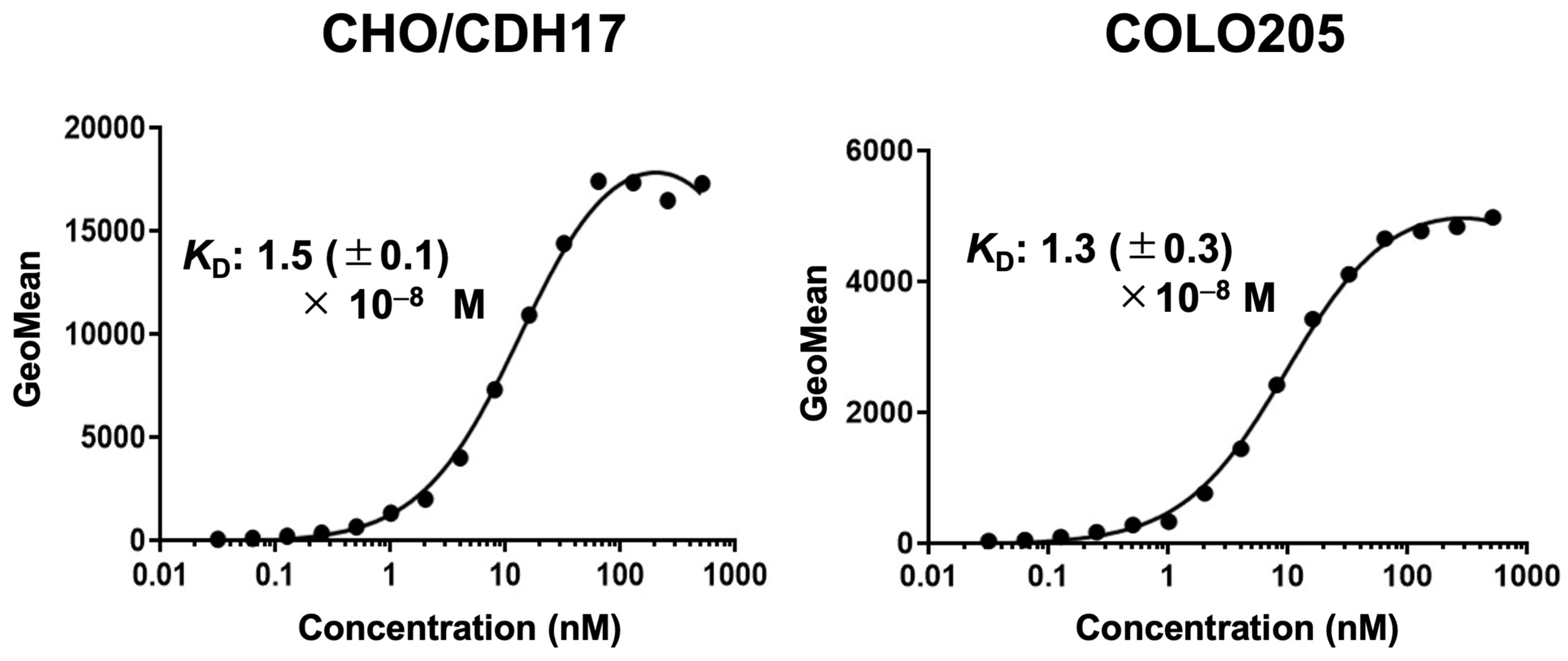
The binding affinity of Ca17Mab-5 was measured with CHO/CDH17 and COLO205 using flow cytometry. The apparent KD values for Ca17Mab-5 with CHO/CDH17 and COLO205 were 1.5 (±0.1) × 10−8 M and 1.3 (±0.3) × 10−8 M, respectively (Figure 5). Supplementary Figure S2 showed the additional information on three independent binding affinity measurements of Ca17Mab-5. The apparent KD value of CDH17/2618 for COLO205 could not be determined because the sigmoid curve did not reach a plateau (Supplementary Figure S3). These results showed that Ca17Mab-5 has moderate binding affinity for exogenous and endogenous CDH17.

3.5. Detection of Exogenous and Endogenous CDH17 by Ca17Mab-5 and CDH17/2618 in Western Blotting

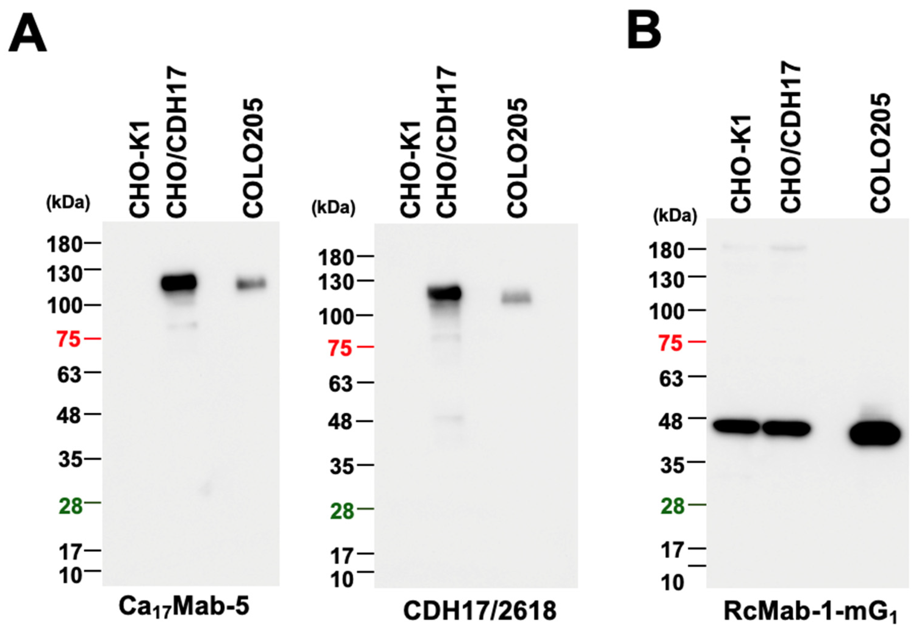
We next tested whether Ca17Mab-5 and CDH17/2618 can be used in Western blotting. As shown in Figure 6A, Ca17Mab-5 and CDH17/2618 detected a band of approximately 120 kDa in CHO/CDH17 cell lysates, whereas no band was detected in parental CHO-K1 cell lysates. Additionally, Ca17Mab-5 and CDH17/2618 detected endogenous CDH17 in COLO205 cell lysates at 120 kDa (Figure 6A). An anti-IDH1 mAb (clone RcMab-1-mG1) served as an internal control (Figure 6B). These results demonstrated that Ca17Mab-5 and CDH17/2618 can detect CDH17 by Western blotting.

3.6. Immunohistochemistry Using Ca17Mab-5 in Formalin-Fixed Paraffin-Embedded Cell Blocks

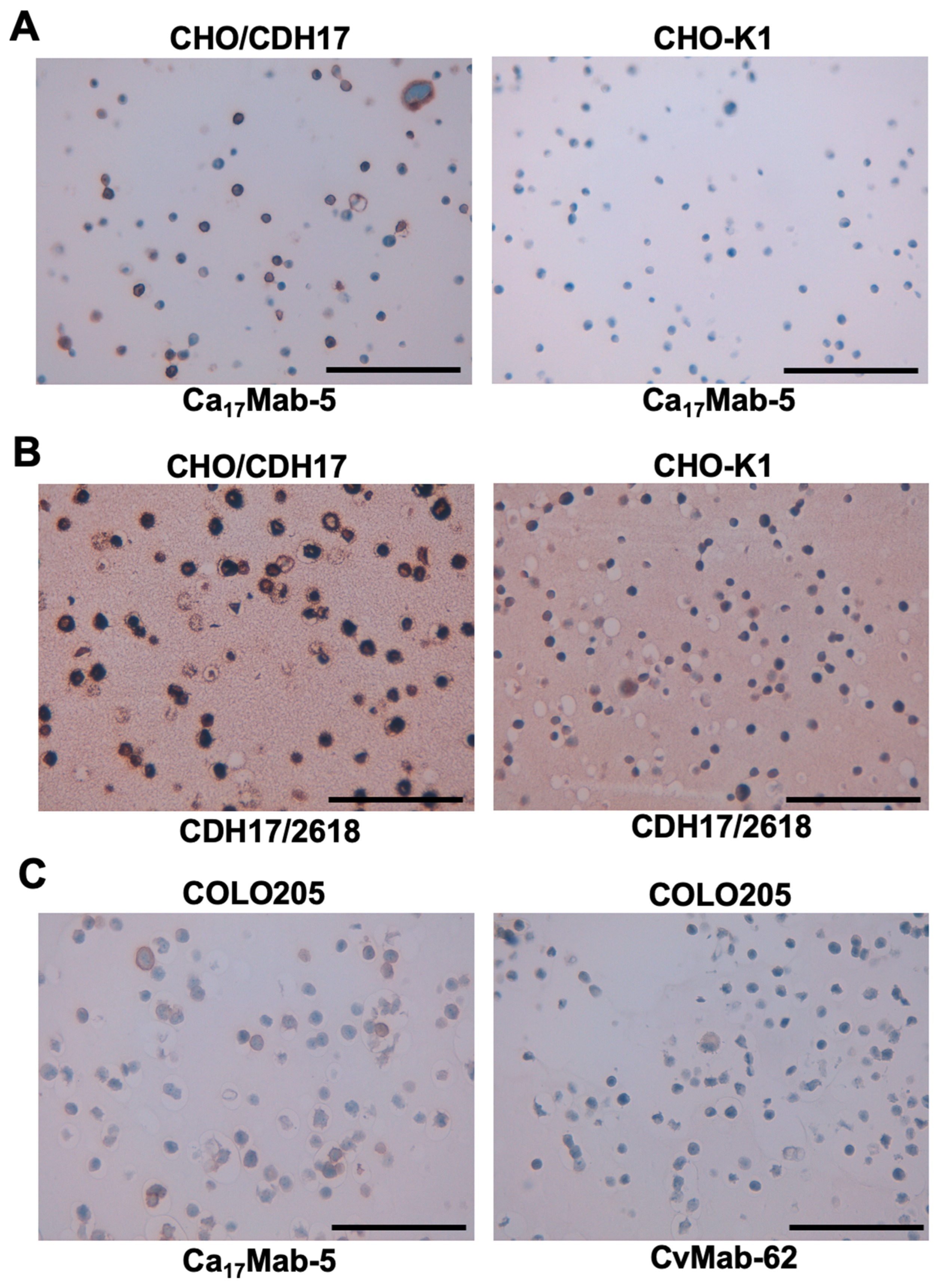
We tested whether Ca17Mab-5 and CDH17/2618 are suitable for IHC in FFPE sections from CHO-K1 and CHO/CDH17. Ca17Mab-5 showed clear membranous staining in CHO/CDH17 but not in CHO-K1 (Figure 7A). In the same experimental setting, CDH17/2618 showed potent reactivity to CHO/CDH17, but weak reactivity to CHO-K1 was also detected (Figure 7B). Furthermore, Ca17Mab-5 also displayed membranous staining in COLO205, but the isotype control mAb (CvMab-62) did not (Figure 7C). These results indicated that Ca17Mab-5 can detect both exogenous and endogenous CDH17 in IHC of FFPE sections from cultured cells.

3.7. Immunohistochemistry Using Ca17Mab-5 in Normal and Tumor Microarrays

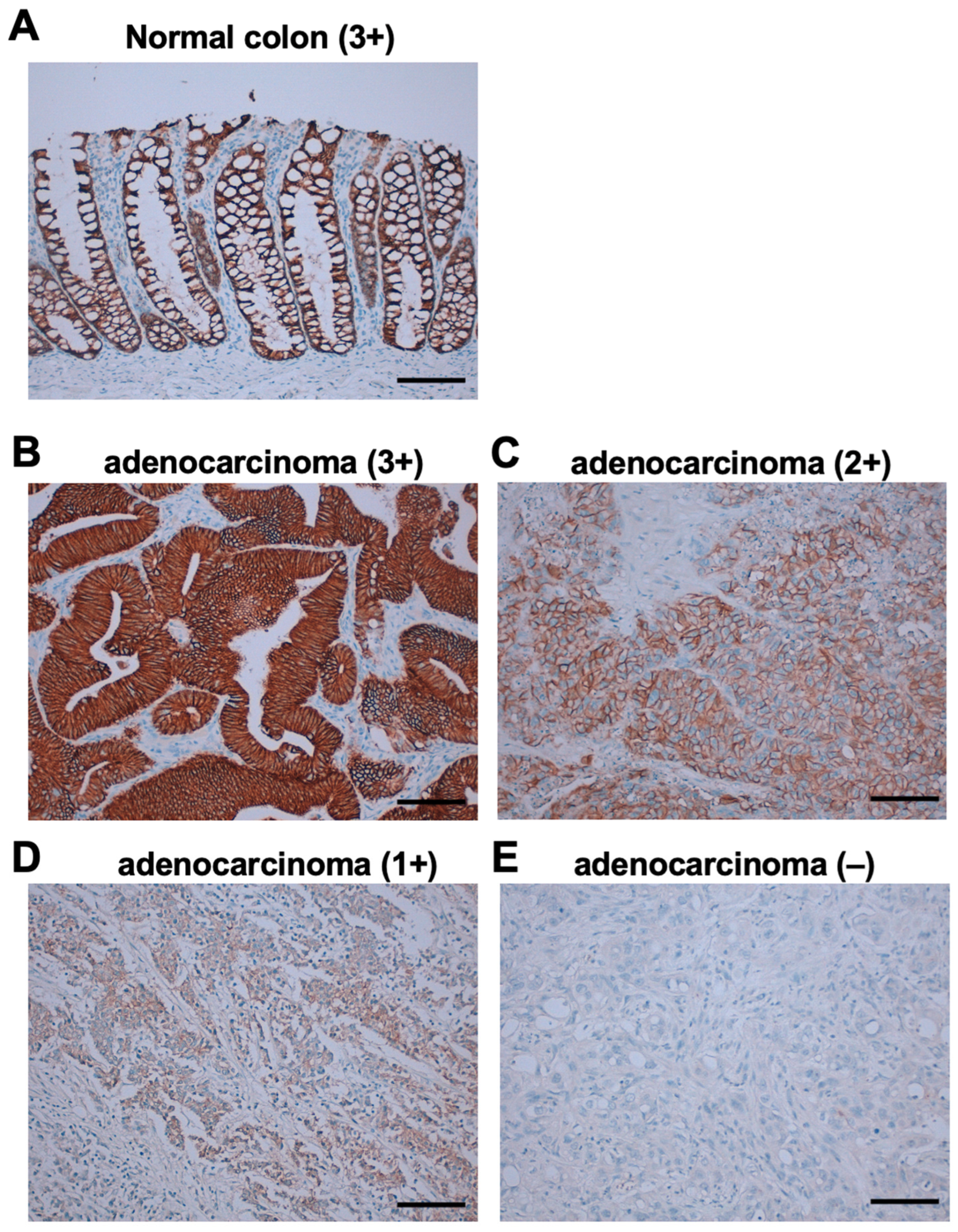
CDH17 expression is reported to be restricted to the small intestinal and colonic epithelium [9]. We investigated the CDH17 expression by Ca17Mab-5 using a human normal organ tissue microarray. As shown in Supplementary Figure S4, Ca17Mab-5 showed a potent membranous staining in small intestinal and colonic epithelia, and the weak staining was also observed in portions of normal pancreas. We next stained colorectal cancer microarrays. As shown in Figure 8A, Ca17Mab-5 exhibited a strong basolateral membranous staining (3+) in normal colon epithelium. In colorectal adenocarcinomas, Ca17Mab-5 showed strong membranous staining (3+) in well-differentiated adenocarcinomas (Figure 8B). The abundance of CDH17 detected by Ca17Mab-5 was reduced in poorly differentiated adenocarcinomas [Figure 8C (moderate intensity, 2+) and D (weak intensity, 1+)]. Adenocarcinomas with no CDH17 expression were also observed (Figure 8E). Table 1 summarizes the result of colorectal cancer microarray (CO483b), which includes Figure 8B–E. Supplementary Table S1 summarizes the results of other colorectal cancer tissue microarrays. Consequently, Ca17Mab-5 stained 133 out of 154 cases (86%, more than +1).
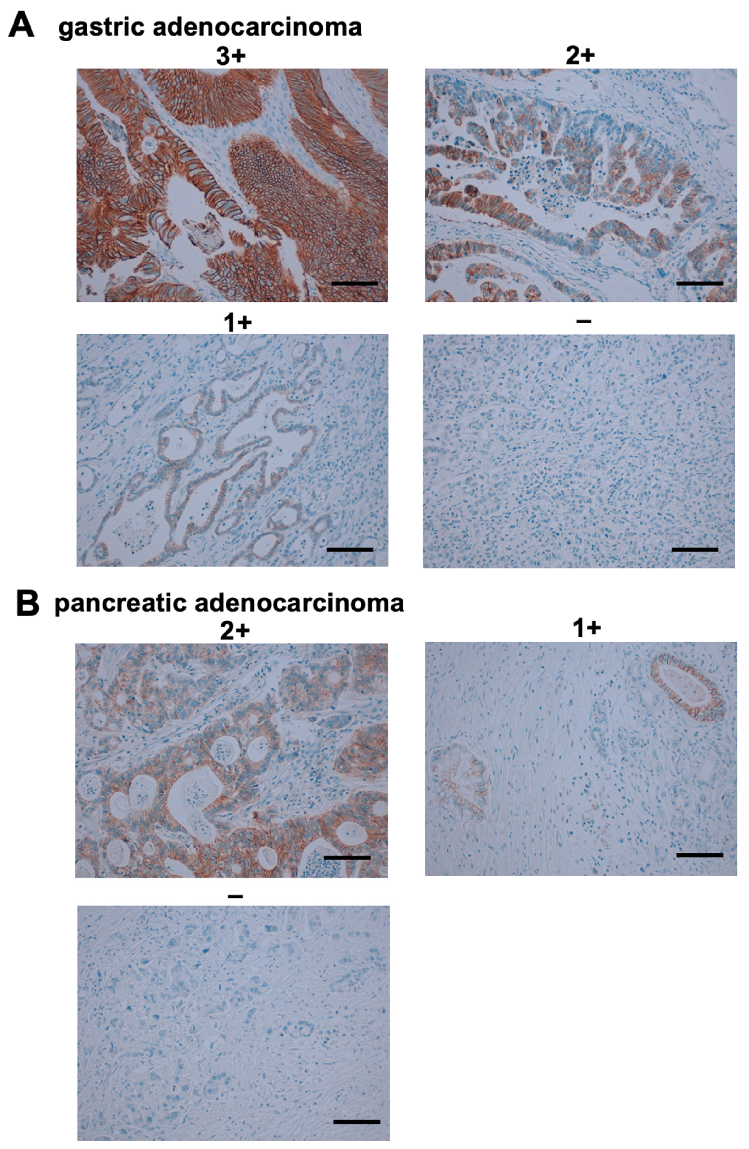
We further stained gastric and pancreatic cancer microarrays using Ca17Mab-5. As shown in Figure 9A, Ca17Mab-5 also exhibited potent membranous staining in well-differentiated gastric adenocarcinomas, and moderate/weak staining was observed. In pancreatic cancer, moderate and weak membranous staining were also observed (Figure 9B). Supplementary Tables S2 and S3 summarize the results of gastric and pancreatic cancer microarrays, respectively. Consequently, Ca17Mab-5 stained 20 out of 72 cases (28%) in gastric cancers and 8 out of 22 cases (36%) in pancreatic cancers. These results indicated that Ca17Mab-5 is suitable for detecting CDH17 in IHC.

4. Discussion

CDH17 is expressed almost exclusively in intestinal epithelial cells of the embryonic and adult small intestine and colon [28]. Consistent with this restricted tissue distribution, CDH17 is regarded as a selective marker of colon cancer cell lines and is expressed in 35 out of 54 epithelial-like colon cancer cell lines included in the Cancer Cell Line Encyclopedia [29]. In this study, we developed novel anti-CDH17 mAbs by immunizing a mouse with CDH17-overexpressed LN229 cells (Figure 1). A clone Ca17Mab-5 recognized both exogenous and endogenous CDH17 in flow cytometry (Figure 2 and Figure 4) and IHC (Figure 7 and Figure 8). The information on tumor differentiation and grade was not available in the microarrays used in this study, which restricts the biological interpretation of the immunohistochemical findings. Additionally, there was no correlation between the IHC score and the T stage (p-value = 0.42).
Ca17Mab-5 was able to detect CDH17 in COLO205 but not HCT116 (Figure 4 and Figure S1), which is consistent with the gene expression profiles of colorectal cancer cell lines in the NCI-60 human cancer cell line panel [29]. Importantly, Ca17Mab-5 specifically recognized CDH17 without detectable cross-reactivity to other 21 cadherins, including classical type I and type II CDHs, and other types of CDHs (Figure 3). Therefore, Ca17Mab-5 will be useful for the specific isolation of CDH17-positive cells via fluorescence-activated cell sorting for basic research.
A commercially available anti-CDH17 mAb (clone CDH17/2618) was developed by immunization of a recombinant fragment (amino acids 242–418) of human CDH17 (https://www.novusbio.com/products/cadherin-17-antibody-cdh17-2618_nbp2-79723?srsltid=AfmBOoqdTWDqYHSRLjuVYR-gDiwQheUveNYpDjFFCxpS_btPMehtSecp#datasheet, accessed on 25 June 2026). CDH17/2618 similarly detected both exogenous and endogenous CDH17 in Western blotting (Figure 6). However, the reactivity to endogenous CDH17 in flow cytometry was low compared to that of Ca17Mab-5 (Figure 4A). Weak reactivities that appear to be in the background were observed at 10 µg/mL (Figure 2B and Figure 4). This may be a reason why the sigmoid curve of CDH17/2618 did not reach a plateau (Supplementary Figure S3). The strategy of CDH17/2618 selection was not described on the above website. Since Ca17Mab-5 was selected by a flow cytometry-based screening (Figure 1C), Ca17Mab-5 showed a reactivity to COLO205, which is thought to be important to target tumor cells in vivo for the development of therapeutic mAb. Furthermore, Ca17Mab-5 can be used for IHC of cell specimens (Figure 7) and tissue microarrays (Figure 8). Notably, IHC was conducted by an automated slide-staining system, VENTANA BenchMark ULTRA PLUS, which ensures reproducible staining conditions and accurate assessment of target expression for diagnosis.
CDH17 is aberrantly expressed not only in colorectal cancer [11,30] but also in gastric cancer [31,32], ovarian cancer [33], hepatocellular carcinoma [34], and pancreatic neuroendocrine tumors [35]. We confirmed the expression of CDH17 in gastric and pancreatic cancers by Ca17Mab-5 (Figure 9). CDH17 transcription is regulated by CDX2, an intestine-specific caudal-related homeobox transcription factor that plays an important role in the regulation of intestinal epithelium homeostasis [36,37]. CDX2 also mediates gastric intestinal metaplasia [38,39], a precancerous lesion defined by the replacement of gastric mucosa with intestinal-like epithelium, with a considerable risk for gastric cancer without effective therapeutic strategies [40,41,42]. There is no information that the CDH17-positive gastric cancers in Figure 9A were derived from gastric intestinal metaplasia. The pathological analysis of CDH17 expression in gastric cancer development from metaplasia is thought to be important.
Functionally, CDH17 has been shown to be a critical regulator of the stemness and chemoresistance through upregulation of the LGR5/Wnt/MYC axis and the glutamine transporter SLC38A5, respectively [17]. An anti-CDH17 RGD mAb (clone 6.6.1), an inhibitor of the α2β1 integrin-CDH17 interaction, suppressed the LGR5 expression and downstream Wnt signaling activity, suggesting that the CDH17-α2β1 integrin interaction promotes the acquisition of stemness in colorectal cancer cells [17,43]. Since the binding epitope of Ca17Mab-5 has not been identified, further studies are essential to determine its epitope and the biological activities of Ca17Mab-5. We previously published several methods to identify the binding epitope of membrane proteins using flow cytometry [44], which will aid the characterization of Ca17Mab-5 and other Ca17Mab clones in the future.
To target CDH17-positive tumors, multiple therapeutic strategies, including mAb monotherapy [43], bispecific Abs such as TRAILR2 × CDH17 [45,46] and CDH3 × CDH17 [47], ADCs [48], immunotoxins [49], radiolabeled agents [50], CAR-T [51,52], and CAR-NK cell therapies [53], have been developed in preclinical studies. Some of these modalities have been evaluated in clinical trials [54]. We previously cloned mAb cDNAs from hybridomas and generated recombinant human IgG1 mAbs to enhance antibody-dependent cellular cytotoxicity (ADCC), and evaluated the antitumor efficacy in human tumor xenograft models [55]. We have successfully cloned the cDNA encoding Ca17Mab-5, and an isotype-converted human IgG1-type Ca17Mab-5 will be generated. In a future study, we will assess the antitumor efficacy using in vitro ADCC assays and tumor xenograft models.
As shown in Figure 8A and Figure S4, and previous observation, CDH17 is predominantly localized to the basolateral membrane of normal intestinal epithelium [52]. This restricted distribution is thought to minimize ADC or CAR-T cell access to normal tissues, thereby reducing the risk of on-target off-tumor toxicities [48,52]. However, there are no preclinical models that accurately predict anti-CDH17 therapy-associated toxicities, raising concerns about on-target off-tumor toxicities in the clinical application of Ca17Mab-5. Adverse effects, including diarrhea and mucositis, could narrow the therapeutic window for clinical applications.
To achieve a favorable therapeutic window with reducing on-target off-tumor toxicities, we have developed cancer-specific mAbs (CasMabs) directed against several tumor-associated antigens, such as human epidermal growth factor receptor 2 (HER2) [56], and successfully identified a cancer-specific epitope of HER2. An anti-HER2 CasMab, H2CasMab-2, was screened based on the cancer-specific binding profile in flow cytometry [56]. H2CasMab-2 recognized HER2-positive breast cancer cells, while exhibiting no detectable reactivity toward various normal cells [56]. Additionally, we elucidated the structural basis of the interaction between H2CasMab-2 and the extracellular domain IV in the HER2 ectodomain [57]. Furthermore, a single-chain variable fragment (scFv) derived from H2CasMab-2 was incorporated into chimeric antigen receptor (CAR) T cells, which exhibited cancer-selective reactivity and potent antitumor activity in preclinical models [57]. H2CasMab-2-derived CAR-T therapy has been evaluated in a phase I clinical trial for patients with HER2-positive advanced solid tumors (NCT06241456). Therefore, these findings underscore the importance of developing CasMabs against CDH17. Moreover, defining their cancer-specific epitopes is a critical step toward the establishment of safe and effective therapeutic anti-CDH17 CasMabs and related modalities. For this purpose, we are going to increase the clones of Ca17Mabs and screen CasMabs against CDH17.

Supplementary Materials

The following supporting information can be downloaded at https://www.mdpi.com/article/10.3390/antib15040059/s1. Figure S1: Flow cytometric analysis using Ca17Mab-5 and CDH17/2618 to detect endogenous CDH17 in HCT116. Figure S2: Additional information on three independent binding affinity measurements of Ca17Mab-5 by flow cytometry. Figure S3: Determination of binding affinity of CDH17/2618 by flow cytometry. Figure S4: Immunohistochemistry using Ca17Mab-5 (5 μg/mL) in a human normal organ tissue microarray. Table S1: Immunohistochemistry of colorectal cancer microarrays by Ca17Mab-5. Table S2: Immunohistochemistry of a gastric cancer microarray (BS01012e) by Ca17Mab-5. Table S3: Immunohistochemistry of a pancreatic cancer microarray (PA484) by Ca17Mab-5.

Author Contributions

Conceptualization, M.K.K. and Y.K. (Yukinari Kato); investigation, R.I., H.S., K.I. and K.Y.; supervision, A.O., Y.K. (Yukio Katori), and Y.K. (Yukinari Kato); writing—original draft preparation, R.I. and H.S.; writing—review and editing, Y.K. (Yukinari Kato); project administration, Y.K. (Yukinari Kato); funding acquisition, H.S. and Y.K. (Yukinari Kato). All authors have read and agreed to the published version of the manuscript.

Funding

This research was supported in part by the Japan Agency for Medical Research and Development (AMED) under Grant Numbers JP26am0521010 (to Y.Kato), JP26ama121008 (to Y.Kato), JP25ama221153 (to Y.Kato), and JP25ama221339 (to Y.Kato), and by the Japan Society for the Promotion of Science (JSPS) Grants-in-Aid for Scientific Research (KAKENHI) grant no. 25K10553 (to Y.Kato) and 26K02289 (to H.S.).

Institutional Review Board Statement

The animal study protocol was approved by the Animal Care and Use Committee of Tohoku University (Permit number: 2022MdA-001; Approval Date: 1 April 2022) for studies involving animals.

Informed Consent Statement

Not applicable.

Data Availability Statement

The data presented in this study are available in the article.

Conflicts of Interest

The authors declare no conflicts of interest.

References

  1. van Roy, F. Beyond E-cadherin: Roles of other cadherin superfamily members in cancer. Nat. Rev. Cancer 2014, 14, 121–134. [Google Scholar] [CrossRef] [PubMed]
  2. Oda, H.; Takeichi, M. Evolution: Structural and functional diversity of cadherin at the adherens junction. J. Cell Biol. 2011, 193, 1137–1146. [Google Scholar] [CrossRef] [PubMed]
  3. Takeichi, M. Cell sorting in vitro and in vivo: How are cadherins involved? Semin. Cell Dev. Biol. 2023, 147, 2–11. [Google Scholar] [CrossRef] [PubMed]
  4. Takeichi, M. Dynamic contacts: Rearranging adherens junctions to drive epithelial remodelling. Nat. Rev. Mol. Cell Biol. 2014, 15, 397–410. [Google Scholar] [CrossRef] [PubMed]
  5. Ratheesh, A.; Yap, A.S. A bigger picture: Classical cadherins and the dynamic actin cytoskeleton. Nat. Rev. Mol. Cell Biol. 2012, 13, 673–679. [Google Scholar] [CrossRef] [PubMed]
  6. Wendeler, M.W.; Drenckhahn, D.; Gessner, R.; Baumgartner, W. Intestinal LI-cadherin acts as a Ca2+-dependent adhesion switch. J. Mol. Biol. 2007, 370, 220–230. [Google Scholar] [CrossRef] [PubMed]
  7. Kreft, B.; Berndorff, D.; Böttinger, A.; Finnemann, S.; Wedlich, D.; Hortsch, M.; Tauber, R.; Gessner, R. LI-cadherin-mediated cell-cell adhesion does not require cytoplasmic interactions. J. Cell Biol. 1997, 136, 1109–1121. [Google Scholar] [CrossRef] [PubMed]
  8. Berndorff, D.; Gessner, R.; Kreft, B.; Schnoy, N.; Lajous-Petter, A.M.; Loch, N.; Reutter, W.; Hortsch, M.; Tauber, R. Liver-intestine cadherin: Molecular cloning and characterization of a novel Ca(2+)-dependent cell adhesion molecule expressed in liver and intestine. J. Cell Biol. 1994, 125, 1353–1369. [Google Scholar] [CrossRef] [PubMed]
  9. Ordóñez, N.G. Cadherin 17 is a novel diagnostic marker for adenocarcinomas of the digestive system. Adv. Anat. Pathol. 2014, 21, 131–137. [Google Scholar] [CrossRef] [PubMed]
  10. Chang, Y.Y.; Yu, L.C.; Yu, I.S.; Jhuang, Y.L.; Huang, W.J.; Yang, C.Y.; Jeng, Y.M. Deletion of cadherin-17 enhances intestinal permeability and susceptibility to intestinal tumour formation. J. Pathol. 2018, 246, 289–299. [Google Scholar] [CrossRef] [PubMed]
  11. Su, M.C.; Yuan, R.H.; Lin, C.Y.; Jeng, Y.M. Cadherin-17 is a useful diagnostic marker for adenocarcinomas of the digestive system. Mod. Pathol. 2008, 21, 1379–1386. [Google Scholar] [CrossRef] [PubMed]
  12. Bartolomé, R.A.; Barderas, R.; Torres, S.; Fernandez-Aceñero, M.J.; Mendes, M.; García-Foncillas, J.; Lopez-Lucendo, M.; Casal, J.I. Cadherin-17 interacts with α2β1 integrin to regulate cell proliferation and adhesion in colorectal cancer cells causing liver metastasis. Oncogene 2014, 33, 1658–1669. [Google Scholar] [CrossRef] [PubMed]
  13. Tha Shrestha, B.; Feng, Y.; Lad, A.; Bates, A.; Chen, J.; Brown, K.; Zeng, F.; Wang, N. The Role of Cadherin 17 (CDH17) in Cancer Progression via Wnt/β-Catenin Signalling Pathway: A Systematic Review and Meta-Analysis. Int. J. Mol. Sci. 2025, 26, 9838. [Google Scholar] [CrossRef] [PubMed]
  14. Casal, J.I.; Bartolomé, R.A. Beyond N-Cadherin, Relevance of Cadherins 5, 6 and 17 in Cancer Progression and Metastasis. Int. J. Mol. Sci. 2019, 20, 3373. [Google Scholar] [CrossRef] [PubMed]
  15. Bartolomé, R.A.; Pintado-Berninches, L.; Martín-Regalado, Á.; Robles, J.; Calvo-López, T.; Ortega-Zapero, M.; Llorente-Sáez, C.; Boukich, I.; Fernandez-Aceñero, M.J.; Casal, J.I. A complex of cadherin 17 with desmocollin 1 and p120-catenin regulates colorectal cancer migration and invasion according to the cell phenotype. J. Exp. Clin. Cancer Res. 2024, 43, 31. [Google Scholar] [CrossRef] [PubMed]
  16. Bartolomé, R.A.; Peláez-García, A.; Gomez, I.; Torres, S.; Fernandez-Aceñero, M.J.; Escudero-Paniagua, B.; Imbaud, J.I.; Casal, J.I. An RGD motif present in cadherin 17 induces integrin activation and tumor growth. J. Biol. Chem. 2014, 289, 34801–34814. [Google Scholar] [CrossRef] [PubMed]
  17. Bartolomé, R.A.; Pintado-Berninches, L.; Robles, J.; Calvo-López, T.; Boukich, I.; Otero-Núñez, P.; Gonzalez-Sancho, J.M.; Casal, J.I. Loss of cadherin 17 downregulates LGR5 expression, stem cell properties and drug resistance in metastatic colorectal cancer cells. Cell Death Dis. 2025, 16, 475. [Google Scholar] [CrossRef] [PubMed]
  18. Katoh, M.; Loriot, Y.; Nakayama, I.; Hamada, A.; Shitara, K.; Katoh, M. Antibody-drug conjugates targeting the cadherin, claudin and nectin families of adhesion molecules. Front. Mol. Med. 2025, 5, 1661016. [Google Scholar] [CrossRef] [PubMed]
  19. Marshall, J.F. Targeting CDH17 in Cancer: When Blocking the Ligand Beats Blocking the Receptor? Clin. Cancer Res. 2018, 24, 253–255. [Google Scholar] [CrossRef] [PubMed]
  20. O’Brien, N.A.; McDermott, M.S.; Zhang, J.; Lu, M.; Gong, K.W.; Hoffstrom, B.; Jia, W.P.; Luo, T.; Madrid, A.M.; Liang, M.; et al. Abstract 1900: TORL-3-600, a novel antibody drug conjugate directed against cadherin 17 (CDH17), has preclinical efficacy in colorectal, gastric, and pancreatic cancer. Cancer Res. 2024, 84, 1900. [Google Scholar] [CrossRef]
  21. Lemech, C.R.; Joubert, W.L.; Parsonson, A.O.; Xu, R.-H.; Shinde, R.; Bhave, P.; Xue, J.; Huang, Y.; Meng, X.; Liu, S.-H. A phase 1, first-in-human study of AMT-676, an anti-CDH17 antibody-drug conjugate, in patients with advanced gastrointestinal tumors. J. Clin. Oncol. 2025, 43, TPS3161. [Google Scholar] [CrossRef]
  22. Lian, W.; Zong, Q.; Deng, C.; Zou, P.; Xiao, L.; Xue, T.; Cai, J. Abstract 2940: Preclinical development of a next generation antibody drug conjugate (ADC) targeting CDH17 for treatment of solid tumors. Cancer Res. 2025, 85, 2940. [Google Scholar] [CrossRef]
  23. Ubukata, R.; Suzuki, H.; Kaneko, M.K.; Kato, Y. Development of novel anti-CDH1/E-cadherin monoclonal antibodies for versatile applications. Biochem. Biophys. Rep. 2026, 45, 102401. [Google Scholar] [CrossRef] [PubMed]
  24. Yamamoto, H.; Suzuki, H.; Kaneko, M.K.; Kato, Y. A Novel Anti-CDH5/VE-Cadherin Monoclonal Antibody (Ca(5)Mab-8) for Flow Cytometry, Western Blotting, and Immunohistochemistry. Monoclon. Antibodies Immunodiagn. Immunother. 2026, 45, 34–44. [Google Scholar] [CrossRef] [PubMed]
  25. Shimizu, K.; Suzuki, H.; Kaneko, M.K.; Kato, Y. Ca13Mab-17, a Novel Anti-Cadherin-13 Monoclonal Antibody for Versatile Applications. Antibodies 2026, 15, 39. [Google Scholar] [CrossRef] [PubMed]
  26. Ubukata, R.; Suzuki, H.; Tanaka, T.; Kaneko, M.K.; Kato, Y. Development of an anti-CDH15/M-cadherin monoclonal antibody Ca(15)Mab-1 for flow cytometry, immunoblotting, and immunohistochemistry. Biochem. Biophys. Rep. 2025, 43, 102138. [Google Scholar] [CrossRef] [PubMed]
  27. Satofuka, H.; Suzuki, H.; Kaneko, M.K.; Kato, Y. Development of Anti-Human Cadherin-26 Monoclonal Antibody, Ca(26)Mab-6, for Flow Cytometry. Monoclon. Antibodies Immunodiagn. Immunother. 2026, 45, 21679436261428408. [Google Scholar] [CrossRef] [PubMed]
  28. Gessner, R.; Tauber, R. Intestinal cell adhesion molecules. Liver-intestine cadherin. Ann. N. Y. Acad. Sci. 2000, 915, 136–143. [Google Scholar] [CrossRef] [PubMed]
  29. Kohn, K.W.; Zeeberg, B.M.; Reinhold, W.C.; Pommier, Y. Gene expression correlations in human cancer cell lines define molecular interaction networks for epithelial phenotype. PLoS ONE 2014, 9, e99269. [Google Scholar] [CrossRef] [PubMed]
  30. Ianole, V.; Giușcă, S.E.; Căruntu, I.D. Evidence for the Prognostic Value of CDH17 Expression in Colorectal Carcinoma. Int. J. Mol. Sci. 2025, 26, 6960. [Google Scholar] [CrossRef] [PubMed]
  31. Ito, R.; Oue, N.; Yoshida, K.; Kunimitsu, K.; Nakayama, H.; Nakachi, K.; Yasui, W. Clinicopathological significant and prognostic influence of cadherin-17 expression in gastric cancer. Virchows Arch. 2005, 447, 717–722. [Google Scholar] [CrossRef] [PubMed]
  32. Ko, S.; Chu, K.M.; Luk, J.M.; Wong, B.W.; Yuen, S.T.; Leung, S.Y.; Wong, J. Overexpression of LI-cadherin in gastric cancer is associated with lymph node metastasis. Biochem. Biophys. Res. Commun. 2004, 319, 562–568. [Google Scholar] [CrossRef] [PubMed]
  33. Huang, L.P.; Yu, Y.H.; Sheng, C.; Wang, S.H. Up-regulation of cadherin 17 and down-regulation of homeodomain protein CDX2 correlate with tumor progression and unfavorable prognosis in epithelial ovarian cancer. Int. J. Gynecol. Cancer 2012, 22, 1170–1176. [Google Scholar] [CrossRef] [PubMed]
  34. Wong, B.W.; Luk, J.M.; Ng, I.O.; Hu, M.Y.; Liu, K.D.; Fan, S.T. Identification of liver-intestine cadherin in hepatocellular carcinoma—A potential disease marker. Biochem. Biophys. Res. Commun. 2003, 311, 618–624. [Google Scholar] [CrossRef] [PubMed]
  35. Johnson, A.; Wright, J.P.; Zhao, Z.; Komaya, T.; Parikh, A.; Merchant, N.; Shi, C. Cadherin 17 is frequently expressed by ‘sclerosing variant’ pancreatic neuroendocrine tumour. Histopathology 2015, 66, 225–233. [Google Scholar] [CrossRef] [PubMed]
  36. Rispal, J.; Escaffit, F.; Trouche, D. Chromatin Dynamics in Intestinal Epithelial Homeostasis: A Paradigm of Cell Fate Determination versus Cell Plasticity. Stem Cell Rev. Rep. 2020, 16, 1062–1080. [Google Scholar] [CrossRef] [PubMed]
  37. Coskun, M.; Troelsen, J.T.; Nielsen, O.H. The role of CDX2 in intestinal homeostasis and inflammation. Biochim. Biophys. Acta 2011, 1812, 283–289. [Google Scholar] [CrossRef] [PubMed]
  38. Bai, Y.Q.; Yamamoto, H.; Akiyama, Y.; Tanaka, H.; Takizawa, T.; Koike, M.; Kenji Yagi, O.; Saitoh, K.; Takeshita, K.; Iwai, T.; et al. Ectopic expression of homeodomain protein CDX2 in intestinal metaplasia and carcinomas of the stomach. Cancer Lett. 2002, 176, 47–55. [Google Scholar] [CrossRef] [PubMed]
  39. Chawengsaksophak, K.; James, R.; Hammond, V.E.; Köntgen, F.; Beck, F. Homeosis and intestinal tumours in Cdx2 mutant mice. Nature 1997, 386, 84–87. [Google Scholar] [CrossRef] [PubMed]
  40. Ge, J.; Chen, Z.; Wu, S.; Yuan, W.; Hu, B.; Chen, Z. A clinicopathological study on the expression of cadherin-17 and caudal-related homeobox transcription factor (CDX2) in human gastric carcinoma. Clin. Oncol. (R. Coll. Radiol.) 2008, 20, 275–283. [Google Scholar] [CrossRef] [PubMed]
  41. Dong, W.; Yu, Q.; Xu, Y. Altered expression of a Li-cadherin in gastric cancer and intestinal metaplasia. Dig. Dis. Sci. 2007, 52, 536–542. [Google Scholar] [CrossRef] [PubMed]
  42. Motoshita, J.; Nakayama, H.; Taniyama, K.; Matsusaki, K.; Yasui, W. Molecular characteristics of differentiated-type gastric carcinoma with distinct mucin phenotype: LI-cadherin is associated with intestinal phenotype. Pathol. Int. 2006, 56, 200–205. [Google Scholar] [CrossRef] [PubMed]
  43. Bartolomé, R.A.; Aizpurua, C.; Jaén, M.; Torres, S.; Calviño, E.; Imbaud, J.I.; Casal, J.I. Monoclonal Antibodies Directed against Cadherin RGD Exhibit Therapeutic Activity against Melanoma and Colorectal Cancer Metastasis. Clin. Cancer Res. 2018, 24, 433–444. [Google Scholar] [CrossRef] [PubMed]
  44. Okada, A.; Suzuki, H.; Arimori, T.; Tanaka, T.; Kaneko, M.K.; Kato, Y. Epitope analysis of an anti-mouse CCR1 monoclonal antibody S15040E using flow cytometry. Biochem. Biophys. Rep. 2025, 44, 102265. [Google Scholar] [CrossRef] [PubMed]
  45. García-Martínez, J.M.; Wang, S.; Weishaeupl, C.; Wernitznig, A.; Chetta, P.; Pinto, C.; Ho, J.; Dutcher, D.; Gorman, P.N.; Kroe-Barrett, R.; et al. Selective Tumor Cell Apoptosis and Tumor Regression in CDH17-Positive Colorectal Cancer Models using BI 905711, a Novel Liver-Sparing TRAILR2 Agonist. Mol. Cancer Ther. 2021, 20, 96–108. [Google Scholar] [CrossRef] [PubMed]
  46. Harding, J.J.; Hofheinz, R.; Élez, E.; Kuboki, Y.; Rasco, D.W.; Cecchini, M.; Shen, L.; He, M.; Archuadze, S.; Chhaya, N.; et al. BI 905711, a TRAILR2/CDH17 Bispecific Antibody, Alone or with Chemotherapy for Patients with Advanced Gastrointestinal Cancers: Phase I Study Findings. Cancer Res. Commun. 2026, 6, 1123–1135. [Google Scholar] [CrossRef] [PubMed]
  47. Synan, A.; Wu, N.C.; Velazquez, R.; Gesner, T.; Logel, C.; Mueller, K.; Green, A.; Barzaghi-Rinaudo, P.; Simmons, Q.; Mercan, S.; et al. A bispecific antibody-drug conjugate targeting pCAD and CDH17 has antitumor activity and improved tumor-specificity. MAbs 2025, 17, 2441411. [Google Scholar] [CrossRef] [PubMed]
  48. Wang, R.; Fang, P.; Chen, X.; Ji, J.; Yu, D.; Mei, F.; Wang, Z.; Zhou, W.; Peng, W.; Wang, R.; et al. Overcoming multidrug resistance in gastrointestinal cancers with a CDH17-targeted ADC conjugated to a DNA topoisomerase inhibitor. Cell Rep. Med. 2025, 6, 102213. [Google Scholar] [CrossRef] [PubMed]
  49. Ding, Y.; Zhou, R.; Shi, G.; Jiang, Y.; Li, Z.; Xu, X.; Ma, J.; Huang, J.; Fu, C.; Zhou, H.; et al. Cadherin 17 Nanobody-Mediated Near-Infrared-II Fluorescence Imaging-Guided Surgery and Immunotoxin Delivery for Colorectal Cancer. Biomater. Res. 2024, 28, 0041. [Google Scholar] [CrossRef] [PubMed]
  50. Mao, C.; Li, S.; Fan, R.; Zhang, J.; Fan, X.; Shentu, Z.; Zhuang, Z.; Gan, L. Development and Characterization of the [(177)Lu]Lu-Labeled Anti-CDH17 Nanobody Derivative for Radioimmunotherapy in the Gastric Cancer Xenograft Model. Mol. Pharm. 2025, 22, 2077–2087. [Google Scholar] [CrossRef] [PubMed]
  51. Greco, B.; El Khoury, R.; Balestrieri, C.; Sirini, C.; Machado, A.; De Girardi, F.; De Rossi, L.; Botrugno, O.A.; Giovannoni, G.; Falcone, L.; et al. Systemic delivery of cadherin 17-specific CAR T cells allows effective and safe targeting of colorectal cancer liver metastases. Sci. Transl. Med. 2025, 17, eadr1928. [Google Scholar] [CrossRef] [PubMed]
  52. Feng, Z.; He, X.; Zhang, X.; Wu, Y.; Xing, B.; Knowles, A.; Shan, Q.; Miller, S.; Hojnacki, T.; Ma, J.; et al. Potent suppression of neuroendocrine tumors and gastrointestinal cancers by CDH17CAR T cells without toxicity to normal tissues. Nat. Cancer 2022, 3, 581–594. [Google Scholar] [CrossRef] [PubMed]
  53. Zheng, L.; Ding, Y.; Xu, X.; Wang, H.; Shi, G.; Li, Y.; He, Y.; Gong, Y.; Zhang, X.; Wei, J.; et al. CDH17-targeting CAR-NK cells synergize with CD47 blockade for potent suppression of gastrointestinal cancers. Acta Pharm. Sin. B 2025, 15, 2559–2574. [Google Scholar] [CrossRef] [PubMed]
  54. Fernandez, B.; Lopez, L.; Matis, T.; Dabernat, S.; Amintas, S. Cadherin 17 and digestive cancers: From diagnostic to therapeutic opportunities. J. Exp. Clin. Cancer Res. 2026, 45, 116. [Google Scholar] [CrossRef] [PubMed]
  55. Suzuki, H.; Ohishi, T.; Tanaka, T.; Kaneko, M.K.; Kato, Y. Anti-HER2 Cancer-Specific mAb, H(2)Mab-250-hG(1), Possesses Higher Complement-Dependent Cytotoxicity than Trastuzumab. Int. J. Mol. Sci. 2024, 25, 8386. [Google Scholar] [CrossRef] [PubMed]
  56. Kaneko, M.K.; Suzuki, H.; Kato, Y. Establishment of a Novel Cancer-Specific Anti-HER2 Monoclonal Antibody H(2)Mab-250/H(2)CasMab-2 for Breast Cancers. Monoclon. Antibodies Immunodiagn. Immunother. 2024, 43, 35–43. [Google Scholar] [CrossRef] [PubMed]
  57. Hosking, M.P.; Shirinbak, S.; Omilusik, K.; Chandra, S.; Kaneko, M.K.; Gentile, A.; Yamamoto, S.; Shrestha, B.; Grant, J.; Boyett, M.; et al. Preferential tumor targeting of HER2 by iPSC-derived CAR T cells engineered to overcome multiple barriers to solid tumor efficacy. Cell Stem Cell 2025, 32, 1087–1101.e4. [Google Scholar] [CrossRef] [PubMed]
Figure 1. Screening of anti-CDH17 mAbs. (A) CDH17-overexpressed LN229 cells (LN229/CDH17) were used to immunize a BALB/cAJcl mouse. (B) After five immunizations, splenocytes were fused with P3U1. (C) Hybridoma culture supernatants were screened by flow cytometry using CHO-K1 and CHO/CDH17. (D) Anti-CDH17 mAb-producing hybridoma clones (Ca17Mabs) were established by limiting dilution.
Antibodies 15 00059 g001
Figure 2. Flow cytometric analysis using Ca17Mab-5 and CDH17/2618. CHO/CDH17 (A) and CHO-K1 (B) were treated with Ca17Mab-5 or CDH17/2618 at the indicated concentrations (red line). The black line represents the negative control (blocking buffer). These cells were incubated with Alexa Fluor 488-conjugated anti-mouse IgG. Fluorescence data were collected using the SA3800 Cell Analyzer. The experiments were conducted twice.
Antibodies 15 00059 g002
Figure 3. Specificity of Ca17Mab-5. (A) The type I CDHs, type II CDHs, a truncated CDH, 7D CDHs, and an atypical CDH-overexpressed CHO-K1 were treated with 10 µg/mL of Ca17Mab-5 (red) or with control blocking buffer (black, negative control), followed by treatment with anti-mouse IgG conjugated with Alexa Fluor 488. (B) Each cadherin expression was confirmed by 10 µg/mL of an anti-CDH1 mAb (clone 67A4), 10 µg/mL of an anti-CDH3 mAb (clone MM0508-9V11), 1 µg/mL of an anti-CDH6 mAb (clone 427909), 1 µg/mL of an anti-CDH17 mAb (clone CDH17/2618), and 0.1 µg/mL of an anti-PA16-tag mAb (clone NZ-33) to detect other CDHs, followed by the treatment with Alexa Fluor 488-conjugated secondary mAbs. The fluorescence data were collected using the SA3800 Cell Analyzer. The experiments were conducted twice.
Antibodies 15 00059 g003
Figure 4. Flow cytometric analysis using Ca17Mab-5 and CDH17/2618 to detect endogenous CDH17. COLO205 (A) and COLO201 (B) were treated with Ca17Mab-5 or CDH17/2618 at the indicated concentrations (red). (C) COLO205 and CDH17-KO COLO205 (BINDS-86) were treated with 10 µg/mL of Ca17Mab-5 and CDH17/2618 (red). The black line represents the negative control (blocking buffer). These cells were incubated with Alexa Fluor 488-conjugated anti-mouse IgG. Fluorescence data were collected using the SA3800 Cell Analyzer. The experiments were conducted twice.
Antibodies 15 00059 g004
Figure 5. Determination of the binding affinity of Ca17Mab-5 by flow cytometry. CHO/CDH17 and COLO205 cells were incubated with serially diluted Ca17Mab-5, then reacted with Alexa Fluor 488-conjugated anti-mouse IgG. Geometric mean fluorescence values were measured using the SA3800 Cell Analyzer and FlowJo software. Average KD values (±standard deviation) from three independent measurements were calculated using GraphPad PRISM 6. Representative graphs are shown.
Antibodies 15 00059 g005
Figure 6. Western blotting using Ca17Mab-5 and CDH17/2618. Cell lysates (10 μg/lane) from CHO-K1, CHO/CDH17, and COLO205 were electrophoresed and transferred to polyvinylidene difluoride membranes. (A) The membranes were incubated with 1 μg/mL of Ca17Mab-5 or 1 μg/mL of CDH17/2618. The detection conditions were the same. (B) An anti-IDH1 mAb (clone RcMab-1-mG1, 2 μg/mL) was used as an internal control. The membranes were further treated with anti-mouse IgG-conjugated with horseradish peroxidase. The experiments were conducted twice.
Antibodies 15 00059 g006
Figure 7. Immunohistochemistry using Ca17Mab-5 and CDH17/2618 in formalin-fixed paraffin-embedded cell blocks. (A) CHO/CDH17 and CHO-K1 sections were treated with 0.2 μg/mL of Ca17Mab-5. (B) CHO/CDH17 and CHO-K1 sections were treated with 0.2 μg/mL of CDH17/2618. (C) COLO205 sections were treated with 2 μg/mL of Ca17Mab-5 or 2 μg/mL of CvMab-62 (IgG1 isotype control). The staining was performed using VENTANA BenchMark ULTRA PLUS with the ultraView Universal DAB Detection Kit. Scale bar = 100 μm. The experiments were conducted twice.
Antibodies 15 00059 g007
Figure 8. Immunohistochemistry using Ca17Mab-5 in colorectal cancer microarrays. The colorectal cancer microarrays were treated with 5 μg/mL of Ca17Mab-5. (A) Normal colon epithelium (strong intensity, 3+). The representative images of colorectal adenocarcinoma with strong intensity (3+, (B)), moderate intensity (2+, (C)), weak intensity (1+, (D)), and no staining (–, (E)) were shown. The staining was performed using VENTANA BenchMark ULTRA PLUS with the ultraView Universal DAB Detection Kit. Scale bar = 100 μm.
Antibodies 15 00059 g008
Figure 9. Immunohistochemistry using Ca17Mab-5 in gastric and pancreatic cancer microarrays. A gastric cancer microarray (BS01012e, (A)) and a pancreatic cancer microarray (PA484, (B)) were treated with 5 μg/mL of Ca17Mab-5. The representative images of adenocarcinomas with strong intensity (3+), moderate intensity (2+), weak intensity (1+), and no staining (−) were shown. The staining was performed using VENTANA BenchMark ULTRA PLUS with the ultraView Universal DAB Detection Kit. Scale bar = 100 μm.
Antibodies 15 00059 g009
Table 1. Immunohistochemistry of a colorectal cancer microarray (CO483b) by Ca17Mab-5.
No.AgeSexPathology DiagnosisTNMCa17Mab-5
167MAdenocarcinomaT2N0M03+
267MAdenocarcinomaT3N1M03+
348MAdenocarcinomaT3N0M03+
458MAdenocarcinomaT3N1M03+
575MAdenocarcinoma with necrosisT3N0M03+
686MAdenocarcinomaT4N1M03+
755MAdenocarcinomaT2N0M03+
838MAdenocarcinomaT4N1M03+
952MAdenocarcinomaT3N1M03+
1046MMucinous adenocarcinomaT3N2M03+
1161MMucinous adenocarcinoma (Figure 8B)T3N0M03+
1255MAdenocarcinomaT3N2M02+
1346FAdenocarcinomaT3N1M03+
1444MAdenocarcinomaT4N1M02+
1531MAdenocarcinomaT4N1M02+
1674FAdenocarcinomaT4N0M03+
1761MAdenocarcinoma (Figure 8E)T3N0M0
1845MAdenocarcinomaT4N1M01+
1958MMucinous adenocarcinomaT3N0M02+
2078MAdenocarcinoma with necrosisT3N1M13+
2169MAdenocarcinomaT4N2M02+
2264FAdenocarcinoma (Figure 8C)T4N1M02+
2382MMucinous adenocarcinomaT4N0M02+
2434MAdenocarcinoma (Figure 8D)T3N0M01+
2550FAdenocarcinomaT4N0M02+
2634FAdenocarcinomaT3N0M01+
2752FAdenocarcinomaT3N2M02+
2853FAdenocarcinomaT2N0M02+
2958FAdenocarcinoma T3N0M0
3059FAdenocarcinomaT3N1M01+
3167MAdenocarcinomaT3N2M01+
3231MAdenocarcinomaT4N0M0
3354FAdenocarcinomaT4N1M0
3462MAdenocarcinomaT2N0M01+
3580MAdenocarcinomaT4N2M02+
3667FAdenocarcinomaT3N0M01+
3752FAdenocarcinomaT3N1M01+
3873MAdenocarcinoma T3N0M0
3975MAdenocarcinomaT4N0M0
4057FAdenocarcinomaT2N0M02+
4130MSigmoid colon tissuenormal2+
4222MSigmoid colon tissuenormal3+
4330MColon tissuenormal3+
4435MColon tissuenormal3+
4545MColon tissue normal3+
4632MAscending colon tissuenormal3+
4732MColon tissuenormal3+
−, no stain; 1+, weak intensity; 2+, moderate intensity; 3+, strong intensity.
Disclaimer/Publisher’s Note: The statements, opinions and data contained in all publications are solely those of the individual author(s) and contributor(s) and not of MDPI and/or the editor(s). MDPI and/or the editor(s) disclaim responsibility for any injury to people or property resulting from any ideas, methods, instructions or products referred to in the content.