Case Report

Safety and Efficacy of Mosunetuzumab: Experience in the Hospital Cardinale Giovanni Panico

by
Giulio Turco
1,
Donatella Tarantino
1,
Antonietta Giuseppa Ferraro
1,
Giuseppina Greco
2 and
Domenico Tricarico
3,*
1
Operative Complex Unit of Hospital Pharmacy Pia Fondazione di Culto e Religione Card. G. Panico, 73039 Tricase, Italy
2
Operative Complex Unit of Hematology Pia Fondazione di Culto e Religione Card. G. Panico, 73039 Tricase, Italy
3
Hospital School of Pharmacy, Department of Pharmacy, Pharmaceutical Sciences, University of Bari Aldo Moro, 70125 Bari, Italy
*
Author to whom correspondence should be addressed.
Antibodies 2026, 15(3), 40; https://doi.org/10.3390/antib15030040
Submission received: 10 February 2026 / Revised: 16 April 2026 / Accepted: 28 April 2026 / Published: 13 May 2026
(This article belongs to the Section Antibody-Based Therapeutics)

Abstract

Background/Objective: Follicular lymphoma (FL) is one of the most common indolent B-cell non-Hodgkin lymphomas (NHL) and is characterized by recurrent relapses despite advances in therapy. Bispecific antibodies that redirect T lymphocytes toward malignant B cells represent a major innovation in the treatment of relapsed or refractory disease. Mosunetuzumab is a CD20×CD3 bispecific antibody that induces T-cell mediated cytotoxicity against B-cell malignancies. In this manuscript, we describe the clinical experience with mosunetuzumab in three patients with relapsed or refractory FL treated at the Hospital Card. G. Panico, Tricase (LE). Methods: Clinical history, prior therapies, treatment responses, and safety outcomes are reported. Results: The cases illustrate the potential efficacy and manageable safety profile of mosunetuzumab in heavily pretreated FL patients. Conclusion: The effectiveness of this drug is confirmed in our center.

1. Introduction

Follicular lymphoma (FL) represents an indolent B-cell malignancy originating from germinal center lymphocytes. Over the last decades, the therapeutic landscape has significantly improved patient survival; however, FL remains a chronic and generally incurable disease characterized by repeated relapses and biological heterogeneity [1]. Some patients experience early disease progression, histologic transformation, or toxicity associated with treatment strategies [1]. Current first- and second-line therapies mainly include chemo-immunotherapy regimens such as bendamustine plus rituximab (BR), bendamustine plus obinutuzumab (BO), and the combination of cyclophosphamide, doxorubicin, vincristine, and prednisone (CHOP) with anti-CD20 monoclonal antibodies. In selected cases, autologous stem cell transplantation (SCT) may represent an additional therapeutic option, particularly in patients experiencing early treatment failure [2].
Despite the availability of multiple therapeutic approaches, FL typically follows a relapsing clinical course, and disease control intervals frequently become shorter with each subsequent line of therapy. Consequently, new treatment modalities with improved efficacy and manageable toxicity are needed. Among emerging immunotherapeutic strategies, bispecific antibodies (bsAbs) have gained considerable attention because they simultaneously target tumor cells and immune effector cells, thereby promoting targeted immune-mediated cytotoxicity.

1.1. Follicular Lymphoma

Follicular lymphoma is the second most frequent subtype of non-Hodgkin lymphoma (NHL) and is characterized by a neoplastic proliferation of germinal center B cells [3]. The development of FL involves a complex and multistep process that includes genetic and epigenetic alterations occurring in B lymphocytes. Tumor tissues typically contain a mixture of small cleaved cells (centrocytes) and larger blastoid cells (Centro blasts), which resemble the cellular composition of normal germinal centers [4].
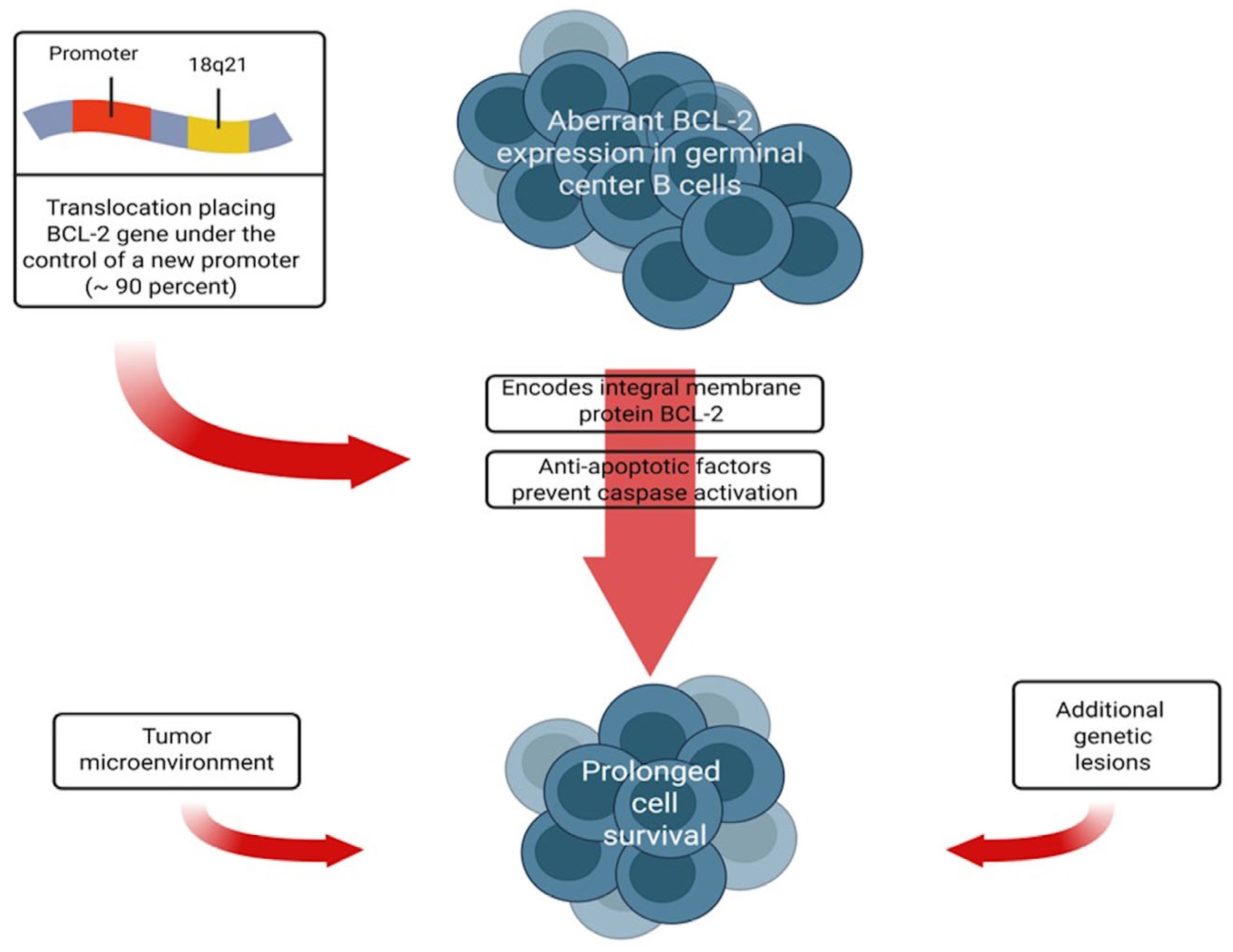
A hallmark molecular event in FL is the chromosomal translocation t(14;18)(q32;q21), which results in overexpression of the anti-apoptotic protein BCL-2 [5]. Although BCL-2 upregulation contributes to prolonged cell survival, this alteration alone is insufficient for malignant transformation. Additional genetic lesions and interactions with the tumor microenvironment are required for disease development and progression [6,7]. Recent genomic studies have highlighted the role of immune cells, stromal components, and cytokine networks in shaping tumor growth and clinical outcome [8,9,10,11,12,13] (Figure 1).
Clinically, FL often presents with painless lymphadenopathy involving cervical, axillary, or inguinal lymph node regions [14,15,16]. Many patients remain asymptomatic at diagnosis, although a minority present with systemic ‘B symptoms’ such as fever, night sweats, and weight loss [17]. Extranodal involvement may occur but is less common, while bone marrow infiltration is frequently observed during staging procedures [18,19,20,21,22,23,24]. The diagnosis of FL is generally established through excisional lymph node biopsy, which allows histological assessment of follicular architecture and cellular morphology. Immunophenotypic analysis and molecular studies may support the diagnosis and help distinguish FL from other lymphoid malignancies [14]. Some infantile FL are often negative to BCL2 [25,26,27].
Immunophenotypic analysis and molecular studies may support the diagnosis and help distinguish FL from other lymphoid malignancies [14]. Several patients show no signs of FL at further evaluation [28,29]. Duodenal-type FL is recognized as a distinct entity confined to the gastrointestinal tract—most frequently the duodenum—and commonly appears as multiple small polyps [30]. Although rare [31,32], it is typically limited to the mucosa, often retains at least a partial follicular growth pattern, and usually displays grade 1–2 cytology [31].
The clinical course of FL is heterogeneous. Some patients experience a prolonged, intermittently active disease trajectory and may remain without treatment for five years or longer [33]. Others present with more extensive involvement and faster growth kinetics, leading to symptoms or organ compromise (e.g., pain, obstruction, or dysfunction) that necessitate therapy [34]. While early studies suggest that certain pathogenic mutations acquired in FL could carry prognostic information, additional evidence is required before routine genome sequencing can be broadly incorporated into standard care pathways [14].
Grading systems differ slightly across classification frameworks. The International Consensus Classification (ICC) grades FL on a 1–3 scale [27], whereas the WHO scheme consolidates disease into classical FL (corresponding to ICC grades 1–3a) and follicular large B-cell lymphoma (ICC grade 3b) [27]. Grade may have prognostic relevance—particularly grade 3b, which is generally considered to follow a more aggressive clinical course compared with other FL grades [35,36].
Comprehensive pretreatment evaluation is required to define disease extent in Stages I–IV, and to capture comorbidities that may influence therapy selection. Beyond history and physical examination, assessment includes review of pathology to confirm diagnostic adequacy, laboratory testing [37,38], and baseline imaging. Imaging may be performed with contrast-enhanced CT or FDG PET/CT, which can help map anatomic involvement and metabolic activity [39,40]. The urgency to initiate therapy varies and should be individualized based on symptoms, organ function, and available options.

1.2. Initial Treatment

Strategy is guided by stage, grade, and distribution of disease. For Stage I FL (grades 1–3a), involved-site radiotherapy with curative intent is generally favored when all sites can be encompassed with acceptable toxicity; if radiotherapy is not feasible, an initial observation approach may be preferred [37,38]. For Stage I grade 3b disease, regimens used for clinically aggressive lymphomas (such as diffuse large B-cell lymphoma) are typically recommended [37,38]. Stage II (grades 1–3a) is often managed similarly to advanced disease, although selected patients may still be offered radiotherapy depending on clinical context [37,38]. Only a minority of FL cases present as Stage I–II (approximately 15–30%) [41,42]. At diagnosis, most patients have Stage II (approximately 11–16%), Stage III (22–33%), or Stage IV disease (26–40%) [43,44]. Management of Stage III–IV FL depends on grade, symptoms/organ dysfunction, tumor burden, and disease kinetics [37,38].
For asymptomatic, low-tumor-burden Stage II–IV FL, single-agent rituximab can be used as an alternative to a watch-and-wait strategy when patients prefer early treatment. Regimens evaluated in randomized studies include four weekly doses (e.g., days 1, 8, 15, 22) or four weekly doses followed by maintenance administration every two months for two years [45,46].
Anti-CD20 immunotherapy (rituximab or obinutuzumab) is a cornerstone for symptomatic FL [47,48]. Common options—ordered from lower to higher intensity—include: (i) rituximab as a defined course; (ii) rituximab plus chemotherapy for 6–8 cycles; and (iii) immunochemotherapy followed by maintenance anti-CD20 therapy for two years. In practice, rituximab-chemotherapy is frequently chosen over rituximab alone because indirect evidence supports a faster and deeper response. Nevertheless, single-agent rituximab remains appropriate for patients with significant comorbidities, lower tumor burden, and/or slowly progressive disease where chemotherapy is undesirable [37,38].
Multiple chemotherapy backbones have been combined with rituximab or obinutuzumab. Bendamustine-rituximab (BR) is commonly preferred because of a favorable toxicity profile and evidence of comparable efficacy to R-CHOP in several studies [37,38]. R-CVP is an alternative but may yield lower response rates. Obinutuzumab can be combined with bendamustine, CHOP, or CVP and followed by maintenance obinutuzumab; in one study, this approach improved progression-free survival but increased toxicity and costs [49]. Fludarabine-based regimens are generally avoided due to high toxicity [50,51]. Lenalidomide-based combinations (e.g., lenalidomide with rituximab or obinutuzumab) are increasingly used, particularly in the relapsed/refractory setting [37,38,49,50,51].
For Relapsed or Refractory Follicular Lymphoma, before initiating therapy for suspected relapse, repeat biopsy is recommended with a low threshold to confirm recurrence and to evaluate for histologic transformation. FDG PET/CT can establish a new baseline and guide biopsy toward the most metabolically active site, which is important because treatment strategy and prognosis differ substantially when transformation is present [51,52].
As with frontline disease, asymptomatic relapse does not necessarily require immediate therapy, but close surveillance is warranted. In general, the same clinical triggers used for initial treatment—symptoms, cytopenias, threatened organ function, or rapid progression—also apply at recurrence or progression [52].
Many patients who relapse more than 24 months after initial immunochemotherapy (or more than 12 months after single-agent rituximab) can anticipate long survival with intermittent treatment, often approaching that of the general population [53]. While cure is uncommon, contemporary therapies frequently achieve complete or partial remissions. The goals of care include symptom control, reversal of cytopenias, and improvement in quality of life. For later relapses, re-treatment with anti-CD20 therapy (rituximab or obinutuzumab)—alone or combined with agents such as lenalidomide—is often favored over repeated intensive immunochemotherapy, although chemotherapy-based options remain acceptable for selected patients when a time-limited intensive strategy is preferred [52,53].

1.3. Bispecific Antibodies

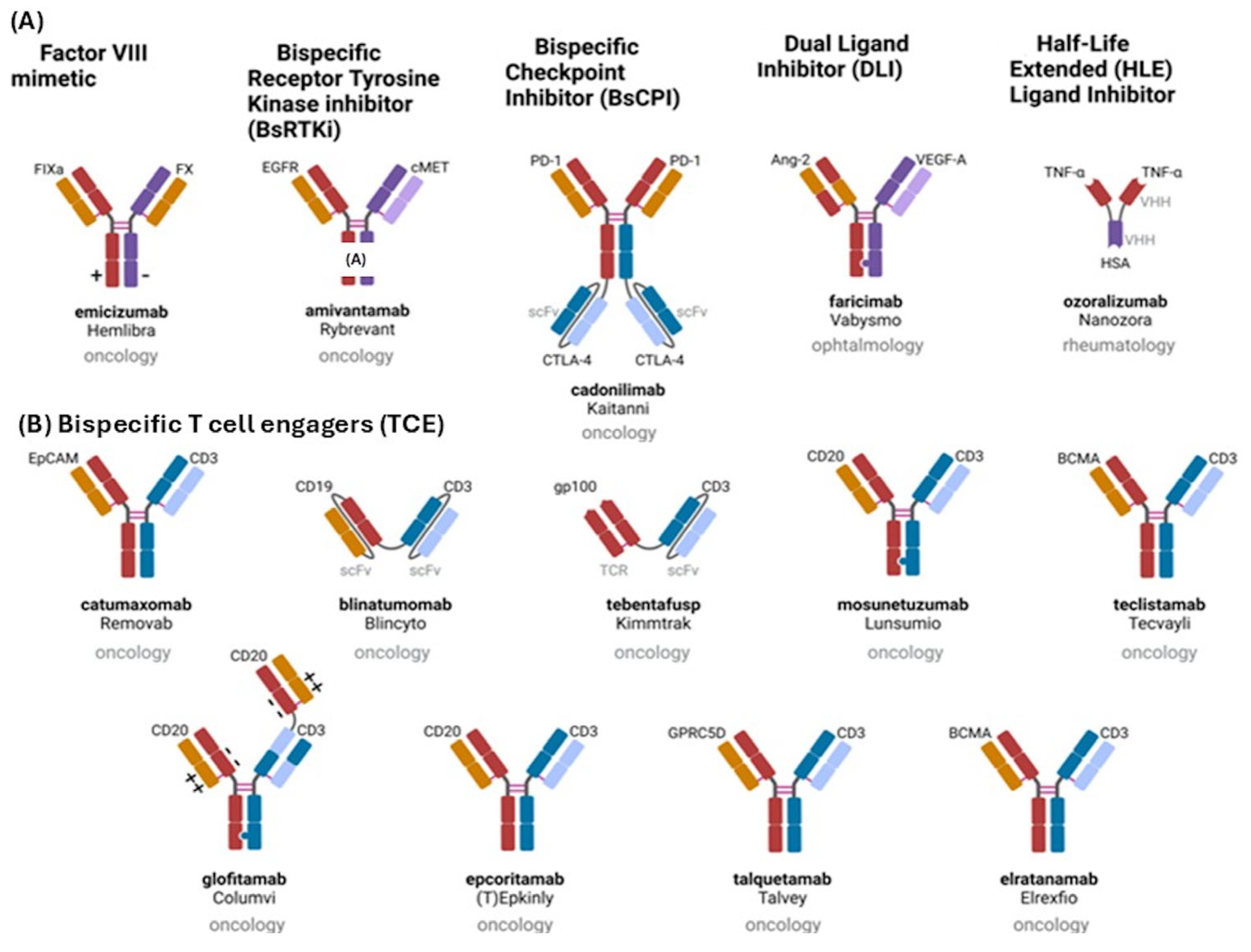
These are engineered molecules capable of simultaneously binding two different antigens or epitopes [54,55,56,57]. In oncology, these agents often link tumor cells with immune effector cells, thereby facilitating targeted immune activation and tumor cell destruction and not target immune reaction. Several structural formats have been developed, including IgG-like and non-IgG-like antibodies, each with specific pharmacological characteristics and clinical advantages (Figure 2) [57,58,59,60].
Although generally manageable, treatment with bispecific antibodies may be associated with immune-related adverse events such as cytokine release syndrome (CRS), immune effector cell-associated neurotoxicity syndrome (ICANS), infusion-related reactions, and opportunistic infections [61,62]. Careful patient monitoring and step-up dosing strategies are therefore important to minimize treatment-related toxicity. The infusion-related reactions are commonly observed with different mAbs, are structure related and have economic impact [63,64,65]. Mosunetuzumab is a CD20×CD3 bispecific antibody designed to redirect T lymphocytes toward CD20-expressing malignant B cells. Through this mechanism, the drug induces T-cell activation and subsequent cytotoxic elimination of lymphoma cells [66,67]. Clinical trials have demonstrated promising efficacy in patients with relapsed or refractory FL who previously received multiple lines of therapy [58,59]. These advances have positioned mosunetuzumab as an important therapeutic option in relapsed or refractory FL, particularly for patients who are not candidates for more intensive treatments such as CAR-T cell therapy. The structure and function of bivalent antibodies are reported in Figure 2 [54,55,56,57,68,69,70].
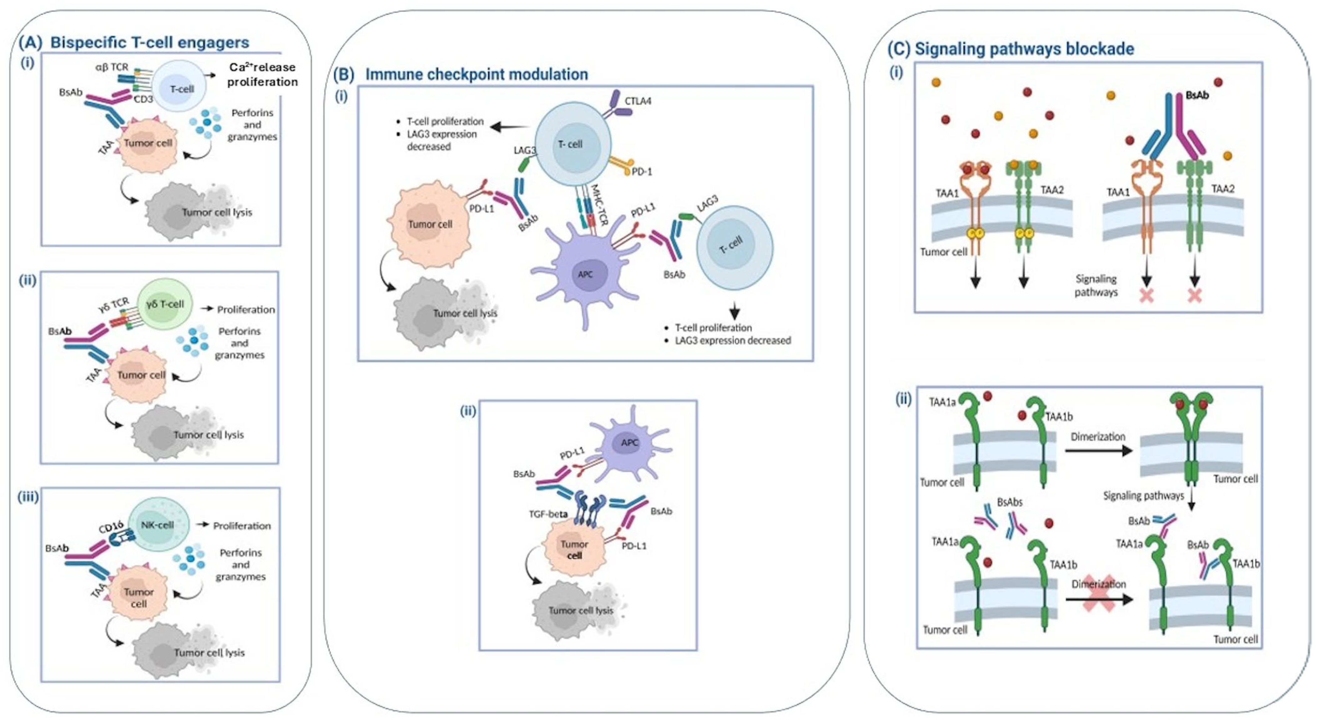
The therapeutic activity of bispecific antibodies (bsAbs) is largely determined by their biological targets, which define their mechanism of action. Based on their functional properties, bsAbs can be broadly categorized into three principal classes (Figure 3A–C).

1.3.1. Immune Cell Engagers

Bispecific T-cell engagers (TCEs) are engineered to physically connect endogenous CD4+ and CD8+ T lymphocytes with tumor cells. This occurs through the simultaneous binding of the CD3ε subunit of the T-cell receptor (TCR) complex and a specific tumor-associated antigen (TAA) expressed on malignant cells [68,69,70]. The resulting immune synapse triggers T-cell activation and leads to the release of cytotoxic mediators and pro-inflammatory cytokines that promote tumor cell destruction [71,72,73,74,75]. This action is mediated by ion channels more specifically calcium release activated channel (CRAC). Indeed, mosunetuzumab activates CD3 signaling increasing short lived calcium release via IP3 on endoplasmic reticulum (ER), activation of calcium channel CRAC with sustained long lasting Ca2+ entry and calcium dependent NFAT transcription with T-cell activation and cytotoxicity [76,77,78,79,80] (Figure 3A).
Immune checkpoint modulation: Another group of bsAbs is designed to target immune checkpoint pathways. Dual immune checkpoint–blocking bsAbs simultaneously bind inhibitory receptors—such as PD-1, CTLA-4, LAG-3, or TIGIT—on the T-cell surface, while the second binding arm interacts with another checkpoint molecule expressed on T cells, tumor cells, or antigen-presenting cells [71,81,82]. By interfering with these inhibitory signals, these bsAbs enhance T-cell activation and restore antitumor immune responses.

1.3.2. Inhibition of Signaling Pathways

BsAbs can also disrupt oncogenic signaling networks by simultaneously targeting two different antigens or distinct epitopes of the same receptor involved in tumor growth pathways. Dual targeting of driver signaling cascades can improve the therapeutic efficacy of bsAbs [83] and may reduce the development of drug resistance in combined therapy [74].
Several mechanisms underlie the ability of bsAbs to interfere with signaling pathways. The primary mechanism involves blocking receptor–ligand interactions, thereby preventing activation of downstream signaling cascades. In addition, bsAbs may induce receptor internalization, limiting receptor cross-linking and dimerization (both homo- and heterodimerization). These effects can ultimately suppress processes such as angiogenesis and tumor cell proliferation. Furthermore, IgG-based bsAbs that retain an Fc domain can activate immune effector mechanisms including antibody-dependent cellular phagocytosis (ADCP) and antibody-dependent cellular cytotoxicity (ADCC), which contribute to apoptosis of tumor cells [72,73].
Importantly, bsAbs may also help overcome resistance to tyrosine kinase inhibitors (TKIs) [84,85]. Resistance can arise through the up-regulation of compensatory signaling pathways, such as activation of the mesenchymal-epithelial transition (MET) pathway observed in epidermal growth factor receptor (EGFR)–mutated non-small cell lung cancer (NSCLC) [74].
Adverse reactions under monitoring are Cytokine Release Syndrome (CRS), immune effector cell-associated neurotoxicity syndrome (ICANS Immune effector cell-associated neurotoxicity syndrome) that is associated with T cells immunoactivities and BBB disruption and infusion-related reactions: (IRRs infusion-related reactions) that can be fatal [86,87,88,89,90,91,92,93]. Opportunistic infections of respiratory track due to gram-negative bacterial and fungal (e.g., Aspergillus spp.), and viral (e.g., cytomegalovirus) infections also occur [93].
Despite the efficacy of monesetuzumab being well established, the approval of mosunetuzumab was based on the results of an international, multicenter, phase II study of 90 patients with FL RR (relapsed refractory follicular lymphoma) after at least two previous lines of systemic therapy [58,59,94]. Mosunetuzumab targeting CD3 and CD20, is approved for patients with FL R/R who have received ≥2 previous lines of treatment [58,59,95,96]. The effectiveness remains to be evaluated in specific contexts.
Insufficient patient care at the Center and the Adverse Drug Reaction (ADR) of this drug can limit the effectiveness of the therapy and require monitoring. In this manuscript we evaluated the effectiveness of mosunetuzumab in our center.

2. Methods

In the A.O. Pia Fondazione di Culto e Religione ‘Card. G. Panico’ in Tricase, (Hospital Card. G. Panico) in the province of Lecce (LE), bispecific antibody therapies have been in use for three years. In particular, bsAb mosunetuzumab, first launched as compassionate use programmed in May 2022 approved at Local Ethical Committee (CEL) of I.R.C.C.S., Oncology Hospital, Giovanni Paolo II Bari, and still in use today as a reimbursable drug authorized by AIFA, was approved for the treatment of eight patients in the Hospital Card. G. Panico.
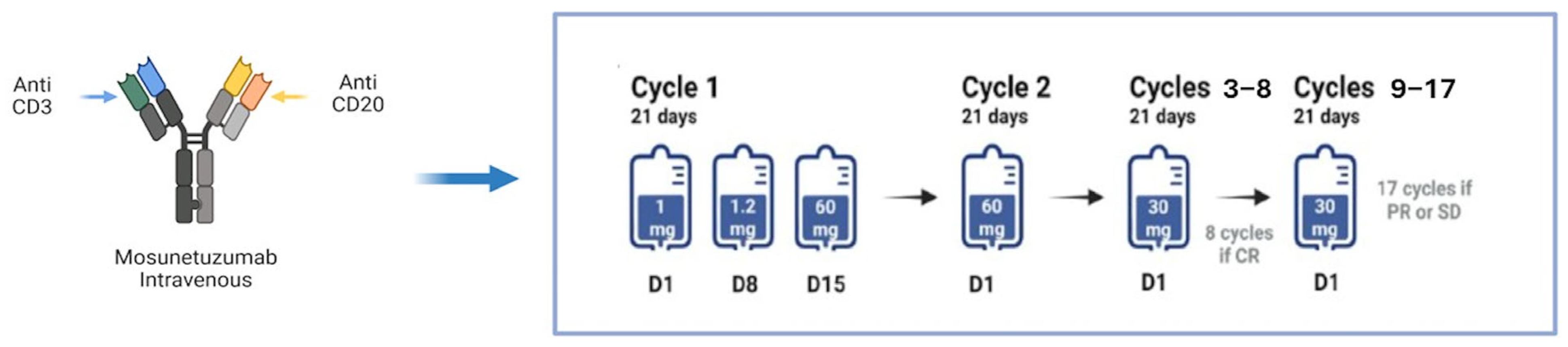
Mosunetuzumab is administered intravenously (EV) in 21-day cycles, with cycle 1 consisting of a stepwise dosage: 1 mg on day 1 of cycle 1, 2 mg on day 8 of cycle 1, 60 mg on day 15 of cycle 1, 60 mg on day 1 of cycle 2, 30 mg on day 1 of cycle 3 and thereafter. Treatment is stopped after cycle 8 for patients with complete response (CR), while patients with partial response (PR) or stable disease (SD) continue treatment for up to 17 cycles (Figure 4) [58,59].
Evaluation of adverse reactions and radiotherapy treatments were based on previous data and protocols [97,98,99,100,101,102,103,104,105,106].

3. Results: Experience in the Hospital Card. G. Panico, Patient Cases

3.1. Patient Cases

Number of patients with relapsed or refractory FL treated with the drug mosunetuzumab, from 2022 to date, in the Hospital Card. G. Panico is eight. Six patients have completed treatment with mosunetuzumab, one patient is being treated with mosunetuzumab, and one patient has changed treatment.
Below is the clinical history of three patients who completed treatment with mosunetuzumab.

3.1.1. Patient 1

Patient 1 was an aged man with hypertension, benign prostatic hyperplasia, chronic obstructive pulmonary disease, a remote smoking history, and a central venous access device. He had previously been treated for B-cell non-Hodgkin lymphoma many years earlier with six cycles of R-CHOP. At relapse, he was diagnosed at our center with follicular lymphoma, stage IIA with bulky abdominal disease (FLIPI-2 score 2; GELF score 2).
Restaging PET/CT at relapse showed metabolically active disease in the left iliac-obturator region and an additional hypermetabolic focus adjacent to the sigmoid colon. Abdominal lymph node biopsy confirmed follicular lymphoma. The patient then received six cycles of bendamustine plus rituximab as second-line therapy and tolerated treatment well, with preserved performance status and no major complications.
Post-treatment PET/CT demonstrated persistent metabolically active disease in the left iliac-obturator region, consistent with refractory lymphoma. Because the disease was refractory to two prior lines of therapy (R-CHOP and bendamustine-rituximab), the patient started mosunetuzumab with standard step-up dosing administered under in-patient observation during cycle 1.
Mosunetuzumab was given for eight cycles, after which treatment was stopped according to protocol because complete response had been achieved. During the first cycle, the patient developed myalgias; pruritic rash involving the axillary, submental, lumbar, and inguinal regions; and fever up to 38.8 °C. These events were consistent with cytokine release syndrome and resolved with supportive management. No subsequent serious toxicities were observed.
End-of-treatment PET/CT showed complete metabolic response, with interval reduction of residual soft tissue along the left iliac vessels and no new sites of disease. Follow-up imaging performed approximately six months after completion of treatment confirmed an ongoing complete remission.

3.1.2. Patient 2

Patient 2 was an adult man with a remote smoking history, congenital solitary kidney, and a central venous access device. He was initially diagnosed with grade 3A, stage IV follicular lymphoma (FLIPI-2 score 2).
Front-line treatment consisted of six cycles of R-CHOP, which produced a marked reduction in nodal disease and complete remission, followed by rituximab maintenance. Approximately three years later, biopsy and PET/CT confirmed relapse as grade 2, stage IVa follicular lymphoma. He then received second-line R-DHAOX with dose reduction of oxaliplatin because of his solitary kidney, followed by autologous stem cell transplantation, and again achieved complete remission.
Approximately 2.5 years after transplantation, PET/CT and biopsy documented a further relapse with metabolically active nodal and soft-tissue disease. Because the lymphoma was refractory to two prior lines of therapy, including transplant-based treatment, the patient started mosunetuzumab with standard step-up dosing and in-patient monitoring during cycle 1.
He completed eight cycles of mosunetuzumab and maintained excellent performance status, with normal hematologic, liver, and kidney function throughout treatment. A mild grade 1 cytokine release syndrome occurred after the first administration and resolved without sequelae.
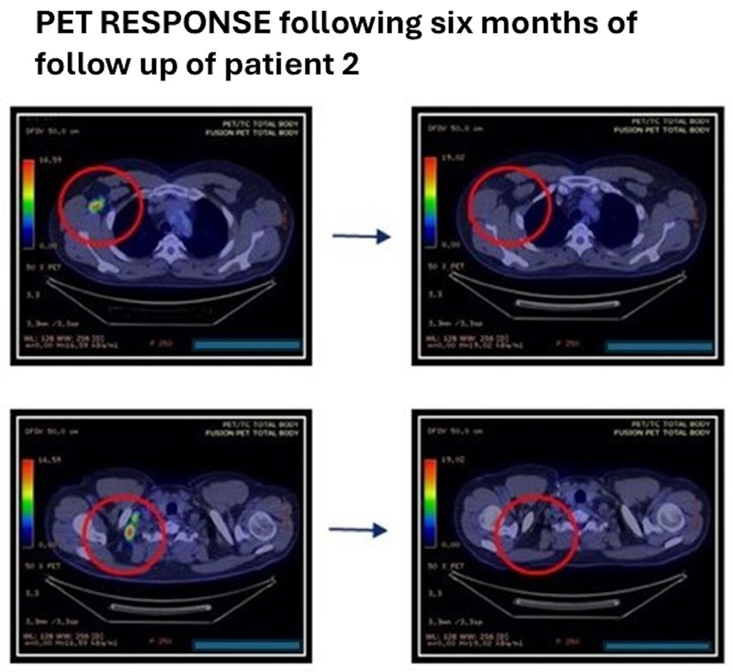
Interim PET/CT after four cycles showed a marked reduction in previously involved cervical and inguinal-femoral lymph nodes, and end-of-treatment PET/CT after eight cycles confirmed complete metabolic response. Follow-up clinical and imaging assessments at approximately 6, 8, and 12 months after treatment completion continued to show complete remission (Figure 5).

3.1.3. Patient 3

Patient 3 was an adult man with anxiety disorder, prior severe hypersensitivity reaction to pegfilgrastim, previous SARS-CoV-2 infection, and a central venous access device. He had grade 2, stage IVa follicular lymphoma (FLIPI-1 score 2; FLIPI-2 score 1).
He received first-line O-CHOP and had persistent disease on post-treatment PET/CT. Second-line salvage therapy with R-DHAP followed by DHAOX was then administered; however, repeat PET/CT showed progressive metabolically active pelvic-abdominal disease. Third-line treatment with rituximab plus lenalidomide was subsequently started; lenalidomide required temporary interruptions because of pruritus and papillomatous skin lesions, which later resolved. Reassessment after this regimen showed stable disease.
Because the lymphoma remained refractory after three prior lines of therapy, the patient started mosunetuzumab with standard step-up dosing and inpatient monitoring during cycle 1.
He completed eight cycles with good general condition, normal cardiopulmonary examination, and no evidence of cytokine release syndrome or immune effector cell-associated neurotoxicity syndrome. Occasional headaches during the first cycle were consistent with his prior history and were not considered treatment related.
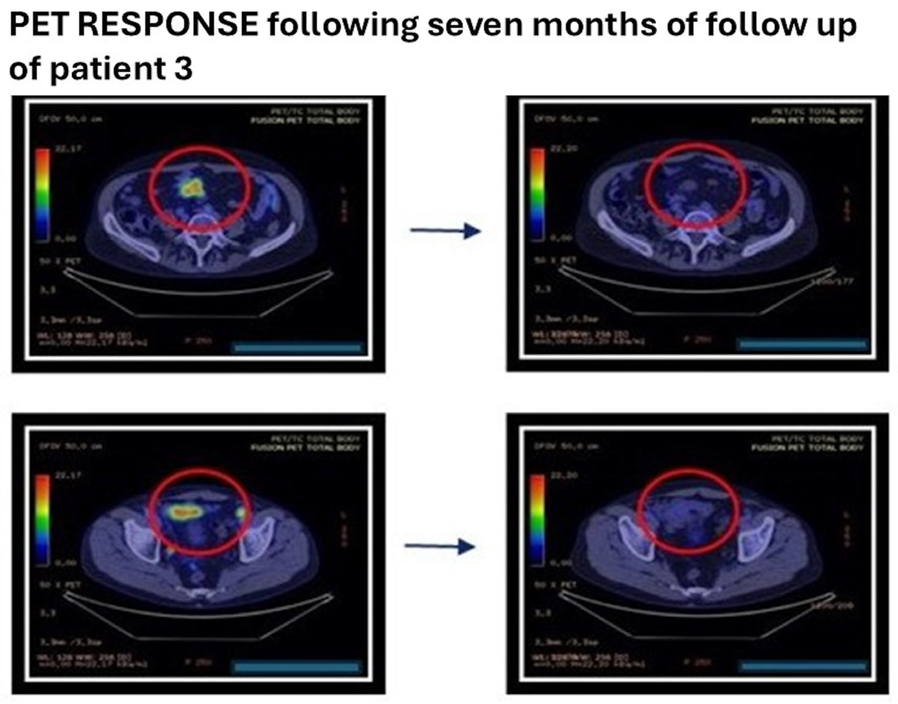
Interim PET/CT after four cycles showed a marked reduction in mesenteric, mesogastric, paravesical, retro vesical, presacral, obturator, and iliac disease sites. End-of-treatment PET/CT after eight cycles demonstrated near-complete disappearance of residual uptake, consistent with complete metabolic response. Follow-up clinical and imaging assessments performed approximately 7, 11, and 13 months after treatment completion confirmed ongoing complete remission (Figure 6).

4. Conclusions

Bispecific antibodies such as mosunetuzumab, a full-length IgG1 CD20×CD3 antibody that redirects T cells against malignant B cells, are expanding the treatment options for relapsed or refractory follicular lymphoma. In this case series, all three heavily pretreated patients achieved complete remission, with durable responses during follow-up and no unexpected safety signals.
The comparative analysis of the GO29781 study shows that the three patients described here meet the eligibility and inclusion criteria established for this single-arm, multi-center, phase I/II clinical trial for the drug mosunetuzumab, which was conducted in 35 countries around the world between 2019 and 2020 prior to its market launch [58,59,94]. The eligibility criteria included being aged 18 years or over, having histologically confirmed follicular lymphoma (grade 1–3a), and having relapsed or not responded to two or more prior lines of treatment, including anti-CD20 therapy and an alkylating agent (Table 1); however, no Italian patients were recruited at that time.
In these three cases, the clinical outcomes were consistent with the primary and secondary endpoints reported in the GO29781 study, with sustained responses, acceptable tolerability, and ongoing remission during follow-up.

Author Contributions

Conceptualization, G.T., D.T. (Donatella Tarantino) and G.G.; methodology, G.T.; validation, D.T. (Domenico Tricarico); formal analysis, A.G.F., D.T. (Donatella Tarantino); resources, G.T. and G.G.; data curation, G.G.; writing—original draft preparation, G.T.; writing—review and editing, G.T., D.T. (Domenico Tricarico), A.G.F.; supervision, D.T. (Domenico Tricarico). D.T. (Domenico Tricarico) is a pharmacologist component of Local Ethical Committee of I.R.C.C.S., Oncology Hospital, Giovanni Paolo II, Bari. All authors have read and agreed to the published version of the manuscript.

Funding

This research received no external funding.

Institutional Review Board Statement

Ethical review and approval were waived for this study, due to: The data are not experimental data; the patients were treated with standard care and treatments, so no approval is needed.

Informed Consent Statement

Informed consent for publication was obtained from all subjects involved in the study.

Data Availability Statement

The data presented in this study are available upon request from the corresponding author due to limited patients permission for diffusion.

Conflicts of Interest

The authors declare no conflicts of interest.

References

  1. Zinzani, P.L.; Muñoz, J.; Trotman, J. Current and Future Therapies for Follicular Lymphoma; Springer: Berlin/Heidelberg, Germany, 2024. [Google Scholar]
  2. Casulo, C.; Friedberg, J.W.; Ahn, K.W.; Flowers, C.; DiGilio, A.; Smith, S.M.; Ahmed, S.; Inwards, D.; Aljurf, M.; Chen, A.I.; et al. Autologous transplantation in follicular lymphoma with early therapy failure: A National LymphoCare Study and Center for International Blood and Marrow Transplant Research analysis. Biol. Blood Marrow Transplant. 2018, 24, 1163–1171. [Google Scholar] [CrossRef] [PubMed]
  3. Swerdlow, S.H.; Campo, E.; Harris, N.L.; Jaffe, E.S.; Pileri, S.A.; Stein, H.; Thiele, J. (Eds.) World Health Organization Classification of Tumours of Haematopoietic and Lymphoid Tissues, revised 4th ed.; IARC: Lyon, France, 2017. [Google Scholar]
  4. Ottensmeier, C.H.; Thompsett, A.R.; Zhu, D.; Wilkins, B.S.; Sweetenham, J.W.; Stevenson, F.K. Analysis of VH genes in follicular and diffuse lymphoma shows ongoing somatic mutation and multiple isotype transcripts in early disease with changes during disease progression. Blood 1998, 91, 4292. [Google Scholar] [CrossRef]
  5. Carreras, J. The pathobiology of follicular lymphoma. J. Clin. Exp. Hematop. 2023, 63, 152–163. [Google Scholar] [CrossRef]
  6. Nuñez, G.; Hockenbery, D.; McDonnell, T.J.; Sorensen, C.M.; Korsmeyer, S.J. Bcl-2 maintains B cell memory. Nature 1991, 353, 71. [Google Scholar] [CrossRef] [PubMed]
  7. McDonnell, T.J.; Deane, N.; Platt, F.M.; Nunez, G.; Jaeger, U.; McKearn, J.P.; Korsmeyer, S.J. Bcl-2-immunoglobulin transgenic mice demonstrate extended B cell survival and follicular lymphoproliferation. Cell 1989, 57, 79. [Google Scholar] [CrossRef] [PubMed]
  8. Bende, R.J.; Smit, L.A.; van Noesel, C.J. Molecular pathways in follicular lymphoma. Leukemia 2007, 21, 18. [Google Scholar] [CrossRef]
  9. Lindhout, E.; Mevissen, M.L.; Kwekkeboom, J.; Tager, J.M.; de Groot, C. Direct evidence that human follicular dendritic cells (FDC) rescue germinal centre B cells from death by apoptosis. Clin. Exp. Immunol. 1993, 91, 330. [Google Scholar] [CrossRef]
  10. Dave, S.S.; Wright, G.; Tan, B.; Rosenwald, A.; Gascoyne, R.D.; Chan, W.C.; Fisher, R.I.; Braziel, R.M.; Rimsza, L.M.; Grogan, T.M.; et al. Prediction of survival in follicular lymphoma based on molecular features of tumor-infiltrating immune cells. N. Engl. J. Med. 2004, 351, 2159. [Google Scholar] [CrossRef]
  11. Glas, A.M.; Knoops, L.; Delahaye, L.; Kersten, M.J.; Kibbelaar, R.E.; Wessels, L.A.; van Laar, R.; van Krieken, J.H.; Baars, J.W.; Raemaekers, J.; et al. Gene-expression and immunohistochemical study of specific T-cell subsets and accessory cell types in the transformation and prognosis of follicular lymphoma. J. Clin. Oncol. 2007, 25, 390. [Google Scholar] [CrossRef]
  12. Coupland, S.E. The challenge of the microenvironment in B-cell lymphomas. Histopathology 2011, 58, 69. [Google Scholar] [CrossRef]
  13. Laurent, C.; Müller, S.; Do, C.; Al-Saati, T.; Allart, S.; Larocca, L.M.; Hohaus, S.; Duchez, S.; Quillet-Mary, A.; Laurent, G.; et al. Distribution, function, and prognostic value of cytotoxic T lymphocytes in follicular lymphoma: A 3-D tissue-imaging study. Blood 2011, 118, 5371. [Google Scholar] [CrossRef]
  14. Solal-Céligny, P.; Roy, P.; Colombat, P.; White, J.; Armitage, J.O.; Arranz-Saez, R.; Au, W.Y.; Bellei, M.; Brice, P.; Caballero, D.; et al. Follicular lymphoma international prognostic index. Blood 2004, 104, 1258. [Google Scholar] [CrossRef]
  15. Martin, A.R.; Weisenburger, D.D.; Chan, W.C.; Ruby, E.I.; Anderson, J.R.; Vose, J.M.; Bierman, P.J.; Bast, M.A.; Daley, D.T.; Armitage, J.O. Prognostic value of cellular proliferation and histologic grade in follicular lymphoma. Blood 1995, 85, 3671. [Google Scholar] [CrossRef] [PubMed]
  16. Anderson, T.; Chabner, B.A.; Young, R.C.; Berard, C.W.; Garvin, A.J.; Simon, R.M.; DeVita, V.T., Jr. Malignant lymphoma. 1. The histology and staging of 473 patients at the National Cancer Institute. Cancer 1982, 50, 2699. [Google Scholar] [CrossRef] [PubMed]
  17. Fernández de Larrea, C.; Martínez-Pozo, A.; Mercadal, S.; García, A.; Gutierrez-García, G.; Valera, A.; Ghita, G.; Colomo, L.; Gainza, E.; Villamor, N.; et al. Initial features and outcome of cutaneous and non-cutaneous primary extranodal follicular lymphoma. Br. J. Haematol. 2011, 153, 334. [Google Scholar] [CrossRef]
  18. Rasmussen, P.K.; Coupland, S.E.; Finger, P.T.; Graue, G.F.; Grossniklaus, H.E.; Honavar, S.G.; McKelvie, P.; Mulay, K.; Prause, J.U.; Ralfkiaer, E.; et al. Ocular adnexal follicular lymphoma: A multicenter international study. JAMA Ophthalmol. 2014, 132, 851. [Google Scholar] [CrossRef]
  19. Weindorf, S.C.; Smith, L.B.; Owens, S.R. Update on gastrointestinal lymphomas. Arch. Pathol. Lab. Med. 2018, 142, 1347. [Google Scholar] [CrossRef]
  20. Louissaint, A., Jr.; Ackerman, A.M.; Dias-Santagata, D.; Ferry, J.A.; Hochberg, E.P.; Huang, M.S.; Lafrate, A.J.; Lara, D.O.; Pinkus, G.S.; Salaverria, I.; et al. Pediatric-type nodal follicular lymphoma: An indolent clonal proliferation in children and adults with high proliferation index and no BCL2 rearrangement. Blood 2012, 120, 2395. [Google Scholar] [CrossRef]
  21. Canioni, D.; Brice, P.; Lepage, E.; Chababi, M.; Meignin, V.; Salles, B.; Xerri, L.; Péaud, P.Y.; Rousselot, P.; Peuchmaur, M.; et al. Bone marrow histological patterns can predict survival of patients with grade 1 or 2 follicular lymphoma: A study from the Groupe d’Etude des Lymphomes Folliculaires. Br. J. Haematol. 2004, 126, 364. [Google Scholar] [CrossRef]
  22. Sarkozy, C.; Baseggio, L.; Feugier, P.; Callet-Bauchu, E.; Karlin, L.; Seymour, J.F.; Lebras, L.; Michallet, A.S.; Offner, F.; Dumas, O.; et al. Peripheral blood involvement in patients with follicular lymphoma: A rare disease manifestation associated with poor prognosis. Br. J. Haematol. 2014, 164, 659. [Google Scholar] [CrossRef] [PubMed]
  23. Flenghi, L.; Bigerna, B.; Fizzotti, M.; Venturi, S.; Pasqualucci, L.; Pileri, S.; Ye, B.H.; Gambacorta, M.; Pacini, R.; Baroni, C.D.; et al. Monoclonal antibodies PG-B6a and PG-B6p recognize, respectively, a highly conserved and a formol-resistant epitope on the human BCL-6 protein amino-terminal region. Am. J. Pathol. 1996, 148, 1543. [Google Scholar]
  24. Pittaluga, S.; Ayoubi, T.A.; Wlodarska, I.; Stul, M.; Cassiman, J.J.; Mecucci, C.; Van Den Berghe, H.; Van De Ven, W.J.; De Wolf-Peeters, C. BCL-6 expression in reactive lymphoid tissue and in B-cell non-Hodgkin’s lymphomas. J. Pathol. 1996, 179, 145. [Google Scholar] [CrossRef]
  25. Campo, E.; Jaffe, E.S.; Cook, J.R.; Quintanilla-Martinez, L.; Swerdlow, S.H.; Anderson, K.C.; Brousset, P.; Cerroni, L.; de Leval, L.; Dirnhofer, S.; et al. The International Consensus Classification of Mature Lymphoid Neoplasms: A report from the Clinical Advisory Committee. Blood 2022, 140, 1229. [Google Scholar] [CrossRef] [PubMed]
  26. Jegalian, A.G.; Eberle, F.C.; Pack, S.D.; Mirvis, M.; Raffeld, M.; Pittaluga, S.; Jaffe, E.S. Follicular lymphoma in situ: Clinical implications and comparisons with partial involvement by follicular lymphoma. Blood 2011, 118, 2976. [Google Scholar] [CrossRef]
  27. Tellier, J.; Menard, C.; Roulland, S.; Martin, N.; Monvoisin, C.; Chasson, L.; Nadel, B.; Gaulard, P.; Schiff, C.; Tarte, K. Human t(14;18)-positive germinal center B cells: A new step in follicular lymphoma pathogenesis? Blood 2014, 123, 3462. [Google Scholar] [CrossRef]
  28. Yoshino, T.; Chott, A. Duodenal-type follicular lymphoma. In WHO Classification of Tumours: Digestive System Tumours, 5th ed.; International Agency for Research on Cancer: Lyon, France, 2019; p. 383. [Google Scholar]
  29. Schmatz, A.I.; Streubel, B.; Kretschmer-Chott, E.; Püspök, A.; Jäger, U.; Mannhalter, C.; Tiemann, M.; Ott, G.; Fischbach, W.; Herzog, P.; et al. Primary follicular lymphoma of the duodenum is a distinct mucosal/submucosal variant of follicular lymphoma: A retrospective study of 63 cases. J. Clin. Oncol. 2011, 29, 1445. [Google Scholar] [CrossRef] [PubMed]
  30. Mori, M.; Kobayashi, Y.; Maeshima, A.M.; Gotoda, T.; Oda, I.; Kagami, Y.; Bennett, S.; Nomoto, J.; Azuma, T.; Yokoyama, H.; et al. The indolent course and high incidence of t(14;18) in primary duodenal follicular lymphoma. Ann. Oncol. 2010, 21, 1500. [Google Scholar] [CrossRef]
  31. Horning, S.J.; Rosenberg, S.A. The natural history of initially untreated low-grade non-Hodgkin’s lymphomas. N. Engl. J. Med. 1984, 311, 1471. [Google Scholar] [CrossRef]
  32. Casulo, C.; Byrtek, M.; Dawson, K.L.; Zhou, X.; Farber, C.M.; Flowers, C.R.; Hainsworth, J.D.; Maurer, M.J.; Cerhan, J.R.; Link, B.K.; et al. Early relapse of follicular lymphoma after rituximab plus cyclophosphamide, doxorubicin, vincristine, and prednisone defines patients at high risk for death: An analysis from the National LymphoCare Study. J. Clin. Oncol. 2015, 33, 2516. [Google Scholar] [CrossRef] [PubMed]
  33. Hans, C.P.; Weisenburger, D.D.; Vose, J.M.; Hock, L.M.; Lynch, J.C.; Aoun, P.; Greiner, T.C.; Chan, W.C.; Bociek, R.G.; Bierman, P.J.; et al. A significant diffuse component predicts for inferior survival in grade 3 follicular lymphoma, but cytologic subtypes do not predict survival. Blood 2003, 101, 2363. [Google Scholar] [CrossRef]
  34. Horn, H.; Schmelter, C.; Leich, E.; Salaverria, I.; Katzenberger, T.; Ott, M.M.; Kalla, J.; Romero, M.; Siebert, R.; Rosenwald, A.; et al. Follicular lymphoma grade 3B is a distinct neoplasm according to cytogenetic and immunohistochemical profiles. Haematologica 2011, 96, 1327. [Google Scholar] [CrossRef] [PubMed]
  35. Cheson, B.D.; Fisher, R.I.; Barrington, S.F.; Cavalli, F.; Schwartz, L.H.; Zucca, E.; Lister, T.A. Recommendations for initial evaluation, staging, and response assessment of Hodgkin and non-Hodgkin lymphoma: The Lugano classification. J. Clin. Oncol. 2014, 32, 3059. [Google Scholar] [CrossRef]
  36. Luminari, S.; Biasoli, I.; Arcaini, L.; Versari, A.; Rusconi, C.; Merli, F.; Spina, M.; Ferreri, A.J.; Zinzani, P.L.; Gallamini, A.; et al. The use of FDG-PET in the initial staging of 142 patients with follicular lymphoma: A retrospective study from the FOLL05 randomized trial of the Fondazione Italiana Linfomi. Ann. Oncol. 2013, 24, 2108. [Google Scholar] [CrossRef]
  37. Link, B.K.; Friedberg, J.W.; Taylor, M.D.; Cerhan, J.R.; Flowers, C.R.; Dillon, H.; Farber, C.M.; Rogers, E.S.; Hainsworth, J.D.; Wong, E.K.; et al. Follicular lymphoma in the United States: First report of the National LymphoCare Study. J. Clin. Oncol. 2009, 27, 1202–1208. [Google Scholar] [CrossRef]
  38. Dinnessen, M.A.W.; van der Poel, M.W.M.; Tonino, S.H.; Visser, O.; Blijlevens, N.M.A.; de Jong, D.; Lam, K.H.; Kersten, M.J.; Lugtenburg, P.J.; Dinmohamed, A.G. Stage-specific trends in primary therapy and survival in follicular lymphoma: A nationwide population-based analysis in the Netherlands, 1989–2016. Leukemia 2021, 35, 1683–1695. [Google Scholar] [CrossRef] [PubMed]
  39. Nooka, A.K.; Nabhan, C.; Zhou, X.; Taylor, M.D.; Byrtek, M.; Miller, T.P.; Friedberg, J.W.; Zelenetz, A.D.; Link, B.K.; Cerhan, J.R.; et al. Examination of the follicular lymphoma international prognostic index (FLIPI) in the National LymphoCare Study (NLCS): A prospective US patient cohort treated predominantly in community practices. Ann. Oncol. 2013, 24, 441–448. [Google Scholar] [CrossRef] [PubMed]
  40. Ardeshna, K.M.; Qian, W.; Smith, P.; Braganca, N.; Lowry, L.; Patrick, P.; Warden, J.; Stevens, L.; Pocock, C.F.; Miall, F.; et al. Rituximab versus a watch-and-wait approach in patients with advanced-stage, asymptomatic, non-bulky follicular lymphoma: An open-label randomised phase 3 trial. Lancet Oncol. 2014, 15, 424. [Google Scholar] [CrossRef]
  41. Cartron, G.; Bachy, E.; Tilly, H.; Daguindau, N.; Pica, G.M.; Bijou, F.; Mounier, C.; Clavert, A.; Damaj, G.L.; Slama, B.; et al. Randomized phase III trial evaluating subcutaneous rituximab for the first-line treatment of low-tumor burden follicular lymphoma: Results of a LYSA study. J. Clin. Oncol. 2023, 41, 3523–3533. [Google Scholar] [CrossRef]
  42. Marcus, R.; Imrie, K.; Belch, A.; Cunningham, D.; Flores, E.; Catalano, J.; Solal-Céligny, P.; Offner, F.; Walewski, J.; Raposo, J.; et al. CVP chemotherapy plus rituximab compared with CVP as first-line treatment for advanced follicular lymphoma. Blood 2005, 105, 1417–1423. [Google Scholar] [CrossRef]
  43. Schulz, H.; Bohlius, J.; Skoetz, N.; Trelle, S.; Kober, T.; Reiser, M.; Dreyling, M.; Herold, M.; Schwarzer, G.; Hallek, M.; et al. Chemotherapy plus rituximab versus chemotherapy alone for B-cell non-Hodgkin’s lymphoma. Cochrane Database Syst. Rev. 2007, 4, CD003805. [Google Scholar] [CrossRef]
  44. Marcus, R.; Davies, A.; Ando, K.; Klapper, W.; Opat, S.; Owen, C.; Phillips, E.; Sangha, R.; Schlag, R.; Seymour, J.F.; et al. Obinutuzumab for the first-line treatment of follicular lymphoma. N. Engl. J. Med. 2017, 377, 1331–1344. [Google Scholar] [CrossRef]
  45. Czuczman, M.S.; Koryzna, A.; Mohr, A.; Stewart, C.; Donohue, K.; Blumenson, L.; Bernstein, Z.P.; McCarthy, P.; Alam, A.; Hernandez-Ilizaliturri, F.; et al. Rituximab in combination with fludarabine chemotherapy in low-grade or follicular lymphoma. J. Clin. Oncol. 2005, 23, 694–704. [Google Scholar] [CrossRef]
  46. Federico, M.; Luminari, S.; Dondi, A.; Tucci, A.; Vitolo, U.; Rigacci, L.; Di Raimondo, F.; Carella, A.M.; Pulsoni, A.; Merli, F.; et al. R-CVP versus R-CHOP versus R-FM for the initial treatment of patients with advanced-stage follicular lymphoma: Results of the FOLL05 trial conducted by the Fondazione Italiana Linfomi. J. Clin. Oncol. 2013, 31, 1506–1513. [Google Scholar] [CrossRef]
  47. Brown, J.R.; Freedman, A.S.; Aster, J.C.; Lister, A.; Connor, R.F. Treatment of Relapsed or Refractory Follicular Lymphoma; CRC Press: Boca Raton, FL, USA, 2025. [Google Scholar]
  48. Maurer, M.J.; Bachy, E.; Ghesquières, H.; Ansell, S.M.; Nowakowski, G.S.; Thompson, C.A.; Inwards, D.J.; Allmer, C.; Chassagne-Clément, C.; Nicolas, E.; et al. Early event status informs subsequent outcome in newly diagnosed follicular lymphoma. Am. J. Hematol. 2016, 91, 1096–1101. [Google Scholar] [CrossRef] [PubMed]
  49. Yamshon, S.; Christos, P.J.; Demetres, M.; Hammad, H.; Leonard, J.P.; Ruan, J. Venous thromboembolism in patients with B-cell non-Hodgkin lymphoma treated with lenalidomide: A systematic review and meta-analysis. Blood Adv. 2018, 2, 1429–1438. [Google Scholar] [CrossRef]
  50. Cheson, B.D.; Chua, N.; Mayer, J.; Dueck, G.; Trněný, M.; Bouabdallah, K.; Fowler, N.; Delwail, V.; Press, O.; Salles, G.; et al. Overall survival benefit in patients with rituximab-refractory indolent non-Hodgkin lymphoma who received obinutuzumab plus bendamustine induction and obinutuzumab maintenance in the GADOLIN study. J. Clin. Oncol. 2018, 36, 2259–2266. [Google Scholar] [CrossRef]
  51. Hiddemann, W.; Barbui, A.M.; Canales, M.A.; Cannell, P.K.; Collins, G.P.; Dürig, J.; Forstpointner, R.; Herold, M.; Hertzberg, M.; Klanova, M.; et al. Immunochemotherapy with obinutuzumab or rituximab for previously untreated follicular lymphoma in the GALLIUM study: Influence of chemotherapy on efficacy and safety. J. Clin. Oncol. 2018, 36, 2395–2404. [Google Scholar] [CrossRef]
  52. van Oers, M.H.J.; Klasa, R.; Marcus, R.E.; Wolf, M.; Kimby, E.; Gascoyne, R.D.; Jack, A.; van ’t Veer, M.B.; Vranovsky, A.; Holte, H.; et al. Rituximab maintenance improves clinical outcome of relapsed/resistant follicular non-Hodgkin lymphoma in patients both with and without rituximab during induction: Results of a prospective randomized phase 3 intergroup trial. Blood 2006, 108, 3295. [Google Scholar] [CrossRef]
  53. van Oers, M.H.J.; van Glabbeke, M.; Giurgea, L.; Klasa, R.; Marcus, R.E.; Wolf, M.; Kimby, E.; van ’t Veer, M.B.; Vranovsky, A.; Holte, H.; et al. Rituximab maintenance treatment of relapsed/resistant follicular non-Hodgkin’s lymphoma: Long-term outcome of the EORTC 20981 phase III randomized intergroup study. J. Clin. Oncol. 2010, 28, 2853. [Google Scholar] [CrossRef] [PubMed]
  54. Herrera, M.; Pretelli, G.; Desai, J.; Garralda, E.; Siu, L.L.; Steiner, T.M.; Au, L. Bispecific antibodies: Advancing precision oncology. Trends Cancer 2024, 10, 893–919. [Google Scholar] [CrossRef] [PubMed]
  55. Chiu, M.L.; Goulet, D.R.; Teplyakov, A.; Gilliland, G.L. Antibody structure and function: The basis for engineering therapeutics. Antibodies 2019, 8, 55. [Google Scholar] [CrossRef]
  56. Brinkmann, U.; Kontermann, R.E. The making of bispecific antibodies. mAbs 2017, 9, 182–212. [Google Scholar] [CrossRef]
  57. Yu, J.; Song, Y.; Tian, W. How to select IgG subclasses in developing anti-tumor therapeutic antibodies. J. Hematol. Oncol. 2020, 13, 45. [Google Scholar] [CrossRef]
  58. Mosunetuzumab Product Information. Available online: https://www.ema.europa.eu/en/documents/product-information/lunsumio-epar-product-information_en.pdf (accessed on 9 February 2026).
  59. Mosunetuzumab-axgb Injection, for Intravenous Use. United States Prescribing Information. US Food and Drug Administration. Available online: https://www.accessdata.fda.gov/drugsatfda_docs/label/2022/761263s000lbl.pdf (accessed on 9 February 2026).
  60. Epcoritamab-bysp Injection. US Food & Drug Administration (FDA) Approved Product Information. US National Library of Medicine. Available online: https://dailymed.nlm.nih.gov/dailymed/index.cfm (accessed on 9 February 2026).
  61. Longhitano, A.P.; Slavin, M.A.; Harrison, S.J.; Teh, B.W. Bispecific antibody therapy, its use and risks for infection: Bridging the knowledge gap. Blood Rev. 2021, 49, 100810. [Google Scholar] [CrossRef]
  62. Park, K.; Sabari, J.K.; Haura, E.B.; Shu, C.A.; Spira, A.; Salgia, R.; Reckamp, K.L.; Sanborn, R.E.; Govindan, R.; Bauml, J.M.; et al. Management of infusion-related reactions (IRRs) in patients receiving amivantamab in the CHRYSALIS study. Lung Cancer 2023, 178, 166–171. [Google Scholar] [CrossRef] [PubMed]
  63. Pierri, C.L.; Bossis, F.; Punzi, G.; De Grassi, A.; Cetrone, M.; Parisi, G.; Tricarico, D. Molecular modeling of antibodies for the treatment of TNFα-related immunological diseases. Pharmacol. Res. Perspect. 2016, 4, e00197. [Google Scholar] [CrossRef]
  64. Mazzuoli, S.; Tricarico, D.; Demma, F.; Furneri, G.; Guglielmi, F.W. Accelerated infliximab infusion: Safety, factors predicting adverse events, patients’ satisfaction and cost analysis. A cohort study in IBD patients. PLoS ONE 2016, 11, e0166443. [Google Scholar] [CrossRef]
  65. Ko, S.; Park, S.; Sohn, M.H.; Jo, M.; Ko, B.J.; Na, J.H.; Yoo, H.; Jeong, A.L.; Ha, K.; Woo, J.R.; et al. An Fc variant with two mutations confers prolonged serum half-life and enhanced effector functions on IgG antibodies. Exp. Mol. Med. 2022, 54, 1850–1861. [Google Scholar] [CrossRef] [PubMed]
  66. Tricarico, D.; Conte Camerino, D.; Govoni, S.; Bryant, S.H. Modulation of rat skeletal muscle chloride channels by activators and inhibitors of protein kinase C. Pflugers Arch. 1991, 418, 500–503. [Google Scholar] [CrossRef]
  67. Van der Merwe, P.A.; Dushek, O. Mechanisms for T cell receptor triggering. Nat. Rev. Immunol. 2011, 11, 47–55. [Google Scholar] [CrossRef] [PubMed]
  68. Van de Donk, N.W.C.J.; Zweegman, S. T-cell-engaging bispecific antibodies in cancer. Lancet 2023, 402, 142–158. [Google Scholar] [CrossRef]
  69. Huang, S.; van Duijnhoven, S.M.J.; Sijts, A.J.A.M.; van Elsas, A. Bispecific antibodies targeting dual tumor-associated antigens in cancer therapy. J. Cancer Res. Clin. Oncol. 2020, 146, 3111–3122. [Google Scholar] [CrossRef]
  70. Ma, J.; Mo, Y.; Tang, M.; Shen, J.; Qi, Y.; Zhao, W.; Huang, Y.; Xu, Y.; Qian, C. Bispecific antibodies: From research to clinical application. Front. Immunol. 2021, 12, 626616. [Google Scholar] [CrossRef]
  71. Mollavelioglu, B.; Aktaş-Çetin, E.; Cabioglu, N.; Abbasov, A.; Özmen, V.; Deniz, G. High co-expression of immune checkpoint receptors PD-1, CTLA-4, LAG-3, TIM-3, and TIGIT on tumor-infiltrating lymphocytes in early-stage breast cancer. World J. Surg. Oncol. 2022, 20, 349. [Google Scholar] [CrossRef] [PubMed]
  72. Zhu, Y.; Choi, S.H.; Shah, K. Multifunctional receptor-targeting antibodies for cancer therapy. Lancet Oncol. 2015, 16, e543–e554. [Google Scholar] [CrossRef]
  73. Gogesch, P.; Dudek, S.; van Zandbergen, G.; Waibler, Z.; Anzaghe, M. The role of Fc receptors on the effectiveness of therapeutic monoclonal antibodies. Int. J. Mol. Sci. 2021, 22, 8947. [Google Scholar] [CrossRef] [PubMed]
  74. Westover, D.; Zugazagoitia, J.; Cho, B.C.; Lovly, C.M.; Paz-Ares, L. Mechanisms of acquired resistance to first- and second-generation EGFR tyrosine kinase inhibitors. Ann. Oncol. 2018, 29, i10–i19. [Google Scholar] [CrossRef] [PubMed]
  75. Shimabukuro-Vornhagen, A.; Gödel, P.; Subklewe, M.; Stemmler, H.J.; Schlößer, H.A.; Schlaak, M.; Kochanek, M.; Böll, B.; von Bergwelt-Baildon, M.S. Cytokine release syndrome. J. Immunother. Cancer 2018, 6, 56. [Google Scholar] [CrossRef]
  76. Smith-Garvin, J.E.; Koretzky, G.A.; Jordan, M.S. T cell activation. Annu Rev. Immunol. 2009, 27, 591–619. [Google Scholar] [CrossRef]
  77. Hogan, P.G.; Lewis, R.S.; Rao, A. Molecular basis of calcium signaling in lymphocytes: STIM and ORAI. Annu Rev. Immunol. 2010, 28, 491–533. [Google Scholar] [CrossRef] [PubMed]
  78. Tricarico, D.; Capriulo, R.; Camerino, D.C. Involvement of K(Ca2+) channels in the local abnormalities and hyperkalemia following the ischemia-reperfusion injury of rat skeletal muscle. Neuromuscul. Disord. 2002, 12, 258–265. [Google Scholar] [CrossRef] [PubMed]
  79. Maqoud, F.; Scala, R.; Hoxha, M.; Zappacosta, B.; Tricarico, D. ATP-sensitive Potassium Channel Subunits in Neuroinflammation: Novel Drug Targets in Neurodegenerative Disorders. CNS Neurol. Disord. Drug. Targets 2022, 21, 130–149. [Google Scholar] [CrossRef] [PubMed]
  80. Tricarico, D.; Mele, A.; Calzolaro, S.; Cannone, G.; Camerino, G.M.; Dinardo, M.M.; Latorre, R.; Conte Camerino, D. Emerging role of calcium-activated potassium channel in the regulation of cell viability following potassium ions challenge in HEK293 cells and pharmacological modulation. PLoS ONE 2013, 8, e69551. [Google Scholar] [CrossRef]
  81. Leclercq-Cohen, G.; Steinhoff, N.; Albertí Servera, L.; Nassiri, S.; Danilin, S.; Piccione, E.; Yángüez, E.; Hüsser, T.; Herter, S.; Schmeing, S.; et al. Dissecting the mechanisms underlying the cytokine release syndrome (CRS) mediated by T-cell bispecific antibodies. Clin. Cancer Res. 2023, 29, 4449–4463. [Google Scholar] [CrossRef]
  82. Markouli, M.; Ullah, F.; Unlu, S.; Omar, N.; Lopetegui-Lia, N.; Duco, M.; Anwer, F.; Raza, S.; Dima, D. Toxicity profile of chimeric antigen receptor T-cell and bispecific antibody therapies in multiple myeloma: Pathogenesis, prevention and management. Curr. Oncol. 2023, 30, 6330–6352. [Google Scholar] [CrossRef]
  83. Ball, K.; Dovedi, S.J.; Vajjah, P.; Phipps, A. Strategies for clinical dose optimization of T-cell-engaging therapies in oncology. mAbs 2023, 15, 2181016. [Google Scholar] [CrossRef]
  84. Antonacci, M.; Di Turi, A.; Miciaccia, M.; Denora, M.; Maqoud, F.; Perrone, M.G.; Scilimati, A.; Tricarico, D. Actions of Midostaurin as Cation Channel and Tyrosine Kinase Inhibitor in Diffuse Intrinsic Pontine Glioma Cell Lines. Cancers 2026, 18, 1066. [Google Scholar] [CrossRef]
  85. Antonacci, M.; Maqoud, F.; Di Turi, A.; Miciaccia, M.; Perrone, M.G.; Scilimati, A.; Tricarico, D. KATP Channel Inhibitors Reduce Cell Proliferation Through Upregulation of H3K27ac in Diffuse Intrinsic Pontine Glioma: A Functional Expression Investigation. Cancers 2025, 17, 358. [Google Scholar] [CrossRef]
  86. Morris, E.C.; Neelapu, S.S.; Giavridis, T.; Sadelain, M. Cytokine release syndrome and associated neurotoxicity in cancer immunotherapy. Nat. Rev. Immunol. 2022, 22, 85–96. [Google Scholar] [CrossRef]
  87. Gu, T.; Hu, K.; Si, X.; Hu, Y.; Huang, H. Mechanisms of immune effector cell-associated neurotoxicity syndrome after CAR-T treatment. WIREs Mech. Dis. 2022, 14, e1576. [Google Scholar] [CrossRef] [PubMed]
  88. Calogiuri, G.; Ventura, M.T.; Mason, L.; Valacca, A.; Buquicchio, R.; Cassano, N.; Vena, G.A. Hypersensitivity reactions to last-generation chimeric, humanized [correction of umanized] and human recombinant monoclonal antibodies for therapeutic use. Curr. Pharm. Des. 2008, 14, 2883–2891. [Google Scholar] [CrossRef]
  89. Rombouts, M.D.; Swart, E.L.; van den Eertwegh, A.J.M.; Crul, M. Systematic review on infusion reactions to and infusion rate of monoclonal antibodies used in cancer treatment. Anticancer Res. 2020, 40, 1201–1218. [Google Scholar] [CrossRef]
  90. Doessegger, L.; Banholzer, M.L. Clinical development methodology for infusion-related reactions with monoclonal antibodies. Clin. Transl. Immunol. 2015, 4, e39. [Google Scholar] [CrossRef]
  91. Cáceres, M.C.; Guerrero-Martín, J.; Pérez-Civantos, D.; Palomo-López, P.; Delgado-Mingorance, J.I.; Durán-Gómez, N. The importance of early identification of infusion-related reactions to monoclonal antibodies. Ther. Clin. Risk Manag. 2019, 15, 965–977. [Google Scholar] [CrossRef] [PubMed]
  92. Laforgia, M.; Laface, C.; Calabrò, C.; Ferraiuolo, S.; Ungaro, V.; Tricarico, D.; Gadaleta, C.D.; Nardulli, P.; Ranieri, G. Peripheral Neuropathy under Oncologic Therapies: A Literature Review on Pathogenetic Mechanisms. Int. J. Mol. Sci. 2021, 22, 1980. [Google Scholar] [CrossRef] [PubMed]
  93. Reynolds, G.; Scheffer Cliff, E.R.; Mohyuddin, G.R.; Popat, R.; Midha, S.; Ng Liet Hing, M.; Harrison, S.J.; Kesselheim, A.S.; Teh, B.W. Infections following bispecific antibodies in myeloma: A systematic review and meta-analysis. Blood Adv. 2023, 7, 5898–5903. [Google Scholar] [CrossRef]
  94. Budde, L.E.; Sehn, L.H.; Matasar, M.; Schuster, S.J.; Assouline, S.; Giri, P.; Kuruvilla, J.; Canales, M.; Dietrich, S.; Fay, K.; et al. Safety and efficacy of mosunetuzumab, a bispecific antibody, in patients with relapsed or refractory follicular lymphoma: A single-arm, multicentre, phase 2 study. Lancet Oncol. 2022, 23, 1055–1065. [Google Scholar] [CrossRef] [PubMed]
  95. Cancemi, G.; Campo, C.; Caserta, S.; Rizzotti, I.; Mannina, D. Single-agent and associated therapies with monoclonal antibodies: What about follicular lymphoma? Cancers 2025, 17, 1602. [Google Scholar] [CrossRef]
  96. Rivas-Delgado, A.; Landego, I.; Falchi, L. The landscape of T-cell engagers for the treatment of follicular lymphoma. Oncoimmunology 2024, 13, 2412869. [Google Scholar] [CrossRef]
  97. Carbone, A.; Roulland, S.; Gloghini, A.; Younes, A.; von Keudell, G.; López Guillermo, A.; Fitzgibbon, J. Follicular lymphoma. Nat. Rev. Dis. Prim. 2019, 5, 83. [Google Scholar] [CrossRef]
  98. Tricarico, D.; Convertino, A.S.; Mehmeti, I.; Ranieri, G.; Leonetti, F.; Laface, C.; Zizzo, N. Inflammatory Related Reactions in Humans and in Canine Breast Cancers, A Spontaneous Animal Model of Disease. Front Pharmacol. 2022, 13, 752098. [Google Scholar] [CrossRef] [PubMed]
  99. Sarkozy, C.; Maurer, M.J.; Link, B.K.; Ghesquieres, H.; Nicolas, E.; Thompson, C.A.; Traverse-Glehen, A.; Feldman, A.L.; Allmer, C.; Slager, S.L.; et al. Cause of death in follicular lymphoma in the first decade of the rituximab era: A pooled analysis of French and US cohorts. J. Clin. Oncol. 2019, 37, 144. [Google Scholar] [CrossRef]
  100. Morschhauser, F.; Nastoupil, L.; Feugier, P.; Schiano de Colella, J.M.; Tilly, H.; Palomba, M.L.; Bachy, E.; Fruchart, C.; Libby, E.N.; Casasnovas, R.O.; et al. Six-year results from RELEVANCE: Lenalidomide plus rituximab (R2) versus rituximab-chemotherapy followed by rituximab maintenance in untreated advanced follicular lymphoma. J. Clin. Oncol. 2022, 40, 3239–3245. [Google Scholar] [CrossRef]
  101. Mauro, G.P.; Medici, C.T.M.; Casimiro, L.C.; Weltman, E. Radiotherapy for early and advanced stages follicular lymphoma. Clinics 2021, 76, e2059. [Google Scholar] [CrossRef]
  102. MacManus, M.; Fisher, R.; Roos, D.; O’Brien, P.; Macann, A.; Davis, S.; Tsang, R.; Christie, D.; McClure, B.; Joseph, D.; et al. Randomized trial of systemic therapy after involved-field radiotherapy in patients with early-stage follicular lymphoma: TROG 99.03. J. Clin. Oncol. 2018, 36, 2918–2925. [Google Scholar] [CrossRef]
  103. Casulo, C.; Larson, M.C.; Lunde, J.J.; Habermann, T.M.; Lossos, I.S.; Wang, Y.; Nastoupil, L.J.; Strouse, C.; Chihara, D.; Martin, P.; et al. Treatment patterns and outcomes of patients with relapsed or refractory follicular lymphoma receiving three or more lines of systemic therapy (LEO CReWE): A multicentre cohort study. Lancet Haematol. 2022, 9, e289–e300. [Google Scholar] [CrossRef] [PubMed]
  104. Maqoud, F.; Zizzo, N.; Attimonelli, M.; Tinelli, A.; Passantino, G.; Antonacci, M.; Ranieri, G.; Tricarico, D. Immunohistochemical, pharmacovigilance, and omics analyses reveal the involvement of ATP-sensitive K+ channel subunits in cancers: Role in drug-disease interactions. Front Pharmacol. 2023, 14, 1115543. [Google Scholar] [CrossRef]
  105. Hill, B.T.; Nastoupil, L.; Winter, A.M.; Becnel, M.R.; Cerhan, J.R.; Habermann, T.M.; Link, B.K.; Maurer, M.J.; Fakhri, B.; Reddy, P.; et al. Maintenance rituximab or observation after frontline treatment with bendamustine-rituximab for follicular lymphoma. Br. J. Haematol. 2019, 184, 524–535. [Google Scholar] [CrossRef]
  106. Wu, Y.; Yi, M.; Zhu, S.; Wang, H.; Wu, K. Recent advances and challenges of bispecific antibodies in solid tumors. Exp. Hematol. Oncol. 2021, 10, 56. [Google Scholar] [CrossRef] [PubMed]
Figure 1. The follicular lymphoma (FL) leads to the replication of a malignant B-cell clone of germinal origin. Most FL tumors have translocations or mutations that result in increased expression of the BCL-2 gene. Approximately 85% of cases have a translocation between the long arm of chromosome 18 and chromosome 14 on the immunoglobulin heavy chain gene resulting in t(14;18) (q32; q21). Overexpression of the anti-apoptotic factor BCL-2 is not sufficient to cause FL and other factors, such as the tumor microenvironment and other genetic lesions, probably contribute to the pathogenesis.
Antibodies 15 00040 g001
Figure 2. Molecular structure of novel engineered antibodies: (A) mimetic of factor VIII, inhibition of dual signaling, bispecific receptor tyrosine kinase (RTK) inhibitor (BsRTI), bispecific checkpoint inhibitor (BsCPI), double ligand inhibitor (DLI), extended half-life ligand inhibitor (HLE). (B) T cell engager (TCE). The symbols + and – indicates the orientation of the interaction sites of the CD3 on the CD20 with ++ head and – – tail, respectively.
Antibodies 15 00040 g002
Figure 3. (A) Bispecific immune cell engagers. (i) Bispecific antibodies that bind both the CD3 subunit of the T-cell receptor (TCR) and a selected tumor-associated antigen (TAA) promote the formation of an immune synapse between T cells and tumor cells. This interaction causes calcium release mediated by CRAC channels that activates T cells, which release perforins and granzymes that induce tumor cell lysis. (ii) Bispecific γδ T-cell engagers simultaneously target Vγ9Vδ2 T-cell receptors and a specific TAA, leading to activation of γδ T cells and cytotoxic killing of tumor cells. (iii) Bispecific natural killer (NK) cell engagers bind CD16 receptors on NK cells together with a TAA on tumor cells, stimulating NK-cell activation and the release of cytotoxic granules that destroy tumor cells. (B) Immune checkpoint modulation. (i) Immune checkpoint–blocking bsAbs can simultaneously bind lymphocyte activation gene-3 (LAG-3) on T cells and programmed death ligand-1 (PD-L1) on tumor cells or antigen-presenting cells (APCs). This dual blockade enhances T-cell activation, promoting the release of perforins and granzymes that mediate tumor cell killing while reducing inhibitory signaling. (ii) Some bsAbs simultaneously target immune checkpoint molecules together with proteins involved in other signaling pathways, thereby integrating immune modulation with direct antitumor signaling inhibition. (C) Signal pathway blockade (X). (i) Certain bsAbs concurrently bind epidermal growth factor receptor (EGFR) and c-MET, inhibiting ligand-induced phosphorylation, suppressing downstream signaling pathways, and promoting receptor degradation. (ii) Biparatopic bsAbs can bind two distinct epitopes on the same target molecule, enhancing the inhibition of receptor activity. Abbreviations: APC, antigen-presenting cell; CD, cluster of differentiation; CTLA-4, cytotoxic T-lymphocyte antigen-4; MHC, major histocompatibility complex; PD-1, programmed death-1. Figure adapted from [70].
Antibodies 15 00040 g003
Figure 4. Mosunetuzumab doses and schedule of administration.
Antibodies 15 00040 g004
Figure 5. Differences Pet patient 2 before treatment with mosunetuzumab and after four cycles of mosunetuzumab therapy. Red circle indicates the tumor area.
Antibodies 15 00040 g005
Figure 6. Differences patient 3 before treatment with mosunetuzumab and after eight cycles of mosunetuzumab therapy. Red circle indicates the tumor area.
Antibodies 15 00040 g006
Table 1. Case-Study Comparison.
GO29781 Single-Arm, Multicenter, Phase II StudyPatient 1Patient 2Patient 3
Grade FLFL (Gr 1–3a)FL (Gr 2a)FL (Gr 3a)FL (Gr 2a)
Age Patients>18 years75 years48 years61 years
Prior RegimensFL R/R to ≥2 prior regimens
≥1 anti-CD20 antibody
≥1 alkylating agent
R-CHOP schedule
R-BENDA schedule
R-CHOP schedule
R-DHAOX schedule
FEAM schedule
ASCT
O-CHOP schedule
R-DHAP schedule
DHAOX schedule
Rituximab and lenalidomide
Therapy Cycles8 cycles if CR after cycles 8
17 cycles if PR/SD after cycles 8
8 cycles8 cycles8 cycles
DosageStep-up dosingStep-up dosingStep-up dosingStep-up dosing
Primary EndpointsComplete Response RateComplete Response RateComplete Response RateComplete Response Rate
Secondary EndpointsPartial Response Rate–Progression Free SurvivalProgression Free SurvivalProgression Free SurvivalProgression Free Survival
Toxicity/Adverse ReactionsCRS/ICANSLower CRSLower CRSNo CRS/ICANS
Abbreviations: FL, NHL; R/R, relapsed or refractory FL; R-CHOP SCHEDULE, rituximab, cyclophosphamide, doxorubicin, vincristine, prednisone; R-BENDA SCHEDULE, rituximab, bendamustine; R-DHAOX SCHEDULE, rituximab, dexamethasone, cytarabine, oxaliplatin; FEAM SCHEDULE, fotemustine, cytarabine, etoposide, melphalan; ASCT, autologous stem cell transplantation in candidate patients; O-CHOP SCHEDULE, obinutuzumab, cyclophosphamide, doxorubicin, vincristine, prednisone; R-DHAP SCHEDULE rituximab, dexamethasone, cytarabine and cisplatin; DHAOX SCHEDULE, dexamethasone, cytarabine, oxaliplatin; CR, complete response; PR, partial response; SD, stable disease; CRS, cytokine release syndrome; ICANS, immune effector cell-associated neurotoxicity syndrome.
Disclaimer/Publisher’s Note: The statements, opinions and data contained in all publications are solely those of the individual author(s) and contributor(s) and not of MDPI and/or the editor(s). MDPI and/or the editor(s) disclaim responsibility for any injury to people or property resulting from any ideas, methods, instructions or products referred to in the content.