Review

IMGT® Nomenclature of Immunoglobulins (IG) or Antibodies and T Cell Receptors (TR): A Common Language for Immunoinformatics and Artificial Intelligence (AI)


by
Marie-Paule Lefranc
1,2,* and
Gérard Lefranc
1,*
1
IMGT®, The International ImMunoGeneTics Information System® (IMGT), Laboratoire d’ImmunoGénétique Moléculaire (LIGM), Institut de Génétique Humaine (IGH), Université de Montpellier (UM), Centre National de la Recherche Scientifique (CNRS), UMR 9002 CNRS-UM, 141 Rue de la Cardonille, CEDEX 5, 34396 Montpellier, France
2
Institut Universitaire de France, 75231 Paris, France
*
Authors to whom correspondence should be addressed.
Antibodies 2026, 15(2), 35; https://doi.org/10.3390/antib15020035
Submission received: 21 February 2026 / Revised: 31 March 2026 / Accepted: 10 April 2026 / Published: 15 April 2026
(This article belongs to the Section Antibody Discovery and Engineering)

Abstract

The immunoglobulins (IG) or antibodies and the T cell receptors (TR) are the antigen receptors of the adaptive immune responses (AIR) of jawed vertebrates (Gnathostomata). IMGT®, the international ImMunoGeneTics information system®, was created in 1989 by Marie-Paule Lefranc (Laboratoire d’ImmunoGénétique Moléculaire (LIGM), Université de Montpellier and CNRS) to deal with and to manage the huge diversity of IG or antibodies and TR. The founding of IMGT® marked the advent of immunoinformatics, a new science which emerged at the interface between immunogenetics and bioinformatics. For the first time, the IG and TR variable (V), diversity (D), joining (J) and constant (C) genes were officially recognized as ‘genes’, as were the conventional genes. The IMGT-ONTOLOGY CLASSIFICATION axiom and the concepts of classification have generated the IMGT nomenclature and the IMGT Scientific chart rules for assigning IMGT names to IG and TR genes and alleles of Homo sapiens and of any other jawed vertebrate species. The IMGT nomenclature is used for genes in locus, in sequences (genomic or rearranged, expressed or not) and in structures enabling comparative immunology, evolutionary immunogenetics, standardized analysis and comparison of IG and TR repertoires analysis in normal or pathologic situations. IMGT nomenclature is used in basic, veterinary, and medical research, in clinical applications (mutation analysis in leukemia and lymphoma), and in therapeutic antibody design, engineering and humanization. By providing consistent and high standard biocuration for the description of the IG and TR loci, genes and alleles, and for the analysis of the IG or antibody and TR-expressed rearranged sequences and proteins and structures, the IMGT nomenclature is the common language for immunoinformatics and artificial intelligence (AI).

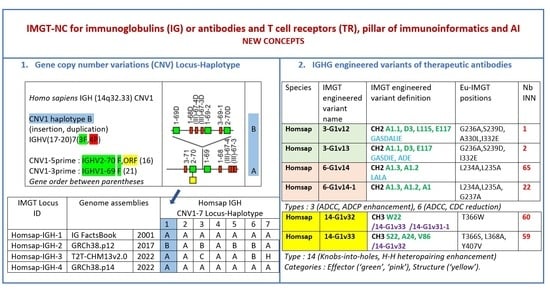
Graphical Abstract

1. Introduction

IMGT®, the international ImMunoGeneTics information system® (IMGT) [1] (https://www.imgt.org), was created in 1989 by Marie-Paule Lefranc (Laboratoire d’ImmunoGénétique Moléculaire (LIGM), Université de Montpellier (UM) and Centre National de la Recherche Scientifique (CNRS) at Montpellier, France. The objective was to deal with and to manage the huge diversity of the immunoglobulins (IG) or antibodies [2] and T cell receptors (TR) [3], which are the antigen receptors (AR) of the adaptive immune responses (AIR) [1,4,5,6]. For the first time, IG and TR variable (V), diversity (D), joining (J) and constant (C) genes [2,3,7,8] were officially recognized as ‘genes’, as were the conventional genes, and accepted in the Human Genome Mapping (HGM10) database. With this major breakthrough, the founding of IMGT® marked the advent of immunoinformatics, a new science which emerged at the interface between immunogenetics and bioinformatics [1,9,10,11,12,13].
The specialist IG and TR nucleotide database, IMGT/LIGM-DB [14,15], was created by LIGM in collaboration with EMBL, using the same accession numbers as the GenBank, EMBL and DDBJ generalist databases (‘GEDI’) for data interoperability of the shared sequences. The first Internet connection of IMGT/LIGM-DB was organized for the 9th International Congress of Immunology (ICI) held in San Francisco, CA (USA) on 23–29 July 1995, marking the 7-year anniversary of the first Internet France–USA connection on the 28 July 1988. The creation of IMGT/LIGM-DB was followed by the development and implementation of other IMGT databases, tools and Web resources for the sequences, genes and structures of the IG or antibodies and TR, which together constitute the IMGT information system [1,9,10,11,12,13,14,15,16,17,18,19,20,21,22,23,24,25,26,27,28,29,30,31,32,33,34,35,36,37,38,39]. The IMGT data are identified, described, classified, numbered, localized, orientated and their source provided, based on the IMGT Scientific chart rules, themselves generated from the IMGT-ONTOLOGY axioms and concepts [1,10,12,25,40,41,42,43,44,45,46,47,48,49,50,51,52,53,54,55,56,57,58,59,60,61,62,63,64,65,66,67,68]. The IMGT standardization, initially characterized for the IG or antibodies and TR of jawed vertebrate species from fish to humans, has been extended to members of the immunoglobulin superfamily (IgSF) other than IG and TR, to the major histocompatibility (MH) proteins, to members of the MH superfamily (MhSF) other than MH and to related proteins of immune interest (RPI). Forty-five years of LIGM expert biocuration and innovation have made IMGT®, the international ImMunoGeneTics information system®, which comprises seven databases, seventeen tools and more than 25,000 pages of Web resources [1,34,35,36,37,38,39], the global reference for IG and TR data (genes, sequences and structures), with the IMGT nomenclature being the common language for immunogenetic and immunoinformatics.
Here, we firstly review, in three sections, the IMGT IG and TR key data which are the antigen receptors of the adaptive immune responses, the IMGT-ONTOLOGY and the IMGT information system which bridge biology and computational spheres, and the IMGT nomenclature (IMGT-NC) of the IG and TR genes and alleles, which marks the birth of immunoinformatics; we then present the IMGT-NC reports for novel IG and TR genes and alleles names, and, in three sections, the IMGT-NC new concepts for biocuration of IG and TR loci from genome assemblies, for gene copy number variations (CNV) with the definition of ‘CNV haplotypes’ and ‘CNV-Locus-Haplotype’, and for engineered IGHG variants for effector properties, half-life and structures of therapeutic antibodies. The last section highlights examples of long term biological contribution of researchers using the IMGT nomenclature, for the genomic and expressed repertoires of the IG and TR of jawed vertebrates, from fish to humans, for antibody engineering, humanization, structure and specificity, demonstrating that IMGT-NC is a common long-term scientific research endeavor, which has provided a foundational and unifying infrastructure for immunogenetics in immunoinformatics, which is now available for artificial intelligence.

2. IMGT Key Data: The IG and TR, Antigen Receptors of the Adaptive Immune Responses

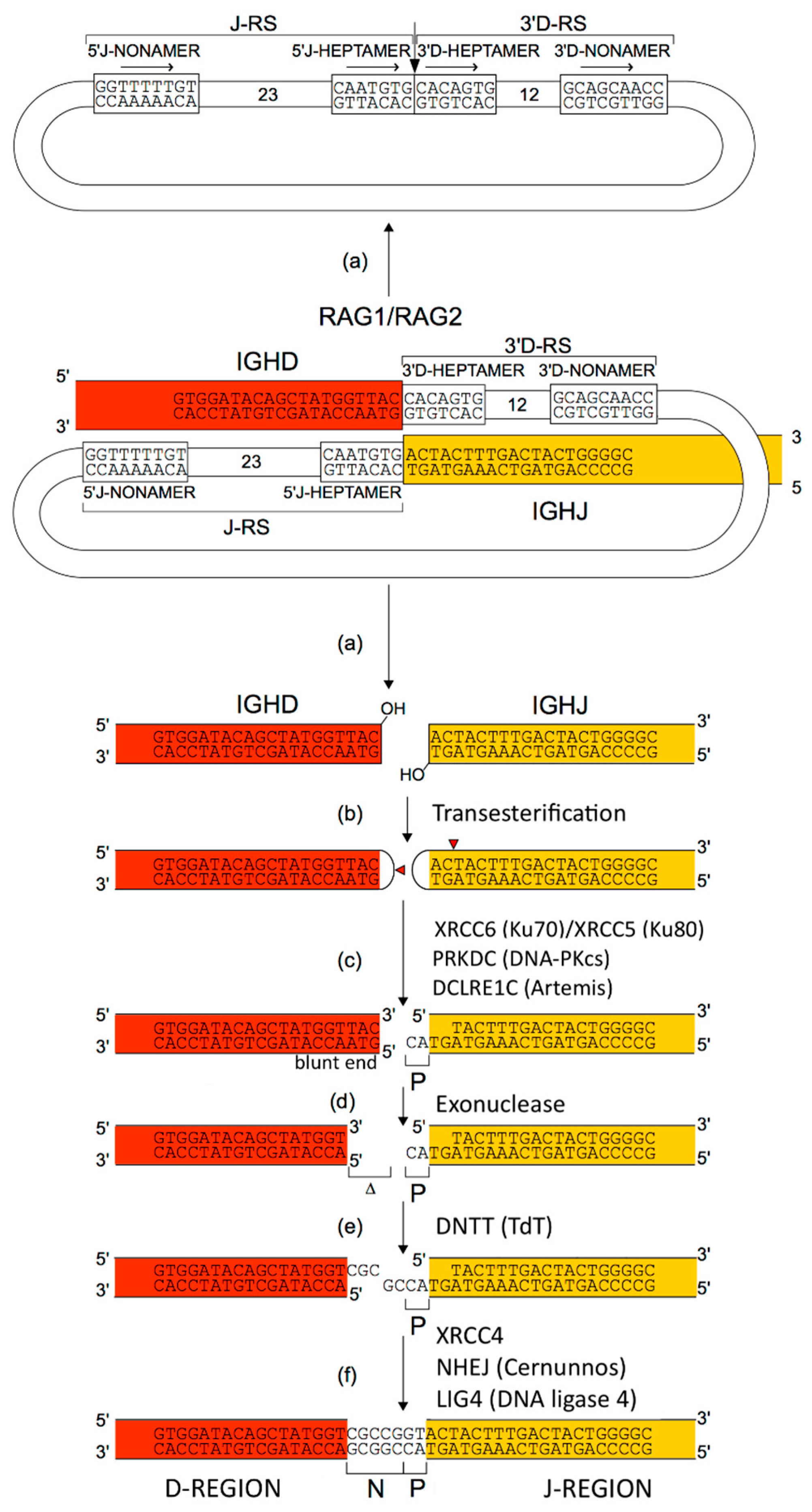
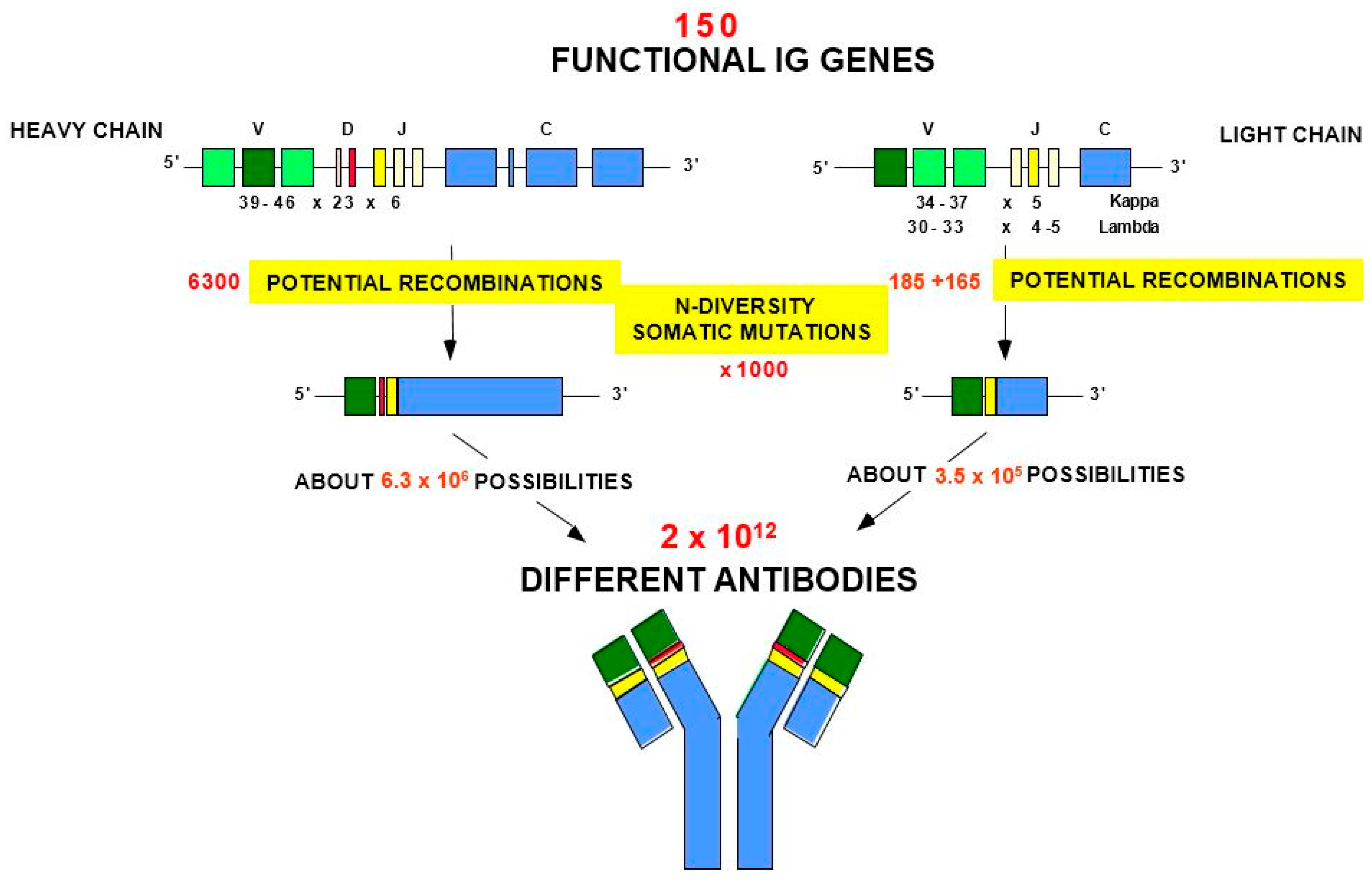
The adaptive immune response (AIR) was acquired by jawed vertebrates (or Gnathostomata) more than 450 million years ago and is found in all extant jawed vertebrate species from fish to humans [1]. The adaptive immune response is characterized by a remarkable immune specificity and memory, which are properties of the B and T cells owing to an extreme diversity of their specific antigen receptors, the IG or antibodies of the B cells (membrane IG) and of the plasmocytes (secreted IG) [2,4,5,6] and the TR [3]. The potential expressed antigen receptor repertoire (ARR) of the adaptive immune responses (AIR) of each individual is estimated to comprise about 2 × 10¹² different IG and TR, and the limiting factor is only the number of B and T cells that an organism is genetically programmed to produce [1]. This huge diversity results from the complex molecular synthesis of the IG and TR chains, and more particularly of the synthesis of the variable domain (V-DOMAIN) that, at the N-terminal end of each chain, recognizes and binds the antigens [1,2,3,4,5,6]. The IG and TR synthesis includes several unique mechanisms that occur at the DNA level: (1) combinatorial rearrangements of the V, D and J genes that code the V-DOMAIN (the number of V, D and J genes in a given IG or TR locus represents the potential genomic repertoire), (2) exonuclease trimming at the ends of the V, D and J genes and random addition of nucleotides by the DNA nucleotidylexotransferase (DNTT, TdT, terminal deoxynucleotidyl transferase) that creates the junctional N-diversity regions (Figure 1) [6] before ligation of the V-(D)-J gene (the V-(D)-J-REGION thus created is then classically transcribed into mRNA and spliced, in the chain transcript, to the C-REGION exon(s) encoded by the C-gene), and (3) later during B cell differentiation, for the IG, two unique mechanisms again at the DNA level, somatic hypermutations (SHM) and class or subclass switch recombination (CSR) [6].
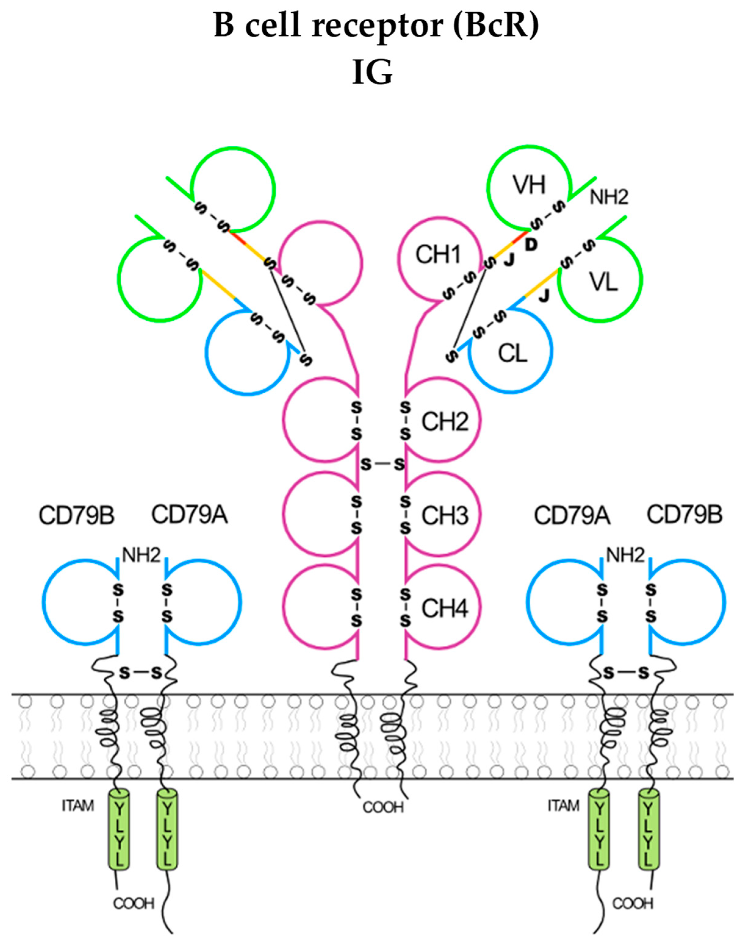
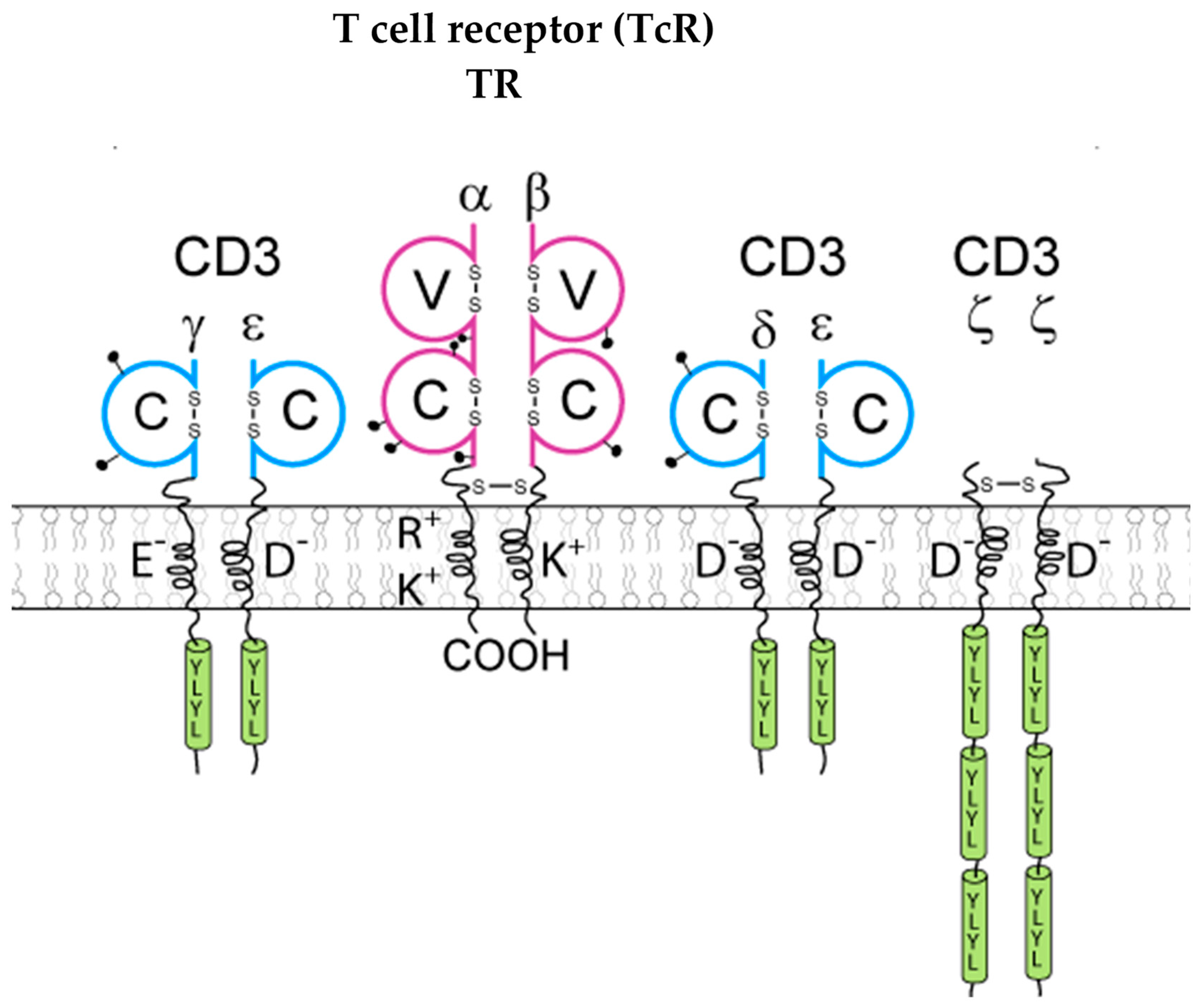
The immunoglobulins (IG) [1,2,4,5,6] or antibodies and the T cell receptors (TR) [1,3] are the antigen receptors of the adaptive immune responses (AIR) [1]. The IG are expressed as B cell receptors (BcR) at the surface of B cells with the coreceptors CD79A/CD79B on mature and memory B cells (Figure 2) whereas the TR are expressed as T cell receptors (TcR) at the surface of T cells with the coreceptors CD3G/CD3E, CD3D/CD3E, and CD247 (alias CD3Z)/CD247 (Figure 3).
The coreceptors of the BcR and TcR transduce the signal intracellularly following the binding of the antigen receptor, IG and TR, respectively, to its antigen [2,3]. The IG recognize antigens in their native (unprocessed) form, whereas the TR recognize processed antigens that are presented as peptides by the highly polymorphic major histocompatibility (MH, in humans HLA for human leucocyte antigens) proteins [1]. At the terminal stage of the B cell differentiation, the IG are secreted by the plasmocytes (or plasma cells, or effector B cells) found in the bone marrow, lymph nodes and spleen [2,6], whereas the TR remain always membranar on the T cells [3].

3. IMGT: An Ontology and a System to Bridge Biology and Computational Spheres

3.1. IMGT-ONTOLOGY: Accuracy and Consistency of the IMGT Data

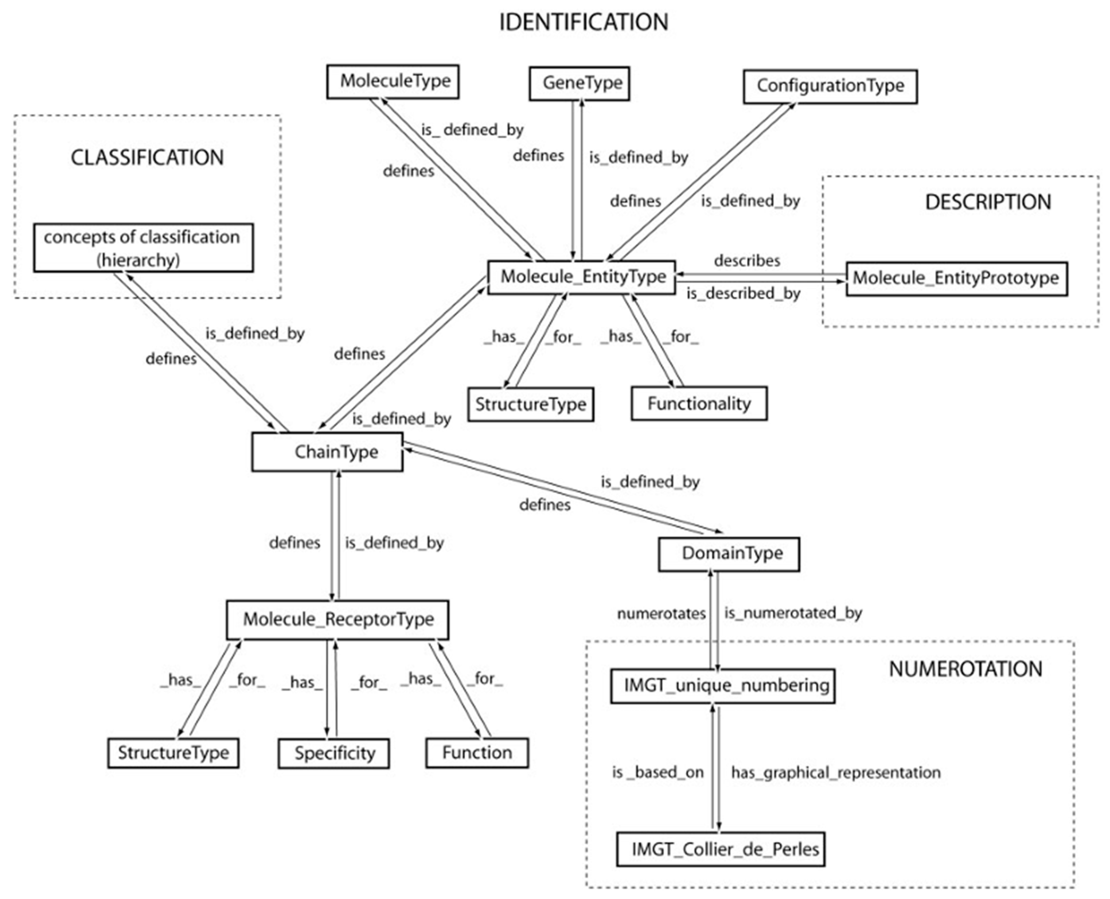
The accuracy and the consistency of the IMGT data (genes, nucleotide and amino acid sequences and structures), as well as the coherence between the different IMGT components (databases, tools and Web resources) are based on IMGT-ONTOLOGY, the first ontology for immunogenetics and immunoinformatics [1,10,12,25,40,41,42,43,44,45,46,47,48,49,50,51,52,53,54,55,56,57,58,59,60,61,62,63,64,65,66,67,68]. The IMGT-ONTOLOGY comprise seven axioms ‘IDENTIFICATION’ [50], ‘DESCRIPTION’ [51], ‘CLASSIFICATION’ [52], ‘NUMEROTATION’ [53,54,55,56,57,58,59,60,61,62,63,64,65,66], ‘LOCALIZATION’ [48], ‘ORIENTATION’ [48,49] and ‘OBTENTION’ [48,49], that postulate that objects, processes and relations have to be identified, described, classified, numerotated, localized, and orientated, and that the way they are obtained has to be determined [40,43,44,47,48,49].
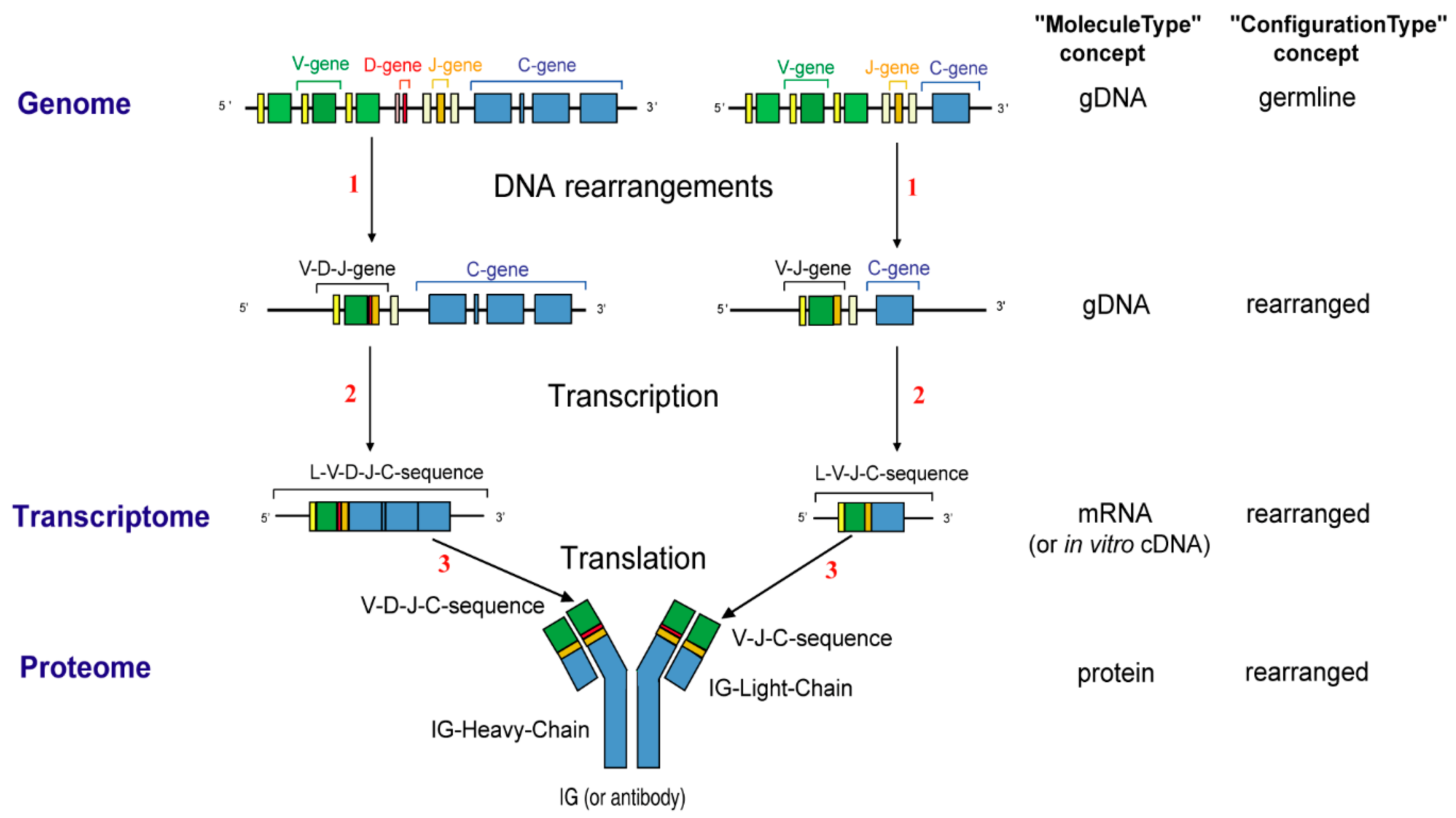
The IMGT-ONTOLOGY seven axioms and generated concepts have been essential for the conceptualization of molecular immunogenetics knowledge through diverse facets (Formal IMGT-ONTOLOGY or IMGT-Kaleidoscope [47]) and for the definition of the IMGT Scientific chart rules (https://www.imgt.org/IMGTScientificChart/) (accessed on 11 February 2026) (IMGT-Choreography) [43,44]. The molecular synthesis of immunoglobulins (IG) or antibodies and the origin of their diversity (Figure 4) is shown, as an example of biological knowledge at the molecular level, based on the IMGT-ONTOLOGY concepts of identification [50] (Figure 5).
Detailed molecular information on the synthesis of the H-mu chains (following D-J and V-D-J rearrangements in the IGH locus), the synthesis of the L-kappa and L-lambda chains (following V-J rearrangements in the IGK and IGL loci), the origin of the variable domain diversity of the immunoglobulins, the class or subclass switch recombination, the regulation of the rearrangements and chain expression, the structural and biological properties of the secreted immunoglobulins, the human IG classes and subclasses, heavy and light chain types, and the Homo sapiens immunoglobulin (IG) chain characteristics taking into account the IMGT concepts have been reviewed elsewhere [6].
The IDENTIFICATION axiom and concepts of identification led to the standardized keywords and qualifiers and to their definitions (e.g., reference sequence (https://www.imgt.org/IMGTScientificChart/SequenceDescription/IMGTreferencesequences.html (accessed on 9 April 2026), clonotype, paratope, epitope, allotype, variant, Fc receptor, FcR) and to the relations for molecule identification [50]) (IMGT standardized keywords: https://www.imgt.org/IMGTScientificChart/SequenceDescription/Keywords.php (accessed on 9 April 2026); IMGT/LIGM-DB qualifiers: https://www.imgt.org/ligmdb/qualifier (accessed on 9 April 2026)).
The DESCRIPTION axiom and concepts of description led to the standardized labels (e.g., V-REGION, complementarity determining region (CDR)-IMGT (CDR1-IMGT to CDR3-IMGT) and framework region (FR-IMGT) (FR1-IMGT to FR4-IMGT)) and to the relations for the prototype descriptions [51] (IMGT/LIGM-DB standardized labels: https://www.imgt.org/ligmdb/label#) (accessed 14 February 2026). IMGT standardized keywords and labels have first been defined for nucleotide sequences and their translation (Supplementary Materials Table S1. List of definitions in the Encyclopedia of Systems Biology). For sequences analyzed only at the amino acid level (e.g., from crystallized three-dimensional (3D) structures), the identification (IMGT keywords) and description (IMGT labels) are done in terms of domains, chains and receptors (IMGT/PROTEIN-DB and IMGT/3Dstructure-DB: Standardized keywords and labels for IG, TR, MH, RPI and FPIA: https://www.imgt.org/IMGTScientificChart/SequenceDescription/IMGT3Dkeywords.html (accessed on 9 April 2026)).
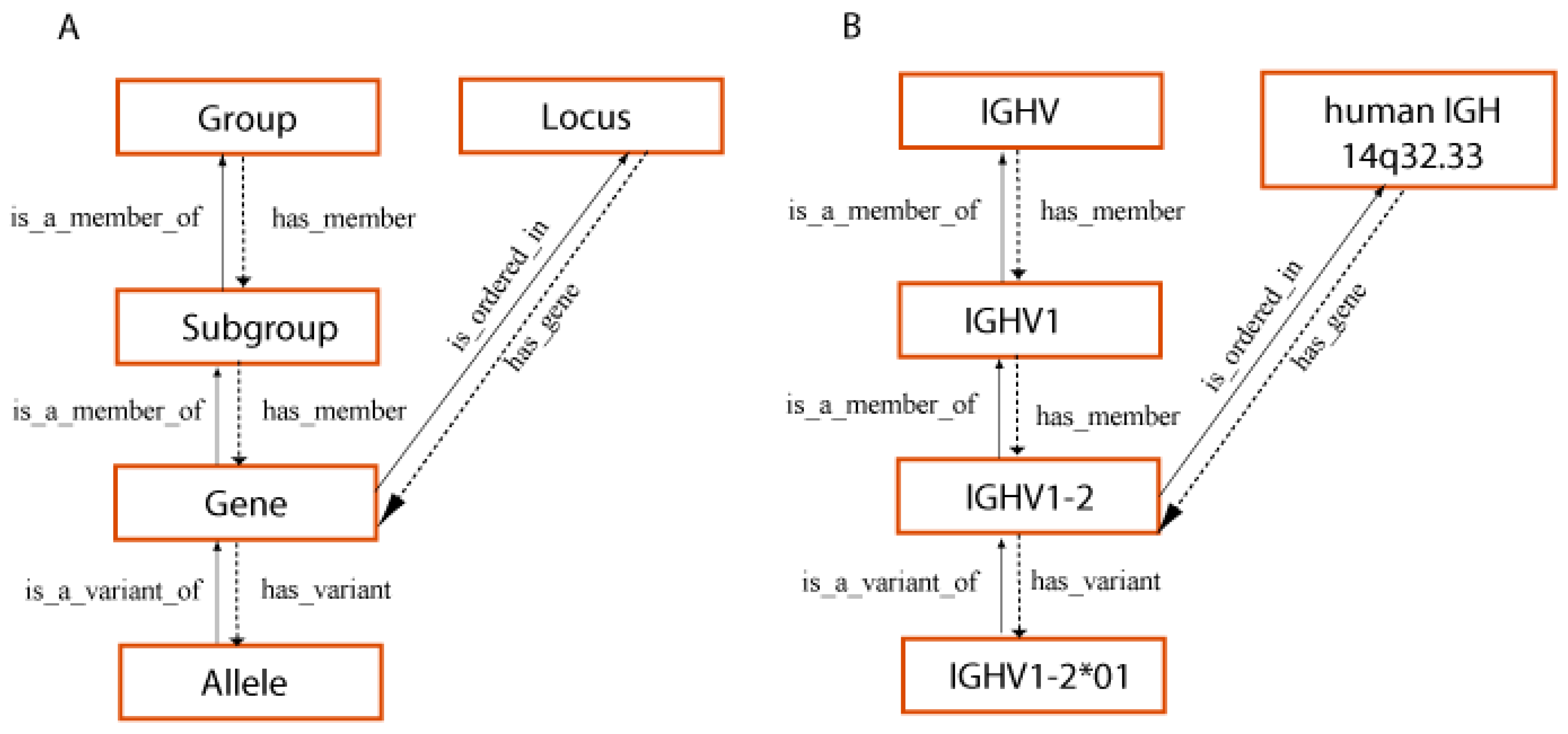
The CLASSIFICATION axiom and concepts of classification [1,52] have formalized the rules of the IMGT standardized gene and allele nomenclature of the IG and TR of jawed vertebrates (Gnathostomata), conceived by LIGM [1,2,3,7,8,52,66,67,68], starting from the early eighties as the ImMunoGeneTics nomenclature (IMGT-NC), officially recognized in 1989 at the 10th Human Genome Mapping (HGM10) Workshop, and demonstrated by the publication of ‘The Immunoglobulin FactsBook’ [2] and ‘The T cell receptor FactsBook’ [3] in 2001 and the creation of the WHO-IUIS Nomenclature Subcommittee for Immunoglobulins and T cell receptors (IMGT-NC) in 2007 [67,68]. The CLASSIFICATION axiom and concepts of classification [1,52] (Figure 6) provide identical IMGT Scientific chart rules regarding the gene and allele nomenclature across jawed vertebrates from fish to humans [1,2,3,7,8,52,66,67,68] and is one of the two pillars of immunoinformatics [66].
The NUMEROTATION axiom and concepts of numerotation [1,53,54,55,56,57,58,59,60,61,62,63,64,65,66] are at the origin of the IMGT unique numbering for the variable (V) domains of the IG and TR [55], of the IMGT unique numbering for the constant (C) domains of the IG and TR [56] and of the IMGT unique numbering of the groove (G) domains of the MH [57], with their graphical representations, the IMGT Colliers de Perles for V, C and G domains [61,62,63,64,65,66]. The IMGT unique numbering for the V and C domains of the IG and TR is valid, respectively, for the V-LIKE and C-LIKE domains of the IgSF members other than IG and TR from vertebrate and invertebrate species [55,56,58,60]. Similarly, the IMGT unique numbering for the G domains of the MH is valid for the G-LIKE domains of the MhSF members other than MH [57,58,60]. The NUMEROTATION axiom and concepts of numerotation [1,53,54,55,56,57,58,59,60,61,62,63,64,65,66], which provide an IMGT unique numbering for V, C and G domains whatever the species, represent the second pillar of immunoinformatics [66] (Table 1). IMGT positions per domain are used in sections of the IMGT Repertoire IG and TR (protein displays, alignments of alleles, CDR-IMGT lengths, allotypes), and to number amino acids involved in paratope/epitope (antigen receptor IG or TR V-domain/target interactions) and in effector properties (antigen receptor IG C-domain/effector binding proteins (e.g., FcγR, complement interactions).

3.2. Relations Between the Concepts of IMGT-ONTOLOGY Implemented in the IMGT Information System

3.2.1. Overall View of the Relations Between Concepts at the Molecular Level

The relations between the concepts of identification, description, classification and numerotation implemented in the databases, tools and web resources of the IMGT® information system [13,14,15,16,17,18,19,20,21,22,23,24,25,26,27,28,29,30,31,32,33,34,35,36,37,38,39] and formalized in IMGT-ONTOLOGY [12,40,41,42,43,44,45,46,47,48,49,50,51,52,53,54,55,56,57,58,59,60,61,62,63,64,65,66,67,68] are summarized in Figure 7.

3.2.2. The ‘Molecule_EntityType’ Concept

The ‘Molecule_EntityType’ concept (Figure 7) allows us to identify any coding molecule of any jawed vertebrate genome, transcriptome and proteome. There are 21 entities defined by: ‘MoleculeType’ (‘gDNA’, ‘mRNA’ (or in vitro ‘cDNA’), ‘protein’), ‘GeneType’ [‘conventional’, and for the IG and TR, ‘variable’ (V), ‘diversity’ (D), ‘joining’ (J) and ‘constant’(C)], and ‘ConfigurationType’ (‘undefined’ for conventional and C genes, ‘germline’ for unrearranged V, D and J genes and ‘rearranged’ for V, D and J genes after DNA rearrangements [1,3]). Three entities, ‘gene’, ‘nt-sequence’ and ‘AA-sequence’, respectively, identify the gDNA, mRNA (or in vitro cDNA) and protein (‘MoleculeType’) of a conventional gene (‘GeneType’), which by definition is an undefined configuration (‘ConfigurationType’). Eighteen entities identify the IG and TR. The ten most classical IG and TR entity types comprise V-gene, D-gene, J-gene, C-gene, V-D-J-gene, V-J-gene, L-V-D-J-C-sequence, L-V-J-C-sequence, V-D-J-C-sequence and V-J-C-sequence (Table 2).
For example, the entity ‘V-gene’ identifies a ‘gDNA’ containing a ‘V’ gene, in ‘germline’ configuration. The entity ‘L-V-J-C-sequence’ identifies a sequence of ‘mRNA’ (or in vitro ‘cDNA’) containing ‘V’, ‘J’ and ‘C’ genes, with V and J in ‘rearranged’ configuration. The less classical eight entities correspond to partial rearrangements or to sterile transcripts [47].
A ‘Molecule_EntityType’ concept entity has two properties, defined by ‘StructureType’ and ‘Functionality’ (Figure 7). The ‘StructureType’ concept allows us to identify instances with a classical organization (‘regular’), from those that have been modified either naturally in vivo (‘orphon’, ‘processed orphon’, ‘unprocessed orphon’, ‘unspliced’, ‘partially spliced’, etc.) or artificially in vitro (‘chimeric’, ‘humanized’, ‘transgene’, etc.). The ‘Functionality’ concept includes five entities: three of them, ‘functional’, ‘ORF’ (open reading frame) and ‘pseudogene’ identify the functionality of ‘Molecule_EntityType’ instances in undefined or germline configuration (conventional genes, C genes, germline V, D and J genes), whereas the two others, ‘productive’ and ‘unproductive’, identify the functionality of ‘Molecule_EntityType’ instances in rearranged configurations (rearranged V, D and J genes, fusion genes resulting from translocations or obtained by biotechnology and/or molecular engineering).

3.2.3. The ‘ChainType’ Concept

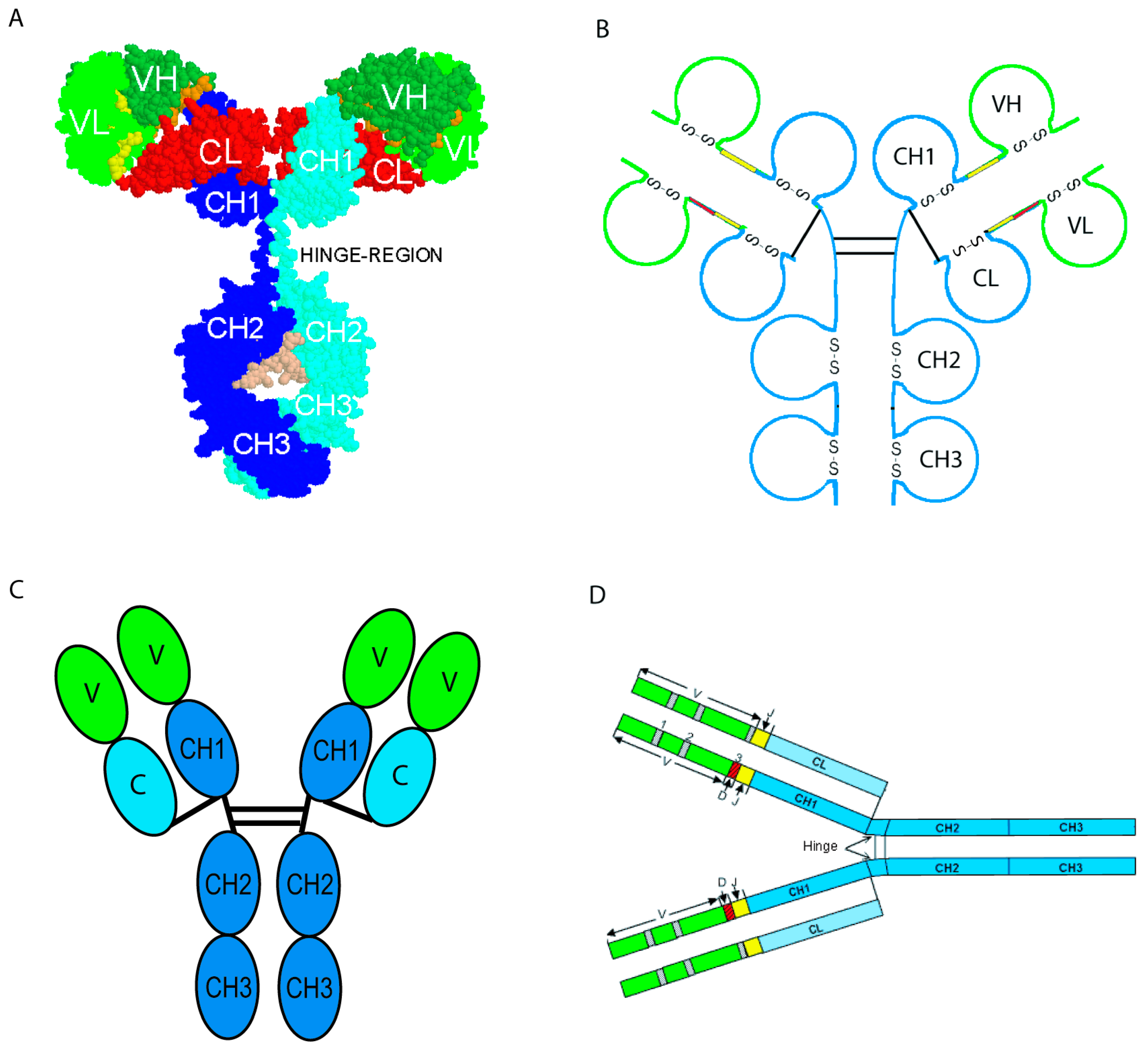
The ‘ChainType’ concept (Figure 7) identifies the type of chain. It is one of the most important concepts of identification for the standardization of genome, transcriptome and proteome data in system biology. Indeed, an instance of the ‘ChainType’ concept is defined not only by an entity of the ‘Molecule_EntityType’ (V-J-C-sequence or V-D-J-C-sequence) but also by an entity of a concept of classification and by a definition in domains, which bridges the gap with 3D structures. Moreover, it is the chain composition which defines the ‘Molecule_ReceptorType’ concept and identifies the type of protein receptor [47]. Thus, IG is an entity of the ‘Molecule_ReceptorType’ concept, defined as comprising four chains, two IG-Heavy and two IG-Light chains (entities of the ‘ChainType’ concept), which are identical two by two and covalently linked. Four graphical representations of an IG or antibody are classically used (Figure 8): (A) 3D molecular model, (B) organization in 12 labeled domains with the conserved intra-domain disulfide bond (S-S) shown for each, (C) representation with each of the 12 domains as an ovoid module (green for the VH and pale green for the VL, blue for the CH1-CH3 and pale blue for the CL (e.g., in IMGT/mAb-DB)), and (D) linear representations of the regions coded by the V (in green), D (in red), J (in yellow) and C (in blue) gene types [66].
By its relation with the concepts of classification, the ‘ChainType’ concept contains a hierarchy of concepts that identify the chain type at different levels of granularity. The finest level of granularity, the ‘GeneLevelChainType’ concept, identifies the chain type by reference to the gene(s), which code(s) the chain (reciprocal relations ‘is_coded_by’ and ‘codes’). The number of instances of the ‘GeneLevelChainType’ concept depends on the number of functional genes and ORF per haploid genome in a given species (in the case of the IG and TR, it is the number of functional and ORF constant genes, which is taken into account). If only the functional genes are considered, the instances of this concept correspond to the isotypes.

3.2.4. The ‘DomainType’ Concept

A chain type instance can also be defined by its constitutive structural units (‘DomainType’concept). A domain is a chain subunit characterized by its 3D structure and, by extension, its amino acid sequence and the nucleotide sequence that encodes it. The ‘DomainType’ concept may theoretically comprise many instances, but so far only the instances that have been carefully characterized by LIGM have been entered in IMGT-ONTOLOGY. The ‘DomainType’concept currently has three major instances fully characterized: V type domain (variable domains of the IG and TR and V-like domains of IgSF superfamily members other than IG or TR) [55], C type domain (constant domains of the IG and TR and C-like domains of IgSF superfamily members other than IG or TR) [56] and G type domain (groove domains of the major histocompatibility (MH) proteins and G-like domains of MhSF superfamily members other than MH) [57] (https://www.imgt.org/IMGTindex/Domain.php) (accessed on 14 February 2026). Other instances of ‘DomainType’ from RPI in IMGT/DomainDisplay [1] include representatives of the A type domain (e.g., [D1] to [D4] of F11 (coagulation factor XI, P03951) and of KLKB1 (kallikrein B1, P039952)), the F type domain (108 sequences of F domains of FN1 (fibronectin 1) and DSCAM (DS cell adhesion molecule)) and the S domain of the scavenger receptor superfamily (SrSF) (e.g., S domains of CD5, CD6, CD163, CD163L1).

3.2.5. The ‘MoleculeReceptorType’ Concept

The properties of the ‘Molecule_ReceptorType’ concept include structure, specificity and function (Figure 6). Thus, instances of the ‘Specificity’ concept identify the antigen recognized by an antigen receptor (IG or TR). The instances of that concept (several hundreds at the present time) can be connected on the one hand, with the ‘Epitope’ concept that identifies the part of the antigen recognized by the antigen receptor and, on the other hand, with the ‘Paratope’ concept that identifies the part of the antigen receptor (IG or TR), which recognizes and binds to the antigen [1]. Instances of the ‘Function’ concept identify the dual function of the IG or antibodies [5] and the modifications linked to the allotypes [66] and to the IMGT engineered variants [66].

3.2.6. The ‘Molecule_EntityPrototype’ Concept (DESCRIPTION)

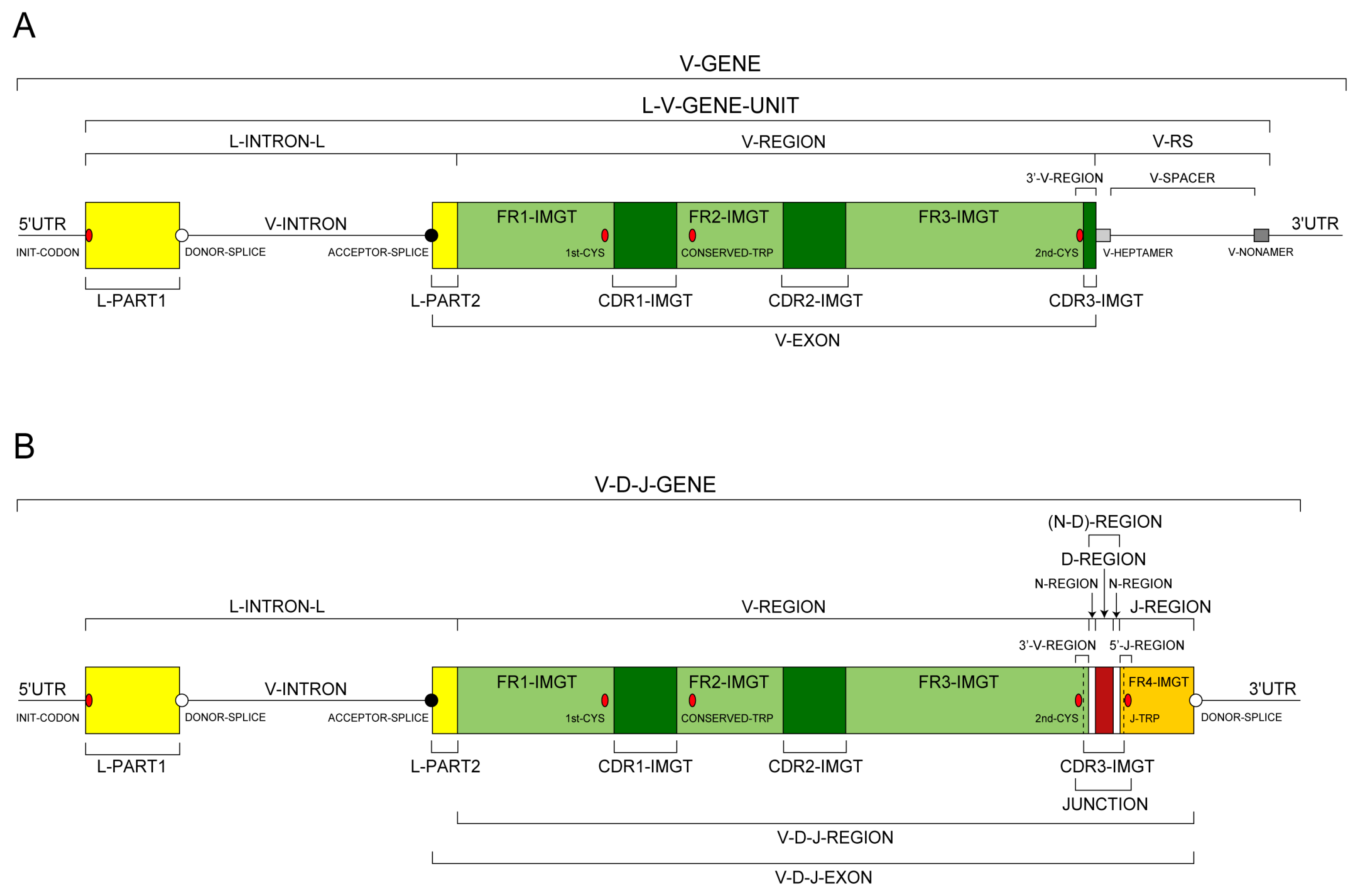
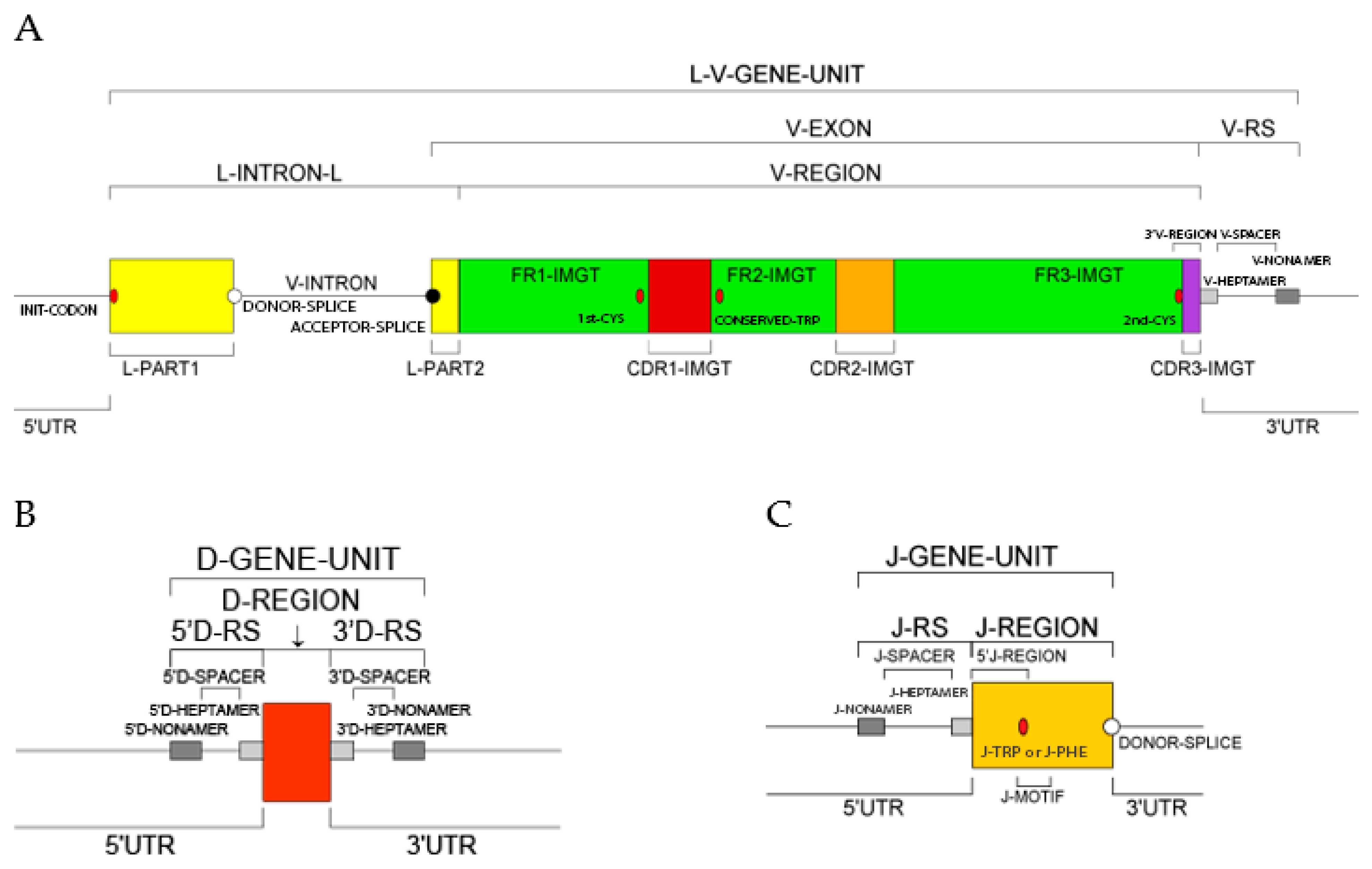
Each one of the 21 instances of the ‘Molecule_EntityType’ concept (IDENTIFICATION axiom) is linked to an instance of the ‘Molecule_EntityPrototype’ concept (DESCRIPTION axiom), by the reciprocal relations ‘is_described_by’ and ‘describes’. Each instance of the ‘Molecule_EntityPrototype’ concept is described with its constitutive motifs and IMGT standardized labels [51] (IMGT Scientific chart > IMGT/IMGT standardized labels https://www.imgt.org/ligmdb/label (accessed 14 February 2026) (DESCRIPTION axiom [51]). Prototypes, for example V-J-GENE and V-D-J-GENE (Figure 9) [66], are available on the IMGT® web site (IMGT Scientific chart https://www.imgt.org/IMGTScientificChart/ (accessed 14 February 2026) > 1. Sequence and 3D structure identification and description > IMGT prototypes table).
Prototypes represent the organizational relationship between labels and give information on the order and expected length (in number of nucleotides) of the labels [51]. This provides rules to verify the manual annotation, and to design automatic annotation tools. Annotation of sequences and 3D structures with these labels constitutes the main part of the expertise. IMGT-specific labels defined for sequences are used to describe specific IG and TR gene organization and protein structures. Interestingly, 64 IMGT-specific labels defined for IG and TR nucleotide sequences have been entered in Sequence Ontology (SO) (https://www.imgt.org/IMGTindex/ontology.php) (accessed 14 February 2026) and 41 essays and four chapters on IMGT labels have been published in Encyclopedia of Systems Biology [36,49].
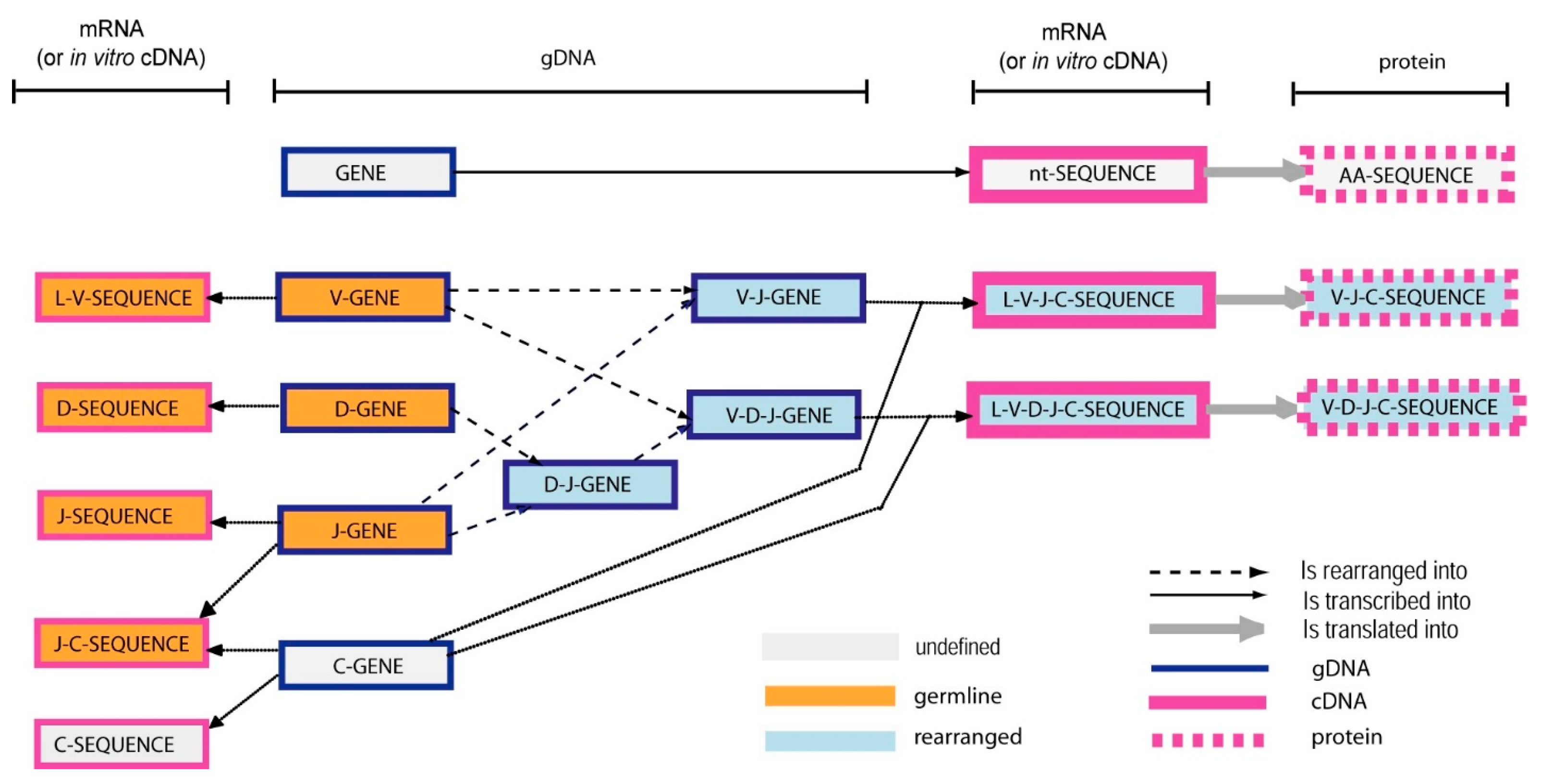
The ‘Molecule_EntityPrototype’ concept is fundamental in IMGT-ONTOLOGY as relations between its instances allow for the representation of the knowledge related to the complex mechanisms of IG and TR gene rearrangements and chain synthesis [1,2,5,6] (Figure 10) [66]. The relation ‘is_rearranged_into’ is specific to the synthesis of the IG and TR. The relations ‘is_transcribed_into’ and ‘is_translated_into’ are general for molecular biology. These three relations allow for the organization of the various instances of the ‘Molecule_EntityPrototype’ concept during the synthesis of the IG and the TR, and in a more general way for the expression of any protein. In addition, by more specific relations, they allow us to take into account the alternative transcripts, the protein isoforms and the post-translational modifications.
More than 500 standardized labels were defined: 221 for the nucleotide sequences (IMGT/LIGM-DB labels https://www.imgt.org/ligmdb/label (accessed on 9 April 2026)) and 285 for the 3D structures (IMGT/3Dstructure-DB labels https://www.imgt.org/IMGTScientificChart/SequenceDescription/IMGT3Dkeywords.html (accessed on 9 April 2026)). A set of 10 relations is necessary and sufficient to compare the localization of the motifs of an instance of the concept ‘Molecule_EntityPrototype’ (Table 3). These relations are part of the concepts of localization (LOCALIZATION axiom) (IMGT Index, https://www.imgt.org/IMGTindex/Localization.php (accessed on 9 April 2026)).

3.3. IMGT®, the International ImMunoGeneTics Information System®: Coherence Between the Components

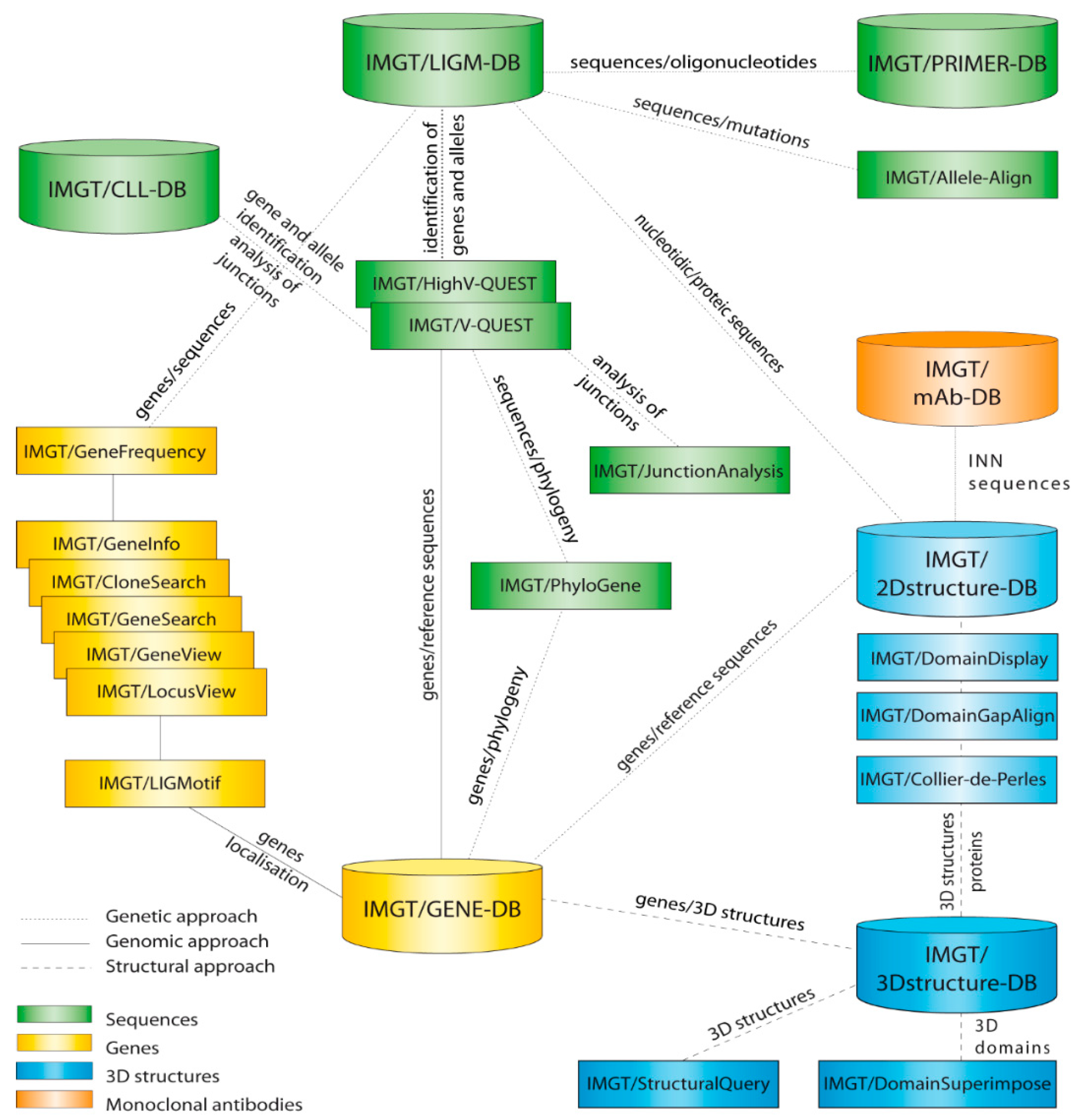
IMGT®, the international ImMunoGeneTics information system® [34,35,36,37,38,39], comprises the IMGT databases [69,70,71,72,73,74,75,76,77] and the IMGT tools [78,79,80,81,82,83,84,85,86,87,88,89,90,91,92,93,94,95,96,97,98,99,100,101,102,103,104,105,106,107] represented as cylinders and rectangles, respectively, in Figure 11. Genomic (genes), genetic (sequences) and structural (structures) components are in yellow, green and blue, respectively [34]. Interactions in the genomic, genetic and structural approaches are represented with continuous, dotted and broken lines, respectively [34].
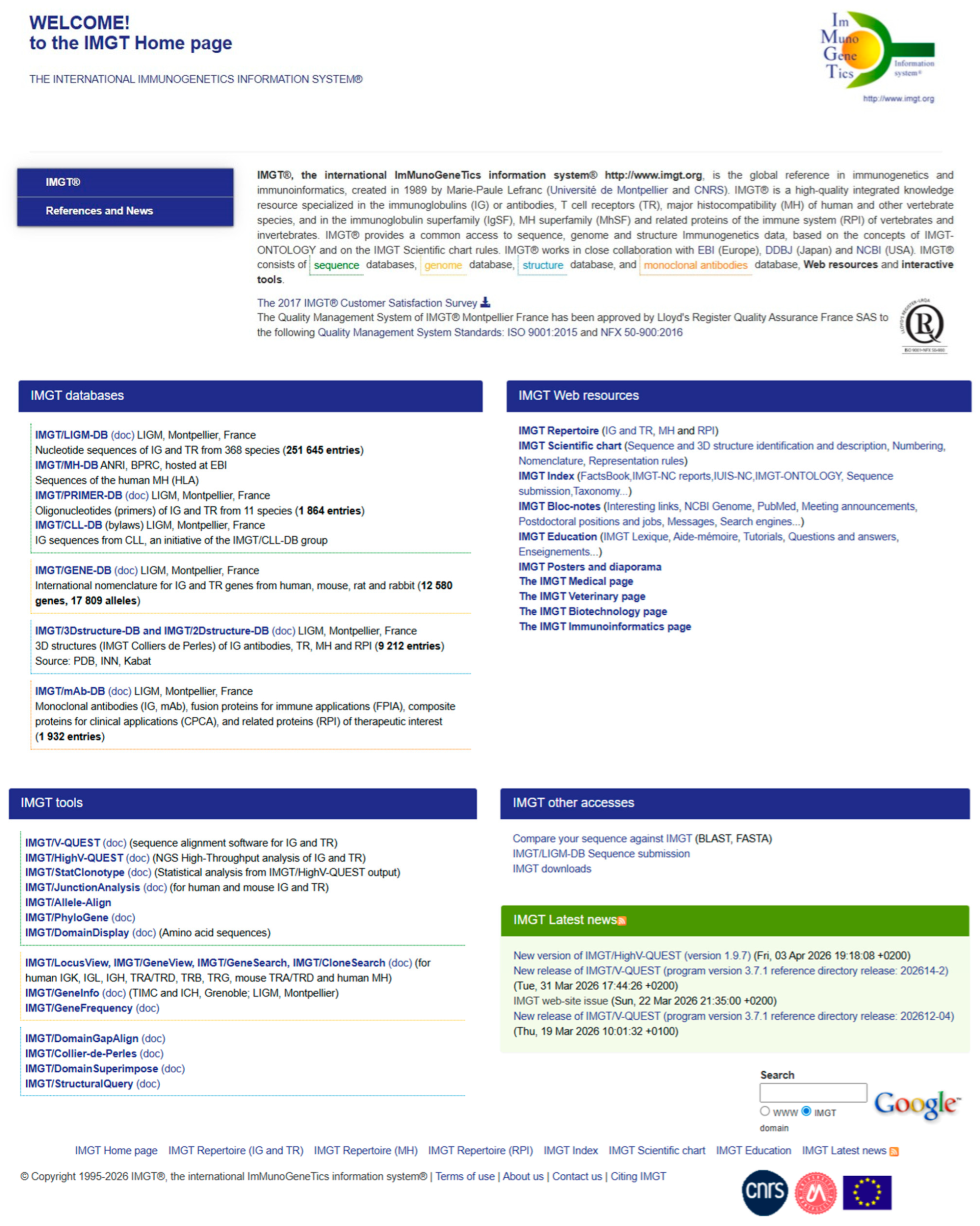
The original IMGT Home page (Figure 12) displays the four sections ‘IMGT databases’, ‘IMGT tools’, ‘IMGT Web resources’ (more than 25,000 pages, previously the ‘IMGT Marie-Paule page’) and ‘IMGT other accesses’ constitutive of the IMGT®, the international information system® (https://www.imgt.org) and gives a direct access to each of the individual seven databases [69,70,71,72,73,74,75,76,77,78], seventeen tools [78,79,80,81,82,83,84,85,86,87,88,89,90,91,92,93,94,95,96,97,98,99,100,101,102,103,104,105,106,107], ten sections of the Web resources [108] or other three accesses. This key feature of the IMGT Home page allows the users, at any time of their queries, to easily return to the IMGT Home page to get direct access to any IMGT database, tool, web resource or other access.
The ‘IMGT databases’ [69,70,71,72,73,74,75,76,77,78] section gives direct access seven databases, three specialized in IG and TR nucleotide sequences (IMGT/LIGM-DB [69], IMGT/PRIMER-DB [70,71], IMGT/CLL-DB [72]), one for IG and TR genes and alleles (IMGT/GENE-DB [73]), and two for amino acid sequence two-dimensional (2D) and three-dimensional (3D) structures (IMGT/2Dstructure-DB and IMGT/3Dstructure-DB [74,75,76]). The IMGT/mAb-DB [77,78] interface allows for the querying of therapeutical monoclonal antibodies (IG, mAb), fusion proteins for immune applications (FPIA), composite proteins for clinical applications (CPCA) and related proteins (RPI) of therapeutic interest (with links to amino acid sequences in IMGT/2Dstructure-DB, and if 3D structures are available, links to IMGT/3Dstructure-DB [74,75,76].
The ‘IMGT® tools’ [78,79,80,81,82,83,84,85,86,87,88,89,90,91,92,93,94,95,96,97,98,99,100,101,102,103,104,105,106,107] include: (1) for nucleotide analysis, IMGT/V-QUEST [79,80,81,82,83,84,98] with the integrated IMGT/JunctionAnalysis [85,86,87,88] and IMGT/Decryption [89] tools and the internal IMGT/Automat [90,91], and for next generation sequencing (NGS), the high-throughput IMGT/HighV-QUEST web portal [78,84,92,93,94,95,98], and the downloadable IMGT/StatClonotype [96,97] package (which allows for statistical pairwise analysis of the diversity and expression of the IMGT clonotypes (AA) and repertoire comparisons in adaptive immune responses); (2) for genome analysis, IMGT/LIGMotif [104] used for the identification and description of new IG and TR genes in large genomic sequences; (3) for amino acid sequence analysis per domain, IMGT/DomainGapAlign [75,78,105,106] and for queries of IMGT/3Dstructure-DB, IMGT/StructuralQuery [74]; and (4) for graphical representation of the domains, IMGT/Collier de Perles [107] (e.g., IMGT Colliers de Perles of the variable (V), constant (C) and groove (G) domains).
The ‘IMGT Web resources’ (previously ‘The Marie-Paule page’) (Figure 12) comprise more than 25,000 pages with direct access to ten sections: IMGT Repertoire (IG and TR, MH, RPI), IMGT Scientific chart (Sequence and 3D structure identification and description, Numbering, Nomenclature, Representation rules), IMGT Index (FactsBook, IMGT-NC reports, IUIS-NC, IMGT-ONTOLOGY, Sequence submission, Taxonomy…), IMGT Bloc-notes (Interesting links, NCBI Genome, PubMed…), IMGT Education (IMGT Lexique, Aide-mémoire (amino acid physicochemical properties [108], splicing sites, Tutorials…), IMGT Posters and diaporama, The IMGT Medical page, The IMGT Veterinary page, The IMGT Biotechnology page, and The IMGT Immunoinformatics page.
The ‘IMGT other accesses’ includes the IMGT/BlastSearch on IMGT sequences databases and the IMGT® downloads (IMGT/LIGM-DB flat files, IMGT/GENE-DB reference sequences in FASTA format, IMGT/3Dstructure-DB flat files, IMGT/V-QUEST reference sequences);
IMGT databases, IMGT tools and examples of items of the IMGT Repertoire (IG and TR) (part of the IMGT Web resources) for the sequences, genes and structures of the IG and TR are listed in Table 4.
The coherence between the components of the system (databases, tools and web resources) is maintained through the use of the standardized IMGT Scientific chart rules (Sequence and 3D structure identification and description, Numbering, Nomenclature, Representation rules) (Figure 12).

4. The IMGT Nomenclature (IMGT-NC) of the IG and TR Genes and Alleles

4.1. Advent of Immunoinformatics

The V, D, J, and C genes which code the antigen receptors were officially recognized as ‘genes’, as were the conventional genes, at HGM10, in New Haven in 1989, marking the creation of IMGT® and the advent of immunoinformatics [1]. The Homo sapiens TRG locus [109] was the first complete antigen receptor locus officially entered in 1989 in the HGM10 database [110,111].
The human (Homo sapiens) IG and TR IMGT gene names [2,3,7,8] were approved by the Human Genome Organization (HUGO) Nomenclature Committee (HGNC) in 1999 [112] and entered in the NCBI gene database (first LocusLink, then EntrezGene, and currently Gene, with reciprocal links with IMGT/GENE-DB).
The manual analysis of the germline nucleotide sequences by the LIGM biocuration team has resulted in 25 publications [113,114,115,116,117,118,119,120,121,122,123,124,125,126,127,128,129,130,131,132,133,134,135,136,137], of which 20 were in the section ‘IMGT Locus in Focus’ of Experimental and Clinical Immunogenetics—on human IGL [114], IGK [115], IGH [117,118], TRB [120,123], TRA [121,122], proteins display IG [119], TR [126], mouse IGK [116,134], TRB [129], TRD [130], teleostei IG [124,125], nomenclature human IGH [131], IGK [132], and IGL [133]. Two were in The Immunologist—locus map IG [127] and TR [128]—one was in Developmental and Comparative Immunology—mouse TRA and TRD [136]—and one chapter was in Molecular Biology of B cells on IGL human and mouse [137], with standardization of gene nomenclature, functionality, and allele polymorphism and the setting of the IMGT Scientific chart rules and IMGT unique numbering.

4.2. Homo sapiens IG and TR Loci, Genes and Alleles: The Immunoglobulin FactsBook and the T Cell Receptor FactsBook References

IG and TR genes are classified in groups defined by the gene type (V, D, J or C) and by the locus to which they belong. The seven major loci, three for IG (IGH, IGK and IGL) [2,6] and four for TR (TRA, TRB, TRD and TRG) [3], are located on different chromosomes, with the particularity of the TRD locus being nestled inside the TRA locus in higher vertebrates [3]. The princeps publications on the IG and TR loci, genes and alleles of the seven human (Homo sapiens) loci are the two FactsBooks published in 2001 [2,3]. The Factsbooks comprise 203 functional and open reading frame (ORF) genes for IG, corresponding to 459 alleles, for a total of 837 sequences [2], and for TR, 168 functional and ORF genes [3]. Entries of the FactsBooks [2,3] provide information on assignment to subgroups and nomenclature, gene definition and functionality, gene location, allelic polymorphism, standardized sequence alignment with protein translation, framework and complementarity determining region (CDR-IMGT) lengths, two-dimensional representations (or Colliers de Perles), IMGT/LIGM-DB and EMBL/GenBank accession numbers, genome database accession numbers (GDB, LocusLink) and key references [2,3]. This information has served as a template for the IMGT Repertoire (IG and TR) https://www.imgt.org/IMGTrepertoire/ (accessed on 9 April 2026)of the IMGT Web resources (the ‘IMGT Marie-Paule page’), which comprises (1) ‘Locus and genes’; (2) ‘Proteins and alleles’; (3) ‘2D and 3D structures’; (4) ‘Probes and RFLP’; (5) ‘Taxonomy’; (6) ‘Gene regulation and expression’; (7) ‘Genes and clinical entities’. Basic IMGT Web resources include ‘Gene tables’, ‘Alignments of alleles’, ‘Protein displays’, ‘Colliers de Perles’, ‘Locus representations’, ‘Potential germline repertoires with CDR-IMGT lengths’, ‘Locus gene order’, copy number variations (CNV) and haplotypes.
This detailed identification, description and classification of the human IG and TR loci, genes and alleles [2,3], using the IMGT Scientific chart rules https://www.imgt.org/IMGTScientificChart/ (accessed on 9 April 2026), is the result of a huge work of annotation and expert analysis, by LIGM, of tens of thousands of nucleotide sequences from phages, cosmids or contigs submitted by the authors to the generalist nucleotide databases (EMBL database, now European Nucleotide Archive (ENA) [138], GenBank [139] and DNA Databank of Japan (DDBJ)) [140]. The annotated sequences were integrated into the then-newly created IMGT/LIGM-DB [14,15], using the EMBL/GenBank/DDBJ accession numbers in order to facilitate interoperability with the generalist nucleotide databases. The ‘Nature’ and ‘Science’ papers on human genome sequencing [141,142], published in 2001, contain limited information on the genes of the antigen receptors of the adaptive immune responses. However a careful analysis of the maps published in these papers allowed us to confirm the chromosomal localizations of the seven main loci—IGH at 14q32.33, IGK at 2p11.2 and IGL at 22q11.2 (for the immunoglobulins), TRA at 14q11.2, TRB at 7q34, TRG at 7p14 and TRD at 14q11.2 (for the T cell receptors)—described in 2001, in the Immunoglobulin FactsBook [2] and in the T cell receptor FactsBook [3], respectively, and determined by an analysis of translocations involving the IG and/or TR loci in leukemia and lymphoma (https://www.imgt.org/IMGTrepertoire/GenesClinical/translocation/human/overview/Hu_overviewpart1.html) (accessed on 14 February 2026).

4.3. Extension of the IMGT Nomenclature to Mus musculus and Fish (Chondrichtyes and Teleostei) IG and TR Genes and Alleles

Based on the paradigm of the human loci (IMGT nomenclature, IMGT unique numbering, IMGT standardized keywords and labels), the seven mouse (Mus musculus) loci with a total of 625 genes (377 IG and 248 TR) [116,129,130,134,136] were characterized and presented at the 19th International Mouse Genome Conference (IMGC) in 2005 (https://www.imgt.org/IMGTposters/IMGC_IG.html and https://www.imgt.org/IMGTposters/IMGC_TR.html) (accessed on 14 February 2026)), and entered in NCBI Gene, with reciprocal links to IMGT/GENE-DB and in Mouse Genome Informatics (MGI).
The analysis of IG genes in four Chondrichthyes and twenty-two different Teleostei species confirmed that the IG and TR paradigm was applicable for fish; however, most sequences were at that time unmapped and were assigned a provisional nomenclature with the letter S [124,125]. The Chondrichthyes and Teleostei light chain which is neither kappa nor lambda was defined as ‘iota’ encoded by genes of the IG iota (IGI) locus which includes IGIV, IGIJ and IGIC groups (https://www.imgt.org/IMGTrepertoire/LocusGenes/genetable/Teleostei/#IGIV) (accessed on 14 February 2026).
Since 1998, novel genes and alleles of any species have been announced in ‘IMGT® Creations and updates’ after validation by IMGT-NC [67,68].

4.4. Management of Genes and Alleles in IMGT/GENE-DB and Corresponding Nucleotide Reference Sequences and Accession Numbers in IMGT/LIGM-DB

Created in 2003, IMGT/GENE-DB [73] provides direct links (access from the Query page) which allow the most frequent requests to be encoded in the form of URL.
A request for a given gene provides (1) the IMGT/GENE-DB entry, (2) the corresponding IMGT/LIGM-DB- nucleotide reference sequence and accession number of each allele of that gene in FASTA format, (3) links to the other IMGT/LIGM-DB sequence(s) with labels in FASTA format, (4) three tables with links to annotated IMGT/LIGM-DB cDNA, to annotated IMGT/LIGM-DB rearranged genomic DNA sequences, and to annotated IMGT/3Dstructure-DB structures and IMGT/2Dstructure amino acid sequences, (5) a table with the gene in genome assemblies with delimitations of the labels of the V-, D-, J- or C-GENE-UNIT.
External links include Nomenclature (HGNC database), Genome databases (NCBI Gene, Ensembl, GeneCards), Protein database (Uniprot), Sequence databases (ENA, DDBJ, GenBank) and for the genome databases, a table in two formats, HTML and CSV format.
On 19 December 2025, IMGT/GENE-DB data include 758 Homo sapiens IG and TR genes and 1825 alleles (507 IG genes and 1319 alleles, 251 TR genes and 506 alleles) with links to HGNC, NCBI Gene, Ensembl, GenAtlas, GeneCards and UniProt, and 1228 Mus musculus IG and TR genes and 1887 alleles (950 IG genes and 1325 alleles, 278 TR genes and 562 alleles) with links to MGI and NCBI Gene. The information, for each IMGT/GENE-DB entry, include: IMGT gene functionality, IMGT gene definition (for Homo sapiens and Mus musculus IG and TR), the HGNC gene definition (identical to the IMGT gene definition), number of alleles, chromosomal localization and IMGT/LIGM-DB reference sequence(s) for allele *01. IMGT/GENE-DB is updated weekly, with downloads available in different formats, in the “IMGT downloads” section.

5. IMGT-NC Reports for Novel IG and TR Genes and Alleles Names

With the increase in genome sequencing and assembly, the starting point for IG and TR gene identification, description and classification has moved from individual sequences (researchers’ submission to generalist databases) to the IG and TR locus identification in NCBI Whole Genome Assemblies (WGS) (submitted by sequencing groups and analyzed by researchers).
In order to allow researchers to go ahead with expression studies and to publish their data with IMGT gene names even if the loci have not yet been annotated in IMGT® or in other specialist databases, thirty IUIS NOM IMGT-NC Reports have been published from 2017 to 2022 (Supplementary Materials Table S2. List of the IMGT-NC reports 1–30). That initiative has allowed scientists to propose IMGT gene names for new IG and TR variable (V), diversity (D), joining (J) and constant (C) genes and alleles, for a given locus of a given species, based on the IMGT Scientific chart rules and the IMGT-ONTOLOGY concepts of classification (CLASSIFICATION axiom).
The submission for an IUIS NOM IMGT-NC Report requires that each gene sequence has an accession number in a generalist database (with localization if large original sequence) and that each V, D, J or C gene sequence has been mapped (cloned from bacterial artificial chromosome (BAC), fosmid, cosmid or phage, or extracted from a referenced genome assembly) (Figure 13).
The label C-GENE-UNIT describes gDNA of an IG or TR C-GENE unit, in undefined configuration, that comprises exon(s) (EXON), coding the C-REGION, and intron(s) (INTRON) if present, from the first nucleotide of the first exon to the STOP-CODON (included) after the last exon.
Recent examples of veterinary IG and TR loci from genome assemblies, analyzed by scientists using gene and allele names validated by the IUIS NOM IMGT-NC, include: dog (Canis lupus familiaris) [143], the first veterinary species with the seven IG and TR loci identified, cat (Felis catus) with the four TR loci [144], rabbit (Oryctolagus cuniculus) TRA locus [145], dolphin (Tursiops truncatus) [146], and salmonid including salmon (Salmo salar) and trout (Oncorhynchus mykiss) duplicated IGH loci [147,148] and TRA/TRD locus [149]. These examples of different species and loci have been key elements in the setting of the NOM IMGT-NC Reports procedure. They also confirm the necessity for databases using these data (for analysis or biocuration) to cite and link to these original IUIS NOM IMGT-NC reports to guarantee interoperability. For example, it is expected that links to the 30 IUIS NOM IMGT-NC reports are added in IMGT® Creations and updates (https://www.imgt.org/IMGTinformation/creations/ (accessed on 9 April 2026)), on the model of ‘25_IMGT-NC_Report_2021-1-0611_Homsap_IGKV_IGLV’ following data annotation and entry of the new genes and alleles in the IMGT system.

6. IMGT-NC New Concepts for Biocuration of IG and TR Loci from Genome Assemblies

6.1. Locus in Genome Assembly

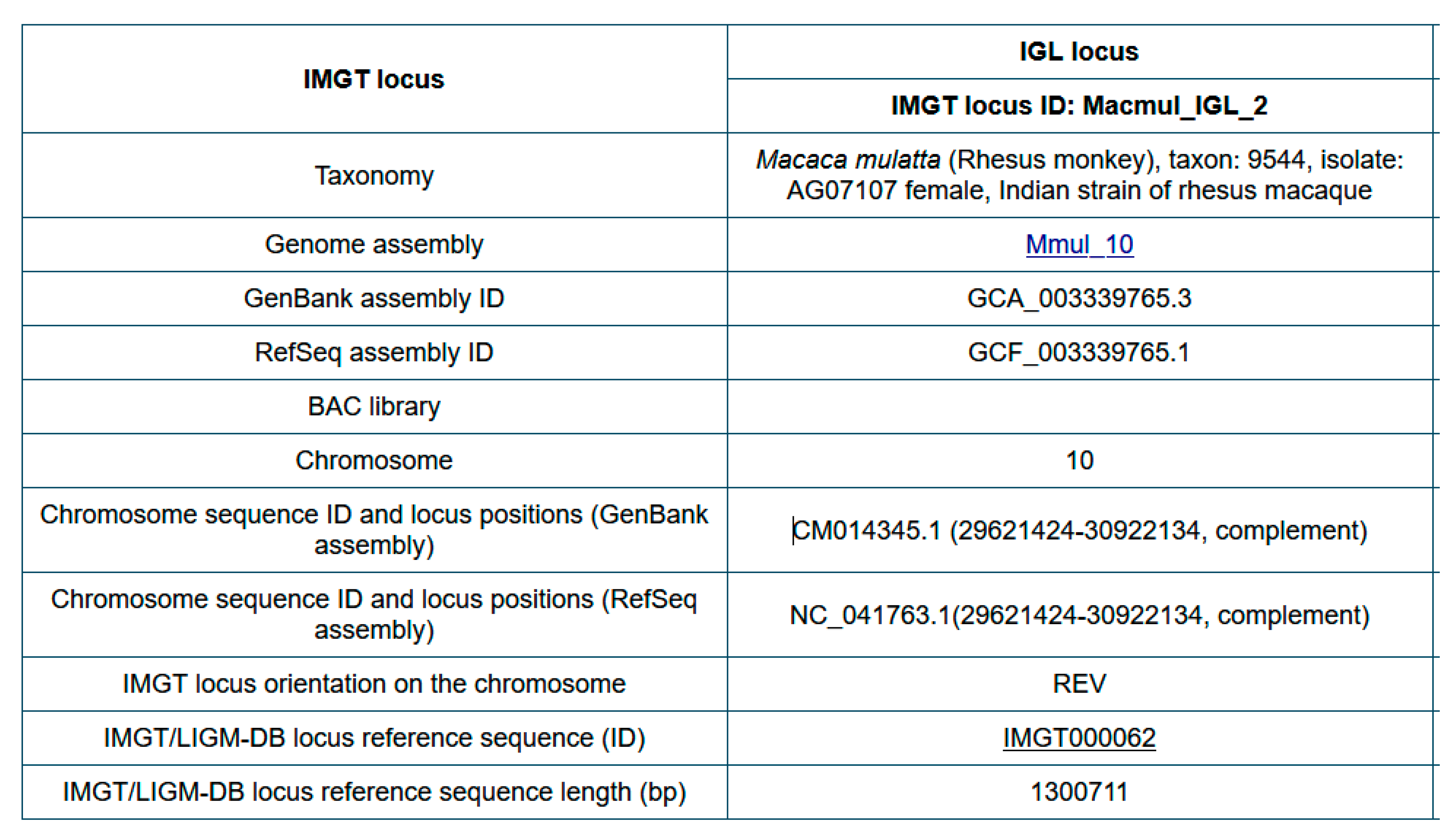
Before starting IMGT biocuration of a new IG or TR locus of a veterinary species, information is collected in ‘Locus in genome assembly’ (Figure 14).
For an easier comparison between loci of different species, and/or between loci of different genomes assemblies (or of different haplotypes, including CNV), the IDENTIFICATION axiom has been enriched by the implementation of ‘IMGT locus ID’ and ‘IMGT/LIGM-DB locus reference sequence (ID)’ (Figure 14) [150].

6.2. IMGT Locus ID and IMGT/LIGM-DB Locus Reference Sequence

An ‘IMGT locus ID’ comprises the 6-letter (or 9-letter) genus and species (or subspecies) Latin names (IMGT taxon abbreviation, e.g., Macmul for Macaca mulatta), the locus type (e.g., IGL) and a chronological increasing number, separated by underscores, for example, Macmul_IGL_2 (Figure 14) [150]. Hyphens instead of underscores may be used in text, e.g., Macmul-IGL-2.
An ‘IMGT/LIGM-DB locus reference sequence’ is an IMGT accession number (‘IMGT’ followed by six digits) which identifies the IMGT/LIGM-DB flat files containing an IG or TR locus (or part of it) extracted from an NCBI genome assembly and presented in its own 5′ to 3′ locus orientation [150]. As a locus may have, on the chromosome, a forward (or ‘Watson’) (FWD) or a reverse (REV) orientation (IMGT Index > Genomic orientation), the sequence orientation in the IMGT accession number flat file is either unchanged (direct) relative to the sequence on the chromosome for an FWD locus, or reverse complemented for a REV locus. For example, the rhesus macaque (Macaca mulatta) IGL locus orientation on chromosome 10 is reverse (REV) and the IMGT/LIGM-DB locus reference sequence in IMGT000062 is reverse-complemented relative to the sequence on chromosome 10.
The information from ‘Locus in genome assembly’ (Figure 14) is reported in the definition lines (DE) of the IMGT/LIGM-DB locus reference accession number. For IMGT000062 it includes: Macaca mulatta (Rhesus monkey), taxon:9544, isolate: AG07107 single Indian origin rhesus female, assembly Mmul_10, 2345051 [UID], GenBank assembly ID: GCA_003339765.3, Refseq assembly ID: GCF_003339765.1, chromosome 10: CM014345.1 (29621424-30922134, complement), IMGT locus ID: Macmul_IGL_2.

6.3. IMGT-LOCUS-UNIT Label and Qualifiers

The label IMGT-LOCUS-UNIT (DESCRIPTION axiom) was created to describe a locus, isolated from a genome assembly, in an IMGT accession number flat file. The definition of the IMGT-LOCUS-UNIT and its qualifiers are given in Table 5.
Three IMGT/LIGM-DB locus reference sequences for the Macaca mulatta (rhesus monkey) IGH (IMGT000064, TPA: BK063715), IGL (IMGT000062, TPA: BK063717,) and IGK (IMGT000063, TPA: BK063716) were created and annotated for the Mmul_10 assembly (GCF_003339765.1).
As an example, the IMGT000062 qualifiers for the IMGT-LOCUS-UNIT (1..1300711) of the Macmul_IGL_2 (IMGT_locus_ID) [150] are the following:
FT/IMGT-LOCUS-UNIT 1..1300711
FT/IMGT_locus_3prime_borne = “RSPH14”
FT/IMGT_locus_3prime_gene = “IGLC7”
FT/IMGT_locus_5prime_borne = “TOP3B”
FT/IMGT_locus_5prime_gene = “IGLV(IV)-127”
FT/IMGT_locus_ID = “Macmul_IGL_2”
FT/IMGT_locus_chromosome = “10”
FT/IMGT_locus_length = “1300711 bp”
FT/IMGT_locus_name = “Macaca mulatta IGL”
FT/IMGT_locus_orientation = “reverse (REV)”
FT/IMGT_locus_positions = “CM014345.1
FT (29621424-30922134 complement)”.

6.4. IMGT Locus 5′ and 3′ Bornes

The IMGT Locus 5′ borne (IMGT_locus_5prime_borne) and the IMGT Locus 3′ borne (IMGT_locus_3prime_borne) (Table 5) are defined for a standardized comparison of the IG and TR locus delimitation across species [150]. The IMGT bornes are genes coding for a protein (other than IG or TR), conserved between species, located upstream of the first gene (for the IMGT 5′ borne) or downstream of the last gene (for the IMGT 3′ borne) of an IG or TR locus (https://www.imgt.org/IMGTrepertoire/LocusGenes/bornes/bornesIGH.html (accessed on 9 April 2026), IMGT Repertoire (IG and TR) > 1. Locus and genes > 3. Locus descriptions > Locus bornes: IGH, IGK, IGL, TRA, TRB, TRG, TRD). If IMGT bornes are not yet identified or are too distant to be included in the locus sequence, a minimal 10 kb sequence is added upstream of the first IG or TR gene in 5′ and/or downstream from the last IG or TR gene in 3′. An overview of the locus IG and TR 5′ and 3′ bornes is shown in Table 6.

6.5. IMGT/GENE-DB Localization in Genome Assemblies

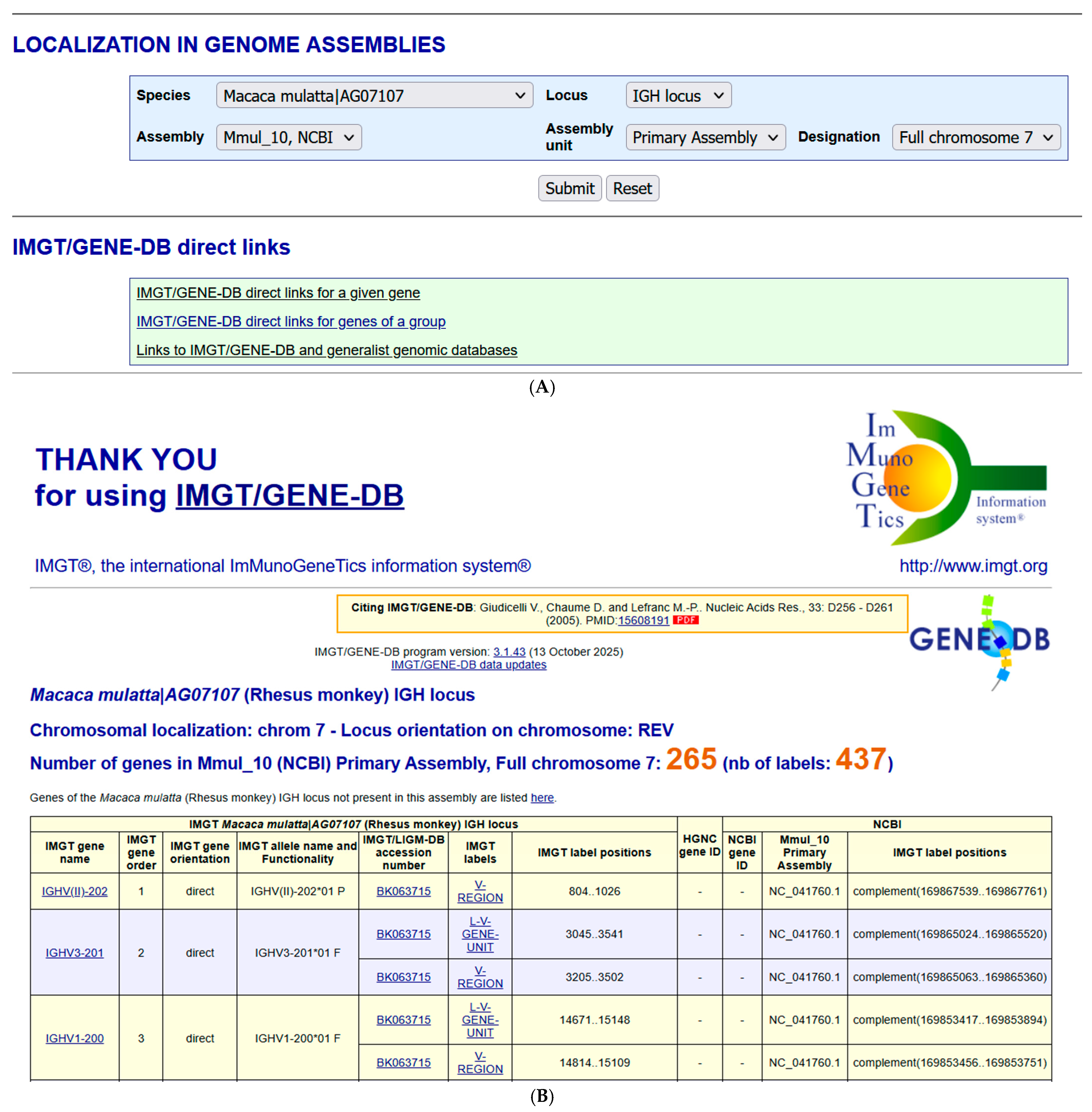
The section “LOCALIZATION IN GENOME ASSEMBLIES” (Figure 11) integrated in 2015 in IMGT/GENE-DB allows, for a given species and a given locus, to query the IMGT IG or TR genes of a given genome assembly. The query Species: Macaca mulatta|AG07107 (AG07107 is the isolate) and Locus: IGH locus shows the availability of IMGT/GENE-DB biocurated genes for the assembly ‘Mmul_10, NCBI’, ‘Primary Assembly’ ‘Full chromosome 70 [150] (Figure 15A).
The results page for the query Species: Macaca mulatta|AG07107 (Rhesus monkey) Locus: IGH (Figure 15B) provides at the top the chromosome localization: ‘chrom7’, the locus orientation on chromosome ‘REV’, the number of genes in Mmul_10 (NCBI) (IMGT/GENE-DB annotated genes) ‘265’ and the number of labels ‘437’. For each gene, the information comprises IMGT gene name, IMGT gene order, IMGT gene orientation (direct (5′ > 3′) or opposite (3′ > 5′) in the locus), IMGT allele name and Functionality (F, ORF or P), IMGT/LIGM-DB accession number, IMGT labels (L-V-GENE-UNIT and V-REGION for a V gene, D-GENE-UNIT and D-REGION for a D gene, J-GENE-UNIT and J-REGION for a J gene, C-GENE-UNIT and C-REGION or the different individual exons for a C gene), IMGT label positions in the IMGT/LIGM-DB accession number, HGNC gene ID (for Homo sapiens), NCBI gene ID (if available), NCBI Mmul_10 Primary Assembly Chromosome (NC) accession number and IMGT label positions in the NC sequence.
The list of genes known to belong to the locus but not localized (NL) in the assembly is also provided in this section as this may correspond to polymorphism by copy number variation, insertion/deletion, or gaps in the assembly.

7. IMGT-NC New Concepts for Gene Copy Number Variations (CNV): ‘CNV Haplotypes’ and ‘CNV-Locus-Haplotype’

7.1. IMGT-NC Gene Copy Number Variation (CNV) Nomenclature and Definition

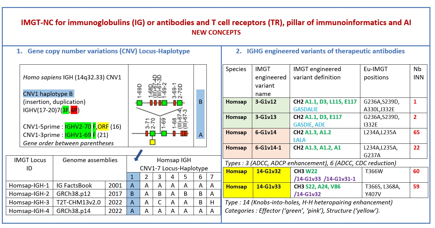
IMGT-NC gene copy number variation (CNV) nomenclature and definition are illustrated with the Homo sapiens IGH locus [2,5,150,151]. Gene copy number variations (CNVs) [150] are numbered from 5′ to 3′ in the locus (IMGT Repertoire (IG and TR) > Locus gene order > Human (Homo sapiens) IGH). Seven CNV are displayed in Table 7, six for the IGHV genes (CNV1 to CNV6) and one (CNV7) for the IGHC genes [150].
The IMGT CNV haplotype nomenclature comprises the genus and species (Latin names in italics) (e.g., Homo sapiens), the locus (e.g., IGH) and the CNV number (e.g., CNV1). A CNV is delimited by a 5prime gene and a 3prime gene; for example, for the Homo sapiens IGH CNV1 haplotype, the CNV1-5prime gene is IGHV2-70 (F, ORF, 16 being the gene order) and the CNV1-3prime gene is IGHV1-69 (F,21) (Table 7) [150].
The IMGT standardized definition of a CNV comprises the group, then between parentheses the order of the start gene (the gene which follows the CNV-5prime), a dash and the order of the end gene (the gene which precedes the CNV-3prime), then the total number of genes involved in the CNV (between the 5prime and 3prime, including RPI gene(s) if present) followed, between parentheses, by the number of IG or TR per functionality (and the number of RPI if present). For instance, the definition of ‘Homo sapiens IGH CNV1’ is ‘IGHV(17–20)7(3F,4P)’ (Table 7) [150]. The letters ‘i’ for insertion, ‘d’ for deletion, and ‘e’ for exchange may be added if needed to indicate the status of a given CNV or that of given gene(s) or groups of genes in a given haplotype by comparison to the haplotype A (e.g., ‘CNV5’ split in ‘IGHV-e1(145–147)’ and ‘IGHV-e2 (148–149.2)’ [150] (Table 7)). The information on gene presence or absence, or insertion/deletion, is provided with colors (orange: genes present in haplotype A (including CNV-5prime and CNV-3prime) and in other haplotypes of a given CNV; pale green: genes absent in haplotype A and other haplotypes, but present by insertion in other haplotype(s) of a given CNV; red: genes absent, by comparison to haplotype A of a given CNV; green: genes present, as insertion by comparison to haplotype A of a given CNV [150] (Table 7)).

7.2. IMGT CNV Haplotypes Illustrated with the Homo sapiens IGH Locus

The description of each CNV haplotype is achieved by comparison with the IMGT Locus representation of the FacstBooks published in 2001 [2,3] (IMGT Repertoire (IG and TR) > 1. Locus and genes > 2. Locus representation > IGH: Human). The genes, localized on the horizontal main line which represents the locus from 5′ to 3′, conventionally, define the ‘haplotype A’ of each individual CNV. A well-characterized CNV example is the Homo sapiens IGH CNV3 IGHV(87–112)26(8F,16P,2RPI), for which six haplotypes A to F [3,150] correspond to polymorphic amplifications of genes, found in individuals of different populations. These polymorphisms are described as a multiple of insertion/deletion between IGHV4-34 (86, CNV3-5prime) and IGHV4-28 (113, CNV3-3prime) (Table 7). The longest haplotype (CNV3 C, 26 genes, 98 kb) corresponds to eight motifs (‘4 pairs of motifs’ or ‘quadruplication’) which each contains three IGHV genes: the first gene of each motif is functional (IGHV4 subgroup gene (green in the right column), starting from 5′prime IGHV4-34, in alternance with IGHV3 subgroup gene (blue in the right column) for the second motif of the pair), followed by a pseudogene of the IGHV3 subgroup and a pseudogene of the IGHV(II) clan. The motifs 2 (IGHV3-33) and 8 (IGHV3-30) are characterized by the presence of GOLGA4P1 and GOLGA4P2 (yellow in the right column), respectively, with GOLGA4P1 containing an Alu sequence amplified in the IGH locus (M.-P. personal communication). The haplotype CNV3 C was built from Han Chinese KC162924, KC162925, AC244456, AC231260 contigs with a detailed IMGT nomenclature and biocuration of the pseudogenes which highlighted the eight paired motifs (or ‘quadruplication’). The graphical representation of the CNV3 haplotypes is available at ‘Human (Homo sapiens) Polymorphism by insertion/deletion between IGHV4-34 and IGHV4-28 (haplotypes A to F) on chromosome 14 (14q32.33)’ (https://www.imgt.org/IMGTrepertoire/index.php?section=LocusGenes&repertoire=locus&species=human&group=IGH/haplotypes#locus) (accessed on 15 December 2025). The haplotype CNV3 C has been observed in T2T-CHM13v2.0 assembly (24 janvier 2022, GCA_009914755.4, hg38). The haplotypes CNV3 A (AB019439, GRCh37) and CNV3 B (AC245166), with 14 genes (50–55 kb), correspond to four motifs (‘2 pairs of motifs’ or ‘duplication’). The haplotype CNV3 D (AC244464, Han Chinese) with seven genes (25 kb) is the smallest haplotype with two motifs (‘1 pair of motifs’). The CNV3 E (20 genes) and CNV3 F (Yoruba, 19 genes) (70–75 kb) correspond to six motifs (‘3 pairs of motifs’ or ‘triplication’).
The Homo sapiens IGH locus on chromosome 14 (14q32.33) is characterized by a remarkable IGH CNV, the CNV7 IGHC(203–211)9(7F,1OP,1P) with seven haplotypes A to G (Table 7), with six of them (haplotypes B to G) corresponding to multigene deletions I to VI, identified on both chromosomes 14 in healthy individuals lacking several subclasses [2,5,150]. Multigene deletions of haplotypes B to G (either identical or different, on both chromosomes in a given individual) are designated I to VI according to the chronological order in which they were found (reviewed in [5]). Deletion I, first identified by the absence of the Gm1 allotypes in a 70-year-old healthy Tunisian woman (TAK3), homozygous for that deletion [152,153], allowed for the ordering of the Homo sapiens IGHC genes in the IGH locus [154,155]. Deletions I and II [152,153,156] (haplotypes IGH CNV7 B and C), found in healthy individuals from consanguineous families, involve highly homologous spots of recombination [157], as also described in a healthy individual (T17) homozygous for deletion III (haplotype IGH CNV7 D) and lacking IgA1, IgG2, IgG4 and IgE [158].
The IGHC duplications comprising the duplication of the IGHG4A in 5′ of IGHG4 (gene order 208.1) (CNV7 H) and the multigene duplications including IGHGP (206.1), IGHG2 (206.2), IGHG4 (206.3), IGHE (206.4), and one IGHA (206.5) are still in progress and not detailed here.

7.3. IMGT Locus CNV-Haplotype: The Example of the IMGT IGH Locus CNV1-7-Haplotype

With its seven CNV haplotypes, the Homo sapiens reference IGH Locus [2] has, by convention, an IMGT Homsap IGH Locus CNV1-7-Haplotype ‘A.A.A.A.A.A.A’, characterized by the haplotype A of the seven CNV involved in the analysis. It is the one obtained from the IGH locus and genes from contig sequences available in the generalist databases and annotated by LIGM in IMGT/LIGM-DB with the Locus representation published in 2001 [2] (Table 8A). The availability of genome assemblies has made it possible to characterize the expected differences in terms of CNV (Table 8B). The ‘IMGT IGH Locus CNV1-7-Haplotype’ from GRCh38.p12 is ‘B.A.B.A.B.B.A’ where the CNV5 B haplotype corresponds to BAC clone sequences [151] from the CHORI-17 BAC library. The ‘IMGT Homsap IGH Locus CNV1-7-Haplotype’ from T2T-CHM13v2.0 is ‘A.A.C.A.A.B.H’ with the duplicated IGHG4A gene, whereas the ‘IMGT Homsap IGH Locus CNV1-7-Haplotype’ from GRCh38.p14 is ‘A.A.A.A.A.A.A’. This has set up the grounds for standardized analysis and comparison of haploid, maternal or paternal chromosomal assemblies.
The CNV representations and the CNV characterization of the IGH, IGK and IGL loci as well as those of the TRA/TRD, TRB and TRG [150] are useful to compare the diversity and polymorphism of the IG and TR loci between individuals of the same species, here Homo sapiens [150], but also to study the evolution of the loci of closely related species, such as Gorilla gorilla gorilla where the same gene order as Homo sapiens can be used. The analysis of IMGT locus CNV-haplotypes which are partial owing to a V-(D)-J rearrangement may provide, for information, the genes and alleles names of the rearranged V, (D) and J genes with their respective gene order, in place of the first missing CNV haplotype.

8. IMGT-NC New Concepts of Engineered IGHG Variants for Effector Properties, Half-Life and Structures of Therapeutic Antibodies

The constant region of the immunoglobulin (IG) or antibody heavy gamma chain is frequently engineered to modify the effector properties of the therapeutic monoclonal antibodies, their half-life and/or their format and structure [159]. The standardized IMGT engineered variant nomenclature [159] has been set up for an easier comparison between engineered antibody variants involved in effector properties (ADCC, ADCP and CDC), half-life and structure of therapeutical monoclonal antibodies [159,160]. The IMGT nomenclature of the IGHG variants is based on a classification in four categories of ‘effector’, ‘half-life’, ‘physicochemical’ and ‘structure’, and in 18 types, assigned to one of the four categories, depending on their property and function types [159]. The ‘effector’ category comprises eight types (1–8): 1. ADCC reduction, 2. ADCC enhancement, 3. ADCC and ADCP enhancement, 4. CDC enhancement, 5. CDC reduction, 6. ADCC and CDC reduction, 7. FcγRIIB binding increase and B cell inhibition (coengagement of antigen and FcγR on the same cell), 8. knock out CH2 84.4 glycosylation (ADCC reduction). Two categories comprise a single type: the ‘half-life’ category includes the type 9 (half-life increase or decrease), whereas the ‘physicochemical’ ‘category’ includes the type 10 (abrogation of binding to Protein A, thermal stability, pI, reduced acid-induced aggregation). The ‘structure’ category comprises eight types (11–18): 11. formation of additional bridge for domain stabilization including scFv, 12. prevention of IgG4 half-IG exchange, 13. hexamerization, 14. knobs-into-holes and enhancement of heteropairing H-H of bispecific antibodies, 15. suppression of inter H-L and/or inter H-H disulfide bridges, 16. site-specific drug attachment, e.g., additional cysteine, 17. enhancement of heteropairing H-L of bispecific antibodies, 18. control of half-IG exchange of bispecific IgG [159,160] (Table 9).
The IMGT engineered variant characterization (Table 10) comprises:
(1)
The IMGT engineered variant name which comprises the species (e.g., Homsap for Homo sapiens), the variant type(s) (number(s) from 1 to 18), the gene name abbreviation (e.g., G1 for IGHG1), the letter ‘v’ with a number (e.g., Homsap 1-G1v1).
(2)
The IMGT engineered variant definition which comprises, for each engineered AA change, the domain (e.g., CH1, CH2 or CH3) or the hinge, the AA in the one-letter abbreviation [108] with its position according to the IMGT unique numbering for C domain [56], followed by the Eu-IMGT position between parentheses, e.g., Homsap 1-G1v1, CH2 P1.4 (233) [159,160]. In the World Health Organisation International Nonproprietary Name (WHO INN) description of therapeutic antibodies [161,162], the Eu-IMGT position is replaced by the position of the AA change in the antibody chain sequence.
(3)
The IMGT AA change(s) with the Eu-IMGT position(s) between parentheses (e.g., CH2 P114 > A (329)).
(4)
The AA change(s) at the Eu-IMGT position(s) (e.g., P329A).
(5)
The IMGT topological motif sequence identifiable in gene and domain with positions according to the IMGT unique numbering [56], followed, between parentheses, by the Eu-IMGT positions; the display of the motif shows the AA involved in the change highlighted in bold, with red before the change and green after the change, respectively (e.g., IGHG1 CH2 1.6–3 APELLGGPS > APPLLGGPS; underlined amino acids in the motif correspond to additional positions in the IMGT unique numbering for the C-domain [56], e.g., APELLG and APPLLG which correspond to 1.6, 1.5, 1.4, 1.3, 1.2 and 1.1).
(6)
‘Property and function’ (at least one and up to three per variant) and 3D structure if available [159,160].

9. IMGT-NC for Common Scientific Research Endeavor

9.1. IMGT-NC a Foundational and Unifying Infrastructure for Immunogenetics and Immunoinformatics

The IMGT nomenclature of the IG and TR gene and allele names, together with the IMGT nomenclature of CNV and haplotypes [150,151,152,153,154,155,156,157,158], the IMGT nomenclature of engineered IGHG variants involved in antibody effector properties and formats [159,160], used in the World Health Organisation International Nonproprietary Name (WHO INN) description of therapeutic antibodies [161,162], and the WHO/IMGT nomenclature of the allotypes [163,164], which bridges serology and proteomics [165,166,167,168,169,170,171,172,173], contribute with the IMGT unique numbering [66] (for V domain [55], C domain [56]) to the common language for IG and TR genes, sequences and structures, as exemplified in antibody engineering and humanization, structural immunology, and therapeutic antibody development [35,36,38,174,175,176,177,178,179,180,181,182,183]. The IMGT numbering has been extended to the groove (G) domain [57] of the major histocompatibility (MH) proteins and MH superfamily (MhSF). They allow for a standardized comparison of IG/Ag complexes, TR/pMH1,/pMH2 and/pMH1-like complexes [66,183,184], including TR-mimic Fab/pMH1 complexes [66,183].

9.2. IMGT Nomenclature for Salmonid IG and TR (Pierre Boudinot, Susana Magadán)

The emergence of high-throughput sequencing in the mid-2000s created new opportunities to investigate IG/TR loci evolution in under-investigated species, such as teleost and reptilian. In early studies aiming to identify immunoglobulin isotypes rather than to produce comprehensive germline reference sets, the IMGT system was instrumental for the resolution of IGHC gene content, exon structure, and isotype diversification in newly sequenced genomes. This was especially important to determine orthology relationships between divergent teleost or reptilian sequences and mammalian isotypes [185,186,187,188,189,190,191]. IMGT-ONTOLOGY and standardized feature annotation facilitated accurate exon definition, identification of conserved Cys/Trp motifs, and characterization of structural features such as hinge regions.
A comprehensive description of loci later became necessary to analyze expressed repertoires in species like rainbow trout and Atlantic salmon. A standardized IG/TR gene nomenclature was then generated by the groups of Pierre Boudinot at the University Paris-Saclay, INRAE (France) and of Susana Magadán at the University de Vigo (Spain) that could be updated and reused by the scientific community [147,148,149,192,193]. IMGT provided a standardized, ontology-driven framework that allowed accurate annotation from reference genome sequences of Salmonids, and efficient definition of VDJ gene nomenclature. Using a consistent set of gene definitions (e.g., what constitutes V subgroups, as well as VDJC genes/alleles) and feature boundaries (leader, FR/CDR, recombination signals (RS), splice sites), a consistent description of the architecture of Oncorhynchus rainbow trout and Atlantic salmon IGH/TR loci was established and published [147,148,149,193]. Particularities of salmonid IGH/TR loci, such as multiple loci in each haplotype and a large number of genes and pseudogenes, were taken into account to create a nomenclature consistent with the complexity of germline repertoires of these species. The IMGT/HighV-QUEST [92,93,94,95,96] was instrumental in the analyses of expressed repertoires in these species [194,195,196,197,198,199,200,201,202,203,204]. The setting of the IMGT nomenclature for Salmonid required (i) knowledge of the bases for analysis and annotation of VDJ genes, early definition of subgroups and definition of rules for nomenclature (germline/potential repertoires) [147,148,149,185,186,187,188,189,190,191,192,193], and has allowed (ii) constitution of the germline reference sets used as a resource for annotation of expressed repertoires [194,195,196,197,198,199,200,201,202,203,204]. This double goal serves as a model for any non-canonical species and opens novel perspectives on repertoire analysis [205].

9.3. IMGT Nomenclature for Less Canonical Antigen Receptors (Michael Criscitiello)

The standardized lingua franca provided by the IMGT is indispensable for work in traditional model species. For the group of Michael Criscitiello at the Texas A&M University (USA) [192,206,207,208,209,210,211,212,213,214,215,216,217,218,219], the curation has been just as necessary when trying to describe less canonical antigen receptors such as cattle immunoglobulin ultralong CDR3H V(D)J junctions [206] or shark loci combining both B and T cell receptor components [213]. The established ground maintained by the IMGT nomenclature gives a firm launch site into discussion of the more divergent and debatable loci that vertebrates have evolved, and we can exploit in antibody and immunotherapeutic engineering. ‘Nomenclature is sometimes viewed as an encumbrance, an onerous chore to move away from the parlance you were taught and adopt someone else’s. I appreciate that others helped me appreciate the need for standard language in communicating complex topics such as antigen receptor loci immunogenetics’. Michael Criscitiello soon became an advocate for and a member of the IUIS-NOM IMGT IG, TR and MH nomenclature committee (IMGT-NC) and worked with the group in efforts to clarify model species loci from zebrafish (Danio rerio) TR [208], to Florida manatee (Trichechus manatus latirostris) IG [209], to bovid ultralong CDR3 components [217], to pinniped California sea lion (Zalophus californianus) MH [219], to teleost IG isotypes [192].

9.4. IMGT Nomenclature for Mammalian TR (Salvatrice Ciccarese)

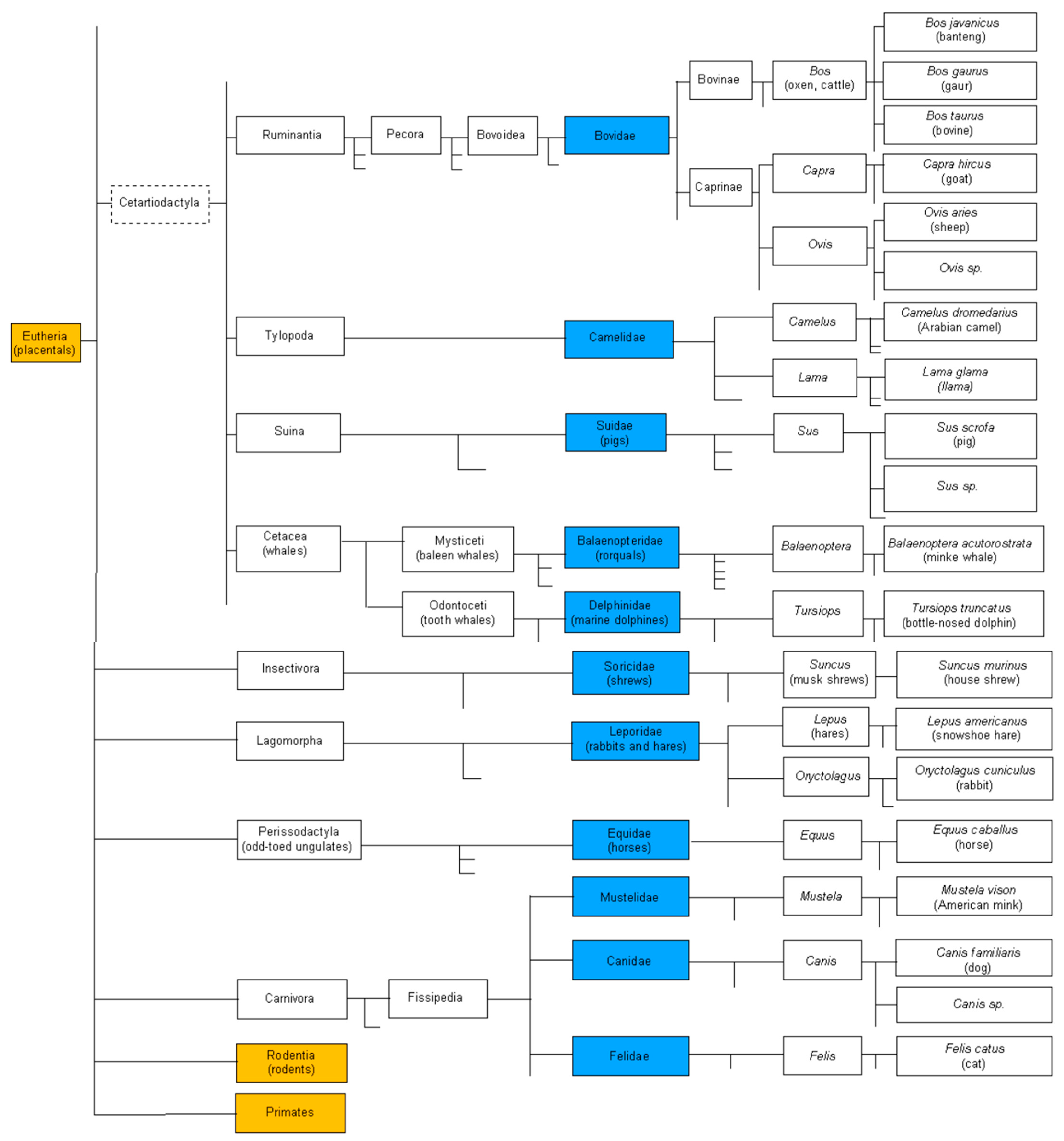
Investigations on mammalian T cell receptors αβ and γδ, conducted in the group of Salvatrice Ciccarese of the University of Bari Aldo Moro (Italy) using the IMGT nomenclature, have progressed through several key stages [146,220,221,222,223,224,225,226,227,228,229,230,231,232,233,234,235,236,237,238,239,240,241,242,243,244,245,246,247,248,249,250,251,252,253]. An initial step was the genomic mapping of the TRGV–TRGJ–TRGC and TRDV–TRDJ–TRDC genes in Bovidae (Figure 16). This analysis revealed the splitting of the TRG locus into two distinct loci, a structural peculiarity of this taxonomic group.
The duplication was clearly demonstrated in Ovis aries (sheep) through BAC clone analysis [220,221,222,223,224,225,226,227,228,229]. Subsequent comparison of the TRG locus organization in Bovidae with the corresponding deduced structure in Canis lupus familiaris (dog) [230], Sus scrofa (pig) [249], Equus caballus (horse) and Equus asinus (donkey) species [251] provided important insights into the evolutionary dynamics of the gamma locus in mammals [245]. A further step involved the analysis of spleen cDNA minilibraries from Camelus dromedarius (dromedary). This study highlighted the occurrence of somatic hypermutation in rearranged TRGV and TRDV genes [232,233,237]. The use of IMGT tools for the analysis of amino acid sequences deduced from spleen transcripts was instrumental in defining both the timing and the modalities of somatic mutation onset. Attention then shifted to the genomic organization and evolutionary analysis of the beta locus (TRB) [234,236], which revealed shared evolutionary patterns among Tylopoda, Ruminantia and Suina. Within the Cetartiodactyla superorder, the characterization of the TRB locus [146], together with the organization of the TRA/TRD and TRG loci in the marine mammal Tursiops truncatus (common bottlenose dolphin) [239], further refined our understanding of the evolutionary landscape of T cell receptor loci. Finally, CD1D-restricted γδ T cell receptors were identified through three-dimensional structural modeling based on C. dromedarius cDNA clones [250]. Protein–protein interactions were analyzed within a TRG–TRD/CD1D complex selected for camel-specific features of antigen receptors, including long CDR3-IMGT regions and the presence of somatic mutations [253]. The IMGT numbering of V [55], C [56] and G [57] domains provided a unifying framework to bridge amino acid sequences, 3D models, structures and functions across species [254,255,256,257].

9.5. IMGT Nomenclature for IG and TR Gene Repertoires in Lymphoproliferative Disorders (Kostas Stamatopoulos)

Investigations of IG and TR gene repertoires in lymphoproliferative disorders using IMGT nomenclature started as early as 2002 under Kostas Stamatopoulos’s initiative at the Centre for Research and Technology Hellas (Greece) and Karolinska Institutet (Sweden) and have continued uninterruptedly ever since [258,259,260,261,262,263,264,265,266,267,268,269,270,271,272,273,274,275,276,277,278,279,280,281,282,283,284,285,286,287,288,289,290,291,292,293,294,295,296,297,298,299]. The first critical step concerned the collaboration between IMGT and ERIC, the European Research Initiative on CLL, with the main aim of developing and maintaining a database with immunogenetic data from patients with Chronic Lymphocytic Leukemia (CLL) attended at academic institutions, to foster high-quality collaborative research on this disease and related disorders. This initiative led to the establishment of IMGT/CLL-DB (https://www.imgt.org/CLLDBInterface/query (accessed on 9 April 2026)), the largest IG gene sequence database globally, currently holding data from more than 80,000 patients with CLL from 54 institutions from all over the world. IMGT/CLL-DB has facilitated several large-scale immunogenetic research projects focused on unraveling the IG gene repertoire in CLL with important biological and clinical implications, leading to several high-impact and highly cited publications [261,264,265,266,267,277,278,288].
The use of the IMGT nomenclature in the collaboration between ERIC and IMGT has been instrumental in advancing the standards of routine diagnostics in CLL. This has been achieved through the continuous refinement of software (indicatively, allowing for accurate annotation of insertions and deletions in rearranged IG genes); prime educational events (hands-on international workshops organized biannually); and recommendations about the analysis of IG gene rearrangements in both a research and a diagnostic setting [275,276,300]. Importantly, these standards are widely used by the global community of scientists and healthcare professionals working on CLL.
Studies of lymphoproliferative disorders enabled by/relying on IMGT nomenclature in software and tools extend beyond CLL to other entities. Indicative examples are offered by large-scale multi-institutional, multi-national projects on the IG gene repertoires of, e.g., mantle-cell lymphoma [258,260], splenic marginal-zone lymphoma [262,263,269,298] and other marginal-zone lymphoproliferations [259,281], amongst others.
Another major area of interest in lymphomas concerns the analysis of bystander T cells in the tumor microenvironment. Also in this context, IMGT nomenclature used in the processing of big datasets originating from high-throughput, next generation sequencing projects have been fundamental to improved molecular characterization of the TR gene repertoire, laying the ground for novel immmunotherapeutic strategies [268,273,285,291].

10. Concluding Remarks

The IMGT nomenclature supports primary immunodeficiency diagnosis, minimal residual disease tracking, vaccine response studies, cancer immunogenetics, and therapeutic antibody design. The major challenge of immunogenetics is how to meet the highest standards and ensure consistency that translates into obvious benefits for both advancing research and providing accurate diagnostic information and helping to find the best and most appropriate therapeutics [300]. The IMGT nomenclature brings a foundational, unifying infrastructure to immunogenetics and immunoinformatics [1] and has made the huge genetic diversity of the IG and TR antigen receptors standardized, computable, and comparable worldwide. It has become the global reference for the IG and TR because it provides a rigorous, universal, and biologically meaningful framework to identify, describe and classify the IG and TR genes, sequences and structures. The standardized IMGT unique numbering for structures is used for AI search [301,302,303,304,305,306,307,308,309,310]. The IMGT nomenclature is used for any IG and TR genes from Homo sapiens [296,311,312] and from any other jawed vertebrate species, enabling comparative immunology, evolutionary studies of immune systems, clinical, veterinary and zoonotic research, and evolutionary immunogenetics. By disseminating the standardized IMGT Scientific chart rules used worldwide to name genes, researchers participate in a global scientific research endeavor for the development of immunogenetics and immunoinformatics with the use of AI. In this context, speaking a common language which bridges genes, sequences, structures and functions ensures that we understand each other and communicate scientific findings effectively while also minimizing misunderstandings and confusion [300].

11. Availability and Citation

Online access to IMGT® databases, tools and web resources is freely available for academics. Authors are encouraged to cite the references quoted in this article. Access to IMGT® databases and tools are under CNRS licenses and contracts for companies.

Supplementary Materials

The following supporting information can be downloaded at: https://www.mdpi.com/article/10.3390/antib15020035/s1, Table S1: List of the IMGT definitions of Encyclopedia of Systems Biology. Springer, New York, NY, 2013. Table S2: List of the IUIS-NOM-IMGT-NC reports (2017–2022).

Author Contributions

Conceptualization, G.L. and M.-P.L.; methodology, G.L. and M.-P.L.; investigation, G.L. and M.-P.L.; resources, M.-P.L.; data curation, M.-P.L.; writing—review and editing, G.L. and M.-P.L.; visualization, M.-P.L. All authors have read and agreed to the published version of the manuscript.

Funding

This research received no external funding.

Data Availability Statement

No new data were created or analyzed in this study.

Acknowledgments

We are very grateful to Salvatrice Ciccarese, Susana Magadán, Michael Criscitiello, Pierre Boudinot and Kostas Stamatopoulos for their scientific contribution to the IMGT nomenclature. We thank Cold Spring Harbor Protocol Press for the pdf of the IMGT Booklet available in IMGT references. IMGT® is a registered trademark of CNRS. IMGT® was funded in part by the BIOMED1 (BIOCT930038), Biotechnology BIOTECH2 (BIO4CT960037), fifth PCRDT Quality of Life and Management of Living Resources (QLG2-2000-01287), and sixth PCRDT Information Science and Technology (ImmunoGrid, FP6 IST-028069) programmes of the European Union (EU). IMGT received financial support from the GIS IBiSA, BioCampus Montpellier, the Région Occitanie (Grand Plateau Technique pour la Recherche (GPTR)), the Agence Nationale de la Recherche (ANR) and the Labex MabImprove (ANR-10-LABX-53-01). IMGT is granted access to the HPC resources of CINES under the allocation [036029] (2010–2026) made by GENCI (Grand Equipement National de Calcul Intensif). IMGT® is currently supported by the Centre National de la Recherche Scientifique (CNRS), the Ministère de l’Enseignement Supérieur et de la Recherche (MESR) and the Université de Montpellier.

Conflicts of Interest

The authors declare no conflicts of interest.

Abbreviations

The following abbreviations are used in this manuscript:
AI Artificial intelligence
AIR Adaptive immune response
AR Antigen receptor (IG and/or TR)
ARR Antigen receptor repertoire
BcR B cell receptor (IG with coreceptors CD79A and CD79B)
C Constant
CDR Complementarity determining region
CNRS Centre National de la Recherche Scientifique
CNV Copy number variation
CPCA Composite protein for clinical applications
CSR Class switch recombination
D Diversity
DDBJ DNA Database of Japan
EMBL European Molecular Biology Laboratory
Fc Fragment crystallizable
FcR Fc receptor
FR Framework region
FPIA Fusion protein for immune applications
ICI International Congress of Immunology
IG Immunoglobulin or antibody
IgSF Immunoglobulin superfamily
IMGT ImMunoGeneTics
IMGT-NC IMGT nomenclature
INN International Nonproprietary Name
IUIS International Union of Immunological Societies
IUIS NOM IUIS Nomenclature committee
J Joining
LIGM Laboratoire d’ImmunoGénétique Moléculaire
MH Major histocompatibility
MhSF MH superfamily
ORF Open reading frame
RPI Related protein of immune interest
SHM Somatic hypermutation
TcR T cell receptor (TR with coreceptors CD3)
TR T cell receptor
UM Université de Montpellier
V Variable
VH Variable domain of heavy (IG chain)
VL Variable domain of light (IG chain)
WHO World Health Organization

References

  1. Lefranc, M.-P. Immunoglobulin and T cell receptor genes: IMGT® and the birth and rise of immunoinformatics. Front. Immunol. 2014, 5, 22. [Google Scholar] [CrossRef] [PubMed]
  2. Lefranc, M.-P.; Lefranc, G. The Immunoglobulin FactsBook; Academic Press: London, UK, 2001; pp. 1–457. [Google Scholar]
  3. Lefranc, M.-P.; Lefranc, G. The T Cell Receptor FactsBook; Academic Press: London, UK, 2001; pp. 1–397. [Google Scholar]
  4. Lefranc, M.-P.; Lefranc, G. Immunoglobulins: 25 years of Immunoinformatics and IMGT-ONTOLOGY. Biomolecules 2014, 4, 1102–1139. [Google Scholar] [CrossRef] [PubMed]
  5. Lefranc, M.-P.; Lefranc, G. IMGT® and 30 years of immunoinformatics insight in antibody V and C domain structure and function. Antibodies 2019, 8, 29. [Google Scholar] [CrossRef]
  6. Lefranc, M.-P.; Lefranc, G. Immunoglobulins or antibodies: IMGT bridging genes, structures and functions. Biomedicines 2020, 8, 319. [Google Scholar] [CrossRef] [PubMed]
  7. Lefranc, M.-P. Nomenclature of the human immunoglobulin genes. In Current Protocols in Immunology; Coligan, J.E., Bierer, B.E., Margulies, D.E., Shevach, E.M., Strober, W., Eds.; John Wiley and Sons: Hoboken, NJ, USA, 2000; pp. A.1P.1–A.1P.37. [Google Scholar]
  8. Lefranc, M.-P. Nomenclature of the human T cell receptor genes. In Current Protocols in Immunology; Coligan, J.E., Bierer, B.E., Margulies, D.E., Shevach, E.M., Strober, W., Eds.; John Wiley and Sons: Hoboken, NJ, USA, 2000; pp. A.1O.1–A.1O.23. [Google Scholar]
  9. Lefranc, M.-P. IMGT, the international ImMunoGeneTics information system®. In Immunoinformatics: Bioinformatic Strategies for Better Understanding of Immune Function; Bock, G., Goode, J., Eds.; Novartis Foundation Symposium 254; John Wiley and Sons: Chichester, UK, 2003; pp. 126–135; discussion pp. 136–142, 216–222, 250–252. [Google Scholar] [PubMed]
  10. Lefranc, M.-P. IMGT-ONTOLOGY and IMGT databases, tools and web resources for immunogenetics and immunoinformatics. Mol. Immunol. 2004, 40, 647–660. [Google Scholar] [CrossRef] [PubMed]
  11. Lefranc, M.-P.; Giudicelli, V.; Regnier, L.; Duroux, P. IMGT, a system and an ontology that bridge biological and computational spheres in bioinformatics. Brief. Bioinform. 2008, 9, 263–275. [Google Scholar] [CrossRef] [PubMed]
  12. Lefranc, M.-P. IMGT-ONTOLOGY, IMGT® databases, tools and Web resources for Immunoinformatics. In Immunoinformatics; Schoenbach, C., Ranganathan, S., Brusic, V., Eds.; Immunomics Reviews, Series of Springer Science and Business Media LLC; Springer: New York, NY, USA, 2008; Chapter 1; pp. 1–18. [Google Scholar]
  13. Lefranc, M.-P. IMGT, the international ImMunoGeneTics information system for immunoinformatics. Methods for querying IMGT® databases, tools and Web resources in the context of immunoinformatics. Mol. Biotechnol. 2008, 40, 101–111. [Google Scholar] [CrossRef]
  14. Lefranc, M.-P.; Giudicelli, V.; Busin, C.; Malik, A.; Déhais, P.; Chaume, D. Une base de données intégrée en Immunogénétique, LIGM-DB/IMGT. In L’Immuno-Intervention. De la Biothérapie à la Thérapie Génique; Chouaib, S., Mencia-Huerta, J.M., Eds.; Institut Henri Beaufour: Les Ulis, France, 1994; pp. 25–32. [Google Scholar]
  15. Lefranc, M.-P.; Giudicelli, V.; Busin, C.; Malik, A.; Mougenot, I.; Déhais, P.; Chaume, D. LIGM-DB/IMGT: An integrated database of Ig and TcR, part of the Immunogenetics database. Ann. N. Y. Acad. Sci. 1995, 764, 47–49. [Google Scholar] [CrossRef] [PubMed]
  16. Giudicelli, V.; Chaume, D.; Bodmer, J.; Müller, W.; Busin, C.; Marsh, S.; Bontrop, R.; Lemaitre, M.; Malik, A.; Lefranc, M.-P. IMGT, the international ImMunoGeneTics database. Nucleic Acids Res. 1997, 25, 206–211. [Google Scholar] [CrossRef] [PubMed]
  17. Lefranc, M.-P.; Giudicelli, V.; Busin, C.; Bodmer, J.; Müller, W.; Bontrop, R.; Lemaitre, M.; Malik, A.; Chaume, D. IMGT, the international ImMunoGeneTics database. Nucleic Acids Res. 1998, 26, 297–303. [Google Scholar] [CrossRef] [PubMed]
  18. Giudicelli, V.; Chaume, D.; Lefranc, M.-P. IMGT/LIGM-DB: A systematized approach for ImMunoGeneTics database coherence and data distribution improvement. ISMB 1998, 6, 59–68. [Google Scholar]
  19. Giudicelli, V.; Chaume, D.; Mennessier, G.; Althaus, H.-H.; Müller, W.; Bodmer, J.; Malik, A.; Lefranc, M.-P. IMGT, the International ImMunoGeneTics Database: A New Design for Immunogenetics Data Access; Cesnik, B., Mc Gray, A.T., Scherrer, J.R., Eds.; IOS Press: Amsterdam, The Netherlands, 1998; Volume 5, pp. 351–355. [Google Scholar]
  20. Lefranc, M.-P. IMGT, the international ImMunoGeneTics database. In Proceedings of the 10th International Congress of Immunology Proceedings, New Delhi, Monduzzi Editore, New Delhi, India, 1–6 November 1998; Talwar, G.P., Nath, I., Ganguly, N.K., Rao, K.V.S., Eds.; Monduzzi Editore: Bologna, Italy, 1998; pp. 105–109. [Google Scholar]
  21. Ruiz, M.; Giudicelli, V.; Lefranc, M.-P. IMGT, base de données internationale en ImMunoGénéTique. Regard Sur La Biochim. 1999, 2, 25–31. [Google Scholar]
  22. Lefranc, M.-P.; Giudicelli, V.; Ginestoux, C.; Bodmer, J.; Müller, W.; Bontrop, R.; Lemaitre, M.; Malik, A.; Barbié, V.; Chaume, D. IMGT, the international ImMunoGeneTics database. Nucleic Acids Res. 1999, 27, 209–212. [Google Scholar] [CrossRef] [PubMed]
  23. Ruiz, M.; Giudicelli, V.; Ginestoux, C.; Stoehr, P.; Robinson, J.; Bodmer, J.; Marsh, S.; Bontrop, R.; Lemaitre, M.; Lefranc, G.; et al. IMGT, the international ImMunoGeneTics database. Nucleic Acids Res. 2000, 28, 219–221. [Google Scholar] [CrossRef] [PubMed]
  24. Lefranc, M.-P. IMGT, the international ImMunoGeneTics database. Nucleic Acids Res. 2001, 29, 207–209. [Google Scholar] [CrossRef] [PubMed]
  25. Chaume, D.; Giudicelli, V.; Lefranc, M.-P. IMGT-ML a XML language for IMGT-ONTOLOGY and IMGT/LIGM-DB data. In CORBA and XML: Towards a Bioinformatics Integrated Network Environment, Proceedings of the NETTAB 2001; Network Tools and Applications in Biology: Genoa, Italy, 2001; pp. 71–75. [Google Scholar]
  26. Lefranc, M.-P. IMGT, the international ImMunoGeneTics database: A high-quality information system for comparative immunogenetics and immunology. Dev. Comp. Immunol. 2002, 26, 697–705. [Google Scholar] [CrossRef] [PubMed]
  27. Lefranc, M.-P. IMGT, the international ImMunoGeneTics database®. Nucleic Acids Res. 2003, 31, 307–310. [Google Scholar] [CrossRef] [PubMed]
  28. Lefranc, M.-P.; Giudicelli, V.; Ginestoux, C.; Chaume, D. IMGT, the international ImMunoGenetics information system®. The reference in immunoinformatics. Stud. Health Technol. Inform 2003, 95, 74–79. [Google Scholar]
  29. Lefranc, M.-P. IMGT® databases, web resources and tools for immunoglobulin and T cell receptor sequence analysis. Leukemia 2003, 17, 260–266. [Google Scholar] [CrossRef] [PubMed]
  30. Lefranc, M.-P. IMGT, the international ImMunoGenetics information system®. In Antibody Engineering Methods and Protocols, 2nd ed.; Lo, B.K.C., Ed.; Humana Press: Totowa, NJ, USA, 2004; Volume 248, Chapter 3, pp. 27–49. [Google Scholar]
  31. Lefranc, M.-P.; Giudicelli, V.; Kaas, Q.; Duprat, E.; Jabado-Michaloud, J.; Scaviner, D.; Ginestoux, C.; Clément, O.; Chaume, D.; Lefranc, G. IMGT, the international ImMunoGeneTics information system®. Nucleic Acids Res. 2005, 33, D593–D597. [Google Scholar] [CrossRef] [PubMed]
  32. Lefranc, M.-P. IMGT, the international ImMunoGeneTics information system: A standardized approach for immunogenetics and immunoinformatics. Immunome Res. 2005, 1, 3. [Google Scholar] [CrossRef] [PubMed][Green Version]
  33. Lefranc, M.-P. IMGT®, the international ImMunoGeneTics information system® for immunoinformatics. Methods for querying IMGT databases, tools and Web resources in the context of immunoinformatics. In Immunoinformatics: Predicting Immunogenicity In Silico; Flower, D.R., Ed.; Methods in Molecular Biology; Humana Press Inc.: Totowa, NJ, USA, 2007; Volume 409, pp. 19–42. [Google Scholar] [CrossRef]
  34. Lefranc, M.-P.; Giudicelli, V.; Ginestoux, C.; Jabado-Michaloud, J.; Folch, G.; Bellahcene, F.; Wu, Y.; Gemrot, E.; Brochet, X.; Lane, J.; et al. IMGT, the international ImMunoGeneTics information system. Nucleic Acids Res. 2009, 37, D1006–D1012. [Google Scholar] [CrossRef] [PubMed]
  35. Lefranc, M.-P. IMGT, the international ImMunoGeneTics information system. Cold Spring Harb. Protoc. 2011, 6, 595–603. [Google Scholar] [CrossRef] [PubMed]
  36. Lefranc, M.-P. IMGT® information system. In Encyclopedia of Systems Biology; Dubitzky, W., Wolkenhauer, O., Cho, K.-H., Yokota, H., Eds.; Springer Science & Business Media, LLC: New York, NY, USA, 2013; pp. 959–964. [Google Scholar] [CrossRef]
  37. Lefranc, M.-P. Antibody informatics: IMGT®, the international ImMunoGeneTics information system®, the international ImMunoGeneTics information system®. Microbiol. Spectr. 2014, 2, 2. [Google Scholar] [CrossRef]
  38. Lefranc, M.-P. Antibody Informatics: IMGT, the International ImMunoGeneTics Information System. In Antibodies for Infectious Diseases; Crowe, J., Boraschi, D., Rappuoli, R., Eds.; ASM Press: Washington, DC, USA, 2015; pp. 363–379. [Google Scholar]
  39. Lefranc, M.-P.; Giudicelli, V.; Duroux, P.; Jabado-Michaloud, J.; Folch, G.; Aouinti, S.; Carillon, E.; Duvergey, H.; Houles, A.; Paysan-Lafosse, T.; et al. IMGT®, the international ImMunoGeneTics information system® 25 years on. Nucleic Acids Res. 2015, 43, D413–D422. [Google Scholar] [CrossRef] [PubMed]
  40. Giudicelli, V.; Lefranc, M.-P. Ontology for Immunogenetics: IMGT-ONTOLOGY. Bioinformatics 1999, 15, 1047–1054. [Google Scholar] [CrossRef] [PubMed]
  41. Giudicelli, V.; Lefranc, M.-P. IMGT-ONTOLOGY: Gestion et découverte de connaissances au sein d’IMGT. In Extraction et Gestion des Connaissances (EGC’2003), Actes des Troisièmes Journées Extraction et Gestion des Connaissances, Lyon, France, 22–24 janvier 2003; Hacid, M.-S., Kodratoff, Y., Boulanger, D., Eds.; Revue des Sciences et Technologies de l’Information, RSTI, Série Revue d’Intelligence Artificielle-Extraction des Connaissances et Apprentissage (RIA-ECA); Hermès Science Publications Lavoisier: Cachan, France, 2023; pp. 13–23. ISBN 2-7462-0631-5. [Google Scholar]
  42. Lefranc, M.-P.; Giudicelli, V.; Ginestoux, C.; Bosc, N.; Folch, G.; Guiraudou, D.; Jabado-Michaloud, J.; Magris, S.; Scaviner, D.; Thouvenin, V.; et al. IMGT-ONTOLOGY for Immunogenetics and Immunoinformatics. Silico Biol. 2004, 4, 17–29. [Google Scholar] [PubMed]
  43. Lefranc, M.-P.; Clément, O.; Kaas, Q.; Duprat, E.; Chastellan, P.; Coelho, I.; Combres, K.; Ginestoux, C.; Giudicelli, V.; Chaume, D.; et al. IMGT-Choreography for Immunogenetics and Immunoinformatics. Silico Biol. 2005, 5, 45–60. [Google Scholar] [PubMed]
  44. Chaume, D.; Giudicelli, V.; Combres, K.; Ginestoux, C.; Lefranc, M.-P. IMGT-Choreography: Processing of complex immunogenetics knowledge. In Computational Methods in Systems Biology CMSB 2004; Danos, V., Schachter, V., Eds.; Lecture Notes in Computer Science; Springer GmbH: Berlin/Heidelberg, Germany, 2005; pp. 73–84. ISBN 3-540-25375-0. [Google Scholar]
  45. Lefranc, M.-P. IMGT-ONTOLOGY and IMGT Databases, Tools and Web Resources for Immunoinformatics. In From the IMGT Genomics, Genetics and Structural Approaches to IMGT-Choreography; Interdisciplinary Seminar Series; Computer Science and IT with/for, Biology; Keet, C.M., Franconi, E., Eds.; Free University of Bozen-Bolzano: Pozzano, Italy, 2005; pp. 26–38. [Google Scholar]
  46. Chaume, D.; Combres, K.; Giudicelli, V.; Lefranc, M.-P. Retrieving factual data and documents using IMGT-ML in the IMGT information system®. Technologies and technological platforms of interest to the field, with emphasis on: Ontologies, Databases and Applications of Semantics in Bioinformatics. In Proceedings of the NETTAB 2005 Workflows Management: New Abilities for the Biological Information Overflow, Naples, Italy, 5–7 October 2005; pp. 47–51. [Google Scholar]
  47. Duroux, P.; Kaas, Q.; Brochet, X.; Lane, J.; Ginestoux, C.; Lefranc, M.-P.; Giudicelli, V. IMGT-Kaleidoscope, the formal IMGT-ONTOLOGY paradigm. Biochimie 2008, 90, 570–583. [Google Scholar] [CrossRef] [PubMed]
  48. Giudicelli, V.; Lefranc, M.-P. IMGT-ONTOLOGY 2012. Front. Genet. 2012, 3, 79. [Google Scholar] [CrossRef]
  49. Giudicelli, V.; Lefranc, M.-P. IMGT-ONTOLOGY. In Encyclopedia of Systems Biology; Dubitzky, W., Wolkenhauer, O., Cho, K.-H., Yokota, H., Eds.; Springer Science & Business Media, LLC: New York, NY, USA, 2013; pp. 964–972. [Google Scholar]
  50. Lefranc, M.-P. From IMGT-ONTOLOGY IDENTIFICATION axiom to IMGT standardized keywords: For immunoglobulins (IG), T cell receptors (TR), and conventional genes. Cold Spring Harb. Protoc. 2011, 6, 604–613. [Google Scholar] [CrossRef]
  51. Lefranc, M.-P. From IMGT-ONTOLOGY DESCRIPTION axiom to IMGT standardized labels: For immunoglobulin (IG) and T cell receptor (TR) sequences and structures. Cold Spring Harb. Protoc. 2011, 6, 614–626. [Google Scholar] [CrossRef]
  52. Lefranc, M.-P. From IMGT-ONTOLOGY CLASSIFICATION axiom to IMGT standardized gene and allele nomenclature: For immunoglobulins (IG) and T cell receptors (TR). Cold Spring Harb. Protoc. 2011, 6, 627–632. [Google Scholar] [CrossRef]
  53. Lefranc, M.-P. Unique database numbering system for immunogenetic analysis. Immunol. Today 1997, 18, 509. [Google Scholar] [CrossRef]
  54. Lefranc, M.-P. The IMGT unique numbering for Immunoglobulins, T cell receptors and Ig-like domains. Immunologist 1999, 7, 132–136. [Google Scholar]
  55. Lefranc, M.-P.; Pommié, C.; Ruiz, M.; Giudicelli, V.; Foulquier, E.; Truong, L.; Thouvenin-Contet, V.; Lefranc, G. IMGT unique numbering for immunoglobulin and T cell receptor variable domains and Ig superfamily V-like domains. Dev. Comp. Immunol. 2003, 27, 55–77. [Google Scholar] [CrossRef] [PubMed]
  56. Lefranc, M.-P.; Pommié, C.; Kaas, Q.; Duprat, E.; Bosc, N.; Guiraudou, D.; Jean, C.; Ruiz, M.; Da Piédade, I.; Rouard, M.; et al. IMGT unique numbering for immunoglobulin and T cell receptor constant domains and Ig superfamily C-like domains. Dev. Comp. Immunol. 2005, 29, 185–203. [Google Scholar] [CrossRef] [PubMed]
  57. Lefranc, M.-P.; Duprat, E.; Kaas, Q.; Tranne, M.; Thiriot, A.; Lefranc, G. IMGT unique numbering for MHC groove G-DOMAIN and MHC superfamily (MhcSF) G-LIKE-DOMAIN. Dev. Comp. Immunol. 2005, 29, 917–938. [Google Scholar] [CrossRef]
  58. Lefranc, M.-P. IMGT Unique Numbering for the Variable (V), Constant (C), and Groove (G) Domains of IG, TR, MH, IgSF, and MhSF. Cold Spring Harb. Protoc. 2011, 6, 633–642. [Google Scholar] [CrossRef] [PubMed]
  59. Lefranc, M.-P. IMGT unique numbering. In Encyclopedia of Systems Biology; Dubitzky, W., Wolkenhauer, O., Cho, K.-H., Yokota, H., Eds.; Springer Science & Business Media, LLC: New York, NY, USA, 2013; pp. 952–959. [Google Scholar]
  60. Lefranc, M.-P. Immunoinformatics of the V, C, and G domains: IMGT® definitive system for IG, TR and IgSF, MH, and MhSF. In Immunoinformatics, 2nd ed.; De, R.K., Tomar, N., Eds.; Humana Press: New York, NY, USA, 2014; Volume 1184, pp. 59–107. [Google Scholar] [CrossRef]
  61. Ruiz, M.; Lefranc, M.-P. IMGT gene identification and Colliers de Perles of human immunoglobulins with known 3D structures. Immunogenetics 2002, 53, 857–883. [Google Scholar] [CrossRef]
  62. Kaas, Q.; Lefranc, M.-P. IMGT Colliers de Perles: Standardized sequence-structure representations of the IgSF and MhcSF superfamily domains. Curr. Bioinform. 2007, 2, 21–30. [Google Scholar] [CrossRef]
  63. Kaas, Q.; Ehrenmann, F.; Lefranc, M.-P. IG, TR and IgSF, MHC and MhcSF: What do we learn from the IMGT Colliers de Perles? Brief. Funct. Genom. 2007, 6, 253–264. [Google Scholar] [CrossRef]
  64. Lefranc, M.-P. IMGT Collier de Perles for the variable (V), constant (C), and groove (G) domains of IG, TR, MH, IgSF, and MhSF. Cold Spring Harb. Protoc. 2011, 6, 643–651. [Google Scholar] [CrossRef]
  65. Lefranc, M.-P. IMGT Collier de Perles. In Encyclopedia of Systems Biology; Dubitzky, W., Wolkenhauer, O., Cho, K.-H., Yokota, H., Eds.; Springer Science & Business Media, LLC: New York, NY, USA, 2013; pp. 944–952. [Google Scholar]
  66. Lefranc, M.-P.; Lefranc, G. IMGT nomenclature and IMGT unique numbering: The two pillars of 35 years of immunoinformatics for immunoglobulins (IG) or antibodies and T cell receptors (TR). In Antibody Engineering; Nevoltris, D., Ollier, R., Eds.; Springer Science and Business Media LLC: New York, NY, USA; Springer Nature: New York, NY, USA, 2026; Volume 2998, Chapter 1, pp. 3–65. [Google Scholar]
  67. Lefranc, M.-P. WHO-IUIS Nomenclature Subcommittee for immunoglobulins and T cell receptors report. Immunogenetics 2007, 59, 899–902. [Google Scholar] [CrossRef]
  68. Lefranc, M.-P. WHO-IUIS Nomenclature Subcommittee for immunoglobulins and T cell receptors report: August 2007, 13th International Congress of Immunology, Rio de Janeiro, Brazil. Dev. Comp. Immunol. 2008, 32, 461–463. [Google Scholar] [CrossRef] [PubMed]
  69. Giudicelli, V.; Duroux, P.; Ginestoux, C.; Folch, G.; Jabado-Michaloud, J.; Chaume, D.; Lefranc, M.-P. IMGT/LIGM-DB, the IMGT comprehensive database of immunoglobulin and T cell receptor nucleotide sequences. Nucleic Acids Res. 2006, 34, D781–D784. [Google Scholar] [CrossRef] [PubMed]
  70. Barbié, V.; Lemaitre, M.; Lefranc, M.-P. IMGT/PRIMER-DB—Une base de données pour la construction de banques combinatoires de fragments d’anticorps et de peptides. In Banques Combinatoires de Peptides et de Fragments d’anticorps à la Surface de Bacteriophages—Peptide and Antibody Combinatorial Phage Display; Atelier 102; INSERM: Paris, France, 1998; pp. 1–7. [Google Scholar]
  71. Folch, G.; Ehrenmann, F.; Lefranc, M.-P. IMGT/PRIMER-DB. NAR Molecular Biology Database Collection Entry Number 505. IMGT/PRIMER-DB Query Page. Available online: https://www.imgt.org/IMGTPrimerDB/ (accessed on 15 December 2025).
  72. Brochet, X. Conception et Intégration d’un Système d’information Dédié à L’analyse et à la Gestion des Séquences Réarrangées des Récepteurs d’antigènes au sein d’IMGT®: Application à la Leucémie Lymphoïde Chronique. Ph.D. Thesis, Université de Montpellier, Montpellier, France, 2008; pp. 1–186. (In French) [Google Scholar]
  73. Giudicelli, V.; Chaume, D.; Lefranc, M.-P. IMGT/GENE-DB: A comprehensive database for human and mouse immunoglobulin and T cell receptor genes. Nucleic Acids Res. 2005, 33, D256–D261. [Google Scholar] [CrossRef] [PubMed]
  74. Kaas, Q.; Ruiz, M.; Lefranc, M.-P. IMGT/3Dstructure-DB and IMGT/StructuralQuery, a database and a tool for immunoglobulin, T cell receptor and MHC structural data. Nucleic Acids Res. 2004, 32, D208–D210. [Google Scholar] [CrossRef] [PubMed]
  75. Ehrenmann, F.; Kaas, Q.; Lefranc, M.-P. IMGT/3Dstructure-DB and IMGT/DomainGapAlign: A database and a tool for immunoglobulins or antibodies, T cell receptors, MHC, IgSF and MhcSF. Nucleic Acids Res. 2010, 38, D301–D307. [Google Scholar] [CrossRef]
  76. Ehrenmann, F.; Lefranc, M.-P. IMGT/3Dstructure-DB: Querying the IMGT database for 3D structures in immunology and immunoinformatics (IG or antibodies, TR, MH, RPI, and FPIA). Cold Spring Harb. Protoc. 2011, 6, 750–761. [Google Scholar] [CrossRef]
  77. Poiron, C.; Wu, Y.; Ginestoux, C.; Ehrenmann, F.; Duroux, P.; Lefranc, M.-P. IMGT/mAb-DB: The IMGT® database for therapeutic monoclonal antibodies. In Proceedings of the 11èmes Journées Ouvertes de Biologie. Informatique et Mathématiques (JOBIM), Montpellier, France, 7–9 September 2010; Poster 13. Available online: http://www.jobim2010.fr/indexe662.html?q=en/node/56 (accessed on 9 April 2026).
  78. Giudicelli, C.; Alamyar, E.; Poiron, C.; Ginestoux, C.; Duroux, P.; Lefranc, M.-P. IMGT/HighV-QUEST, IMGT/DomainGapAlign and IMGT/mAb-DB: Novel IMGT contribution for antibody engineering and humanization. In Proceedings of the Antibody Engineering, IBC 21st Annual International Conference, San Diego, CA, USA, 5–9 December 2010. [Google Scholar]
  79. Giudicelli, V.; Chaume, D.; Lefranc, M.-P. IMGT/V-QUEST, an integrated software for immunoglobulin and T cell receptor V-J and V-D-J rearrangement analysis. Nucleic Acids Res. 2004, 32, W435–W440. [Google Scholar] [CrossRef]
  80. Giudicelli, V.; Lefranc, M.-P. Interactive IMGT on-line tools for the analysis of immunoglobulin and T cell receptor repertoires. In New Research on Immunology; Veskler, B.A., Ed.; Nova Science Publishers Inc.: New York, NY, USA, 2005; pp. 77–105. [Google Scholar]
  81. Brochet, X.; Lefranc, M.-P.; Giudicelli, V. IMGT/V-QUEST: The highly customized and integrated system for IG and TR standardized V-J and V-D-J sequence analysis. Nucleic Acids Res. 2008, 36, W503–W508. [Google Scholar] [CrossRef] [PubMed]
  82. Giudicelli, V.; Lefranc, M.-P. IMGT® standardized analysis of immunoglobulin rearranged sequences. In Immunoglobulin Gene Analysis in Chronic Lymphocytic Leukemia; Ghia, P., Rosenquist, R., Davi, F., Eds.; Wolters Kluwer Health Italy Ltd.: Milan, Italy, 2008; Chapter 2; pp. 33–52. [Google Scholar]
  83. Giudicelli, V.; Brochet, X.; Lefranc, M.-P. IMGT/V-QUEST: IMGT standardized analysis of the immunoglobulin (IG) and T cell receptor (TR) nucleotide sequences. Cold Spring Harb. Protoc. 2011, 6, 695–715. [Google Scholar] [CrossRef] [PubMed]
  84. Alamyar, E.; Duroux, P.; Lefranc, M.-P.; Giudicelli, V. IMGT® tools for the nucleotide analysis of immunoglobulin (IG) and T cell receptor (TR) V-(D)-J repertoires, polymorphisms, and IG mutations: IMGT/V-QUEST and IMGT/HighV-QUEST for NGS. In Immunogenetics; Christiansen, F.T., Tait, B.D., Eds.; Humana Press: New York, NY, USA, 2012; Volume 882, pp. 569–604. [Google Scholar] [CrossRef]
  85. Yousfi Monod, M.; Giudicelli, V.; Chaume, D.; Lefranc, M.-P. IMGT/JunctionAnalysis: The first tool for the analysis of the immunoglobulin and T cell receptor complex V-J and V-D-J JUNCTIONs. Bioinformatics 2004, 20, i379–i385. [Google Scholar] [CrossRef] [PubMed]
  86. Bleakley, K.; Giudicelli, V.; Wu, Y.; Lefranc, M.-P.; Biau, G. IMGT standardization for statistical analyses of T cell receptor junctions: The TRAV-TRAJ example. Silico Biol. 2006, 6, 573–588. [Google Scholar] [CrossRef]
  87. Bleakley, K.; Lefranc, M.-P.; Biau, G. Recovering probabilities for nucleotide trimming processes for T cell receptor TRA and TRG V-J junctions analyzed with IMGT tools. BMC Bioinform. 2008, 9, 408. [Google Scholar] [CrossRef]
  88. Giudicelli, V.; Lefranc, M.-P. IMGT/JunctionAnalysis: IMGT standardized analysis of the V-J and V-D-J junctions of the rearranged immunoglobulins (IG) and T cell receptors (TR). Cold Spring Harb. Protoc. 2011, 6, 716–725. [Google Scholar] [CrossRef]
  89. Rollin, M.; Giudicelli, V.; Lefranc, M.-P. IMGT/JunctionAnalysis: IMGT JUNCTION Decryption Values for (3′V)3′{N}[5′(D)3′{N}]5′(5′J). In IMGT/JunctionAnalysis Documentation. 2019. Available online: https://www.imgt.org/IMGT_jcta/decryption (accessed on 19 December 2023).
  90. Giudicelli, V.; Protat, C.; Lefranc, M.-P. The IMGT strategy for the automatic annotation of IG and TR cDNA sequences: IMGT/Automat. In Proceedings of the European Conference on Computational Biology (ECCB 2003), Data and Knowledge Bases, Poster DKB_31, ECCB; Institut National de Recherche en Informatique et en Automatique INRIA: Paris, France, 2003; pp. 103–104. [Google Scholar]
  91. Giudicelli, V.; Chaume, D.; Jabado-Michaloud, J.; Lefranc, M.-P. Immunogenetics sequence annotation: The strategy of IMGT based on IMGT-ONTOLOGY. Stud. Health Technol. Inform. 2005, 116, 3–8. [Google Scholar]
  92. Alamyar, E.; Giudicelli, V.; Duroux, P.; Lefranc, M.-P. IMGT/HighV-QUEST: A high-throughput system and web portal for the analysis of rearranged nucleotide sequences of antigen receptors—High-throughput version of IMGT/V-QUEST. In Proceedings of the 11èmes Journées Ouvertes de Biologie, Informatique et Mathématiques (JOBIM), Montpellier, France, 7–9 September 2010. [Google Scholar]
  93. Alamyar, E.; Giudicelli, V.; Li, S.; Duroux, P.; Lefranc, M.-P. IMGT/HighV-QUEST: The IMGT® web portal for immunoglobulin (IG) or antibody and T cell receptor (TR) analysis from NGS high throughput and deep sequencing. Immunome Res. 2012, 8, 26. [Google Scholar]
  94. Li, S.; Lefranc, M.-P.; Miles, J.J.; Alamyar, E.; Giudicelli, V.; Duroux, P.; Freeman, J.D.; Corbin, V.D.A.; Scheerlinck, J.-P.; Frohman, M.A.; et al. IMGT/HighV QUEST paradigm for T cell receptor IMGT clonotype diversity and next generation repertoire immunoprofiling. Nat. Commun. 2013, 4, 2333. [Google Scholar] [CrossRef]
  95. Giudicelli, V.; Duroux, P.; Lavoie, A.; Aouinti, S.; Lefranc, M.-P.; Kossida, S. From IMGT-ONTOLOGY to IMGT/HighV-QUEST for NGS immunoglobulin (IG) and T cell receptor (TR) repertoires in autoimmune and infectious diseases. Autoimmune Infect. Dis. 2015, 1, 1–15. [Google Scholar] [CrossRef]
  96. Aouinti, S.; Malouche, D.; Giudicelli, V.; Kossida, S.; Lefranc, M.-P. IMGT/HighV-QUEST statistical significance of IMGT clonotype (AA) diversity per gene for standardized comparisons of next generation sequencing immunoprofiles of immunoglobulins and T cell receptors. PLoS ONE 2015, 10, e0142353. [Google Scholar] [CrossRef] [PubMed]
  97. Aouinti, S.; Giudicelli, V.; Duroux, P.; Malouche, D.; Kossida, S.; Lefranc, M.-P. IMGT/StatClonotype for pairwise evaluation and visualization of NGS IG and TR IMGT clonotype (AA) diversity or expression from IMGT/HighV-QUEST. Front. Immunol. 2016, 7, 339. [Google Scholar] [CrossRef] [PubMed]
  98. Giudicelli, V.; Duroux, P.; Kossida, S.; Lefranc, M.-P. IG and TR single chain fragment variable (scFv) sequence analysis: A new advanced functionality of IMGT/V-QUEST and IMGT/HighV-QUEST. BMC Immunol. 2017, 18, 35. [Google Scholar] [CrossRef] [PubMed]
  99. Hemadou, A.; Giudicelli, V.; Smith, M.L.; Lefranc, M.-P.; Duroux, P.; Kossida, S.; Heiner, C.; Hepler, N.L.; Kuijpers, J.; Groppi, A.; et al. Pacific Biosciences Sequencing and IMGT/HighV-QUEST analysis of full-length single chain Fragment variable from an in vivo selected phage-display combinatorial library. Front. Immunol. 2017, 8, 1796. [Google Scholar] [CrossRef]
  100. Han, S.Y.; Antoine, A.; Howard, D.; Chang, B.; Chang, W.S.; Slein, M.; Deikus, G.; Kossida, S.; Duroux, P.; Lefranc, M.-P.; et al. Coupling of single molecule, long read sequencing with IMGT/HighV-QUEST analysis expedites identification of SIV gp140-specific antibodies from scFv phage display libraries. Front. Immunol. 2018, 9, 329. [Google Scholar] [CrossRef]
  101. Elemento, O.; Lefranc, M.-P. IMGT/PhyloGene: An online software package for phylogenetic analysis of immunoglobulin and T cell receptor genes. Dev. Comp. Immunol. 2003, 27, 763–779. [Google Scholar] [CrossRef]
  102. Baum, P.; Pasqual, N.; Thuderoz, F.; Hierle, V.; Chaume, D.; Lefranc, M.-P.; Jouvin-Marche, E.; Marche, N.; Demongeot, J. IMGT/GeneInfo: Enhancing V(D)J recombination database accessibility. Nucleic Acids Res. 2004, 32, D51–D54. [Google Scholar] [CrossRef]
  103. Baum, T.P.; Hierle, V.; Pascal, N.; Bellahcene, F.; Chaume, D.; Lefranc, M.-P.; Jouvin-Marche, E.; Marche, P.N.; Demongeot, J. IMGT/GeneInfo: T cell receptor gamma TRG and delta TRD genes in database give access to all TR potential V(D)J recombinations. BMC Bioinform. 2006, 7, 224. [Google Scholar] [CrossRef]
  104. Lane, J.; Duroux, P.; Lefranc, M.-P. From IMGT-ONTOLOGY to IMGT/LIGMotif: The IMGT standardized approach for immunoglobulin and T cell receptor gene identification and description in large genomic sequences. BMC Bioinform. 2010, 11, 223. [Google Scholar] [CrossRef]
  105. Ehrenmann, F.; Lefranc, M.-P. IMGT/DomainGapAlign: IMGT standardized analysis of amino acid sequences of variable, constant, and groove domains (IG, TR, MH, IgSF, MhSF). Cold Spring Harb. Protoc. 2011, 6, 737–749. [Google Scholar] [CrossRef] [PubMed]
  106. Ehrenmann, F.; Lefranc, M.-P. IMGT/DomainGapAlign: The IMGT® tool for the analysis of IG, TR, MHC, IgSF and MhcSF domain amino acid polymorphism. In Immunogenetics; Christiansen, F., Tait, B., Eds.; Humana Press: Totowa, NJ, USA, 2012; Volume 882, pp. 605–633. [Google Scholar] [CrossRef]
  107. Ehrenmann, F.; Giudicelli, V.; Duroux, P.; Lefranc, M.-P. IMGT/Collier de Perles: IMGT standardized representation of domains (IG, TR, and IgSF variable and constant domains, MH and MhSF groove domains). Cold Spring Harb. Protoc. 2011, 6, 726–736. [Google Scholar] [CrossRef]
  108. Pommié, C.; Levadoux, S.; Sabatier, R.; Lefranc, G.; Lefranc, M.-P. IMGT standardized criteria for statistical analysis of immunoglobulin V-REGION amino acid properties. J. Mol. Recognit. 2004, 17, 17–32. [Google Scholar] [CrossRef]
  109. Lefranc, M.-P.; Rabbitts, T.H. The human T-cell receptor gamma (TRG) genes. Trends Biochem. Sci. 1989, 14, 214–218. [Google Scholar] [CrossRef] [PubMed]
  110. Lefranc, M.-P. The joining segments and constant region genes of the human T-cell receptor gamma chain and linkage of the variable and constant regions. Human Gene Mapping 10. New Haven Conference (1989). Tenth International Workshop on Human Gene Mapping. New Haven, Etats-Unis, 11–17 juin 1989. Cytogenet. Cell Genet. 1989, 51, 1031 (A2337). [Google Scholar]
  111. Lefranc, M.-P. The variable region genes of the human T-cell receptor gamma chain. Human Gene Mapping 10. New Haven Conference (1989). Tenth International Workshop on Human Gene Mapping. New Haven, Etats-Unis, 11–17 juin 1989. Cytogenet. Cell Genet. 1989, 51, 1031 (A2338). [Google Scholar]
  112. Wain, H.M.; Bruford, E.A.; Lovering, R.C.; Lush, M.J.; Wright, M.W.; Povey, S. Guidelines for human gene nomenclature. Genomics 2002, 79, 464–470. [Google Scholar] [CrossRef]
  113. Lefranc, M.-P. IMGT Locus in Focus: A new section of Experimental and clinical Immunogenetics. Exp. Clin. Immunogenet. 1998, 15, 1–7. [Google Scholar] [CrossRef] [PubMed]
  114. Pallarès, N.; Frippiat, J.-P.; Giudicelli, V.; Lefranc, M.-P. The human immunoglobulin lambda variable (IGLV) genes and joining (IGLJ) segments. Exp. Clin. Immunogenet. 1998, 15, 8–18. [Google Scholar] [CrossRef] [PubMed]
  115. Barbié, V.; Lefranc, M.-P. The human immunoglobulin kappa variable (IGKV) genes and joining (IGKJ) segments. Exp. Clin. Immunogenet. 1998, 15, 171–183. [Google Scholar] [CrossRef] [PubMed]
  116. Martinez, C.; Lefranc, M.-P. The mouse (Mus musculus) immunoglobulin kappa variable (IGKV) genes and joining (IGKJ) segments. Exp. Clin. Immunogenet. 1998, 15, 184–193. [Google Scholar] [CrossRef] [PubMed]
  117. Pallarès, N.; Lefebvre, S.; Contet, V.; Mastuda, F.; Lefranc, M.-P. The human immunoglobulin heavy variable (IGHV) genes. Exp. Clin. Immunogenet. 1999, 16, 36–60. [Google Scholar] [CrossRef] [PubMed]
  118. Ruiz, M.; Pallarès, N.; Contet, V.; Barbié, V.; Lefranc, M.-P. The human immunoglobulin heavy diversity (IGHD) and joining (IGHJ) segments. Exp. Clin. Immunogenet. 1999, 16, 173–184. [Google Scholar] [CrossRef] [PubMed]
  119. Scaviner, D.; Barbié, V.; Ruiz, M.; Lefranc, M.-P. Protein displays of the human immunoglobulin heavy, kappa and lambda variable and joining regions. Exp. Clin. Immunogenet. 1999, 16, 234–240. [Google Scholar] [CrossRef] [PubMed]
  120. Folch, G.; Lefranc, M.-P. The human T cell Receptor Beta Variable (TRBV) genes. Exp. Clin. Immunogenet. 2000, 17, 42–54. [Google Scholar] [CrossRef] [PubMed]
  121. Scaviner, D.; Lefranc, M.-P. The human T cell Receptor Alpha Variable (TRAV) Genes. Exp. Clin. Immunogenet. 2000, 17, 83–96. [Google Scholar] [CrossRef] [PubMed]
  122. Scaviner, D.; Lefranc, M.-P. The human T cell Receptor Alpha joining (TRAJ) Genes. Exp. Clin. Immunogenet. 2000, 17, 97–106. [Google Scholar] [CrossRef] [PubMed]
  123. Folch, G.; Lefranc, M.-P. The human T cell Receptor Beta Diversity (TRBD) and Beta Joining (TRBJ) Genes. Exp. Clin. Immunogenet. 2000, 17, 107–114. [Google Scholar] [CrossRef] [PubMed]
  124. Artero, S.; Lefranc, M.-P. The Teleostei Immunoglobulin Heavy IGH Genes. Exp. Clin. Immunogenet. 2000, 17, 148–161. [Google Scholar] [CrossRef] [PubMed]
  125. Artero, S.; Lefranc, M.-P. The Teleostei Immunoglobulin Light IGL1 and IGL2 V, J and C Genes. Exp. Clin. Immunogenet. 2000, 17, 162–172. [Google Scholar] [CrossRef] [PubMed]
  126. Folch, G.; Scaviner, D.; Contet, V.; Lefranc, M.-P. Protein displays of the Human T cell Receptor Alpha, Beta, Gamma and Delta Variable and Joining Regions. Exp. Clin. Immunogenet. 2000, 17, 205–215. [Google Scholar] [CrossRef] [PubMed]
  127. Lefranc, M.-P. Locus maps and genomic repertoire of the human immunoglobulin genes. Immunologist 2000, 8, 80–87. [Google Scholar]
  128. Lefranc, M.-P. Locus maps and genomic repertoire of the human T-cell receptor genes. Immunologist 2000, 8, 72–79. [Google Scholar]
  129. Bosc, N.; Lefranc, M.-P. The Mouse (Mus musculus) T-cell receptor beta variable (TRBV), diversity (TRBD) and joining (TRBJ) genes. Exp. Clin. Immunogenet. 2000, 17, 216–228. [Google Scholar] [CrossRef] [PubMed]
  130. Bosc, N.; Contet, V.; Lefranc, M.-P. The mouse (Mus musculus) T cell receptor delta variable (TRDV), diversity (TRDD) and joining (TRDJ) genes. Exp. Clin. Immunogenet. 2001, 18, 51–58. [Google Scholar] [CrossRef] [PubMed]
  131. Lefranc, M.-P. Nomenclature of the human immunoglobulin heavy (IGH) genes. Exp. Clin. Immunogenet. 2001, 18, 100–116. [Google Scholar] [CrossRef] [PubMed]
  132. Lefranc, M.-P. Nomenclature of the human immunoglobulin kappa (IGK) genes. Exp. Clin. Immunogenet. 2001, 18, 161–174. [Google Scholar] [CrossRef] [PubMed]
  133. Lefranc, M.-P. Nomenclature of the human immunoglobulin lambda (IGL) genes. Exp. Clin. Immunogenet. 2001, 18, 242–254. [Google Scholar] [CrossRef] [PubMed]
  134. Martinez-Jean, C.; Folch, G.; Lefranc, M.-P. Nomenclature and overview of the mouse (Mus musculus and Mus sp.) immunoglobulin kappa (IGK) genes. Exp. Clin. Immunogenet. 2001, 18, 255–279. [Google Scholar] [CrossRef] [PubMed]
  135. Warr, G.W.; Clem, W.; Söderhäll, K. The International ImMunoGeneTics database IMGT. Dev. Comp. Immunol. 2003, 27, 1. [Google Scholar] [CrossRef] [PubMed]
  136. Bosc, N.; Lefranc, M.-P. IMGT Locus in Focus: The mouse (Mus musculus) T cell receptor alpha (TRA) and delta (TRD) variable genes. Dev. Comp. Immunol. 2003, 27, 465–497. [Google Scholar] [CrossRef] [PubMed]
  137. Lefranc, M.-P.; Lefranc, G. Immunoglobulin lambda (IGL) genes of human and mouse. In Molecular Biology of B Cells; Honjo, T., Alt, F.W., Neuberger, M.S., Eds.; Academic Press: Cambridge, MA, USA; Elsevier Science: Amsterdam, The Netherlands, 2004; pp. 37–59. [Google Scholar]
  138. Amid, C.; Alako, B.T.F.; Balavenkataraman Kadhirvelu, V.; Burdett, T.; Burgin, J.; Fan, J.; Harrison, P.W.; Holt, S.; Hussein, A.; Ivanov, E.; et al. The European Nucleotide Archive in 2019. Nucleic Acids Res. 2020, 48, D70–D76. [Google Scholar] [CrossRef]
  139. Sayers, E.W.; Cavanaugh, M.; Clark, K.; Ostell, J.; Pruitt, K.D.; Karsch-Mizrachi, I. GenBank. Nucleic Acids Res. 2020, 48, D84–D86. [Google Scholar] [PubMed]
  140. Fukuda, A.; Kodama, Y.; Mashima, J.; Fujisawa, T.; Ogasawara, O. DDBJ update: Streamlining submission and access of human data. Nucleic Acids Res. 2021, 49, D71–D75. [Google Scholar] [CrossRef]
  141. Lander, E.S.; Linton, L.M.; Birren, B.; Nusbaum, C.; Zody, M.C.; Baldwin, J.; Devon, K.K.; Dewar, K.; Doyle, M.; FitzHugh, W.; et al. Initial sequencing and analysis of the human genome. Nature 2001, 409, 860–921. [Google Scholar] [CrossRef] [PubMed]
  142. Venter, J.C.; Adams, M.D.; Myers, E.W.; Li, P.W.; Mural, R.J.; Sutton, G.G.; Smith, H.O.; Yandell, M.; Evans, C.A.; Holt, R.A.; et al. The sequence of the human genome. Science 2001, 291, 1304–1351. [Google Scholar] [CrossRef]
  143. Martin, J.; Ponstingl, H.; Lefranc, M.-P.; Archer, J.; Sargan, D.; Bradley, A. Comprehensive annotation and evolutionary insights into the canine (Canis lupus familiaris) antigen receptor loci. Immunogenetics 2018, 70, 223–236. [Google Scholar] [CrossRef]
  144. Radtanakatikanon, A.; Keller, S.M.; Darzentas, N.; Moore, P.F.; Folch, G.; Nguefack Ngoune, V.; Lefranc, M.-P.; Vernau, W. Topology and expressed repertoire of the Felis catus T cell receptor loci. BMC Genom. 2020, 21, 20. [Google Scholar] [CrossRef]
  145. Mondot, S.; Lantz, O.; Lefranc, M.-P.; Boudinot, P. The T cell receptor (TRA) locus in the rabbit (Oryctolagus cuniculus): Genomic features and consequences for invariant T cells. Eur. J. Immunol. 2019, 49, 2146–2158. [Google Scholar] [CrossRef] [PubMed]
  146. Linguiti, G.; Kossida, S.; Pierri, C.L.; Jabado-Michaloud, J.; Folch, G.; Massari, S.; Lefranc, M.-P.; Ciccarese, S.; Antonacci, R. The T cell receptor (TRB) locus in Tursiops truncatus: From sequence to structure of the alpha/beta heterodimer in the human/dolphin comparison. Genes 2021, 12, 571. [Google Scholar] [CrossRef] [PubMed]
  147. Magadan, S.; Krasnov, A.; Hadi-Saljoqi, S.; Afanasyev, S.; Mondot, S.; Lallias, D.; Castro, R.; Salinas, I.; Sunyer, O.; Hansen, J.; et al. Standardized IMGT® nomenclature of Salmonidae IGH genes, the paradigm of Atlantic salmon and rainbow trout: From genomics to repertoires. Front. Immunol. 2019, 10, 2541. [Google Scholar] [CrossRef]
  148. Magadan, S.; Mondot, S.; Palti, Y.; Gao, G.; Lefranc, M.-P.; Boudinot, P. Genomic analysis of a second rainbow trout line (Arlee) leads to an extended description of the IGH VDJ gene repertoire. Dev. Comp. Immunol. 2021, 118, 103998. [Google Scholar] [CrossRef] [PubMed]
  149. Edholm, E.S.; Fenton, C.G.; Mondot, S.; Paulssen, R.H.; Lefranc, M.-P.; Boudinot, P.; Magadan, S. Profiling the T cell receptor alpha/delta locus in Salmonids. Front. Immunol. 2021, 12, 753960. [Google Scholar] [CrossRef] [PubMed]
  150. Lefranc, M.-P.; Lefranc, G. IMGT® Homo sapiens IG and TR loci, gene order, CNV and haplotypes: New concepts as a paradigm for jawed vertebrates genome assemblies. Biomolecules 2022, 12, 381. [Google Scholar] [CrossRef] [PubMed]
  151. Watson, C.T.; Steinberg, K.M.; Huddleston, J.; Warren, R.L.; Malig, M.; Schein, J.; Willsey, A.J.; Joy, J.B.; Scott, J.K.; Graves, T.A.; et al. Complete haplotype sequence of the human immunoglobulin heavy-chain variable, diversity, and joining genes and characterization of allelic and copy-number variation. Am. J. Hum. Genet. 2013, 92, 530–546. [Google Scholar] [CrossRef] [PubMed]
  152. Lefranc, G.; Chaabani, H.; Van Loghem, E.; Lefranc, M.-P.; De Lange, G.; Helal, A.N. Simultaneous absence of the human IgG1, IgG2, IgG4 and IgA1 subclasses: Immunological and immunogenetical considerations. Eur. J. Immunol. 1983, 13, 240–244. [Google Scholar] [CrossRef]
  153. Lefranc, M.-P.; Lefranc, G.; Rabbitts, T.H. Inherited deletion of immunoglobulin heavy chain constant region genes in normal human individuals. Nature 1982, 300, 760–762. [Google Scholar] [CrossRef]
  154. Flanagan, J.G.; Rabbitts, T.H. Arrangement of human immunoglobulin heavy chain constant region genes implies evolutionary duplication of a segment containing gamma, epsilon and alpha genes. Nature 1982, 300, 709–713. [Google Scholar] [CrossRef]
  155. Rabbitts, T.H.; Flanagan, J.G.; Lefranc, M.-P. Flexibility and change within the human immunoglobulin gene locus. In Genetic Rearrangement Proceedings of the Fifth John Innes Symposium “Biological Consequences of DNA Structure and Genome Arrangement”; Chater, K.F., Cullis, C.A., Hopwood, D.A., Johnston, A.W.B., Woolhouse, H.W., Eds.; Croom Helm: London, UK, 1983; pp. 143–154. [Google Scholar]
  156. Lefranc, M.-P.; Lefranc, G.; de Lange, G.; Out, T.A.; van den Broek, P.J.; van Nieuwkoop, J.; Radl, J.; Helal, A.N.; Chaabani, H.; van Loghem, E.; et al. Instability of the human immunoglobulin heavy chain constant region locus indicated by different inherited chromosomal deletions. Mol. Biol. Med. 1983, 1, 207–217. [Google Scholar] [PubMed]
  157. Keyeux, G.; Lefranc, G.; Lefranc, M.-P. A multigene deletion in the human IGH constant region locus involves highly homologous hot spots of recombination. Genomics 1989, 5, 431–441. [Google Scholar] [CrossRef]
  158. Wiebe, V.; Helal, A.; Lefranc, M.-P.; Lefranc, G. Molecular analysis of the T17 immunoglobulin CH multigene deletion (del A1-GP-G2-G4-E). Hum. Genet. 1994, 93, 520–528. [Google Scholar] [CrossRef] [PubMed]
  159. Lefranc, M.-P.; Lefranc, G. IMGT® nomenclature of engineered IGHG variants involved in antibody effector properties and formats. Antibodies 2022, 11, 65. [Google Scholar] [CrossRef] [PubMed]
  160. Lefranc, M.-P.; Lefranc, G. Using IMGT unique numbering for IG allotypes and Fc-engineered variants of effector properties and half-life of therapeutic antibodies. Immunol. Rev. 2024, 328, 473–506. [Google Scholar] [CrossRef] [PubMed]
  161. Lefranc, M.-P. Antibody nomenclature: From IMGT-ONTOLOGY to INN definition. Mabs 2011, 3, 1–2. [Google Scholar] [CrossRef] [PubMed]
  162. Guimaraes Koch, S.S.; Thorpe, R.; Kawasaki, N.; Lefranc, M.-P.; Malan, S.; Martin, A.C.R.; Mignot, G.; Plückthun, A.; Rizzi, M.; Shubat, S.; et al. International nonproprietary names for monoclonal antibodies: An evolving nomenclature system. Mabs 2022, 14, 2075078. [Google Scholar] [CrossRef] [PubMed]
  163. Lefranc, M.-P.; Lefranc, G. Human Gm, Km and Am allotypes and their molecular characterization: A remarkable demonstration of polymorphism. In Immunogenetics; Christiansen, F., Tait, B., Eds.; Humana Press: Totowa, NJ, USA, 2012; Volume 882, pp. 635–680. [Google Scholar] [CrossRef]
  164. Lefranc, M.-P.; Lefranc, G. Human Gm, Km, and Am Allotypes: WHO/IMGT nomenclature and IMGT unique numbering for immunoinformatics and therapeutical antibodies. BioMedInformatics 2023, 3, 649–690. [Google Scholar] [CrossRef]
  165. Lefranc, G.; Rivat, L.; Rivat, C.; Loiselet, J.; Ropartz, C. Evidence for “deleted” or “silent” genes homozygous at the locus coding for the constant region of the gamma3 chain. Am. J. Hum. Genet. 1976, 28, 51–61. [Google Scholar] [PubMed]
  166. Lalouel, J.M.; Loiselet, J.; Lefranc, G.; Chayban, D.; Chakhachiro, L.; Rivat, L.; Ropartz, C. Genetic differentiation among Lebanese communities. Acta Anthropog. 1976, 1, 15–33. [Google Scholar]
  167. Lefranc, G.; Loiselet, J.; Rivat, L.; Ropartz, C. Gm, Km and Isf in the Lebanese population. Acta Anthropog. 1976, 1, 34–45. [Google Scholar]
  168. Geha, R.S.; Malakian, A.; Lefranc, G.; Chaiban, D.; Serre, J.L. Immunologic reconstitution in severe combined immunodeficiency following transplantation with parental bone marrow. Pediatrics 1976, 58, 451–455. [Google Scholar] [CrossRef] [PubMed]
  169. Magdelaine-Beuzelin, C.; Kaas, Q.; Wehbi, V.; Ohresser, M.; Jefferis, R.; Lefranc, M.P.; Watier, H. Structure-function relationships of the variable domains of monoclonal antibodies approved for cancer treatment. Crit. Rev. Oncol. Hematol. 2007, 64, 210–225. [Google Scholar] [CrossRef] [PubMed]
  170. Jefferis, R.; Lefranc, M.-P. Human immunoglobulin allotypes: Possible implications for immunogenicity. MAbs 2009, 1, 332–338. [Google Scholar] [CrossRef] [PubMed]
  171. Magdelaine-Beuzelin, C.; Vermeire, S.; Goodall, M.; Baert, F.; Noman, M.; Assche, G.V.; Ohresser, M.; Degenne, D.; Dugoujon, J.-M.; Jefferis, R.; et al. IgG1 heavy chain-coding gene polymorphism (G1m allotypes) and development of antibodies-to-infliximab. Pharmacogenetics Genom. 2009, 19, 383–387. [Google Scholar] [CrossRef] [PubMed][Green Version]
  172. Dechavanne, C.; Guillonneau, F.; Chiappetta, G.; Sago, L.; Lévy, P.; Salnot, V.; Guitard, E.; Ehrenmann, F.; Broussard, C.; Chafey, P.; et al. Mass spectrometry detection of G3m and IGHG3 alleles and follow-up of differential mother and neonate IgG3. PLoS ONE 2012, 7, e46097. [Google Scholar] [CrossRef]
  173. Dambrun, M.; Dechavanne, C.; Emmanuel, A.; Aussenac, F.; Leduc, M.; Giangrande, C.; Vinh, J.; Dugoujon, J.-M.; Lefranc, M.-P.; Guillonneau, F.; et al. Human immunoglobulin heavy gamma chain polymorphisms: Molecular confirmation of proteomic assessment. Mol. Cell. Proteom. 2017, 16, 824–839. [Google Scholar] [CrossRef]
  174. Lefranc, M.-P. Antibody databases and tools: The IMGT® experience. In Therapeutic Monoclonal Antibodies: From Bench to Clinic; An, Z., Ed.; John Wiley & Sons, Inc.: Hoboken, NJ, USA, 2009; pp. 91–114. [Google Scholar]
  175. Lefranc, M.-P. Antibody databases: IMGT®, a French platform of world-wide interest [in French]. Bases de données anticorps: IMGT®, une plate-forme française d’intérêt mondial. Médecine/Sciences 2009, 25, 1020–1023. [Google Scholar] [CrossRef][Green Version]
  176. Ehrenmann, F.; Duroux, P.; Giudicelli, V.; Lefranc, M.-P. Standardized sequence and structure analysis of antibody using IMGT®. In Antibody Engineering; Kontermann, R., Dübel, S., Eds.; Springer: Berlin/Heidelberg, Germany, 2010; Volume 2, Chapter 2, pp. 11–31. [Google Scholar]
  177. Lefranc, M.-P.; Ehrenmann, F.; Ginestoux, C.; Duroux, P.; Giudicelli, V. Use of IMGT® databases and tools for antibody engineering and humanization. In Antibody engineering; Chames, P., Ed.; Humana Press: Totowa, NJ, USA, 2012; Volume 907, pp. 3–37. [Google Scholar]
  178. Shirai, H.; Prades, C.; Vita, R.; Marcatili, P.; Popovic, B.; Xu, J.; Overington, J.P.; Hirayama, K.; Soga, S.; Tsunoyama, K.; et al. Antibody informatics for drug discovery. Biochim. Biophys. Acta 2014, 1844, 2002–2015. [Google Scholar] [CrossRef]
  179. Alamyar, E.; Giudicelli, V.; Duroux, P.; Lefranc, M.-P. Antibody V and C domain sequence, structure and interaction analysis with special reference to IMGT®. In Monoclonal Antibodies: Methods and Protocols, 2nd ed.; Ossipow, V., Fisher, N., Eds.; Humana Press: Totowa, NJ, USA, 2014; Volume 1131, pp. 337–381. [Google Scholar] [CrossRef]
  180. Lefranc, M.-P. How to use IMGT® for therapeutic antibody engineering. In Handbook of Therapeutic Antibodies, 2nd ed.; Dübel, S., Reichert, J., Eds.; Volume 1: Defining the Right Antibody Composition; Wiley: Hoboken, NJ, USA, 2014; Volume 4, Chapter 10, pp. 229–264. [Google Scholar]
  181. Lefranc, M.-P. IMGT® immunoglobulin repertoire analysis and antibody humanization. In Molecular Biology of B Cells, 2nd ed.; Alt, F.W., Honjo, T., Radbruch, A., Reth, M., Eds.; Academic Press, Elsevier Ltd.: London, UK, 2014; Chapter 26; pp. 481–514. ISBN 978-0-12-397933-9. [Google Scholar]
  182. Marillet, S.; Lefranc, M.-P.; Boudinot, P.; Cazals, F. Novel structural parameters of Ig-Ag complexes yield a quantitative description of interaction specificity and binding affinity. Front. Immunol. 2017, 8, 34. [Google Scholar] [CrossRef]
  183. Lefranc, M.-P.; Lefranc, G. Antibody sequence and structure analyses using IMGT®: 30 years of immunoinformatics. In Computer-Aided Antibody Design: Methods and Protocols; Tsumoto, K., Kuroda, D., Eds.; Springer Science+Business Media, LLC: New York, NY, USA, 2023; Volume 2552, pp. 3–59. [Google Scholar] [CrossRef] [PubMed]
  184. Lefranc, M.-P.; Lefranc, G. IMGT/3Dstructure-DB: T-cell receptor TR paratope and peptide/major histocompatibility pMH contact sites and epitope. Methods Mol. Biol. 2022, 2453, 533–570. [Google Scholar] [CrossRef] [PubMed]
  185. Boudinot, P.; Boubekeur, S.; Benmansour, A. Primary structure and complementarity-determining region (CDR) 3 spectratyping of rainbow trout TCRbeta transcripts identify ten Vbeta families with Vbeta6 displaying unusual CDR2 and differently spliced forms. J. Immunol. 2002, 169, 6244–6252. [Google Scholar] [CrossRef] [PubMed]
  186. Gambón-Deza, F.; Sánchez-Espinel, C.; Magadán-Mompó, S. Presence of an unique IgT on the IGH locus in three-spined stickleback fish (Gasterosteus aculeatus) and the very recent generation of a repertoire of VH genes. Dev. Comp. Immunol. 2010, 34, 114–122. [Google Scholar] [CrossRef] [PubMed]
  187. Magadán-Mompó, S.; Sánchez-Espinel, C.; Gambón-Deza, F. Immunoglobulin heavy chains in medaka (Oryzias latipes). BMC Evol. Biol. 2011, 11, 165. [Google Scholar] [CrossRef]
  188. Gambón-Deza, F.; Sánchez-Espinel, C.; Mirete-Bachiller, S.; Magadán-Mompó, S. Snakes antibodies. Dev. Comp. Immunol. 2012, 38, 1–9. [Google Scholar] [CrossRef] [PubMed]
  189. Magadán-Mompó, S.; Zimmerman, A.M.; Sánchez-Espinel, C.; Gambón-Deza, F. Immunoglobulin light chains in medaka (Oryzias latipes). Immunogenetics 2013, 65, 387–396. [Google Scholar] [CrossRef][Green Version]
  190. Magadán-Mompó, S.; Sánchez-Espinel, C.; Gambón-Deza, F. IgH loci of American alligator and saltwater crocodile shed light on IgA evolution. Immunogenetics 2013, 65, 531–541. [Google Scholar] [CrossRef] [PubMed]
  191. Magadán-Mompó, S.; Sánchez-Espinel, C.; Gambón-Deza, F. Immunoglobulin genes of the turtles. Immunogenetics 2013, 65, 227–237. [Google Scholar] [CrossRef]
  192. Dornburg, A.; Ota, T.; Criscitiello, M.F.; Salinas, I.; Sunyer, J.O.; Magadán, S.; Boudinot, P.; Xu, Z.; Flajnik, M.F.; Singer, A.; et al. From IgZ to IgT: A call for a common nomenclature for immunoglobulin heavy chain genes of ray-finned fish. Zebrafish 2021, 18, 343–345. [Google Scholar] [CrossRef] [PubMed]
  193. Boudinot, P.; Novas, S.; Jouneau, L.; Mondot, S.; Lefranc, M.-P.; Grimholt, U.; Magadán, S. Evolution of T cell receptor beta loci in salmonids. Front Immunol. 2023, 14, 1238321. [Google Scholar] [CrossRef] [PubMed]
  194. Boudinot, P.; Boubekeur, S.; Benmansour, A. Rhabdovirus infection induces public and private T cell responses in teleost fish. J. Immunol. 2001, 167, 6202–6209. [Google Scholar] [CrossRef] [PubMed]
  195. Bernard, D.; Six, A.; Rigottier-Gois, L.; Messiaen, S.; Chilmonczyk, S.; Quillet, E.; Boudinot, P.; Benmansour, A. Phenotypic and functional similarity of gut intraepithelial and systemic T cells in a teleost fish. J. Immunol. 2006, 176, 3942–3949. [Google Scholar] [CrossRef]
  196. Castro, R.; Jouneau, L.; Pham, H.P.; Bouchez, O.; Giudicelli, V.; Lefranc, M.-P.; Quillet, E.; Benmansour, A.; Cazals, F.; Six, A.; et al. Teleost fish mount complex clonal IgM and IgT responses in spleen upon systemic viral infection. PLoS Pathog. 2013, 9, e1003098. [Google Scholar] [CrossRef]
  197. Magadan, S.; Jouneau, L.; Touzel, M.P.; Marillet, S.; Chara, W.; Six, A.; Quillet, E.; Mora, T.; Walczak, A.M.; Cazals, F.; et al. Origin of public memory B cell clones in fish after antiviral vaccination. Front Immunol. 2018, 9, 2115. [Google Scholar] [CrossRef]
  198. Magadan, S.; Jouneau, L.; Boudinot, P.; Salinas, I. Nasal vaccination drives modifications of nasal and systemic antibody repertoires in rainbow trout. J. Immunol. 2019, 203, 1480–1492. [Google Scholar] [CrossRef] [PubMed]
  199. Navelsaker, S.; Magadan, S.; Jouneau, L.; Quillet, E.; Olesen, N.J.; Munang’ANdu, H.M.; Boudinot, P.; Evensen, Ø. Sequential immunization with heterologous viruses does not result in attrition of the B cell memory in rainbow trout. Front. Immunol. 2019, 10, 2687. [Google Scholar] [CrossRef] [PubMed]
  200. Castro, R.; Navelsaker, S.; Collet, B.; Jouneau, L.; Bochet, P.; Quillet, E.; Evensen, Ø.; Sunyer, J.O.; Fillatreau, S.; Bruhns, P.; et al. Cutting Edge: Neutralizing Public Antibody Responses Are an Ancient Form of Defense Conserved in Fish and Mammals. J. Immunol. 2021, 207, 371–375. [Google Scholar] [CrossRef] [PubMed]
  201. Castro, R.; Magadán, S.; Jouneau, L.; Mhana, V.; Pham, H.P.; Mariotti-Ferrandiz, E.; Six, A.; Huetz, F.; Boudinot, P. Clonotypic IgH response against systemic viral infection in pronephros and spleen of a Teleost fish. J. Immunol. 2022, 208, 2573–2582. [Google Scholar] [CrossRef]
  202. Shibasaki, Y.; Afanasyev, S.; Fernández-Montero, A.; Ding, Y.; Watanabe, S.; Takizawa, F.; Lamas, J.; Fontenla-Iglesias, F.; Leiro, J.M.; Krasnov, A.; et al. Cold-blooded vertebrates evolved organized germinal center-like structures. Sci. Immunol. 2023, 8, eadf1627. [Google Scholar] [CrossRef]
  203. Jiménez-Guerrero, R.; Karlsen, C.; Boudinot, P.; Afanasyev, S.; Mørkøre, T.; Krasnov, A. Differentiation and traffic of IgM+ B cells between focal dark spots in skeletal muscle of Atlantic salmon, lymphoid and adipose tissues. Fish Shellfish Immunol. 2023, 139, 108858. [Google Scholar] [CrossRef]
  204. Porter, D.; Collins, C.; Mazzolini, A.; Jouneau, L.; Mhanna, V.; Coiffier, C.; Peruzzi, M.; Jaszczyszyn, Y.; Mariotti-Ferrandiz, E.; Huetz, F.; et al. Divergent B-cell repertoire remodelling by mRNA, DNA and live attenuated vaccines in fish. NPJ Vaccines 2025, 10, 166. [Google Scholar] [CrossRef]
  205. Six, A.; Mariotti-Ferrandiz, M.E.; Chaara, W.; Magadan, S.; Pham, H.P.; Lefranc, M.-P.; Mora, T.; Thomas-Vaslin, V.; Walczak, A.M.; Boudinot, P. The past, present, and future of immune repertoire biology—The rise of next-generation repertoire analysis. Front. Immunol. 2013, 4, 413. [Google Scholar] [CrossRef] [PubMed]
  206. Wang, F.; Ekiert, D.C.; Ahmad, I.; Yu, W.; Zhang, Y.; Bazirgan, O.; Torkamani, A.; Raudsepp, T.; Mwangi, W.; Criscitiello, M.F.; et al. Reshaping antibody diversity. Cell 2013, 153, 1379–1393. [Google Scholar] [CrossRef] [PubMed]
  207. de los Rios, M.; Criscitiello, M.F.; Smider, V.V. Structural and genetic diversity in antibody repertoires from diverse species. Curr. Opin. Struct. Biol. 2015, 33, 27–41. [Google Scholar] [CrossRef] [PubMed]
  208. Seelye, S.L.; Chen, P.L.; Deiss, T.C.; Criscitiello, M.F. Genomic organization of the zebrafish (Danio rerio) T cell receptor alpha/delta locus and analysis of expressed products. Immunogenetics 2016, 68, 365–379. [Google Scholar] [CrossRef] [PubMed]
  209. Breaux, B.; Deiss, T.C.; Chen, P.L.; Cruz-Schneider, M.P.; Sena, L.; Hunter, M.E.; Bonde, R.K.; Criscitiello, M.F. The Florida manatee (Trichechus manatus latirostris) immunoglobulin heavy chain suggests the importance of clan III variable segments in repertoire diversity. Dev. Comp. Immunol. 2017, 72, 57–68. [Google Scholar] [CrossRef] [PubMed]
  210. Breaux, B.; Hunter, M.E.; Cruz-Schneider, M.P.; Sena, L.; Bonde, R.K.; Criscitiello, M.F. The Florida manatee (Trichechus manatus latirostris) T cell receptor loci exhibit V subgroup synteny and chain-specific evolution. Dev. Comp. Immunol. 2018, 85, 71–85. [Google Scholar] [CrossRef] [PubMed]
  211. Ott, J.A.; Castro, C.D.; Deiss, T.C.; Ohta, Y.; Flajnik, M.F.; Criscitiello, M.F. Somatic hypermutation of T cell receptor α chain contributes to selection in nurse shark thymus. eLife 2018, 7, e28477. [Google Scholar] [CrossRef] [PubMed]
  212. Deiss, T.C.; Vadnais, M.; Wang, F.; Chen, P.L.; Torkamani, A.; Mwangi, W.; Lefranc, M.-P.; Criscitiello, M.F.; Smider, V.V. Immunogenetic factors driving formation of ultralong VH CDR3 in Bos taurus antibodies. Cell. Mol. Immunol. 2019, 16, 53–64. [Google Scholar] [CrossRef] [PubMed]
  213. Deiss, T.C.; Breaux, B.; Ott, J.A.; Daniel, R.A.; Chen, P.L.; Castro, C.D.; Ohta, Y.; Flajnik, M.F.; Criscitiello, M.F. Ancient Use of Ig Variable Domains Contributes Significantly to the TCRδ Repertoire. J. Immunol. 2019, 203, 1265–1275. [Google Scholar] [CrossRef] [PubMed]
  214. Ott, J.A.; Harrison, J.; Flajnik, M.F.; Criscitiello, M.F. Nurse shark T-cell receptors employ somatic hypermutation preferentially to alter alpha/delta variable segments associated with alpha constant region. Eur. J. Immunol. 2020, 50, 1307–1320. [Google Scholar] [CrossRef] [PubMed]
  215. Ott, J.A.; Ohta, Y.; Flajnik, M.F.; Criscitiello, M.F. Lost structural and functional inter-relationships between Ig and TCR loci in mammals revealed in sharks. Immunogenetics 2021, 73, 17–33. [Google Scholar] [CrossRef] [PubMed]
  216. Ott, J.A.; Haakenson, J.K.; Kelly, A.R.; Christian, C.; Criscitiello, M.F.; Smider, V.V. Evolution of surrogate light chain in tetrapods and the relationship between lengths of CDR H3 and VpreB tails. Front. Immunol. 2022, 13, 1001134. [Google Scholar] [CrossRef] [PubMed]
  217. Ott, J.A.; Mitchell, C.; Sheppard, M.; Deiss, T.C.; Horton, J.M.C.; Haakenson, J.K.; Huang, R.; Kelley, A.R.; Davis, B.W.; Derr, J.N.; et al. Evolution of immunogenetic components encoding ultralong CDR H3. Immunogenetics 2023, 75, 323–339. [Google Scholar] [CrossRef] [PubMed]
  218. Flajnik, M.F.; Stanfield, R.L.; Verissimo, A.; Neely, H.R.; Muñoz-Mérida, A.; Criscitiello, M.F.; Wilson, I.A.; Ohta, Y. Origin of immunoglobulins and T cell receptors: A candidate gene for invasion by the RAG transposon. Sci. Adv. 2025, 11, eadw1273. [Google Scholar] [CrossRef] [PubMed]
  219. Duckworth, E.E.M.; Romoser, K.R.; Ott, J.A.; Deiss, T.C.; Gulland, F.M.D.; Criscitiello, M.F. Using PacBio SMRT data for identification of class I MHC alleles in a wildlife species, Zalophus californianus (California sea lion). Infect. Genet. Evol. 2021, 88, 104700. [Google Scholar] [CrossRef] [PubMed]
  220. Massari, S.; Lipsi, M.R.; Vonghia, G.; Antonacci, R.; Ciccarese, S. T-cell receptor TCRG1 and TCRG2 clusters map separately in two different regions of sheep chromosome 4. Chromosome Res. 1998, 6, 419–420. [Google Scholar] [CrossRef] [PubMed]
  221. Massari, S.; Antonacci, R.; Lanave, C.; Ciccarese, S. Genomic organization of sheep TRDJ segments and their expression in the δ-chain repertoire thymus. Immunogenetics 2000, 52, 1–8. [Google Scholar] [CrossRef] [PubMed]
  222. Miccoli, M.C.; Lipsi, M.R.; Massari, S.; Lanave, C.; Ciccarese, S. Exon-intron organization of TRGC genes in sheep. Immunogenetics 2001, 53, 416–422. [Google Scholar] [CrossRef] [PubMed]
  223. Miccoli, M.C.; Antonacci, R.; Vaccarelli, G.; Lanave, C.; Massari, S.; Cribiu, E.P.; Ciccarese, S. Evolution of TRG clusters in cattle and sheep genomes as drawn from the structural analysis of the ovine TRG2@ locus. J. Mol. Evol. 2003, 57, 52–62. [Google Scholar] [CrossRef] [PubMed]
  224. Vaccarelli, G.; Miccoli, M.C.; Lanave, C.; Massari, S.; Cribiu, E.P.; Ciccarese, S. Genomic organization of the sheep TRG1@ locus and comparative analyses of Bovidae and human variable genes. Gene 2005, 357, 103–114. [Google Scholar] [CrossRef] [PubMed]
  225. Miccoli, M.C.; Vaccarelli, G.; Lanave, C.; Cribiu, E.P.; Ciccarese, S. Comparative analyses of sheep and human TRG joining regions: Evolution of J genes in Bovidae is driven by sequence conservation in their promoters for germline transcription. Gene 2005, 355, 67–78. [Google Scholar] [CrossRef] [PubMed]
  226. Antonacci, R.; Lanave, C.; Del Faro, L.; Vaccarelli, G.; Ciccarese, S.; Massari, S. Artiodactyl emergence is accompanied by the birth of an extensive pool of diverse germline TRDV1 genes. Immunogenetics 2005, 57, 254–266. [Google Scholar] [CrossRef] [PubMed]
  227. Antonacci, R.; Vaccarelli, G.; Di Meo, G.P.; Piccinni, B.; Miccoli, M.C.; Cribiu, E.P.; Perucatti, A.; Iannuzzi, L.; Ciccarese, S. Molecular in situ hybridization analysis of sheep and goat BAC clones identifies the transcriptional orientation of T cell receptor gamma genes on chromosome 4 in Bovids. Vet. Res. Commun. 2007, 31, 977–983. [Google Scholar] [CrossRef] [PubMed]
  228. Vaccarelli, G.; Miccoli, M.C.; Antonacci, R.; Pesole, G.; Ciccarese, S. Genomic organization and recombinational unit duplication-driven evolution of ovine and bovine T cell receptor gamma loci. BMC Genom. 2008, 9, 81. [Google Scholar] [CrossRef] [PubMed] [PubMed Central]
  229. Antonacci, R.; Di Tommaso, S.; Lanave, C.; Cribiu, E.P.; Ciccarese, S.; Massari, S. Organization, structure and evolution of 41 kb of genomic DNA spanning the D-J-C region of the sheep TRB locus. Mol. Immunol. 2008, 45, 493–509. [Google Scholar] [CrossRef] [PubMed]
  230. Massari, S.; Bellahcene, F.; Vaccarelli, G.; Carelli, G.; Mineccia, M.; Lefranc, M.-P.; Antonacci, R.; Ciccarese, S. The deduced structure of the T cell receptor gamma locus in Canis lupus familiaris. Mol. Immunol. 2009, 46, 2728–2736. [Google Scholar] [CrossRef] [PubMed]
  231. Di Tommaso, S.; Antonacci, R.; Ciccarese, S.; Massari, S. Extensive analysis of D-J-C arrangements allow the identification of different mechanisms enhancing the diversity in sheep T cell receptor β-chain repertoire. BMC Genom. 2010, 11, 3. [Google Scholar] [CrossRef] [PubMed] [PubMed Central]
  232. Antonacci, R.; Mineccia, M.; Lefranc, M.-P.; Ashmaoui, H.M.; Lanave, C.; Piccinni, B.; Pesole, G.; Hassanane, M.S.; Massari, S.; Ciccarese, S. Expression and genomic analyses of Camelus dromedarius T cell receptor delta (TRD) genes reveal a variable domain repertoire enlargement due to CDR3 diversification and somatic mutation. Mol. Immunol. 2011, 48, 1384–1396. [Google Scholar] [CrossRef] [PubMed]
  233. Vaccarelli, G.; Antonacci, R.; Tasco, G.; Yang, F.; Giordano, L.; El Ashmaoui, H.M.; Hassanane, M.S.; Massari, S.; Casadio, R.; Ciccarese, S. Generation of diversity by somatic mutation in the Camelus dromedarius T cell receptor gamma variable domains. Eur. J. Immunol. 2012, 42, 3416–3428. [Google Scholar] [CrossRef] [PubMed]
  234. Mineccia, M.; Massari, S.; Linguiti, G.; Ceci, G.; Ciccarese, S.; Antonacci, R. New insight into the genomic structure of dog T cell receptor beta (TRB) locus inferred from the expression analysis. Dev. Comp. Immunol. 2012, 37, 279–293. [Google Scholar] [CrossRef] [PubMed]
  235. Massari, S.; Ciccarese, S.; Antonacci, R. Structural and comparative analysis of the T cell receptor gamma (TRG) locus in Oryctolagus cuniculus. Immunogenetics 2012, 64, 773–779. [Google Scholar] [CrossRef] [PubMed]
  236. Antonacci, R.; Giannico, F.; Ciccarese, S.; Massari, S. Genomic characteristics of the T cell receptor (TRB) locus in the rabbit (Oryctolagus cuniculus) revealed by comparative and phylogenetic analyses. Immunogenetics 2014, 66, 255–266. [Google Scholar] [CrossRef] [PubMed]
  237. Ciccarese, S.; Vaccarelli, G.; Lefranc, M.-P.; Tasco, G.; Consiglio, A.; Casadio, R.; Linguiti, G.; Antonacci, R. Characteristics of the somatic hypermutation in the Camelus dromedarius T cell receptor gamma (TRG) and delta (TRD) variable domains. Dev. Comp. Immunol. 2014, 46, 300–313. [Google Scholar] [CrossRef] [PubMed]
  238. Piccinni, B.; Massari, S.; Jambrenghi, A.; Giannico, F.; Lefranc, M.-P.; Ciccarese, S.; Antonacci, R. Sheep (Ovis aries) T cell receptor alpha (TRA) and delta (TRD) genes and genomic organization of the TRA/TRD locus. BMC Genom. 2015, 16, 709. [Google Scholar] [CrossRef] [PubMed]
  239. Linguiti, G.; Antonacci, R.; Tasco, G.; Grande, F.; Casadio, R.; Massari, S.; Castelli, V.; Consiglio, A.; Lefranc, M.-P.; Ciccarese, S. Genomic and expression analyses of Tursiops truncatus T cell receptor gamma (TRG) and alpha/delta (TRA/TRD) loci reveal a similar basic public γδ repertoire in dolphin and human. BMC Genom. 2016, 17, 634. [Google Scholar] [CrossRef] [PubMed]
  240. Antonacci, R.; Bellini, M.; Pala, A.; Mineccia, M.; Hassanane, M.S.; Ciccarese, S.; Massari, S. The occurrence of three D-J-C clusters within the dromedary TRB locus highlights a shared evolution in Tylopoda, Ruminantia and Suina. Dev. Comp. Immunol. 2017, 76, 105–119. [Google Scholar] [CrossRef] [PubMed]
  241. Antonacci, R.; Bellini, M.; Castelli, V.; Ciccarese, S.; Massari, S. Data characterizing the genomic structure of the T cell receptor (TRB) locus in Camelus dromedarius. Data Brief 2017, 14, 507–514. [Google Scholar] [CrossRef] [PubMed]
  242. Massari, S.; Bellini, M.; Ciccarese, S.; Antonacci, R. Overview of the Germline and Expressed Repertoires of the TRB Genes in Sus scrofa. Front. Immunol. 2018, 9, 2526. [Google Scholar] [CrossRef] [PubMed]
  243. Ciccarese, S.; Burger, P.A.; Ciani, E.; Castelli, V.; Linguiti, G.; Plasil, M.; Massari, S.; Horin, P.; Antonacci, R. The Camel Adaptive Immune Receptors Repertoire as a Singular Example of Structural and Functional Genomics. Front. Genet. 2019, 10, 997. [Google Scholar] [CrossRef] [PubMed]
  244. Antonacci, R.; Bellini, M.; Linguiti, G.; Ciccarese, S.; Massari, S. Comparative Analysis of the TRB Locus in the Camelus Genus. Front. Genet. 2019, 10, 482. [Google Scholar] [CrossRef] [PubMed]
  245. Antonacci, R.; Massari, S.; Linguiti, G.; Jambrenghi, A.C.; Giannico, F.; Lefranc, M.-P.; Ciccarese, S. Evolution of the T-Cell Receptor (TR) Loci in the Adaptive Immune Response: The Tale of the TRG Locus in Mammals. Genes 2020, 11, 624. [Google Scholar] [CrossRef] [PubMed]
  246. Antonacci, R.; Linguiti, G.; Burger, P.; Castelli, V.; Pala, A.; Fitak, R.; Massari, S.; Ciccarese, S. Comprehensive genomic analysis of the dromedary T cell receptor gamma (TRG) locus and identification of a functional TRGC5 cassette. Dev. Comp. Immunol. 2020, 106, 103614. [Google Scholar] [CrossRef] [PubMed]
  247. Giannico, F.; Massari, S.; Jambrenghi, A.C.; Soriano, A.; Pala, A.; Linguiti, G.; Ciccarese, S.; Antonacci, R. The expansion of the TRB and TRG genes in domestic goats (Capra hircus) is characteristic of the ruminant species. BMC Genom. 2020, 21, 623. [Google Scholar] [CrossRef] [PubMed]
  248. Massari, S.; Linguiti, G.; Giannico, F.; D’addabbo, P.; Ciccarese, S.; Antonacci, R. The genomic organisation of the TRA/TRD locus validates the peculiar characteristics of dromedary δ-chain expression. Genes 2021, 12, 544. [Google Scholar] [CrossRef] [PubMed]
  249. Linguiti, G.; Giannico, F.; D’Addabbo, P.; Pala, A.; Caputi Jambrenghi, A.; Ciccarese, S.; Massari, S.; Antonacci, R. The organization of the pig T-cell receptor gamma (TRG) locus provides insights into the evolutionary patterns of the TRG genes across Cetartiodactyla. Genes 2022, 13, 177. [Google Scholar] [CrossRef] [PubMed]
  250. Linguiti, G.; Tragni, V.; Pierri, C.L.; Massari, S.; Lefranc, M.-P.; Antonacci, R.; Ciccarese, S. 3D structures inferred from cDNA clones identify the CD1D-Restricted γδ T cell receptor in dromedaries. Front. Immunol. 2022, 13, 928860. [Google Scholar] [CrossRef] [PubMed]
  251. Massari, S.; Giannico, F.; Paolillo, N.V.; Pala, A.; Jambrenghi, A.C.; Antonacci, R. Genomic and comparative analysis of the T cell receptor gamma locus in two Equus species. Front. Immunol. 2023, 14, 1264949. [Google Scholar] [CrossRef] [PubMed]
  252. Massari, S.; Giannico, F.; Paolillo, N.V.; Pala, A.; Jambrenghi, A.C.; Antonacci, R. A comprehensive analysis of the genomic and expressed repertoire of the T-cell receptor beta chain in Equus caballus. Animals 2024, 14, 2817. [Google Scholar] [CrossRef]
  253. Ciccarese, S.; Lefranc, M.-P.; Perrone, G.C.M.; D’Addabbo, P.; Pierri, C.L. Three-Dimensional Modeling of Camelus dromedarius T cell Receptor Gamma (TRG)_Delta (TRD)/CD1D complex reveals different binding interactions depending on the TRD CDR3 length. Antibodies 2025, 14, 46. [Google Scholar] [CrossRef] [PubMed] [PubMed Central]
  254. Glusman, G.; Rowen, L.; Lee, I.; Boysen, C.; Roach, J.C.; Smit, A.F.; Wang, K.; Koop, B.F.; Hood, L. Comparative genomics of the human and mouse T cell receptor loci. Immunity 2001, 15, 337–349. [Google Scholar] [CrossRef] [PubMed]
  255. Herzig, C.; Blumerman, S.; Lefranc, M.-P.; Baldwin, C. Bovine T cell receptor gamma variable and constant genes: Combinatorial usage by circulating gammadelta T cells. Immunogenetics 2006, 58, 138–151. [Google Scholar] [CrossRef] [PubMed]
  256. Gerritsen, B.; Pandit, A.; Zaaraoui-Boutahar, F.; van den Hout, M.C.G.N.; van IJcken, W.F.J.; de Boer, R.J.; Andeweg, A.C. Characterization of the ferret TRB locus guided by V, D, J, and C gene expression analysis. Immunogenetics 2020, 72, 101–108. [Google Scholar] [CrossRef] [PubMed]
  257. Zhou, H.; Jiko, C.; Gerle, C.; Lefranc, M.-P.; Katoh, K.; Standley, D.M. From benchmarking alignment of genome assemblies to IMGT annotation: The paradigm of the bovine Bos taurus T cell receptor (TRG) locus. BMC Genom. 2025, 26, 809. [Google Scholar] [CrossRef] [PubMed]
  258. Hadzidimitriou, A.; Agathangelidis, A.; Darzentas, N.; Murray, F.; Delfau-Larue, M.-H.; Pedersen, L.B.; Lopez, A.N.; Dagklis, A.; Rombout, P.; Beldjord, K.; et al. Is there a role for antigen selection in mantle cell lymphoma? Immunogenetic support from a series of 807 cases. Blood 2011, 118, 3088–3095. [Google Scholar] [CrossRef] [PubMed]
  259. DDagklis, A.; Ponzoni, M.; Govi, S.; Cangi, M.G.; Pasini, E.; Charlotte, F.; Vino, A.; Doglioni, C.; Davì, F.; Lossos, I.S.; et al. Immunoglobulin gene repertoire in ocular adnexal lymphomas: Hints on the nature of the antigenic stimulation. Leukemia 2012, 26, 814–821. [Google Scholar] [CrossRef] [PubMed]
  260. Navarro, A.; Clot, G.; Royo, C.; Jares, P.; Hadzidimitriou, A.; Agathangelidis, A.; Bikos, V.; Darzentas, N.; Papadaki, T.; Salaverria, I.; et al. Molecular subsets of mantle cell lymphoma defined by the IGHV mutational status and SOX11 expression have distinct biologic and clinical features. Cancer Res. 2012, 72, 5307–5316. [Google Scholar] [CrossRef] [PubMed]
  261. Kostareli, E.; Gounari, M.; Janus, A.; Murray, F.; Brochet, X.; Giudicelli, V.; Pospisilova, S.; Oscier, D.; Foroni, L.; di Celle, P.F.; et al. Antigen receptor stereotypy across B-cell lymphoproliferations: The case of IGHV4-59/IGKV3-20 receptors with rheumatoid factor activity. Leukemia 2012, 26, 1127–1131. [Google Scholar] [CrossRef] [PubMed]
  262. Bikos, V.; Darzentas, N.; Hadzidimitriou, A.; Davis, Z.; Hockley, S.; Traverse-Glehen, A.; Algara, P.; Santoro, A.; Gonzalez, D.; Mollejo, M.; et al. Over 30% of patients with splenic marginal zone lymphoma express the same immunoglobulin heavy variable gene: Ontogenetic implications. Leukemia 2012, 26, 1638–1646. [Google Scholar] [CrossRef] [PubMed]
  263. Bikos, V.; Stalika, E.; Baliakas, P.; Darzentas, N.; Davis, Z.; Traverse-Glehen, A.; Dagklis, A.; Kanellis, G.; Anagnostopoulos, A.; Tsaftaris, A.; et al. Selection of antigen receptors in splenic marginal-zone lymphoma: Further support from the analysis of the immunoglobulin light-chain gene repertoire. Leukemia 2012, 26, 2567–2569. [Google Scholar] [CrossRef] [PubMed]
  264. Agathangelidis, A.; Darzentas, N.; Hadzidimitriou, A.; Brochet, X.; Murray, F.; Yan, X.J.; Davis, Z.; van Gastel-Mol, E.J.; Tresoldi, C.; Chu, C.C.; et al. Stereotyped B-cell receptors in one-third of chronic lymphocytic leukemia: A molecular classification with implications for targeted therapies. Blood 2012, 119, 4467–4475. [Google Scholar] [CrossRef] [PubMed]
  265. Xochelli, A.; Agathangelidis, A.; Kavakiotis, I.; Minga, E.; Sutton, L.A.; Baliakas, P.; Chouvarda, I.; Giudicelli, V.; Vlahavas, I.; Maglaveras, N.; et al. Immunoglobulin heavy variable (IGHV) genes and alleles: New entities, new names and implications for research and prognostication in chronic lymphocytic leukaemia. Immunogenetics 2015, 67, 61–66. [Google Scholar] [CrossRef] [PubMed]
  266. Baliakas, P.; Agathangelidis, A.; Hadzidimitriou, A.; Sutton, L.A.; Minga, E.; Tsanousa, A.; Scarfò, L.; Davis, Z.; Yan, X.J.; Shanafelt, T.; et al. Not all IGHV3-21 chronic lymphocytic leukemias are equal: Prognostic considerations. Blood 2015, 125, 856–859. [Google Scholar] [CrossRef] [PubMed]
  267. Baliakas, P.; Hadzidimitriou, A.; Sutton, L.-A.; Minga, E.; Agathangelidis, A.; Nichelatti, M.; Tsanousa, A.; Scarfò, L.; Davis, Z.; Yan, X.-J.; et al. Clinical effect of stereotyped B-cell receptor immunoglobulins in chronic lymphocytic leukaemia: A retrospective multicentre study. Lancet Haematol. 2014, 1, e74–e84. [Google Scholar] [CrossRef] [PubMed]
  268. Vardi, A.; Agathangelidis, A.; Stalika, E.; Karypidou, M.; Siorenta, A.; Anagnostopoulos, A.; Rosenquist, R.; Hadzidimitriou, A.; Ghia, P.; Sutton, L.A.; et al. Antigen selection shapes the T-cell repertoire in chronic lymphocytic leukemia. Clin. Cancer Res. 2016, 22, 167–174. [Google Scholar] [CrossRef] [PubMed]
  269. Bikos, V.; Karypidou, M.; Stalika, E.; Baliakas, P.; Xochelli, A.; Sutton, L.A.; Papadopoulos, G.; Agathangelidis, A.; Papadopoulou, E.; Davis, Z.; et al. An immunogenetic signature of ongoing antigen interactions in splenic marginal zone lymphoma expressing IGHV1-2*04 receptors. Clin. Cancer Res. 2016, 22, 2032–2040. [Google Scholar] [CrossRef] [PubMed]
  270. Kavakiotis, I.; Xochelli, A.; Agathangelidis, A.; Tsoumakas, G.; Maglaveras, N.; Stamatopoulos, K.; Hadzidimitriou, A.; Vlahavas, I.; Chouvarda, I. Integrating multiple immunogenetic data sources for feature extraction and mining somatic hypermutation patterns: The case of “towards analysis” in chronic lymphocytic leukaemia. BMC Bioinform. 2016, 17, 173. [Google Scholar] [CrossRef] [PubMed][Green Version]
  271. Rosenquist, R.; Rosenwald, A.; Du, M.Q.; Gaidano, G.; Groenen, P.; Wotherspoon, A.; Ghia, P.; Gaulard, P.; Campo, E.; Stamatopoulos, K. European Research Initiative on CLL (ERIC) and the European Association for Haematopathology (EAHP). Clinical impact of recurrently mutated genes on lymphoma diagnostics: State-of-the-art and beyond. Haematologica 2016, 101, 1002–1009. [Google Scholar] [CrossRef] [PubMed]
  272. Stamatopoulos, K.; Agathangelidis, A.; Rosenquist, R.; Ghia, P. Antigen receptor stereotypy in chronic lymphocytic leukemia. Leukemia 2017, 31, 282–291. [Google Scholar] [CrossRef] [PubMed]
  273. Vardi, A.; Vlachonikola, E.; Karypidou, M.; Stalika, E.; Bikos, V.; Gemenetzi, K.; Maramis, C.; Siorenta, A.; Anagnostopoulos, A.; Pospisilova, S.; et al. Restrictions in the T-cell repertoire of chronic lymphocytic leukemia: High-throughput immunoprofiling supports selection by shared antigenic elements. Leukemia 2017, 31, 1555–1561. [Google Scholar] [CrossRef] [PubMed]
  274. Sutton, L.A.; Hadzidimitriou, A.; Baliakas, P.; Agathangelidis, A.; Langerak, A.W.; Stilgenbauer, S.; Pospisilova, S.; Davis, Z.; Forconi, F.; Davi, F.; et al. Immunoglobulin genes in chronic lymphocytic leukemia: Key to understanding the disease and improving risk stratification. Haematologica 2017, 102, 968–971. [Google Scholar] [CrossRef] [PubMed]
  275. Langerak, A.W.; Brüggemann, M.; Davi, F.; Darzentas, N.; Van Dongen, J.J.M.; Gonzalez, D.; Cazzaniga, G.; Giudicelli, V.; Lefranc, M.-P.; Giraud, M.; et al. High-Throughput Immunogenetics for Clinical and Research Applications in Immunohematology: Potential and Challenges. J. Immunol. 2017, 198, 3765–3774. [Google Scholar] [CrossRef]
  276. Rosenquist, R.; Ghia, P.; Hadzidimitriou, A.; Sutton, L.A.; Agathangelidis, A.; Baliakas, P.; Darzentas, N.; Giudicelli, V.; Lefranc, M.-P.; Langerak, A.W.; et al. Immunoglobulin gene sequence analysis in chronic lymphocytic leukemia: Updated ERIC recommendations. Leukemia 2017, 31, 1477–1481. [Google Scholar] [CrossRef] [PubMed]
  277. Xochelli, A.; Baliakas, P.; Kavakiotis, I.; Agathangelidis, A.; Sutton, L.A.; Minga, E.; Ntoufa, S.; Tausch, E.; Yan, X.J.; Shanafelt, T.; et al. Chronic Lymphocytic Leukemia with Mutated IGHV4-34 Receptors: Shared and Distinct Immunogenetic Features and Clinical Outcomes. Clin. Cancer Res. 2017, 23, 5292–5301. [Google Scholar] [CrossRef] [PubMed]
  278. Baliakas, P.; Mattsson, M.; Hadzidimitriou, A.; Minga, E.; Agathangelidis, A.; Sutton, L.A.; Scarfo, L.; Davis, Z.; Yan, X.J.; Plevova, K.; et al. No improvement in long-term survival over time for chronic lymphocytic leukemia patients in stereotyped subsets #1 and #2 treated with chemo(immuno)therapy. Haematologica 2018, 103, e158–e161. [Google Scholar] [CrossRef] [PubMed]
  279. Agathangelidis, A.; Psomopoulos, F.; Stamatopoulos, K. Stereotyped B Cell Receptor Immunoglobulins in B Cell Lymphomas. Methods Mol. Biol. 2019, 1956, 139–155. [Google Scholar] [CrossRef] [PubMed]
  280. Agathangelidis, A.; Rosenquist, R.; Davi, F.; Ghia, P.; Belessi, C.; Hadzidimitriou, A.; Stamatopoulos, K. Immunoglobulin Gene Analysis in Chronic Lymphocytic Leukemia. Methods Mol. Biol. 2019, 1881, 51–62. [Google Scholar] [CrossRef] [PubMed]
  281. Xochelli, A.; Bikos, V.; Polychronidou, E.; Galigalidou, C.; Agathangelidis, A.; Charlotte, F.; Moschonas, P.; Davis, Z.; Colombo, M.; Roumelioti, M.; et al. Disease-biased and shared characteristics of the immunoglobulin gene repertoires in marginal zone B cell lymphoproliferations. J. Pathol. 2019, 247, 416–421. [Google Scholar] [CrossRef] [PubMed]
  282. Gemenetzi, K.; Agathangelidis, A.; Zaragoza-Infante, L.; Sofou, E.; Papaioannou, M.; Chatzidimitriou, A.; Stamatopoulos, K. B Cell Receptor Immunogenetics in B Cell Lymphomas: Immunoglobulin Genes as Key to Ontogeny and Clinical Decision Making. Front. Oncol. 2020, 10, 67. [Google Scholar] [CrossRef] [PubMed]
  283. Vardi, A.; Vlachonikola, E.; Papazoglou, D.; Psomopoulos, F.; Kotta, K.; Ioannou, N.; Galigalidou, C.; Gemenetzi, K.; Pasentsis, K.; Kotouza, M.; et al. T-cell dynamics in chronic lymphocytic leukemia under different treatment modalities. Clin. Cancer Res. 2020, 26, 4958–4969. [Google Scholar] [CrossRef] [PubMed]
  284. Kotouza, M.T.; Gemenetzi, K.; Galigalidou, C.; Vlachonikola, E.; Pechlivanis, N.; Agathangelidis, A.; Sandaltzopoulos, R.; Mitkas, P.A.; Stamatopoulos, K.; Chatzidimitriou, A.; et al. TRIP—T cell receptor/immunoglobulin profiler. BMC Bioinform. 2020, 21, 422. [Google Scholar] [CrossRef] [PubMed]
  285. Agathangelidis, A.; Galigalidou, C.; Scarfò, L.; Moysiadis, T.; Rovida, A.; Vlachonikola, E.; Sofou, E.; Psomopoulos, F.; Vardi, A.; Ranghetti, P.; et al. High-throughput analysis of the T cell receptor gene repertoire in low-count monoclonal B cell lymphocytosis reveals a distinct profile from chronic lymphocytic leukemia. Haematologica 2020, 105, e515. [Google Scholar] [CrossRef] [PubMed]
  286. Maity, P.C.; Bilal, M.; Koning, M.T.; Young, M.; van Bergen, C.A.M.; Renna, V.; Nicolò, A.; Datta, M.; Gentner-Göbel, E.; Barendse, R.S.; et al. IGLV3-21*01 is an inherited risk factor for CLL through the acquisition of a single-point mutation enabling autonomous BCR signaling. Proc. Natl. Acad. Sci. USA 2020, 117, 4320–4327. [Google Scholar] [CrossRef] [PubMed]
  287. Rovida, A.; Maccalli, C.; Scarfò, L.; Dellabona, P.; Stamatopoulos, K.; Ghia, P. Exploiting B-cell Receptor stereotypy to design tailored immunotherapy in chronic lymphocytic leukemia. Clin. Cancer Res. 2021, 27, 729–739. [Google Scholar] [CrossRef] [PubMed]
  288. Agathangelidis, A.; Chatzidimitriou, A.; Gemenetzi, K.; Giudicelli, V.; Karypidou, M.; Plevova, K.; Davis, Z.; Yan, X.J.; Jeromin, S.; Schneider, C.; et al. Higher-order connections between stereotyped subsets: Implications for improved patient classification in CLL. Blood 2021, 137, 1365–1376. [Google Scholar] [CrossRef] [PubMed]
  289. Galigalidou, C.; Zaragoza-Infante, L.; Chatzidimitriou, A.; Stamatopoulos, K.; Psomopoulos, F.; Agathangelidis, A. Purpose-Built Immunoinformatics for BcR IG/TR Repertoire Data Analysis. Methods Mol. Biol. 2022, 2453, 585–603. [Google Scholar] [CrossRef] [PubMed]
  290. Kaufman, M.; Yan, X.-J.; Li, W.; Ghia, E.M.; Langerak, A.W.; Rassenti, L.Z.; Belessi, C.; Kay, N.E.; Davi, F.; Byrd, J.C.; et al. Impact of the Types and Relative Quantities of IGHV Gene Mutations in Predicting Prognosis of Patients with Chronic Lymphocytic Leukemia. Front. Oncol. 2022, 12, 897280. [Google Scholar] [CrossRef] [PubMed]
  291. Vlachonikola, E.; Pechlivanis, N.; Karakatsoulis, G.; Sofou, E.; Gkoliou, G.; Jeromin, S.; Stavroyianni, N.; Ranghetti, P.; Scarfo, L.; Österholm, C.; et al. A T cell receptor gene repertoire profiles in subgroups of patients with chronic lymphocytic leukemia bearing distinct genomic aberrations. Front. Oncol. 2023, 13, 1097942. [Google Scholar] [CrossRef] [PubMed]
  292. Sofou, E.; Vlachonikola, E.; Zaragoza-Infante, L.; Brüggemann, M.; Darzentas, N.; Groenen, P.J.T.A.; Hummel, M.; Macintyre, E.A.; Psomopoulos, F.; Davi, F.; et al. Clonotype definitions for immunogenetic studies: Proposals from the EuroClonality NGS Working Group. Leukemia 2023, 37, 1750–1752. [Google Scholar] [CrossRef] [PubMed]
  293. Gkoliou, G.; Agathangelidis, A.; Karakatsoulis, G.; Lalayanni, C.; Papalexandri, A.; Medina, A.; Genuardi, E.; Chlichlia, K.; Hatjiharissi, E.; Papaioannou, M.; et al. Differences in the immunoglobulin gene repertoires of IgG versus IgA multiple myeloma allude to distinct immunopathogenetic trajectories. Front. Oncol. 2023, 13, 1123029. [Google Scholar] [CrossRef] [PubMed]
  294. Agathangelidis, A.; Roussos, A.; Kardamiliotis, K.; Psomopoulos, F.; Stamatopoulos, K. Stereotyped B-cell receptor immunoglobulins in B-cell lymphomas. Methods Mol. Biol. 2025, 2865, 125–143. [Google Scholar] [CrossRef] [PubMed]
  295. Davi, F.; Langerak, A.W.; de Septenville, A.L.; Kolijn, P.M.; Hengeveld, P.J.; Chatzidimitriou, A.; Bonfiglio, S.; Sutton, L.A.; Rosenquist, R.; Ghia, P.; et al. Immunoglobulin gene analysis in chronic lymphocytic leukemia in the era of next generation sequencing. Leukemia 2020, 34, 2545–2551. [Google Scholar] [CrossRef] [PubMed]
  296. Wood, R.K.; Elsharawi, I.; Goudie, M.; Bruyère, H.; Rahmani, M.; Gillan, T.L. The ABCs of IGHV Testing in Chronic Lymphocytic Leukaemia: Current Recommendations, Ongoing Challenges, and Future Directions. Int. J. Lab. Hematol. 2025; online ahead of print. [CrossRef] [PubMed]
  297. Agathangelidis, A.; Chatzikonstantinou, T.; Stamatopoulos, K. B-cell receptor immunoglobulin stereotypy in chronic lymphocytic leukemia: Key to understanding disease biology and stratifying patients. Semin. Hematol. 2024, 61, 91–99. [Google Scholar] [CrossRef] [PubMed]
  298. Zaragoza-Infante, L.; Agathangelidis, A.; Iatrou, A.; Junet, V.; Pechlivanis, N.; Karypidou, M.; Koletsa, T.; Karakatsoulis, G.; Bruscaggin, A.; Davis, Z.; et al. Antigen selection reflected in the subclonal architecture of the B-cell receptor immunoglobulin gene repertoire in splenic marginal zone lymphoma. Hemasphere 2025, 9, e70147. [Google Scholar] [CrossRef] [PubMed]
  299. Vlachonikola, E.; Pechlivanis, N.; Karakatsoulis, G.; Degano, M.; Psomopoulos, F.; Crisanti, A.; Tonon, G.; Ghia, P.; Stamatopoulos, K.; Lavezzo, E.; et al. Imprints of somatic hypermutation on B-cell receptor immunoglobulins post-infection versus post-vaccination against SARS-CoV-2. Immunohorizons 2025, 9, vlaf021. [Google Scholar] [CrossRef] [PubMed]
  300. Stamatopoulos, K.; Bruford, E.; Campo, E.; Lefranc, M.-P. Immunogenetics in hematopathology and hematology: Why a common language is important. Leukemia 2024, 38, 1474–1476. [Google Scholar] [CrossRef] [PubMed]
  301. Raybould, M.I.J.; Marks, C.; Krawczyk, K.; Taddese, B.; Nowak, J.; Lewis, A.P.; Bujotzek, A.; Shi, J.; Deane, C.M. Five computational developability guidelines for therapeutic antibody profiling. Proc. Natl. Acad. Sci. USA 2019, 116, 4025–4030. [Google Scholar] [CrossRef] [PubMed]
  302. Olsen, T.H.; Boyles, F.; Deane, C.M. Observed Antibody Space: A diverse database of cleaned, annotated, and translated unpaired and paired antibody sequences. Protein Sci. 2022, 31, 141–146. [Google Scholar] [CrossRef] [PubMed]
  303. Hummer, A.M.; Abanades, B.; Deane, C.M. Advances in computational structure-based antibody design. Curr. Opin. Struct. Biol. 2022, 74, 102379. [Google Scholar] [CrossRef] [PubMed]
  304. Hummer, A.M.; Deane, C.M. Designing stable humanized antibodies. Nat. Biomed. Eng. 2024, 8, 3–4. [Google Scholar] [CrossRef] [PubMed]
  305. Outeiral, C.; Deane, C.M. Perfecting antibodies with language models. Nat. Biotechnol. 2024, 42, 185–186. [Google Scholar] [CrossRef] [PubMed]
  306. Erasmus, M.F.; Spector, L.; Ferrara, F.; DiNiro, R.; Pohl, T.J.; Perea-Schmittle, K.; Wang, W.; Tessier, P.M.; Richardson, C.; Turner, L.; et al. AIntibody: An experimentally validated in silico antibody discovery design challenge. Nat. Biotechnol. 2024, 42, 1637–1642. [Google Scholar] [CrossRef] [PubMed]
  307. Kenlay, H.; A Dreyer, F.; Kovaltsuk, A.; Miketa, D.; Pires, D.; Deane, C.M. Large scale paired antibody language models. PLoS Comput. Biol. 2024, 20, e1012646. [Google Scholar] [CrossRef] [PubMed]
  308. Dreyer, F.A.; Schneider, C.; Kovaltsuk, A.; Cutting, D.; Byrne, M.J.; Nissley, D.A.; Kenlay, H.; Marks, C.; Errington, D.; Gildea, R.J.; et al. Computational design of therapeutic antibodies with improved developability: Efficient traversal of binder landscapes and rescue of escape mutations. MAbs 2025, 17, 2511220. [Google Scholar] [CrossRef] [PubMed]
  309. Smirnova, A.O.; Miroshnichenkova, A.M.; Belyaeva, L.D.; Kelmanson, I.V.; Lebedev, Y.B.; Mamedov, I.Z.; Chudakov, D.M.; Komkov, A.Y. Novel bimodal TRBD1-TRBD2 rearrangements with dual or absent D-region contribute to TRB V-(D)-J combinatorial diversity. Front. Immunol. 2023, 14, 1245175. [Google Scholar] [CrossRef] [PubMed]
  310. Nagano, Y.; Chain, B. tidytcells: Standardizer for TR/MH nomenclature. Front. Immunol. 2023, 14, 1276106. [Google Scholar] [CrossRef] [PubMed]
  311. Yang, Y.-H.; Yao, C.-Y.; Lin, M.-J.; Huang, K.-T.; Lin, Y.-H.; Huang, Y.-H.; Chiu, I.-H.; Lai, S.-K.; Chen, C.-Y.; Yang, Y.-C.; et al. Unmasking human T cell receptor germline diversity: 335 novel alleles identified in 47 Pangenome reference individuals using the gAIRR Suite. J. Adv. Res. 2026. [Google Scholar] [CrossRef] [PubMed]
  312. Marsden, A.A.; Corcoran, M.; Hedestam, G.K.; Garrett, N.; Karim, S.S.A.; Moore, P.L.; Kitchin, D.; Morris, L.; Scheepers, C. Novel polymorphic and copy number diversity in the antibody IGH locus of South African individuals. Immunogenetics 2025, 77, 6. [Google Scholar] [CrossRef]
Figure 1. Molecular events generating the junction N-diversity, in a rearrangement between a D gene and a J gene in the IGH locus [6]. (a) The enzymatic RAG1/RAG2 protein complex recognizes the recombination signal (RS) sites and cuts the germline DNA between the heptamer and the coding region. The signal joint sequence (upward arrow) is eliminated as an excision circle, whereas at each coding end, here 3′D and 5′J ends (downward arrow), (b) a hairpin is formed by transesterification. (c) The hairpin is cut by the protein complex (X-ray repair cross complementing 6 (XRCC6, Ku70)/X-ray repair cross complementing 5 (XRCC5, Ku80), protein kinase DNA-activated catalytic subunit (PRKDC, DNA-PKcs, DNA-PK), DNA cross-link repair 1C (DCLRE1C, Artemis)). Depending on the position of the cut-off site, a blunt end or P nucleotides are obtained. (d) An exonuclease eliminates nucleotides at the coding ends. (e) DNA nucleotidylexotransferase (DNTT, TdT, terminal deoxynucleotidyl transferase) adds N nucleotides, preferably ‘g’ and ‘c’ at random without template. (f) Finally, the rearranged DNA is repaired and ends are ligated by the ligase complex (X-ray repair cross complementing 4 (XRCC4), non-homologous end joining factor 1 (NHEJ1, Cernunnos), LIG4 (DNA ligase 4, ligase IV DNA ATP-dependent). D-REGION IGHD (red), J-REGION IGHJ (yellow). (With permission from M-P. Lefranc and G. Lefranc, LIGM, Founders and Authors of IMGT®, the international ImMunoGeneTics information system®, http://www.imgt.org).
Antibodies 15 00035 g001
Figure 2. B cell receptor (BcR) [2,6]. The B cell receptor (BcR) on the surface of mature B cells comprises an immunoglobulin (IG) or antibody, here IgM, as a monomer H2L2, anchored in the membrane of the B cell (membrane IG or mIG) and the CD79 signaling coreceptors constituted of two heterodimers CD79A/CD79B (BcR = mIG + dimeric CD79A/CD79B coreceptors). VH (green), CH1, CH2, CH3 and CH4 (pink) indicate the domains of the H-mu chains of the IgM. Depending on the light chain type, L-kappa or L-lambda, VL (green) and CL (blue) correspond to V-kappa and C-kappa, or to V-lambda and C-lambda, respectively. The CD79A and CD79B belong to the IgSF by their extracellular C-like domains (blue). ITAM motifs (green cylinders, not to scale) are indicated schematically by the letters YLYL with Y (tyrosyl) and L (leucyl or isoleucyl) amino acids. ITAM are rich in tyrosines and with a consensus (D/E)xxYxx(L/I)x6–8Yxx(L/I) [6]. Cross-linking of the BcR induces the tyrosylphosphorylation of the ITAM on the cytoplasmic region of CD79A and CD79B, and the signaling cascade leading to B cell activation, by recruitment of signaling molecules which belong to families of protein tyrosine kinases (PTKs), the Src family (LYN, BLK, FYN), the Syk family (SYK) and the Tec family (BTK), and provide signal transmission (with permission from M-P. Lefranc and G. Lefranc, LIGM, Founders and Authors of IMGT®, the international ImMunoGeneTics information system®, https://www.imgt.org).
Antibodies 15 00035 g002
Figure 3. T cell receptor (TcR) [3]. The T cell receptor (TcR) on the surface of T cells comprises a heterodimeric T cell receptor (TR) alpha–beta (or gamma–delta), anchored in the membrane of the T cells and the CD3 signaling coreceptors constituted of two heterodimers CD3G/CD3E and CD3D/CD3E and of the homodimer CD247 (alias CD3Z) [1,3]. Depending on the TR chain type, alpha and beta, or gamma and delta, the variable (V) domains (pink) are V-alpha and V-beta, or V-gamma and V-delta, and the constant (C) domains (pink) are C-alpha and C-beta, or C-gamma and C-delta, respectively. The CD3G, CD3D, CD3E belong to the IgSF due to their extracellular C-like domains (blue). The N-glycosylation sites are those of a TR alpha–beta. Positively charged (lysyl K+, arginyl R+) and negatively charged (glutamyl E, aspartyl D) amino acids of the transmembrane region are shown. ITAM motifs (green cylinders, not to scale) are indicated schematically by the letters YLYL for Y (tyrosyl) and L (leucyl or isoleucyl) amino acids. (With permission from M-P. Lefranc and G. Lefranc, LIGM, Founders and Authors of IMGT®, the international ImMunoGeneTics information system®, https://www.imgt.org).
Antibodies 15 00035 g003
Figure 4. Molecular synthesis of the immunoglobulins (IG) or antibodies and origin of their diversity [2,6]. The gene numbers are from the human (Homo sapiens) IGH, IGK and IGL loci [5]. The molecular mechanisms creating the diversity of the antigen receptors of the adaptive immune responses include the combinatorial diversity (V-(D)-J rearrangements), the junctional diversity (including the N-diversity), the somatic hypermutations (SHM) and the pairing of the heavy and light chains. Altogether the mechanisms of diversity that occur at the DNA level in the B cell result in about 6.3 × 10⁶ and about 3.5 × 10⁶ possibilities of heavy and light chains, respectively, and the pairing of one heavy chain with one light chain (the antibody is made of two identical heavy and light chains) results in a potential repertoire of 2 × 10¹² different antibodies. The chain pairing results from two inter-H-L and from inter-H-H disulfide bridges which depend on IG or antibody class or subclass. Only the core gene regions are represented. V = V-REGION (in green), D = D-REGION, J = J-REGION, C = C-REGION (in blue). The gene regions involved in the H chain V-D-J and L chain V-J rearrangements are highlighted: V (in dark green), D (in red) and J (in yellow). (With permission from M-P. Lefranc and G. Lefranc, LIGM, Founders and Authors of IMGT®, the international ImMunoGeneTics information system®, https://www.imgt.org).
Antibodies 15 00035 g004
Figure 5. An example of biological knowledge at the molecular level: the synthesis of an IG or antibody in humans [2,6]. Compared with Figure 3, molecular entity types of the IG synthesis (IDENTIFICATION axiom) [50] in jawed vertebrates (Gnathostomata) are indicated as keywords. (1) DNA rearrangements (is_rearranged_into). (2) Transcription (is_transcribed_into). (3) Translation (is_translated_into). IMGT standardized keywords, generated from the concepts of identification, identify the entity types based on the MoleculeType and the ConfigurationType. The MoleculeType is gDNA, mRNA (or in vitro cDNA) or protein. The ConfigurationType is undefined (conventional gene or C-gene), germline (V-gene, D-gene and J-gene) or rearranged. The rearranged entities include V-D-J-gene (label L-V-D-J-GENE) and V-J-gene (label L-V-J-GENE) (gDNA), L-V-D-J-C-sequence and L-V-J-C-sequence (cDNA), V-D-J-C-chain and V-J-C-chain (protein) [50]. The functionality of undefined and germline entities is functional (F), open reading frame (ORF) or pseudogene (P) [50]. The functionality of rearranged entities is productive or unproductive [50]. L = L-REGION (shown in ‘Transcriptome’) (light yellow), V = V-REGION (in green), D = D-REGION, J = J-REGION, C = C-REGION (in blue). The gene core regions involved in the V-D-J (heavy chain) and V-J (light chain) rearrangements are highlighted: V (in dark green), D (in red) and J (in orange yellow). (With permission from M-P. Lefranc and G. Lefranc, LIGM, Founders and Authors of IMGT®, the international ImMunoGeneTics information system®, https://www.imgt.org).
Antibodies 15 00035 g005
Figure 6. Concepts of classification for IMGT gene and allele nomenclature (CLASSIFICATION axiom) [52,67,68]. (A) Hierarchy of the concepts of classification and their relations. The definition of the reciprocal relations between concepts can be read, from one concept to the other, either ascending the hierarchy (solid arrows) or descending the hierarchy (dotted arrows). (B) Examples of concept instances for each concept of classification. The concept instances are associated with an instance of the “Taxon” concept (IDENTIFICATION axiom), and more precisely for the “Gene” and “Allele” concepts to an instance of the “Species” concept (here, Homo sapiens) [1,2,3,6,7,8]. The “Locus” concept is a concept of localization (LOCALIZATION axiom) [48]. It is shown with the reciprocal relations to the “Gene” concept. (With permission from M-P. Lefranc and G. Lefranc, LIGM, Founders and Authors of IMGT®, the international ImMunoGeneTics information system®, https://www.imgt.org).
Antibodies 15 00035 g006
Figure 7. Relations between the concepts of IMGT-ONTOLOGY implemented in the IMGT system. Main IMGT-ONTOLOGY concepts of identification [50] generated from the IDENTIFICATION axiom [50] and their relations with concepts generated from the DESCRIPTION [51], CLASSIFICATION [52] and NUMEROTATION [55,56,57,58,64,65] axioms, at the molecular level. (With permission from M-P. Lefranc and G. Lefranc, LIGM, Founders and Authors of IMGT®, the international ImMunoGeneTics information system®, https://www.imgt.org).
Antibodies 15 00035 g007
Figure 8. Identification of an IG or antibody as an entity of the ‘Molecule_ReceptorType’ made of four chains, two IG-Heavy-Chain and two IG-Light-Chain (‘ChainType’concept) (IDENTIFICATION) [50]. The four representations, although different, allow us to identify an IG as a receptor of four chains, themselves organized in domains (‘DomainType’ concept). VH and VL are V type domains, coded by the V-D-J-region and V-J-region, respectively. CL, CH1, CH2 and CH3 are C type domains. The C type domain codes the constant region (CL for the IG-Light-Chain and CH1, hinge, CH2 and CH3 for the IG-Heavy-Chain. (A) IG or antibody 3D molecular model with labeled VH, CH1, HINGE-REGION, CH2, CH3 for the heavy (H) chains and VL, CL for the light (L) chains, (B) IG or antibody organization in twelve IG labeled domains (green border for the four V domains and blue border for the eight C domains), with the conserved intra-domain disulfide bridge (S-S) for each. Each H-chain is connected to an L-chain by a H-L bridge. The two H-chains are connected by H-H disulfide bridges (here, two) at the hinge level, (C) IMGT IG or antibody representation with each of the twelve domains shown as an ovoid module (green for the VH and pale green for the VL, blue for the CH1, CH2 and CH3 and pale blue for the CL), used to illustrate the different formats of therapeutic antibodies (e.g., in IMGT/mAb-DB), (D) IG or antibody with a linear representation of the regions coded by the V, D, J and C gene types. The V, D, J regions which encode the variable domains are highlighted: VH is encoded by a rearranged V-D-J-region, and VL by a rearranged V-J-region, where V is in green, D is in red and J in yellow. This representation, schematized as a Y shape, is frequently used to represent an IG or antibody. (With permission from M-P. Lefranc and G. Lefranc, LIGM, Founders and Authors of IMGT®, the international ImMunoGeneTics information system®, https://www.imgt.org).
Antibodies 15 00035 g008
Figure 9. IMGT® standardized labels of the “V-GENE” and “V-D-J-GENE” “Molecule_ EntityPrototype” (DESCRIPTION). (A) “V-GENE” prototype. The genomic (gDNA) molecular organization of a V-GENE (germline configuration) highlights the 5′UTR, L-PART1 (yellow), V-INTRON, V-EXON (made of the L-PART2 (yellow) and V-REGION (green)) and 3′UTR (starting with the V-RS). The V-REGION includes FR1-IMGT (with 1st-CYS), CDR1-IMGT, FR2-IMGT (with CONSERVED-TRP), CDR2-IMGT, FR3-IMGT (with 2nd-CYS) and germline CDR3-IMGT. (B) “V-D-J-GENE” prototype. The genomic (gDNA) molecular organization of a V-D-J-GENE (rearranged configuration) features a V-D-J-REGION which encodes a VH domain. Owing to the V-D-J rearrangement, the V-REGION is joined to an additional (N-D)-REGION (D-REGION (red) between N-REGION (white)) and J-REGION (pale orange). The CDR3-IMGT extends between 2nd-CYS and J-TRP (which are the CDR3-IMGT anchors and belong to the FR3-IMGT and FR4-IMGT, respectively). The JUNCTION includes the two anchors 2nd-CYS and J-TRP and therefore is two amino acids longer than the CDR3-IMGT. The coding region from J-TRP (included) to the DONOR-SPLICE of the J-REGION is the FR4-IMGT which corresponds to the G-STRAND of the V-DOMAIN. Thirty-nine labels (27 for “V-GENE” and 32 for “V-D-J-GENE”), of which 20 are shared and twelve relations [51] are necessary and sufficient for a complete description of these prototypes. (With permission from M-P. Lefranc and G. Lefranc, LIGM, Founders and Authors of IMGT®, the international ImMunoGeneTics information system®, https://www.imgt.org).
Antibodies 15 00035 g009
Figure 10. The ‘Molecule_EntityPrototype’ concept of description (DESCRIPTION) [51]. A ‘Molecule_EntityPrototype’ is generated for the description of each Molecule_EntityType. Nineteen entities are defined (labels in capital letters). Three of them, ‘GENE’, ‘nt-SEQUENCE’ and ‘AA-SEQUENCE’, correspond to conventional genes. Sixteen entities allow the description of the IG and TR: six for gDNA, germline (‘V-GENE’, ‘D-GENE’, ‘J-GENE’), undefined (‘C-GENE’), and rearranged (‘V-D-J-GENE’, ‘V-J-GENE’), two for mRNA and for in vitro cDNA (‘L-V-D-J-C-SEQUENCE’, ‘L-V-J-C-SEQUENCE’), two for protein (‘V-D-J-C-SEQUENCE’, ‘V-J-C-SEQUENCE’), one for partial rearrangement (‘D-J-GENE’) and five for sterile germline transcripts, e.g., L-V-SEQUENCE (left column). The ten entities involving IG and TR V, D and J genes show the transition from germline genes to rearranged genes, followed by their transcription into mRNA, and then by the translation into protein sequences. They include V-GENE, D-GENE, J-GENE (orange background), which rearrange into V-J-GENE, D-J-GENE and V-D-J-GENE (blue background) and are transcribed, with the C-GENE (undefined configuration, no background color), into L-V-J-C-SEQUENCE and L-V-D-J-C-SEQUENCE, and translated and processed into V-J-C-SEQUENCE and V-D-J-C-SEQUENCE. (With permission from M-P. Lefranc and G. Lefranc, LIGM, Founders and Authors of IMGT®, the international ImMunoGeneTics information system®, https://www.imgt.org).
Antibodies 15 00035 g010
Figure 11. IMGT®, the international ImMunoGenetics information system® databases and tools, https://www.imgt.org [1,34,35,36,37,38,39]. IMGT® comprises seven IMGT databases [69,70,71,72,73,74,75,76,77,78] (shown as cylinders), seventeen online IMGT tools [78,79,80,81,82,83,84,85,86,87,88,89,90,91,92,93,94,95,96,97,98,99,100,101,102,103,104,105,106,107] (shown as rectangles) for genes (in yellow), sequences (in green) and structures (in blue), all available from the original IMGT® Home page (Figure 8). IMGT/mAb-DB [77,78] has been online since 4 December 2009. IMGT/HighV-QUEST for next-generation sequencing (NGS) high-throughput sequence analysis, created in October 2010, has been available on the web since 22 November 2010 [78]. (With permission from M-P. Lefranc and G. Lefranc, LIGM, Founders and Authors of IMGT®, the international ImMunoGeneTics information system®, https://www.imgt.org).
Antibodies 15 00035 g011
Figure 12. IMGT Home page of the IMGT®, the international ImMunoGenetics information system®, https://www.imgt.org [1,34,35,36,37,38,39]. The IMGT Home page displays four sections which give direct access to each of the individual seven ‘IMGT databases [69,70,71,72,73,74,75,76,77,78]’, seventeen ‘IMGT tools’ [78,79,80,81,82,83,84,85,86,87,88,89,90,91,92,93,94,95,96,97,98,99,100,101,102,103,104,105,106,107] (with dotted lines in green for sequences, yellow for genes, and blue for structures), ten sections of the ‘IMGT Web resources’ and three ‘IMGT other accesses’, and the ‘IMGT Latest news’.
Antibodies 15 00035 g012
Figure 13. Prototypes with IMGT standardized labels [51]. (A) L-V-GENE-UNIT. This label describes gDNA of an IG or TR V-GENE unit, in germline configuration, that comprises L-PART1 (yellow), V-INTRON, V-EXON and V-RS. The V-EXON includes L-PART2 (yellow) and the V-REGION with FR1-IMGT, FR2-IMGT and FR3-IMGT (green), and CDR1-IMGT, CDR2-IMGT and germline CDR3-IMGT (here, red, orange and purple, respectively for an IGH V-GENE https://www.imgt.org/IMGTScientificChart/RepresentationRules/colormenu.php (accessed on 9 April 2026)). (B) D-GENE-UNIT. This label describes gDNA of an IG or TR D-GENE unit, in germline configuration, that comprises 5′D-RS, D-REGION (red) and 3′D-RS. (C) J-GENE-UNIT. This label describes gDNA of an IG or TR J-GENE unit, in germline configuration, that comprises 5′J-RS and J-REGION (pale orange). Definitions of the IMGT standardized labels are available at https://www.imgt.org/ligmdb/label (accessed on 9 April 2026). Abbreviations: L: leader, RS: recombination signal. (With permission from M-P. Lefranc and G. Lefranc, LIGM, Founders and Authors of IMGT®, the international ImMunoGeneTics information system®, https://www.imgt.org).
Antibodies 15 00035 g013
Figure 14. Locus in genome assembly with the example of the IMGT locus ID: Macmul_IGL_2 [150]. Rhesus monkey (Macaca mulatta) IGL locus, IMGT Repertoire (IG and TR) 1. Locus and genes. > 3. Locus descriptions > Locus in genome assembly > IGL: Rhesus monkey https://www.imgt.org/IMGTrepertoire/index.php?section=LocusGenes&repertoire=locusAssembly&species=rhesus_monkey&group=IGL (accessed on 14 December 2025). Only the last annotated locus in genome assembly is shown in the figure; annotated loci of previous assemblies validated by IMGT-NC are available online on the right of the displayed locus. (With permission from M-P. Lefranc and G. Lefranc, LIGM, Founders and Authors of IMGT®, the international ImMunoGeneTics information system®, https://www.imgt.org) (accessed on 14 December 2025).
Antibodies 15 00035 g014
Figure 15. IMGT/GENE-DB Localization in genome assemblies [150]. (A) Query for Macaca mulatta|AG07107 (Species, AG07107 isolate) and IGH (Locus) showing the availability of IMGT/GENE-DB biocurated genes for the assembly ‘Mmul_10, NCBI’. (B) Top of the results page for the query. IMGT alleles of a given gene are defined by the number which follows the asterisk (i.e., *01). The alternating colors are for better readability. (With permission from M–P. Lefranc and G. Lefranc, LIGM, Founders and Authors of IMGT®, the international ImMunoGeneTics information system®, https://www.imgt.org) (accessed on 14 December 2025).
Antibodies 15 00035 g015
Figure 16. IMGT taxonomy tree: Eutheria (placentals). The “Cetartiodactyla” NCBI taxonomy level of classification groups together the previously designated “Artiodactyla” and the “Cetacea”. A dotted rectangle indicates that the corresponding NCBI taxonomy level of classification is not mentioned in IMGT flat files (and therefore cannot be queried in IMGT/LIGM-DB). A non-limited vertical line indicates that there are more than 5 subdivisions. Available IMGT taxonomy detailed trees are in orange. Families are in blue. https://www.imgt.org/IMGTrepertoire/Taxonomy/vertebrates/eutheria.html (accessed on 9 April 2026). (With permission from M-P. Lefranc and G. Lefranc, LIGM, Founders and Authors of IMGT®, the international ImMunoGeneTics information system®, https://www.imgt.org).
Antibodies 15 00035 g016
Table 1. IMGT-ONTOLOGY axioms, concepts, IMGT Scientific chart rules and examples of IMGT expertise data concepts.
IMGT-ONTOLOGY
Axioms
IMGT-ONTOLOGY
Concepts
IMGT Scientific Chart
Rules
Examples of IMGT Expertise Data Concepts
IDENTIFICATION
axiom [50]
Concepts of
identification
[50]
IMGT standardized keywords
(e.g., reference sequence,
clonotype, paratope, epitope,
allotype, variant, Fc receptor, FcR) (1)
Molecule type, receptor type, chain type, gene type,
configuration type,
molecule entity type,
functionality [50]
DESCRIPTION
axiom [51]
Concepts of
description
[51]
IMGT standardized labels and
annotations (e.g., V-REGION,
CDR-IMGT, FR-IMGT, antibody description)
IMGT prototypes
Core (V-, D-, J-, C-)
Prototypes
Labels for sequences
Labels for 2D and 3D structures
[51]
CLASSIFICATION
axiom [52]
‘one of the two
pillars of
immunoinformatics’
[66]
Concepts of
classification
(group, subgroup, gene, allele)
[52]
IMGT standardized IG and TR gene nomenclature (group,
subgroup, gene, allele) [2,6,12,13]
IMGT clans (between species)
Gene tables
Alignments of alleles
Tables of alleles
IMGT Reference sequences
directories (IG and TR for all jawed vertebrate species)
NUMEROTATION
axiom [53,54,55,56,57,58,59,60,61,62,63,64,65,66]
‘one of the two
pillars of
immunoinformatics’
[66]
Concepts of
numerotation
(IMGT unique
numbering, IMGT
Collier de Perles)
[53,54,55,56,57,58,59,60,61,62,63,64,65,66]
IMGT unique numbering for:
V- and V-LIKE-DOMAIN [55]
C- and C-LIKE-DOMAIN [56]
G- and G-LIKE-DOMAIN [57]
IMGT Colliers de Perles [61,62,63,64,65,66]
Protein displays
IMGT Colliers de Perles for V, C and G domains
FR-IMGT and CDR-IMGT
delimitations
LOCALIZATION
axiom
Concepts of
localization
[48]
5′ borne and 3′ borne
Copy number variation
(CNV)
Chromosomal localization
Locus representation
Standardized keywords
ORIENTATION
axiom [48,49]
Concepts of
orientation
[48,49]
Orientation of genomic instances relative to each other (i.e.,
centromeric, telomeric, 5′, 3′)
Chromosome orientation
Locus orientation
Gene orientation
DNA strand orientation
Domain beta-strand orientation
OBTENTION
axiom [48,49]
Standardized origin
Standardized
methodology [48,49]
Standardized origin
Standardized methodology
References
(1) Owing to the diversity and multiplicity of the Fc gamma receptors which belong to the related proteins of immune interest (RPIs), and in the absence of standardized sequence characterization in functional analysis, these receptors are usually identified with keywords, for example for Homo sapiens, FcγR, FcγRI, FcγRII, FcγRIII, etc. However, it should be noted that, when there is no ambiguity as to the interactive chain involved, the HGNC gene name should be used (FCGR1A, FCGR2A, FCGR2B, FCRG2C, FCGR3A and FCGR3B). This rule is applied for the neonatal Fc receptor (FcRn), which is made of the interactive Fc gamma receptor and transporter (FCGRT) chain associated with B2M.
Table 2. The ten most classical IG and TR molecule entity types and associated prototypes are shown with examples of gene and allele names. Molecule entity types are identified with IMGT standardized keywords [50] (IDENTIFICATION axiom [50]). Molecule entity prototypes are described with the corresponding longest IMGT standardized label (in capital letters [51] (DESCRIPTION axiom [51])). Examples of Homo sapiens (Homsap) IG gene and allele names [2] illustrate the IMGT standardized nomenclature [52]. Allele names of a given gene include the gene name followed by an asterisk (*) and a number starting chronologically from 01. (CLASSIFICATION axiom [52]). F: functional, ORF: open reading frame, P: pseudogene.
IDENTIFICATION
(IMGT Standardized Keywords) [50]
DESCRIPTION
(IMGT Standardized
Labels) [51]
CLASSIFICATION
(IMGT Standardized
Nomenclature) [52]
Molecule Entity
Type
Molecule
Type
Gene TypeConfiguration
Type
FunctionalityMolecule Entity
Prototype
Gene and Allele
Name (IG Examples)
V-genegDNAVgermlineF, ORF, PV-GENEHomsap IGHV1-2*01
D-genegDNADgermlineF, ORF, PD-GENEHomsap IGHD1-1*01
J-genegDNAJgermlineF, ORF, PJ-GENEHomsap IGHJ1*01,
Homsap IGKJ1*01,
Homsap IGLJ2*01…
C-genegDNACundefinedF, ORF, PC-GENEHomsap IGHM*01,
Homsap IGHD*01,
Homsap IGHG1*01…
V-D-J-genegDNAV, D, Jrearrangedproductive or unproductiveV-D-J-GENEHomsap IGHV1-2*01-
IGHD1-1*01-IGHJ1*01
V-J-genegDNAV, Jrearrangedproductive or unproductiveV-J-GENEHomsap IGKV1-5*01-
IGKJ1*01
L-V-D-J-C-sequencecDNAV, D, J, Crearrangedproductive
or unproductive
L-V-D-J-C-SEQUENCEHomsap IGHV1-2*01-
IGHD1-1*01-IGHJ1*01-IGHM*01
L-V-J-C-sequencecDNAV, J, Crearrangedproductive or unproductiveL-V-J-C-SEQUENCEHomsap IGKV1-5*01-
IGKJ1*01-IGKC*01
V-D-J-C-sequence
(chain or isotype)
proteinV, D, J, Crearrangedproductive or unproductiveV-D-J-C-SEQUENCE
(or H-MU,
H-DELTA,
H-GAMMA1…)
Homsap IGHV1-2*01-
IGHD1-1*01-
IGHJ1*01-IGHM*01
V-J-C-sequence
(chain or isotype)
proteinV, J, Crearrangedproductive or unproductiveV-J-C-SEQUENCE)
(L-KAPPA,
L-LAMBDA2…)
Homsap IGKV1-5*01-
IGKJ1*01-IGKC*01,
Homsap IGLV2-8*01-
IGLJ2*01-IGLC2*01
Table 3. Relations for sequence description (LOCALIZATION axiom) [11,12].
RelationReciprocal Relation
‘adjacent_at_its_5_prime_to’‘adjacent_at_its_3_prime_to’
‘included_with_same_5_prime_in’‘includes_with_same_5_prime’
‘included_with_same_3_prime_in’‘includes_with_same_3_prime’
‘overlaps_at_its_3_prime_with’‘overlaps_at_its_5_prime_with’
‘included_in’‘includes’
Table 4. IMGT databases, IMGT tools and IMGT Repertoire (IG and TR) (part of the IMGT Web resources). The core IMGT references databases and tools and IMGT Repertoire sections are in bold.
IMGT DatabasesIMGT ToolsIMGT Repertoire IG and TR)
SequencesIMGT/LIGM-DB [69]
IMGT/MH-DB (hosted at EBI)
IMGT/PRIMER-DB [70,71]
IMGT/CLL-DB a [72]
IMGT/V-QUEST [79,80,81,82,83,84,98]
IMGT/JunctionAnalysis [85,86,87,88]
IMGT/Decryption (internal) [89]
IMGT/Automat (internal) [90,91]
IMGT/HighV-QUEST [78,84,92,93,94,95,98]
IMGT/StatClonotype [96,97]
IMGT/PhyloGene [101]
IMGT/Allele-Align
IMGT/DomainDisplay [1]
Proteins and alleles’ [34]:
Alignments of alleles IG [2] and TR [3]
Tables of alleles
CDR-IMGT lengths
Protein displays [2,3]
Allotypes
Isotypes
Engineered variants
GenesIMGT/GENE-DB [73]IMGT/LIGMotif [104]
IMGT/LocusView
IMGT/GeneView
IMGT/GeneSearch
IMGT/CloneSearch
IMGT/GeneInfo [102,103]
IMGT/GeneFrequency
Locus and Genes’ [34]:
Chromosomal localizations [2,3]
Locus representations [2,3]
Locus descriptions
Locus in genome assembly, locus bornes, gene order, CNV
Gene exon/intron organization
Gene exon/intron splicing sites
Gene tables, Clans
Potential germline repertoires
Lists of IG and TR genes, groups, loci and orphons
Correspondence between
Nomenclatures [2,3]
StructuresIMGT/2Dstructure-DB
IMGT/3Dstructure-DB [74,75,76]
IMGT/DomainGapAlign [75,78,105,106]
IMGT/DomainDisplay [1]
IMGT/DomainSuperimpose
IMGT/StructuralQuery [74]
IMGT/Collier-de-Perles [107]
2D and 3D structures’ [34]:
IMGT classes for amino acid
physicochemical properties [108]
IMGT Colliers de Perles (2D representations on one layer or two layers [61,62,63,64,65]
3D representations.
IMGT Colliers de Perles reference profiles [108]
FR-IMGT and CDR-IMGT lengths
Strands, loops and helices lengths
Therapeutical mAb,
FPIA, CPCA
IMGT/mAb-DB [77,78]Links to IMGT/2Dstructure-DB
Links to IMGT/3Dstructure-DB
Format structure representations
a IMGT/CLL-DB [72] contains IG sequences of chronic lymphocytic leukemia (CLL) patients, analyzed by IMGT/V-QUEST.
Table 5. The IMGT-LOCUS-UNIT label and its associated qualifiers and definitions [150].
IMGT-LOCUS-UNIT Label
and Associated IMGT Qualifiers
Definition
IMGT label aIMGT-LOCUS-UNITgDNA of an immunoglobulin (IG) or T cell receptor (TR)
IMGT locus unit from chromosome genomic assembly,
that starts at the 5 prime (5′) end of the most 5′ IG or TR
GENE-UNIT in the IMGT-LOCUS-UNIT and ends at the
3 prime (3′) end of the most 3′ IG or TR GENE-UNIT in the locus
IMGT qualifiers bIMGT_locus_3prime_borne cName of the gene identified as the 3 prime (3′) borne of
an IMGT-LOCUS-UNIT
IMGT_locus_3prime_geneIMGT gene name of the most 3 prime (3′) IG or TR
GENE-UNIT of an IMGT-LOCUS-UNIT
IMGT_locus_5prime_borne cName of the gene identified as the 5 prime (5′) borne of
an IMGT-LOCUS-UNIT
IMGT_locus_5prime_geneIMGT gene name of the most 5 prime (5′) IG or TR GENE-UNIT of an IMGT-LOCUS-UNIT
IMGT_locus_IDIdentifier of an IMGT-LOCUS-UNIT comprising the
IMGT_locus_name and a chronological number,
separated with underscores
IMGT_locus_chromosomeChromosome identifier (with band or section if known)
IMGT_locus_lengthLength of an IMGT-LOCUS-UNIT in base pairs (bp) in
the sequence
IMGT_locus_nameName of an IMGT-LOCUS-UNIT that includes the
genus and species Latin names and the IMGT locus type
(i.e., in higher vertebrates: IGH, IGK, IGL, TRA, TRB, TRG, TRD)
IMGT_locus_orientationOrientation of an IMGT-LOCUS-UNIT on a
chromosome, is either ‘forward (FWD)’ or ‘reverse
(REV)’
IMGT_locus_positionsNCBI chromosome sequence accession with positions of
the IMGT-LOCUS-UNIT
a IMGT/LIGM-DB labels: https://www.imgt.org/ligmdb/label (accessed on 14 December 2025). b IMGT/LIGM-DB qualifiers: https://www.imgt.org/ligmdb/qualifier (accessed on 14 December 2025). c IMGT Borne: https://www.imgt.org/IMGTindex/IMGTborne.php (accessed on 14 December 2025).
Table 6. IMGT Locus 5′ and 3′ bornes of the IG and TR loci from mammal species [150] (updated 13 February 2026).
IMGT Locus 5′ BorneIMGT Locus 3′ Borne
Gene NameOccurrence
/Nb of Species
Gene NameOccurrence
/Nb of Species
IGH N.d. a 11/11TMEM121Transmembrane protein 1218/11
N.d. a 3/11
IGKPAX8paired box 811/15RPIAribose
5-phosphate
isomerase A
15/15
N.d. a 4/15
IGLTOP3BDNA
topoisomerase
III
6/13RSPH14radial spoke
head 14
homolog
9/13
SLC5A1solute carrier
family 5
member 1
3/13VPREB3V-set pre-B cell
surrogate light
chain 3
3/13
N.d. a 4/13N.d. a 1/13
TRA/TRDOR10G3olfactory
receptor 10G3
8/12DAD1defender
against cell
death
12/12
N.d. a 4/12
TRBMOXD2monooxygenase
DBH-like 2
14/15EPHB6EPH receptor
B6
15/15
N.d. a 1/15
TRG AMPH amphiphysin13/14STARD3NLSTARD3
N-terminal like
14/14
N.d. a 1/14
a N.d.: Not defined.
Table 7. Homo sapiens IGH locus CNV haplotypes [150].
CNV IGHV GenesFctGene OrderABCDEFG
CNV1-5primeIGHV2-70F, ORF16 Antibodies 15 00035 i001
Homo sapiens
IGH CNV1
IGHV (17–20)
7 (3F,4 P)
IGHV1-69DF17
IGHV1-68DP 17.1
IGHV(III)-67-4DP17.2
IGHV(III)-67-3DP17.3
IGHV1-69-2F18
IGHV3-69-1P19
IGHV2-70DF20
CNV1-3primeIGHV1-69F21
AB
CNV2-5primeIGHV4-39F70 Antibodies 15 00035 i002
Homo sapiens
IGH CNV2
IGHV (71–80)
10 (2F,2O,6P)
IGHV1-38-4ORF71
IGHV(III)-38-1DP72
IGHV3-38-3ORF73
IGHV(III)-44DP74
IGHV(III)-43-1DP75
IGHV3-43DF76
IGHV3-42DP77
IGHV7-40DP78
IGHV4-38-2F79
IGHV(III)-38-1P80
CNV2-3primeIGHV3-38ORF81
ABCDEF
CNV3-5primeIGHV4-34F86 Antibodies 15 00035 i003
Homo sapiens
IGH CNV3
IGHV (87–112) 26 (8F,16P,2RPI)
IGHV3-33-2P87
IGHV(II)-33-1P88
IGHV3-33F89
GOLGA4P1nr90
IGHV3-32P91
IGHV(II)-31-1P92
IGHV4-31F93
IGHV3-30-52P94
IGHV(II)-30-51P95
IGHV3-30-5F96
IGHV3-30-42P97
IGHV(II)-30-41P98
IGHV4-30-4F99
IGHV3-30-33P100
IGHV(II)-30-32P101
IGHV3-30-3F102
IGHV3-30-22P103
IGHV(II)-30-21P104
IGHV4-30-2F105
IGHV4-30-1F106
IGHV3-30-2P107
IGHV(II)-30-1P108
IGHV3-30F109
GOLGA4P2nr110
IGHV3-29P111
IGHV(II)-28-1P112
CNV3-3primeIGHV4-28F113
ABCDEF
CNV4-5primeIGHV1-24F120
Homo sapiens
IGH CNV4
IGHV (121–123)3 (1F,2P)
IGHV3-23DF121 Antibodies 15 00035 i004
IGHV(III)-22-2DP122
IGHV(II)-22-1DP123
CNV4-3primeIGHV3-23F124
A B
CNV5-5primeIGHV3-11F,P144 Antibodies 15 00035 i005
Homo sapiens
IGH CNV5
IGHV (145–149.2)
8 (4F,4P), split into:
IGHV-e1 (145–147)3 (2F,1P)
IGHV-e2 (148–149.2)5 (2F,3P)
IGHV2-10P145
IGHV3-9F146
IGHV1-8F147
IGHV5-10-1F148
IGHV(III)-10-2P148.1
IGHV3-64DF149
IGHV3-7-2P149.1
IGHV(II)-7-1P149.2
CNV5-3primeIGHV3-7F150
CNV6-5primeIGHV2-5F154 Antibodies 15 00035 i006
IGH CNV6IGHV7-4-1F155
IGHV (155–155.1)2(1F,1P)IGHV(II)-4-4 *P155.1
CNV6-3primeIGHV4-4F156
* IGHV(II)-4-4 is present in the CNV6 B insertion in 3′ of IGHV7-4-1.
A BCDEFG
IIIIIIIVVVI
Homo sapiens
IGH CNV7
IGHC (203–211)
9 (7F,1OP,1 P)
IGHG3F203 Antibodies 15 00035 i007
IGHG1F204
IGHEP1P205
IGHA1F206
IGHGPORF, P207
IGHG2F208
IGHG4F209
IGHEF210
IGHA2F211
Present in haplotype A and in other haplotype(s) of a given CNV Absent (deletion by comparison to haplotype A of a given CNV)
Absent in haplotype A but present by insertion in other haplotype(s) of a given CNV Present (insertion by comparison to haplotype A of a given CNV
Genes present in haplotype A (including CNV-5prime and CNV-3prime) and in other haplotypes of a given CNV are shown in orange. A pale green color indicates the absence of a gene in haplotype A and other haplotypes, but present by insertion in other haplotype(s) of a given CNV. Genes absent, by comparison to haplotype A of a given CNV, are shown in red. Genes present, as insertion by comparison to haplotype A of a given CNV, are shown in green. Pseudogenes are written in red. F: functional, ORF: open reading frame, P: pseudogene. Letters A to G indicate the CNV haplotypes. For the CNV3, the column on the right of the haplotypes highlights the CNV3 paired motifs of the ‘duplication’, ‘triplication’ and ‘quadruplication’, with colors based on the IGHV gene functionality and subgroup or clan: green (F, IGHV4 subgroup) or blue (F, IGHV3 subgroup), dark orange (P, IGHV3 subgroup), dark red (IGHV(II) clan). The two golgin genes (GOLGA4P1 in IGHV3-33 motif and GOLGA4P2 in IGHV3-30 motif) are in yellow. An Alu sequence is present in GOLGA4P1 (M-P Lefranc, personal communication). For the CNV7, the graphical representation of the IGHC cluster from IGHG3 to IGHA2 and the multigene deletions I to VI corresponds to the haplotypes CNV7 A to G.
Table 8. Homo sapiens IG and TR IMGT locus biocuration and CNV haplotypes in genome assemblies with the example of the IGH locus (at 14q32.33). (A) IG and TR IMGT Locus representations and NCBI, EBI genome assemblies of the Homo sapiens IGH locus. (B) IMGT Homo sapiens IGH Locus CNV1-7 Haplotypes from genome assemblies.
(A)
IMGT
Locus ID
Homo sapiens
IG and TR IMGT
Locus
Representations [2,3],
IGH Genome
Assemblies (NCBI, EBI)
SynonymsAvailabilityIMGT/LIGM-DB
IG and TR reference sequences and submission to HGNC,
IGH GenBank
Assembly ID
Homo sapiens
IG and TR Locus,
Localization and Orientation on Chromosome [2,3],
IGH locus in Genome assemblies
Homsap-IGH-1The immunoglobulin FactsBooks [2],
The T cell receptor FactsBook [3]
2001IMGT/LIGM-DB IG and TR sequences annotation [2,3].
LIGM submission of 203 IG and 168 TR F and ORF (V,D,J,C) genes to HGNC [2,3].
IGH 14q32.33 (REV) [2]
IGK 2p11.2 (REV) [2]
IGL 22q11.2 (FWD) [2]
TRA 14q11.2 (FWD) [3]
TRB 7q34 (FWD) [3]
TRG 7p14 (REV) [3]
TRD 14q11.2 (FWD) [3]
Homsap-IGH-2GRCh38.p12 21 December 2017GCA_000001405.27
GCF_000001405.38
CM000676.2
105586437106879844, complement
(NC_000014.9)
IMGT000035,
1293408 bp
Homsap-IGH-3T2T-CHM13v2.0hg38 24 January 2022GCA_009914755.4
GCF_009914755.1
CP068264.2
99830032-101161492, complement
IMGT000110,
1331461 bp
Homsap-IGH-4GRCh38.p14hg383 February 2022GCA_000001405.29
GCF_000001405.40
CM000676.1:
106040491-107298051, complement
(NC_000014.8)
IMGT000113,
1249050 bp
F: functional, ORF: open reading frame. FWD: forward locus orientation on chromosome. REV: reverse locus orientation on chromosome.
(B)
IMGT locus IDIMGT Locus representations.
NCBI Genomes
assemblies
IMGT/LIGM-DB
IGH locus accessionnumbers
IMGT/LIGM-DB IGH locus
length (bp)
Homsap IGH locus
CNV1 to CNV7
1234567
Homsap-IGH-1FactsBook [2]LIGM manual annotation of Homsap IGH genes locus [2]AAAAAAA
Homsap-IGH-2GRCh38.p12IMGT0000351293408BABABBA
Homsap-IGH-3T2T-CHM13v2.0IMGT0001101331461AACAABH
Homsap-IGH-4GRCh38.p14IMGT0001131249050AAAAAAA
The letter in each column of a given row indicates the haplotype of the corresponding individual CNV (CNV1 to CNV7) for a given Homsap-IGH locus (Table 7). The 7 letters (separated by dots, in text) define the ‘IMGT Locus CNV1-7 Haplotype’ of a given Homsap-IGH locus.
Table 9. IMGT-NC engineered IGHG variant categories and types defined by their properties and functions [159,160].
Variant CATEGORIESVariant TypesProperty and Function Type
Effector1antibody-dependent cellular cytotoxicity (ADCC) reduction.
2antibody-dependent cellular cytotoxicity (ADCC) enhancement.
3antibody-dependent cellular cytotoxicity (ADCC) and antibody-dependent cellular phagocytosis (ADCP) enhancement.
4complement-dependent cytotoxicity (CDC) enhancement.
5complement-dependent cytotoxicity (CDC) reduction.
6antibody-dependent cellular cytotoxicity (ADCC) and complement-dependent cytotoxicity (CDC) reduction.
7FcγRIIB binding increase and B cell inhibition (coengagement of antigen and FcγR on the same cell).
8knock out CH2 84.4 glycosylation (ADCC reduction).
Half-life9half-life increase or decrease.
Physicochemical properties10abrogation of binding to Protein A, thermal stability, pI, reduced acid-induced aggregation
Structure11additional intrachain disulfide bridge for domain or scFv stabilization.
12prevention of IgG4 half-IG exchange, AA changes or insertion at the elbow of crossovers.
13hexamerization.
14enhancement of heteropairing H-H of bispecific antibodies, knobs-into-holes, charge steering, additional H-H disulfide bridge.
15suppression of inter H-L and/or inter H-H disulfide bridges.
16site-specific drug attachment, e.g., additional cysteine.
17enhancement of heteropairing H-L of bispecific antibodies.
18control of H chain expression or half-IG exchange of bispecific IgG, subclass amino acid changes.
Table 10. Examples of IMGT engineered IGHG variants found in therapeutical antibodies [159,160]. The different columns correspond to the items of the standardized variant characterization detailed above.
TypeSpeciesIMGT Engineered Variant NameIMGT Engineered Variant DefinitionIMGT Amino Acid Changes on IGHG
CH Domain a,b
Amino Acid Change
at the
Eu-IMGT
Positions
IMGT Topological
Motifs Identifiable in Gene and Domain, with Positions
According to the IMGT Unique
Numbering c
1. Property and Function2. Property and Function
6Homsap6-G1v4CH2
A114
CH2
P114 > A (329)

P329A
IGHG1 CH2
FG 105–117 (322–332)
KVSNKA..LPAPI > KVSNKA..LAAPI
ADCC
reduction.
Reduces FcγR binding.
CDC
reduction.
Reduces C1q binding.
6Homsap6-G2v3CH2
A1.2,
A1,
S2,
A30,
L92,
S115,
S116
CH2
V1.2 > A (235),
G1 > A (237),
P2 > S (238),
H30 > A (268),
V92 > L (309),
A115 > S (330),
P116 > S (331).
G2sigma

V235A,
G237A,
P238S,
H268A,
V309L,
A330S,
P331S
IGHG2 CH2
1.6–3 (231–239)
AP.PVAGPS >
AP.PAAASS
23–31 (261–269)
CVVVDVSHE >
CVVVDVSAE
89–96 (306–313)
LTVVHQDW >
LTVLHQDW
FG 105–117 (322–332)
KVSNKG..LPAPI >
KVSNKA..LPSSI
ADCC
reduction.
Reduces FcγR binding.
Undetectable ADCC and ADCP.
CDC
reduction.
Reduces C1q binding.
Undetectable CDC.
6Homsap6-G4v4CH2
A1.3,
A1.2
CH2
F1.3 > A (234),
L1.2 > A (235)
FALA

F234A
L235A
IGHG4 CH2
1.6–3 (231–239)
APEFLGGPS >
APEAAGGPS
ADCC
reduction.
Reduces FcγR binding.
CDC
reduction.
Reduces C1q binding.
9Homsap9-G1v21CH2
Y15.1,
T16,
E18
CH2
M15.1 > Y (252),
S16 > T (254),
T18 > E (256)
YTE

M252Y,
S254T,
T256E
IGHG1 CH2
13–18 (249–256)
DTLMISRT >
DTLYITRE
Half-life increase
Enhances FCGRT binding at pH 6.0.
12Homsap12-G4v5hinge
P10
hinge
S10 > P (228)

S228P
IGHG4 hinge
1–12 (216–230)
ESKYGPPCPSCP >
ESKYGPPCPPCP
(G1-like)
Prevents in vivo and in vitro IgG4 half-IG exchange
8Homsap8-G4v36CH2
Q84.4
CH2
N84.4 > Q (297)

N297Q
IGHG4 CH2
83–86 (292–303)
REEQFN..STYRVV >
REEQFQ..STYRVV
ADCC
reduction.
Reduces FcγR binding
Owing to the absence of N-glycosylation at CH2 84.4.
a Engineered amino acid changes are in bold (red before the change, green after the change). When the standardized IMGT variant nomenclature is used in antibody descriptions with available amino acid sequences (e.g., WHO INN proposed and recommended lists [161,162]), positions in the antibody chains are added between parentheses instead of the Eu-IMGT positions. b Alias variant names found in the literature are written in blue. c The topological motif (highlighted in yellow) is shown before and after the AA change(s). Amino acids of the motifs at additional positions in the IMGT unique numbering for C-domain [56] (by comparison to the V-domain IMGT unique numbering [55]) are underlined, if present. The background color indicates a reduction (pink color) or an enhancement (green color) of the involved ‘Effector’ property and function. For other properties and functions, background colors refer to the category ‘Structure’ (yellow), ‘Half-life’ (pale blue color) or ‘Physicochemical’ (pale orange). (With permission from M-P. Lefranc and G. Lefranc, LIGM, Founders and Authors of IMGT®, the international ImMunoGeneTics information system®, https://www.imgt.org).
Disclaimer/Publisher’s Note: The statements, opinions and data contained in all publications are solely those of the individual author(s) and contributor(s) and not of MDPI and/or the editor(s). MDPI and/or the editor(s) disclaim responsibility for any injury to people or property resulting from any ideas, methods, instructions or products referred to in the content.