Systematic Review

Diagnosis of Systemic Rheumatic Disease Using the Connective Tissue Disease Screen

1
Department of Rheumatology, Hôpital Erasme, H.U.B, Université Libre de Bruxelles, 1070 Brussels, Belgium
2
Laboratory of Immunology, Laboratoire Hospitalier Universitaire de Bruxelles—Université Libre de Bruxelles (LHUB-ULB), 1000 Bruxelles, Belgium
*
Author to whom correspondence should be addressed.
These authors contributed equally to this work.
Antibodies 2025, 14(3), 56; https://doi.org/10.3390/antib14030056
Submission received: 15 May 2025 / Revised: 9 June 2025 / Accepted: 24 June 2025 / Published: 2 July 2025
(This article belongs to the Section Antibody-Based Diagnostics)

Abstract

Connective tissue diseases (CTDs) comprise a heterogeneous group of autoimmune conditions characterized by diverse clinical manifestations and autoantibody profiles, posing significant diagnostic challenges. This systematic review and meta-analysis evaluated the diagnostic performance of automated connective tissue disease screening assays, commonly known as CTD screens, in diagnosing systemic rheumatic diseases. Eleven studies, including cohort and case–control designs, involving a total of 2384 CTD-positive patients, 8972 controls without CTD, and 679 healthy blood donors, were analyzed. The results demonstrated a pooled sensitivity of 79.36% and specificity of 90.79% for Elia® CTD-screen, and a sensitivity of 87.23% and specificity of 83.56% for QuantaFlash® CTD-screen. These tests exhibited varied sensitivity across individual CTDs, with excellent specificity for distinguishing CTD patients from healthy controls. Despite their utility, CTD screens should not be solely relied upon for diagnosis due to limitations in positive predictive value, particularly in low-prevalence populations. Clinical context and expert rheumatological evaluation remain indispensable. Optimizing the use of CTD screens can enhance diagnostic efficiency, reduce unnecessary testing, and mitigate patient anxiety and healthcare costs. Further research focusing on integrating these assays with clinical evaluation is recommended.

1. Introduction

Connective tissue diseases (CTDs) encompass a heterogeneous group of rheumatic diseases with a broad spectrum of clinical and biological characteristics characterized by organ involvement [1,2]. As such, specific and characteristic clinical signs and symptoms as well as autoantibodies define specific connective tissue diseases [3]. There are mainly five defined autoimmune connective tissue diseases: systemic lupus erythematosus (SLE), systemic sclerosis (SSC), myositis, rheumatoid arthritis (RA), and Sjogren’s syndrome (SS). One of the principal conundrums in defining and classifying CTD lies in the fact that some patients portray features of autoimmune disease but do not satisfy classification criteria for a defined CTD. These patients are diagnosed as undifferentiated connective tissue disease (UCTD). In addition, other patients have criteria for two or more defined autoimmune conditions and are diagnosed as having an “overlap syndrome”, of which mixed connective tissue disease (MCTD) is part of it [3].
Due to their heterogeneity and complexity, the diagnosis of CTD is often challenging [2]. Autoantibodies are a helpful tool in enabling the diagnosis of CTD as some autoantibodies are clearly associated with a specific phenotype of CTD [1]. Antinuclear antibodies (ANAs) are valuable laboratory markers for screening systemic rheumatic diseases [4].
However, autoantibodies carry certain limitations, making their interpretation sometimes difficult. In particular, autoantibodies can be associated with more than one disease [1]. Moreover, ANAs can also be found in patients with several broad diseases and even in healthy individuals [5]. Although ANA positivity shows high sensitivity for several systemic rheumatic diseases, their presence is non-specific and may result from environmental exposures, malignancies, drugs, or infections [6].
In current clinical practice, there are several lines of evidence corroborating the inappropriate use of ANA screening, thereby leading to unnecessary visits and economic costs, as well as patient anxiety. Understanding how to use ANAs is cardinal to reduce unnecessary referrals and costly workups [6]. ANA screening should be avoided in patients with low pretest probabilities for ANA-associated rheumatic diseases [7]. Nevertheless, early in the disease, patients with systemic rheumatic disease often present with vague symptoms such as fatigue, joint pain, or muscle weakness, with a wide array of possible diagnoses [8]. Whether ANAs are useful biomarkers in this particular clinical setting, in particular for the identification of early disease and management, is still debated [9].
ANAs remain useful for ANA-associated systemic rheumatic disease diagnosis. Indirect immunofluorescence (IIF) assay on cultured human epithelial carcinoma cells (Hep-2 cell) has been used as a gold standard method [10]. However, it has been shown that some subtypes of ANA, especially anti-SSA/Ro and anti-JO 1 antibodies, may be overlooked by IIF. Furthermore, IIF requires experienced and well-trained analysts, is time-consuming, and shows high inter-observational variability [9]. Recently, commercially available automated CTD-associated ANA screening assays, the so-called “CTD screen”, have been developed, allowing the simultaneous detection of several antibodies [8]. CTD screen has a higher specificity but a lower sensitivity than IIF [8]. CTD screen was shown to be excellent for patients with SLE, and the combination with IIF was more effective for the diagnosis of systemic rheumatic diseases [5,10].
The use and interpretation of the CTD screening test according to cut-offs (“negative”, “doubtful”, “positive”) is very challenging for the clinician. The aim of this study was to analyze the use and contribution of searching ANAs and CTD screen to diagnose systemic rheumatic disease with a systematic review of the literature with a meta-analysis.

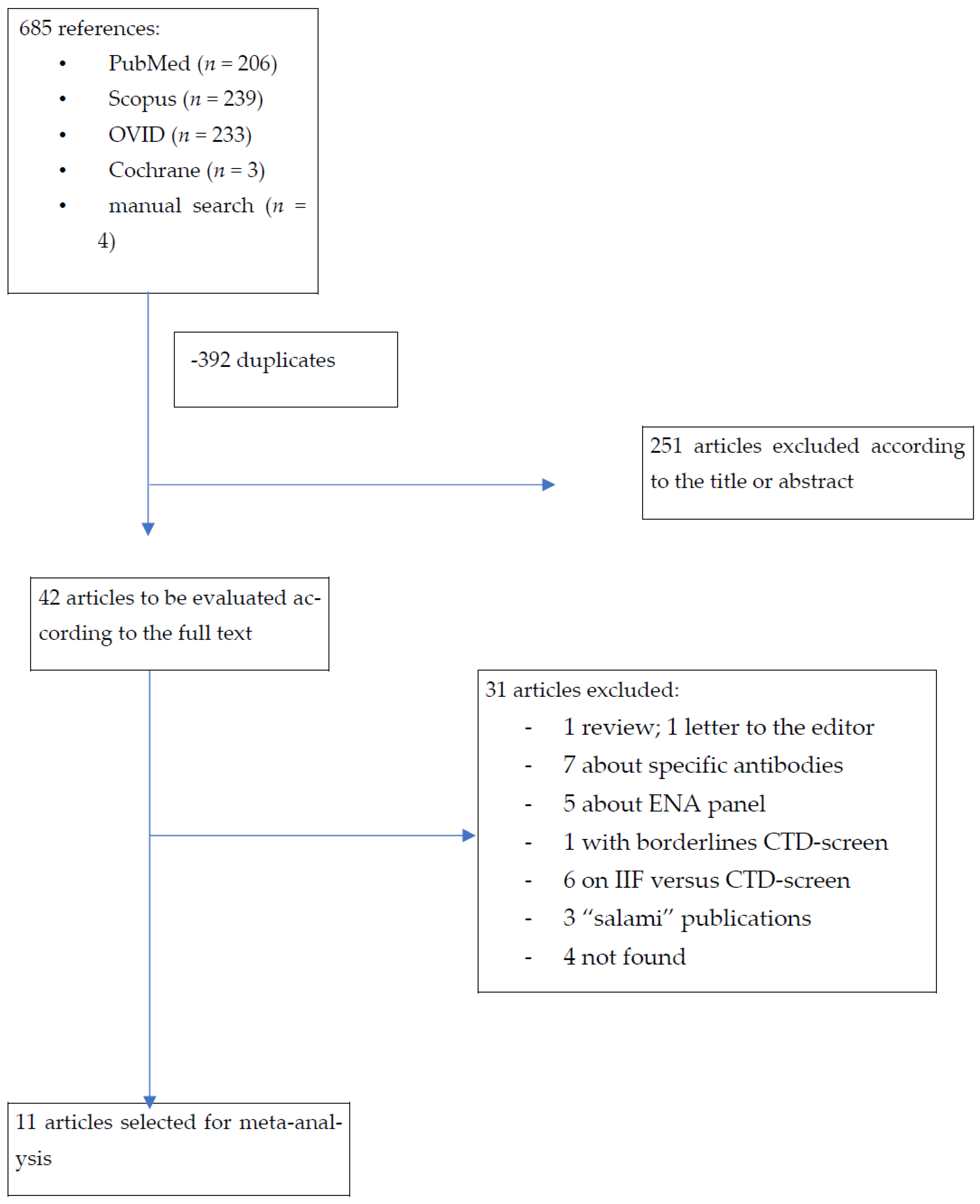
2. Materials and Methods

A systematic review of the literature was carried out according to the Cochrane Collaboration for a systematic review of diagnostic tests studies. Automated research was conducted with different MeSH and free text so that an equation was obtained for each database: PubMed, Scopus, Ovid, and Cochrane Library databases (from inception to 24 February 2021), without either language or publication period restrictions.Antibodies 14 00056 i001
The search was completed with manual search of used references in firm documentations or journals including the CTD screen. All cohorts or case–control studies having considered CTD screen as diagnostic tool for connective tissue disease were imported in a spreadsheet software program (Excel) using a bibliographic management software (Zotero. Version4) to be selected according to title and abstract by two independent investigators after removing the duplicates. The two investigators compared their results to discuss discrepancies, and a third party was designated in case of a disagreement. Selected studies were evaluated on full text for eligibility for inclusion in the systematic review of the literature. Moreover, studies had to specify the CTD screen positivity rate to allow the meta-analysis of data.
Figure 1 shows the flowchart of the selection of the different references used in this meta-analysis. Inclusion criteria were all case–control or cohort studies assessing CTD screen to diagnose connective tissue disease. A total of 685 potentially eligible references were identified according to our different equations and research strategy. After the exclusion of 392 duplicated articles, titles and abstracts from the 251 remaining articles were analyzed. From this, 42 articles were analyzed from the full text and 31 references were excluded because they did not satisfy our criteria (not case–control or cohort studies). Finally, 11 articles were included for more analyses. This study was conducted in accordance with the Preferred Reporting Items for Systematic Reviews and Meta-Analyses (PRISMA) guidelines.This systematic review was conducted according to PRISMA guidelines.

Statistical Analysis
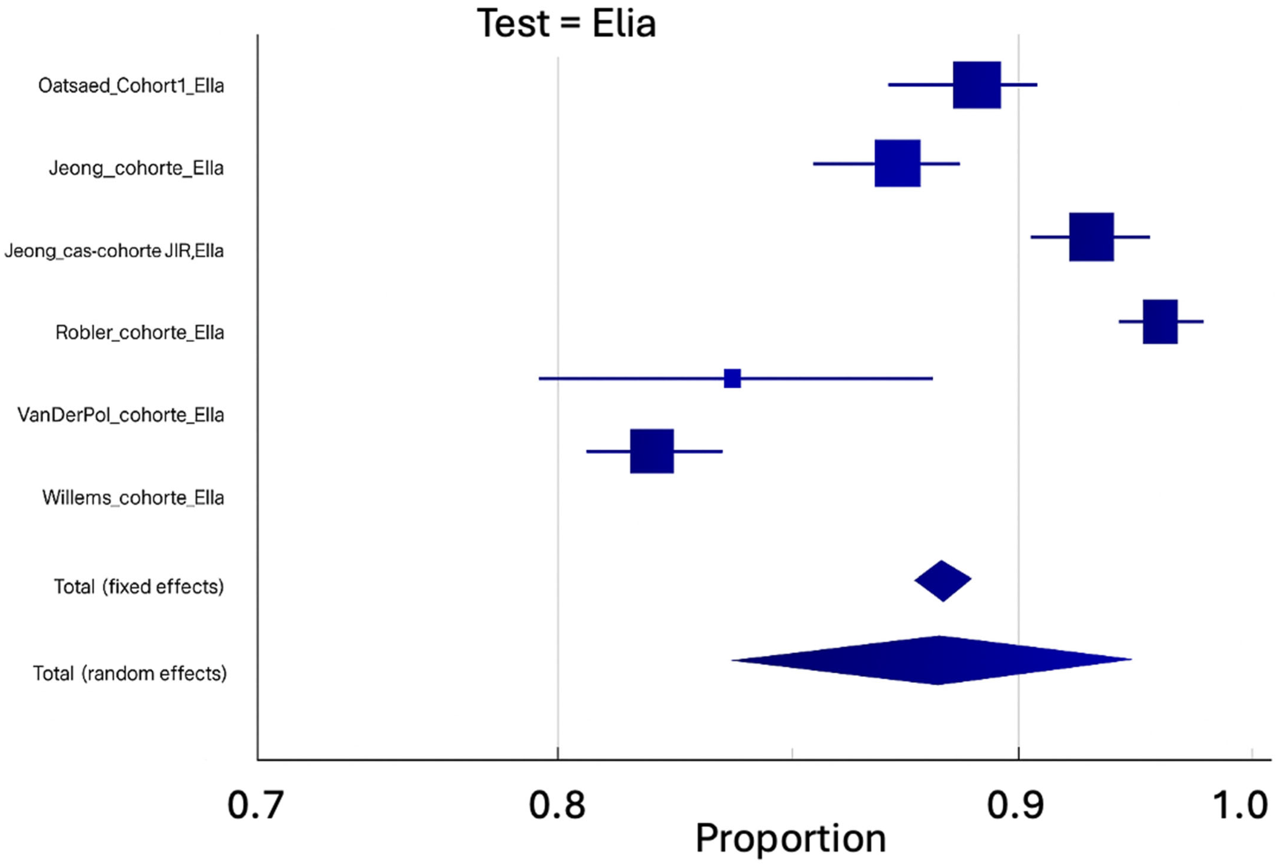
CTD screen positivity rates were systematically extracted for sub-groups of interest provided in the studies. When unavailable, positivity rates were calculated from other available data. Sensitivity, specificity, and their 95% confidence intervals were calculated for each type of study (cohort or case–control) and each commercial kit. Quality of studies was evaluated according the QUADAS-2 score tool (Quality Assessment of Diagnostic Accuracy Studies) [11]. Because of the study design bias, the random-effects model was used to obtain pooled sensitivity and specificity and their confidence interval. Results were presented as Forest’s diagrams. In each Forest plot, symbol size corresponds with sample size. To evaluate the heterogeneity of included studies in the meta-analysis, we used the I2. A value of 0 to 25% of the I2 value represents non-significant heterogeneity, 26 to 50% represents poor heterogeneity, 51 to 75% represents moderate values of heterogeneity, and >75% represents high heterogeneity. Publication bias was evaluated with visual inspection of the funnel plot and Egger and Begg’s tests [12,13]. Statistical analysis was performed with MedCalc (MedCalc Softwares, Belgium). Statistical significance was considered significant for a p-value < 0.05.

3. Results

From the 11 articles selected for meta-analysis, 15 datasets were analyzed according to the type of recruitment (8 cohort studies and 7 case–control studies) or the kit used (9 Elia® CTD-screen, 5 QuantaFlash® CTD-screen, 1 Varelisa® CTD-screen, and 1 Elia® and/or QuantaFlash® CTD-screen). The 11 studies encompassed 2384 patients diagnosed with CTD, 8972 patients without CTD, and 679 blood donors (Table 1).
Quality assessment of the studies according the QUADAS-2 score tool was performed (Table 2). Overall, quality assessment indicated that studies were moderate quality, in particular for patients’ selection. Indeed, several studies carried out case–control studies and resorted to patients from blood banks. Moreover, in some studies, final diagnosis was confirmed only in IIF patients and/or CTD-screen positive and in part of CTD-screen negative [8,14,15]. Jeong et al. did not clearly explain their patients’ selection [5,10].
Pooled sensitivity and specificity for Elia® CTD-screen (Se: 79.36%; Sp: 90.79%) and QuantaFlash® CTD-screen tests (Se: 87.23%; Sp: 87.14%) were extracted from the data of nine and four studies, respectively. Table 3 shows operational performance for each type of CTD.
Regarding Elia® CTD-screen, in over six studies including only cohort studies, the prevalence of CTD on a 8109 patient sample was 10.22% (IC: 6.9–14.1). For those same studies, a sensitivity of 79.25% (IC: 74.26–83.84) and a specificity of 88.3% (IC: 83.81–92.14) were established (Table 3) (Figure 2A,B). By relating this prevalence with Elia® CTD-screen diagnosis performance (LR+: 6.77; LR−: 0.23), we established a post-test probability to have a CTD of 43.5% if the test was positive, and of 2.6% if negative. Sensitivity and specificity for a cohort study using the QuantaFlash® CTD-screen test were unable to be pooled, but a single cohort study was extracted.

4. Discussion

This meta-analysis provides a comprehensive and quantitative assessment of the diagnostic performance of CTD-screen assays in the context of systemic autoimmune rheumatic diseases. These conditions, including systemic lupus erythematosus (SLE), systemic sclerosis (SSc), SS, myositis, and undifferentiated connective tissue diseases (UCTDs), pose significant diagnostic challenges due to their overlapping clinical manifestations and variable autoantibody profiles [1,2,3]. The ability to detect disease-specific antibodies early in the disease process is critical to guide accurate diagnosis. This meta-analysis identified 11 studies (cohort studies and case–control studies) studying the performance of CTD screen to diagnose systemic rheumatic diseases [5,8,10,14,15,16,17,18,19,20,21]. This showed a sensitivity of 79.36% and a specificity of 90.79% for Elia® and a sensitivity of 87.23% and a specificity of 83.56% for QuantaFlash®. Among cohort studies including all hospital laboratory requests, we found a prevalence of pooled CTD of 10.33%, a sensitivity of 79.25%, and a specificity of 88.3% for the Elia® CTD screen. Since only one cohort study was carried out with QuantaFlash® CTD screen, it was not possible to establish pooled probabilities for this kit. These findings highlight the utility of CTD screens as complementary tools in the diagnostic process but also underscore their limitations if used in isolation. Their strength lies in complementing clinical evaluation and more established serological methods. With proper application, these assays can enhance diagnostic efficiency, minimize unnecessary testing, and ultimately contribute to better patient care.
The transition of indirect immunofluorescence (IIF) from primary clinical application to a confirmatory and research role represents a significant paradigm shift in autoimmune diagnostics. As stated in our methodology, “indirect immunofluorescence (IIF) assay on cultured human epithelial carcinoma cells (Hep-2 cell) has been used as a gold standard method. However, it has been shown that some subtypes of ANA, especially anti-SSA/Ro and anti-JO 1 antibodies, may be overlooked by IIF. Furthermore, IIF requires experienced and well-trained analysts, is time-consuming, and shows high inter-observational variability”.
This evolution occurred due to several converging factors: technical limitations where IIF misses certain clinically important antibodies (anti-SSA/Ro, anti-Jo1); practical constraints including the requirement for specialized expertise, time-intensive protocols, and significant inter-observer variability; and automation advantages where CTD screens enable simultaneous detection of multiple antibodies with standardized, reproducible results suitable for high-throughput clinical laboratories.
However, this shift should not be interpreted as IIF becoming obsolete. Rather, IIF has evolved into a confirmatory and specialized diagnostic tool that provides irreplaceable pattern-specific diagnostic intelligence, while automated CTD screens have assumed the role of efficient initial screening in appropriate clinical contexts.
The practical application of these diagnostic modalities to clinical specimens requires specific technical considerations that are essential for optimal diagnostic performance. For serum-based testing, standard venipuncture with serum separation is followed by serial dilutions (typically 1:80 to 1:160 for IIF, automated dilution for CTD screens). IIF application involves serum application to HEp-2 cell substrates, incubation, washing, fluorescent secondary antibody application, and expert microscopic interpretation. CTD screen application utilizes automated liquid handling systems with standardized reagent addition, incubation protocols, and optical detection systems. For tissue-based diagnosis, while our study focuses on serum-based assays, direct immunofluorescence on tissue biopsies (particularly skin and kidney) provides complementary diagnostic information, especially valuable in lupus nephritis assessment and cutaneous manifestations of systemic autoimmune diseases. This approach detects in situ immune complex deposition and complements serum-based antibody detection. The integration of both approaches—serum-based antibody detection and tissue-based immune complex visualization—provides comprehensive diagnostic assessment, though tissue-based methods were beyond the scope of this meta-analysis.
The detection of ANA is important to help in the diagnosis of CTD, but it should be used with care. Because of its good sensitivity, ANA detection provides a good screening test, but ANAs are frequently found in healthy individuals or in patients with other diseases, with a prevalence between 5 and 30% depending upon studied populations [22]. High clinical suspicion is necessary to avoid useless and costly investigations in the case of false positive tests. In this way, a sensible approach in using an ANA test is essential, and testing should only be conducted in the appropriate clinical context, with patients’ characteristic symptoms and clinical examination ideally coming before laboratory tests. Another consideration is the potential utility of CTD screens in the early identification of patients with UCTDs or overlap syndromes. These patient subsets often evolve over time into defined autoimmune conditions, and early serological signals can be pivotal in risk stratification and longitudinal monitoring. As such, CTD screens may have prognostic as well as diagnostic value, though this hypothesis warrants further investigation in prospective cohort studies. The economic implications of ANA and CTD screen testing in apparently healthy individuals present a significant healthcare challenge. The economic burden of inappropriate screening includes unnecessary specialist referrals generating substantial healthcare costs, patient anxiety and quality-of-life impacts from false-positive results, cascade testing and follow-up investigations in low-probability patients, and healthcare system resource diversion from patients with genuine clinical need. Cost-effectiveness analysis suggests that ANA and CTD screen testing should be reserved for patients with intermediate-to-high pretest probability based on clinical presentation. The positive predictive value in low-prevalence populations renders population-based screening economically unsustainable and clinically counterproductive. High clinical suspicion is necessary to avoid useless and costly investigations in cases of false positive tests.
Screening value in asymptomatic individuals is therefore limited not only by poor positive predictive value but also by the substantial economic and psychological costs associated with false-positive results in healthy populations. A significant opportunity for improving diagnostic performance lies in integrating CTD screens with readily available hematologic parameters. Complete blood count (CBC) abnormalities are frequently present in connective tissue diseases and can enhance diagnostic accuracy when combined with serological testing. Key hematologic parameters in CTD include lymphopenia (common in SLE and other CTDs), thrombocytopenia (particularly in SLE and antiphospholipid syndrome), anemia of chronic disease (prevalent across CTDs), and elevated inflammatory markers (ESR, CRP). Calculated ratios such as platelet–lymphocyte ratio (PLR) can provide additional diagnostic value when integrated with immunological markers. The combination of CBC parameters with ELISA/CTD screen results may be mandatory for optimal diagnostic accuracy in rheumatic disorders. This integrated approach leverages the complementary information provided by cellular immune dysfunction (reflected in CBC abnormalities) and humoral immune activation (detected by antibody testing). Economic advantages of this combined approach include utilization of routinely ordered laboratory tests without additional cost, enhanced diagnostic accuracy potentially reducing the need for additional testing, and improved risk stratification for specialist referral decisions. Our analysis supports a comprehensive diagnostic strategy combining three complementary assessment domains: ANA/CTD antibody testing for humoral immune activation detection, complete blood count analysis for cellular immune dysfunction assessment, and clinical findings integration for phenotypic disease manifestation evaluation. This triple approach offers several advantages: comprehensive immune system assessment addressing both humoral and cellular components, enhanced sensitivity and specificity through multi-parameter integration, cost-effective utilization of standard laboratory tests, and improved clinical decision making through integrated data interpretation. While achieving 100% sensitivity and specificity remains unlikely given the complexity of autoimmune diseases and existence of seronegative cases, this integrated approach significantly improves diagnostic accuracy compared to single-parameter testing. The synergistic effect of combining serological, hematologic, and clinical parameters provides a more robust diagnostic framework than relying on any single testing modality. Implementation considerations include standardized protocols for parameter integration, training for multi-parameter interpretation, quality assurance across testing domains, and cost-effectiveness validation in diverse clinical settings. Our findings support a structured, tiered approach to autoimmune diagnostics that optimizes both diagnostic accuracy and resource utilization. CTD screens should serve as initial diagnostic tools specifically in patients with intermediate-to-high pretest probability for connective tissue disease, not as broad population screening instruments. This targeted approach addresses the fundamental limitation that positive predictive value remains modest in low-prevalence populations. All positive CTD screen results should be confirmed by indirect immunofluorescence (IIF) to maximize diagnostic accuracy and provide crucial pattern-specific diagnostic information. This confirmation step is essential because automated CTD screens, while efficient and standardized, cannot provide the diagnostic intelligence offered by expert pattern recognition. IIF pattern recognition provides diagnostic intelligence that cannot be replicated by solid-phase assays. Specific immunofluorescence patterns offer valuable diagnostic clues that guide both diagnosis and subsequent testing strategies:
  • Peripheral/homogeneous patterns are strongly associated with anti-dsDNA antibodies and systemic lupus erythematosus.
  • Centromere patterns are highly suggestive of limited systemic sclerosis.
  • Nucleolar patterns are characteristic of systemic sclerosis, particularly diffuse cutaneous forms.
  • Speckled patterns encompass various specificities including anti-Sm, anti-RNP, anti-SSA/Ro, and anti-SSB/La antibodies.
Accurate interpretation of these patterns requires experienced laboratory personnel and should be integrated with clinical assessment. The diagnostic accuracy demonstrated in our meta-analysis for CTD screens should therefore be viewed as complementary to, rather than a replacement for, expert IIF interpretation.
CTD screens are valuable in the initial diagnostic phase for detecting systemic autoimmune diseases. However, CTD screens are not recommended for monitoring disease progression or treatment response. For longitudinal patient management, it is essential to adopt a personalized approach based on the involved organ system and utilize appropriate disease-specific markers. This phase-specific approach optimizes both clinical outcomes and healthcare resource allocation.
The increased prevalence of non-organ-specific autoantibodies—particularly antinuclear antibodies (ANA)—has been observed in individuals with X-chromosome aneuploidies, such as Klinefelter syndrome [23]. This finding underscores that autoantibody production may be influenced by genetic factors independent of autoimmune disease, thereby complicating standard diagnostic interpretation.
A positive ANA result alone is insufficient to establish a diagnosis of systemic autoimmune disease. ANA positivity can occur in a wide range of conditions—including infections, neoplasms, drug exposures, and even among healthy individuals—with its prevalence varying according to demographic, genetic, and methodological factors.
Based on our findings and the need for practical implementation guidance, we propose the following integrated diagnostic algorithm:
  • Clinical Assessment: Comprehensive evaluation including symptoms, physical examination, and organ system involvement.
  • Pretest Probability Determination: Low probability—avoid testing; intermediate–high probability—proceed with a multi-parameter approach.
  • Multi-Parameter Testing: CTD screen + CBC with calculated ratios + inflammatory markers.
  • Result Integration: Combined interpretation of serological and hematologic findings.
  • Confirmation Strategy: Positive CTD screen confirmed with IIF and expert pattern interpretation.
  • Clinical Correlation: Rheumatological integration of all findings with clinical presentation.
  • Monitoring Framework: Disease-specific, organ-based follow-up rather than repeat CTD screening.
The QUADAS-2 assessment showed that study quality was generally moderate, with most concerns focused on patient selection and reference standard application. In this context, clinicians should be cautious in generalizing the performance of these tests to all patient populations and should consider the risk of spectrum bias, particularly in settings outside of tertiary care or specialist centers. Despite these limitations, the diagnostic accuracy of the Elia® and QuantaFlash® CTD screens remains robust, particularly when used judiciously. Another strength of our study is the inclusion of both cohort and case–control studies across a wide geographic and clinical range. This diversity strengthens the generalizability of our findings but also introduces heterogeneity. While pooled estimates provide meaningful summary statistics, the individual study designs and selection criteria varied, particularly regarding the definition of control populations and reference standards for final diagnosis. In some studies, diagnoses were only confirmed in patients with positive CTD screens or IIF, introducing potential verification bias. Moreover, a few studies included healthy blood donors as controls, which may overestimate specificity due to the lack of clinical ambiguity in this population.
An important implication of this work is the necessity to interpret CTD screen results within the framework of pretest probability. In clinical settings with low disease prevalence, such as primary care or general internal medicine, the positive predictive value of ANA and CTD screen testing is modest. This is consistent with previous studies demonstrating high ANA positivity rates in healthy individuals, ranging from 5% to 30%, depending on demographic and methodological variables. As a result, indiscriminate testing can lead to overdiagnosis, unnecessary specialist referrals, and unwarranted patient anxiety. Conversely, in rheumatology clinics or in patients with suggestive clinical features, these tests can meaningfully contribute to the diagnostic process when integrated into a structured clinical assessment.
Our results indicate that the Elia® CTD-screen had a pooled sensitivity of 79.36% and specificity of 90.79%, while the QuantaFlash® assay demonstrated slightly higher sensitivity (87.23%) with slightly lower specificity (83.56%). These findings suggest that both assays can contribute to the detection of systemic rheumatic diseases, but their use must be contextualized. Sensitivity and specificity varied among different CTDs. For example, primary Sjögren’s syndrome and mixed connective tissue disease (Sharp’s syndrome) showed the highest sensitivities, while autoimmune myositis exhibited relatively lower sensitivity. These results reflect the underlying differences in antibody production and the composition of each test panel and suggest that no single test can reliably identify all CTDs with equal accuracy.
According to Abeles et al., 90% of referred patients to the Rheumatology Department had no evidence of rheumatic disease associated to ANAs identification [7], which means that the prevalence of CTD is only of 10% and so the majority of people with positive ANA testing are not affected by a systemic rheumatic disease. Moreover, ANA positivity has a low positive predictive value, which can be explained by using tests with suboptimal performance in groups with low probability clinical presentation [14].
The term ‘CTD screen’ may be misleading and overly promotional, which can be confusing because we cannot really consider it as a screening test or use it as a diagnostic test. CTD-screen may be interesting to help CTD diagnosis, but clinician expertise remains, essential especially because some CTDs are seronegative.

5. Conclusions

ANAs are a cornerstone in detection of connective tissue disease, but they are widely spread in the healthy population too. CTD-screen does not have diagnostic performance if used alone in clinical practice for the detection of CTD. But this test could help specialists to make diagnosis or perform other complementary exams. Currently, CTD diagnosis is complex, and specialist assessment remains essential. The optimal diagnostic approach integrates multiple complementary modalities: automated CTD screens for efficient initial detection, expert IIF interpretation for pattern-specific diagnostic intelligence, hematologic parameter analysis for cellular immune assessment, and comprehensive clinical evaluation for phenotypic disease characterization. This multi-parameter strategy maximizes diagnostic accuracy while optimizing healthcare resource utilization through targeted testing, appropriate confirmation protocols, and recognition of the distinct roles of different diagnostic modalities in the evolving landscape of autoimmune diagnostics. The findings of this meta-analysis support a paradigm shift toward integrated, multi-parameter autoimmune diagnostics that combines the efficiency of modern automated systems with the irreplaceable value of expert interpretation and comprehensive clinical assessment. This approach acknowledges both the technical evolution of diagnostic capabilities and the economic imperatives of sustainable healthcare delivery while maintaining the diagnostic accuracy essential for optimal patient care. Future research should explore the integration of clinical prediction tools with serological testing to optimize diagnostic algorithms. Machine learning and artificial intelligence could assist in combining clinical variables, imaging, and serology to improve diagnostic precision and reduce false-positive rates. Additionally, cost-effectiveness studies are needed to evaluate the long-term impact of CTD screen utilization on healthcare resource allocation and patient outcomes.

Author Contributions

Conceptualization: D.P. and M.S.; Methodology: A.K., N.K., D.P. and M.S.; software: N.K., A.K. and D.P.; validation: A.K., D.P. and M.S.; formal analysis: N.K., D.P. and J.S. Investigation: N.K., A.K. and J.S.; resources: J.S., D.P. and M.S.; Data curation D.P. and M.S.; project administration: M.S. and D.P.; writing and review: A.K., D.P. and M.S. All authors have read and agreed to the published version of the manuscript.

Funding

No funding was received.

Institutional Review Board Statement

Erasme hospital ethics approved this study (p2021/056).

Informed Consent Statement

Not applicable.

Data Availability Statement

No new data was created in this study.

Conflicts of Interest

The authors declare no conflicts of interest.

References

  1. Mulhearn, B.; Tansley, S.L.; McHugh, N.J. Autoantibodies in connective tissue disease. Best Pract. Res. Clin. Rheumatol. 2020, 34, 101462. [Google Scholar] [CrossRef] [PubMed]
  2. Didier, K.; Bolko, L.; Giusti, D.; Toquet, S.; Robbins, A.; Antonicelli, F.; Servettaz, A. Autoantibodies Associated With Connective Tissue Diseases: What Meaning for Clinicians? Front. Immunol. 2018, 9, 541. [Google Scholar] [CrossRef]
  3. Pepmueller, P.H. Undifferentiated Connective Tissue Disease, Mixed Connective Tissue Disease, and Overlap Syndromes in Rheumatology. Mo. Med. 2016, 113, 136–140. [Google Scholar] [PubMed]
  4. Bossuyt, X.; De Langhe, E.; Borghi, M.O.; Meroni, P.L. Understanding and interpreting antinuclear antibody tests in systemic rheumatic diseases. Nat. Rev. Rheumatol. 2020, 16, 715–726. [Google Scholar] [CrossRef] [PubMed]
  5. Jeong, S.; Yang, H.; Hwang, H. Evaluation of an automated connective tissue disease screening assay in Korean patients with systemic rheumatic diseases. PLoS ONE 2017, 12, e0173597. [Google Scholar] [CrossRef]
  6. Volkmann, E.R.; Taylor, M.; Ben-Artzi, A. Using the antinuclear antibody test to diagnose rheumatic diseases: When does a positive test warrant further investigation? South. Med. J. 2012, 105, 100–104. [Google Scholar] [CrossRef]
  7. Abeles, A.M.; Abeles, M. The clinical utility of a positive antinuclear antibody test result. Am. J. Med. 2013, 126, 342–348. [Google Scholar] [CrossRef]
  8. Willems, P.; De Langhe, E.; Claessens, J.; Westhovens, R.; Van Hoeyveld, E.; Poesen, K.; Vanderschueren, S.; Blockmans, D.; Bossuyt, X. Screening for connective tissue disease-associated antibodies by automated immunoassay. Clin. Chem. Lab. Med. 2018, 56, 909–918. [Google Scholar] [CrossRef]
  9. Robier, C.; Amouzadeh-Ghadikolai, O.; Stettin, M.; Reicht, G. Comparison of the Clinical Utility in the Detection of Anti-Nuclear Antibodies Between the Elia CTD Screen and Indirect Immunofluorescence on Hep-2 Cells: A Review of the Literature. Isr. Med. Assoc. J. IMAJ 2018, 20, 700–702. [Google Scholar]
  10. Jeong, S.; Hwang, H.; Roh, J.; Shim, J.E.; Kim, J.; Kim, G.-T.; Tag, H.-S.; Kim, H.-S. Evaluation of an Automated Screening Assay, Compared to Indirect Immunofluorescence, an Extractable Nuclear Antigen Assay, and a Line Immunoassay in a Large Cohort of Asian Patients with Antinuclear Antibody-Associated Rheumatoid Diseases: A Multicenter Retrospective Study. J. Immunol. Res. 2018, 2018, 9094217. [Google Scholar]
  11. Whiting, P.F.; Rutjes, A.W.S.; Westwood, M.E.; Mallett, S.; Deeks, J.J.; Reitsma, J.B.; Leeflang, M.M.G.; Sterne, J.A.C.; Bossuyt, P.M.M. QUADAS-2 Group QUADAS-2: A revised tool for the quality assessment of diagnostic accuracy studies. Ann. Intern. Med. 2011, 155, 529–536. [Google Scholar] [CrossRef] [PubMed]
  12. Egger, M.; Davey Smith, G.; Schneider, M.; Minder, C. Bias in meta-analysis detected by a simple, graphical test. BMJ 1997, 315, 629–634. [Google Scholar] [CrossRef] [PubMed]
  13. Begg, C.B.; Mazumdar, M. Operating characteristics of a rank correlation test for publication bias. Biometrics 1994, 50, 1088–1101. [Google Scholar] [CrossRef] [PubMed]
  14. Bizzaro, N.; Brusca, I.; Previtali, G.; Alessio, M.G.; Daves, M.; Platzgummer, S.; Cinquanta, L.; Paura, G.; Infantino, M.; Manfredi, M.; et al. The association of solid-phase assays to immunofluorescence increases the diagnostic accuracy for ANA screening in patients with autoimmune rheumatic diseases. Autoimmun. Rev. 2018, 17, 541–547. [Google Scholar] [CrossRef]
  15. Robier, C.; Amouzadeh-Ghadikolai, O.; Stettin, M.; Reicht, G. Comparison of the clinical utility of the Elia CTD Screen to indirect immunofluorescence on Hep-2 cells. Clin. Chem. Lab. Med. 2016, 54, 1365–1370. [Google Scholar] [CrossRef]
  16. Op De Beeck, K.; Vermeersch, P.; Verschueren, P.; Westhovens, R.; Mariën, G.; Blockmans, D.; Bossuyt, X. Detection of antinuclear antibodies by indirect immunofluorescence and by solid phase assay. Autoimmun. Rev. 2011, 10, 801–808. [Google Scholar] [CrossRef]
  17. van der Pol, P.; Bakker-Jonges, L.E.; Kuijpers, J.H.S.A.M.; Schreurs, M.W.J. Analytical and clinical comparison of two fully automated immunoassay systems for the detection of autoantibodies to extractable nuclear antigens. Clin. Chim. Acta Int. J. Clin. Chem. 2018, 476, 154–159. [Google Scholar] [CrossRef]
  18. Claessens, J.; Belmondo, T.; De Langhe, E.; Westhovens, R.; Poesen, K.; Hüe, S.; Blockmans, D.; Mahler, M.; Fritzler, M.J.; Bossuyt, X. Solid phase assays versus automated indirect immunofluorescence for detection of antinuclear antibodies. Autoimmun. Rev. 2018, 17, 533–540. [Google Scholar] [CrossRef]
  19. Bentow, C.; Lakos, G.; Rosenblum, R.; Bryant, C.; Seaman, A.; Mahler, M. Clinical performance evaluation of a novel, automated chemiluminescent immunoassay, QUANTA Flash CTD Screen Plus. Immunol. Res. 2015, 61, 110–116. [Google Scholar] [CrossRef]
  20. Alsaed, O.S.; Alamlih, L.I.; Al-Radideh, O.; Chandra, P.; Alemadi, S.; Al-Allaf, A.-W. Clinical utility of ANA-ELISA vs. ANA-immunofluorescence in connective tissue diseases. Sci. Rep. 2021, 11, 8229. [Google Scholar] [CrossRef]
  21. Lopez-Hoyos, M.; Rodriguez-Valverde, V.; Martinez-Taboada, V. Performance of Antinuclear Antibody Connective Tissue Disease Screen. Ann. N. Acad. Sci. 2007, 1109, 322–329. [Google Scholar] [CrossRef] [PubMed]
  22. Pisetsky, D.S. Antinuclear antibody testing—Misunderstood or misbegotten? Nat. Rev. Rheumatol. 2017, 13, 495–502. [Google Scholar] [CrossRef] [PubMed]
  23. Panimolle, F.; Tiberti, C.; Spaziani, M.; Riitano, G.; Lucania, G.; Anzuini, A.; Lenzi, A.; Gianfrilli, D.; Sorice, M.; Radicioni, A.F. Non-organ-specific autoimmunity in adult 47, XXY Klinefelter patients and higher-grade X-chromosome aneuploidies. Clin. Exp. Immunol. 2021, 205, 316–325. [Google Scholar] [CrossRef] [PubMed]
Figure 1. Flowchart of the selection of the different studies used in this meta-analysis.
Antibodies 14 00056 g001
Figure 2. (A) Funnel and Forest plot for cohort sensitivity using the Elia® CTD-screen test. (B) Funnel and Forest plot for cohort specificity using the Elia® CTD-screen test.
Antibodies 14 00056 g002aAntibodies 14 00056 g002bAntibodies 14 00056 g002cAntibodies 14 00056 g002d
Table 1. Results from the studies included in the meta-analysis.
All Connective Tissue DiseasesSystemic Lupus ErythematosusPrimary Sjogren’s SyndromeSystemic SclerodermaDermato-PolymyositisUndifferentiated Connective Tissue DiseasesControls Without Connective Tissue DiseasesBlood Donor
AuthorCTDpostotalCTDpostotalCTDpostotalCTDpostotalCTD
pos
totalCTD
pos
totalCTD negtotalCTD negtotal
Bizarro
Cohort study
Elia-QuantaFlash
2823689112311112847662539812
Willems
Cohort study
Elia
17021662834345546312178818042197
Robier
Cohort study
Elia
6585212817171011 14415231623
Van der Pool
Cohort study
Elia
667243441316244444210250
Van der Pool
Cohort study
QuantaFlash
717244441516444444190250
Van der Pool
Case–control study
Elia
1041203640333417231823
Van der Pool
Case–control study
QuantaFlash
1011203840323417231423
Lopez-Hoyos
Case–control study
Varelisa
19325415220230411111 19821895105
Bentow
Case–control study
QuantaFlash
139178799824302130 1520192204140146
Jeong
Cohort study
Elia
46623135 2 2 13238981031
Jeong
Cohort study
Elia
91112576716191321 559241003
Claessens
Case–control study
Elia
38648095119596518122025502626748767276279
Claessens
Case–control study
QuantaFlash
412480102119596519222033502626675767262279
Op De Beeck
Case–control study
Elia
17123659803236506911281313409422145149
Olsaed
Cohort study
Elia
150201?142?24?15?10 11121257
? unknown
Table 2. Quality assessment of the studies according to the QUADAS-2 score tool.
Author
and Test
YearCountry
And Study
Quality Assessment of the Studies (QUADAS2)
Risk of BiasApplicability Concerns
Patients’ SelectionIndex TestStandard Reference Flow and TimingPatients’ SelectionIndex TestStandard Reference
Bizzaro
Elia-Quanta
2018Italy
Cohort study
Antibodies 14 00056 i002Antibodies 14 00056 i003Antibodies 14 00056 i003Antibodies 14 00056 i003Antibodies 14 00056 i002Antibodies 14 00056 i002Antibodies 14 00056 i002
Willems
Elia
2018Belgium
Cohort study
Antibodies 14 00056 i002Antibodies 14 00056 i002Antibodies 14 00056 i002Antibodies 14 00056 i002Antibodies 14 00056 i002Antibodies 14 00056 i002Antibodies 14 00056 i002
Robier
Elia
2016Austria
Cohort study
Antibodies 14 00056 i002Antibodies 14 00056 i002Antibodies 14 00056 i002Antibodies 14 00056 i002Antibodies 14 00056 i002Antibodies 14 00056 i002Antibodies 14 00056 i002
Van der Pool
Elia
2018Netherlands
Cohort study
Antibodies 14 00056 i002Antibodies 14 00056 i002Antibodies 14 00056 i002Antibodies 14 00056 i002Antibodies 14 00056 i002Antibodies 14 00056 i002Antibodies 14 00056 i002
Van der Pool
Quanta
2018Netherlands
Cohort study
Antibodies 14 00056 i002Antibodies 14 00056 i002Antibodies 14 00056 i002Antibodies 14 00056 i002Antibodies 14 00056 i002Antibodies 14 00056 i002Antibodies 14 00056 i002
Van der Pool
Elia
2018Netherlands
Case–control study
Antibodies 14 00056 i003Antibodies 14 00056 i002Antibodies 14 00056 i002Antibodies 14 00056 i002Antibodies 14 00056 i003Antibodies 14 00056 i002Antibodies 14 00056 i002
Van der Pool
Quanta
2018Netherlands
Case–control study
Antibodies 14 00056 i003Antibodies 14 00056 i002Antibodies 14 00056 i002Antibodies 14 00056 i002Antibodies 14 00056 i003Antibodies 14 00056 i002Antibodies 14 00056 i002
Lopez-Hoyos
Varelisa
2007Spain
Case–control study
Antibodies 14 00056 i003Antibodies 14 00056 i002Antibodies 14 00056 i002Antibodies 14 00056 i002Antibodies 14 00056 i003Antibodies 14 00056 i002Antibodies 14 00056 i002
Bentow
Quanta
2015International
Case–control study
Antibodies 14 00056 i003Antibodies 14 00056 i002Antibodies 14 00056 i002Antibodies 14 00056 i002Antibodies 14 00056 i003Antibodies 14 00056 i002Antibodies 14 00056 i002
Jeong
Elia
2018Korea
Cohort study
?Antibodies 14 00056 i002Antibodies 14 00056 i002Antibodies 14 00056 i002?Antibodies 14 00056 i002Antibodies 14 00056 i002
Jeong
Elia
2017Korea
Cohort study
?Antibodies 14 00056 i002Antibodies 14 00056 i002Antibodies 14 00056 i002?Antibodies 14 00056 i002Antibodies 14 00056 i002
Claessens
Elia
2018International
Case–control study
Antibodies 14 00056 i003Antibodies 14 00056 i002Antibodies 14 00056 i002Antibodies 14 00056 i002Antibodies 14 00056 i003Antibodies 14 00056 i002Antibodies 14 00056 i002
Claessens
Quanta
2018International
Case–control study
Antibodies 14 00056 i003Antibodies 14 00056 i002Antibodies 14 00056 i002Antibodies 14 00056 i002Antibodies 14 00056 i003Antibodies 14 00056 i002Antibodies 14 00056 i002
Op De Beeck
Elia
2011Belgium
Case–control study
Antibodies 14 00056 i003Antibodies 14 00056 i002Antibodies 14 00056 i002Antibodies 14 00056 i002Antibodies 14 00056 i003Antibodies 14 00056 i002Antibodies 14 00056 i002
Alsaed
Elia
2018Qatar
Cohort
Antibodies 14 00056 i002Antibodies 14 00056 i002Antibodies 14 00056 i002Antibodies 14 00056 i002Antibodies 14 00056 i002Antibodies 14 00056 i002Antibodies 14 00056 i002
Legend: Antibodies 14 00056 i003 High Antibodies 14 00056 i002 low ? unclear.
Table 3. Operational performance for each type of CTD.
ELIA© CTD-SCREEN
Sensitivity for…Number of StudiesNumber of CasesPooled SensitivityIC95%QI2EggerBegg
Connective tissue disease diagnosis9158479.3675.61–82.8823.5466.02%NSNS
Lupus erythematosus diagnosis849682.9876.49–88.6022.5268.92%NSNS
Primary Sjogren’s syndrome diagnosis723291.4386.69–95.218.235627.15%NSNS
Systemic scleroderma diagnosis741177.4470.40–87.7811.2146.46%NSNS
Autoimmune myositis diagnosis612360.9543.13–77.3716.62469.92NSNS
Sharp’s syndrome diagnosis78393.0577.92–99.8024.1375.14NSNS
Specificity for …
Clinic controls
(no connective tissue disease)
7855091.0586.59–94.69298.6597.66NSNS
Healthy controls (blood donor)242898.1896.09–99.491.700541.19p
<0.001
NS
Total pooled controls8812290.7986.69–94.20233.2897NSNS
ELIA© Pooled CohortsNumber of StudiesNumber of CasesPooled %IC95%QI2EggerBegg
Prevalence of connective tissue disease6810910.226.90–14.10141.6396.47NSNS
Sensitivity674879.2574.26–83.8412.6460.43NSNS
Specificity6736188.383.81–92.14151.7196.7NSNS
QUANTAFLASH© CTD-SCREEN
Sensitivity for…Number of StudiesNumber of CasesPooled SensitivityIC95%QI2EggerBegg
Connective tissue disease diagnosis485087.2379.10–93.5825.2888.13NSNS
Lupus erythematosus diagnosis430191.1380.51–97.8320.9485.68NSNS
Primary Sjogren’s syndrome diagnosis414589.183.31–93.793.24717.61NSNS
Systemic scleroderma diagnosis427780.5268.07–90.487.89261.93%NSNS
Autoimmune myositis diagnosis37768.20552.30–82.183.3640.62%NSNS
Sharp’s syndrome diagnosis35091.6567.02–99.949.2178.28NSNS
Specificity for …
Clinic controls
(no connective tissue disease)
379683.5675.18–90.5013.379185.05NSNS
Healthy controls (blood donor)242594.47492.11–96.440.58710p
<0.001
NS
Total pooled controls3122187.1477.18–94.5635.1594.31NSNS
NS: Not significant.
Disclaimer/Publisher’s Note: The statements, opinions and data contained in all publications are solely those of the individual author(s) and contributor(s) and not of MDPI and/or the editor(s). MDPI and/or the editor(s) disclaim responsibility for any injury to people or property resulting from any ideas, methods, instructions or products referred to in the content.

