Article

Stress Marker Response in the Manila Clam, Ruditapes philippinarum, After Exposure to Sediment Liming


1
Department of Environmental and Prevention Sciences, and Centro Ricerche Inquinamento Fisico Chimico Microbiologico Ambienti Alta Sterilità, University of Ferrara, Via Luigi Borsari 46, 44121 Ferrara, Italy
2
Tekne Hub Research Center, University of Ferrara, 44122 Ferrara, Italy
3
Department of Chemical, Pharmaceutical and Agricultural Sciences, University of Ferrara, Via Luigi Borsari 46, 44121 Ferrara, Italy
4
Department of Architecture, Via Ghiara 36, 44121 Ferrara, Italy
*
Author to whom correspondence should be addressed.
Water 2026, 18(7), 776; https://doi.org/10.3390/w18070776
Submission received: 13 February 2026 / Revised: 13 March 2026 / Accepted: 22 March 2026 / Published: 25 March 2026
(This article belongs to the Section Oceans and Coastal Zones)

Abstract

Beach sands may harbor human pathogens and antibiotic resistance genes, prompting the proposal of low-dose quicklime (CaO; 1–3% w/w) as a remediation strategy to improve microbiological quality in highly contaminated areas. After application, CaO is converted into calcium carbonate (CaCO3), yet the ecological effects of this residual compound on benthic fauna remain poorly understood. This study evaluated the short-term impact of CaCO3-enriched sediment (3% w/w) on the Manila clam, Ruditapes philippinarum, under controlled mesocosm conditions. Adult clams were exposed for one week, and survival, burrowing behavior, feeding- and metabolism-related parameters (clearance, ingestion, absorption efficiency and rate, ammonia excretion), and oxidative stress (malondialdehyde, MDA) were assessed using a hierarchical design, with a tank as the experimental unit. No significant differences were detected between control and CaCO3-enriched treatments for any measured endpoint. Survival remained high, functional responses showed overlapping ranges, and MDA levels did not differ significantly between groups. Although limited to short-term exposure and a single concentration, these findings suggest that residual CaCO3 derived from quicklime application did not induce detectable adverse effects in adult R. philippinarum under the tested conditions. Further long-term and multi-species studies are needed to confirm ecological safety.

1. Introduction

Coastal beach environments harbor highly dynamic and complex microbial ecosystems, whose balance is influenced by sediment, water, and living organisms’ characteristics [1,2]. Consistent with this, the sand microbial community can be altered, potentially leading to its imbalance and favoring the increase in potential human pathogens [3,4]. Despite the significant threat this poses to human health, beach sand is not routinely monitored, unlike seawater, whose quality is regulated under the European Union Bathing Water Directive [5]. Notably, beachgoers often spend more time in contact with sand than with seawater, increasing the risk of direct (skin contact) or indirect (entry through eyes, ears, lungs, or mouth) exposure to pathogens [6].
High-throughput 16S rDNA sequencing has shown that microbial communities in sand and seawater cluster into distinct clades, with complex and difficult-to-predict dynamics involving both native (autochthonous) and transient (allochthonous) microorganisms [6,7,8,9,10]. Recreational exposure to contaminated sand may contribute to gastrointestinal illness via the fecal–oral route, particularly when sand contains elevated levels of fecal indicator bacteria (FIB), such as fecal coliforms, Escherichia coli, and enterococci. These microorganisms originate from human and animal feces and can reach beach sand through water transport or direct animal deposition [11,12].
Recent studies have also documented the presence of additional human-associated bacteria, fungi, viruses, and parasites in beach sand. For example, Staphylococcus spp., whose abundance correlates strongly with beach crowding, may increase infection risk through skin lesions [13]. Moreover, so-called “flesh-eating” bacteria, including Streptococcus, Klebsiella, Clostridium, Escherichia, and Aeromonas species, have been detected in coastal sands; these pathogens are capable of causing severe soft tissue infections such as necrotizing fasciitis [13]. Fungal and viral pathogens have also been identified in up to 23% of beach sand samples, including Trichophyton mentagrophytes and T. rubrum, Aspergillus spp., Candida spp., as well as enteroviruses and reoviruses, which are associated with respiratory and gastrointestinal infections [13].
Based on these observations, we recently characterized the microbiome of a specific stretch of the Italian coastline located in the northern Adriatic Sea, between the Po Delta and the Reno River estuary. Due to the confidential nature of that study, the exact location of the sampling site remains undisclosed. Due to their proximity to animal farms and urban areas along their course, these rivers may act as reservoirs of human and animal pathogens as well as antimicrobial resistance (AMR), potentially influencing the sand microbiome. Our results, obtained using both culture-based and molecular approaches, revealed a high bacterial load (>103 CFU/g), including members of the Enterobacteriaceae family and the genera Pseudomonas aeruginosa, Enterococcus, and Staphylococcus. The detected levels exceeded safety thresholds established for water and sand by WHO guidelines, such as the 60 CFU/g limit for intestinal enterococci in beach sand [14].
Most Enterococcus species were likely of animal origin; specifically, the presence of E. gallinarum suggests contamination from nearby farms as well as from synanthropic avian species, such as seagulls and coastal wading birds, whereas E. casseliflavus may point to broader environmental or agricultural sources.
Several human pathogens were also identified, including Vibrio cholerae, Shigella dysenteriae, Yersinia enterocolitica, Staphylococcus epidermidis, Staphylococcus hominis, and Escherichia coli, likely originating from the wastewater discharges of coastal urban centers in the immediate vicinity. Notably, multiple antibiotic resistance genes (ARGs) were detected within the sand microbiome, conferring resistance primarily to aminoglycosides, macrolides, lincosamides, streptogramins, and vancomycin. These findings indicate the widespread presence of multidrug-resistant (MDR) bacteria in beach sand [4].
As increasing evidence has documented the presence of potential pathogens in beach sand [4,15], various sand decontamination strategies have been proposed, including sand removal, chlorine treatment [11], and the application of quicklime (CaO). CaO has been widely used for the stabilization of highly contaminated wastewater sludge [16] and has more recently been explored for food decontamination [17].
We recently evaluated quicklime as a rapid sand decontamination strategy both in vitro and in situ, demonstrating that the addition of CaO to sand (1–3% w/w) can significantly reduce microbial contamination [4]. In vitro, CaO inactivated >99% of bacteria and fungi within 24 h and completely inactivated high loads of Modified Vaccinia virus Ankara (MVA) and Enterovirus 71 (EV71), two viruses recognized for their high resistance to disinfection. The antimicrobial effect was likely driven by rapid heat generation and alkaline pH shifts occurring when CaO reacts with even small amounts of water (≤15% w/w) [4].
Field trials confirmed these findings, showing up to a 90% reduction in baseline microbial contamination in a dose-dependent manner, with effects persisting for up to one month [4]. Notably, several core microbial taxa potentially associated with human disease (such as Cutibacterium spp., Nocardioides spp., Pseudomonas aeruginosa/stutzeri, Bordetella petrii, and Microbacterium sp. Y-01) showed marked decreases in abundance following treatment [4]. Ongoing open-field studies further support the effectiveness of CaO in controlling beach sand microbial contamination (Caselli, unpublished data).
All treatments were applied at a safe distance from the waterline to prevent direct contact between CaO and seawater, ensuring that the study focused exclusively on testing the effect of limestone on the sand’s bacterial component without interference from marine water. Beyond our specific experimental conditions, in the event of sand disinfection treatments using limestone, CaO would be converted into CaCO3 during the reaction process; this byproduct could subsequently come into contact with organisms inhabiting the water–sand interface, such as mollusks and other benthic invertebrates. Carbonate minerals in Northern Adriatic sediments derive predominantly from the erosion of surrounding carbonate-rich mountain belts and their transport through an extensive fluvial network into this semi-enclosed basin. The Eastern Alps and Dolomites represent the principal source for the northern sector (Veneto–Friuli), where rivers such as the Adige and Piave supply sediments rich in dolomite and calcite [18,19]. Farther south, the Po River, draining the Central Alps and the Northern Apennines, delivers a substantial carbonate load to the Emilia-Romagna coast and deltaic system. This terrigenous input is complemented by local biogenic production, as mollusks, foraminifera, and other calcifying organisms contribute additional calcite to the sedimentary budget [18,19,20].
In geochemical datasets, carbonate abundance is commonly expressed as CaO, which does not indicate free calcium oxide but represents total calcium bound within mineral lattices, mainly calcite (CaCO3) and dolomite (CaMg(CO3)2), following standard XRF reporting conventions. From a stoichiometric perspective, calcite corresponds to ~56 wt% CaO, whereas dolomite contributes ~30 wt% CaO (plus MgO), so measured CaO values directly reflect sediment mineralogy and provenance. Across the Northern Adriatic, carbonates show marked spatial variability [19,20]. Total CaO generally increases eastward toward the Croatian coast (10.15–42.15%), while the Po delta shelf is characterized by lower mean carbonate contents (~11%), ranging from 2.4% near the delta to ~32% in northern prodelta muds and decreasing offshore to 5–9% in sandy shelf sediments. Along the Romagna coast, dolomite contents are comparatively low (2–10.6%, mean ~7%) due to limited inputs from local rivers [18,19,20]. Regarding the clam rearing area (Sacca di Goro), CaCO3 concentrations in the sediments are relatively variable, ranging between 8% and 15% (based on a total of 139 sediment samples analyzed) [21]. The observed variability in carbonate content is mainly related to local environmental conditions, particularly to differences in the abundance and frequency of bioclastic fractions.
While the carbonate content in Adriatic sediments is naturally variable, its presence does not justify unregulated artificial additions of CaCO3 or CaO. Under the Italian D.Lgs. 152/2006 [22], any sediment intervention must adhere to the Precautionary Principle, ensuring the protection of benthic communities and water quality. This Italian decree transposes a rigorous EU framework, primarily the Marine Strategy Framework Directive (MSFD) 2008/56/EC [23], which mandates achieving Good Environmental Status (GES) by monitoring anthropogenic impacts on physico-chemical parameters like pH. Furthermore, the Water Framework Directive (WFD) 2000/60/EC [24] requires that remediation or “liming” treatments prove non-toxicity toward Biological Quality Elements (BQEs), such as bivalve mollusks. Even for naturally occurring substances, industrial-scale applications of CaCO3 or CaO fall under the REACH Regulation (EC 1907/2006) [25], necessitating eco-toxicological risk assessments for benthic and pelagic compartments. Consequently, this study evaluates the impact of CaCO3-enriched sediments on the functional performance and oxidative stress of the Manila clam (Ruditapes philippinarum), a key bioindicator for the Emilia-Romagna coast.

2. Materials and Methods

2.1. Experimental Setup

Manila clams Ruditapes philippinarum (N = 120) were obtained from a hatchery (Naturedulis) in Goro (Italy). Specimens ranged between 25–40 mm in shell length and had a fresh weight between 6–14 g. Commercial CaCO3 powder (Fassa Bortolo, Spresiano, Italy), with physico-chemical properties comparable to exhausted CaO (Caselli, unpubl. data), was first homogenized with dried natural sediment from the Sacca di Goro at 3% (w/w), the upper limit of the 1–3% range typically used for beach sand decontamination [4], using a mechanical stirrer. This concentration was selected to represent a conservative worst-case scenario, corresponding to the maximum dosage currently applied in remediation practices.
The experimental setup comprised six 18 L aquaria, each equipped with an aerator (Amtra MOUSE3) to ensure adequate oxygenation and water circulation. Three tanks received 3 kg of sieved (1 mm mesh) natural sediment (control), while three were filled with the CaCO3-enriched sediment (treatment), forming a bottom layer of approximately 5 cm. Tanks were then filled with artificial seawater prepared from reverse-osmosis water (Acqua Tecnica, Syntesis Micro 100) and a commercial marine salt mixture (Sea Salt Aquaforest) to achieve a salinity of 28 psu. Sediment granulometry was determined in one randomly selected control tank and one randomly selected treatment tank by wet sieving and pipette analysis [26]. In the same tanks, water alkalinity was also measured by titration according to the standard method [27]. All tanks were operated for one week without organisms to allow system equilibration.
Clams were visually inspected for vitality based on the rapidity of valve closure in response to mechanical stimulation, a widely accepted indicator of bivalve physiological condition [28], and were then randomly allocated to the tanks (20 individuals per tank), yielding three control and three treatment replicates. Clams were acclimated for one week under controlled conditions, with 10% partial water renewal every other day and daily compensation of evaporative losses using RO water. Clams were fed daily with a microalgal diet consisting of a live culture of the diatom Chaetoceros gracilis. Algae were delivered three times per day at 8 h intervals using two programmable four-channel dosing pumps connected to the aquaria. Each feeding event supplied 200 mL per aquarium at a concentration of approximately 6 × 10⁶ cells mL−1. Throughout the entire experimental period, water temperature and dissolved oxygen were measured daily using a Pocket Oxygen Meter (FireSting®-GO2, PyroScience), salinity with a Digital ATC LED Refractometer (AQPET), and nitrite (N–NO2) concentrations with a HANNA® Multiparameter Photometer.

2.2. Functional and Metabolic Responses

Following the one-week exposure period, functional measurements were performed within the subsequent 24 h. Feeding was suspended 12 h prior to the trials to allow gut clearance and standardize physiological status among individuals. From each aquarium, five clams were randomly selected for functional assays. All parameters were calculated according to the literature [29,30,31,32]. Mortality rate (MR) was calculated as follows:
MR = (N(d)/N(t)) × 100
where N(d) is the number of dead individuals and N(t) the total number of clams.
Burrowing time (BT) was recorded as the time (min) required for a manually extracted individual to fully re-burrow into the sediment.
Clearance rate (CR) was estimated as follows:
CR = (V/N) × [(ln C0 − ln C(t))/t]
where V is the water volume (L), N the number of clams, C0 and C(t) the phytoplankton concentrations at times 0 and t (cells mL−1), and t the duration of the experiment (h). Each test lasted 1 h.
Ingestion rate (IR) was calculated as follows:
IR = CR × C(org)
where C(org) is the organic concentration of the microalgal suspension (mg L−1).
Absorption efficiency (AE) was determined using the Conover [33] ratio method:
AE = [1 − (F(o)/F(i))] × 100
where F(i) is the organic fraction of the ingested food (%) and F(o) the organic fraction of the feces (%).
Absorption rate (AR) was computed as follows:
AR = IR × (AE/100)
Ammonia excretion rate (ER) was calculated using:
ER = [(NH4+(t) − NH4+0) × V]/(N × t)
where NH4+0 and NH4+(t) are the ammonia concentrations (µmol h−1) at times 0 and t, V is the water volume (L), N is the number of clams, and t is the duration (h). Each trial lasted 2 h. Ammonia concentrations were quantified using the phenol–hypochlorite spectrophotometric method [34]. To account for differences in body size, all rates were standardized to individual wet weight.

2.3. Oxidative Stress Marker Quantification

Oxidative stress was evaluated by quantifying malondialdehyde (MDA), a widely recognized biomarker of lipid peroxidation and cellular stress [35,36,37,38]. MDA was measured in tissue lysates from CaCO3-treated and control clams. Five randomly selected clams were analyzed from each aquarium: clams were rinsed with physiological solution (0.9% w/v NaCl), immediately sacrificed by rapid freezing in liquid nitrogen to minimize cellular stress, and stored at −80 °C until processing. Soft tissues were dissected from each individual, weighed, and thoroughly homogenized by mechanical disruption using sterile scalpels and scissors in 2 mL of sterile physiological solution. The homogenate was centrifuged at 14,000 rpm for 10 min at 4 °C, and the supernatant was collected and stored at −80 °C until analysis.
MDA content in 30 samples was determined using a competitive Malondialdehyde (MDA) BioAssay ELISA kit (USBiological Life Sciences, Salem, MA, USA), following the manufacturer’s instructions. Because the assay is validated for serum, plasma, and other biological fluids, preliminary tests were conducted to identify the optimal dilution for clam lysates. Samples were analyzed at a 1:2 dilution using the buffer provided in the kit, and all lysates were assayed in duplicate. The assay was performed in microtiter plates pre-coated with MDA. MDA present in standards or samples competed with the immobilized analyte for binding to the biotinylated antibody. After washing to remove unbound components, HRP–streptavidin conjugate was added and the plate was incubated. TMB substrate was then applied, and the reaction was stopped with sulfuric acid. Optical density was measured immediately at 450 nm using a microplate reader.
MDA concentrations (range 1.953–500 ng mL−1) were calculated by comparing sample absorbance values with a standard curve. The mean of duplicate readings was calculated for each standard and sample, and the zero-standard optical density was subtracted. A four-parameter logistic (4-PL) curve was generated using CurveExpert Professional (Hyams Development), and sample concentrations were obtained by interpolation from the fitted curve. MDA levels were expressed as ng mL−1, as specified by the manufacturer’s protocol. Tissue samples were homogenized using a fixed tissue-to-volume ratio, and all samples were processed under identical conditions. This approach allowed a reliable comparison of relative MDA levels between experimental groups.

2.4. Statistical Analyses

For all analyses, the tank (n = 3 per treatment) was considered the experimental unit. Functional parameters were analyzed using a hierarchical (nested) linear mixed-effects model, with Treatment as a fixed factor and Tank nested within Treatment as a random factor. Similarly, biochemical responses (MDA) were evaluated by averaging individual measurements within each tank to obtain a single tank mean; these means were compared between treatments using an unpaired t-test with Welch’s correction. This dual approach ensured that the hierarchical structure of the sampling was respected across all endpoints.

3. Results

3.1. Functional and Metabolic Responses

Table 1 summarizes sediment and water parameters in control and treatment tanks. The addition of 3% (w/w) CaCO3 caused only minor non-significant granulometric changes: fine sand was slightly increased (72.15% to 72.96%), while medium and very fine sand appeared proportionally decreased. The sediment remained sand-dominated, with negligible shifts in grain-size distribution. Sediment pH and water alkalinity also showed negligible variations.
Clam mortality was monitored throughout the experiment; no deaths occurred during acclimation, and only one individual died in the treatment during CaCO3 exposure (1.66%), while no mortality was recorded in the control group.
Burrowing time ranged from 11 to 35 min, with comparable mean values between control and CaCO3-enriched tanks (Table 2). Similarly, clearance rate and ingestion rate showed overlapping ranges across groups. Absorption efficiency (65–84%) and absorption rate (0.010–0.019 g(a) g−1 h−1) also exhibited similar patterns in both treatments. Ammonia excretion rate varied between 0.10 and 0.54 µmol g−1 h−1, with no evident differences between groups.
As summarized in Table 2, ANOVA did not detect significant treatment effects for any of the measured functional parameters (all p > 0.05).

3.2. Oxidative Stress Marker

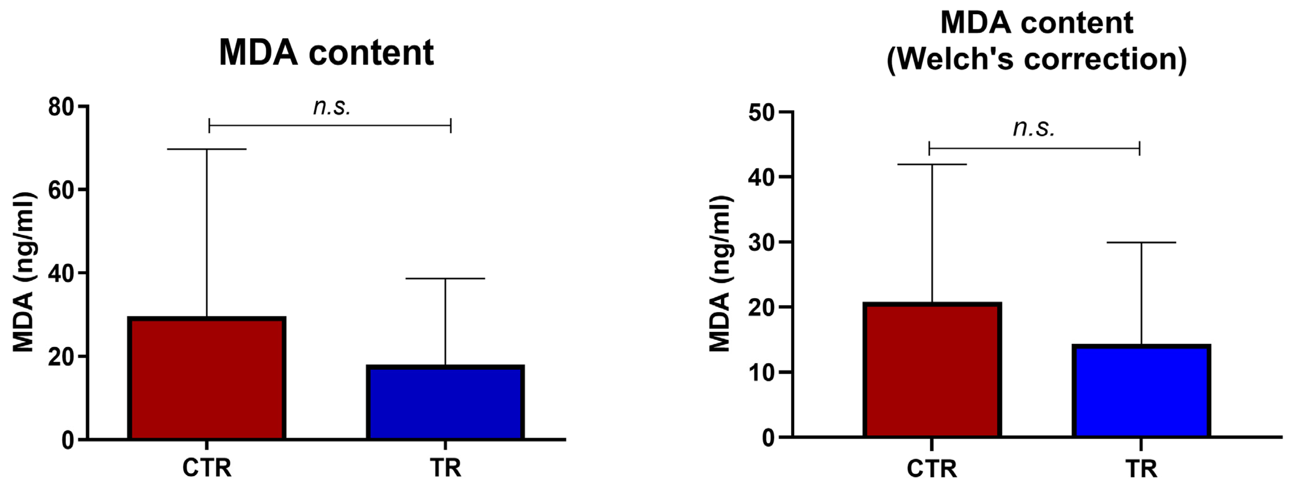
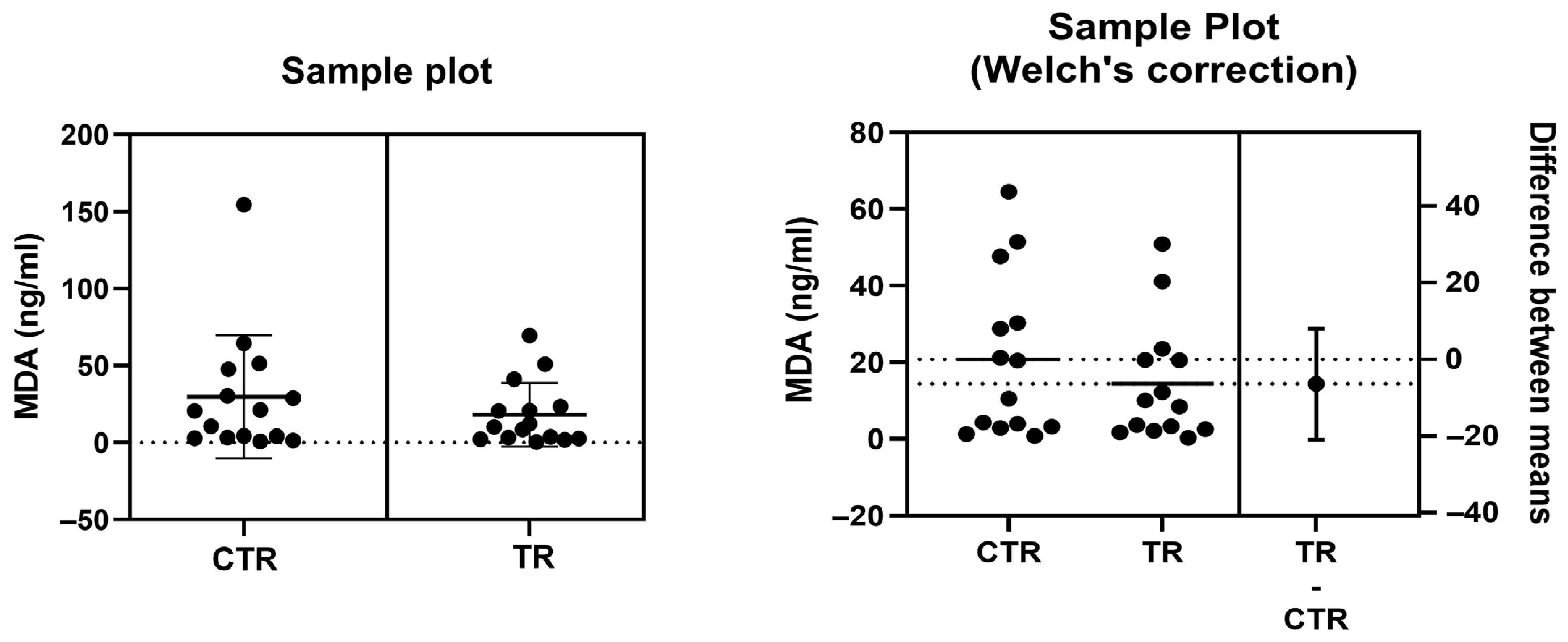
The extent of cellular oxidative stress responses, in terms of MDA content, was assessed in the whole soft body of Ruditapes philippinarum clam species, following growth in sand with or without 3% CaCO3 (w/w) (TR group and CTR groups, respectively). The MDA concentration values obtained for each tested clam are plotted in Figure 1.
Two outlier samples were eliminated in total (one per group), due to the high value, by the statistical GraphPad 10.5.0 software for outliers’ identification (ROUT, Q = 1%), and the remaining values were used to construct the graphs shown in Figure 2.
Overall, no statistically significant differences were observed between the CTR and TR groups, as the CTR group exhibited MDA mean levels corresponding to 20.43 ± 40.05 ng mL−1, and the TR group displayed MDA levels corresponding to 18.03 ± 20.67 ng mL−1.
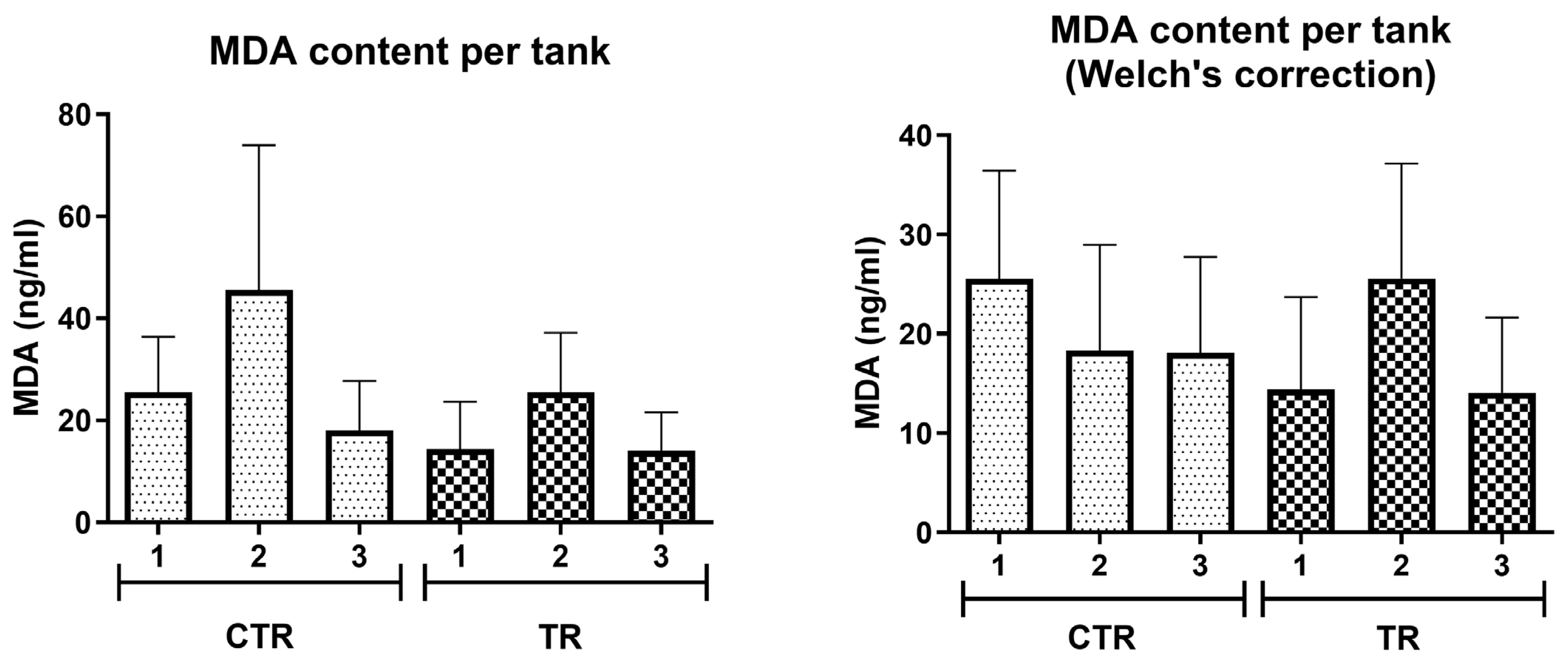
Moreover, to avoid pseudoreplication, an additional analysis was performed at the tank level (Figure 3). Protein levels measured in individual clams were averaged per tank, and tank means were used as independent biological replicates for statistical comparisons. In line with the results shown above, mean values were comparable between groups, and no statistically significant differences were detected (unpaired t-test with Welch’s correction, p = 0.60).

4. Discussion

4.1. Liming of Marine Sediments

CaO has recently been proposed as a remediation tool for sand decontamination due to its demonstrated in vitro and in situ effectiveness [4]. However, before considering routine application, the potential effects of residual calcium carbonate (CaCO3), formed after CaO treatment, on coastal fauna must be evaluated. The ecological implications of CaO-derived residues remain largely unexplored [4,39], and no studies have specifically assessed their impact on benthic infauna such as mollusks, despite the known sensitivity of these organisms to changes in sediment geochemistry. For example, Ruditapes philippinarum juveniles exposed to low-pH sediments exhibited increased metal bioaccumulation [40], and this species is widely recognized as a sentinel organism responsive to environmental stressors in transitional waters [41]. Moreover, Ca-based amendments can significantly modify sediment pH, redox conditions, and contaminant dynamics [42], potentially affecting benthic communities.
Although sediment acidification effects are well documented [40], no study has combined alkaline treatments (e.g., CaO-derived CaCO3) with ecotoxicological evaluation in infaunal bivalves. To address this gap, we investigated whether CaCO3 residues from CaO applications pose risks to a commercially important bivalve, assessing vitality, behavior, feeding- and metabolism-related functions, and oxidative stress. Tests were conducted in the absence or presence of 3% (w/w) CaCO3 added to sand to simulate the upper application level of CaO in situ [4], a concentration previously shown not to alter sediment pH [4].

4.2. Functional and Metabolic Responses

The observed functional and metabolic stability of R. philippinarum indicates that CaCO3 addition did not induce detectable stress. These results align with D.Lgs. 152/2006 [22] and the MSFD (2008/56/EC) [23] goal of maintaining Good Environmental Status. Furthermore, the lack of adverse effects supports the WFD (2000/60/EC) [24] requirement to protect Biological Quality Elements and satisfies the risk assessment principles of the REACH Regulation (EC 1907/2006) [25] regarding the introduction of substances into marine habitats.
Mortality remained extremely low (one individual in the treatment group; none in controls) and was likely attributable to individual variability or handling stress rather than to the experimental treatment itself. All physiological indicators, burrowing time, clearance and ingestion rates, absorption efficiency and absorption rate, and ammonia excretion, showed overlapping ranges, and no statistically significant differences were detected between control and CaCO3-enriched sediments. These parameters collectively describe key aspects of bivalve performance, from feeding and digestion to metabolic waste release, and are widely recognized as sensitive proxies of sublethal stress [43]. Their overall consistency across treatments indicates the absence of measurable short-term physiological impairment associated with CaCO3 enrichment. The value of such integrative physiological testing lies in its ability to reveal subtle functional disruptions that may not be reflected in mortality alone. In the present case, the stability of all measured endpoints supports the interpretation that the tested CaCO3 concentration did not compromise short-term organismal performance.
This contrasts with the marked sublethal impairment reported by Munari and Mistri [44] for the same species exposed to copper released from antifouling paints. In their study, conducted under a comparable experimental framework (similar seasonal collections, acclimation procedures, and laboratory exposure conditions), clams exposed to 10 µg Cu L−1 exhibited a significant reduction in clearance rate, increased oxygen consumption, and a consequent decline in scope for growth. These responses reflected clear metabolic and nutritional destabilization, consistent with the well-established toxicity of Cu to filter-feeding bivalves. The comparison highlights how integrated physiological endpoints can effectively discriminate between harmful stressors and environmentally neutral amendments.
While the findings of Munari and Mistri [44] raised explicit concerns for shellfish farming in the Northern Adriatic, the present results, within the limits of short-term laboratory exposure, do not indicate comparable adverse effects of CaCO3 enrichment. Although longer-term and field-based assessments would be necessary for a comprehensive evaluation, the evidence obtained here suggests that 3% (w/w) sedimentary CaCO3 addition, under controlled conditions, does not impair the short-term functional performance of R. philippinarum and may therefore be compatible with sediment management strategies when carefully regulated.

4.3. Oxidative Stress Marker

Oxidative stress was assessed by quantifying lipid peroxidation (LPO), a well-recognized pathway of cellular damage in both animals and plants. During oxidative stress, unstable lipid peroxides degrade into reactive end-products such as malondialdehyde (MDA) and 4-hydroxynonenal (4-HNE). Because MDA is a relatively stable end-product of lipid oxidation, its measurement is widely used to estimate oxidative damage in biological systems [38,39]. MDA, which exhibits mutagenic and cytotoxic properties, has been employed as a biomarker of oxidative stress in a variety of matrices, including blood, urine, and tissue lysates [45,46].
In aquatic organisms, and particularly in bivalves, MDA has frequently been used to evaluate physiological responses to environmental stressors. Elevated MDA levels have been reported in clams exposed to heavy metals, industrial contaminants, carbon-based nanomaterials, and microplastics, supporting its application as an indicator of oxidative damage in these taxa [46,47]. Experimental studies have also documented dose-dependent increases in MDA under seasonal or thermal stress, reflecting lipid peroxidation associated with environmental challenges [36,48]. Traditionally, oxidative damage in bivalves has been quantified using TBARS-based assays, with results normalized to tissue mass or protein content to enable inter-study comparisons.
In the present study, MDA was quantified using a competitive ELISA adapted to clam tissue lysates and expressed as ng mL−1 according to the manufacturer’s protocol. As all samples were processed using the same tissue-to-volume ratio and extraction procedure, the method allows consistent relative comparisons between groups, although direct comparison with studies using different normalization approaches should be made cautiously.
MDA levels did not differ significantly between CaCO3-treated and control clams. In agreement with the absence of detectable alterations in functional and behavioral parameters, these results do not indicate a measurable increase in oxidative stress under the tested conditions. While limited to short-term laboratory exposure, the findings suggest that residual CaCO3, at the concentration examined, did not elicit detectable oxidative damage in R. philippinarum. Specifically, the stability of MDA levels supports the hypothesis that the addition of CaCO3 to the sediment remained within the physiological tolerance limits of the species. While the absence of MDA accumulation does not strictly rule out the activation of subtle antioxidant defenses, it indicates that any such metabolic adjustments were sufficient to maintain cellular homeostasis. Consequently, under the experimental conditions tested, CaCO3 enrichment appears to be compatible with the oxidative health of the organisms, as it did not trigger the degradative pathways typically associated with environmental stress.

4.4. Implications and Future Research Needs

Several limitations of the present study should be acknowledged. The experiment was conducted under controlled laboratory conditions, using a single CaCO3 concentration (3% w/w) and a short exposure period (one week) on adult clams only. While this design allowed for a focused assessment of short-term functional and oxidative responses, it does not capture potential effects arising from longer-term exposure, different life stages, or varying environmental scenarios.
Future research should include chronic and multi-generational experiments testing a range of CaCO3 concentrations and sediment types under more variable temperature and salinity regimes. Expanding the suite of biological endpoints would further strengthen ecological interpretation, for example, by incorporating additional oxidative stress markers (e.g., antioxidant enzyme activities), detoxification biomarkers, energy budget metrics (e.g., scope for growth), and molecular indicators of stress response. Field-based assessments in natural coastal systems would also help validate laboratory findings and support a more comprehensive evaluation of the ecological implications of CaCO3-enriched sediments.
Further investigations should evaluate whether the disposal or reuse of CaCO3-enriched sediments in coastal environments may entail secondary effects or potential benefits. The addition of calcium-based materials (e.g., limestone, lime, or quicklime) to marine systems has been proposed within the framework of Ocean Alkalinity Enhancement to increase buffering capacity and counteract ocean acidification [49]. In aquaculture settings, such as R. philippinarum farming areas, carbonate enrichment could theoretically influence carbonate chemistry and shell formation dynamics, given the dependence of calcification on bicarbonate availability [50,51,52]. For instance, sediment buffering with carbonate shell hash has been proposed as a mitigation strategy against sediment acidification in Mercenaria mercenaria [53].
Under the short-term experimental conditions adopted here, CaCO3-enriched sediment did not produce detectable adverse effects on adult R. philippinarum. However, these findings should be considered preliminary. Longer exposure periods, multiple concentrations, and a broader range of biological endpoints will be necessary to provide a more comprehensive ecological assessment.

5. Conclusions

The data collected did not show significant differences between clams maintained in untreated sediment and those exposed to sediment enriched with 3% (w/w) CaCO3. Across the measured endpoints, including survival, functional performance, and oxidative stress, responses remained within comparable ranges under the tested conditions. Although these findings are limited to a one-week exposure and a single CaCO3 concentration, they suggest that residual CaCO3 derived from quicklime application did not produce detectable short-term effects on adult R. philippinarum under controlled laboratory conditions. While further studies are required to evaluate longer-term exposures, different life stages, and broader ecological contexts, the present results provide preliminary evidence that CaCO3 residues, at the concentration examined, are unlikely to cause acute functional impairment in this species. These observations contribute to the assessment of quicklime-based remediation strategies, supporting their potential compatibility with benthic fauna when carefully managed and appropriately controlled.

Author Contributions

Conceptualization, E.C. and M.M.; methodology, E.C., M.M. and C.M.; formal analysis, I.S. and F.C.; investigation, I.S., F.C. and M.D.; resources, E.C.; data curation, I.S., F.C. and E.C.; writing—original draft preparation, I.S., M.M. and F.C.; writing—review and editing, E.C., M.M. and C.M.; supervision, E.C. and M.M.; project administration, E.C. and M.B.; funding acquisition, E.C., M.B. and M.M. All authors have read and agreed to the published version of the manuscript.

Funding

This research was partially funded by FASSA Bortolo s.r.l. (Protocol n. 104319 Rep. n.13) to E.C., and by the European Union—Next Generation EU, Mission 4 Component 1 CUP C63C22000570001 to M.M.

Data Availability Statement

The original contributions presented in this study are included in the article. Further inquiries can be directed to the corresponding author.

Acknowledgments

We would like to acknowledge the excellent technical assistance and critical contribution of Sante Mazzacane, Matteo Bisi, Antonella Volta, and Francesco Viroli. The authors would like to thank three anonymous reviewers for their valuable suggestions and constructive feedback, which greatly improved the quality of this manuscript.

Conflicts of Interest

The authors declare that this study received funding from FASSA Bortolo srl, and European Union—Next Generation EU. The funders were not involved in the study design, collection, analysis, interpretation of data, the writing of this article or the decision to submit it for publication.

Abbreviations

The following abbreviations are used in this manuscript:
MDAMalondialdehyde
MRMortality rate
CRClearance rate
IRIngestion rate
AEAbsorption efficiency
ARAbsorption rate
ERAmmonia excretion rate

References

  1. Boudreau, B.P.; Huettel, M.; Forster, S.; Jahnke, R.A.; McLachlan, A.; Middelburg, J.J.; Nielsen, P.; Sansone, F.; Taghon, G.; Van Raaphorst, W.; et al. Permeable Marine Sediments: Overturning an Old Paradigm. Eos Trans. Am. Geophys. Union 2001, 82, 133–136. [Google Scholar] [CrossRef]
  2. Weiskerger, C.J.; Brandão, J.; Ahmed, W.; Aslan, A.; Avolio, L.; Badgley, B.D.; Boehm, A.B.; Edge, T.A.; Fleisher, J.M.; Heaney, C.D.; et al. Impacts of a Changing Earth on Microbial Dynamics and Human Health Risks in the Continuum between Beach Water and Sand. Water Res. 2019, 162, 456–470. [Google Scholar] [CrossRef]
  3. Magalhães, E.A.; de Jesus, H.E.; Pereira, P.H.F.; Gomes, A.S.; Santos, H.F. dos Beach Sand Plastispheres Are Hotspots for Antibiotic Resistance Genes and Potentially Pathogenic Bacteria Even in Beaches with Good Water Quality. Environ. Pollut. 2024, 344, 123237. [Google Scholar] [CrossRef] [PubMed]
  4. Soffritti, I.; D’Accolti, M.; Bini, F.; Mazziga, E.; Volta, A.; Bisi, M.; Rossi, S.; Viroli, F.; Balzani, M.; Petitta, M.; et al. Characterization of the Pathogenic Potential of the Beach Sand Microbiome and Assessment of Quicklime as a Remediation Tool. Microorganisms 2023, 11, 2031. [Google Scholar] [CrossRef]
  5. European Parliament European Parliament Directive 2006/7/EC of the European Parliament and of the Council of 15 February 2006 Concerning the Management of Bathing Water Quality and Repealing Directive 76/160/EEC. Available online: https://eur-lex.europa.eu/legal-content/EN/TXT/HTML/?uri=CELEX:02006L0007-20140101 (accessed on 1 December 2025).
  6. King, N.; Leonard, M. A Review of the Human Health Risks from Microbial Hazards in Recreational Beach Sand. Int. J. Environ. Res. Public Health 2025, 22, 1537. [Google Scholar] [CrossRef] [PubMed]
  7. Whitman, R.L.; Harwood, V.J.; Edge, T.A.; Nevers, M.B.; Byappanahalli, M.; Vijayavel, K.; Brandão, J.; Sadowsky, M.J.; Alm, E.W.; Crowe, A.; et al. Microbes in Beach Sands: Integrating Environment, Ecology and Public Health. Rev. Environ. Sci. Biotechnol. 2014, 13, 329–368. [Google Scholar] [CrossRef]
  8. Halliday, E.; McLellan, S.L.; Amaral-Zettler, L.A.; Sogin, M.L.; Gast, R.J. Comparison of Bacterial Communities in Sands and Water at Beaches with Bacterial Water Quality Violations. PLoS ONE 2014, 9, e90815. [Google Scholar] [CrossRef] [PubMed]
  9. Miksch, S.; Meiners, M.; Meyerdierks, A.; Probandt, D.; Wegener, G.; Titschack, J.; Jensen, M.A.; Ellrott, A.; Amann, R.; Knittel, K. Bacterial Communities in Temperate and Polar Coastal Sands Are Seasonally Stable. ISME Commun. 2021, 1, 29. [Google Scholar] [CrossRef]
  10. Abdool-Ghany, A.A.; Klaus, J.S.; Sosa Villegas, L.E.; D’Alessio, T.; Gidley, M.L.; Sinigalliano, C.D.; Gaston, C.; Solo-Gabriele, H.M. Microbial Communities in the Water Surface Microlayer and Associations with Microbes in Aerosols, Beach Sand, and Bulk Water. FEMS Microbiol. Ecol. 2023, 99, fiad039. [Google Scholar] [CrossRef]
  11. Valério, E.; Santos, M.L.; Teixeira, P.; Matias, R.; Mendonça, J.; Ahmed, W.; Brandão, J. Microbial Source Tracking as a Method of Determination of Beach Sand Contamination. Int. J. Environ. Res. Public Health 2022, 19, 7934. [Google Scholar] [CrossRef]
  12. Tamponi, C.; Knoll, S.; Tosciri, G.; Salis, F.; Dessì, G.; Cappai, M.G.; Varcasia, A.; Scala, A. Environmental Contamination by Dog Feces in Touristic Areas of Italy: Parasitological Aspects and Zoonotic Hazards. Am. J. Trop. Med. Hyg. 2020, 103, 1143. [Google Scholar] [CrossRef] [PubMed]
  13. Buczek, M. Sandy Beach Microbes: The Good, the Bad, and the Flesh-Eating. Available online: https://asm.org/articles/2017/august/sandy-beach-microbes-the-good-the-bad-and-the-fles (accessed on 1 December 2025).
  14. WHO. Recreational Water Quality Guidelines. 2021. Available online: https://www.who.int/news/item/13-07-2021-who-launches-guidelines-for-recreational-water-quality-as-summer-heats-up (accessed on 1 December 2025).
  15. Potrykus, M.; Kurpas, M.; Gałęzowska, G.; Gajewska, M. Linking Chemical Contamination to Composition of Bacterial Communities in Urban Beach Sands of a Brackish Sea under Anthropogenic Pressure. Environ. Pollut. 2025, 381, 126596. [Google Scholar] [CrossRef] [PubMed]
  16. Malcheva, B.Z.; Petrov, P.G.; Stefanova, V.V. Microbiological Control in Decontamination of Sludge from Wastewater Treatment Plant. Processes 2022, 10, 406. [Google Scholar] [CrossRef]
  17. Choi, H.Y.; Bang, I.H.; Kang, J.H.; Min, S.C. Development of a Microbial Decontamination System Combining Washing with Highly Activated Calcium Oxide Solution and Antimicrobial Coating for Improvement of Mandarin Storability. J. Food Sci. 2019, 84, 2190–2198. [Google Scholar] [CrossRef]
  18. De Lazzari, A.; Rampazzo, G.; Pavoni, B. Geochemistry of sediments in the Northern and Central Adriatic Sea. Estuar. Coast. Shelf Sci. 2004, 59, 429–440. [Google Scholar] [CrossRef]
  19. Boldrin, A.; Langone, L.; Miserocchi, S.; Turchetto, M.; Acri, F. Po River plume on the Adriatic continental shelf: Dispersion and sedimentation of dissolved and suspended matter during different river discharge rates. Mar. Geol. 2005, 222–223, 135–158. [Google Scholar] [CrossRef]
  20. Ravaioli, M.; Alvisi, F.; Menegazzo Vitturi, L. Dolomite as a tracer for sediment transport and deposition on the northwestern Adriatic continental shelf (Adriatic Sea, Italy). Cont. Shelf Res. 2003, 23, 1359–1377. [Google Scholar] [CrossRef]
  21. Dal Cin, R.; Pambianchi, P. I Sedimenti Della Sacca di Goro (Delta del Po). In Studio Integrato Della Sacca di Goro; Bencivelli, S., Castaldi, N., Eds.; FrancoAngeli: Milan, Italy, 1991; pp. 253–263. [Google Scholar]
  22. Decreto Legislativo 3 Aprile 2006, n. 152, Norme in Materia Ambientale, Gazzetta Ufficiale Serie Generale n. 88 (14 April 2006) Suppl. Ord. n. 96; in Force 29 April 2006. Available online: https://www.gazzettaufficiale.it/dettaglio/codici/materiaAmbientale (accessed on 9 January 2026).
  23. European Commission. Directive 2008/56/EC of the European Parliament and of the Council of 17 June 2008 establishing a framework for community action in the field of marine environmental policy (Marine Strategy Framework Directive). Off. J. Eur. Union 2008, L164, 19–40. [Google Scholar]
  24. European Commission. Directive 2000/60/EC of the European Parliament and of the Council of 23 October 2000 establishing a framework for Community action in the field of water policy. Off. J. Eur. Union 2000, L327, 1–73. [Google Scholar]
  25. European Commission. Regulation (EC) No 1907/2006 of the European Parliament and of the Council of 18 December 2006 concerning the Registration, Evaluation, Authorisation and Restriction of Chemicals (REACH). Off. J. Eur. Union 2006, L396, 1–849. [Google Scholar]
  26. Folk, R.L. Petrology of Sedimentary Rocks; Hemphill Publishing Company: Austin, TX, USA, 1980. [Google Scholar]
  27. APAT; IRSA-CNR. Metodi Analitici per le Acque, Vol. 1—Sezione 2000: Parametri Fisici, Chimici e Chimico-Fisici; 2010. Acidità e Alcalinità; APAT: Rome, Italy; IRSA-CNR: Rome, Italy, 2003; pp. 115–122.
  28. Helm, M.M.; Bourne, N.; Lovatelli, A. Hatchery Culture of Bivalves: A Practical Manual; FAO Fisheries Technical Paper No. 471; FAO: Rome, Italy, 2004. [Google Scholar]
  29. Coughlan, J. The Estimation of Filtering Rate from the Clearance of Suspensions. Mar. Biol. 1969, 2, 356–358. [Google Scholar] [CrossRef]
  30. Bayne, B.L.; Hawkins, A.J.S.; Navarro, E. Feeding and digestion by the mussel Mytilus edulis L. (Bivalvia: Mollusca) in mixtures of silt and algal cells at low concentration. J. Exp. Mar. Biol. Ecol. 1987, 111, 1–22. [Google Scholar] [CrossRef]
  31. Hawkins, A.J.S.; Bayne, B.L.; Bougrier, S.; Héral, M.; Iglesias, J.I.P.; Navarro, E.; Smith, R.F.M.; Urrutia, M.B. Some general relationships in comparing the feeding physiology of suspension-feeding bivalve molluscs. J. Exp. Mar. Biol. Ecol. 1998, 219, 87–103. [Google Scholar] [CrossRef]
  32. Navarro, J.M.; Velasco, L.A. Comparison of two methods for measuring filtration rate in filter-feeding bivalves. J. Mar. Biol. Assoc. U.K. 2003, 83, 553–558. [Google Scholar] [CrossRef]
  33. Conover, R.J. Assimilation of Organic Matter by Zooplankton. Limnol. Oceanogr. 1966, 11, 338–345. [Google Scholar] [CrossRef]
  34. Solórzano, L. Determination of Ammonia in Natural Waters by the Phenol Hypochlorite Method. Limnol. Oceanogr. 1969, 14, 799–801. [Google Scholar] [CrossRef]
  35. Cid-Samamed, A.; Correa-Duarte, M.Á.; Mariño-López, A.; Diniz, M.S. Exposure to Oxidized Multi-Walled CNTs Can Lead to Oxidative Stress in the Asian Freshwater Clam Corbicula fluminea (Müller, 1774). Int. J. Mol. Sci. 2023, 24, 16122. [Google Scholar] [CrossRef]
  36. Zhang, X.; Wang, X.; Yan, B. Single and Combined Effects of Phenanthrene and Polystyrene Microplastics on Oxidative Stress of the Clam (Mactra veneriformis). Sci. Total Environ. 2021, 771, 144728. [Google Scholar] [CrossRef]
  37. Cruz, D.; Almeida, Â.; Calisto, V.; Esteves, V.I.; Schneider, R.J.; Wrona, F.J.; Soares, A.M.V.M.; Figueira, E.; Freitas, R. Caffeine Impacts in the Clam Ruditapes philippinarum: Alterations on Energy Reserves, Metabolic Activity and Oxidative Stress Biomarkers. Chemosphere 2016, 160, 95–103. [Google Scholar] [CrossRef]
  38. Zhang, J.; Wang, N.; Zhang, Z.; Gao, Y.; Dong, J.; Gao, X.; Yuan, H.; Li, X. The Combined Effects of Toxic Microcystis aeruginosa and Thermal Stress on the Edible Clam (Corbicula fluminea): Insights into Oxidative Stress Responses and Molecular Networks. Antioxidants 2023, 12, 1901. [Google Scholar] [CrossRef] [PubMed]
  39. Peng, W.; Li, X.; Xiao, S.; Fan, W. Review of Remediation Technologies for Sediments Contaminated by Heavy Metals. J. Soils Sediments 2018, 18, 1701–1719. [Google Scholar] [CrossRef]
  40. López, I.R.; Kalman, J.; Vale, C.; Blasco, J. Influence of Sediment Acidification on the Bioaccumulation of Metals in Ruditapes philippinarum. Environ. Sci. Pollut. Res. Int. 2010, 17, 1519–1528. [Google Scholar] [CrossRef] [PubMed]
  41. Moschino, V.; Delaney, E.; Da Ros, L. Assessing the Significance of Ruditapes Philippinarum as a Sentinel for Sediment Pollution: Bioaccumulation and Biomarker Responses. Environ. Pollut. 2012, 171, 52–60. [Google Scholar] [CrossRef]
  42. Zang, Y.; Yan, P.; Ren, T.; Ding, S.; Sun, S.; Shen, J.; Wang, X.; He, S. Enhanced In-Situ Sediment Remediation by Calcium Peroxide Coupled with Zero-Valent Iron: Simultaneous Nitrogen Removal and Phosphorus Stabilization. Sci. Total Environ. 2024, 956, 177327. [Google Scholar] [CrossRef]
  43. Iglesias, J.I.P.; Urrutia, M.B.; Navarro, E.; Ibarrola, I. Measuring Feeding and Absorption in Suspension-Feeding Bivalves: An Appraisal of the Biodeposition Method. J. Exp. Mar. Bio. Ecol. 1998, 219, 71–86. [Google Scholar] [CrossRef]
  44. Munari, C.; Mistri, M. Effect of copper on the scope for growth of clams (Tapes philippinarum) from a farming area in the Northern Adriatic Sea. Mar. Environ. Res. 2007, 64, 347–357. [Google Scholar] [CrossRef]
  45. Del Rio, D.; Stewart, A.J.; Pellegrini, N. A Review of Recent Studies on Malondialdehyde as Toxic Molecule and Biological Marker of Oxidative Stress. Nutr. Metab. Cardiovasc. Dis. 2005, 15, 316–328. [Google Scholar] [CrossRef]
  46. Cordiano, R.; Di Gioacchino, M.; Mangifesta, R.; Panzera, C.; Gangemi, S.; Minciullo, P.L. Malondialdehyde as a Potential Oxidative Stress Marker for Allergy-Oriented Diseases: An Update. Molecules 2023, 28, 5979. [Google Scholar] [CrossRef]
  47. Geret, F.; Serafim, A.; Bebianno, M.J. Antioxidant Enzyme Activities, Metallothioneins and Lipid Peroxidation as Biomarkers in Ruditapes decussatus? Ecotoxicology 2003, 12, 417–426. [Google Scholar] [CrossRef] [PubMed]
  48. Zhang, J.; Chen, H.; Tong, T.; Liu, R.; Yan, S.; Liang, X.; Martyniuk, C.J.; Zha, J. Comparative Toxicogenomics of Benzotriazole Ultraviolet Stabilizers at Environmental Concentrations in Asian Clam (Corbicula fluminea): Insight into Molecular Networks and Behavior. J. Hazard. Mater. 2023, 447, 130811. [Google Scholar] [CrossRef]
  49. Kheshgi, H.S. Sequestering Atmospheric Carbon Dioxide by Increasing Ocean Alkalinity. Energy 1995, 20, 915–922. [Google Scholar] [CrossRef]
  50. Banni, M.; Bouraoui, Z.; Ghedira, J.; Clearandeau, C.; Jebali, J.; Boussetta, H.; Banni, M.; Bouraoui, Z.; Ghedira, J.; Jebali, J.; et al. Seasonal Variation of Oxidative Stress Biomarkers in Clams Ruditapes decussatus Sampled from Tunisian Coastal Areas. Environ. Monit. Assess. 2008, 155, 119–128. [Google Scholar] [CrossRef] [PubMed]
  51. Pernet, F.; Dupont, S.; Gattuso, J.P.; Metian, M.; Gazeau, F. Cracking the Myth: Bivalve Farming Is Not a CO2 Sink. Rev. Aquac. 2025, 17, e12954. [Google Scholar] [CrossRef]
  52. Ninokawa, A.T.; Saley, A.M.; Shalchi, R.; Gaylord, B. Multiple Carbonate System Parameters Independently Govern Shell Formation in a Marine Mussel. Commun. Earth Environ. 2024, 5, 273. [Google Scholar] [CrossRef]
  53. Curtin, T.P.; Volkenborn, N.; Dwyer, I.P.; Aller, R.C.; Zhu, Q.; Gobler, C.J. Buffering Muds with Bivalve Shell Significantly Increases the Settlement, Growth, Survival, and Burrowing of the Early Life Stages of the Northern Quahog, Mercenaria mercenaria, and Other Calcifying Invertebrates. Estuar. Coast. Shelf Sci. 2022, 264, 107686. [Google Scholar] [CrossRef]
Figure 1. MDA values (ng/mL) measured by ELISA assay in clam lysates of Control (CTR) and Treatment (TR) groups. The graph shows all samples analyzed, before (left) and after (right) outliers’ removal. Outliers identified by GraphPad ROUT test at Q = 1%. Dotted lines represent zero values (left and right), and difference between TR and CTR (right).
Water 18 00776 g001
Figure 2. MDA values (ng/mL) measured by ELISA assay in clam lysates of Control (CTR) and Treatment (TR) groups (n.s., not significant). The results are expressed as mean values ± S.D. The graph shows values obtained before (left) and after (right) removal of outliers. Outliers identified by GraphPad ROUT test at Q = 1%.
Water 18 00776 g002
Figure 3. Tank-level representation of MDA values (ng/mL) in clam tissues. Each bar represents one tank (corresponding to 5 clams). Results are expressed as mean ± SE. Outliers identified by GraphPad ROUT test at Q = 1%.
Water 18 00776 g003
Table 1. Sediment and water parameters in control (CTR) and CaCO3-treated (TR) tanks.
SampleParameterCTR (*)TR (*)
WaterTemperature (°C)13 ± 313 ± 3
pH8.32 ± 0.138.36 ± 0.11
Dissolved oxygen (mg/L)8.3 ± 0.38.2 ± 0.3
Salinity (psu)28 ± 128 ± 1
N–NO2 (µM)0.68 ± 0.240.63 ± 0.18
Alkalinity (CaCO3, mg/L)128120
SedimentpH8.168.10
Medium sand (%)24.2923.58
Fine sand (%)72.1572.96
Very fine sand (%)2.992.90
Clay (%)0.570.55
Note: (*) results are expressed as mean values ± SD.
Table 2. Clams’ functional parameters in control (CTR) and CaCO3-treated (TR) tanks.
ParameterCTR (*)TR (*)Fp
Burrowing time (min)22.2 ± 3.723.0 ± 7.60.0250.88
Clearance rate (L h−1 g−1)0.093 ± 0.0010.097 ± 0.0060.500.52
Ingestion rate (g(a) g−1 h−1)0.416 ± 0.0010.416 ± 0.0010.001.00
Absorption efficiency (%)75.1 ± 2.374.3 ± 3.60.130.74
Absorption rate (g(a) g−1 h−1)0.0146 ± 0.00080.0150 ± 0.00120.360.58
NH4 excretion rate (µmol g−1 h−1)0.304 ± 0.0980.296 ± 0.0600.010.94
Notes: (*) Results are expressed as the mean values ± SD obtained in CTR or TR groups (each group = 3 tanks). Statistical significance was tested by one-way ANOVA (df = 1, 4).
Disclaimer/Publisher’s Note: The statements, opinions and data contained in all publications are solely those of the individual author(s) and contributor(s) and not of MDPI and/or the editor(s). MDPI and/or the editor(s) disclaim responsibility for any injury to people or property resulting from any ideas, methods, instructions or products referred to in the content.