Article

Biodegradation and Metabolic Pathways of Thiamethoxam and Atrazine Driven by Microalgae

1
College of Water Resources and Civil Engineering, Inner Mongolia Agricultural University, Hohhot 010018, China
2
Institute of Water Ecological Environment, Chinese Research Academy of Environmental Sciences, Beijing100012, China
3
School of Material Science and Engineering, Beihang University, Beijing 100191, China
4
Inner Mongolia Alge Life Science Co., Ltd., Ulanqab 011800, China
5
Inner Mongolia Key Lab of Molecular Biology, College of Basic Medical Science, Inner Mongolia Medical University, Hohhot 010059, China
*
Author to whom correspondence should be addressed.
Water 2026, 18(3), 304; https://doi.org/10.3390/w18030304
Submission received: 23 December 2025 / Revised: 20 January 2026 / Accepted: 22 January 2026 / Published: 24 January 2026

Abstract

Pesticide residues from agriculture pose persistent threats to ecosystems and human health. Precipitation and surface runoff facilitate the transport of pesticide residues, leading to their subsequent accumulation in lakes and rivers. Microalgae-based bioremediation offers a promising and environmentally friendly approach for degrading and detoxifying these residues. This study employed liquid chromatography–mass spectrometry (LC-MS) to determine pesticide residues in various microalgal solutions. Using three-dimensional excitation-emission matrix (3D-EEM) spectroscopy and fluorescence regional integration (FRI), we quantified the dynamics of dissolved organic matter (DOM) and its relationship with pesticide degradation in the microalgal system. Over time, Tolypothrix tenuis exhibited the highest degradation rate for THX (95.7%), while Anabaena showed the most effective degradation for ATZ (53.8%). Based on structural analysis of degradation products, three potential degradation pathways for THX and ATZ under microalgae action were proposed. Moreover, the degradation process may also involve reactive oxygen species and intracellular enzymes. Hydroxylation and carboxylation were the primary reactions involved in THX degradation, leading to ring opening and subsequent mineralization. In ATZ, the initially removed groups included methyl and carbonyl groups, with the final products undergoing hydroxylation and subsequent mineralization to water and carbon dioxide. This study, conducted within the context of aquatic environmental protection, investigates the threat of pesticide residues to aquatic ecosystems. It further elucidates the associated environmental impacts and degradation mechanisms from a microalgal perspective.

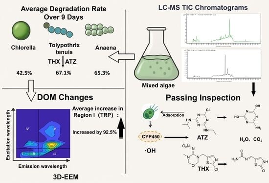
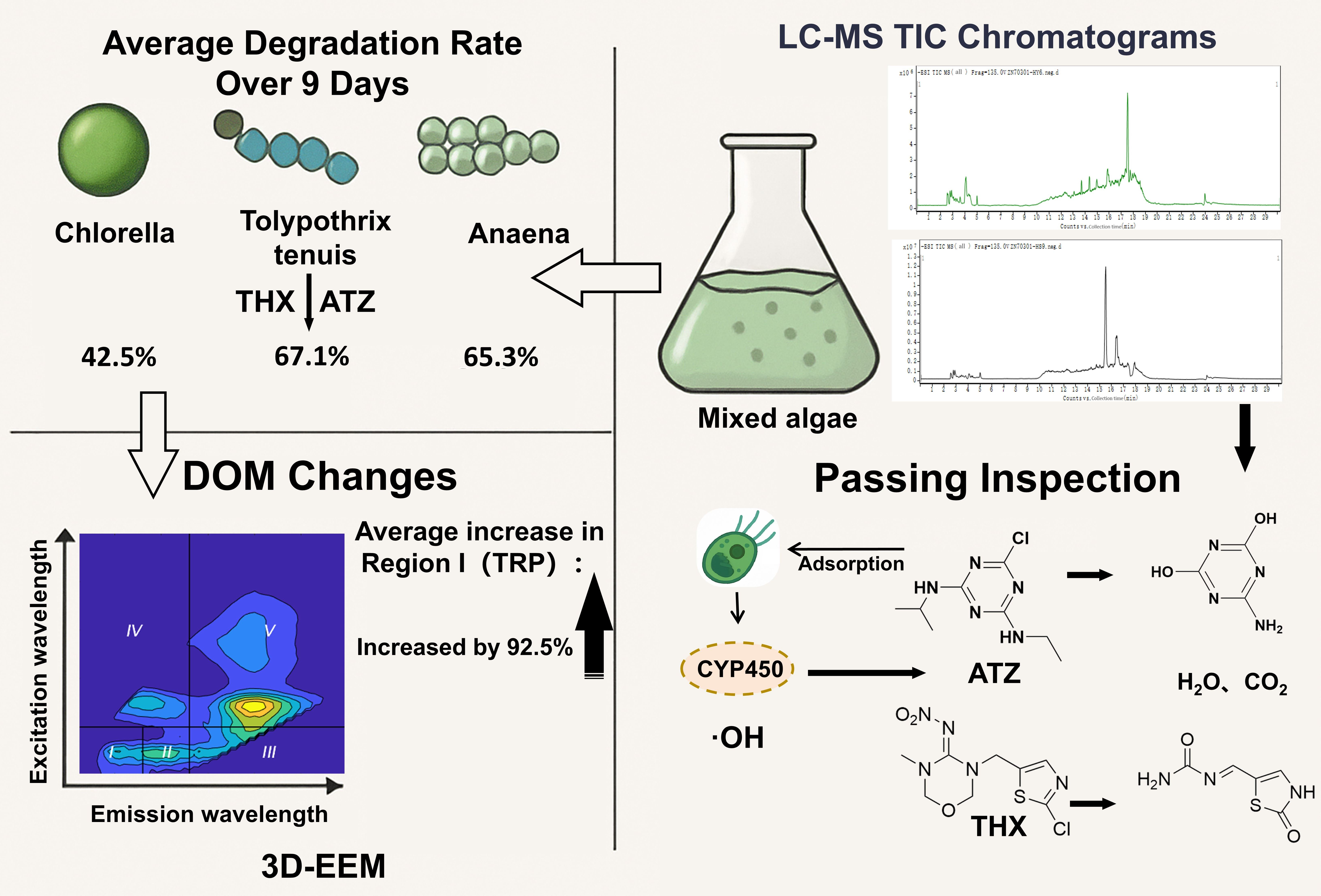
Graphical Abstract

1. Introduction

Approximately 2 million tons of pesticides are used annually in global agricultural production to ensure crop yields [1]. Although pesticides help control pests and boost crop yields, their persistence and mobility enable them to leach into groundwater and surface water. This transport harms both soil and aquatic environments [2,3,4,5]. As two typical pesticides in China, thiamethoxam (THX) and atrazine (ATZ) are primarily applied to crops through soil drenching, foliar spraying, and seed treatment [6,7]. THX, a neonicotinoid insecticide, is widely employed in agricultural practices owing to its high efficacy, selectivity, and persistence [8]. Under different environmental conditions, the half-life of THX ranges from several weeks to months [9], leading to accumulation in the environment with concentrations reaching up to 7.44 mg/kg [10]. The presence of neonicotinoid pesticides in the environment poses risks to soil microorganisms [11], bees [12], aquatic invertebrates [3], birds [13], and even humans [14]. Owing to its stable structure, ATZ is resistant to natural mineralization, resulting in a half-life of up to 700 days [15]. ATZ induces toxic effects and endocrine disruption in animals, leading to developmental defects in fish and amphibians [16,17,18]. Therefore, developing efficient and safe pesticide degradation technologies is critical to ensure food security, promote sustainable agriculture, and mitigate the threats pesticide residues pose to ecosystems and human health [19].
Several emerging pesticide removal technologies have been developed, yet each presents certain limitations. Cold plasma technology achieves pesticide oxidation through direct reactions or reactive oxygen species (ROS)-generated chain reactions, but its widespread application is hindered by high operational costs and complex processes [20,21]. Bimetallic ZIF-derived magnetic carbon materials adsorb neonicotinoid pesticides via pore filling, hydrogen bonding, and Π-Π interactions, yet they risk secondary heavy metal pollution [22]. Regarding the synergistic biodegradation by microalgae, research predominantly focuses on microalgae–bacteria consortia, which can enhance contaminant removal rates by 40% to 90%. However, these systems carry cultivation risks such as ecological invasion and the spread of antibiotic resistance genes. Studies on multi-microalgal consortia, in contrast, remain insufficiently explored [23]. In comparison, biodegradation offers an environmentally sustainable solution by utilizing microorganisms to metabolize pollutants as sources of carbon, nitrogen, and phosphorus, with minimal adverse environmental impact [24]. Among these, microalgae demonstrate significant potential in pollution remediation due to their pesticide tolerance and degradation capacity. Different microalgae exhibit distinct response processes to pesticides, with degradation efficiency closely linked to their physiological characteristics. Their strong environmental adaptability enables them to establish symbiotic relationships with heterotrophic organisms to maintain degradation activity under metabolic stress. For instance, Phaeodactylum tricornutum degrades high concentrations of dichlorvos and glyphosate by utilizing organophosphates in cellular metabolism [25], while Isochrysis galbana effectively removes glyphosate and exhibits enhanced growth and biomass accumulation at concentrations of 25–75 μg/L [26]. A microalgae-based system developed by Avila et al. achieved 74–97% removal rates for pesticides including chlorpyrifos, oxadiazole, and cypermethrin [27]. Microalgae degrade pesticides through multiple mechanisms, including bioaccumulation and biosorption [28], enzymatic degradation [29], and redox reactions [25]. Key enzymes involved in biodegradation comprise hydrolases, oxidoreductases, and transferases [30], which facilitate detoxification through hydroxylation, dehydrogenation, oxidation, and reduction [31]. Additionally, microalgae can contribute to indirect pesticide degradation via ROS generation. However, high pesticide concentrations may inhibit microalgal growth and compromise remediation efficiency because of toxic effects [32]. Association with bacteria enhances the organism’s tolerance to multiple pollutants, including microplastics and heavy metals, as well as its stress resistance under enzyme-inhibiting conditions [33,34,35].
During their growth, development, and reproduction, microalgae synthesize substantial amounts of algal organic matter (AOM), part of which remains attached to the cell surface or is secreted into the surrounding environment as extracellular organic matter (EOM) [36]. Composed primarily of proteins, carbohydrates, and humic acids, these biomacromolecules can effectively mitigate the toxic effects of hazardous chemicals on algal cells [37]. To elucidate the interaction mechanisms between AOM and exogenous pollutants such as antibiotics and pesticides, multiple analytical techniques have been employed for AOM characterization. Among these, fluorescence spectroscopy has been widely adopted in organic matter studies due to its high sensitivity, minimal sample damage, and operational simplicity [38]. In particular, the FRI method enables the division of three-dimensional fluorescence spectra of microalgal aqueous environments into several characteristic regions, with volumetric integration of fluorescence intensity in each region, thereby allowing quantitative characterization of the composition and properties of AOM [5].
Prior studies have explored the degradation performance of various microalgae (particularly Chlorella) towards pesticides. However, there is a scarcity of experiments examining multiple microalgae and multiple pesticides under standardized conditions, and comparative assessments with mixed microalgae systems are notably lacking. This study systematically investigates the degradation efficiency and mechanisms of two pesticides, THX and ATZ, by different microalgae under high-concentration conditions. LC-MS is employed to quantify degradation rates, elucidate transformation pathways, and assess the toxicity evolution of degradation products. Meanwhile, chlorophyll-a content, determined via UV spectrophotometry, is used to evaluate the growth response of microalgae under pesticide stress. Furthermore, the relationships between degradation kinetics and key environmental parameters—including DOM, pH, and EC—are analyzed to examine the feedback effect of microalgal growth on ambient conditions. Furthermore, the environmental effects of the microalgae degradation process were analyzed, including changes in indicators such as EC, pH, and DOM. This aims to screen for superior algal species with enhanced capabilities for pesticide degradation and environmental remediation.

2. Materials and Methods

2.1. Biological Material and Chemical Reagents

The microalgae species used in this study included: (i) Chlorella pyrenoidosa, a green alga (Chlorophyta), from the Chlorellaceae family; (ii) Tolypothrix tenuis Kützing ex, a cyanobacteria, from the Tolypothrichaceae family; and (iii) Anabaena, cyanobacterias, from the Nostocaceae family.
All algal strains were obtained from Inner Mongolia Alge Life Science Co., Ltd. (Ulanqab, China). The microalgae were cultivated in BG-11 medium.
The thiamethoxam (THX) and atrazine (ATZ) standards used in the experiment were sourced from the Agricultural Product Quality Standards Research Center of the Ministry of Agriculture and Rural Affairs.

2.2. Experimental Design

The experiment was conducted in October 2024 at the cultivation laboratory of Inner Mongolia Alge Life Science Co., Ltd. It aimed to investigate how different microalgae degrade typical pesticide residues. The microalgae species used included three monocultures—C. pyrenoidosa, T. tenuis, and Anabaena sp.—along with a mixed algal consortium (2:1:1 ratio of the respective species). The initial chlorophyll-a concentration of microalgae was adjusted to 10 mg/L for all groups using UV spectrophotometry (UV-1780, Shimadzu, Kyoto, Japan). To ensure enzymatic reactions proceeded under mild conditions, the experimental flasks were maintained at 25 °C, pH 7, and a light intensity of 8000 lx, with continuous aeration and twice-daily shaking. All microalgal culture flasks were cleaned, autoclaved, and then used. Purified water was used throughout the experiment, and the cultures were maintained under aseptic conditions [39].
All experimental groups were conducted under illumination with aeration, including: (i) MT (mixed algae degrading thiamethoxam), MA (mixed algae degrading atrazine); (ii) CT (C. pyrenoidosa degrading thiamethoxam), CA (C. pyrenoidosa degrading atrazine); (iii) AT (Anabaena sp. degrading thiamethoxam), AA (Anabaena sp. degrading atrazine); (iv) and TT (T. tenuis degrading thiamethoxam) and TA (T. tenuis degrading atrazine). (v) CP (control groups without microalgae included a light-exposed pesticide control, with air supplied), and (vi) CD (the control group was kept in darkness without microalgae, with air supplied). All experiments were performed in triplicate to minimize experimental error [40]. The pesticide concentration was uniformly set to 2 mg/L in all experiments to highlight the influence of the sole variable—microalgal species—on the degradation efficiency [17].

2.3. Experiment and Analysis

2.3.1. Pesticide Quantification and Degradation Product Analysis

The corresponding atrazine and thiamethoxam standard solutions were added to the culture medium. On days 1, 3, 6, and 9 after addition, 200 mL aliquot samples were collected and filtered through 0.45 μm membranes for pesticide quantification and simultaneous degradation product analysis.
For sample pretreatment, solid phase extraction was performed using HLB cartridges (500 mg/6 mL). The cartridges were first activated with methanol and ultrapure water. The aqueous samples were then loaded at a constant flow rate of 3–5 mL/min for extraction. The extracted samples were analyzed using liquid chromatography-tandem mass spectrometry (Shimadzu triple quadrupole mass spectrometer, LCMS-8050, Kyoto, Japan) for pesticide quantification [41]. The system was equipped with an Athena UHPLC C18 120A chromatographic column (2.1 × 50 mm, 1.8 μm, Shanghai, China). For quality control and data measurement assurance, the recovery of each spiked sample, method detection limit (MDL), method quantification limit (MQL), and parallel samples were analyzed, as shown in Table S2. The detection signal-to-noise ratios (S/N) for thiamethoxam and atrazine were 1.14 and 2.2, respectively.
Structural identification of pesticide metabolic intermediates was performed using liquid chromatography–mass spectrometry (LC-MS) [23]. The liquid chromatography system (Agilent 1260 Infinity II Prime, Waldbronn, Germany) was equipped with a ZORBAX Eclipse Plus C18 column (Agilent Technologies, Santa Clara, CA, USA) maintained at 40 °C. The mobile phase consisted of 0.1% formic acid in water (phase A) and acetonitrile (phase B). Mass spectrometric detection was carried out using an Agilent 6530 Q-TOF (Agilent Technologies, Santa Clara, CA, USA) mass spectrometer with a Dual AJS electrospray ionization (ESI) source. The spray voltage was set at 4.0 kV for both positive and negative ionization modes. Data were acquired in Full MS/dd-MS (data-dependent acquisition) top 10 mode: primary full-scan MS spectra were recorded at a resolution of 70,000 (mass range 200–6000 m/z), followed by fragmentation of the ten most intense precursor ions (resolution 17,500, start mass 50 m/z) using stepped collision energies (HCD) of 15, 30, and 45 eV to obtain comprehensive fragmentation information.

2.3.2. Determination of Microalgal Chlorophyll Content

One hundred milliliters of culture solution was filtered through a 0.45 μm glass fiber filter. The filter membrane was then immersed in 90% acetone for chlorophyll extraction and centrifuged at 4000 rpm for 10 min. Absorbance of the microalgae solution was measured using a Hitachi U-1900 UV-Vis spectrophotometer (Tokyo, Japan) at wavelengths of 750 nm, 664 nm, 647 nm, and 630 nm (A750, A664, A647 and A630), with 90% acetone serving as the blank. The absorbance values at 664 nm, 647 nm, and 630 nm were corrected by subtracting the absorbance at 750 nm to account for turbidity. The concentration of chlorophyll-a was calculated using the following formula [42]:
ρ 1 = 11.85 A 664 1.54 A 647 0.08 A 630
The mass concentration of chlorophyll-a in the sample (mg/L) is calculated according to Formula (2):
ρ = ρ 1 × v V × L
where: ρ: mass concentration of chlorophyll-a in the sample, mg/L; ρ1:mass concentration of chlorophyll-a in the test specimen, μg/L; v: volume of acetone 90%, L; V: volume of water sample, L; L: light path of cuvette, cm. The initial biomass was standardized to a chlorophyll-a concentration of 10 mg/L, as determined by UV spectrophotometry. The initial cell density was adjusted to 3–4 × 10⁵ cells/mL, based on counts obtained using microscopy.

2.3.3. Kinetic Analysis of Pesticide Degradation by Microalgae

The first-order kinetic model is a mathematical representation describing processes in which the reaction rate is directly proportional to the reactant concentration. It is widely applied in fields such as biodegradation, pharmaceutical metabolism, and microbial growth. First-order degradation kinetics was employed to characterize the pesticide degradation profiles, as described by the following equation [43]:
d C t d t = k C t
t 1 2 = L n 2 k
where Ct represents the reactant concentration (μg/L), k denotes the degradation rate constant, and t indicates the reaction time (days).

2.3.4. Three-Dimensional Excitation-Emission Matrix (3D-EEM) Analysis

The 3D-EEM fluorescence spectra of water samples before and after irradiation were recorded using a fluorescence spectrophotometer. The measurement parameters were set as follows: excitation (Ex) wavelength range of 220–450 nm at 5 nm intervals, emission (Em) wavelength range of 250–600 nm at 1 nm intervals, excitation and emission slit widths of 5 nm and 10 nm, respectively. Raman and Rayleigh scattering effects were mathematically eliminated from the obtained spectra. Ultrapure water was used as a blank control for background correction. The processed fluorescence data were subsequently used to determine the relative abundances of different fluorescent components through grating processing [44].
Quantitative analysis of the EEM was performed using the FRI method in MATLAB R2024b. The integration volume (ϕi) for each fluorescence region was calculated, representing the integral of the spectral area and fluorescence intensity within that region. To enable comparison, ϕi was normalized to the dissolved organic carbon (DOC) concentration to account for sample concentration effects and was further normalized to mitigate the shoulder peak effect in overlapping regions, resulting in the standardized integration volume (ϕi,n). Finally, the percentage (Pi,n) of each standardized volume (ϕi,n) relative to the total standardized integration volume (ϕT,n) was computed. The five types of DOM are Aromatic Protein I: Tyrosine-like (TYR), Aromatic Protein II: Tryptophan-like (TRP), Fulvic acid-like (FUL), Microbial byproduct-like (MBP), and Humic acid-like (HS). The specific partitioning is shown in Figure S1 [45].
ϕ i , n = M F i ϕ i = M F i e x e m I λ e x λ e m d λ e x λ e m
where ϕi,n is the integrated reference volume (au·nm2) of fluorescence region i; ϕi is the integrated volume (au·nm) of region i; Iex, λem) represents the fluorescence intensity (au) at excitation wavelength λex and emission wavelength λem; and MFi is the multiplication factor, defined as the reciprocal of the fractional integrated area of fluorescence region i relative to the total integrated area of all regions.

2.3.5. Data Analysis

The experimental data were plotted and processed using Origin 2024 software. In IBM SPSS Statistics 27, a two-way ANOVA was performed to evaluate the statistical significance of degradation differences, followed by Duncan’s multiple-range test for post hoc comparisons. The significance level was set at p < 0.05 [36].

3. Results

3.1. Differences in Pesticide Tolerance and Degradation Performance Among Microalgae

3.1.1. Variations in Chlorophyll-a Content of Microalgae Under Pesticide Stress

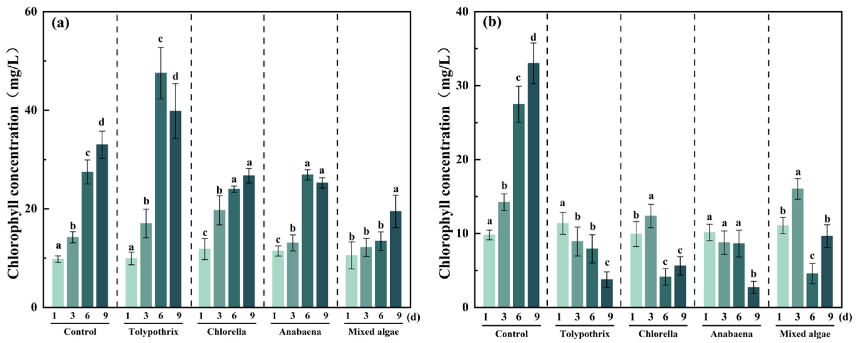
Chlorophyll content, an indicator of photosynthetic efficiency and growth status, was measured spectrophotometrically over time under various different pesticide treatments [46]. The growth patterns of microalgae varied significantly depending on the pesticide (p < 0.05; Figure 1). Under THX exposure, all four microalgal cultures exhibited an overall increase in chlorophyll content, with an average rise of 159%. Among them, T. tenuis. exhibited the largest increase (300%) (Figure 1a), while the mixed culture exhibited the smallest (84%). In contrast, ATZ treatment reduced chlorophyll levels in all groups by an average of 49% (Figure 1b). The most pronounced inhibition was observed in C. pyrenoidosa (73% decrease), whereas the mixed culture was the least affected (12.9% decrease). By day 9, the chlorophyll level in the THX group was 5.12 times that of the ATZ group. These results indicate that THX was degraded by the three microalgae and concurrently promoted growth and stress resistance. Conversely, ATZ inhibited microalgal growth, with C. pyrenoidosa being the most susceptible and the mixed culture demonstrating relatively greater tolerance.

3.1.2. Analysis of Pesticide Degradation Based on First-Order Kinetic Modeling

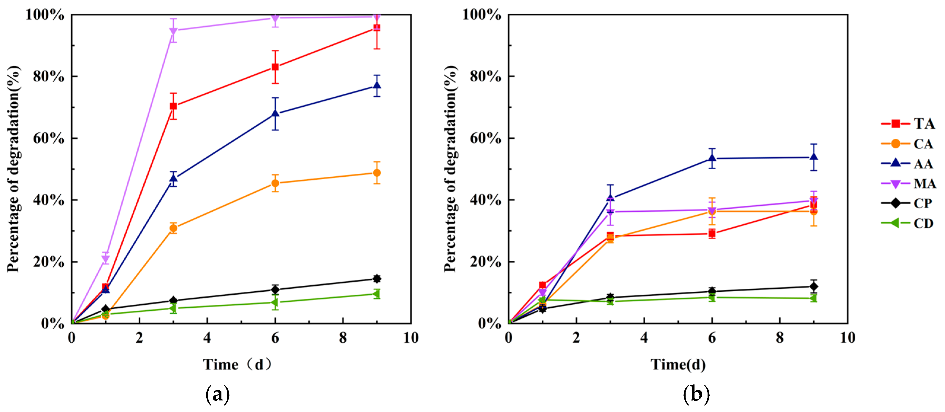
Different microalgal species and their consortium exhibited marked variation in their capacity to degrade ATZ and THX over time (Figure 2). The TT group was the most effective in degrading THX, achieving a removal rate of 95.57% after 9 days. In contrast, AA group was the most effective in degrading ATZ, with a 53.79% removal rate over the same period. CA group and TA group showed comparable efficiency in degrading ATZ, with removal rates of 36.3% and 38.4%, respectively. In contrast, the microalgal consortium exhibited excellent performance for both pesticides, achieving removal rates of 99.3% for THX and 39.8% for ATZ within 9 days. Degradation rates in all groups peaked on day 3 and then gradually declined (Figure S1). In the CP group, the degradation rates of ATZ and THX reached 12% and 14.5%, respectively, over 9 days. The average degradation rates under light were 1.12 and 1.54 times higher than those in the dark for ATZ and THX, respectively, indicating a more pronounced light-enhanced effect on THX degradation. Although the process involved air sparging, the low volatility and high water solubility of the pesticides suggest that the results reasonably reflect realistic conditions [47]. In the CD group, both pesticides showed minimal concentration changes, with degradation rates of only 9.61% and 8.16% after 9 days, respectively.
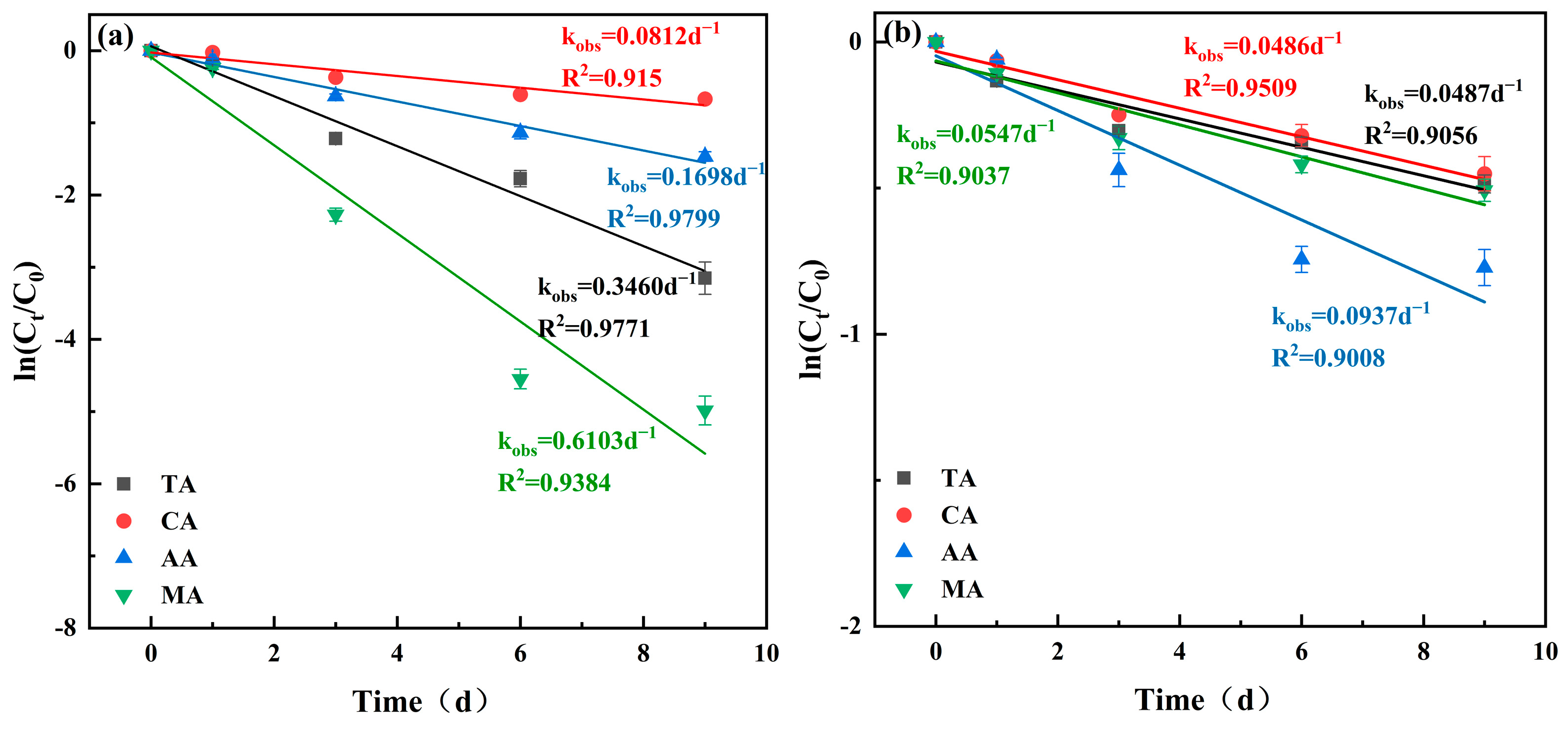
Comparison of the fitting results using first-order and second-order kinetic models shows that the degradation processes of both ATZ and THX are well described by a pseudo-first-order kinetic model (Figure 3) [43]. The high coefficients of determination (R2 ≥ 0.9) indicated a good fit for the model, with detailed results provided in Table S1. Significant interspecific differences (p < 0.05) were observed in the degradation rate constants. The half-lives of ATZ and THX in the microalgal treatments were 11.4 and 4.9 days, respectively, which were considerably shorter than those under pesticide-only illuminated conditions (40.1 and 51 days) and dark conditions (114 and 80 days). Among the single-species treatments, TT group was the most effective in degrading THX, exhibiting the shortest half-life (2 days) and the highest rate constant (0.346 d−1), followed by AT group (k = 0.1698 d−1, t1/2 = 4.08 days). CT group presented a longer half-life for THX (8.53 days, k = 0.0812 d−1). For ATZ degradation, Anabaena sp. was the most effective (k = 0.0937 d−1, t1/2 = 7.4 days), while C. pyrenoidosa and TA group exhibited similar, lower efficiencies (k ≈ 0.0486 d−1, t1/2 ≈ 14.25 days). The MT group demonstrated the best performance for THX degradation (t1/2 = 1.14 days, k = 0.613 d−1), outperforming all single species. For ATZ, the consortium (t1/2 = 12.67 days, k = 0.0547 d−1) also displayed a better degradation capability than CA group and TA group.

3.1.3. Analysis of Variations in DOM Concentration and Composition in Microalgal Environments

The FRI method was applied to the EEMs to enable a semi-quantitative comparison of fluorescent signals in the microalgal water samples [45,48]. The integrated volume of each region was calculated to determine its relative contribution (Figures S2 and S3). This approach facilitated the analysis of variation patterns in different DOM components and their relationships with the pesticide degradation rates.
During THX degradation, the TRP component constituted the highest average proportion (34.7%) of DOM across all four microalgal groups. The overall DOM content exhibited an increasing trend, with an average rise of 92.5%. T. tenuis exhibited the most significant increase (215%; Figure S2). Specifically, the TYR and TRP components increased markedly by averages of 186% and 196%, respectively, whereas HS showed the smallest average increase (28%). During ATZ degradation, the MBP component was the most dominant (30%), while TYR was the least (8.8%). The total DOM content increased over time in the consortium and T. tenuis groups, with the latter showing the highest rise (109%). DOM content decreased in the Anabaena sp. group by 12.6% and in the C. pyrenoidosa group by 76.5%. Analysis of the control and treatment groups revealed substantial DOM concentration shifts associated with microalgal growth. By day 9 in the THX environment, average DOM content had increased 43% relative to day 1, accompanied by a raised upper limit of fluorescence intensity. This suggests that the presence of THX has a relatively weak inhibitory effect on AOM release from various microalgae, indicating higher tolerance to THX [49]. Conversely, in the ATZ environment, the DOM content decreased by an average of 67.1% (Figure S3), with concentrations distributed within a lower, more constrained range (Figure S4). This indicates that ATZ generally and significantly inhibits AOM release, likely because its addition restricts microalgal growth, reproduction, and metabolic activity [50], thereby reducing AOM excretion and ultimately lowering the ambient DOM level. Exposure to ATZ induces cell death and lysis. Substances released from the cells are rapidly degraded, thereby reducing DOM levels [51].

3.2. Influence of Environmental Factors on the Degradation Rate

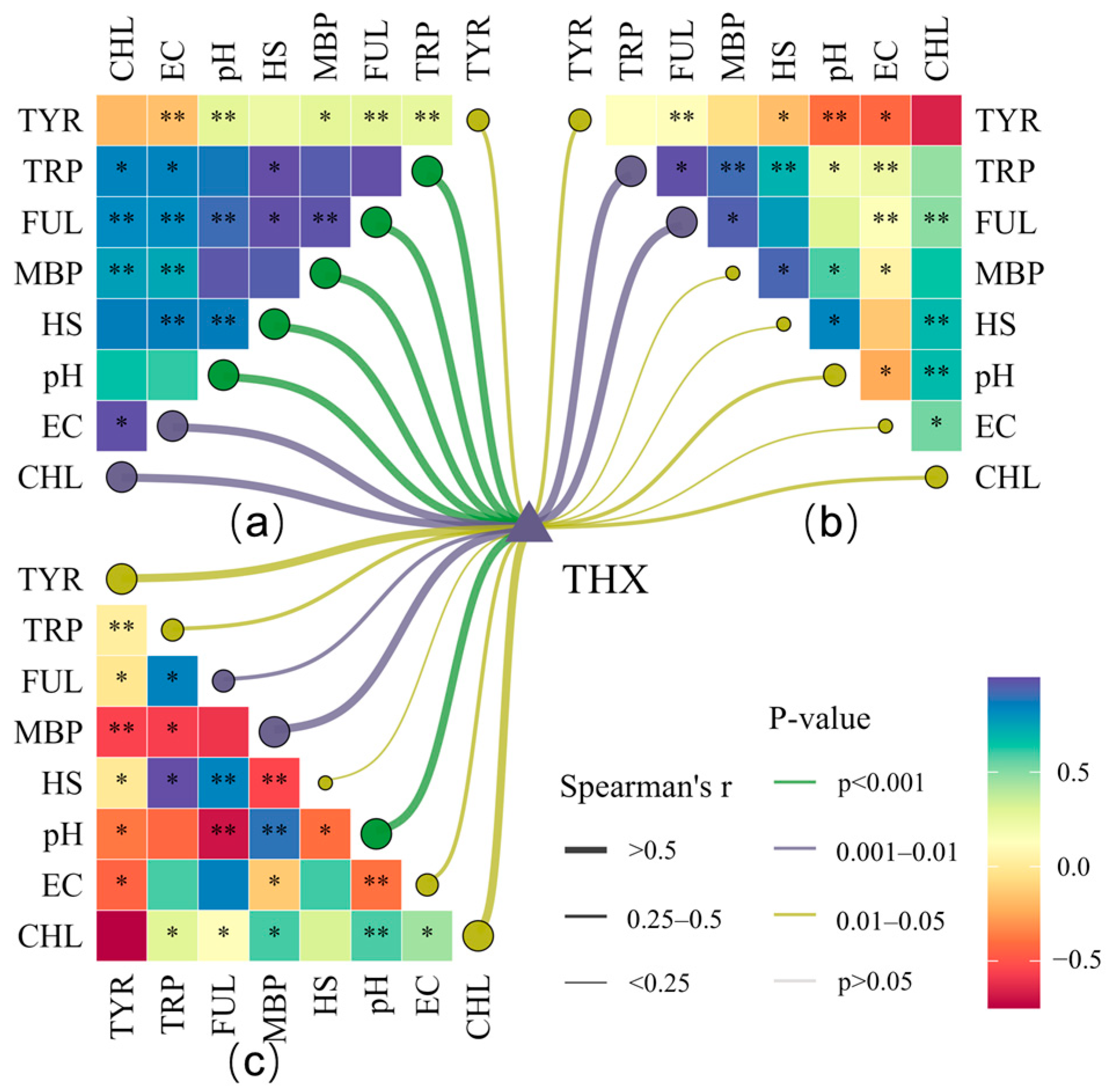
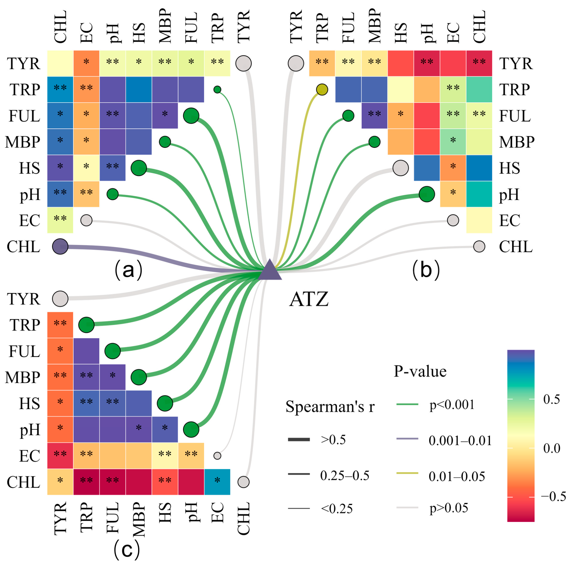
To identify potential associations among DOM composition, microalgal growth status, and pesticide degradation rate, we performed an exploratory correlation analysis using Spearman‘s rank correlation coefficient (Figure 4 and Figure 5). In the heatmaps, blue and red denote significant positive (r > 0.5) and negative (r < −0.5) correlations, respectively. In Anabaena sp., four DOM components—TRP, FUL, MBP, and HS—exhibited a significant positive correlation with the THX degradation rate (r > 0.9, p < 0.01; Figure 5a). Conversely, in T. tenuis, TRP and FUL were significantly negatively correlated with the THX degradation rate (r < −0.6, p < 0.01). In C. pyrenoidosa, the levels of MBP, along with pH and CHL, were positively associated with THX degradation (r > 0.8, p < 0.01). Furthermore, THX degradation efficiency co-varied with CHL content across all three microalgae, indicating an association where higher cellular activity co-occurred with a higher degradation rate. Among the three microalgae, only Anabaena sp. exhibited a significant positive correlation between its degradation rate and EC (r > 0.9, p < 0.01). Additionally, in this species, all DOM components except TYR were positively correlated with both EC and pH.
Anabaena sp. exhibited negative correlations between its ATZ degradation rate and the concentrations of all five DOM components, pH, and CHL (Figure 5a). In T. tenuis, the ATZ degradation rate exhibited a significant negative correlation with TRP and FUL (Figure 5b), but a significant positive correlation with pH (r = 0.81, p < 0.001). Conversely, in C. pyrenoidosa, the ATZ degradation rate presented significant positive correlations with TRP, FUL, MBP, HS, and pH (r > 0.9, p < 0.01), while correlating negatively with CHL and TYR (Figure 5c). Notably, a significant positive correlation was observed between TRP and chlorophyll content. As a key component of DOM, fluctuations in TRP substances may reflect changes in overall microbial metabolic activity. Therefore, its observed co–variation with chlorophyll-a could support the hypothesis that the cellular metabolic state—which potentially regulates the activity of relevant catabolic enzyme systems—varies in concert with biomass, thereby influencing degradation efficiency [52].

3.3. Elucidation of the Microalgal Pesticide Degradation Pathway

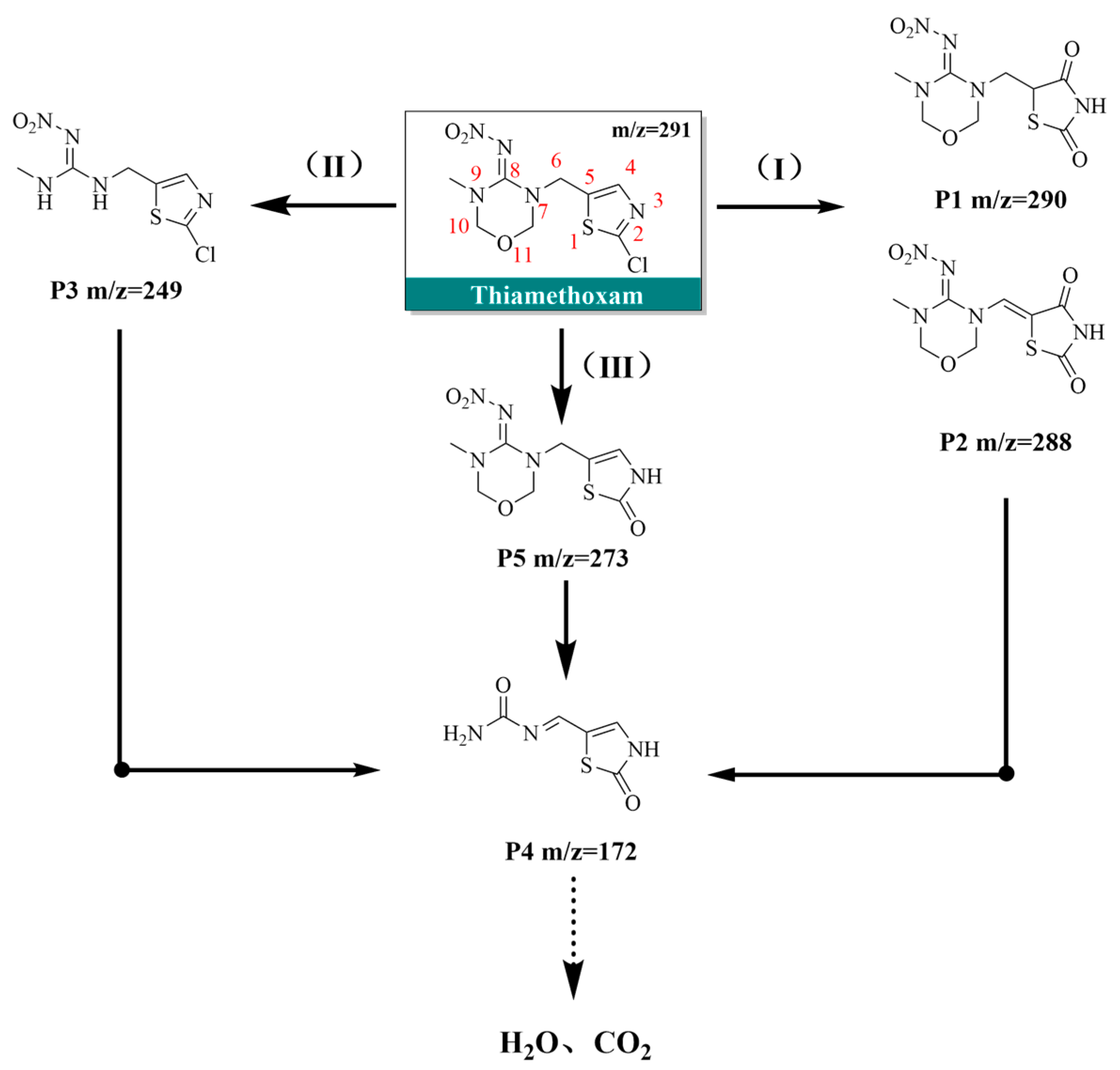
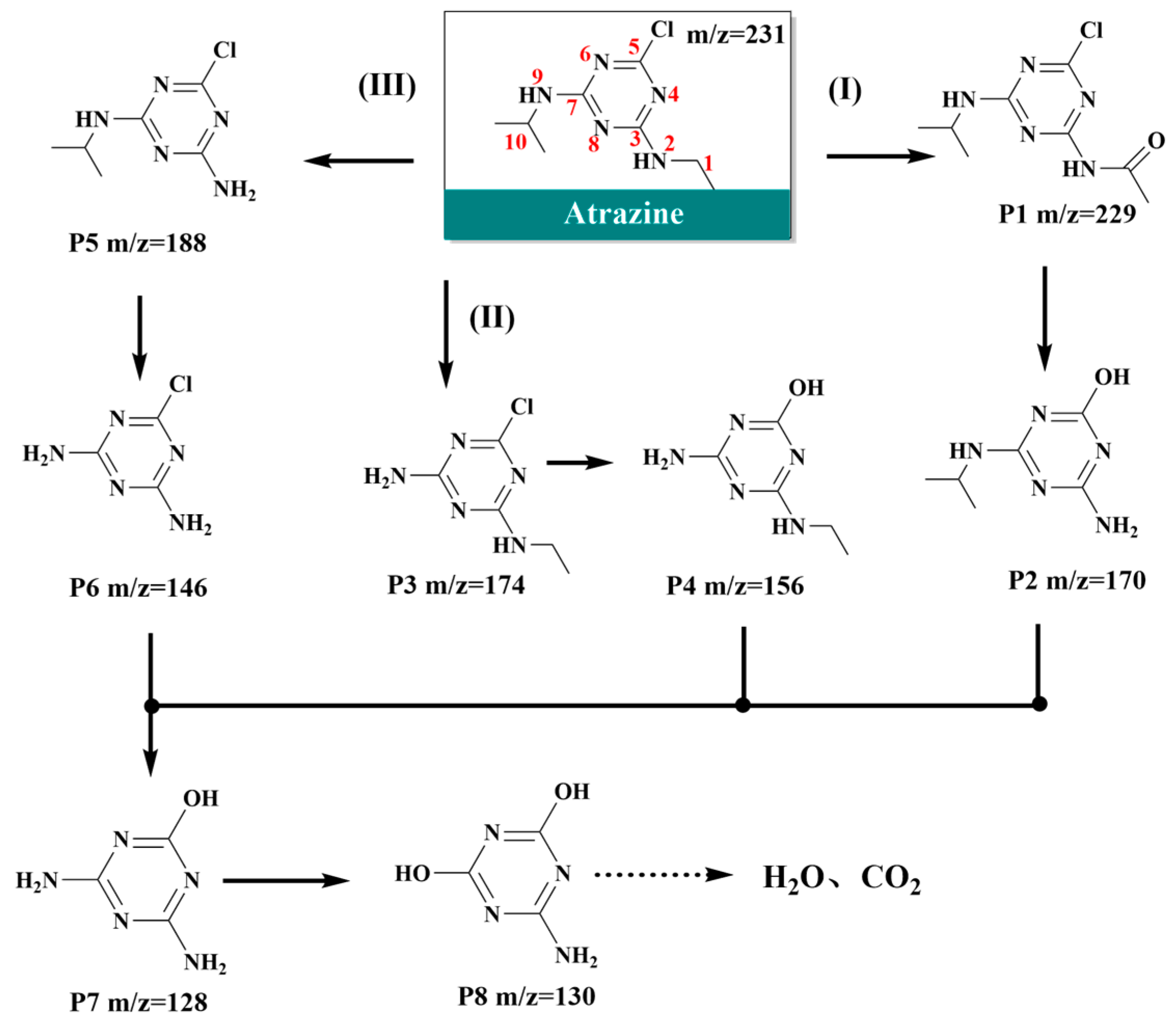
LC-MS/MS analysis identified characteristic metabolites for both ATZ and THX during degradation (Tables S4 and S5). The dynamic changes in these metabolites revealed the primary products and reaction types involved [53], with the proposed structural changes and degradation pathways illustrated in Figure 6 and Figure 7. As shown in Figure 6, pathway (I) of THX is initiated by the substitution of the chlorine atom at the 2-position and a carboxylation reaction, leading to the formation of two intermediate products, P1 and P2. This process is likely catalyzed by the intracellular enzyme CYP450 [23]. These intermediates then undergo oxidative cleavage at the carbonyl group (position 4), the carbon–nitrogen double bond (position 8), and the carbon–nitrogen single bond (position 9), facilitated by enzymes and hydroxyl radicals. This is followed by ring-opening reactions at the nitrogen atoms (positions 7 and 9), leading to the formation of product P4 [14]. In Pathway (II), the chlorine atom (position 2) and the nitroimino group (position 8) may have been substituted via carboxylation reactions, likely catalyzed by ROS and enzymes, concurrent with ring-opening at positions 7 and 9 to form P4 [54]. Pathway (III) is derived from Pathway (II), differing in the sequence of functional group substitution and ring-opening. Ultimately, product P4 is mineralized to water and carbon dioxide [55].
Key reactions in ATZ degradation included carboxylation, demethylation, and hydroxylation [56]. In Pathway (I), carboxylation at the 1-position—potentially catalyzed by enzyme systems such as CYP450 or peroxidases—yields product P1 [57]. Subsequently, the amide bond is cleaved by ·OH and phospholipases (Figure 7), leading to decarboxylation and the formation of P2 [58]. Pathway (II) proceeds via enzymatic loss of the isopropyl group at the 9-position and substitution of the chlorine atom at the 5-position, sequentially generating P3 (deisopropylatrazine) and P4. Finally, oxidative removal of the ethyl group at the 2-position produces P7.
Pathway (III) represents an alternative sequence for the substitution of the ethyl, isopropyl, and chlorine groups. The amino group on the final product P7 undergoes hydroxylation, likely mediated by ROS and enzymes, to form P8, which is ultimately mineralized to water and carbon dioxide. Furthermore, product P8 lacks active groups such as chlorine atoms, ethylamino groups and isopropylamino groups, which causes it to lose the ability to efficiently bind to the target of the plant PS II, and its toxicity has significantly decreased [59]. In summary, hydroxyl radicals (·OH), primarily generated in the chloroplasts and mitochondria of growing microalgae [60], along with enzymatic reactions, may play crucial roles in pesticide degradation. CYP450, in particular, are considered key agents potentially facilitating hydroxylation and carboxylation [61].

4. Discussion

4.1. Microalgal Response and Degradation Mechanisms to Pesticides

In this study, C. pyrenoidosa achieved 9-day degradation rates of 48.8% for THX and 36.3% for ATZ, which were lower than the previously reported 87–96% removal for a mixed pesticide solution at 0.1 mg/L [28]. This discrepancy is likely owing to the higher concentrations used herein, as ATZ removal efficiency by microalgae has been shown to decrease with increasing concentration [29]. In that study, the primary removal processes was bioaccumulation. At elevated concentrations, enhanced intracellular pesticide accumulation exacerbates cytotoxicity, thereby impairing the degradation of both ATZ and THX. Mechanistically, high ATZ concentrations inhibit photosynthetic electron transport in PSII. This inhibition can lead to ROS accumulation, which may exceed the quenching capacity of photoprotective compounds such as carotenoids, xanthophylls, and tocopherols. This results in severe oxidative damage to proteins, lipids, and pigments, ultimately causing membrane disruption and cell death [62]. Pesticides generally impact microalgae by disrupting photosynthesis, thereby affecting growth and morphology [52]. Under laboratory conditions, chlorophyll content exhibits a strong linear correlation with cell density, validating its use as a reliable biomarker for assessing biomass fluctuations under pesticide stress [63].
In this study, Anabaena sp. demonstrated the highest degradation efficiency for ATZ (53.79% at 2 mg/L over 9 days), outperforming Chlamydomonas mexicana (14–36%) reported by Kabra et al. [40]. The decline in chlorophyll content between days 6 and 9 may be attributed to the depletion of nutrients. NH4-N concentration and COD/TN ratio directly affect the growth rate of microalgae, which would reduce their tolerance and degradation capacity [64]. Despite a decrease in DOM concentration under ATZ stress, Anabaena sp. exhibited superior resistance and degradation performance among the single-species treatments. This resilience may stem from its classification as Cyanobacteria; its prokaryotic structure lacks typical pesticide targets such as the chloroplast membrane and mitochondrial electron transport chain found in eukaryotic algae, thereby conferring greater tolerance to ATZ. This aligns with previous observations of cyanobacterial tolerance to, and degradation of, high concentrations of organochlorine pesticides [65]. Thus, Anabaena sp. shows greater potential for survival and degradation under high ATZ concentrations compared to T. tenuis and C. pyrenoidosa. Meanwhile, T. tenuis exhibited robust degradation and growth for both pesticides, achieving a 95.7% removal rate for THX. The distinct physiological responses to ATZ among the three microalgae may result from differences in cell wall and membrane lipid composition, which influence pesticide uptake rates and subsequent physiological impacts [29]. Furthermore, the higher octanol-water partition coefficient (log Kow) of ATZ (2.5–2.7) compared to THX (−0.13–0.26) indicates greater lipophilicity, facilitating easier cellular entry and posing a heightened threat to microalgae at equivalent concentrations.

4.2. Effect of Microalgal Synergism on the Degradation Process

Synergistic degradation by the microalgal consortium significantly enhanced THX removal efficiency, exceeding the average rate of single species by 34.5%. Furthermore, during ATZ degradation, the consortium exhibited a distinct growth pattern in the later phase, characterized by a notable rebound in chlorophyll content. This enhancement is likely attributable to the diversification of metabolic pathways, including enzymatic degradation, oxidation, reduction, and hydrolysis. Hydrolytic enzymes, for instance, can cleave ester or amide bonds in pesticide molecules, facilitating further breakdown [66]. Pesticide adsorption by microalgae, often a passive process independent of algal activity [67], is augmented in consortia through the inclusion of cells with varying sizes. Surface functional groups (e.g., -OH, -NH2, -PO32−, -COO) on different microalgae further aid in sequestering positively charged toxic compounds [68]. Synergistic systems, such as Anabaena-Chlorella, demonstrate accelerated growth and photosynthesis, boosting overall cellular metabolism [69] and mitigating pesticide toxicity over time. Consortia like Chlorococcum-Scenedesmus can not only transform endosulfan to less toxic endosulfan sulfate but also degrade organophosphates and mineralize them into bioavailable phosphorus [66]. In summary, multi-algal consortia hold a clear advantage over single species, primarily through interspecific metabolic complementarity that boosts degradation efficiency, coordinated antioxidant defense, and rapid mineralization of intermediates, collectively forming a more robust and efficient pesticide degradation network. While the synergistic processes among different microalgae species remain unclear, insights can be drawn from previously studied algae–bacteria consortia to enhance degradation efficiency. The immobilization of microalgae using polyvinyl alcohol-sodium alginate (PVA-SA) and polyvinyl alcohol-sodium sulfonate-polybutadiene (PVA-SA-PDI) carriers leverages the inherent adsorption capacity of PDI to concentrate pesticides and pharmaceuticals. This approach also helps retain microalgal and bacterial cells, provides a protective barrier against external toxicity, and reduces damage to the photosynthetic system, thereby alleviating oxidative stress [70,71]. Furthermore, incorporating carbon-coated magnetic γ-Fe2O3 nanoparticles into a multi-microalgae system enables their spontaneous adsorption onto the surfaces of various microalgae species [72]. Under illumination, these nanoparticles generate abundant photogenerated electrons, which subsequently enhance the activity of PSII and stimulate the synthesis of ATP and NADPH. This process activates relevant intracellular enzymes, such as carboxylesterases, to facilitate the hydrolysis of the target pesticides [73].

4.3. Response and Feedback of Microalgae-Mediated Degradation to Environmental Factor Variations

In the THX degradation groups, chlorophyll content and DOM concentration in all four microalgae showed a clear increasing trend (Figure 5), suggesting that pesticide exposure may be associated with enhanced microalgal metabolic activity [74]. External stress can stimulate microalgae to produce and release AOM into the environment. This AOM can bind with pesticide stressors, reducing their bioavailability and toxicity [75,76]. During THX degradation, the cleavage of the nitroimino group and carbon–nitrogen double bond generates NO3/NO2 and NH4+, which may serve as nitrogen sources to promote microalgal growth [77], thereby accelerating metabolic activity and AOM release. Nitrite has been identified as a key photosensitizer for ·OH generation, a process that can further enhance pesticide photodegradation [78]. This aligns with findings that C. pyrenoidosa increased its lipid and biomass yield by over 10% under high concentrations of glyphosate and imidacloprid [79,80]. Notably, due to its high water solubility, THX is prone to leaching from farmland [81] and persists in the environment (half-life: 7–353 days) [9]. Its entry into water bodies can stimulate massive DOM production by microalgae. Upon degradation, this DOM can become bioavailable to bacteria and phytoplankton, potentially triggering eutrophication [82]. As shown in Figure 4 and Figure 5, DOM components such as TRP, HS, and FUL displayed significant positive correlations with the degradation rates of ATZ and THX. This finding supports the proposed crucial role of DOM in the photodegradation of organic compounds [83]. The structure and composition of DOM can influence ·OH generation [84]. As an important radical source, humic substances in DOM can absorb sunlight (300–500 nm), reach an excited state, and generate ROS that degrade THX and ATZ [85]. This is further corroborated by T. tenuis, which achieved the highest THX degradation rate (95.7%) among single-species groups and also exhibited the highest total DOM fluorescence intensity on day 9.

5. Conclusions

This laboratory-scale study investigated the degradation characteristics of C. pyrenoidosa, T. tenuis, and Anabaena sp. under high concentrations of ATZ and THX. The results revealed significant interspecific differences in degradation efficiency and growth kinetics. T. tenuis was most effective in degrading THX, whereas Anabaena sp. excelled at ATZ degradation. The microalgal consortium demonstrated a notable synergistic effect. The degradation process may involve dual pathways: CYP450-catalyzed reactions and ·OH attack, which likely lead to dealkylation/hydroxylation and C-N bond cleavage, respectively, and are proposed to culminate in complete mineralization to CO2 and H2O. Fluorescence regional integration analysis showed that THX stress nearly doubled the total DOM, whereas ATZ exposure reduced DOM by about two-thirds. This reveals the differential effects of the two pollutants on algal metabolism and highlights the dual role of DOM in enhancing tolerance and facilitating degradation processes. A promising direction for future research lies in integrating microalgal genetic engineering with polymer immobilization technologies to bolster stress resistance, thereby enabling the development of highly efficient, stable, and easily recoverable symbiotic systems. These findings establish an experimental basis for developing microalgae-based bioremediation technologies targeting pesticide contamination.

Supplementary Materials

The following supporting information can be downloaded at: https://www.mdpi.com/article/10.3390/w18030304/s1, Table S1: EEM Region Division and Classification. Table S2: Method detection limit (MDL), method quantification limit (MQL). Table S3: First-order kinetic parameters for the degradation of atrazine and thiamethoxam by microalgae. Table S4: Intermediate products from thiamethoxam degradation. Table S5: Intermediate products from atrazine degradation. Figure S1: Pesticide degradation rate over time (a) degradation rate of thiamethoxam over time (b) degradation rate of atrazine over time. Figure S2: Regional integration plot of DOM in thiamethoxam by microalgae over time. Figure S3: Regional integration plot of DOM in atrazine by microalgae over time. Figure S4: DOM concentration distribution trends in different microalgae.

Author Contributions

Conceptualization, Y.W., Q.M., F.Y., H.L., W.F. and P.D.; methodology, Y.W.; software, Y.W. and F.Y.; validation, Y.L. and Y.W.; formal analysis, Z.F., T.P.; investigation, Y.W.; resources, Q.M., F.Y., H.L., W.F. and P.D.; data curation, Y.W.; writing—original draft preparation, Y.W.; writing—review and editing, Y.W. and F.Y.; visualization, W.F.; supervision, Y.L.; project administration, Q.M., F.Y. and H.L.; funding acquisition, P.D. and F.Y. All authors have read and agreed to the published version of the manuscript.

Funding

This research was funded by the National Natural Science Foundation of China Joint Fund for Research on Yellow River Water (U2443210), the National Key Research and Development Program Project (2024YFD1501004) and Key Research and Development and Achievement Transformation Plan Project of the Autonomous Region for 2025 (2025YFHH0145).

Data Availability Statement

The original contributions presented in this study are included in the article and Supplementary Materials. Further inquiries can be directed to the corresponding author.

Conflicts of Interest

Author Pengcheng Duan is employed by Inner Mongolia Alge Life Science Co., Ltd. The remaining authors declare that the research was conducted in the absence of any commercial or financial relationships that could be construed as a potential conflict of interest.

Abbreviations

The following abbreviations are used in this manuscript:
LC-MSLiquid chromatography–mass spectrometry
3D-EEMThree-dimensional excitation-emission matrix
DOMDissolved organic matter
THXThiamethoxam
ATZAtrazine
AOMAlgal organic matter
EOMExtracellular organic matter
MT Mixed algae degrading thiamethoxam
MA Mixed algae degrading atrazine
CTC. pyrenoidosa degrading thiamethoxam
CA C. pyrenoidosa degrading atrazine
AT Anabaena sp. degrading thiamethoxam
AA Anabaena sp. degrading atrazine
TTT. tenuis degrading thiamethoxam
TAT. tenuis degrading atrazine
CPControl groups without microalgae included a light-exposed pesticide control, with air supplied
CDThe control group was kept in darkness without microalgae, with air supplied
TYRAromatic protein I: tyrosine-like
TRPAromatic protein II: tryptophan-like
FULFulvic acid-like
MBPMicrobial byproduct-like
HSHumic acid-like
ROSReactive oxygen species
CYP450Cytochrome P450 enzymes

References

  1. Tang, F.H.M.; Wyckhuys, K.A.G.; Li, Z.; Maggi, F.; Silva, V. Transboundary impacts of pesticide use in food production. Nat. Rev. Earth Environ. 2025, 6, 383–400. [Google Scholar] [CrossRef] [Scilit]
  2. Moreno, M.T.; Rodríguez-Amaro, R.; Mellado, J.M.R.; Mayén, M.; Guardeño, F.J. Imidazolinone and triazine herbicides in soils in relation to the complexes formed with Cu(II) ions. Comptes Rendus Chim. 2018, 21, 884–889. [Google Scholar] [CrossRef] [Scilit]
  3. Morrissey, C.A.; Mineau, P.; Devries, J.H.; Sanchez-Bayo, F.; Liess, M.; Cavallaro, M.C.; Liber, K. Neonicotinoid contamination of global surface waters and associated risk to aquatic invertebrates: A review. Environ. Int. 2015, 74, 291–303. [Google Scholar] [CrossRef] [Scilit]
  4. Anderson, J.C.; Dubetz, C.; Palace, V.P. Neonicotinoids in the Canadian aquatic environment: A literature review on current use products with a focus on fate, exposure, and biological effects. Sci. Total Environ. 2015, 505, 409–422. [Google Scholar] [CrossRef] [Scilit]
  5. Chen, L.; Hu, X.; Yang, Y.; Jiang, C.; Bian, C.; Liu, C.; Cai, T. Degradation of atrazine and structurally related s-triazine herbicides in soils by ferrous-activated persulfate: Kinetics, mechanisms and soil-types effects. Chem. Eng. J. 2018, 351, 523–531. [Google Scholar] [CrossRef] [Scilit]
  6. Jeschke, P.; Nauen, R.; Schindler, M.; Elbert, A. Overview of the status and global strategy for neonicotinoids. J. Agric. Food Chem. 2011, 59, 2897–2908. [Google Scholar] [CrossRef] [Scilit]
  7. Rajendran, K.; Pujari, L.; Ethiraj, K. Biodegradation and Bioremediation of S-Triazine Herbicides. In Environmental Biotechnology; Gothandam, K.M., Ranjan, S., Dasgupta, N., Lichtfouse, E., Eds.; Springer International Publishing: Cham, Switzerland, 2021; Volume 3, pp. 31–54. [Google Scholar]
  8. Wang, X.; Liu, Q. Spatial and Temporal Distribution Characteristics of Triazine Herbicides in Typical Agricultural Regions of Liaoning, China. Bull. Environ. Contam. Toxicol. 2020, 105, 899–905. [Google Scholar] [CrossRef] [Scilit]
  9. Goulson, D. REVIEW: An overview of the environmental risks posed by neonicotinoid insecticides. J. Appl. Ecol. 2013, 50, 977–987. [Google Scholar] [CrossRef] [Scilit]
  10. Zhang, P.; He, M.; Wei, Y.; Zhao, Y.; Mu, W.; Liu, F. Comparative soil distribution and dissipation of phoxim and thiamethoxam and their efficacy in controlling Bradysia odoriphaga Yang and Zhang in Chinese chive ecosystems. Crop Prot. 2016, 90, 1–8. [Google Scholar] [CrossRef] [Scilit]
  11. Wu, C.; Wang, Z.; Ma, Y.; Luo, J.; Gao, X.; Ning, J.; She, D. Influence of the neonicotinoid insecticide thiamethoxam on soil bacterial community composition and metabolic function. J. Hazard. Mater. 2021, 405, 124275. [Google Scholar] [CrossRef] [Scilit] [PubMed]
  12. Orčić, S.M.; Čelić, T.V.; Purać, J.S.; Vukašinović, E.L.; Kojić, D.K. Acute toxicity of sublethal concentrations of thiacloprid and clothianidin to immune response and oxidative status of honey bees. Apidologie 2022, 53, 50. [Google Scholar] [CrossRef] [Scilit]
  13. Hallmann, C.A.; Foppen, R.P.; van Turnhout, C.A.; de Kroon, H.; Jongejans, E. Declines in insectivorous birds are associated with high neonicotinoid concentrations. Nature 2014, 511, 341–343. [Google Scholar] [CrossRef] [Scilit]
  14. Yao, Y.; Huang, C.; Yang, Y.; Li, M.; Ren, B. Electrochemical removal of thiamethoxam using three-dimensional porous PbO2-CeO2 composite electrode: Electrode characterization, operational parameters optimization and degradation pathways. Chem. Eng. J. 2018, 350, 960–970. [Google Scholar] [CrossRef] [Scilit]
  15. Comber, S.D.W. Abiotic persistence of atrazine and simazine in water. Pestic. Sci. 1999, 55, 696–702. [Google Scholar] [CrossRef]
  16. Horzmann, K.A.; Lin, L.F.; Taslakjian, B.; Yuan, C.; Freeman, J.L. Embryonic atrazine exposure and later in life behavioral and brain transcriptomic, epigenetic, and pathological alterations in adult male zebrafish. Cell Biol. Toxicol. 2021, 37, 421–439. [Google Scholar] [CrossRef] [Scilit]
  17. Hou, X.; Wang, X.; Ou, Y.; Yan, L.; Liu, H.; Li, X.; Shang, M. Response of acetochlor degradation and bacterial community in black soil to the application of vermicompost. Biol. Fertil. Soils 2025, 61, 13–26. [Google Scholar] [CrossRef] [Scilit]
  18. Ali, S.; Ullah, M.I.; Sajjad, A.; Shakeel, Q.; Hussain, A. Environmental and Health Effects of Pesticide Residues. In Sustainable Agriculture Reviews 48; Inamuddin, Ahamed, M.I., Lichtfouse, E., Eds.; Springer: Cham, Switzerland, 2021; pp. 199–219. [Google Scholar]
  19. Dagnew, M.; Xue, Q.; Zhang, J.; Wang, Z.; Zhou, A.; Li, M.; Zhao, C. A Review of Various Advanced Oxidation Techniques for Pesticide Degradation for Practical Application in Aqueous Environments. Sustainability 2025, 17, 4710. [Google Scholar] [CrossRef] [Scilit]
  20. Kumar, A.; Škoro, N.; Gernjak, W.; Puač, N. Cold atmospheric plasma technology for removal of organic micropollutants from wastewater—A review. Eur. Phys. J. D 2021, 75, 283. [Google Scholar] [CrossRef] [Scilit]
  21. Sojithamporn, P.; Leksakul, K.; Sawangrat, C.; Charoenchai, N.; Boonyawan, D. Degradation of Pesticide Residues in Water, Soil, and Food Products via Cold Plasma Technology. Foods 2023, 12, 4386. [Google Scholar] [CrossRef] [Scilit]
  22. Bi, L.; Shi, L.; Wang, K.; Li, J.; Tao, X.; Liu, P.; Li, J. In-depth analysis of the removal mechanisms behind neonicotinoid pesticides and bimetal ZIF derived-magnetic carbon. Sep. Purif. Technol. 2025, 354, 129292. [Google Scholar] [CrossRef] [Scilit]
  23. Abate, R.; Oon, Y.S.; Oon, Y.L.; Bi, Y. Microalgae-bacteria nexus for environmental remediation and renewable energy resources: Advances, mechanisms and biotechnological applications. Heliyon 2024, 10, e31170. [Google Scholar] [CrossRef] [Scilit]
  24. Dar, M.A.; Baba, Z.A.; Kaushik, G. A review on phorate persistence, toxicity and remediation by bacterial communities. Pedosphere 2022, 32, 171–183. [Google Scholar] [CrossRef] [Scilit]
  25. Dai, S.; Feng, W.; Song, F.; Li, T.; Tao, Y.; Yang, F.; Miao, Q.; Duan, P.; Liao, H.; Shi, H.; et al. Review of biological algal fertilizer technology: Alleviating salinization, sequestering carbon, and improving crop productivity. Bioresour. Technol. 2025, 429, 132507. [Google Scholar] [CrossRef] [Scilit] [PubMed]
  26. Solomonova, E.S.; Shoman, N.Y.; Akimov, A.I. Hormesis effect of the herbicide glyphosate on growth and lipid synthesis in the microalga Isochrysis galbana, an object of industrial cultivation. Aquac. Int. 2024, 32, 7253–7261. [Google Scholar] [CrossRef] [Scilit]
  27. Avila, R.; Peris, A.; Eljarrat, E.; Vicent, T.; Blánquez, P. Biodegradation of hydrophobic pesticides by microalgae: Transformation products and impact on algae biochemical methane potential. Sci. Total Environ. 2021, 754, 142114. [Google Scholar] [CrossRef] [Scilit]
  28. Mishaqa, E.-S. Biosorption Potential of the Microchlorophyte Chlorella vulgaris for Some Pesticides. J. Fertil. Pestic. 2017, 8, 177. [Google Scholar]
  29. Ramírez, J.R.G.; Muñoz, L.A.I.; Balagurusamy, N.; Ramírez, J.E.F.; Hernández, L.A.; Campos, J.C. Microbiology and Biochemistry of Pesticides Biodegradation. Int. J. Mol. Sci. 2023, 24, 15969. [Google Scholar] [CrossRef] [Scilit]
  30. de Morais, M.G.; Zaparoli, M.; Lucas, B.F.; Costa, J.A.V. Microalgae for bioremediation of pesticides: Overview, challenges, and future trends. In Algal Biotechnology; Springer: Berlin/Heidelberg, Germany, 2022; pp. 63–78. [Google Scholar]
  31. Li, H.; Meng, F. Efficiency, mechanism, influencing factors, and integrated technology of biodegradation for aromatic compounds by microalgae: A review. Environ. Pollut. 2023, 335, 122248. [Google Scholar] [CrossRef] [Scilit]
  32. Matos, D.; Sá, C.; Cardoso, P.; Pires, A.; Rocha, S.M.; Figueira, E. The role of volatiles in Rhizobium tolerance to cadmium: Effects of aldehydes and alcohols on growth and biochemical endpoints. Ecotoxicol. Environ. Saf. 2019, 186, 109759. [Google Scholar] [CrossRef] [Scilit]
  33. Hamimed, S.; Merazka, R.; Keroui, M.; Chatti, A. Insights into PET-Microplastics effect on pathogenic bacteria. World J. Microbiol. Biotechnol. 2025, 41, 468. [Google Scholar] [CrossRef] [Scilit]
  34. Feng, J.R.; Ni, H.G. Effects of heavy metals and metalloids on the biodegradation of organic contaminants. Environ. Res. 2024, 246, 118069. [Google Scholar] [CrossRef] [Scilit]
  35. Srinivasulu, M.; Maddela, N.R.; Chandra, S.M.; Shankar, P.C.; Rangaswamy, V.; Prasad, R. Microbe-pesticide interactions: Soil enzyme analysis and bacterial degradation of chlorpyrifos. Environ. Chem. Ecotoxicol. 2024, 6, 180–191. [Google Scholar] [CrossRef] [Scilit]
  36. Fang, J.; Ma, J.; Yang, X.; Shang, C. Formation of carbonaceous and nitrogenous disinfection by-products from the chlorination of Microcystis aeruginosa. Water Res. 2010, 44, 1934–1940. [Google Scholar] [CrossRef] [Scilit]
  37. Aquino, S.F.; Stuckey, D.C. Soluble microbial products formation in anaerobic chemostats in the presence of toxic compounds. Water Res. 2004, 38, 255–266. [Google Scholar] [CrossRef] [Scilit]
  38. Li, T.; Cao, X.; Wu, Z.; Liu, J.; Hu, B.; Chen, H.; Li, B. Biotransformation of nitrogen and tetracycline by counter-diffusion biofilm system: Multiple metabolic pathways, mechanism, and slower resistance genes enrichment. Chem. Eng. J. 2023, 474, 145637. [Google Scholar] [CrossRef] [Scilit]
  39. Pandey, S.; Narayanan, I.; Vinayagam, R.; Selvaraj, R.; Varadavenkatesan, T.; Pugazhendhi, A. A review on the effect of blue green 11 medium and its constituents on microalgal growth and lipid production. J. Environ. Chem. Eng. 2023, 11, 109984. [Google Scholar] [CrossRef] [Scilit]
  40. Kabra, A.N.; Ji, M.K.; Choi, J.; Kim, J.R.; Govindwar, S.P.; Jeon, B.H. Toxicity of atrazine and its bioaccumulation and biodegradation in a green microalga, Chlamydomonas mexicana. Environ. Sci. Pollut. Res. 2014, 21, 12270–12278. [Google Scholar] [CrossRef] [Scilit]
  41. Zhang, S.; Zhou, J.; Du, R.; Zheng, Y.; Liu, Y.; Xu, H. Research Progress on Detection of Pesticide Residues in Food by High-resolution Mass Spectrometry. J. Instrum. Anal. 2023, 42, 502–509. [Google Scholar]
  42. Johan, F.; Jafri, M.Z.; Lim, H.S.; Maznah, W.O.W. Laboratory measurement: Chlorophyll-a concentration measurement with acetone method using spectrophotometer. In Proceedings of the 2014 IEEE International Conference on Industrial Engineering and Engineering Management, Selangor, Malaysia, 9–12 December 2014; pp. 744–748. [Google Scholar]
  43. Manassero, A.; Passalia, C.; Negro, A.C.; Cassano, A.E.; Zalazar, C.S. Glyphosate degradation in water employing the H2O2/UVC process. Water Res. 2010, 44, 3875–3882. [Google Scholar] [CrossRef] [Scilit] [PubMed]
  44. Song, K.; Shang, Y.; Wen, Z.; Jacinthe, P.-A.; Liu, G.; Lyu, L.; Fang, C. Characterization of CDOM in saline and freshwater lakes across China using spectroscopic analysis. Water Res. 2019, 150, 403–417. [Google Scholar] [CrossRef] [Scilit] [PubMed]
  45. Chen, W.; Westerhoff, P.; Leenheer, J.A.; Booksh, K. Fluorescence Excitation—Emission Matrix Regional Integration to Quantify Spectra for Dissolved Organic Matter. Environ. Sci. Technol. 2003, 37, 5701–5710. [Google Scholar] [CrossRef] [Scilit]
  46. Lazcano-Hernández, H.E.; Aguilar, G.; Dzul-Cetz, G.A.; Patiño, R.; Arellano-Verdejo, J. Off-line and on-line optical monitoring of microalgal growth. PeerJ 2019, 7, e7956. [Google Scholar] [CrossRef] [Scilit]
  47. Hilton, M.J.; Jarvis, T.D.; Ricketts, D.C. The degradation rate of thiamethoxam in European field studies. Pest Manag. Sci. 2016, 72, 388–397. [Google Scholar] [CrossRef] [Scilit]
  48. Sun, H.; Peng, Y.; Wang, S.; Ma, J. Achieving nitritation at low temperatures using free ammonia inhibition on Nitrobacter and real-time control in an SBR treating landfill leachate. J. Environ. Sci. 2015, 30, 157–163. [Google Scholar] [CrossRef] [Scilit]
  49. Fayaz, T.; Rana, S.S.; Goyal, E.; Ratha, S.K.; Renuka, N. Harnessing the potential of microalgae-based systems for mitigating pesticide pollution and its impact on their metabolism. J. Environ. Manag. 2024, 357, 120723. [Google Scholar] [CrossRef] [Scilit]
  50. Sun, C.; Xu, Y.; Hu, N.; Ma, J.; Sun, S.; Cao, W.; Zhao, Y. To evaluate the toxicity of atrazine on the freshwater microalgae Chlorella sp. using sensitive indices indicated by photosynthetic parameters. Chemosphere 2020, 244, 125514. [Google Scholar] [CrossRef] [Scilit]
  51. Yang, L.; Li, H.; Zhang, Y.; Jiao, N. Environmental risk assessment of triazine herbicides in the Bohai Sea and the Yellow Sea and their toxicity to phytoplankton at environmental concentrations. Environ. Int. 2019, 133, 105175. [Google Scholar] [CrossRef] [Scilit] [PubMed]
  52. Niraula, A.; Danesh, A.; Merindol, N.; Meddeb-Mouelhi, F.; Desgagné-Penix, I. Aromatic Amino Acids: Exploring Microalgae as a Potential Biofactory. BioTech 2025, 14, 6. [Google Scholar] [CrossRef] [Scilit] [PubMed]
  53. Gao, J.; Zhao, X.; Li, C.; Luo, Z.; Zhang, L.; Xiao, Z.; Sun, X. Efficient atrazine degradation through ZnFe2O4-catalyzed peroxymonosulphate activation. Sci. Rep. 2025, 15, 15184. [Google Scholar] [CrossRef] [Scilit] [PubMed]
  54. Wang, H.; Zhan, J.; Gao, L.; Yu, G.; Komarneni, S.; Wang, Y. Kinetics and mechanism of thiamethoxam abatement by ozonation and ozone-based advanced oxidation processes. J. Hazard. Mater. 2020, 390, 122180. [Google Scholar] [CrossRef] [Scilit] [PubMed]
  55. Mori, T.; Ohno, H.; Ichinose, H.; Kawagishi, H.; Hirai, H. White-rot fungus Phanerochaete chrysosporium metabolizes chloropyridinyl-type neonicotinoid insecticides by an N-dealkylation reaction catalyzed by two cytochrome P450s. J. Hazard. Mater. 2021, 402, 123831. [Google Scholar] [CrossRef] [Scilit]
  56. Zhang, B.; Zhang, J.; Wang, Y.; Qu, J.; Jiang, Z.; Zhang, X.; Zhang, Y. Biodegradation of atrazine with biochar-mediated functional bacterial biofilm: Construction, characterization and mechanisms. J. Hazard. Mater. 2024, 465, 133237. [Google Scholar] [CrossRef] [Scilit]
  57. Abdelmonem, B.H.; Abdelaal, N.M.; Anwer, E.K.E.; Rashwan, A.A.; Hussein, M.A.; Ahmed, Y.F.; Abdelnaser, A. Decoding the Role of CYP450 Enzymes in Metabolism and Disease: A Comprehensive Review. Biomedicines 2024, 12, 1467. [Google Scholar] [CrossRef] [Scilit] [PubMed]
  58. Vingiani, G.M.; De Luca, P.; Ianora, A.; Dobson, A.D.W.; Lauritano, C. Microalgal Enzymes with Biotechnological Applications. Mar. Drugs 2019, 17, 459. [Google Scholar] [CrossRef] [Scilit]
  59. Dorne, J.L.; Doerge, D.R.; Vandenbroeck, M.; Fink-Gremmels, J.; Mennes, W.; Knutsen, H.K.; Vernazza, F.; Castle, L.; Edler, L.; Benford, D. Recent advances in the risk assessment of melamine and cyanuric acid in animal feed. Toxicol. Appl. Pharmacol. 2013, 270, 218–229. [Google Scholar] [CrossRef] [Scilit]
  60. Demidchik, V. Mechanisms of oxidative stress in plants: From classical chemistry to cell biology. Environ. Exp. Bot. 2015, 109, 212–228. [Google Scholar] [CrossRef] [Scilit]
  61. Zhang, L.; Sun, F.; Wu, D.; Yan, W.; Zhou, Y. Biological conversion of sulfamethoxazole in an autotrophic denitrification system. Water Res. 2020, 185, 116156. [Google Scholar] [CrossRef] [Scilit] [PubMed]
  62. Hess, F.D. Light-dependent herbicides: An overview. Weed Sci. 2000, 48, 160–170. [Google Scholar] [CrossRef] [Scilit]
  63. Zhang, Z.; Chen, Q.; Chen, B.; Dong, T.; Chen, M. Toxic effects of pesticides on the marine microalga Skeletonema costatum and their biological degradation. Sci. China Earth Sci. 2023, 66, 663–674. [Google Scholar] [CrossRef] [Scilit]
  64. Bellver, M.; Ruales, E.; Díez-Montero, R.; Casas, M.E.; Matamoros, V.; Ferrer, I. Natural pigments and biogas recovery from cyanobacteria grown in treated wastewater. Fate of organic microcontaminants. Water Res. 2025, 273, 123005. [Google Scholar] [CrossRef] [Scilit] [PubMed]
  65. Sarasa-Buisán, C.; Guío, J.; Castro, C.; Bes, M.T.; Fillat, M.F.; Peleato, M.L.; Sevilla, E. Contributions on Lindane Degradation by Microcystis aeruginosa PCC 7806. Water 2022, 14, 1219. [Google Scholar] [CrossRef] [Scilit]
  66. Touliabah, H.E.; El-Sheekh, M.M.; Ismail, M.M.; El-Kassas, H. A Review of Microalgae- and Cyanobacteria-Based Biodegradation of Organic Pollutants. Molecules 2022, 27, 1141. [Google Scholar] [CrossRef] [Scilit] [PubMed]
  67. Saket, P.; Kashyap, M.; Bala, K.; Joshi, A. Microalgae and bio-polymeric adsorbents: An integrative approach giving new directions to wastewater treatment. Int. J. Phytoremediation 2022, 24, 536–556. [Google Scholar] [CrossRef] [Scilit] [PubMed]
  68. Xiong, Q.; Hu, L.-X.; Liu, Y.-S.; Zhao, J.-L.; He, L.-Y.; Ying, G.-G. Microalgae-based technology for antibiotics removal: From mechanisms to application of innovational hybrid systems. Environ. Int. 2021, 155, 106594. [Google Scholar] [CrossRef] [Scilit]
  69. Castro-Severyn, J.; Fortt, J.; Sierralta, M.; Alegria, P.; Donoso, G.; Choque, A.; Avellaneda, A.M.; Pardo-Esté, C.; Saavedra, C.P.; Stoll, A.; et al. Rhizospheric bacteria from the Atacama Desert hyper-arid core: Cultured community dynamics and plant growth promotion. Microbiol. Spectr. 2024, 12, e0005624. [Google Scholar] [CrossRef] [Scilit]
  70. Yang, M.; Chen, Y.; Yang, Y.; Yan, Y. Nonlinear relationship and threshold-based zones between ecosystem service supply-demand ratio and land use intensity: A case study of the Beijing-Tianjin-Hebei region, China. J. Clean. Prod. 2024, 481, 144148. [Google Scholar] [CrossRef] [Scilit]
  71. Liang, L.; Bai, X.; Hua, Z. Enhancement of the immobilization on microalgae protective effects and carbamazepine removal by Chlorella vulgaris. Environ. Sci. Pollut. Res. 2022, 29, 79567–79578. [Google Scholar] [CrossRef] [Scilit]
  72. Wang, J.; Chen, X.; Sun, H.; Li, X.; Lu, Q.; Qin, P.; Yang, Y.; Lai, D.; Luo, L.; Peng, X.; et al. Accelerated removal and mechanism of tetracycline from water using immobilized bacteria combined with microalgae. J. Environ. Chem. Eng. 2025, 13, 115424. [Google Scholar] [CrossRef] [Scilit]
  73. Zhang, X.; Xu, X.; Fu, Q.; Cao, X.; He, Y.; Wang, G.; Fan, J.; An, X. Self-assembled microalgae-photosensitized biohybrids enabling solar-driven directed phosphate conversion from malathion. Water Res. 2026, 288, 124787. [Google Scholar] [CrossRef] [Scilit]
  74. Aji, A.; Sidik, F.; Lin, J.L. Minimization of algogenic organic matter from cyanobacteria-laden water by electrochemical oxidation: Molecular degradation signature of disinfection by-products precursors by electro-oxidation and electro-Fenton. Sustain. Environ. Res. 2024, 34, 28. [Google Scholar] [CrossRef] [Scilit]
  75. Xiaohan, A.; Xiaojie, T.; Pingping, X.; Yixue, X.; Yonghong, B. Physiological Activity of Chlamydomonas nivalis under Mercury Stress and Analysis of Algal-Derived Organic Matter. J. Ecotoxicol. 2023, 18, 245–256. [Google Scholar]
  76. Zhang, F.; Ye, N.; Wang, S.; Meng, Y.; Fang, H.; Wang, Z.; Wang, D.G. Dissolved Organic Matter Modulates Algal Oxidative Stress and Membrane System Responses to Binary Mixtures of Nano-Metal-Oxides (nCeO2, nMgO and nFe3O4) and Sulfadiazine. Nanomaterials 2019, 9, 712. [Google Scholar] [CrossRef] [Scilit] [PubMed]
  77. Xingru, C.; Shiqi, F.; Shuang, W.; Wenwen, Z.; Chao, S.; Jun, L. Research Advances in Microorganisms and Metabolic Pathways for Degrading Neonicotinoid Insecticides. J. Biotechnol. 2022, 38, 4462–4497. [Google Scholar]
  78. An, J.; Jiang, Y.; Cao, H.; Yi, C.; Li, S.; Qu, M.; Liu, G. Photodegradation of glyphosate in water and stimulation of by-products on algae growth. Ecotoxicol. Environ. Saf. 2023, 263, 115211. [Google Scholar] [CrossRef] [Scilit]
  79. Deng, Z.; Zhu, J.; Yang, L.; Zhang, Z.; Li, B.; Xia, L.; Wu, L. Microalgae fuel cells enhanced biodegradation of imidacloprid by Chlorella sp. Biochem. Eng. J. 2022, 179, 108327. [Google Scholar] [CrossRef] [Scilit]
  80. Jaiswal, K.K.; Kumar, V.; Vlaskin, M.S.; Nanda, M. Impact of glyphosate herbicide stress on metabolic growth and lipid inducement in Chlorella sorokiniana UUIND6 for biodiesel production. Algal Res. 2020, 51, 102071. [Google Scholar] [CrossRef] [Scilit]
  81. Oanh, N.T.; Duc, H.D. Degradation of Thiamethoxam by Mixed Culture of Phanerochaete sp. Th1 and Ensifer sp. Th2. Appl. Biochem. Microbiol. 2023, 59, 858–866. [Google Scholar] [CrossRef] [Scilit]
  82. Huntscha, S.; Stravs, M.A.; Bühlmann, A.; Ahrens, C.H.; Frey, J.E.; Pomati, F.; Poiger, T. Seasonal Dynamics of Glyphosate and AMPA in Lake Greifensee: Rapid Microbial Degradation in the Epilimnion During Summer. Environ. Sci. Technol. 2018, 52, 4641–4649. [Google Scholar] [CrossRef] [Scilit]
  83. Li, Y.; Pan, Y.; Lian, L.; Yan, S.; Song, W.; Yang, X. Photosensitized degradation of acetaminophen in natural organic matter solutions: The role of triplet states and oxygen. Water Res. 2017, 109, 266–273. [Google Scholar] [CrossRef] [Scilit]
  84. Li, X.; Guo, M.; Wang, Y.; Liu, G.; Fu, Q. Molecular insight into the release of phosphate from dissolved organic phosphorus photo-mineralization in shallow lakes based on FT-ICR MS analysis. Water Res. 2022, 222, 118859. [Google Scholar] [CrossRef] [Scilit]
  85. Hu, S.; Ye, L.; Tian, B.J.; Li, J.Y.; Zhang, A.Y.; Zhao, L.; Zhang, C.; Jiang, C.; Lin, Z.X.; Da, W.; et al. Humic substances mediated superior photochemical pollutant conversion on defective TiO2 in environmentally relevant matrices: The key roles of oxygen vacancy in surface interactions, oxidant activation and radical generation. Sci. Total Environ. 2024, 921, 171145. [Google Scholar] [CrossRef] [Scilit] [PubMed]
Figure 1. Variation in Chlorophyll-a Concentration of Microalgae (a) under thiamethoxam exposure; (b) under atrazine exposure. Different lowercase letters (a, b, c, d) above the bars indicate statistically significant differences among groups (p < 0.05), whereas bars sharing a common letter are not significantly different from each other.
Water 18 00304 g001
Figure 2. Temporal profiles of pesticide degradation rates by different microalgae: (a) thiamethoxam; (b) atrazine. MT (mixed algae degrading thiamethoxam), MA (mixed algae degrading atrazine); CT (C. pyrenoidosa degrading thiamethoxam), CA (C. pyrenoidosa degrading atrazine); AT (Anabaena sp. degrading thiamethoxam), AA (Anabaena sp. degrading atrazine); TT (T. tenuis degrading thiamethoxam), TA (T. tenuis degrading atrazine); CP (control groups without microalgae included a light-exposed pesticide control, with air supplied); CD (the control group was kept in darkness without microalgae, with air supplied).
Water 18 00304 g002
Figure 3. Pseudo-first-order kinetic modeling of pesticide degradation by microalgae: (a) thiamethoxam; (b) atrazine. MT (mixed algae degrading thiamethoxam), MA (mixed algae degrading atrazine); CT (C. pyrenoidosa degrading thiamethoxam), CA (C. pyrenoidosa degrading atrazine); AT (Anabaena sp. degrading thiamethoxam), AA (Anabaena sp. degrading atrazine); TT (T. tenuis degrading thiamethoxam), TA (T. tenuis degrading atrazine).
Water 18 00304 g003
Figure 4. Correlation analysis between thiamethoxam degradation rate and environmental parameters in different microalgae: (a) Anabaena sp.; (b) T. tenuis; (c) C. pyrenoidosa. *: It indicates that this correlation is statistically significant (p < 0.05); **: It indicates that this correlation is highly significant (p < 0.01).
Water 18 00304 g004
Figure 5. Correlation analysis between atrazine degradation rate and environmental parameters in different microalgae: (a) Anabaena sp.; (b) T. tenuis; (c) C. pyrenoidosa.*: It indicates that this correlation is statistically significant (p < 0.05); **: It indicates that this correlation is highly significant (p < 0.01).
Water 18 00304 g005
Figure 6. Proposed degradation pathway of thiamethoxam.
Water 18 00304 g006
Figure 7. Proposed degradation pathway of atrazine.
Water 18 00304 g007
Disclaimer/Publisher’s Note: The statements, opinions and data contained in all publications are solely those of the individual author(s) and contributor(s) and not of MDPI and/or the editor(s). MDPI and/or the editor(s) disclaim responsibility for any injury to people or property resulting from any ideas, methods, instructions or products referred to in the content.