Article

Methodology Based on Raman Spectroscopy for Detection and Quantification of Lubricant and Diesel Oils in Saline Water

by
Guilherme Mendes de Andrade
1,2,
Luciana Lopes Guimarães
1,2,*,
Letícia Parada Moreira
1,
Walber Toma
1,2,
Vinicius Roveri
2,3,4,
Marcos Tadeu Tavares Pacheco
1,5,6 and
Landulfo Silveira, Jr.
1,5,6,*
1
Programa de Pós-Graduação em Sustentabilidade de Ecossistemas Costeiros e Marinhos, Universidade Santa Cecília (UNISANTA), Rua Oswaldo Cruz, 266, C28, Santos 11045-040, SP, Brazil
2
Laboratório de Pesquisa em Produtos Naturais, Universidade Santa Cecília (UNISANTA), Rua Oswaldo Cruz, 266, C21, Santos 11045-040, SP, Brazil
3
Environmental Management Department, Universidade Metropolitana de Santos (UNIMES), Avenida Conselheiro Nébias, 536, Santos 11045-002, SP, Brazil
4
Centro Interdisciplinar de Investigação Marinha e Ambiental (CIIMAR/CIMAR), Avenida General Norton de Matos S/N, 4450-208 Matosinhos, Portugal
5
Biomedical Engineering Institute, Universidade Anhembi Morumbi (UAM), Rua Casa do Ator, 275, São Paulo 04546-001, SP, Brazil
6
Center for Innovation, Technology and Education (CITÉ), Parque de Inovação Tecnológica—PIT São José dos Campos, Estrada Dr. Altino Bondensan, 500, São José dos Campos 12247-016, SP, Brazil
*
Authors to whom correspondence should be addressed.
Water 2025, 17(22), 3289; https://doi.org/10.3390/w17223289
Submission received: 18 October 2025 / Revised: 12 November 2025 / Accepted: 17 November 2025 / Published: 18 November 2025

Abstract

Oil and its derivatives affect marine ecosystems due to pollution. Analytical methods for detecting oils and greases in saline water can identify oil-derived pollutants in seas and oceans, supporting the preservation and recovery of water quality. This study describes a methodology based on Raman spectroscopy to quantify oil in saline water. Specific seriate volumes of synthetic lubricating oil (SLO) and diesel fuel oil (DFO) were added to a beaker containing 1000 mL of saline water. A magnetic stirrer was used to create vortex, where the added oil dispersed uniformly over the surface and created a thin film. Raman spectra of the surface’s film were obtained by a spectrometer (830 nm, 350 mW) at a fixed position with reference to the beaker border, in triplicate. Two spectral models were developed; one based on the intensity of the peak at ~1400–1500 cm−1 and another based on partial least squares regression (PLSR). Both spectral models enabled the quantification of SLO and DFO at concentrations ranging from 25.6 to 307 mg/L, and from 16.8 to 205 mg/L, respectively, with correlation coefficients as high as r = 0.99. The results highlight the potential of using Raman spectroscopy for analyzing oil in environmental water samples.

1. Introduction

The current global population is estimated to be about 7.8 billion people, with projections indicating an increase to 9.7 billion by 2050 and a possible peak of 11 billion by 2100. In parallel, the urban population alone is expected to reach 6.7 billion individuals, representing approximately 68.4% of the total by 2050 [1]. In this sense, the emerging population urbanization and the increasing industrialization growth in developing countries have tremendous consequences in different perspectives and generate concern regarding the natural environment, especially for the quality and security of aquatic ecosystems (rivers and oceans) [2,3]. As human activity strongly depends on water, researchers have shown that the sustainability of coastal marine ecosystems is threatened, including ecosystem quality and integrity [4,5,6].
Among the various residues and pollutants present in marine ecosystems, oil and its derivatives stand out as major stressors, generating severe and large-scale impacts on ecological integrity. These substances persist in the environment due to their limited natural biodegradation, leading to long-term disturbances in trophic interactions, habitat quality, and ecosystem functioning [7]. Currently, marine water pollution by oil and its derivatives is a major challenge to be solved, where it is estimated that approximately 6 million tons of oil pollutants reached the oceans from 1970 to 2020 [8]. Oil spills occur eventually through accidents on oil platforms and oil tankers, and through common operations of oil transportation, floating vessels, and during boat and ship operations; also, effluents may carry diffuse pollution containing oil that are released into the environment without prior treatment, as the growth of cities and lack of urban planning can contribute to this aggravation [9,10]. Contamination of the oceans by oil causes damage not only to marine life and water quality but also affects coastal populations, food chains, human health, tourism, and the economy in general [11,12,13].
In recent decades, advances in analytical techniques and methods have enabled the detection and identification of total petroleum hydrocarbons in aquatic ecosystems. The analytical approaches most frequently employed include gravimetric analysis, gas chromatography coupled with flame ionization detection (GC-FID), gas chromatography combined with mass spectrometric (GC-MS) detection, fluorescence-based methods, immunoassay (IMA), visible and ultraviolet (UV) spectrophotometry, infrared (IR) adsorption, and Raman spectroscopy [14,15,16,17]. Current challenges lie in the development of more sustainable techniques for the identification of contaminants in water, which should allow rapid analysis, minimize waste generation, and reduce costs [18,19,20]. In this context, the Raman spectroscopy technique has emerged as a promising tool for comparative analysis, since it requires little or no sample pretreatment or preparation, generates no waste, operates with reduced costs, and provides useful chemical information in real time [21].
Raman spectroscopy is based on the inelastic scattering of an incident light beam by a given substance, where incident light polarizes the substance’s molecules, and the interaction of the molecular vibration with the incident light during polarization results in light scattering with photons’ energy different from the incident one [22]. These scattered photons (the Raman signal) can be detected with a spectrometer by measuring their intensity and wavelength distribution, making it possible to obtain information regarding the molecular composition of the substance [23]. The micro-Raman technique has been employed to identify and characterize microplastics in both surface waters and groundwater bodies. In the cited study, microplastic particles in surface and groundwater were classified and chemically identified as polyethylene, polypropylene, polyethylene terephthalate, and polystyrene, and authors found more microplastics in shallow groundwater and surface waters than in deep boreholes [24]. The highly sensitive surface enhanced Raman spectroscopy (SERS) has been proposed for environmental applications, such as monitoring water quality contaminated with multi-pollutants using edge-deployable, multi-modal nano-sensor arrays combined with a deep learning models, aimed at enabling real-time prediction of heavy metals (e.g., Pb2+), organic micropollutants (e.g., atrazine), and nanoplastics [25]. The potential of biochar derived from common kitchen waste as a sustainable and efficient adsorbent for copper removal from contaminated water was evaluated by Raman spectroscopy. The Raman analysis revealed variations in the D and G bands, indicating modifications in graphitic ordering and defect density, especially for silver nitrate-activated biochar. These spectroscopic results supported the findings from IR absorption spectroscopy, showing that structural enhancement and increased surface functionalization contributed to the superior copper removal efficiency observed [26].
The detection of soil and water pollution by means of Raman spectroscopy has been proposed [27,28]. In this view, Almaviva et al. [27] applied Raman spectroscopy to liquid samples contaminated with nitrates, phosphates, pesticides, and their metabolites, as well as for the detection of polycyclic aromatic hydrocarbons, including benzo(a)pyrene. Their study introduced a methodology based on the “coffee-ring effect” which allows pre-concentrating of analytes without any pretreatment of the samples, allowing fast and in situ detection of water pollutants at concentrations close to or below regulatory limits. Similarly, Cao et al. [28] demonstrated the use of dual-excitation Raman spectroscopy combined with microscopy for on-site detection of petroleum derivatives and in situ monitoring of petroleum content in soil and groundwater. They achieved limits of detection as low as 94 ppm in soil samples and 0.46 ppm in groundwater, and the technique was able to track petroleum transformation at the soil–groundwater interface during chemical oxidation remediation, providing valuable insights into degradation processes in contaminated environments.
Although Raman spectroscopy has been applied to detect petroleum and other contaminants in different environmental contexts and in several matrices including water [27,28,29,30], the use of the technique for monitoring petroleum-derived pollutants in marine waters is still limited, where only a few studies explore the use of Raman spectroscopy for quantifying oils directly in natural waters such as marine water, thus highlighting the need for specific methodological advances. In this context, the present study aimed to describe a simple and effective methodology based on Raman spectroscopy to identify and quantify automotive lubricating oil and diesel fuel oil in samples of saline water, simulating both acute contamination events, such as diesel fuel spills and engine lubricant leakage during ferry boat operations, and diffuse pollution from runoff near vehicle maintenance sites. By establishing this methodology, Raman spectroscopy may become a practical tool to support environmental monitoring programs, enabling faster responses to oil contamination events and contributing to more effective strategies for marine pollution control.

2. Materials and Methods

2.1. Standardization of the Mass and Volume of Lubricating Oil and Diesel Oil in Saline Water

The full experiment was carried out during a workday in a laboratory with controlled temperature (~25 °C) and stable humidity (~50%). These environmental conditions were not sufficient to expect modification of oil’s viscosity, solubility, and oxidation status.
Samples of synthetic lubricating oil (SLO) 5W30 API-SN and diesel fuel oil (DFO) S10 were used in this study. The SLO sample was from a new package and the DFO was readily obtained in a plastic package suitable for fuel transportation from a fuel station. Marine water (5 L) was donated by the Guarujá Acqua Mundo Aquarium, which routinely collects seawater from Enseada Beach (Guarujá, SP, Brazil), and the measured salinity was 35 ppt (~35 PSU). In the context of the presented methodology, the term “saline water” is used as a synonym of “marine water” thorough the text.
A procedure was developed to standardize the volume of each oil sample to be added to the saline water in the experiments, as described in Supplementary Material S1. The procedure was developed to obtain the mass of both SLO and DFO per liter of saline water (mg/L) to be used for Raman spectroscopy experiments.

2.2. Raman Spectroscopy for Quantification of Oil Concentrations

The Raman spectra were obtained using a dispersive Raman spectrometer (Dimension P-1, Lambda Solutions Inc., Waltham, MA, USA) connected to a 3 m long optical Raman probe (Vector probe, Lambda Solutions Inc.). The probe has an optical focus of about 10 mm from its excitation and collection distal end, and its use for Raman signal collection helps to maintain the light excitation/collection geometry. The excitation wavelength of the laser (830 nm; near-infrared), and the laser power (350 mW at the distal tip of the probe) were found not to thermally degrade the samples. The spectrometer has a spectrograph with grating (1200 lines/mm) and a charge-coupled device (CCD) camera (1320 × 100 pixels, deep cooled to −75 °C), resulting in a spectral range of 400–1800 cm−1 with an estimated spectral resolution of about 4 cm−1.
To obtain the standard spectrum of SLO and DFO, the Raman spectra of pure oil samples (SLO and DFO) were obtained by placing 80 µL of each oil type into an aluminum sample holder with wells of about 5 mm in diameter with the aid of a pipette. The Raman probe was positioned at a focal distance of 10 mm perpendicular to the sample’s surface. The spectra obtained from pure oils served as references to identify characteristic Raman peaks of each compound, which were compared to the spectra obtained when the oils were added to saline water.
Each oil sample was added to a beaker filled with 1000 mL of saline water in seriate quantities, in a procedure that was detailed in the Supplementary Material S1. For the SLO, the final concentrations were 25.6, 51.4, 77.2, 154, and 307 mg/L, and for the DFO, the final concentrations were 16.8, 34.0, 51.2, 103, and 205 mg/L. The difference in the values for SLO and DFO was due to different densities and viscosities as a result of the methodology employed to obtain the mass added to a volume of saline water (the number of drops that was allowed to fall under gravity into the beaker with water). The beaker with each mixture of oil in water was placed on a magnetic stirrer under constant stirring at 300 rpm (Figure 1). This procedure generated a vortex in the water at the center of the beaker, thus providing the formation of a thin film of oil at the water surface. Then, Raman spectra were collected at the water surface, with the probe positioned at two-thirds of the distance from the vortex center to the beaker border. Please refer to the Supplementary Material S1 for further details. It is noteworthy to mention that the term “concentration” refers to the mass of oil added to a volume of saline water (mg/L) under the experimental conditions established in this study. However, it constitutes a heterogeneous mixture since oil is nonpolar and water is polar. As both lubricant and fuel oil’s densities are lower than the saline water density, the oil becomes a supernatant; the procedure then involves the stirring of the mixture to form a thin film of oil. Despite the film’s formation, the amount of oil added to the saline water was still expressed in terms of its concentration rather than thickness.
The Raman system was set to obtain spectra from 3 s scans and 10 accumulations (total 30 s) to improve the signal-to-noise ratio. Each spectrum was recorded in triplicate for each sample’s concentration for statistical purposes. Raw spectra were corrected by the spectral response of the spectrograph, and pre-processing routines (cosmic ray removal and baseline correction) were applied prior to spectral analysis.

2.3. Quantification of the Oil Added to the Saline Water via Principal Component Regression (PCR) and Partial Least Squares Regression (PLSR)

To estimate the concentration of the oils added to saline water, the intensities of the most prominent peaks of SLO and DFO in the ~1440–1450 cm−1 region were obtained. These intensities were compared to the corresponding concentrations of both oils diluted in saline water. Then, two regression methods were employed; the first was a univariate linear model based on the intensity of the prominent peak at ~1440–1450 cm−1 region and the second was a multivariate linear model based on the partial least squares regression (PLSR). In the first model, the intensity of the prominent peak (dependent variable, y) was plotted versus the concentration of the oil (independent variable, x) and a linear regression function was adjusted to the data points. The linear function (y = a·x + b, where a is the angular coefficient, and b is the intercept) was obtained as well as the Pearson’s correlation coefficient (r). Then, the function can be used to determine the concentration of new samples. The PLSR is a multivariate linear regression model that uses the whole spectra instead of a single peak intensity and the concentrations to obtain the covariance between the dependent (spectra) and the independent variables (concentrations) of the dataset, thus finding which spectral features are more aligned with the concentrations.
The univariate linear model was implemented in Microsoft Excel and the multivariate PLSR model was implemented in the freeware software Chemoface version 1.73 (http://www.ufla.br/chemoface, accessed on 23 October 2024) [31]. For the PLSR, leave-one-out cross-validation was used, as the number of latent variables used to model the multivariate regression was defined as the ones with high correlation coefficient between the real and the predicted concentrations.

3. Results and Discussion

3.1. Raman Spectra of Synthetic Lubricating Oil (SLO) and Diesel Fuel Oil (DFO) Added to Saline Water

The Raman spectra of saline water, SLO, and DFO are presented in Figure 2 and Figure 3. The Raman spectrum of saline water in Figure 2 shows a strong peak at 981 cm−1, assigned to the vibration of the anion sulfate, SO42– (S–O stretching at around 980 cm−1) [32,33], and represents one of the major anions found in marine waters after Na+ and Cl. In fact, the anions Na+ and Cl present no Raman scattering (monatomic ions lack internal vibration) despite the higher concentration than the sulfate. A broad band with peak at 1642 cm−1 is assigned to the water vibration mode (H–O–H bending mode at around 1640 cm−1) [33].
The Raman spectra of pure SLO and saline water with different concentrations of SLO added to it are seen in Figure 3. The spectra revealed a similar profile as observed by Passoni et al. [21] for automotive lubricant oils. The spectra showed bands in the region of 1000–1200 cm−1 (peaks at 1065, 1080 and 1151 cm−1), which can be assigned to skeletal C–C vibration and CH3 rocking. The peak observed in the region from 1300 to 1350 cm−1 (peak at 1302 cm−1) is assigned to the CH2 in-plane twisting deformation. The intense Raman band with peaks at 1441 and 1458 cm−1 corresponding to CH vibration modes (CH2 and CH3 bending and deformation modes, methylene and methyl, respectively) is a common peak found in hydrocarbon molecules such as lubricating oils. In the spectra of SLO added to saline water (Figure 3B), the intensity of the cited peaks, particularly the ones at 1032, 1441, and 1458 cm−1, was found to increase with the increased concentration of SLO added to water. The intensity of the spectral features of saline water (sulfate peak at 981 cm−1 and broad band of water at 1642 cm−1) was found to be unaltered.
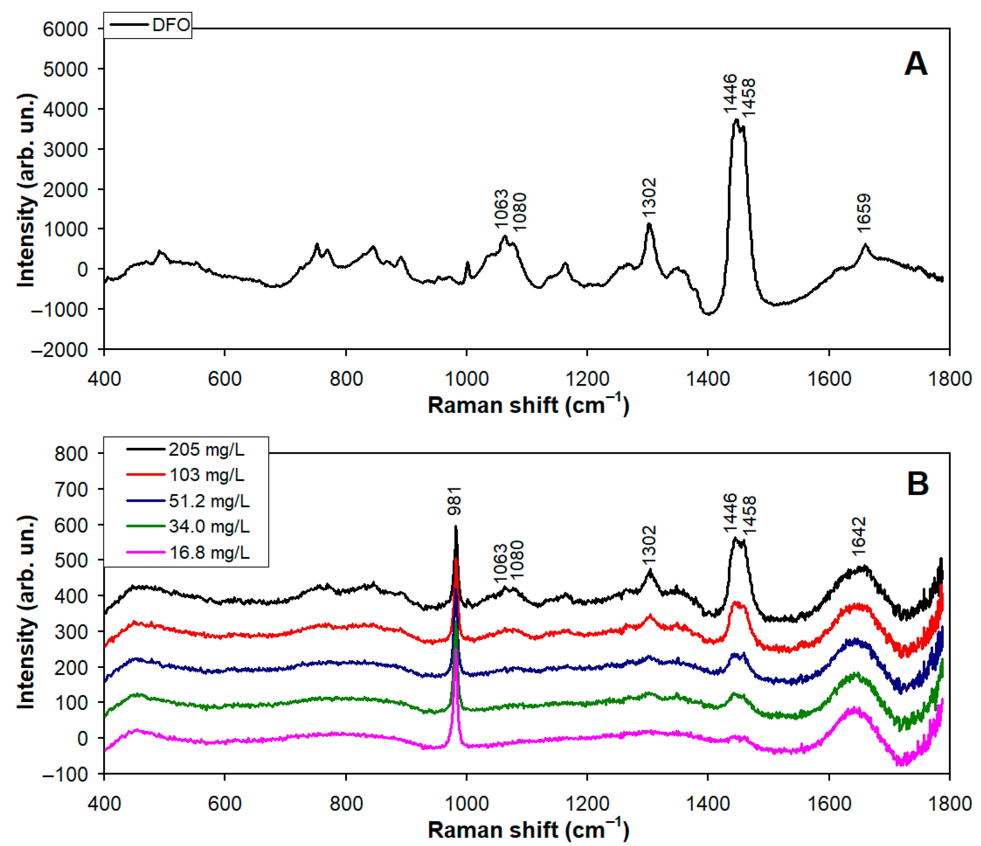
The Raman spectra of pure DFO and the saline water with different concentrations of DFO added are seen in Figure 4. The spectra showed more complex characteristic features than the SLO, but some bands appear at similar positions for both the DFO and SLO, especially the ones with peaks at 1063, 1080, 1302, 1446, and 1458 cm−1. The peak at 1659 cm−1 can be assigned to C=C stretching from olefins (alkenes) [21,34]. In the spectra of DFO added to saline water (Figure 4B), the intensity of the cited peaks, particularly the ones at 1302, 1446, and 1458 cm−1, was found to increase with the increased concentration of DFO added to water. The intensity of the saline water features at 1642 and 981 cm−1 remained unaltered. Notably, there was a lower concentration of DFO with regard to SLO when comparing both dilution curves. This was due to the dilution method used (detailed in Supplementary Material S1), where a number of drops of SLO and DFO were added to the water to obtain the different concentrations, but as each oil presents different viscosity and density values, there was difference in the volume of a single drop between the two oils.

3.2. Quantification of Synthetic Lubricating Oil and Diesel Fuel Oil Added to Saline Water

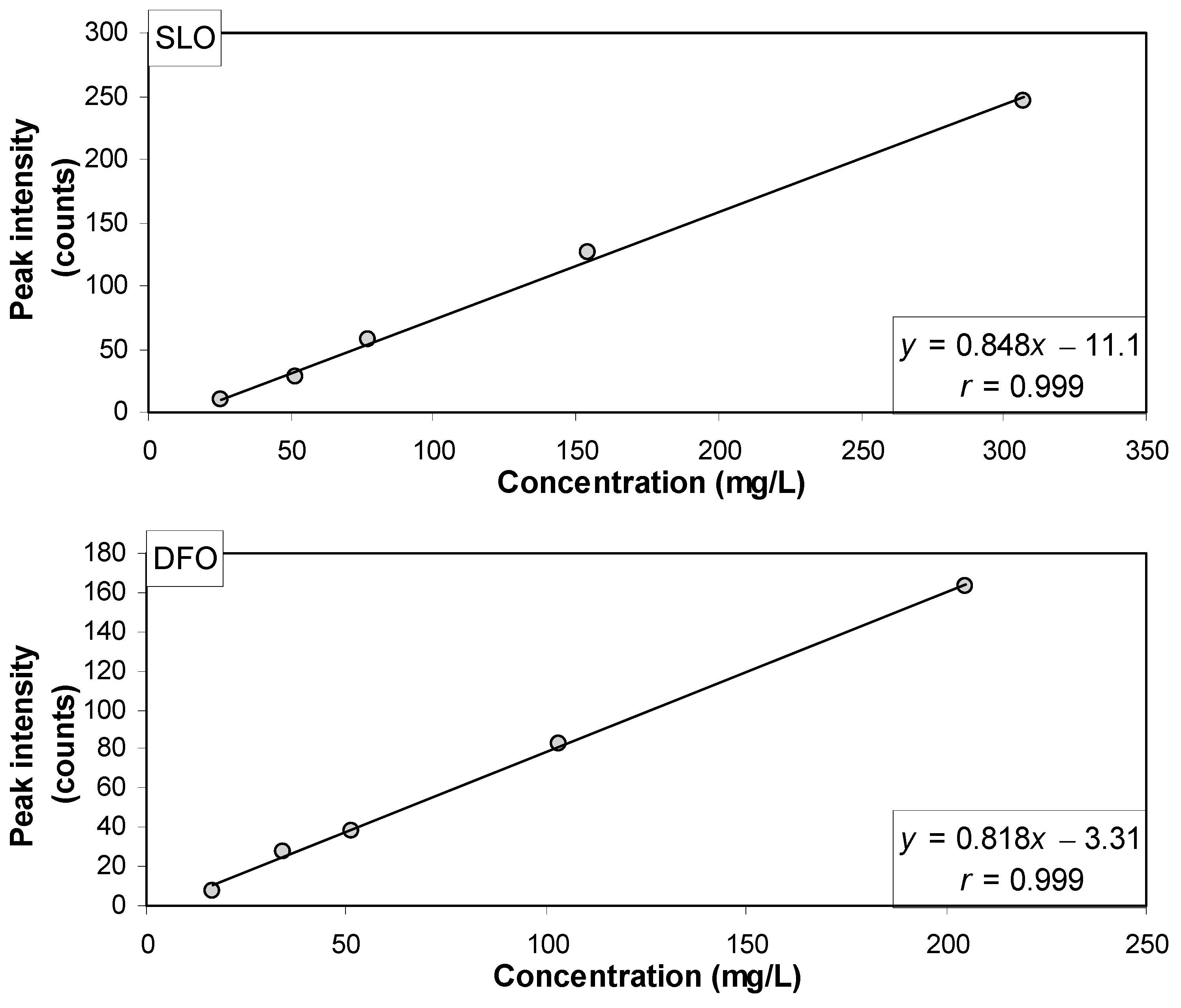
The spectra seen in Figure 3 and Figure 4 indicated proportionality between the intensity of the Raman features of SLO and DFO and the concentration of oils added to the water; therefore, two regression models were developed to quantify the amount of oil added. In the univariate linear regression model, the intensity of the most intense peak at ~1440–1450 cm−1 (methylene and methyl bending and deformation modes) was plotted versus the oil concentration added to the water. Figure 5 shows the scatter plot of the intensities of the most intense peak of SLO and DFO versus the respective concentrations in saline water; the linear curve that fits the data and the correlation coefficients for SLO and DFO are seen in the plots, and the extremely high correlation coefficients (r = 0.999 for both SLO and DFO) indicate that the intensities of the most intense peaks of methylene and methyl could be used to precisely quantify the oil added to the saline water. The linear curves seen in Figure 5 were used to estimate the concentration of the oils added to the saline water [x = (y + 11.1)/0.848 and x = (y + 3.31)/0.818 for SLO and DFO, respectively, where x is the predicted concentration and y is the intensity of the peak at ~1400–1500 cm−1 region], and the results are summarized in Table 1.
The second regression model, based on the PLSR applied to the Raman spectra (independent variables) and the concentrations (dependent variables), using the leave-one-out cross-validation, showed r = 0.997 for SLO and r = 0.987 for DFO using the first latent variable, which means that a single set of spectral features (mostly the oil features) is responsible for the differences seen in the spectra of the dilutions. The scatter plot of the concentrations of oil in each sample and the respective concentrations estimated by the PLSR model are presented in Figure 6. The estimated concentrations of oil provided by the PLSR model in the spectrum of each sample are summarized in Table 1.
The linearity and the extremely high correlation coefficient of the regression curves for both models (Figure 5 and Figure 6), particularly for the univariate, peak intensity-based model, suggest that the concentration of oil added to the saline water could be precisely estimated by measuring the Raman spectra directly on the thin film layer of oil at the water surface when the magnetic stirring system is turned on, forming a vortex at the water surface and spreading a regular and well-defined thin layer of oil over the water surface that could be precisely measured by the Raman probe.

3.3. Raman Spectroscopy as a Tool for Monitoring Oil Contamination in a Saline Environment

The methodology proposed in this study was based on the Raman spectroscopy technique to obtain the spectra of a thin film layer of oil at the saline water surface when a vortex was generated in the water by a magnetic stirrer with constant speed at the center of the beaker. The method was able to detect the presence of SLO in the concentration range of 25.6 to 307 mg/L and of DFO in the concentration range of 16.8 to 205 mg/L, with prediction error on the order of 8.0 and 11 mg/L for SLO and DFO, respectively, using the PLSR model. The results suggest that methodology could be exploited to obtain the concentrations of different oil types in saline waters, since the Brazilian legislation sets the limit of 20 mg/L for detecting oils of mineral origin and 100 mg/L for hydrocarbons soluble in hexane (e.g., oils and greases) [35].
The correlation coefficient provides auxiliary information to verify the “quality” of the regression curve, in order to verify if the proposed model can adequately describe the phenomenon; in linear models, the r-values range from 0 to 1, and values close to 1 indicate that the proposed methodology reliably describes the phenomenon [36]. The univariate linear regression model based on the intensity of the most intense peak of methylene and methyl showed the highest r-value (r = 0.999 for both oils), and the values are close to the ones found in the PLSR model (r > 0.99 for both oils). The prediction error of 8.0 and 11 mg/dL (SLO and DFO, respectively) can be suitable for the analysis of oils added to saline water.
Water pollution by oils and other petroleum-derived substances is associated with negative environmental, human health, and economic impacts [37]. Oils, greases, and waste from the production of petrol derivatives and ship operations make up the most dangerous group of pollutants released into the sea [38] and marine water can be contaminated by these substances in different ways. Fires in port terminals, ship collisions, cleaning of cargo and fuel tanks, engine failure and other incidents with seagoing transport ships can result in marine pollution [39]. In fact, oils and greases are usually found in water at low concentrations; most of the time, they are not visible to the naked eye (a thin film which is refractive when illuminated by white light is well-known evidence of the presence of oil). These substances can undergo a combined modification of physical–chemical and biological processes in water through spreading, evaporation, dispersion, dissolution, saponification, oxidation and degradation, accumulation as persistent residues, and biodegradation by microorganisms [40]. Therefore, the identification and quantification of such pollutants in water is of extreme importance for pollution characterization and control.
Previous studies employed Raman spectroscopy to investigate automotive lubricant oils (ALOs). Passoni et al. [21] evaluated the differences in the viscosity and type of base oil (mineral, semi-synthetic, and synthetic) in commercial ALOs by Raman spectroscopy and found spectral differences in the spectra which were dependent on the base oil used in each formulation, as well as differences in the additive packages. The authors also found that the viscosity at low and high temperatures (SAE specifications) could be determined and discriminated by differences in the spectral features, depending on the temperature specification. Bezerra et al. [34] evaluated thermal degradation of mineral, semi-synthetic, and synthetic ALOs and identified peaks assigned to amorphous carbon and changes (mainly decrease in the intensity) in peaks that were attributed to the thermal degradation of the base oil. Exploratory analysis by PCA showed that modifications in the spectral features were linearly correlated to the time of heating, and thus a regression model based on PCA was able to predict the time of heating. These studies characterized the strong Raman scattering that occurs in ALO samples and evidenced the possibilities of using the spectral information for the identification of base oil type, as well as for quantification of the degradation process.
Several approaches have been developed over the years, regarding the identification and analysis of oils, greases, and fuels present in or added to water [29,41,42]. An old review by Stenstrom, Fam, and Silverman (1986) [43] highlighted that IR spectroscopy can provide reproducible, accurate, easier, and faster identification and quantification of oils and greases. In a relatively recent publication, Adeniji et al. [44] provided a comprehensive overview of the analytical methods used to determine total petroleum hydrocarbons (TPHCs) in water and sediment. The authors discuss the physicochemical behavior of hydrocarbons in aquatic systems, their natural and anthropogenic sources, and the extraction and detection techniques commonly employed, including liquid–liquid and solid-phase extraction, IR spectroscopy, gravimetry, and gas chromatography with FID or MS detection. The authors emphasize that no single method is universally applicable, as accuracy and sensitivity depend on the sample matrix and analytical objectives. The review also compiles global data on TPHC concentrations, highlighting higher accumulation in sediments than in water, and underscores the need for standardized protocols to improve comparability among studies. Regarding recent original studies, Sosnowski et al. [45] built a rugged handheld fluorescence spectrometry device along with machine learning techniques to identify ocean oil spill samples. The technique could classify oil according to type (crude oil, light fuel oil, heavy fuel oil, and lubricant oil) and according to its saturate, aromatic, resin, and asphaltene contents. Shankar et al. [46] used a tiered approach to investigate lubricant oil in seawater using thin-layer chromatography-flame ionization detection, Fourier-transform infrared (FT-IR) spectroscopy, and GC-MS. The authors could identify the FT-IR peaks assigned to photo oxidation (carboxylic acids, phenols, saturated esters, and alcohols). Jager et al. [29] tested the feasibility of using solid-phase microextraction (SPME) combined with direct Raman spectroscopy for the quantitative determination of petroleum hydrocarbons and fuels (gasoline, jet fuel, fuel oil) added to raw river water. The SPME/Raman method provided sensitive, reproducible, and solvent-free quantification of petroleum hydrocarbons in water, with detection limits in the low ppm range and results comparable to standard chromatographic and infrared methods. Fadzil et al. [47] investigated the levels of oil and grease and total petroleum hydrocarbons in port waters to assess environmental contamination using UV fluorescence spectroscopy. Water samples collected from several points within the port were analyzed and results showed elevated concentrations of oils and greases and total petroleum hydrocarbons, indicating significant pollution from petroleum residues. Fluorescence-based methods generate faster and more direct results since the fluorescence spectra can be captured almost instantly, without the requirement of chromatographic separation of the oil content [45,47]. FT-IR spectroscopy presents sensitivity to detect hydrocarbons and degradation, but the readings are sensitive to humidity and can produce false positive results due to a strong absorbance of water [43,46]. Conversely, analytical methods such as GC-MS and GC-FID have longer operational times and higher analytical costs for sample analysis [48,49].
A major problem in detecting oil in seawater using fluorescence spectroscopy is the sensitivity of the method. Seawater contains phytoplankton, pigments, and colored and fluorescent dissolved organic matter. These natural constituents absorb and fluoresce in the same spectral region as the oil; therefore, the spectra partially overlap, impairing the results [50,51,52]. Despite this, Baszanowska and Otremba [53] showed that oily substances can be detected in natural seawater by fluorescence spectroscopy, despite the partial overlap of peaks originating from substances naturally present in the sea and from substances that leach out from the oil. UV–visible spectrophotometry is an analytical method that has shown advantages over other methods previously used to determine the presence of oils and greases in water and soil; these advantages include shorter response times, greater sensitivity, more selectivity, and better detection limits and simplicity [54].
While these traditional methods are effective for determining total petroleum hydrocarbon content, they require expensive laboratory equipment, and obtaining the analysis results can be time-consuming [55]. Each of the established methods has advantages and disadvantages, so a technique should only be chosen after considering the objectives and type of information or data needed for each application [44]. Considering an oil spill case, portable spectroscopy devices would be very acceptable for immediate on-site sample characterization, without the need to send samples for laboratory testing [45]. Since oil spills can worsen dramatically as the oil is spread by the action of winds and sea currents [56], Bill et al. [57] emphasize the importance of using methods capable of quickly classifying and characterizing oil samples, preferably at the oil spill site.
The technique of liquid–liquid gravimetric was applied by Roveri et al. [58] to detect the presence of oils and greases in water at two points close to diffusers of the submarine outfall of a Brazilian coastal city. This is a situation where the methodology based on Raman spectroscopy technique could be applied for oil analysis. In fact, portable Raman instruments with fiber optic probes similar to those used in this work can be adapted to aquatic or air (drone) vehicles for in situ monitoring and analysis (where the probe could be adapted to a fluctuating device or a buoy), and measuring the film thickness formed by the oil instead of the mass per volume. It is noteworthy to mention that Raman lidar systems for detecting oil films in water in situ have been demonstrated [59]. Therefore, techniques based on Raman scattering are advantageous due to their relatively high sensitivity; there is no need for sample preparation, and there is low interference from water.
This study sheds light on an extremely important area of pollution monitoring and control, where Raman spectroscopy could bring advantages such as real-time analysis (detection and quantification), so that one can immediately take actions to mitigate environmental impacts. Raman spectroscopy technique is a very promising method for the label-free analysis of oils and greases, due to its low cost, minimal or no need for sample preparation, and no residual waste, providing fast and accurate results.

4. Conclusions

This work proposed the quantification of SLO and DFO in saline water by means of Raman spectroscopy and two spectral models: (a) univariate linear regression model using the intensity of the methylene and methyl (CH2 and CH3) bands at ~1440/1450 cm−1, and (b) a multivariate PLSR model applied to the whole spectra. In the range of concentrations of 25.6–307 mg/L for synthetic lubricating oil and 16.8–205 mg/L for diesel fuel oil added to saline water, both models achieved correlation coefficients as high as r = 0.99 and prediction errors of 8.0 and 11 mg/L for SLO and DFO, respectively, suggesting that the Raman features of both oils can be easily distinguished from the Raman features of saline water (sulfate S–O stretching peak at 981 cm−1 and water H–O–H bending peak at 1642 cm−1).
The concentrations of the oils evaluated in this study can be compared to the limit of 20 mg/L for oils of mineral origin and up to 100 mg/L for substances soluble in hexane (oils and greases) according to CONAMA Resolution No. 430/2011 [35]. Overall, the findings provide evidence that Raman spectroscopy is a reliable technique for the detection and quantification of petroleum-derived pollutants in saline water, offering new perspectives for its application in environmental monitoring and oil spill assessment.

Supplementary Materials

The following supporting information can be downloaded at https://www.mdpi.com/article/10.3390/w17223289/s1, Figure S1: Setup developed to standardize the amount of synthetic lubricating oil (SLO) and diesel fuel oil (DFO) to be added to the samples.; Table S1: Standardization of mass values of synthetic lubricating oil (SLO) and diesel fuel oil (DFO); S1: Standardization of the mass and volume of synthetic lubricating oil (SLO) and diesel fuel oil (DFO); S2: Samples of saline water added with SLO and DFO.

Author Contributions

G.M.d.A.: conceptualization, investigation, and methodology; L.L.G.: conceptualization, formal analysis, and writing; L.P.M.: original draft preparation, review, and editing; W.T., V.R., and M.T.T.P.: writing, review and editing; L.S.J.: conceptualization, formal analysis, investigation, and supervision. All authors have read and agreed to the published version of the manuscript.

Funding

L. Silveira Jr. acknowledges FAPESP (São Paulo Research Foundation) for granting the Raman spectrometer (Process No. 2009/01788-5), CNPq (Brazilian National Council for Scientific and Technological Development) for the productivity fellowship (Process No. 314167/2021-8), and Ânima Institute (AI) for the research fellowship. L. P. Moreira acknowledges CAPES (Coordination for the Improvement of Higher Education Personnel) for the scholarship (Finance Code 001).

Data Availability Statement

The original contributions presented in this study are included in the article and its Supplementary Materials. Further inquiries can be directed to the corresponding authors, and the data can be provided upon reasonable request.

Conflicts of Interest

The authors declare no conflicts of interest.

References

  1. United Nations. Department of Economic and Social Affairs. World Urbanization Prospects 2018. Available online: https://population.un.org/wup/ (accessed on 10 July 2025).
  2. Fang, C.; Cui, X.; Li, G.; Bao, C.; Wang, Z.; Ma, H.; Sun, S.; Li, H.; Luo, K.; Ren, Y. Modeling regional sustainable development scenarios using the Urbanization and Eco-environment Coupler: Case study of Beijing-Tianjin-Hebei urban agglomeration, China. Sci. Total Environ. 2019, 689, 820–830. [Google Scholar] [CrossRef] [PubMed]
  3. Munir, K.; Ameer, A. Assessing nonlinear impact of urbanization, economic growth, technology, and trade on environment: Evidence from African and Asian emerging economies. GeoJournal 2022, 87, 2195–2208. [Google Scholar] [CrossRef]
  4. Freeman, L.A.; Reide Corbett, D.; Fitzgerald, A.M.; Lemley, D.A.; Quigg, A.; Steppe, C.N. Impacts of urbanization and development on estuarine ecosystems and water quality. Estuaries Coasts 2019, 42, 1821–1838. [Google Scholar] [CrossRef]
  5. Zhai, T.; Wang, J.; Fang, Y.; Qin, Y.; Huang, L.; Chen, Y. Assessing ecological risks caused by human activities in rapid urbanization coastal areas: Towards an integrated approach to determining key areas of terrestrial-oceanic ecosystems preservation and restoration. Sci. Total Environ. 2020, 708, 135153. [Google Scholar] [CrossRef]
  6. Fan, X.; Zhang, L.; Yuan, L.; Guo, B.; Zhang, Q.; Huang, H. Urbanization and water quality dynamics and their spatial correlation in coastal margins of mainland China. Ecol. Indic. 2022, 138, 108812. [Google Scholar] [CrossRef]
  7. Silva, I.A.; Almeida, F.C.G.; Souza, T.C.; Bezerra, K.G.O.; Durval, I.J.B.; Converti, A.; Sarubbo, L.A. Oil spills: Impacts and perspectives of treatment technologies with focus on the use of green surfactants. Environ. Monit. Assess. 2022, 194, 1–29. [Google Scholar] [CrossRef]
  8. ITOPF—The International Tanker Owners Pollution Federation. Tanker Spill Statistics 2020. Available online: https://www.itopf.org/news/news/tanker-spill-statistics-2021/ (accessed on 10 July 2025).
  9. Chen, J.; Zhang, W.; Wan, Z.; Li, S.; Huang, T.; Fei, Y. Oil spills from global tankers: Status review and future governance. J. Clean. Prod. 2019, 227, 20–32. [Google Scholar] [CrossRef]
  10. Little, D.I.; Sheppard, S.R.J.; Hulme, D.A. Perspective on oil spills: What we should have learned about global warming. Ocean Coast Manag. 2021, 202, 105509. [Google Scholar] [CrossRef]
  11. Yu, L.; Han, M.; He, F. A review of treating oily wastewater. Arab. J. Chem. 2017, 10, S1913–S1922. [Google Scholar] [CrossRef]
  12. Andrews, N.; Bennett, N.J.; Le Billon, P.; Green, S.J.; Cisneros-Montemayor, A.M.; Amongin, S.; Gray, N.J.G.; Sumaila, U.R. Oil, fisheries and coastal communities: A review of impacts on the environment, livelihoods, space and governance. Energy Res. Soc. Sci. 2021, 75, 102009. [Google Scholar] [CrossRef]
  13. Câmara, S.F.; Pinto, F.R.; da Silva, F.R.; Soares, M.O.; de Paula, T.M. Socioeconomic vulnerability of communities on the Brazilian coast to the largest oil spill (2019–2020) in tropical oceans. Ocean Coast Manag. 2021, 202, 105506. [Google Scholar] [CrossRef]
  14. Reddy, C.M.; Quinn, J.G. GC-MS analysis of total petroleum hydrocarbons and polycyclic aromatic hydrocarbons in seawater samples after the North Cape oil spill. Mar. Pollut. Bull. 1999, 38, 126–135. [Google Scholar] [CrossRef]
  15. Cortes, J.E.; Suspes, A.; Roa, S.; González, C.; Castro, H.E. Total petroleum hydrocarbons by gas chromatography in Colombian waters and soils. Am. J. Environ. Sci. 2012, 8, 396–402. [Google Scholar] [CrossRef]
  16. Muthukumar, A.; Idayachandiran, G.; Kumaresan, S.; Kumar, T.A.; Balasubramanian, T. Petroleum hydrocarbons (PHC) in sediments of three different ecosystems from Southeast Coast of India. Int. J. Pharm. Biol. Arch. 2013, 4, 543–549. [Google Scholar]
  17. Okparanma, R.N.; Mouazen, A.M. Determination of total petroleum hydrocarbon (TPH) and polycyclic aromatic hydrocarbon (PAH) in soils: A review of spectroscopic and nonspectroscopic techniques. Appl. Spectrosc. Rev. 2013, 48, 458–486. [Google Scholar] [CrossRef]
  18. Li, T.; Xu, H.; Zhang, Y.; Zhang, H.; Hu, X.; Sun, Y.; Gu, X.; Luo, J.; Zhou, D.; Gao, B. Treatment technologies for selenium contaminated water: A critical review. Environ. Pollut. 2022, 299, 118858. [Google Scholar] [CrossRef] [PubMed]
  19. Gavrilescu, M.; Demnerová, K.; Aamand, J.; Agathos, S.; Fava, F. Emerging pollutants in the environment: Present and future challenges in biomonitoring, ecological risks and bioremediation. N. Biotechnol. 2015, 32, 147–156. [Google Scholar] [CrossRef] [PubMed]
  20. Ferreiro, V.; Gómez-Motos, I.; Lombrana, J.I.; de Luis, A.; Villota, N.; Ros, O.; Etxebarria, N. Contaminants of emerging concern removal in an effluent of wastewater treatment plant under biological and continuous mode ultrafiltration treatment. Sustainability 2020, 12, 725. [Google Scholar] [CrossRef]
  21. Passoni, D.D.J.; Pacheco, M.T.T.; Silveira, L. Raman spectroscopy for the identification of differences in the composition of automobile lubricant oils related to SAE specifications and additives. Instrum. Sci. Technol. 2020, 49, 164–181. [Google Scholar] [CrossRef]
  22. de Bettignies, P. Optics/instrumentation: Micro-Raman spectroscopy: Theory and application. Phys. Sci. Rev. 2020, 5, 20190027. [Google Scholar] [CrossRef]
  23. Tsukada, S.; Fujii, Y. Multivariate curve resolution for angle-resolved polarized Raman spectroscopy of ferroelectric crystals. Jpn. J. Appl. Phys. 2020, 59, SKKA03. [Google Scholar] [CrossRef]
  24. Perraki, M.; Skliros, V.; Mecaj, P.; Vasileiou, E.; Salmas, C.; Papanikolaou, I.; Stamatis, G. Identification of microplastics using µ-Raman spectroscopy in surface and groundwater bodies of SE Attica, Greece. Water 2024, 16, 843. [Google Scholar] [CrossRef]
  25. Xi, Z.; Nicolas, R.; Wei, J. An edge-deployable multi-modal nano-sensor array coupled with deep learning for real-time, multi-pollutant water quality monitoring. Water 2025, 17, 2065. [Google Scholar] [CrossRef]
  26. Kataya, G.; Issa, M.; El Charif, Z.; Cornu, D.; Taleb, B.; Bechelany, M.; Hijazi, A. Enhanced copper adsorption with sustainable biochar derived from kitchen waste. Water 2025, 17, 1887. [Google Scholar] [CrossRef]
  27. Almaviva, S.; Artuso, F.; Giardina, I.; Lai, A.; Pasquo, A. Fast detection of different water contaminants by Raman spectroscopy and surface-enhanced Raman spectroscopy. Sensors 2022, 22, 8338. [Google Scholar] [CrossRef]
  28. Cao, S.; Zhan, G.; Wei, K.; Zhou, B.; Zhang, H.; Gao, T.; Zhang, L. Raman spectroscopic and microscopic monitoring of on-site and in-situ remediation dynamics in petroleum contaminated soil and groundwater. Water Res. 2023, 233, 119777. [Google Scholar] [CrossRef]
  29. Jager, M.J.; McClintic, D.P.; Tilotta, D.C. Measurement of petroleum fuel contamination in water by solid-phase microextraction with direct Raman spectroscopic detection. Appl. Spectrosc. 2000, 54, 1617–1623. [Google Scholar] [CrossRef]
  30. Ahmadjian, M.; Brown, C.W. Feasibility of remote detection of water pollutants and oil slicks by laser-excited Raman spectroscopy. Environ. Sci. Technol. 1973, 7, 452–453. [Google Scholar] [CrossRef]
  31. Nunes, C.A.; Freitas, M.P.; Pinheiro, A.C.M.; Bastos, S.C. Chemoface: A novel free user-friendly interface for chemometrics. J. Braz. Chem. Soc. 2012, 23, 2003–2010. [Google Scholar] [CrossRef]
  32. Fontana, M.D.; Mabrouk, K.B.; Kauffmann, Y.H. Spectroscopic Properties of Inorganic and Organometallic Compounds: Techniques, Materials and Applications; Yarwood, J., Douthwaite, R., Duckett, S., Eds.; RSC Publishing: Cambridge, UK, 2013; Volume 44, pp. 40–67. [Google Scholar] [CrossRef]
  33. Zhang, X.; Walz, P.M.; Kirkwood, W.J.; Hester, K.C.; Ussler, W.; Peltzer, E.T.; Brewer, P.G. Development and deployment of a deep-sea Raman probe for measurement of pore water geochemistry. Deep-Sea Res. I Oceanogr. Res. Pap. 2010, 57, 297–306. [Google Scholar] [CrossRef]
  34. Bezerra, A.C.M.; Coelho, N.M.A.; Bertelli, F.; Pacheco, M.T.T.; Silveira, L. Temperature-induced chemical changes in lubricant automotive oils evaluated using Raman spectroscopy. Appl. Spectrosc. 2020, 75, 145–155. [Google Scholar] [CrossRef]
  35. Ministério do Meio Ambiente. CONAMA—Conselho Nacional do Meio-Ambiente. Resolução No. 430 de 13 de maio de 2011. Dispõe sobre as condições e padrões de lançamento de efluentes, complementa e altera a Resolução No. 357, de 17 de março de 2005, do Conselho Nacional do Meio Ambiente—CONAMA. Available online: https://www.legisweb.com.br/legislacao/?id=114770 (accessed on 10 July 2025).
  36. Matos, M.A. Manual operacional para a regressão linear. Faculdade de Engenharia da Universidade do Porto. 1995. Available online: https://paginas.fe.up.pt/~mam/regressao.pdf (accessed on 20 July 2024).
  37. Nowak, P.; Kucharska, K.; Kamiński, M. Ecological and health effects of lubricant oils emitted into the environment. Int. J. Environ. Res. Public Health 2019, 16, 3002. [Google Scholar] [CrossRef] [PubMed]
  38. Deja, A.; Ulewicz, R.; Kyrychenko, Y. Analysis and assessment of environmental threats in maritime transport. Transp. Res. Procedia 2021, 55, 1073–1080. [Google Scholar] [CrossRef]
  39. Łapko, A.; Strulak-Wójcikiewicz, R.; Landowski, M.; Wieczorek, R. Management of waste collection from yachts and tall ships from the perspective of sustainable water tourism. Sustainability 2018, 11, 121. [Google Scholar] [CrossRef]
  40. Kuppusamy, S.; Maddela, N.R.; Megharaj, M.; Venkateswarlu, K. Total Petroleum Hydrocarbons; Springer: Cham, Switzerland, 2019; Volume 1, Chapter 3; pp. 57–77. [Google Scholar] [CrossRef]
  41. Strother, T.; Lowry, S.; Bravo, B. Application Note 52439. Measurement of Dispersed Oil in Water Using an Infrared Analysis Method. 2013. Available online: https://analysis.rs/wp-content/uploads/2022/01/9132023968-min.pdf (accessed on 10 July 2025).
  42. Stevens, D.; Shi, Q.; Hsu, C.S. Novel analytical technique for petroleum biomarker analysis. Energy Fuels 2013, 27, 167–171. [Google Scholar] [CrossRef]
  43. Stenstrom, M.K.; Fam, S.; Silverman, G.S. Analytical methods for quantitative and qualitative determination of hydrocarbons and oil and grease in water and wastewater. Environ. Sci. Technol. Lett. 1986, 7, 625–636. [Google Scholar] [CrossRef]
  44. Adeniji, A.O.; Okoh, O.O.; Okoh, A.I. Analytical methods for the determination of the distribution of total petroleum hydrocarbons in the water and sediment of aquatic systems: A review. J. Chem. 2017, 2017, 5178937. [Google Scholar] [CrossRef]
  45. Sosnowski, K.; Loh, A.; Zubler, A.V.; Shir, H.; Ha, S.Y.; Yim, U.H.; Yoon, J.Y. Machine learning techniques for chemical and type analysis of ocean oil samples via handheld spectrophotometer device. Biosens. Bioelectron. X 2022, 10, 100128. [Google Scholar] [CrossRef]
  46. Shankar, R.; Jung, J.H.; Loh, A.; An, J.G.; Ha, S.Y.; Yim, U.H. Environmental significance of lubricant oil: A systematic study of photooxidation and its consequences. Water Res. 2020, 168, 115183. [Google Scholar] [CrossRef]
  47. Fadzil, M.F.; Yun, P.S.; Razal, A.R.; Chee, P.S.; Suratman, S.; Dagang, N.S.; Tahir, N.M. Oil and grease and total petroleum hydrocarbons in the waters of Ramsar Gazetted mangrove area, Johor. J. Sustain. Sci. Manag. 2017, 12, 30–39. [Google Scholar]
  48. Krupcík, J.; Oswald, P.; Oktavec, D.; Armstrong, D.W. Calibration of GC-FID and IR spectrometric methods for determination of high boiling petroleum hydrocarbons in environmental samples. Water Air Soil Pollut. 2004, 153, 329–341. [Google Scholar] [CrossRef]
  49. Kim, M.; Hong, S.H.; Won, J.; Yim, U.H.; Jung, J.H.; Ha, S.Y.; An, J.G.; Joo, C.; Kim, E.; Han, G.M.; et al. Petroleum hydrocarbon contaminations in the intertidal seawater after the Hebei Spirit oil spill—Effect of tidal cycle on the TPH concentrations and the chromatographic characterization of seawater extracts. Water Res. 2013, 47, 758–768. [Google Scholar] [CrossRef]
  50. Drozdowska, V.; Freda, W.; Baszanowska, E.; Rudź, K.; Darecki, M.; Heldt, J.; Toczek, H. Spectral properties of natural and oil-polluted Baltic seawater—Results of measurements and modelling. Eur. Phys. J. Spec. Top. 2013, 222, 2157–2170. [Google Scholar] [CrossRef]
  51. Drozdowska, V.; Wrobel, I.; Markuszewski, P.; Makuch, P.; Raczkowska, A.; Kowalczuk, P. Study on organic matter fractions in the surface microlayer in the Baltic Sea by spectrophotometric and spectrofluorometric methods. Ocean Sci. 2017, 13, 633–647. [Google Scholar] [CrossRef]
  52. Lopes, R.; Miranda, M.L.; Schuette, H.; Gassmann, S.; Zielinski, O. Microfluidic approach for controlled ultraviolet treatment of colored and fluorescent dissolved organic matter. Spectrochim. Acta Part A Mol. Biomol. Spectrosc. 2020, 239, 118435. [Google Scholar] [CrossRef] [PubMed]
  53. Baszanowska, E.; Otremba, Z. Fluorometric detection of oil traces in a sea water column. Sensors 2022, 22, 2039. [Google Scholar] [CrossRef] [PubMed]
  54. Khorshid, Z.B.; Doroodmand, M.M.; Abdollahi, S. UV-Vis. spectrophotometric method for oil and grease determination in water, soil and different mediates based on emulsion. Microchem. J. 2021, 160, 105620. [Google Scholar] [CrossRef]
  55. Kuppusamy, S.; Maddela, N.R.; Megharaj, M.; Venkateswarlu, K. Total Petroleum Hydrocarbons; Springer: Cham, Switzerland, 2020; Volume 1, Chapter 2; pp. 29–55. [Google Scholar] [CrossRef]
  56. Yim, U.H.; Khim, J.S.; Kim, M.; Jung, J.H.; Shim, W.J. Environmental impacts and recovery after the Hebei Spirit oil spill in Korea. Arch. Environ. Contam. Toxicol. 2017, 73, 47–54. [Google Scholar] [CrossRef] [PubMed]
  57. Bills, M.V.; Loh, A.; Sosnowski, K.; Nguyen, B.T.; Ha, S.Y.; Yim, U.H.; Yoon, J.Y. Handheld UV fluorescence spectrophotometer device for the classification and analysis of petroleum oil samples. Biosens. Bioelectron. 2020, 159, 112193. [Google Scholar] [CrossRef]
  58. Roveri, V.; Guimarães, L.L.; Correia, A.T. Spatial and temporal evaluation of the urban runoff water flowing into recreational areas of Guarujá, São Paulo State, Brazil. Int. J. River Basin Manag. 2022, 20, 93–109. [Google Scholar] [CrossRef]
  59. Shangguan, M.; Yang, Z.; Shangguan, M.; Lin, Z.; Liao, Z.; Guo, Y.; Liu, C. Remote sensing oil in water with an all-fiber underwater single-photon Raman lidar. Appl. Opt. 2023, 62, 5301–5305. [Google Scholar] [CrossRef] [PubMed]
Figure 1. Schematic diagram for obtaining the Raman spectra of the oil samples (SLO and SFO) added to a beaker containing 1000 mL of saline water; a thin film of oil was formed by constant stirring of the samples at 300 rpm, and the spectrum was collected at the water surface.
Water 17 03289 g001
Figure 2. Raman spectrum of saline water. The labeled peak is assigned to the anion sulfate (SO42−) [27,28].
Water 17 03289 g002
Figure 3. Raman spectra of (A) pure (non-diluted) SLO and (B) saline water with different concentrations of SLO added as follows: 25.6, 51.4, 77.2, 154, and 307 mg/L.
Water 17 03289 g003
Figure 4. Raman spectra of (A) pure (non-diluted) DFO and (B) saline water with different concentrations of DFO added as follows: 16.8, 34.0, 51.2, 103, and 205 mg/L.
Water 17 03289 g004
Figure 5. Scatter plot of the intensities of the most intense peak of SLO and DFO at the region of ~1440–1450 cm−1 versus the respective concentrations of both oils diluted in saline water. The linear curves and the correlation coefficients are also displayed.
Water 17 03289 g005
Figure 6. Scatter plot of the concentrations of oil predicted by the PLSR models using the spectra of SLO and DFO versus the respective concentrations of both oils in saline water. The number of latent variables for the highest correlation was 1 for both models, suggesting that a single spectral feature (the Raman bands of oil) is responsible for providing the correlation between the different concentration and the spectral changes due to the different concentrations.
Water 17 03289 g006
Table 1. Results of the estimated concentration of SLO and DFO added to the saline water for each spectral model (linear and PLSR) and error (mass added minus estimated concentration). * The linear model was based on the intensity of the peak at ~1440–1450 cm−1, and the PLSR model was based on the regression of the latent variables obtained from the covariance between the independent variable (spectra) and the dependent variable (concentration of added oil).
Type of OilMass Added (mg)Concentration Estimated by Each Spectral Model (mg/L)
Linear *Error (mg)PLSR *Error (mg)
SLO25.623.8−1.825.90.3
51.446.6−4.848.8−2.6
77.279.92.778.81.6
15416171628
307304−3294−13
DFO16.812.5−4.314.7−2.1
34.037.73.739.15.1
51.251.20.052.41.2
10310521074
205204−1194−11
Disclaimer/Publisher’s Note: The statements, opinions and data contained in all publications are solely those of the individual author(s) and contributor(s) and not of MDPI and/or the editor(s). MDPI and/or the editor(s) disclaim responsibility for any injury to people or property resulting from any ideas, methods, instructions or products referred to in the content.