Article

Impact of Hydrodynamic Conditions on the Production and Distribution of Extracellular Polymeric Substance in River Biofilms

1
College of Environmental Science and Engineering, Yancheng Institute of Technology, Yancheng 224003, China
2
Jiangsu Province Engineering Research Center of Intelligent Environmental Protection Equipment, Yancheng Institute of Technology, Yancheng 224051, China
3
Yancheng Environmental Monitoring Center of Jiangsu Province, Yancheng 224002, China
*
Author to whom correspondence should be addressed.
Water 2023, 15(21), 3821; https://doi.org/10.3390/w15213821
Submission received: 26 September 2023 / Revised: 30 October 2023 / Accepted: 30 October 2023 / Published: 1 November 2023
(This article belongs to the Special Issue Water Quality, Ecological Health and Ecosystem Restoration)

Abstract

:
The extracellular polymeric substance (EPS) plays a key factor in biofilm formation. However, the research on the importance of each EPS fraction is mainly concentrated in the activated sludge field. In this study, biofilms were cultivated under different hydrodynamic conditions in indoor flumes, and the important regulatory effects of dissolved EPS (SB-EPS), loosely bound EPS (LB-EPS), and tightly bound EPS (TB-EPS) on biofilm formation were investigated. The results indicated that the ratios of soluble EPS (S-EPS), loosely bound EPS (LB-EPS), and tightly bound EPS (TB-EPS) were 27:74:108 in the turbulent flow, 38:48:71 in the transitional flow, and 89:51:51 in the laminar flow. Regarding proportion, TB-EPS and LB-EPS were secreted more in the turbulent flow, while S-EPS was secreted slightly more in the laminar flow. S-EPS lacks the structural strength provided by bound EPS. Under the special bonding effects of LB-EPS and TB-EPS, many microcolonies join to form biofilms. The polysaccharide content in the EPS of biofilms remained dominant under all conditions. Polysaccharides are the core of biofilm formation, which enhance bacterial aggregation and make biofilm dense. Through the mutual verification of the results in the microscopic and macroscopic fields, the mechanism of biofilm formation was further elucidated, especially, in Stage IV, due to the special bonding effects of LB-EPS and TB-EPS, many colonies adhere to the mature biofilm. Further studies are required to investigate the extracellular polysaccharides and proteins in EPS along with their properties in biofilms.

Graphical Abstract

1. Introduction

River biofilms growing on any carrier in water are an important part of lotic ecosystems. Biofilm formation is a dynamic process, which occurs when microorganisms adhere to the extracellular polymeric substance (EPS) secreted by themselves. The biofilm develops a wavy shape and eventually forms a complex three-dimensional (3D) network [1,2]. The formation of biofilm occurs through different processes in different environments, and their structural and morphological characteristics adapt to the environment during their formation [3,4,5].
At different stages of interaction between the microorganisms and carrier surface, EPS is required for microorganisms to secrete extracellular structures of biofilms; it contributes to the initial adhesion of microorganisms, maintenance of the biofilm structure, and separation of the aggregates surrounded by EPS [6,7]. The morphology and structure of biofilms at different stages show typical characteristics. The major components of EPS in biofilms are generally extracellular polysaccharides and extracellular proteins [8]. The adhesion of EPS to the surface of the carrier indicates the beginning of biofilm formation and plays a key factor in biofilm research [6,9]. EPS can be divided into dissolved EPS (S-EPS) and bound EPS according to the effort required to separate it and the spatial distribution of the combination with microorganisms [10,11]. S-EPS can act as a transport medium for nutrients, facilitating their diffusion through the biofilm matrix [12]. Bound EPS is further divided into loosely bound EPS (LB-EPS) and tightly bound EPS (TB-EPS) [13,14,15]. Bound EPS is closely bound with cells, while soluble EPS is weakly bound with cells [10]. However, excessive LB-EPS may deteriorate cell attachment and weaken the floc structure [16]. TB-EPS had better bioflocculation activity compared to S-EPS and LB-EPS [17]. However, studies on LB-EPS and TB-EPS have mostly focused on activated sludge [10,18], which has led to limited research on the regulatory significance of LB-EPS and TB-EPS on biofilm formation.
This study aimed to investigate the important regulatory effects of SB-EPS, LB-EPS, and TB-EPS on biofilm formation. The distribution and production of EPS at different stages of biofilm formation were analyzed under the regulation of hydrodynamic factors to better examine the mechanism of biofilm formation. Based on the images of the confocal laser scanning microscope (CLSM), the 3D dynamic distribution of macromolecular subcomponents (polysaccharides, proteins, etc.) of the biofilm cells and EPS matrix were mapped, and quantitative data on the structural characteristics of the dynamic process of biofilm formation were obtained. The contents of proteins and polysaccharides in the S-EPS, LB-EPS, and TB-EPS fractions were determined by chemical analyses, simultaneously. Through the mutual verification of the results in the above microscopic and macroscopic fields, the relationships between the dynamic changes in the production and distribution of EPS, and the hydrodynamic conditions during biofilm formation were evaluated to further elucidate the mechanism of biofilm formation and to provide a better theoretical basis and guidance for the application of biofilm in river ecological restoration.

2. Materials and Methods

2.1. Biofilm Cultivation

The microcosms consisted of three identical Plexiglass flumes connected to two shared Plexiglass reservoirs (80 L). The experimental conditions maintained for biofilm cultivation were the same as those described in a previous study [19]. The detailed information for conditions of biofilm cultivation was provided in Text S1, the hydraulic characteristics of flumes is listed in Table S1.

2.2. Biofilm Analysis

2.2.1. Sampling and Preprocessing

Biofilms grown on slides were sampled on days 8, 15, 32, 48, and 60. For CLSM examination, biofilms were observed in situ. Samples collected for extracting the EPS of biofilms were scraped and then sonicated with water to a uniform suspension and freeze-dried for further analyses.

2.2.2. Fluorescence Labeling and CLSM Image

The three dyes were added to the biofilms in the following order: a 4′, 6-diamidino-2-phenylindole (DAPI) solution (5 mg L−1, 100 μL) was kept in a shaker for 5 min. Secondly, the solution was conjugated with tetramethylrhodamine (ConA-TMR) solution (50 mg L−1, 100 μL) and incubated for 30 min. Then a fluorescein-isothiocyanate (FITC) solution (100 mg L−1, 100 μL) was added. The mixture was shaken for 1 h. During the process, the biofilms were incubated in a dark room. After each of the three staining processes, the sample was washed twice using phosphate-buffered saline (PBS) to remove the excess stain [20]. The detailed information for fluorescence labeling was provided in Text S2. Then, the images of stained biofilms were taken using a laser scanning microscope (LSM) 710 Confocal Microscope (Carl Zeiss, Jena, Germany) under 40× magnification.

2.2.3. Extraction of Extracellular Polymeric Substance

EPS was extracted step by step using cation exchange resin (Dowex Marathon C, strongly acidic, Type Na), following the procedure described by Pan et al. [20], which was based on the methods described in other studies [21,22]. The three EPS fractions, which were extracted from the biofilm by steps, were preserved at −20 °C.
The contents of polysaccharides and proteins of the extracted EPS were determined using the anthrone-sulfuric acid method [23] and the Folin phenol reagent method [24], respectively. The values were assessed as the mass of protein or polysaccharide/m2 of the surface area of the biofilm.

3. Results and Discussion

3.1. The Composition and Content of EPS in Biofilms

The total EPS of the biofilm was determined by summing up the polysaccharides and proteins extracted from the biofilm [25]. The total content of EPS in the biofilm was the highest in the turbulent flow (Figure 1a,b), followed by that in the transitional flow and laminar flow. These findings were similar to those of another study [26], which indicated that the larger shear force in the turbulent flow could stimulate the secretion of extracellular polysaccharides and proteins, thereby forming a relatively dense biofilm. This occurs because the intensity of the horizontal flow and vertical turbulence on the surface of the biofilm under turbulent conditions increases with changes in time and space.
On day 48, the ratios of S-EPS, LB-EPS, and TB-EPS were 27:74:108 in the turbulent flow, 38:48:71 in the transitional flow, and 89:51:51 in the laminar flow. From these ratios, EPS, especially TB-EPS and LB-EPS, were secreted in large quantities in the turbulent flow. The ratio of polysaccharide and protein was 1.38 and 1.22, respectively; the polysaccharide content was also high, which enhanced bacterial agglomeration and condensed the biofilm [1,27]. In the laminar flow, the formation and secretion of S-EPS were slightly higher, and the polysaccharide content was dominant (the polysaccharide-to-protein ratio was 1.36). Figure 1b shows the similarity of the ratio of polysaccharide to protein in all forms of EPS to that on day 48 (shown in Figure 1a); several studies found that viscoelastic matrix polysaccharide was at the core of biofilms and flow pattern formation [28]. Overall, the form and quantity of EPS were considerably affected by hydrodynamic forces [29].
As shown in Figure 1a,b, the content of proteins and polysaccharides in S-EPS, LB-EPS, and TB-EPS were compared. The content of proteins and polysaccharides of S-EPS in the biofilm on the 48th and 60th days in the laminar flow were higher than those determined in the turbulent flow, probably because S-EPS is a soluble EPS and is susceptible to external environment interference [3]. In the turbulent flow, the higher flow velocity of water might affect the biofilm and remove a part of S-EPS. S-EPS was higher in the biofilms in the laminar flow. However, in the laminar flow, the structure of the biofilm is loose, and S-EPS can be easily removed during dyeing due to the unstable structure of the biofilm because of the presence of a few TB-EPS and LB-EPS [29].
The content of proteins and polysaccharides of LB-EPS was relatively higher in the turbulent flow, probably because the properties of LB-EPS are between TB-EPS and S-EPS. Compared to S-EPS, LB-EPS is less affected by the turbulent flow and has a buffering effect on the changes in the external environment [3,30].
Because it binds tightly to the cell surface, TB-EPS is less susceptible to changes in the external environment. TB-EPS is immune to the effect of the turbulent flow. In the turbulent flow, when the content of LB-EPS gradually increases, microorganisms can accumulate in large numbers, thus secreting more TB-EPS. This is also the key reason for the overall high levels of TB-EPS in a turbulent flow. Because the content of LB-EPS under turbulent conditions was high and the overall biofilm structure was dense [20], the importance of LB-EPS for maintaining the structural stability of the biofilm was also verified from another perspective [11], which showed that EPS is the skeleton of biofilms. From the perspective of the biofeedback mechanism, when environmental conditions change (i.e., the environment is the turbulent flow), the greater secretion of EPS by microorganisms might be attributed to biological stress [31]. This stress effectively protects microorganisms from environmental changes and partly facilitates the interaction between the environment and microorganisms. Thus, biofilms are the most successful mode of survival [6]. EPS forms a protective umbrella that surrounds microorganisms [32,33].

3.2. Distribution of EPS in Biofilms

3.2.1. Distribution of TB-EPS and Bacteria around Algae

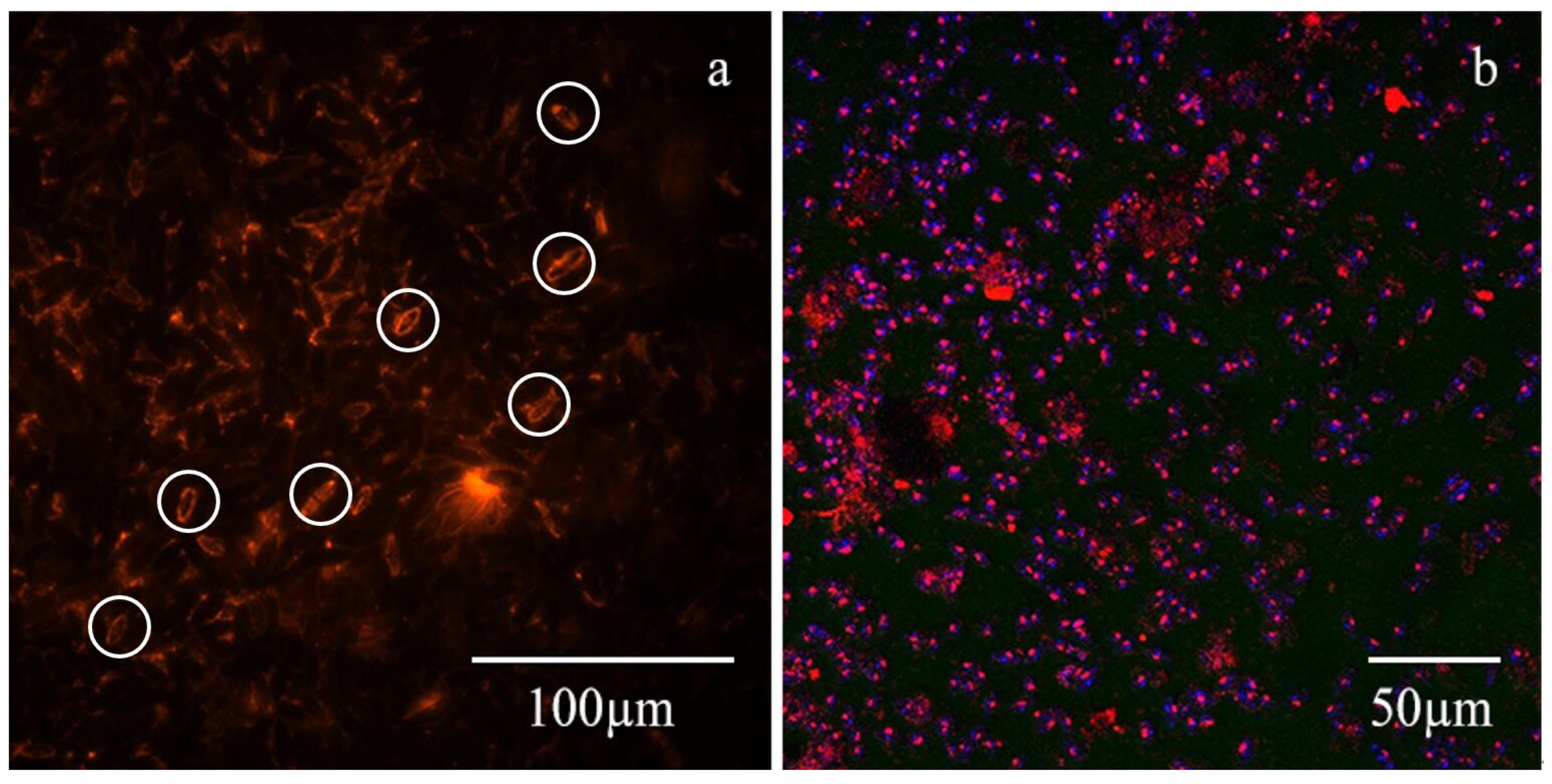
For analyzing multi-fluorescence labeled biofilm samples, the split menu was used in the Zeiss ZEN 2012 v 1.1.2.0 software (Zeiss, Jena, Germany) to capture the single-color fluorescence independent focus, and the merged menu was used to obtain the multi-fluorescence synthesis (Figure 2a). Additionally, the sequential scanning program and spectral analysis technique were used to overcome the channel spectral cross interference.
As shown in Figure 2a, the exopolysaccharides (red) were predominantly distributed around the algae (diatoms), some of which were marked in the white circle. Di Pippo et al. [34] also found similar results in their study. Other studies found that when the EPS secreted by diatoms was firmly attached to the carrier, some bacteria or small colonies gathered around the diatoms. The EPS secreted by diatoms is used by bacteria, while small zooplankton feed on bacteria [35]. The position of bacteria around the extracellular polysaccharide is shown in Figure 2b, which also confirms our results and indicates the importance of EPS for bacterial adhesion and biofilm formation. From the definition of EPS and our observations (Figure 2), we found that the EPS fractions mostly belonged to the TB-EPS group. This further supported our conclusion that TB-EPS and polysaccharide contents were high under turbulent flow conditions.

3.2.2. The Relationship between the Distribution of EPS in Biofilms and Biofilm Formation

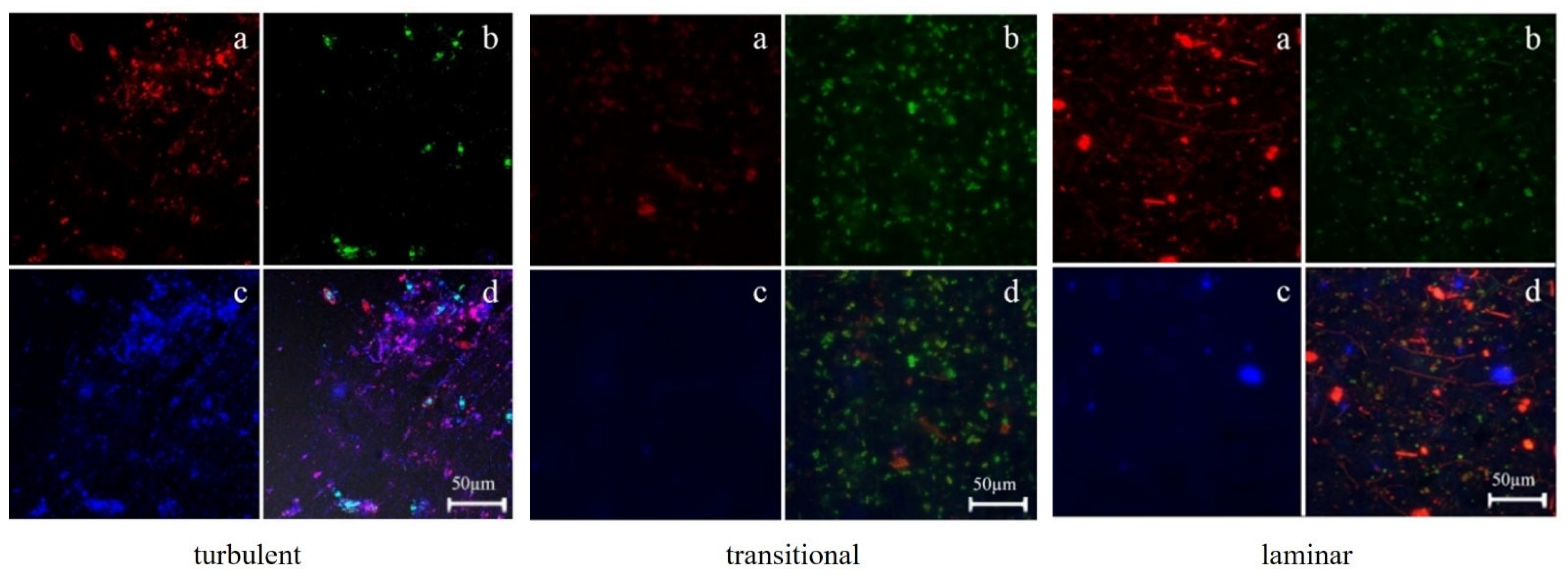
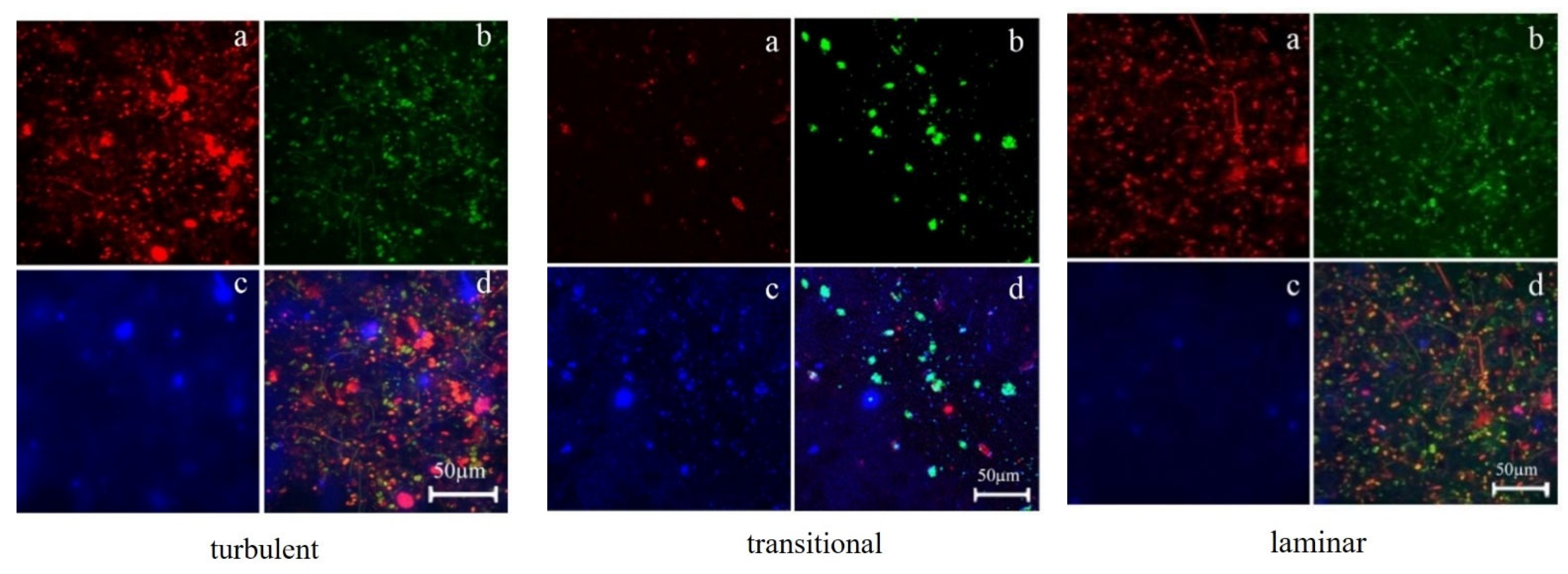
The CLSM images were analyzed using the Zeiss ZEN 2012 v 1.1.2.0 software (Zeiss). The function of “Maximum Intensity Projection” was used to obtain the CLSM images of the biofilms. The “split” function was used to decompose the images, and the images of extracellular polysaccharide (extracellular protein), bacteria, and a combination of the three substances were obtained. After further growth of the biofilm, the CLSM data showed that the biofilm thickness increased after the adhesion of algae and bacteria embedded within the biofilm matrix (Figure 3, Figure 4, Figure 5 and Figure 6).
By using CLSM for dynamic observation of biofilms, the CLSM images showed that the biofilm cultured after the 48th day did not change much compared to that on the 48th day, which might be because the biofilm became too thick. However, the CLSM technique can be used to perform more accurate qualitative and quantitative analyses of EPS in early biofilms [36]. After 48 days, the biofilms (dense or thick biofilms) were embedded in paraffin and frozen; a medical animal, plant, and human tissue slicer was used to cut thin sections of the biofilm. However, all the experiments failed. Therefore, due to the technical limitations, it is not possible to obtain CLSM images that are different between biofilms cultured after 48 days and those cultured on the 48th day. However, based on the dynamic monitoring of the growth cycle of the biofilm, the CLSM image of the biofilm cultured on the 48th day can better reflect the dynamics of biofilm formation under the three flow states, which include the EPS, bacterial distribution, and biofilm structure.
Under the same water flow condition, the content of extracellular polysaccharides in the biofilms during the same period exceeded that of extracellular proteins (Figure 3 and Figure 4), which was consistent with the results of the distribution extraction method, i.e., the content of extracellular polysaccharides and extracellular proteins, as shown in Figure 1; this further supported the results. According to the properties and distribution characteristics of various forms of EPS, most of the EPS in the figure should be LB-EPS and TB-EPS, which is related to S-EPS being soluble in water, hence, S-EPS being easy to be washed off during dyeing [3]. Several images showed the same characteristics.
The contents of extracellular polysaccharides, extracellular proteins, and bacteria detected on the biofilm on the 8th day were low (Figure 3). Low levels of EPS were secreted, most of which were S-EPS. However, S-EPS lacks the structural strength provided by bound EPS. This can lead to the detachment of cells from the biofilm [3]. Then, EPS flowed into the water rapidly, and some of them were washed off during dyeing. Under turbulent flow conditions, the effects of water drag force were not conducive to biological adhesion; thus, EPS and bacteria were relatively less. The flow velocity of the transitional flow was lesser than that of the turbulent flow, which helped the microorganisms adhere to the surface of the carrier because of the combination of sedimentation, convection, and diffusion [37]. Under laminar flow conditions, firstly, gravity and other functions play a key role, especially because many suspended particles carry microorganisms from the water [38]. Therefore, microorganisms under such conditions get deposited on the surface of the carrier due to the action of various suspended particles. Secondly, most of the EPS was S-EPS, which can act as a transport medium for nutrients, facilitating their diffusion through the biofilm matrix. This allows microorganisms to access essential nutrients and maintain metabolic activity. Thirdly, homogeneous surface coverage by S-EPS renders easier the adhesion of new planktonic/daughter cells [3]. These may be the reasons for the higher biomass and thicker biofilm under laminar flow conditions. At this stage, the contents of extracellular proteins and polysaccharides of bacteria were higher than those under other hydrodynamic conditions, and the least under turbulent conditions. Additionally, due to the effect of the fractions and content of EPS, biological adhesion in biofilms may fall off again and return to water. Hence, this stage was defined as Stage I, i.e., the initial adhesion stage of biofilm formation [3,39,40,41].
The contents of extracellular polysaccharides and proteins and the number of bacteria monitored in the biofilm on day 15 increased (Figure 4). EPS and bacteria in the biofilm increased the most under turbulent conditions during the corresponding period. This might have occurred because microorganisms secrete more EPS under stimulation to minimize hydraulic resistance under conditions that are unsuitable for adhesion [5]. The EPS and bacteria in the biofilms cultured under transitional and laminar flow conditions were relatively scattered and independent of each other, as shown in Figure 4. However, the distribution of EPS and bacteria under turbulent flow conditions was highly uneven, and small colonies appeared locally due to the secretion and distribution of LB-EPS under such conditions. LB-EPS has good flocculability and settleability [14], and thus, it promotes biological agglomeration and adhesion of living extracellular polymers, which form a layer of hydrogel that covers the cell surface [11,15]. Under such conditions, biofilm formation enters the irreversible adhesion stage (stage II), where EPS strongly influences biological adhesion. Overall, the results of this study provided strong support to the previous finding that EPS is the “House of Biofilm Cells” of the biofilm [33], especially since the flocculation and bonding characteristics of LB-EPS play a bridging role. The EPS forms the skeleton of the biofilm and provides functional characteristics, such as bonding and bridging, to regulate the formation and structure of the biofilm [42].
The number of biofilms and microorganisms as well as the content of EPS increased considerably on day 32 (Figure 5). From the perspective of the distribution of EPS during this period, biofilms, microorganisms, and EPS were distributed around algae. EPS was mainly secreted by TB-EPS. Under laminar and transitional flow conditions, the distribution of microorganisms and EPS superimposed in a 3D shape (Figure 5) is relatively independent of each other. This might be because LB-EPS in the biofilms formed in this period were secreted to a lesser extent under these two conditions. However, at this time, the distribution of bacteria and EPS showed a distinct 3D layer. Another study found that, with an increase in bacteria, EPS continued to increase, and osmotic pressure increased, resulting in expansion of the gel and biofilm area [38]. Many microcolonies formed in this period. Hence, this biofilm was categorized as a Stage III biofilm [3,38].
The biofilm cultured until the 48th day formed the 3D structure (Figure 6), especially in the turbulent flow. The microorganisms were tightly wrapped by EPS. The content of extracellular polysaccharides was considerably high; however, the quantity of extracellular protein was negligible. When the actual quantity of EPS was measured, no difference was found between the quantities of extracellular proteins and extracellular polysaccharides (Figure 1a). This might be because the adhesion of extracellular polysaccharides during the dyeing of dense spaces provides an advantage while staining, which affects the infiltration of the FITC stain; hence, the level of stained extracellular protein was low [43]. The biofilm under the conditions of transitional flow and laminar flow was reduced multiple times when scanned by CLSM. As EPS was relatively independent of each other, the field of vision was expanded for thorough observation. The biofilm in the transitional flow had an obvious sense of layers, and the bacteria were clustered. In the laminar flow, the content of extracellular proteins and polysaccharides and the number of bacteria in the biofilms were high, and they were evenly distributed. The results of the previous analysis showed that the content of TB-EPS was high, while that of LB-EPS remained relatively low (consistent with the data in Figure 1a), without the flocculability and settleability of LB-EPS. S-EPS in the biofilm was partially removed by washing during the dyeing process. Therefore, as shown in the CLSM diagram of the biofilm under the laminar flow, the three substances were relatively independent of each other and had small colonies, but there was more quantity of EPS than that under transitional flow conditions. This stage was the maturity stage (Stage IV) of biofilm formation [3,38,41].
A comparison of the images in the turbulent flow (Figure 3, Figure 4, Figure 5 and Figure 6) indicated that the content and distribution of extracellular polysaccharides in the biofilms formed on the 15th, 32nd, and 48th days, and the distribution of S-EPS in the initial stage was more than that of S-EPS in the other stages. At the early stage of biofilm growth, the distribution of extracellular polysaccharides in biofilms in the turbulent flow was more evenly scattered and uniform in the laminar flow. For mature biofilms, the distribution of extracellular polysaccharides in biofilms in the turbulent flow appeared to be uniform and tightly wrapped together. However, the distribution of exopolysaccharides in the laminar flow was still uniform but still independent. LB-EPS played a key role in biofilm formation, showing strong flocculation ability [11]. Therefore, more bacteria adhered to the biofilm and continued to secrete more TB-EPS, which was consistent with the changing trend of various EPS fractions, as shown in Figure 1.
Along with Figure 3, Figure 4, Figure 5 and Figure 6, the CLSM images of the biofilms in the transitional flow were compared. The content of extracellular polysaccharides, extracellular proteins, bacteria, and algae was increased. After the algae were attached, their number increased considerably on days 8 and 15, and EPS was secreted. The green fluorescence observed on the 32nd day was mostly emitted after the extracellular proteins were stained. Also, bacterial adhesion occurred to a greater extent during this period. Thus, under hydrodynamic conditions, bacteria and algae adhere to the carrier, secrete EPS, adhere to more bacteria and algae, and continue to secrete EPS and promote biofilm formation [9].
Along with Figure 3, Figure 4, Figure 5 and Figure 6, the images of biofilms in the laminar flow were compared. Both algae and EPS were increased, though the content of EPS in the biofilm was lower. This might be attributed to the high levels of S-EPS (soluble in water) under such conditions. Therefore, the biofilm structure might be unstable. S-EPS is more susceptible to shear forces, such as fluid flow or physical disturbance. Based on the distribution of EPS around algae and the data shown in Figure 1, most EPS in the CLSM images were LB-EPS and TB-EPS, which resulted in the formation of a compact and stable biofilm structure that is not susceptible to external interference under turbulent conditions. Additionally, EPS and bacteria were evenly distributed in the whole process, and the bacterial micelles were dispersed, which was also related to the flocculability and settleability of LB-EPS. Bound EPS forms a matrix that holds the microbial cells together, providing structural stability to the biofilm. It helps in maintaining the biofilm’s architecture and prevents cell detachment. Bound EPS may hinder the diffusion of signaling molecules, reducing intercellular communication within the biofilm. These also verified that there were more bound EPS in the biofilm under turbulent flow conditions, resulting in a dense biofilm structure and obvious restriction of dissolved oxygen mass transfer [44].

4. Conclusions

Hydrodynamic conditions affect the composition of EPS, thus affecting the biofilm formation. Multi-disciplinary methods provide complementary insights into the production and distribution of EPS in biofilms. Through the mutual verification of the results in the microscopic and macroscopic fields, the relationships between the dynamic changes in the production and distribution of EPS, and the hydrodynamic conditions during biofilm formation were evaluated to further elucidate the mechanism of biofilm formation. To summarize, the biofilm formation process can be described as follows: bacteria and algae attach randomly and secret less EPS. In Stage II, higher-level EPS is secreted, and the adhesion of bacteria and algae enters the irreversible adhesion formation stage. In Stage III, the formation of microcolonies and biofilms enter the high-speed formation stage. In Stage IV, under the special bonding effect of LB-EPS and TB-EPS, many colonies adhere to the mature biofilm with a 3D structure. We found that TB-EPS and LB-EPS were heavily secreted under turbulent flow conditions, while slightly more S-EPS was secreted in the laminar flow. S-EPS lacks the structural strength provided by bound EPS. Therefore, the structure of biofilms cultured under the laminar flow was relatively loose. When the content of LB-EPS gradually increases, microorganisms can accumulate in large numbers, thus secreting more TB-EPS. This is also the key reason for the overall high levels of TB-EPS in a turbulent flow. The content of LB-EPS under turbulent conditions was high and the overall biofilm structure was dense. Polysaccharides dominated the EPS in biofilms under three hydrodynamic conditions. They were at the core of biofilm formation enhanced bacterial aggregation, and further increased the biofilm density. To better characterize and parameterize the effect of EPS on the biofilm, future studies are suggested to investigate the composition and properties of extracellular polysaccharides and proteins in EPS, which better regulate river biofilm growth and to better apply biofilm technologies in river ecological restoration projects.

Supplementary Materials

The following supporting information can be downloaded at: https://www.mdpi.com/article/10.3390/w15213821/s1, Table S1: Hydraulic Characteristics of Flumes. References [45,46,47,48] are citied in the Supplementary Materials.

Author Contributions

M.P.: conceptualization, writing—original draft; H.L.: validation, methodology, review and editing; X.H.: investigation, methodology; S.J.: data curation; Y.D.: methodology; W.M.: visualization; X.L.: data curation; J.Q.: methodology; J.Y.: methodology; Z.W.: investigation. All authors have read and agreed to the published version of the manuscript.

Funding

This research was funded by Major Project of the Natural Science Foundation of Jiangsu University, China (No. 22KJA610006).

Data Availability Statement

Not applicable.

Acknowledgments

The authors appreciate the support of the Major Project of the Natural Science Foundation of Jiangsu University, China (No. 22KJA610006).

Conflicts of Interest

The authors declare that they have no known competing financial interests or personal relationships that could have appeared to influence the work reported in this paper.

Nomenclature

EPSextracellular polymeric substance
SB-EPSdissolved EPS
LB-EPSloosely bound EPS
TB-EPStightly bound EPS
3Dthree-dimensional
CLSMconfocal laser scanning microscope
LSMlaser scanning microscope
FITCfluorescein-isothiocyanate concanavalin
ConA-TMRA conjugated with tetramethylrhodamine
DAPI4′,6-diamidino-2-phenylindole
Llitre
PBSphosphate-buffered saline

References

  1. Flemming, H.C.; Wingender, J.; Szewzyk, U.; Steinberg, P.; Rice, S.A.; Kjelleberg, S. Biofilms: An emergent form of bacterial life. Nat. Rev. Microbiol. 2016, 14, 563–575. [Google Scholar] [CrossRef] [PubMed]
  2. Risse-Buhl, U.; Anlanger, C.; Kalla, K.; Neu, T.R.; Noss, C.; Lorke, A.; Weitere, M. The role of hydrodynamics in shaping the composition and architecture of epilithic biofilms in fluvial ecosystems. Water Res. 2017, 127, 211–222. [Google Scholar] [CrossRef] [PubMed]
  3. Janissen, R.; Murillo, D.M.; Niza, B.; Sahoo, P.K.; Nobrega, M.M.; Cesar, C.L.; Temperini, M.L.A.; Carvalho, H.F.; de Souza, A.A.; Cotta, M.A. Spatiotemporal distribution of different extracellular polymeric substances and filamentation mediate Xylella fastidiosa adhesion and biofilm formation. Sci. Rep. 2015, 5, 9856. [Google Scholar] [CrossRef] [PubMed]
  4. Jafari, M.; Desmond, P.; Van Loosdrecht, M.C.M.; Derlon, N.; Morgenroth, E.; Picioreanu, C. Effect of biofilm structural deformation on hydraulic resistance during ultrafiltration: A numerical and experimental study. Water Res. 2018, 145, 375–387. [Google Scholar] [CrossRef]
  5. Desmond, P.; Best, J.P.; Morgenroth, E.; Derlon, N. Linking composition of extracellular polymeric substances (EPS) to the physical structure and hydraulic resistance of membrane biofilms. Water Res. 2018, 132, 211–221. [Google Scholar] [CrossRef]
  6. Flemming, H.C.; Wingender, J. The biofilm matrix. Nat. Rev. Microbiol. 2010, 8, 623–633. [Google Scholar] [CrossRef] [PubMed]
  7. Osella, M.; Tans, S.J.; Cosentino Lagomarsino, M. The Peculiar Functions of the Bacterial Extracellular Matrix. Trends Microbiol. 2017, 25, 250–256. [Google Scholar] [CrossRef]
  8. Sun, P.; Zhang, J.; Esquivel-Elizondo, S.; Ma, L.; Wu, Y. Uncovering the flocculating potential of extracellular polymeric substances produced by periphytic biofilms. Bioresour. Technol. 2018, 248 Pt B, 56–60. [Google Scholar] [CrossRef]
  9. Battin, T.J.; Besemer, K.; Bengtsson, M.M.; Romani, A.M.; Packmann, A.I. The ecology and biogeochemistry of stream biofilms. Nat. Rev. Microbiol. 2016, 14, 251–263. [Google Scholar] [CrossRef]
  10. Sheng, G.P.; Yu, H.Q.; Li, X.Y. Extracellular polymeric substances (eps) of microbial aggregates in biological wastewater treatment systems: A review. Biotechnol. Adv. 2010, 28, 882–894. [Google Scholar] [CrossRef]
  11. Zhang, P.; Fang, F.; Chen, Y.P.; Shen, Y.; Zhang, W.; Yang, J.X.; Li, C.; Guo, J.S.; Liu, S.Y.; Huang, Y.; et al. Composition of EPS fractions from suspended sludge and biofilm and their roles in microbial cell aggregation. Chemosphere 2014, 117, 59–65. [Google Scholar] [CrossRef] [PubMed]
  12. Zhang, H.; Lu, X.; Song, L.; Zhang, L. Effects of loosely bound eps release and floc reconstruction on sludge dewaterability. Water Air Soil Pollut. 2018, 229, 27. [Google Scholar] [CrossRef]
  13. Sheng, G.P.; Yu, H.Q. Characterization of extracellular polymeric substances of aerobic and anaerobic sludge using three-dimensional excitation and emission matrix fluorescence spectroscopy. Water Res. 2006, 40, 1233–1239. [Google Scholar] [CrossRef] [PubMed]
  14. Chen, Y.P.; Zhang, P.; Guo, J.S.; Fang, F.; Li, C. Functional groups characteristics of eps in biofilm growing on different carriers. Chemosphere 2013, 92, 633–638. [Google Scholar] [CrossRef]
  15. Xing, L.; Yang, J.; Ni, B.J.; Yang, C.; Yuan, C.; Li, A. Insight into the generation and consumption mechanism of tightly bound and loosely bound extracellular polymeric substances by mathematical modeling. Sci. Total Environ. 2022, 811, 152359. [Google Scholar] [CrossRef]
  16. Li, X.Y.; Yang, S.F. Influence of loosely bound extracellular polymeric substances (EPS) on the flocculation, sedimentation and dewaterability of activated sludge. Water Res. 2007, 41, 1022–1030. [Google Scholar] [CrossRef]
  17. Yu, G.H.; He, P.J.; Shao, L.M. Characteristics of extracellular polymeric substances (EPS) fractions from excess sludges and their effects on bioflocculability. Bioresour. Technol. 2009, 100, 3193–3198. [Google Scholar] [CrossRef]
  18. Basuvaraj, M.; Fein, J.; Liss, S.N. Protein and polysaccharide content of tightly and loosely bound extracellular polymeric substances and the development of a granular activated sludge floc. Water Res. 2015, 82, 104–117. [Google Scholar] [CrossRef]
  19. Pan, M.; Liu, X.; Ma, W.; Li, X.; Li, H.; Ding, C.; Chen, Y.; Chen, R. The effect of hydrodynamics on the succession of autotrophic and heterotrophic organisms of biofilms in river ecosystems. Water Sci. Technol. 2021, 83, 63–76. [Google Scholar] [CrossRef]
  20. Pan, M.; Li, H.Z.; Han, X.H.; Ma, W.X.; Li, X.; Guo, Q.Y.; Yang, B.R.; Ding, C.; Ma, Y.W. Effects of hydrodynamic conditions on the composition, spatiotemporal distribution of different extracellular polymeric substances and the architecture of biofilms. Chemosphere 2022, 307, 135965. [Google Scholar] [CrossRef]
  21. Romaní, A.M.; Fund, K.; Artigas, J.; Schwartz, T.; Sabater, S.; Obst, U. Relevance of Polymeric Matrix Enzymes During Biofilm Formation. Microb. Ecol. 2008, 56, 427–436. [Google Scholar] [CrossRef] [PubMed]
  22. Proia, L.; Romaní, A.M.; Sabater, S. Nutrients and light effects on stream biofilms: A combined assessment with CLSM, structural and functional parameters. Hydrobiologia 2012, 695, 281–291. [Google Scholar] [CrossRef]
  23. Johnson, J.A.; Fusaro, R.M. The quantitative enzymic determination of animal liver glycogen. Anal. Biochem. 1966, 15, 140–149. [Google Scholar] [CrossRef] [PubMed]
  24. Waterborg, J.H. The Lowry Method for Protein Quantitation. In The Protein Protocols Handbook, 3rd ed.; Humana Press Inc.: Totowa, NJ, USA, 2009; pp. 7–9. [Google Scholar]
  25. Ras, M.; Lefebvre, D.; Derlon, N.; Paul, E.; Girbal-Neuhauser, E. Extracellular Polymeric Substances diversity of biofilms grown under contrasted environmental conditions. Water Res. 2011, 45, 1529–1538. [Google Scholar] [CrossRef]
  26. Battin, T.J.; Kaplan, L.A.; Newbold, J.D.; Cheng, X.; Hansen, C. Effects of Current Velocity on the Nascent Architecture of Stream Microbial Biofilms. Appl. Environ. Microbiol. 2003, 69, 5443–5452. [Google Scholar] [CrossRef]
  27. Orgad, O.; Oren, Y.; Walker, S.L.; Herzberg, M. The role of alginate in Pseudomonas aeruginosa EPS adherence, viscoelastic properties and cell attachment. Biofouling 2011, 27, 787–798. [Google Scholar] [CrossRef]
  28. Chew, S.C.; Kundukad, B.; Seviour, T.; Van der Maarel, J.R.; Yang, L.; Rice, S.A.; Doyle, P.; Kjelleberg, S. Dynamic remodeling of microbial biofilms by functionally distinct exopolysaccharides. mBio 2014, 5, e01536-14. [Google Scholar] [CrossRef]
  29. Nadell, C.D.; Ricaurte, D.; Yan, J.; Drescher, K.; Bassler, B.L. Flow environment and matrix structure interact to determine spatial competition in Pseudomonas aeruginosa biofilms. Elife 2017, 6, e21855. [Google Scholar] [CrossRef]
  30. Zhao, W.; Yang, S.; Huang, Q.; Peng, C. Bacterial cell surface properties: Role of loosely bound extracellular polymeric substances (LB-EPS). Colloids Surf. B Biointerfaces 2015, 128, 600–607. [Google Scholar] [CrossRef]
  31. Besemer, K.; Singer, G.; Limberger, R.; Chlup, A.K.; Hochedlinger, G.; Hodl, I.; Baranyi, C.; Battin, T.J. Biophysical controls on community succession in stream biofilms. Appl. Environ. Microbiol. 2007, 73, 4966–4974. [Google Scholar] [CrossRef]
  32. Flemming, H.C.; Wingender, J. The Crucial Role of Extracellular Polymeric Substances in Biofilms; IWA Publishing: London, UK, 2003; pp. 178–210. [Google Scholar]
  33. Flemming, H.C.; Neu, T.R.; Wozniak, D.J. The EPS Matrix: The “House of Biofilm Cells”. J. Bacteriol. 2007, 189, 7945–7947. [Google Scholar] [CrossRef] [PubMed]
  34. Di Pippo, F.; Ellwood, N.T.W.; Gismondi, A.; Bruno, L.; Rossi, F.; Magni, P.; De Philippis, R. Characterization of exopolysaccharides produced by seven biofilm-forming cyanobacterial strains for biotechnological applications. J. Appl. Phycol. 2013, 25, 1697–1708. [Google Scholar] [CrossRef]
  35. Goldsborough, L.G.; Hickman, M. A comparison of periphytic algal biomass and community structure on Scirpus validus and on a morphologically similar artificial substratum. J. Phycol. 1991, 27, 196–206. [Google Scholar] [CrossRef]
  36. Halan, B.; Buehler, K.; Schmid, A. Biofilms as living catalysts in continuous chemical syntheses. Trends Biotechnol. 2012, 30, 453–465. [Google Scholar] [CrossRef] [PubMed]
  37. Li, J.; Busscher, H.J.; Norde, W.; Sjollema, J. Analysis of the contribution of sedimentation to bacterial mass transport in a parallel plate flow chamber. Colloids Surf. B Biointerfaces 2011, 84, 76–81. [Google Scholar] [CrossRef]
  38. Sauer, K. The genomics and proteomics of biofilm formation. Genome Biol. 2003, 4, 219–224. [Google Scholar] [CrossRef]
  39. Carniello, V.; Peterson, B.W.; van der Mei, H.C.; Busscher, H.J. Physico-chemistry from initial bacterial adhesion to surface-programmed biofilm growth. Adv. Colloid Interface Sci. 2018, 261, 1–14. [Google Scholar] [CrossRef]
  40. Busscher, H.J.; van der Mei, H.C. Microbial adhesion in flow displacement systems. Clin. Microbiol. Rev. 2006, 19, 127–141. [Google Scholar] [CrossRef]
  41. Muhammad, M.H.; Idris, A.L.; Fan, X.; Guo, Y.; Huang, T. Beyond risk: Bacterial biofilms and their regulating approaches. Front. Microbiol. 2020, 11, 928. [Google Scholar] [CrossRef]
  42. Cogan, N.G.; Keener, J.P. The role of the biofilm matrix in structural development. Math. Med. Biol. 2004, 21, 147–166. [Google Scholar] [CrossRef]
  43. Barranguet, C.; Beusekom, S.A.M.V.; Veuger, B.; Neu, T.R.; Manders, E.M.M.; Sinke, J.J.; Admiraal, W. Studying undisturbed autotrophic biofilms: Still a technical challenge. Aquat. Microb. Ecol. 2004, 34, 1–9. [Google Scholar] [CrossRef]
  44. Pan, M.; Li, H.; Han, X.; Quan, G.; Ma, W.; Guo, Q.; Li, X.; Yang, B.; Ding, C.; Chen, Y.; et al. Effect of hydrodynamics on the transformation of nitrogen in river water by regulating the mass transfer performance of dissolved oxygen in biofilm. Chemosphere 2023, 312, 137013. [Google Scholar] [CrossRef] [PubMed]
  45. Singer, G.; Besemer, K.; Hödl, I.; Chlup, A.; Hochedlinger, G.; Stadler, P.; Battin, T.J. Microcosm design and evaluation to study stream microbial biofilms. Limnol. Oceanogr. Methods 2006, 4, 436–447. [Google Scholar] [CrossRef]
  46. Chen, M.Y.; Lee DJ Tay, J.H. Distribution of extracellular polymeric substances in aerobic granules. Appl. Microbiol. Biotechnol. 2006, 73, 1463–1469. [Google Scholar] [CrossRef]
  47. Chen, M.Y.; Lee, D.J.; Tay, J.H.; Show, K.Y. Staining of extracellular polymeric substances and cells in bioaggregates. Appl. Microbiol. Biotechnol. 2007, 75, 467–474. [Google Scholar] [CrossRef]
  48. Yu, G.H.; Tang, Z.; Xu, Y.C.; Shen, Q.R. Multiple Fluorescence Labeling and Two Dimensional FTIR–13C NMR Heterospectral Correlation Spectroscopy to Characterize Extracellular Polymeric Substances in Biofilms Produced during Composting. Environ. Sci. Technol. 2011, 45, 9224–9231. [Google Scholar] [CrossRef]
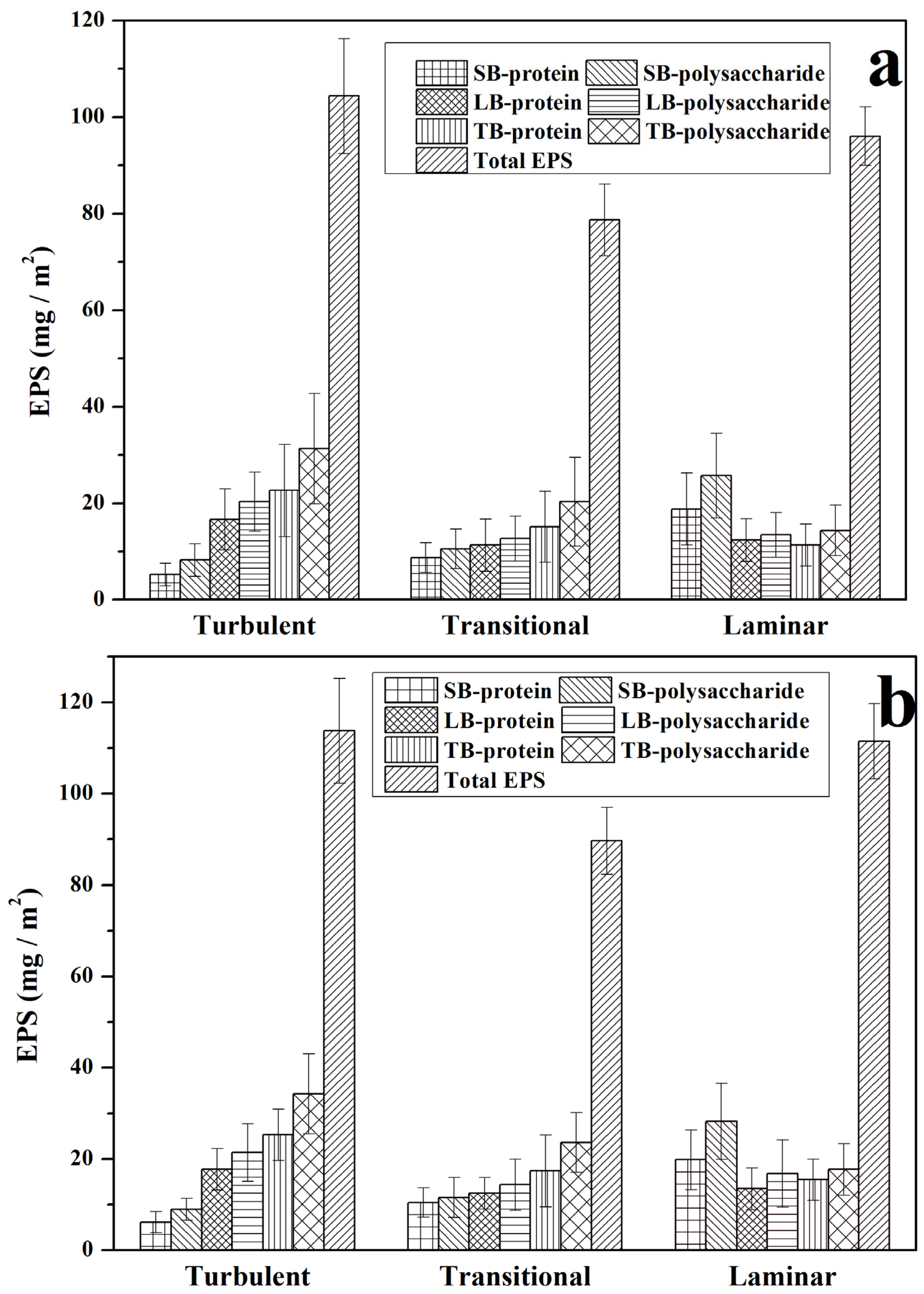
Figure 1. The contents of polysaccharides and proteins in the S-EPS, LB-EPS, and TB-EPS under three hydrodynamic conditions during the biofilm formation. The biofilm was cultivated on (a) the 48th and (b) 60th days.
Water 15 03821 g001
Figure 2. The CLSM images of extracellular polysaccharides and bacteria in the biofilm cultured in the turbulent flow. The ones marked in the white circle in (a) represent algae (diatom). (a) α-extracellular polysaccharides; and (b) a merged image of α-polysaccharide and bacteria. Red indicates α-extracellular polysaccharides (ConA-TMR); Blue indicates bacteria (DAPI).
Water 15 03821 g002
Figure 3. The CLSM maximum overlay projection images of the growth of biofilms under three hydrodynamic conditions after the 8th day of cultivation. (a) Red indicates α-extracellular polysaccharides; (b) Green indicates extracellular proteins; (c) Blue indicates bacteria; and (d) Merged images of (ac).
Water 15 03821 g003
Figure 4. The CLSM maximum overlay projection images of the growth of biofilms under three hydrodynamic conditions after the 15th day of cultivation. (a) Red indicates α-extracellular polysaccharide; (b) Green indicates extracellular protein; (c) Blue indicates bacteria; and (d) Merged images of (ac).
Water 15 03821 g004
Figure 5. The CLSM maximum overlay projection images of the growth of biofilms under three hydrodynamic conditions after the 32nd day of cultivation. (a) Red indicates α-extracellular polysaccharide; (b) Green indicates extracellular protein; (c) Blue indicates bacteria; and (d) Merged images of (ac).
Water 15 03821 g005
Figure 6. The CLSM maximum overlay projection images of the growth of biofilms under three hydrodynamic conditions after the 48th day of cultivation. (a) Red indicates α-extracellular polysaccharide; (b) Green indicates extracellular protein; (c) Blue indicates bacteria; and (d) Merged images of (ac).
Water 15 03821 g006
Disclaimer/Publisher’s Note: The statements, opinions and data contained in all publications are solely those of the individual author(s) and contributor(s) and not of MDPI and/or the editor(s). MDPI and/or the editor(s) disclaim responsibility for any injury to people or property resulting from any ideas, methods, instructions or products referred to in the content.