Article

Water and Sediments of an Acidic Hot Spring—Distinct Differentiation with Regard to the Microbial Community Composition and Functions

by
Anastasia I. Maltseva
,
Alexandra A. Klyukina
*,
Alexander G. Elcheninov
,
Nikolay V. Pimenov
,
Igor I. Rusanov
,
Ilya V. Kublanov
,
Tatiana V. Kochetkova
and
Evgeny N. Frolov
Winogradsky Institute of Microbiology, Federal Research Center of Biotechnology, Russian Academy of Sciences, 60-Let Oktyabrya Prospect, 7, Bld. 2, 119071 Moscow, Russia
*
Author to whom correspondence should be addressed.
Water 2023, 15(19), 3415; https://doi.org/10.3390/w15193415
Submission received: 30 August 2023 / Revised: 23 September 2023 / Accepted: 26 September 2023 / Published: 28 September 2023
(This article belongs to the Section Biodiversity and Functionality of Aquatic Ecosystems)

Abstract

:
Over the last half-century, microbial communities of the Kamchatka hot springs have been largely studied using molecular, radioisotopic, and cultural approaches. Generally, these results were obtained for mixed samples of water with sediments, for only hydrothermal water, or for only sediment samples. Simultaneous comparative analysis of the microbial communities of water and sediments was performed for only one Kamchatka hot spring with circumneutral pH. Here, the microbial communities of both sediments and water (separately) of hot spring #4229 (the Uzon Caldera, Kamchatka) with a temperature of 50–56 °C and pH of 3.2 were analyzed by 16S rRNA gene V4 fragment amplicon sequencing. It was revealed that the microbial community of sediments was represented by uncultured phylogenetically deep-branching lineages of archaea, such as ARK-15 within Thermoplasmatota and ‘Ca. Marsarchaeales’ from the Thermoproteota phyla. Metagenome analysis showed that these archaea most probably carried out the degradation of organic matter. The microbial community of the hot water is represented by thermoacidophilic, (micro)aerobic, chemolithoautotrophic, hydrogen- and sulfur-oxidizing bacteria of the genera Hydrogenobaculum (phylum Aquificota) and Acidithiobacillus (phylum Pseudomonadota). Radioisotopic tracing and DNA-stable-isotope probing techniques proved their role as primary producers in the hot spring. The experiment revealed significant differences in the composition and functions of the microbial communities of sediments and water through the example of a typical acidic hot spring in Kamchatka.

1. Introduction

Hot springs are unique ecosystems with a huge diversity of thermophilic prokaryotes inhabiting them. Hot springs are associated with zones of volcanic activity or higher tectono-magmatic activity. The largest active volcanic region on Earth, called the Pacific Ring of Fire, is spread from the Aleutian Islands to New Zealand and Chile. The Kamchatka Peninsula is a part of the Ring and includes the Uzon Caldera, which is located in the central part of the Eastern Kamchatka on the territory of the Kronotsky Nature Reserve. Uzon is the site of the largest manifestations of geothermal activity in Kamchatka, with numerous hot springs, fumaroles, mud pots, and mud volcanoes, as well as a relatively newly appeared geyser [1]. The caldera contains all major types of hydrothermal springs (chloride-sodium, chloride-sulfate, sulfate-chloride, sulfate, and bicarbonate), distinguishing in chemical composition and a wide range of temperatures from 20 to 97 °C and pH from 2.0 to 8.5 [1,2,3,4]. The variety of physicochemical parameters of hot springs provides a high diversity of microbial communities, which serves as a source for the isolation and characterization of novel thermophilic microorganisms [1,5].
The primary producers in hot springs, in contrast to meso- and psychrophilic ecosystems, are not only photosynthetic organisms but also chemolithoautotrophs. Microbial photosynthesis in thermal springs is limited by temperature: in neutral and alkaline environments, the boundary is 73 °C; in acidic, it is 56 °C [6,7,8]. Cyanobacteria, which carry out oxygenic photosynthesis, dominate in Kamchatka hot springs with temperatures up to 50 °C, followed by purple and green sulfur bacteria. In hot springs with temperatures from 50 to 73 °C, non-sulfur filamentous bacteria of the phylum Chloroflexota, namely representatives of the genera Chloroflexus and Roseiflexus, carry out anoxygenic photosynthesis and could represent a significant part of the community in hot springs with temperatures of up to 62 °C [9,10]. In contrast, chemolithoautotrophy using the energy of redox reactions is limited only by the boiling point of the water in terrestrial hot springs. Hydrothermal fluids, rich in reduced inorganic compounds, first of all, sulfur compounds and hydrogen, provide opportunities for chemolithoautotrophs to dominate in hot vent environments. Among them, bacteria of the phylum Aquificota are the most numerous primary producers that are widely spread in hydrothermal springs all over the world [11,12,13,14,15,16,17,18,19,20]. In Kamchatka hot springs, Aquificota representatives account for up to 85% or more of the whole microbial community [12,18]. In hot springs with temperatures between 60 and 97 °C and pH above 5.0, members of the genus Sulfurihydrogenibium, belonging to this phylum, are the most common [18,20,21,22,23], while bacteria of the genus Hydrogenobaculum inhabit acidic springs [18,20]. An anaerobic hyperthermophilic bacterium Caldimicrobium rimae (phylum Desulfobacterota), able to assimilate CO2, is also quite numerous in the hot springs of the Uzon Caldera with water temperatures from 60 to 90 °C [16,18].
Organic matter, produced by autotrophic organisms, and organic compounds from the outside are metabolized in hydrothermal environments by a diverse group of heterotrophic prokaryotes, mainly facultative or strict anaerobes [1,24]. Representatives of heterotrophic bacteria of the Dictyoglomota, Thermotogota, Bacillota, Caldisericota, Bacteroidota, and Deinococcota phyla are the most common [3,22,23,25]. Among cultivated archaea with heterotrophic metabolism, representatives of the phylum Thermoproteota, such as the Fervidicoccus or Caldisphaera species, as well as the Conexivisphaera species (=the Terrestrial Hot Spring Crenarchaeotic Group or THSCG) dominate in acidic hot springs [1,18]. In hot springs with circumneutral pH, archaea of the genera Pyrobaculum and Thermoproteus (both from Thermoproteota), as well as uncultivated representatives of Korarchaeia (phylum Thermoproteota) and Nanoarchaeota occur [12,16,26].
Thus, over the last half-century, classical microbiological, radioisotopic, and molecular-biological studies have succeeded extensively in studying the microbial metabolism and communities of Uzon hot springs. However, these results were obtained either only for thermal water samples, only for sediments, or microbial mats/filamentous “streamers”, or for a mix of water and sediments [2,4,12,18,20,26,27,28]. Previously, only one work was devoted to the comparison of water and sediment microbial communities separately in the Bourlyashchy Pool of the Uzon Caldera [16]. That study showed the significant difference in the composition and function of microbial communities in water and sediments, where lithoautotrophic representatives of the phyla Aquificota (affiliated to the genera Sulfurihydrogenibium and Hydrogenobacter) and Thermoproteota (genus Pyrobaculum) predominated in the water, whereas Desulfobacterota and Aquificota occupied the sediments. Bourlyashchy is a very special spring in the caldera. It is the hottest one there (with a temperature of above 90 °C) with pH 6.0–7.0, while the majority of hot springs are mildly acidic with moderate temperatures (54–72 °C) [18]. Characterization of the latter with regard to a separate study of microbial community structures in water and sediments is still absent, which may lead to incorrect conclusions about the role of a particular microorganism in microbial associations.
Here, we studied separately the microbial communities of water and sediments of an acidic spring with moderate temperatures using radioisotopic tracing, DNA-stable-isotope probing (DNA-SIP), and the next-generation sequencing (NGS) techniques, including metagenomic and 16S rRNA gene sequences analyses, in order to demonstrate the significant difference in their composition and functions.

2. Materials and Methods

2.1. Characteristics of the Sampling Site

The sediments or water samples of hot spring #4229 (N54.50077111°, E160.00748449°) were collected on the East Thermal Field of the Uzon Caldera (the Kronotsky National Park) in July 2021. Hot spring #4229 is located in the central part of an acidic stream called Izumrudny and has a circle shape about 0.5 m in diameter with gray-yellow sandy-clay sediments (Figure 1). The water in the spring was muddy with a gray-yellow color. The temperature of water and sediments varied from 50 to 56 °C depending on the site. The pH value of the hot spring water was 3.2. The emanation of gases in the form of bubbles rising from the hot spring bottom was observed. Hot spring #4229 received water from an upstream spring and then flowed into the acidic Izumrudny stream. The Izumrudny stream, together with associated hot springs and #4229, was located inside a ravine with clay slopes. The upper part of the ravine was covered with plants, which indicates the entry of allochthonous organic matter into hot springs. Moreover, small depressions in the clay filled with water and cyanobacterial mats were located around the Izumrudny stream and its hot springs.

2.2. DNA Extraction and 16S rRNA Gene Sequencing

For DNA isolation, sediment or water samples were taken aseptically with spoon or spatula in 2 mL Eppendorf tubes with screw caps and then fixed with a stabilizing buffer (100 mM EDTA, 100 mM Tris·HCl, 150 mM NaCl). The water sample was collected at a distance of 25 cm from the edge of the hot spring and from a depth of 5 cm. Sediments were collected at the edge of the hot spring from a depth of 10 cm. During transportation and storage, the fixed samples were maintained at +4 °C and then stored at −20 °C until DNA was extracted. DNA isolation from water (designated as 4229w) and sediment (designated as 4229s) samples fixed with stabilizing buffer was performed using a commercial FastDNA Spin Kit for Soil (MP Biomedicals, Santa Ana, CA, USA) according to the manufacturer’s instructions, including the step of mechanical cell disruption using a FastPrep-24™ 5G homogenizer (MP Biomedicals, USA).
Amplicon libraries of the V4 hypervariable region of the 16S rRNA gene were prepared according to Gohl et al. [29]. The following primer system was used: 515F (5′-GTGBCAGCMGCCGCGGCGGTAA-3′) [30] and 806R (5′-GGACTACNVGGGGTMTCTAATCC-3′) [31]. Libraries were prepared in two replicates for each sample. High-throughput sequencing of the obtained libraries was performed using the MiSeq Reagent Micro Kit v2 (300-cycles) MS-103-1002 (Illumina, San Diego, CA, USA) on a MiSeq System (Illumina, USA).
Primary raw reads were processed using QIIME 2 (version 2019.1) [32] and analyzed using the SILVAngs service (https://ngs.arb-silva.de/silvangs/; SILVA138.1 database, accessed on 19 November 2022) [33]. Taxonomic classification was performed using GTDB taxonomy (https://gtdb.ecogenomic.org/).

2.3. Sequencing and Analysis of the 4229s Metagenome

Libraries for metagenomic sequencing were prepared using the KAPA HyperPlus kit (Roche, Basel, Switzerland). Paired-end sequencing of metagenome 4229s was performed using the Illumina NovaSeq 6000 platform (Illumina, San Diego, CA, USA). Adapter trimming was done using Cutadapt (https://github.com/marcelm/cutadapt). Further reads were filtered in CLC Genomics Workbench v.10 (Qiagen, Venlo, The Netherlands) using the Trim tool (quality limit = 0.03, maximum ambiguous nucleotides = 2, minimum length = 80).
The metagenome was assembled using metaSpades v.3.15.5 [34]; scaffolds with lengths below 200bp were discarded from the assembly. The recovery of metagenome-assembled genomes (MAGs) was done using the metaWRAP v1.3.2 pipeline [35] with three tools, CONCOCT v.1.1.0 [36], MaxBin2 v.2.2.7 [37], and metaBAT2 v.2.12.1 [38]. The following bins consolidation was done using the Bin_refinement module of metaWRAP with a threshold of 50% completeness and 10% contamination estimated CheckM [39]. Taxonomic classification of MAGs was performed using GTDB-Tk v.2.1.1 [40] with the GTDB r207 database as a reference.
The rapid annotation subsystem technology (RAST) [41] was used to perform a general functional annotation of each of the obtained MAGs. The number of genes that were detected by RAST was counted in each functional category for each MAG. R studio using the library ggplot2 v.3.3.5 (ggplot2.tidyverse.org) was used for visualization of the absolute abundance of the subsystem feature counts.
The heatmaps were generated using R studio using the library ggplot2 v.3.3.5 (ggplot2.tidyverse.org).

2.4. DNA Stable-Isotope Probing (DNA-SIP)

In situ incubation of the samples was performed using stable isotope-labeled 13C-carbonate (enrichment of Na213CO3—85%; All-regional association “Izotop”, Russia) and was carried out in 1-L bottles with gas-tight butyl rubber stoppers and screw caps filled with a sample of water from the hot spring with no headspace. Two milliliters of 1M Na213CO3 solution was added to each tube with a syringe through the stopper. Further, the sample bottles were placed at the sampling site and incubated for 6 and 24 h. Each series of measurements had a control sample with unlabeled carbonate to help confirm the enrichment of specific organisms in the 13C-exposed samples. After incubation, the samples were fixed with 6 mL of 2 M NaOH solution and filtered using Sterivex™ filters (Merck Millipore, Darmstadt, Germany) with a pore diameter of 0.22 microns for cell deposition. The filters, together with the cells, were fixed with stabilizing buffer (100 mM EDTA, 100 mM Tris·HCl, 150 mM NaCl) and transported to the laboratory. During transportation and storage, the fixed samples were maintained at +4 °C and then stored at −20 °C until DNA was extracted.
Further processing of the samples was conducted according to the previously described method [42,43] without ethidium bromide addition to a gradient solution of CsCl. To separate light and heavy DNA, ultracentrifugation was performed in an 8.9 mL OptiSeal Polypropylene Tube (16 × 60 mm—56 Pk) using Optima XPN-80 Ultracentrifuge (Beckman Coulter Inc., Brea, CA, USA). Ultracentrifugation conditions were 44,100 rpm (177,000× g) at 20 °C for 40 h with vacuum, maximum acceleration, and without brake (takes an additional 2 h to stop). After ultracentrifugation, the retrieval of DNA was carried out by gradient fractionation from gradients without EtBr. Eighteen fractions with a volume of 0.5 mL were obtained. DNA concentration was determined in each fraction using the Qubit DNA HS Assay Kit (Thermo Fisher Scientific, Waltham, MA, USA). According to the measurement results, fractions with a high concentration of DNA were selected. One fraction was obtained in the control samples, while two fractions were obtained in the experimental samples. These fractions were used for subsequent high-throughput sequencing by the 16S rRNA gene (V4 region).

2.5. Radioisotopic Tracing Experiments

The rates of autotrophic CO2 fixation were measured by means of the radioisotopic technique using 14C-bicarbonate. Radioisotope experiments were performed in three replications in 17 mL Hungate tubes with screw caps that were filled completely with the water sample. To each tube, 0.2 mL of 14C-bicarbonate (28 × 104 Bq) was added with a syringe through the stopper. Incubation was performed in situ at the sampling site for 0, 6, 24, 48, and 72 h, which also allowed tracing the dynamics of the process. After incubation, the samples were fixed with 1 mL of 2 M NaOH solution. Each series of measurements had an abiotic control, which was a sample with the initial addition of NaOH solution before the incubation began. The subsequent treatment was performed as described previously [44].

3. Results

3.1. The Microbial Communities of Sediments and Water in Hot Spring #4229

The taxonomic composition of the microbial communities of sediments or water of hot spring #4229 was determined by NGS data analysis based on 16S rRNA gene sequencing (Figure 2). More than 87% of the microbial population of the sediments (4229s) was represented by archaea, mainly affiliated with the uncultured lineages. The most numerous was the ARK-15 group, with the order level belonging to the phylum Thermoplasmatota, and the uncultured ‘Candidatus Marsarchaeales’ order from the phylum Thermoproteota, representing 41% and 37% of the microbial community, respectively. Another group of archaea, Conexivisphaerales order (=THSCG), comprised 7% of the sediments’ microbial community. Among bacterial components in sediments, members of the genera Mesoaciditoga (3%), Hydrogenobaculum (2%), and Desulfurella (1%) accounted for the greatest number of sequences. The dominant component of the microbial community of hydrothermal water (4229w) was members of the genus Hydrogenobaculum (70% of the total composition). Representatives of the genera Acidithiobacillus (11%), Thiomonas (7%), and Desulfurella (7%) were also abundant.

3.2. Metagenome Analysis of the Sediments Samples 4229s

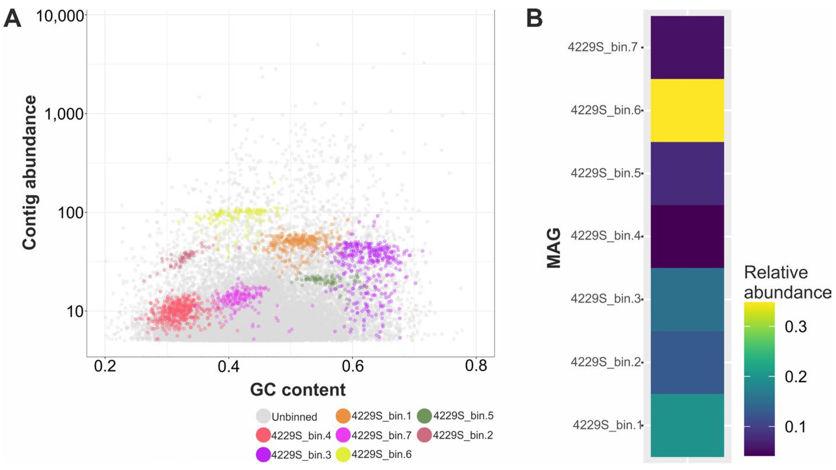
The metagenome assembly of 4229s included 56,941 contigs with a total size of 30,957,030 bp(N50 = 659 bp). Seven MAGs were obtained from the metagenome during the binning process (Figure 3A). Relative abundances of MAGs were 19.7%—4229S_bin.1, 12.7%—4229S_bin.2, 15.3%—4229S_bin.3, 4.1%—4229S_bin.4, 7.7%—4229S_bin.5, 34.8%—4229S_bin.6, and 5.6%—4229S_bin.7 (Figure 3B).
The completeness levels of MAGs were relatively high 64.89–100%, while contamination levels were low, 0-1.25% (Table 1). All seven MAGs were affiliated to the Archaea domain (Table 1). Five MAGs belonged to the Thermoproteota phylum, including ‘Ca. Marsarchaeales’ order (4229S_bin.1 and 4229S_bin.5), Conexivisphaerales order (4229S_bin.3 and 4229S_bin.6), and Caldisphaera genus (4229S_bin.4). Each of Thermoplasmatota (ARK-15 order) and Nanoarchaeota (Parvarchaeales order) phyla were represented by a single MAG, 4229S_bin.2 and 4229S_bin.7, respectively.
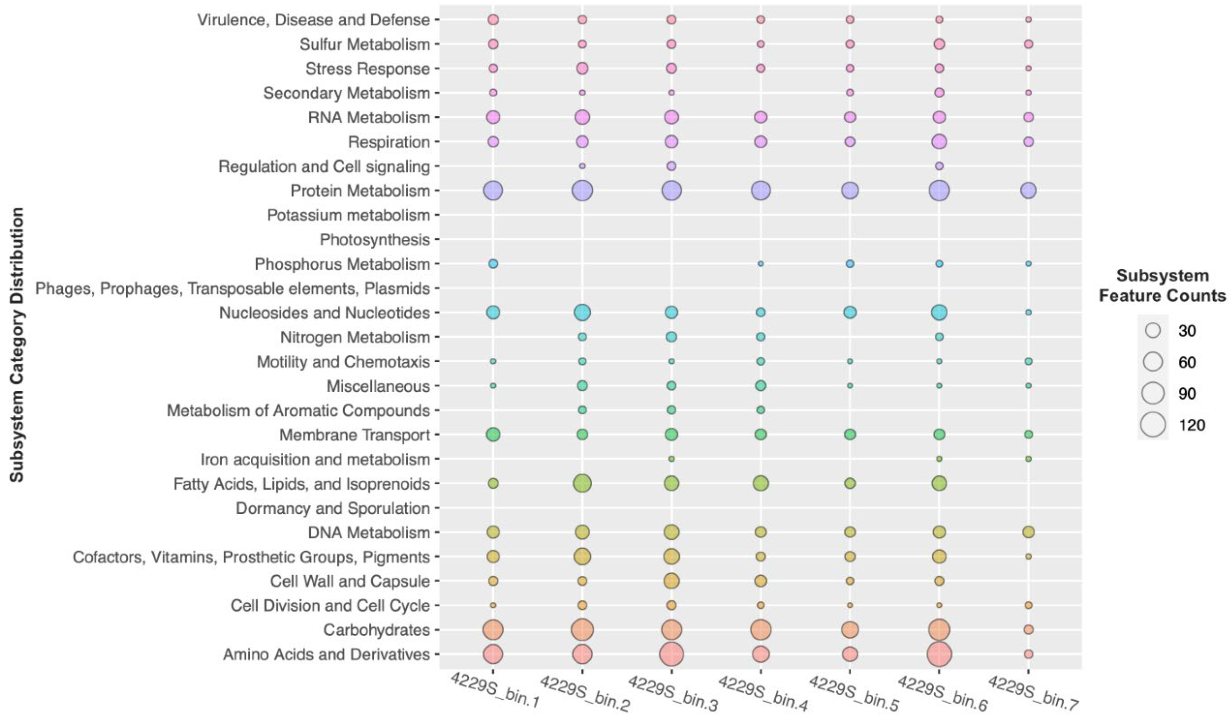
The obtained MAGs were screened for the genes of enzymes of CO2 fixation pathways. This analysis showed that the MAGs lacked the genes encoding enzymes of all known autotrophic pathways: the Calvin–Benson–Bassham cycle, the Wood–Ljungdahl pathway, the reductive glycine pathway, two variants of the reductive tricarboxylic acid cycle, the 3-hydroxypropionate bicycle, the 3-hydroxypropionate/4-hydroxybutyrate cycle, and the dicarboxylate/4-hydroxybutyrate cycle. At the same time, in all obtained MAGs, a large number of genes encoding enzymes involved in the metabolism of proteins, amino acids, carbohydrates, lipids, and fatty acids were identified (Figure 4).

3.3. DNA Stable-Isotope Probing of the Water Samples 4229w

After 6 h of incubation (Figure 2) of the control sample without 13C-carbonate, the share of representatives of the genus Hydrogenobaculum in the microbial community increased from 70% to 88%, while the share of other bacteria decreased (Acidithiobacillus—from 11% to 8%, Thiomonas—from 7% to 0.7%, and Desulfurella—from 7% to 2%). The DNA obtained from the sample with the addition of 13C-carbonate was separated into light and heavy fractions in a gradient solution of CsCl. Analysis of heavy DNA fraction showed that 86% of all sequences belonged to the bacteria of the genus Hydrogenobaculum and 11% to the genus Acidithiobacillus.
After 24 h of incubation (Figure 2) of the control sample, the share of prokaryotes affiliated with the Hydrogenobaculum genus decreased to 60%, while the fractions of the genera Acidithiobacillus and Thiomonas increased up to 15% and 17%, respectively. Analysis of heavy DNA fractions showed that 42% of all sequences belonged to the bacteria of the genus Hydrogenobaculum, 27% to the genus Acidithiobacillus, and 27% to the genus Thiomonas. Thus, it was shown that after 6 h of incubation, the growth rate of Hydrogenobaculum was significantly reduced, but the growth rate of Acidithiobacillus and Thiomonas increased.

3.4. Rates of Carbon Assimilation in the Water Samples 4229w

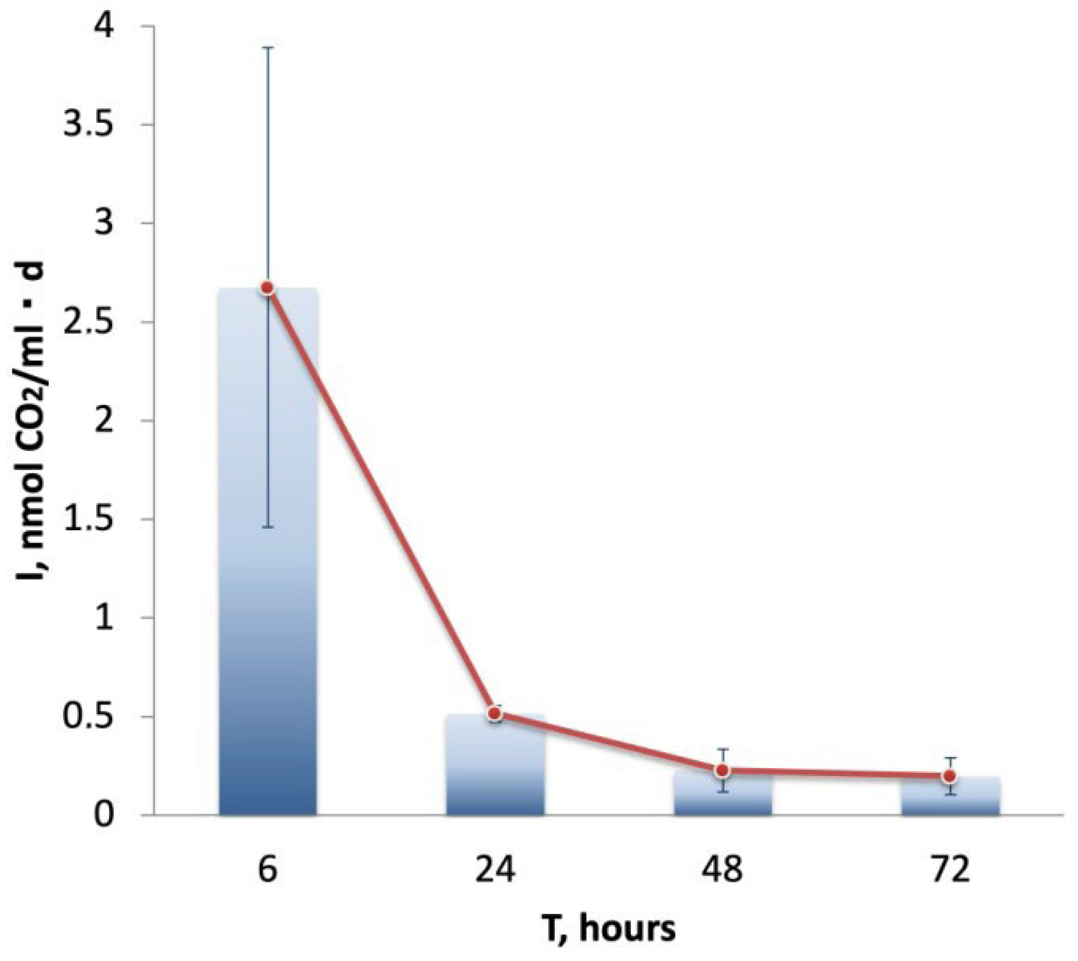
The intensity of the dark assimilation of carbon dioxide in the water samples 4229w was measured dynamically (Figure 5). The maximum rate of carbon dioxide fixation was observed for the first 6 h of incubation and was 2.67 ± 1.21 nmol CO2/(mL day), after which the intensity of this process decreased sharply to a final value of 0.2 ± 0.09 nmol CO2/(mL day).

4. Discussion

By its physicochemical parameters, hot spring #4229 is a typical thermal spring of the Uzon. Although the microbial communities of many Uzon hydrothermal springs have been studied using NGS and other approaches, the vast majority of these works were based on narrow studies of individual water, sediment, or microbial mat samples or, alternatively, they considered total microbial communities of springs. This is one of the few articles where a comparative analysis of microbial communities of water and sediments of the same hot spring was carried out. The 16S rRNA-based taxonomic profiling showed that the microbial community of hot spring #4229 sediments was represented by archaea, mainly affiliated with uncultured lineages. Bacteria comprised only 7% of the sediments’ microbiome. Actually, the ratio between the bacterial and archaeal components in various samples of the Uzon Caldera hot springs varies widely. The archaeal component may consist of 1% to 90% of a total microbial community [4,12,16,18,20,22,26,45]. An archaeal component is often represented there by uncultured phylogenetically deep-branching lineages. Among them, for example, uncultured representatives of the phyla Thermoplasmatota, Nanoarchaeota, or Thermoproteota are the most numerous groups in hot springs [4,12,18,23,26,45,46,47,48,49]. In the considered case, the most abundant archaeal component in hot spring #4229 sediments (41% of the sediments’ microbiome) was an uncultured phylogenetically deep-branching group within the phylum Thermoplasmatota, designated as ARK-15 at the order level. Archaea of this group were detected previously in another hot spring in the Uzon caldera, and the genome of one of them was assembled from the spring metagenome [23]. As it was shown, the ARK-15 MAG was dominated by genes involved in carbohydrate, proteins, and aromatic compounds metabolism. The second archaeal component of the sediments’ microbial community (37%) was another uncultured archaeal phylogenetic group—the order ‘Ca. Marsarchaeales’ within the class Thermoprotei of the phylum Thermoproteota. MAG analysis of some representatives of these archaea indicated an obligate heterotrophic type of metabolism, such as hydrolysis of peptides and complex carbohydrates [50]. The third archaeal component of the sediments’ microbial community (7%) was representatives of the order Conexivisphaerales within the class Nitrososphaeria of the phylum Thermoproteota. Cultivated representatives of this group (Conexivisphaera calida) could not oxidize ammonium and fix inorganic carbon. Instead, it is an anaerobic organoheterotroph, which utilizes yeast extract and peptides [51]. MAG analysis from this study also indicated the chemoorganoheterotrophic metabolism of all archaea inhabited #4229 sediments. From the above, it appears that the sediments’ microbiome, consisting mainly of thermoacidophilic archaea, most likely carries out the destruction of organic matter in hot spring #4229.
The microbial community of the hydrothermal water differed significantly from the microbial community of sediments and was almost exclusively represented by bacteria (99% of the total microbial population). Representatives of the genus Hydrogenobaculum were the dominant component, comprising 70% of the water microbial community. The only cultivated representative of this genus is a thermoacidophilic obligate chemolithoautotrophic bacterium Hydrogenobaculum acidophilum that was isolated from a solfataric field in Japan [52,53]. H. acidophilum uses hydrogen, reduced sulfur compounds, or arsenite as electron donors, oxygen as an electron acceptor, and fixes CO2 via the reductive tricarboxylic acid cycle. The obtained results of DNA-SIP confirmed that bacteria of the genus Hydrogenobaculum were the primary producers in hot spring #4229. In this respect, the hot spring #4229 is similar to many well-studied moderately acidic hot springs around the world, where Hydrogenobaculum representatives are the dominant component [11,13,14,15,16,17,18,19,20,27,54,55,56,57,58,59,60,61]. DNA-SIP in water samples of acid hot springs of the Yellowstone National Park also demonstrated that Hydrogenobaculum species acted as primary producers [7,62].
The next predominant bacteria in the water microbial community of hot spring #4229 were representatives of the genus Acidithiobacillus (phylum Pseudomonadota). The DNA-SIP results demonstrated the autotrophic metabolism of these bacteria; however, their growth rates were probably lower compared to Hydrogenobaculum ones. The genus Acidithiobacillus comprises seven validly published species, and all of them are acidophilic chemolithoautotrophic aerobes oxidizing reduced sulfur compounds, ferrous iron or hydrogen, and fixing CO2 via the Calvin-Benson-Bassham cycle [63,64,65]. Representatives of the genus Acidithiobacillus occur worldwide in sulfur- and iron-rich environments with low pH, including a diverse range of natural (acid rock drainage, sulfur springs, including hot springs, acidic soils, etc.) and industrial settings (ore concentrates, pulps, and leaching solutions of the mining industry, etc.) [20,22,28,57,64,65,66,67,68,69,70,71,72]. Most of them are mesophiles [73], with only one moderate thermophilic species, Acidithiobacillus caldus, with the temperature range for growth at 32–52 °C [74]. At the upper boundary, it fits the conditions of hot spring #4229.
The third most abundant component of the water microbial community was bacteria related to Thiomonas (phylum Pseudomonadota). Some of the validly published species of this genus are thermophiles [75,76,77]; all of them grow aerobically and are capable of chemolithoautotrophic growth on reduced sulfur compounds, hydrogen, iron or arsenite, or heterotrophic growth on a wide range of organic compounds, or mixotrophic growth on several organic compounds, thiosulfate and/or hydrogen [75,77,78,79,80,81,82]. Thiomonas strains are mainly ubiquitous in arsenic-contaminated environments like acid mine drainage, and iron and sulfur-rich environments with a pH range from moderately acidic to neutral and an optimum temperature range of 25–30 °C [80,83,84,85]. In different hydrothermal environments, representatives of the Thiomonas genus were often detected as a minor component in water communities of acid hot springs [17,19,26,86,87,88]. The DNA-SIP results obtained in the present study did not show the inclusion of heavy carbon in the DNA affiliated with the genus Thiomonas during the first six hours of incubation. Significant 13C inclusion in the DNA was observed only after 24 h of incubation, when the rates of carbon dioxide dark assimilation decreased sharply. Therefore, it can be assumed that bacteria of the genus Thiomonas grow heterotrophically or mixotrophically in the water of hot spring #4229.
Bacteria related to Desulfurella (phylum Campylobacterota) were another noticeable component of the water microbial community of hot spring #4229. This genus comprises five validly published species, mostly isolated from Kamchatka hot springs [89,90]. They are moderate thermophilic, obligately anaerobic, and facultatively chemolithoautotrophic bacteria able to grow with hydrogen, acetate, or fatty acids by sulfur respiration, as well as ferment pyruvate [91]. Desulfurella sp. are frequently detected in hydrothermal environments all over the world [3,11,17,23,26,27,67], where they accompany Hydrogenobaculum, Acidithiobacillus, and Thiomonas representatives, which is well compared with the present study. However, during the entire period of incubation, no significant 13C inclusion in the DNA affiliated with the genus Desulfurella was observed. That is why one can assume that the representatives of this genus were introduced into the hot spring water from the below anaerobic zones and do not grow under aerobic conditions.
The intensity of carbon dioxide dark assimilation is a reliable indicator of the activity of the chemolithoautotrophic microbial community. Therefore, the rate of CO2 fixation by the radioisotope method using 14C-bicarbonate was determined for the water of hot spring #4229. To calculate the rate of microbial processes measured by the radioisotope method accurately, it is essential to select an incubation time, at which the amount of labeled substrate consumed is directly proportional to the incubation time. The maximum rate of carbon dioxide fixation was observed after the first 6 h of incubation, followed by its decrease by an order of magnitude. The reduction in the rate of carbon dioxide fixation is probably related to the depletion of substrates for chemolithoautotrophic growth. The obtained results were consistent with previously published data on rates of dark CO2 fixation in various hot springs of the Uzon Caldera that varied significantly in the range from 0.1 to 54.7 nmol CO2/(mL day) and achieved a maximum rate in microbial black sulfidic mat samples [16,18,92,93,94]. The same rates of CO2 fixation were observed for acidic hot springs in Yellowstone National Park [7,95].

5. Conclusions

This work revealed significant differences in the composition and functions of the microbial communities of sediments and water of one of the typical moderately acidic hot springs in Kamchatka. The sediments’ community consists mainly of the uncultured phylogenetically deep-branching lineages of archaea, such as ARK-15 from Thermoplasmatota, ‘Ca. Marsarchaeales’ and Conexivisphaerales from Thermoproteota degrading organic matter. The high number of these archaea in hot spring #4229 makes it a promising source for their isolation. The microbial community of the hydrothermal water was dominated by aerobic, chemolithoautotrophic, hydrogen- and sulfur-oxidizing thermoacidophilic bacteria of the genera Hydrogenobaculum and Acidithiobacillus, which are responsible for the primary production of organic matter in the studied hot spring. In addition, the results emphasize the importance of conducting microbiological, radioisotopic, and molecular-biological studies separately for water, sediments, and microbial mats of hot springs. This approach will allow comparing the structure and functionality of microbial communities inhabiting different fractions, as well as understanding more deeply the role of a particular microorganism in microbial associations.

Author Contributions

A.I.M., T.V.K. and E.N.F. obtained the samples from Kamchatka. A.I.M., N.V.P. and I.I.R. performed radiotracing experiments. A.A.K. and A.I.M. isolated DNA from the sampling sites and performed high-throughput 16S rRNA genes amplicon sequencing. A.G.E., T.V.K. and E.N.F. made bioinformatic analysis. A.I.M. and E.N.F. did DNA stable-isotope probing. Writing—original draft preparation, A.I.M.; writing—review and editing, T.V.K., A.A.K., E.N.F. and I.V.K. All authors have read and agreed to the published version of the manuscript.

Funding

This research was funded by a grant of the Russian Science Foundation #21-14-00242 (Frolov E.N.). The work of Kochetkova T.V. on metagenome analysis was supported by a grant of the Russian Science Foundation #23-14-00312. In addition, the work of Elcheninov A.G., Maltseva A.I. and Klyukina A.A. on 16S rRNA gene sequencing, as well as the work of Pimenov N.V. and Rusanov I.I. on radiotracing experiments were funded by the Ministry of Science and Higher Education of the Russian Federation.

Data Availability Statement

All obtained 16S rRNA amplicons sequencing data were deposited in NCBI BioProject PRJNA1004267. MAGs obtained from #4229s metagenome are available in NCBI BioProject PRJNA1002483.

Acknowledgments

We are grateful to the staff of the Kronotsky Nature Reserve for their assistance in the organization of field studies in the Uzon Caldera. We thank Evgenii Kulikov (Winogradsky Institute of Microbiology, Federal Research Center of Biotechnology, Russian Academy of Sciences) for his help in ultracentrifugation.

Conflicts of Interest

The authors declare no conflict of interest.

References

  1. Kochetkova, T.V.; Podosokorskaya, O.A.; Elcheninov, A.G.; Kublanov, I.V. Diversity of thermophilic prokaryotes inhabiting Russian natural hot springs. Microbiology 2022, 91, 3–31. [Google Scholar] [CrossRef]
  2. Perevalova, A.A.; Kolganova, T.V.; Birkeland, N.K.; Schleper, C.; Bonch-Osmolovskaya, E.A.; Lebedinsky, A.V. Distribution of Crenarchaeota representatives in terrestrial hot springs of Russia and Iceland. Appl. Environ. Microbiol. 2008, 74, 7620–7628. [Google Scholar] [CrossRef] [PubMed]
  3. Rozanov, A.S.; Bryanskaya, A.V.; Malup, T.K.; Meshcheryakova, I.A.; Lazareva, E.V.; Taran, O.P.; Ivanisenko, T.V.; Ivanisenko, V.A.; Zhmodik, S.M.; Kolchanov, N.A.; et al. Molecular analysis of the benthos microbial community in Zavarzin thermal spring (Uzon Caldera, Kamchatka, Russia). BMC Genom. 2014, 15, S12. [Google Scholar] [CrossRef] [PubMed]
  4. Dobretsov, N.L.; Lazareva, E.V.; Zhmodik, S.M.; Bryanskaya, A.V.; Morozova, V.V.; Tikunova, N.V.; Peltek, S.E.; Karpov, G.A.; Taran, O.P.; Ogorodnikova, O.L.; et al. Geological, hydrogeochemical, and microbiological characteristics of the Oil site of the Uzon caldera (Kamchatka). Russ. Geol. Geophys. 2015, 56, 39–63. [Google Scholar] [CrossRef]
  5. Bonch-Osmolovskaya, E.A. Studies of thermophilic microorganisms at the Institute of Microbiology, Russian Academy of Sciences. Microbiology 2004, 73, 644–658. [Google Scholar] [CrossRef]
  6. Brock, T.D. Thermophilic Microorganisms and Life at High Temperatures; Springer: Berlin, Germany, 1978; p. 233. [Google Scholar]
  7. Boyd, E.S.; Leavitt, W.D.; Geesey, G.G. CO2 uptake and fixation by a thermoacidophilic microbial community attached to precipitated sulfur in a geothermal spring. Appl. Environ. Microbiol. 2009, 75, 4289–4296. [Google Scholar] [CrossRef]
  8. Kees, E.D.; Murugapiran, S.K.; Bennett, A.C.; Hamilton, T.L. Distribution and Genomic Variation of Thermophilic Cyanobacteria in Diverse Microbial Mats at the Upper Temperature Limits of Photosynthesis. mSystems 2022, 26, e0031722. [Google Scholar] [CrossRef]
  9. Bonch-Osmolovskaya, E.A.; Gorlenko, V.M.; Karpov, G.A.; Starynin, D.A. Anaerobic destruction of the organic matter in microbial mats of the Thermofillnyi Spring (Uzon Caldera, Kamchatka). Microbiology 1987, 56, 812–818. [Google Scholar]
  10. Gorlenko, V.M.; Kachalkin, V.A.; Bonch-Osmolovskaya, E.A.; Starynin, D.A. Production processes in microbial cenoses of the Thermofilnyi hot spring. Microbiology 1987, 56, 692–697. [Google Scholar]
  11. Jackson, C.R.; Langner, H.W.; Donahoe-Christiansen, J.; Inskeep, W.P.; McDermott, T.R. Molecular analysis of microbial community structure in an arsenite-oxidizing acidic thermal spring. Environ. Microbiol. 2001, 3, 532–542. [Google Scholar] [CrossRef]
  12. Reigstad, L.J.; Jorgensen, S.L.; Schleper, C. Diversity and abundance of Korarchaeota in terrestrial hot springs of Iceland and Kamchatka. ISME J. 2010, 4, 346–356. [Google Scholar] [CrossRef]
  13. Hou, W.; Wang, S.; Dong, H.; Jiang, H.; Briggs, B.R.; Peacock, J.P.; Huang, Q.; Huang, L.; Wu, G.; Zhi, X.; et al. A comprehensive census of microbial diversity in hot springs of Tengchong, Yunnan Province China using 16S rRNA gene pyrosequencing. PLoS ONE 2013, 8, e53350. [Google Scholar] [CrossRef] [PubMed]
  14. Inskeep, W.P.; Jay, Z.J.; Tringe, S.G.; Herrgård, M.J.; Rusch, D.B. The YNP Metagenome project: Environmental parameters responsible for microbial distribution in the Yellowstone geothermal ecosystem. Front. Microbiol. 2013, 6, 67. [Google Scholar] [CrossRef]
  15. Takacs-Vesbach, C.; Inskeep, W.P.; Jay, Z.J.; Herrgard, M.J.; Rusch, D.B.; Tringe, S.G.; Kozubal, M.A.; Hamamura, N.; Macur, R.E.; Fouke, B.W.; et al. Metagenome sequence analysis of filamentous microbial communities obtained from geochemically distinct geothermal channels reveals specialization of three aquificales lineages. Front. Microbiol. 2013, 29, 84. [Google Scholar] [CrossRef] [PubMed]
  16. Chernyh, N.A.; Mardanov, A.V.; Gumerov, V.M. Microbial life in Bourlyashchy, the hottest thermal pool of Uzon Caldera, Kamchatka. Extremophiles 2015, 19, 1157–1171. [Google Scholar] [CrossRef] [PubMed]
  17. Lin, K.H.; Liao, B.Y.; Chang, H.W.; Huang, S.W.; Chang, T.Y.; Yang, C.Y.; Wang, Y.B.; Lin, Y.T.; Wu, Y.W.; Tang, S.L.; et al. Metabolic characteristics of dominant microbes and key rare species from an acidic hot spring in Taiwan revealed by metagenomics. BMC Genom. 2015, 16, 1029. [Google Scholar] [CrossRef]
  18. Merkel, A.Y.; Pimenov, N.V.; Rusanov, I.I.; Slobodkin, A.I.; Slobodkina, G.B.; Tarnovetckii, I.Y.; Frolov, E.N.; Dubin, A.V.; Perevalova, A.A.; Bonch-Osmolovskaya, E.A. Microbial diversity and autotrophic activity in Kamchatka hot springs. Extremophiles 2017, 21, 307–317. [Google Scholar] [CrossRef]
  19. Cousins, C.R.; Fogel, M.; Bowden, R.; Crawford, I.; Boyce, A.; Cockell, C.; Gunn, M. Biogeochemical probing of microbial communities in a basalt-hosted hot spring at Kverkfjöll volcano, Iceland. Geobiology 2018, 16, 507–521. [Google Scholar] [CrossRef]
  20. Mardanov, A.V.; Gumerov, V.M.; Beletsky, A.V.; Ravin, N.V. Microbial diversity in acidic thermal pools in the Uzon Caldera, Kamchatka. Antonie Van Leeuwenhoek 2018, 111, 35–43. [Google Scholar] [CrossRef]
  21. O’Neill, A.H.; Liu, Y.; Ferrera, I.; Beveridge, T.J.; Reysenbach, A.L. Sulfurihydrogenibium rodmanii sp. nov., a sulfur-oxidizing chemolithoautotroph from the Uzon Caldera, Kamchatka Peninsula, Russia, and emended description of the genus Sulfurihydrogenibium. Int. J. Syst. Evol. Microbiol. 2008, 58, 1147–1152. [Google Scholar] [CrossRef]
  22. Wemheuer, B.; Taube, R.; Akyol, P.; Wemheuer, F.; Daniel, R. Microbial diversity and biochemical potential encoded by thermal spring metagenomes derived from the Kamchatka peninsula. Archaea 2013, 2013, 136714. [Google Scholar] [CrossRef]
  23. Wilkins, L.G.E.; Ettinger, C.L.; Jospin, G.; Eisen, J.A. Metagenome assembled genomes provide new insight into the microbial diversity of two thermal pools in Kamchatka, Russia. Sci. Rep. 2019, 9, 3059. [Google Scholar] [CrossRef] [PubMed]
  24. Zavarzin, G.A.; Svetlichny, V.A.; Bonch-Osmolovskaya, E.A. Extreme thermophiles from hydrotherm. In Recent Advances in Microbial Ecology; Hattori, T., Ed.; Japan Science Society Press: Tokyo, Japan, 1989; pp. 33–37. [Google Scholar]
  25. Kublanov, I.V.; Perevalova, A.A.; Slobodkina, G.B.; Lebedinsky, A.V.; Bidzhieva, S.K.; Kolganova, T.V.; Kaliberda, E.N.; Rumsh, L.D.; Haertlé, T.; Bonch-Osmolovskaya, E.A. Biodiversity of thermophilic prokaryotes with hydrolytic activities in hot springs of Uzon Caldera, Kamchatka (Russia). Appl. Environ. Microbiol. 2009, 75, 286–291. [Google Scholar] [CrossRef]
  26. Gumerov, V.M.; Mardanov, A.V.; Beletsky, A.V.; Bonch-Osmolovskaia, E.A.; Ravin, N.V. Molecular analysis of microbial diversity in the Zavarzin Spring, Uzon Caldera, Kamchatka. Microbiology 2011, 80, 244–251. [Google Scholar] [CrossRef]
  27. Burgess, E.A.; Unrine, J.M.; Mills, G.L.; Romanek, C.S.; Wiegel, J. Comparative geochemical and microbiological characterization of two thermal pools in the Uzon Caldera, Kamchatka, Russia. Microb. Ecol. 2012, 63, 471–489. [Google Scholar] [CrossRef]
  28. Menzel, P.; Gudbergsdóttir, S.R.; Rike, A.G.; Lin, L.; Zhang, Q.; Contursi, P.; Moracci, M.; Kristjansson, J.K.; Bolduc, B.; Gavrilov, S.; et al. Comparative metagenomics of eight geographically remote terrestrial hot springs. Microb. Ecol. 2015, 70, 411–424. [Google Scholar] [CrossRef] [PubMed]
  29. Gohl, D.M.; MacLean, A.; Hauge, A.; Becker, A.; Walek, D.; Beckman, K.B. An optimized protocol for high-throughput amplicon-based microbiome profiling. Protoc. Exch. 2016. [Google Scholar] [CrossRef]
  30. Hugerth, L.W.; Wefer, H.A.; Lundin, S.; Jakobsson, H.E.; Lindberg, M.; Rodin, S.; Engstrand, L.; Andersson, A.F. DegePrime, a program for degenerate primer design for broad-taxonomic-range PCR in microbial ecology studies. Appl. Environ. Microbiol. 2014, 80, 5116–5123. [Google Scholar] [CrossRef]
  31. Merkel, A.Y.; Tarnovetskii, I.Y.; Podosokorskaya, O.A.; Toshchakov, S.V. Analysis of 16S rRNA primer systems for profiling of thermophilic microbial communities. Microbiology 2019, 88, 671–680. [Google Scholar] [CrossRef]
  32. Bolyen, E.; Rideout, J.R.; Dillon, M.R.; Bokulich, N.A.; Abnet, C.C.; Al-Ghalith, G.A.; Alexander, H.; Alm, E.J.; Arumugam, M.; Asnicar, F.; et al. Reproducible, interactive, scalable and extensible microbiome data science using QIIME 2. Nat. Biotechnol. 2019, 37, 852–857. [Google Scholar] [CrossRef]
  33. Quast, C.; Pruesse, E.; Yilmaz, P.; Gerken, J.; Schweer, T.; Yarza, P.; Peplies, J.; Glöckner, F.O. The SILVA ribosomal RNA gene database project: Improved data processing and web-based tools. Nucleic Acids Res. 2013, 41, D590–D596. [Google Scholar] [CrossRef] [PubMed]
  34. Nurk, S.; Meleshko, D.; Korobeynikov, A.; Pevzner, P.A. MetaSPAdes: A New Versatile Metagenomic Assembler. Genome Res. 2017, 27, 824–834. [Google Scholar] [CrossRef]
  35. Uritskiy, G.V.; DiRuggiero, J.; Taylor, J. MetaWRAP-a Flexible Pipeline for Genome-Resolved Metagenomic Data Analysis. Microbiome 2018, 6, 158. [Google Scholar] [CrossRef] [PubMed]
  36. Alneberg, J.; Bjarnason, B.S.; de Bruijn, I.; Schirmer, M.; Quick, J.; Ijaz, U.Z.; Lahti, L.; Loman, N.J.; Andersson, A.F.; Quince, C. Binning metagenomic contigs by coverage and composition. Nat. Methods 2014, 11, 1144–1146. [Google Scholar] [CrossRef]
  37. Wu, Y.-W.; Simmons, B.A.; Singer, S.W. MaxBin 2.0: An Automated Binning Algorithm to Recover Genomes from Multiple Metagenomic Datasets. Bioinformatics 2016, 32, 605–607. [Google Scholar] [CrossRef] [PubMed]
  38. Kang, D.D.; Li, F.; Kirton, E.; Thomas, A.; Egan, R.; An, H.; Wang, Z. MetaBAT 2: An Adaptive Binning Algorithm for Robust and Efficient Genome Reconstruction from Metagenome Assemblies. PeerJ 2019, 26, e7359. [Google Scholar] [CrossRef]
  39. Parks, D.H.; Imelfort, M.; Skennerton, C.T.; Hugenholtz, P.; Tyson, G.W. CheckM: Assessing the Quality of Microbial Genomes Recovered from Isolates, Single Cells, and Metagenomes. Genome Res. 2015, 25, 1043–1055. [Google Scholar] [CrossRef]
  40. Chaumeil, P.A.; Mussig, A.J.; Hugenholtz, P.; Parks, D.H. GTDB-Tk v2: Memory friendly classification with the Genome Taxonomy Database. Bioinformatics 2022, 38, 5315–5316. [Google Scholar] [CrossRef]
  41. Aziz, R.K.; Bartels, D.; Best, A.A.; DeJongh, M.; Disz, T.; Edwards, R.A.; Formsma, K.; Gerdes, S.; Glass, E.M.; Kubal, M.; et al. The RAST Server: Rapid annotations using subsystems technology. BMC Genom. 2008, 9, 75. [Google Scholar] [CrossRef]
  42. Neufeld, J.D.; Vohra, J.; Dumont, M.G.; Lueders, T.; Manefield, M.; Friedrich, M.W.; Murrell, J.C. DNA stable-isotope probing. Nat. Protoc. 2007, 2, 860–866. [Google Scholar] [CrossRef]
  43. Dunford, E.A.; Neufeld, J.D. DNA stable-isotope probing (DNA-SIP). J. Vis. Exp. 2010, 2, 2027. [Google Scholar]
  44. Pimenov, N.V.; Bonch-Osmolovskaya, E.A. In situ activity studies in thermal environments. Methods Microbiol. 2006, 35, 29–53. [Google Scholar]
  45. Mardanov, A.V.; Gumerov, V.M.; Beletsky, A.V.; Perevalova, A.A.; Karpov, G.A.; Bonch-Osmolovskaya, E.A.; Ravin, N.V. Uncultured archaea dominate in the thermal groundwater of Uzon Caldera, Kamchatka. Extremophiles 2011, 15, 365–372. [Google Scholar] [CrossRef] [PubMed]
  46. Hohn, M.J.; Hedlund, B.P.; Huber, H. Detection of 16S rDNA sequences representing the novel phylum “Nanoarchaeota”: Indication for a wide distribution in high temperature biotopes. Syst. Appl. Microbiol. 2002, 25, 551–554. [Google Scholar] [CrossRef]
  47. Auchtung, T.A.; Shyndriayeva, G.; Cavanaugh, C.M. 16S rRNA phylogenetic analysis and quantification of Korarchaeota indigenous to the hot springs of Kamchatka, Russia. Extremophiles 2011, 15, 105–116. [Google Scholar] [CrossRef]
  48. Perevalova, A.A.; Kublanov, I.V.; Baslerov, R.V.; Zhang, G.; Bonch-Osmolovskaya, E.A. Brockia lithotrophica gen. nov., sp. nov., an anaerobic thermophilic bacterium from a terrestrial hot spring. Int. J. Syst. Evol. Microbiol. 2013, 63, 479–483. [Google Scholar] [CrossRef]
  49. Golyshina, O.V.; Bargiela, R.; Toshchakov, S.V.; Chernyh, N.A.; Ramayah, S.; Korzhenkov, A.A.; Kublanov, I.V.; Golyshin, P.N. Diversity of “Ca. Micrarchaeota” in two distinct types of acidic environments and their associations with Thermoplasmatales. Genes 2019, 10, 461. [Google Scholar] [CrossRef]
  50. Kozubal, M.A.; Romine, M.; Jennings, R.D.; Jay, Z.J.; Tringe, S.G.; Rusch, D.B.; Beam, J.P.; McCue, L.A.; Inskeep, W.P. Geoarchaeota: A new candidate phylum in the Archaea from high-temperature acidic iron mats in Yellowstone National Park. ISME J. 2012, 7, 622–634. [Google Scholar] [CrossRef]
  51. Kato, S.; Ohnishi, M.; Nagamori, M.; Yuki, M.; Takashina, T.; Ohkuma, M.; Itoh, T. Conexivisphaera calida gen. nov., sp. nov., a thermophilic sulfur- and iron-reducing archaeon, and proposal of Conexivisphaeraceae fam. nov., Conexivisphaerales ord. nov., and Conexivisphaeria class. nov. in the phylum Thaumarchaeota. Int. J. Syst. Evol. Microbiol. 2021, 71, 004595. [Google Scholar] [CrossRef]
  52. Shima, S.; Suzuki, K.I. Hydrogenobacter acidophilus sp. nov., a thermoacidophilic, aerobic, hydrogen-oxidizing bacterium requiring elemental sulfur for growth. Int. J. Syst. Bacteriol. 1993, 43, 703–708. [Google Scholar] [CrossRef]
  53. Eder, W.; Huber, R. New isolates and physiological properties of the Aquificales and description of Thermocrinis albus sp. nov. Extremophiles 2002, 6, 309–318. [Google Scholar] [CrossRef] [PubMed]
  54. Donahoe-Christiansen, J.; D’Imperio, S.; Jackson, C.R.; Inskeep, W.P.; McDermott, T.R. Arsenite-oxidizing Hydrogenobaculum strain isolated from an acid-sulfate-chloride geothermal spring in Yellowstone National Park. Appl. Environ. Microbiol. 2004, 70, 1865–1868. [Google Scholar] [CrossRef] [PubMed]
  55. Reysenbach, A.L.; Banta, A.; Civello, S.; Daly, J.; Mitchel, K.; Lalonde, S.; Konhauser, K.; Rodman, A.; Rustercholdz, K.; Takacs-Vesbach, C. Aquificales in Yellowstone National Park. In Geothermal Biology and Geochemistry in YNP; Inskeep, W.P., McDermott, T.R., Eds.; Montana State University Publications: Bozeman, MT, USA, 2005; pp. 129–142. [Google Scholar]
  56. Spear, J.R.; Walker, J.J.; McCollom, T.M.; Pace, N.R. Hydrogen and bioenergetics in the Yellowstone geothermal ecosystem. Proc. Natl. Acad. Sci. USA 2005, 15, 102–107. [Google Scholar] [CrossRef]
  57. Mathur, J.; Bizzoco, R.W.; Ellis, D.G.; Lipson, D.A.; Poole, A.W.; Levine, R.; Kelley, S.T. Effects of abiotic factors on the phylogenetic diversity of bacterial communities in acidic thermal springs. Appl. Environ. Microbiol. 2007, 73, 2612–2623. [Google Scholar] [CrossRef]
  58. D’Imperio, S.; Lehr, C.R.; Oduro, H.; Druschel, G.; Kühl, M.; McDermott, T.R. Relative importance of H2 and H2S as energy sources for primary production in geothermal springs. Appl. Environ. Microbiol. 2008, 74, 5802–5808. [Google Scholar] [CrossRef]
  59. Hall, J.R.; Mitchell, K.R.; Jackson-Weaver, O.; Kooser, A.S.; Cron, B.R.; Crossey, L.J.; Takacs-Vesbach, C.D. Molecular characterization of the diversity and distribution of a thermal spring microbial community by using rRNA and metabolic genes. Appl. Environ. Microbiol. 2008, 74, 4910–4922. [Google Scholar] [CrossRef] [PubMed]
  60. Hamamura, N.; Macur, R.E.; Korf, S.; Ackerman, G.; Taylor, W.P.; Kozubal, M.; Reysenbach, A.L.; Inskeep, W.P. Linking microbial oxidation of arsenic with detection and phylogenetic analysis of arsenite oxidase genes in diverse geothermal environments. Environ. Microbiol. 2009, 11, 421–431. [Google Scholar] [CrossRef]
  61. Inskeep, W.P.; Rusch, D.B.; Jay, Z.J.; Herrgard, M.J.; Kozubal, M.A.; Richardson, T.H.; Macur, R.E.; Hamamura, N.; Jennings, R.; Fouke, B.W.; et al. Metagenomes from high-temperature chemotrophic systems reveal geochemical controls on microbial community structure and function. PLoS ONE 2010, 5, e9773. [Google Scholar] [CrossRef]
  62. Jennings, R.M.; Moran, J.J.; Jay, Z.J.; Beam, J.P.; Whitmore, L.M.; Kozubal, M.A.; Kreuzer, H.W.; Inskeep, W.P. Integration of Metagenomic and Stable Carbon Isotope Evidence Reveals the Extent and Mechanisms of Carbon Dioxide Fixation in High-Temperature Microbial Communities. Front. Microbiol. 2017, 3, 88. [Google Scholar] [CrossRef]
  63. Valdes, J.; Quatrini, R.; Hallberg, K.; Dopson, M.; Valenzuela, P.D.; Holmes, D.S. Draft genome sequence of the extremely acidophilic bacterium Acidithiobacillus caldus ATCC 51756 reveals metabolic versatility in the genus Acidithiobacillus. J. Bacteriol. 2009, 191, 5877–5878. [Google Scholar] [CrossRef]
  64. Nunez, H.; Covarrubias, P.C.; Moya-Beltran, A.; Issotta, F.; Atavales, J.; Acuña, L.G.; Johnson, D.B.; Quatrini, R. Detection, identification and typing of Acidithiobacillus species and strains: A review. Res. Microbiol. 2016, 167, 555–567. [Google Scholar] [CrossRef] [PubMed]
  65. Nunez, H.; Moya-Beltran, A.; Covarrubias, P.C.; Issotta, F.; Cárdenas, J.P.; González, M.; Atavales, J.; Acuña, L.G.; Johnson, D.B.; Quatrini, R. Molecular systematics of the genus Acidithiobacillus: Insights into the phylogenetic structure and diversification of the taxon. Front. Microbiol. 2017, 8, 30. [Google Scholar] [CrossRef] [PubMed]
  66. Fliermans, C.B.; Brock, T.D. Ecology of sulfur-oxidizing bacteria in hot acid soils. J. Bacteriol. 1972, 111, 343–350. [Google Scholar] [CrossRef] [PubMed]
  67. Burton, N.P.; Norris, P.R. Microbiology of acidic, geothermal springs of Montserrat: Environmental rDNA analysis. Extremophiles 2000, 4, 315–320. [Google Scholar] [CrossRef]
  68. Hamamura, N.; Olson, S.H.; Ward, D.M.; Inskeep, W.P. Diversity and functional analysis of bacterial communities associated with natural hydrocarbon seeps in acidic soils at Rainbow Springs, Yellowstone National Park. Appl. Environ. Microbiol. 2005, 71, 5943–5950. [Google Scholar] [CrossRef]
  69. Ni, Y.Q.; He, K.Y.; Bao, J.T.; Yang, Y.; Wan, D.S.; Li, H.Y. Genomic and phenotypic heterogeneity of Acidithiobacillus spp. strains isolated from diverse habitats in China. FEMS Microbiol. Ecol. 2008, 64, 248–259. [Google Scholar] [CrossRef]
  70. Aditiawati, P.; Yohandini, H.; Madayanti, F. Microbial diversity of acidic hot spring (kawah hujan B) in geothermal field of kamojang area, west java-Indonesia. Open Microbiol. J. 2009, 3, 58–66. [Google Scholar] [CrossRef]
  71. Urbieta, M.S.; González, T.E.; Aguilera, A.; Giaveno, M.A.; Donati, E. First prokaryotic biodiversity assessment using molecular techniques of an acidic river in Neuquén, Argentina. Microb. Ecol. 2012, 64, 91–104. [Google Scholar] [CrossRef]
  72. Sahm, K.; John, P.; Nacke, H.; Wemheuer, B.; Grote, R.; Daniel, R.; Antranikian, G. High abundance of heterotrophic prokaryotes in hydrothermal springs of the Azores as revealed by a network of 16S rRNA gene-based methods. Extremophiles 2013, 17, 649–662. [Google Scholar] [CrossRef]
  73. Kelly, D.P.; Wood, A.P. Reclassification of some species of Thiobacillus to the newly designated genera Acidithiobacillus gen. nov., Halothiobacillus gen. nov. and Thermithiobacillus gen. nov. Int. J. Syst. Evol. Microbiol. 2000, 2, 511–516. [Google Scholar] [CrossRef]
  74. Hallberg, K.B.; Lindstrom, E.B. Characterization of Thiobacillus caldus sp. nov., a moderately thermophilic acidophile. Microbiology 1994, 140, 3451–3456. [Google Scholar] [CrossRef] [PubMed]
  75. Shooner, F.; Bousquet, J.; Tyagi, R.D. Isolation, phenotypic characterization, and phylogenetic position of a novel, facultatively autotrophic, moderately thermophilic bacterium, Thiobacillus thermosulfatus sp. nov. Int. J. Syst. Bacteriol. 1996, 46, 409–415. [Google Scholar] [CrossRef] [PubMed]
  76. Kelly, D.P.; Uchino, Y.; Huber, H.; Amils, R.; Wood, A.P. Reassessment of the phylogenetic relationships of Thiomonas cuprina. Int. J. Syst. Evol. Microbiol. 2007, 57, 2720–2724. [Google Scholar] [CrossRef] [PubMed]
  77. Vesteinsdottir, H.; Reynisdottir, D.B.; Orlygsson, J. Thiomonas islandica sp. nov., a moderately thermophilic, hydrogen- and sulfur-oxidizing betaproteobacterium isolated from a hot spring. Int. J. Syst. Evol. Microbiol. 2011, 61, 132–137. [Google Scholar] [CrossRef] [PubMed]
  78. London, J.; Rittenberg, S.C. Thiobacillus perometabolis nov. sp., a non-autotrophic thiobacillus. Arch. Mikrobiol. 1967, 59, 218–225. [Google Scholar] [CrossRef]
  79. Moreira, D.; Amils, R. Phylogeny of Thiobacillus cuprinus and other mixotrophic thiobacilli: Proposal for Thiomonas gen. nov. Int. J. Syst. Bacteriol. 1997, 47, 522–528. [Google Scholar] [CrossRef]
  80. Panda, S.K.; Jyoti, V.; Bhadra, B.; Nayak, K.C.; Shivaji, S.; Rainey, F.A.; Das, S.K. Thiomonas bhubaneswarensis sp. nov., an obligately mixotrophic, moderately thermophilic, thiosulfate-oxidizing bacterium. Int. J. Syst. Evol. Microbiol. 2009, 59, 2171–2175. [Google Scholar] [CrossRef]
  81. Slyemi, D.; Moinier, D.; Brochier-Armanet, C.; Bonnefoy, V.; Johnson, D. Characteristics of a phylogenetically ambiguous, arsenic-oxidizing Thiomonas sp., Thiomonas arsenitoxydans strain 3As(T) sp. nov. Arch. Microbiol. 2011, 193, 439–449. [Google Scholar] [CrossRef]
  82. Gerasimchuk, A.L.; Ivasenko, D.A.; Kasymova, A.A.; Frank, Y.A. Selective cultivation of bacterial strains with lipolytic and hydrocarbon-oxidizing activity from bottom sediments of the Ob River, Western Siberia. Vavilov J. Genet. Breed. 2022, 26, 449–457. [Google Scholar] [CrossRef]
  83. Bruneel, O.; Personné, J.C.; Casiot, C.; Leblanc, M.; Elbaz-Poulichet, F.; Mahler, B.J.; Le Flèche, A.; Grimont, P.A. Mediation of arsenic oxidation by Thiomonas sp. in acid-mine drainage (Carnoulès, France). J. Appl. Microbiol. 2003, 95, 492–499. [Google Scholar] [CrossRef]
  84. Duquesne, K.; Lieutaud, A.; Ratouchniak, J.; Muller, D.; Lett, M.C.; Bonnefoy, V. Arsenite oxidation by a chemoautotrophic moderately acidophilic Thiomonas sp.: From the strain isolation to the gene study. Environ. Microbiol. 2008, 10, 228–237. [Google Scholar] [CrossRef]
  85. Akob, D.M.; Hallenbeck, M.; Beulig, F.; Fabisch, M.; Küsel, K.; Keffer, J.L.; Woyke, T.; Shapiro, N.; Lapidus, A.; Klenk, H.P.; et al. Mixotrophic iron-oxidizing Thiomonas isolates from an acid mine drainage-affected creek. Appl. Environ. Microbiol. 2022, 86, e01424–20. [Google Scholar]
  86. Macur, R.E.; Langer, H.W.; Kocar, B.D.; Inskeep, W.P. Linking geochemical processes with microbial community analysis: Successional dynamics in an arsenic-rich, acid-sulphate-chloride geothermal spring. Geobiology 2004, 2, 163–177. [Google Scholar] [CrossRef]
  87. Inskeep, W.P.; McDermott, T.R. Geomicrobiology of acidsulfate-chloride springs in Yellowstone National Park. In Geothermal Biology and Geochemistry in YNP; Inskeep, W.P., McDermott, T.R., Eds.; Thermal Biology Institute: Bozeman, MT, USA, 2005; pp. 143–162. [Google Scholar]
  88. Wilson, M.S.; Siering, P.L.; White, C.L.; Hauser, M.E.; Bartles, A.N. Novel archaea and bacteria dominate stable microbial communities in North America’s Largest Hot Spring. Microb. Ecol. 2008, 56, 292–305. [Google Scholar] [CrossRef] [PubMed]
  89. Bonch-Osmolovskaya, E.A.; Sokolova, T.G.; Kostrikina, N.A.; Zavarzin, G.A. Desulfurella acetivorans gen. nov., sp. nov., a new thermophilic sulfur-reducing bacterium. Arch. Microbiol. 1990, 153, 151–155. [Google Scholar] [CrossRef]
  90. Miroshnichenko, M.L.; Rainey, F.A.; Hippe, H.; Chernyh, N.A.; Kostrikina, N.A.; Bonch-Osmolovskaya, E.A. Desulfurella kamchatkensis sp. nov. and Desulfurella propionica sp. nov., new sulfur-respiring thermophilic bacteria from Kamchatka thermal environments. Int. J. Syst. Bacteriol. 1998, 2, 475–479. [Google Scholar] [CrossRef] [PubMed]
  91. Florentino, A.P.; Brienza, C.; Stams, A.J.M.; Sánchez-Andrea, I. Desulfurella amilsii sp. nov., a novel acidotolerant sulfur-respiring bacterium isolated from acidic river sediments. Int. J. Syst. Evol. Microbiol. 2016, 66, 1249–1253. [Google Scholar] [CrossRef]
  92. Bonch-Osmolovskaya, E.A.; Myroshichenko, M.L.; Slobodkin, A.I.; Sokolova, T.G.; Kostrikina, N.A.; Prokofeva, M.I.; Rusanov, I.I.; Pimenov, N.V.; Karpov, G.A.; Zavarzina, D.G. Biodiversity of anaerobic lithotrophic prokaryotes in terrestrial hot springs of Kamchatka. Microbiology 1999, 68, 398–406. [Google Scholar]
  93. Pimenov, N.V. Radioisotope studies of microbial activity in the hot springs of the Uzon Caldera (Kamchatka). Proc. Winogradsky Inst. Microbiol. 2011, 16, 144–159. [Google Scholar]
  94. Frolov, E.N.; Kublanov, I.V.; Toshchakov, S.; Samarov, N.I.; Novikov, A.A.; Lebedinsky, A.V.; Bonch-Osmolovskaya, E.A.; Chernyh, N.A. Thermodesulfobium acidiphilum sp. nov., a new thermoacidophilic sulfate-reducing chemoautotrophic bacterium from a Kamchatkan thermal site. Int. J. Syst. Evol. Microbiol. 2016, 67, 1482–1485. [Google Scholar] [CrossRef]
  95. Urschel, M.R.; Kubo, M.D.; Hoehler, T.M.; Peters, J.W.; Boyd, E.S. Carbon source preference in chemosynthetic hot spring communities. Appl. Environ. Microbiol. 2015, 81, 3834–3847. [Google Scholar] [CrossRef] [PubMed]
Figure 1. Location of hot spring #4229 and the sampling site.
Water 15 03415 g001
Figure 2. Relative abundance of taxonomic groups of prokaryotes in sediments (4229s) or water (4229w) samples of the acidic hot spring #4229 and DNA stable-isotope probing incubated for 6 (SIP.6h) and 24 (SIP.24h) hours.
Water 15 03415 g002
Figure 3. (A) coverage and GC contents of assembled contigs of the 4229s metagenome; (B) relative abundance of MAGs obtained from the 4229s metagenome.
Water 15 03415 g003
Figure 4. Absolute abundance of the subsystem feature counts found in the functional categories established by RAST (rapid annotation using subsystem technology) for each of the representative MAGs.
Water 15 03415 g004
Figure 5. The rates of dark carbon dioxide assimilation in the water samples 4229w.
Water 15 03415 g005
Table 1. Characteristics of MAGs from the 4229s metagenome.
MAGSize, MbpCompleteness, %Contamination, %Taxonomical Assignment (According to the GTDB Taxonomy)
4229S_bin.11.3687.620.93Thermoproteota/Thermoproteia/’Ca. Marsarchaeales’/NA
4229S_bin.21.231000.93Thermoplasmatota/Thermoplasmata/ARK-15/NA
4229S_bin.31.5491.590Thermoproteota/Nitrososphaeria/Conexivisphaerales/Conexivisphaeraceae/Conexivisphaera
4229S_bin.41.1480.610Thermoproteota/Thermoproteia/Sulfolobales/Acidilobaceae/Caldisphaera
4229S_bin.50.9986.990Thermoproteota/Thermoproteia/’Ca. Marsarchaeales’/NA
4229S_bin.61.2690.810Thermoproteota/Nitrososphaeria/Conexivisphaerales/NA
4229S_bin.70.4864.891.25Nanoarchaeota/Nanoarchaeia/Parvarchaeales/Parvarchaeacea/NA
Disclaimer/Publisher’s Note: The statements, opinions and data contained in all publications are solely those of the individual author(s) and contributor(s) and not of MDPI and/or the editor(s). MDPI and/or the editor(s) disclaim responsibility for any injury to people or property resulting from any ideas, methods, instructions or products referred to in the content.