Aquatic Toxicity of Photocatalyst Nanoparticles to Green Microalgae Chlorella vulgaris


Abstract
1. Introduction
2. Chlorella vulgaris—Culture, Monitoring and Growth Parameters
2.1. Chlorella vulgarisMaintenance Medium and Culturing Parameters
2.2. Chlorella vulgaris Growth Monitoring
2.2.1. The cell Concentration
- (1)
- UV-VIS spectrophotometric optical density (OD), estimating chromophore formation in liquid culture and quantitative evaluation using Beer–Lambert low [46] at different wavelengths of 480, 649, and 665 nm is a parameter that measured the cell concentration [47]. This parameter depends on the type of culture (autotrophic, heterotrophic, or mixotrophic) and CO2 concentration, light penetration, and the presence or absence of inorganic or organic compounds can affect it [48]. If the algae concentration is too low, they will not be established in an anaerobic condition of cultures because the dissolved oxygen quantity is too low for the overall cell respiration [49]. For this mechanism, when concentration hits 1.9 × 10⁷ cell mL−1, it is important to keep an active growth phase with density around 1–2 × 10⁴ cell mL−1.
- (2)
- Neubauer haemocytometry using Neubauer improved chamber is currently used as a standard in laboratories for manual cell counting under a light microscope. The literature mentions that the Neubauer chamber showed the best overall performance [50].
- (3)
- Automatic cell counting is based on image analysis to count the particles present on the image.
- (4)
- Particles are counted by flow cytometry for the fast quantification of fluorescent particles excited with a fluorescent light source.
2.2.2. Biomass Quantification
2.2.3. Pigments
2.2.4. Chemistry and Composition of Chlorella vulgaris
3. Toxicity of Photocatalyst Nanoparticles to Chlorella vulgaris
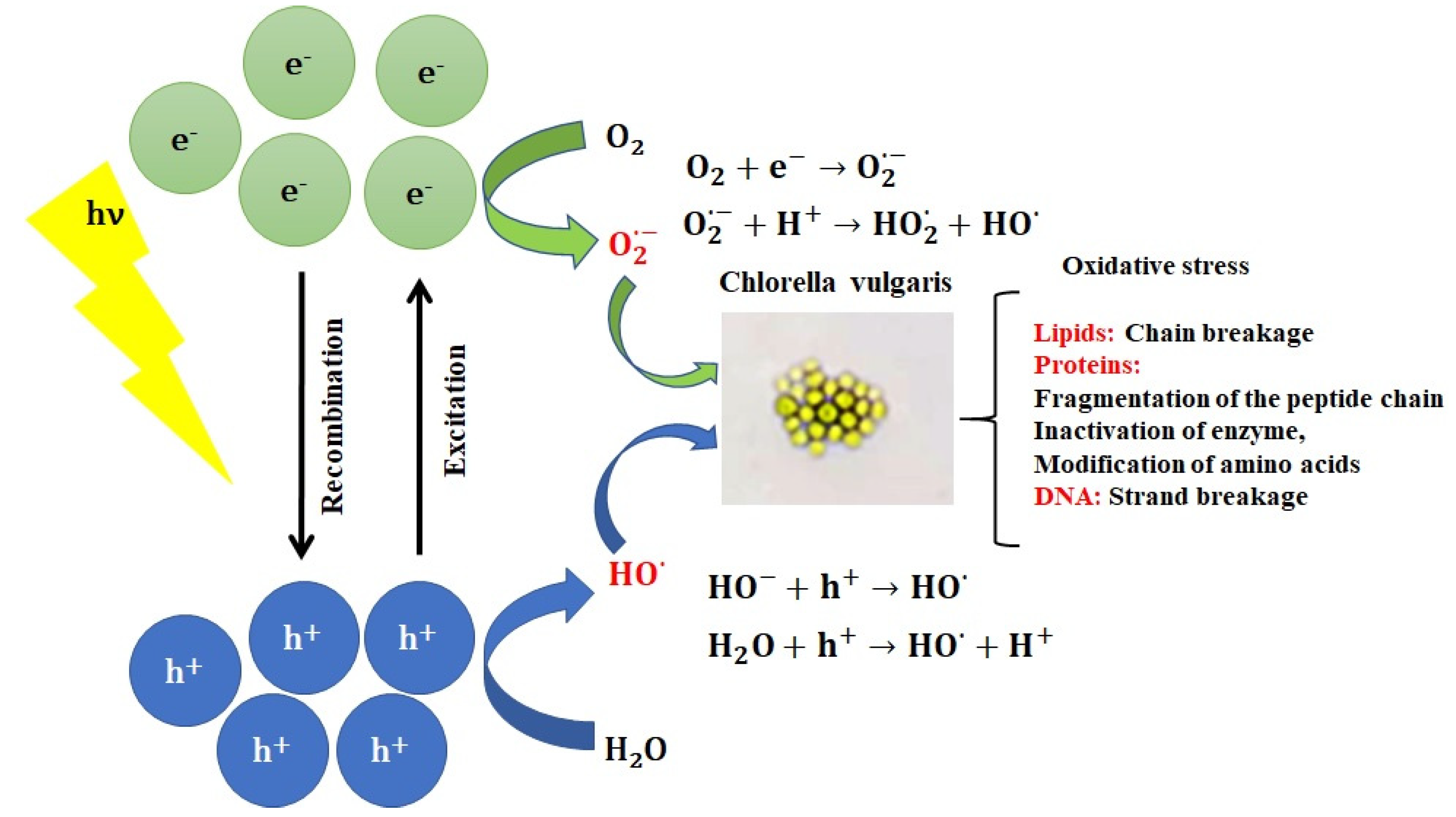
4. Mechanism of Photocatalyst Nanoparticles during Oxidative Stress of Algae
5. Conclusion and Perspectives
Author Contributions
Funding
Data Availability Statement
Conflicts of Interest
References
- Raizada, P.; Sudhaik, A.; Singh, P. Photocatalytic water decontamination using graphene and ZnO coupled photocatalysts: A review. Mater. Sci. Energy Technol. 2019, 2, 509–525. [Google Scholar] [CrossRef]
- Murgolo, S.; Franz, S.; Arab, H.; Bestetti, M.; Falletta, E.; Mascolo, G. Degradation of emerging organic pollutants in wastewater effluents by electrochemical photocatalysis on nanostructured TiO2 meshes. Water Res. 2019, 164, 114920. [Google Scholar] [CrossRef] [PubMed]
- Khan, I.; Saeed, K.; Khan, I. Nanoparticles: Properties, Applications and Toxicities. Arab. J. Chem. 2019, 12, 908–931. [Google Scholar] [CrossRef]
- Masciangioli, T.; Zhang, W.-X. Peer Reviewed: Environmental Technologies at the Nanoscale. Environ. Sci. Technol. 2003, 37, 102A–108A. [Google Scholar] [CrossRef] [PubMed]
- Corma, A.; Atienzar, P.; García, H.; Chane-Ching, J.-Y. Hierarchically mesostructured doped CeO2 with potential for solar-cell use. Nat. Mater. 2004, 3, 394–397. [Google Scholar] [CrossRef]
- Sangchay, W. The Self-cleaning and Photocatalytic Properties of TiO2 Doped with SnO2 Thin Films Preparation by Sol-gel Method. Energy Procedia 2016, 89, 170–176. [Google Scholar] [CrossRef]
- Guzmán, K.A.D.; Taylor, M.R.; Banfield, J.F. Environmental Risks of Nanotechnology: National Nanotechnology Initiative Funding, 2000−2004. Environ. Sci. Technol. 2006, 40, 1401–1407. [Google Scholar] [CrossRef]
- Ali, I.; Alharbi, O.M.L.; Tkachev, A.; Galunin, E.; Burakov, A.; Grachev, V.A. Water treatment by new-generation graphene materials: Hope for bright future. Environ. Sci. Pollut. Res. 2018, 25, 7315–7329. [Google Scholar] [CrossRef]
- Golobič, M.; Jemec, A.; Drobne, D.; Romih, T.; Kasemets, K.; Kahru, A. Upon Exposure to Cu Nanoparticles, Accumulation of Copper in the IsopodPorcellio scaberIs Due to the Dissolved Cu Ions Inside the Digestive Tract. Environ. Sci. Technol. 2012, 46, 12112–12119. [Google Scholar] [CrossRef]
- Khoshnamvand, M.; Ashtiani, S.; Chen, Y.; Liu, J. Impacts of organic matter on the toxicity of biosynthesized silver nanoparticles to green microalgae Chlorella vulgaris. Environ. Res. 2020, 185, 109433. [Google Scholar] [CrossRef]
- Baker, T.J.; Tyler, C.R.; Galloway, T.S. Impacts of metal and metal oxide nanoparticles on marine organisms. Environ. Pollut. 2014, 186, 257–271. [Google Scholar] [CrossRef] [PubMed]
- Moreno-Garrido, I.; Pérez, S.; Blasco, J. Toxicity of silver and gold nanoparticles on marine microalgae. Mar. Environ. Res. 2015, 111, 60–73. [Google Scholar] [CrossRef] [PubMed]
- Safi, C.; Zebib, B.; Merah, O.; Pontalier, P.-Y.; Vaca-Garcia, C. Morphology, composition, production, processing and applications of Chlorella vulgaris: A review. Renew. Sustain. Energy Rev. 2014, 35, 265–278. [Google Scholar] [CrossRef]
- Yamamoto, M.; Fujishita, M.; Hirata, A.; Kawano, S. Regeneration and maturation of daughter cell walls in the autospore-forming green alga Chlorella vulgaris (Chlorophyta, Trebouxiophyceae). J. Plant Res. 2004, 117, 257–264. [Google Scholar] [CrossRef] [PubMed]
- Júnior, W.G.M.; Gorgich, M.; Corrêa, P.S.; Martins, A.A.; Mata, T.M.; Caetano, N.S. Microalgae for biotechnological applications: Cultivation, harvesting and biomass processing. Aquaculture 2020, 528, 735562. [Google Scholar] [CrossRef]
- Yamamoto, M.; Kurihara, I.; Kawano, S. Late type of daughter cell wall synthesis in one of the Chlorellaceae, Parachlorella kessleri (Chlorophyta, Trebouxiophyceae). Planta 2005, 221, 766–775. [Google Scholar] [CrossRef]
- Oukarroum, A.; Bras, S.; Perreault, F.; Popovic, R. Inhibitory effects of silver nanoparticles in two green algae, Chlorella vulgaris and Dunaliella tertiolecta. Ecotoxicol. Environ. Saf. 2012, 78, 80–85. [Google Scholar] [CrossRef]
- OECD. Test No. 201: Freshwater Alga and Cyanobacteria, Growth Inhibition Test, OECD Guidelines for the Testing of Chemicals; Section 2; OECD Publishing: Paris, France, 2011. [Google Scholar]
- Wehr, J.D. Algae: Anatomy, Biochemistry, and Biotechnology by Barsanti, L. & Gualtieri, P.J. Phycology 2007, 43, 412–414. [Google Scholar]
- Serra-Maia, R.; Bernard, O.; Gonçalves, A.; Bensalem, S.; Lopes, F. Influence of temperature on Chlorella vulgaris growth and mortality rates in a photobioreactor. Algal Res. 2016, 18, 352–359. [Google Scholar] [CrossRef]
- Žitnik, M.; Šunta, U.; Torkar, K.G.; Klemenčič, A.K.; Atanasova, N.; Bulc, T.G. The study of interactions and removal efficiency of Escherichia coli in raw blackwater treated by microalgae Chlorella vulgaris. J. Clean. Prod. 2019, 238, 117865. [Google Scholar] [CrossRef]
- Mayo, A.W. Effects of temperature and pH on the kinetic growth of unialga Chlorella vulgaris cultures containing bacteria. Water Environ. Res. 1997, 69, 64–72. [Google Scholar] [CrossRef]
- Filali, R.; Tebbani, S.; Dumur, D.; Isambert, A.; Pareau, D.; Lopes, F. Identification of the growth model parameters for a culture of Chlorella vulgaris in a photobioreactor. IFAC Proc. Vol. 2010, 43, 431–436. [Google Scholar] [CrossRef]
- Bélanger-Lépine, F.; Tremblay, A.; Huot, Y.; Barnabé, S. Cultivation of an algae-bacteria consortium in wastewater from an industrial park: Effect of environmental stress and nutrient deficiency on lipid production. Bioresour. Technol. 2018, 267, 657–665. [Google Scholar] [CrossRef] [PubMed]
- Khoeyi, Z.A.; Seyfabadi, J.; Ramezanpour, Z. Effect of light intensity and photoperiod on biomass and fatty acid composition of the microalgae, Chlorella vulgaris. Aquac. Int. 2011, 20, 41–49. [Google Scholar] [CrossRef]
- Salgueiro, J.L.; Pérez, L.; Maceiras, R.; Sánchez, A.; Cancela, A. Bioremediation of Wastewater Using Chlorella vulgaris Microalgae: Phosphorus and Organic Matter. Int. J. Environ. Res. 2016, 10, 465–470. [Google Scholar]
- Kube, M.; Mohseni, A.; Fan, L.; Roddick, F. Impact of alginate selection for wastewater treatment by immobilised Chlorella vulgaris. Chem. Eng. J. 2019, 358, 1601–1609. [Google Scholar] [CrossRef]
- González, L.E.; Cañizares, R.O.; Baena, S. Efficiency of ammonia and phosphorus removal from a colombian agroindustrial wastewater by the microalgae Chlorella vulgaris and Scenedesmus dimorphus. Bioresour. Technol. 1997, 60, 259–262. [Google Scholar] [CrossRef]
- Lam, M.K.; Yusoff, M.I.; Uemura, Y.; Lim, J.W.; Khoo, C.G.; Lee, K.T.; Ong, H.C. Cultivation of Chlorella vulgaris using nutrients source from domestic wastewater for biodiesel production: Growth condition and kinetic studies. Renew. Energy 2017, 103, 197–207. [Google Scholar] [CrossRef]
- Daliry, S.; Hallajisani, A.; Mohammadi Roshandeh, J.; Nouri, H.; Golzary, A. Investigation of Optimal Condition for Chlorella vulgaris Microalgae Growth. Glob. J. Environ. Sci. Manag. 2017, 3, 217–230. [Google Scholar]
- Gong, N.; Shao, K.; Che, C.; Sun, Y. Stability of nickel oxide nanoparticles and its influence on toxicity to marine algae Chlorella vulgaris. Mar. Pollut. Bull. 2019, 149, 110532. [Google Scholar] [CrossRef]
- Sreesai, S.; Pakpain, P. Nutrient Recycling by Chlorella vulgaris from Septage Effluent of the Bangkok City, Thailand. ScienceAsia 2007, 33, 293–299. [Google Scholar] [CrossRef]
- Wang, C.-Y.; Fu, C.-C.; Liu, Y.-C. Effects of using light-emitting diodes on the cultivation of Spirulina platensis. Biochem. Eng. J. 2007, 37, 21–25. [Google Scholar] [CrossRef]
- Yeh, K.-L.; Chang, J.-S. Nitrogen starvation strategies and photobioreactor design for enhancing lipid content and lipid production of a newly isolated microalga Chlorella vulgaris ESP-31: Implications for biofuels. Biotechnol. J. 2011, 6, 1358–1366. [Google Scholar] [CrossRef]
- Kim, D.G.; Lee, C.; Park, S.-M.; Choi, Y.-E. Manipulation of light wavelength at appropriate growth stage to enhance biomass productivity and fatty acid methyl ester yield using Chlorella vulgaris. Bioresour. Technol. 2014, 159, 240–248. [Google Scholar] [CrossRef] [PubMed]
- Munir, N.; Imtiaz, A.; Sharif, N.; Naz, S. Optimization of Growth Conditions of Different Algal Strains and Determination of Their Lipid Contents. J. Anim. Plant. Sci. 2015, 25, 546–553. [Google Scholar]
- Griffiths, M.J.; Garcin, C.; Van Hille, R.; Harrison, S.T. Interference by pigment in the estimation of microalgal biomass concentration by optical density. J. Microbiol. Methods 2011, 85, 119–123. [Google Scholar] [CrossRef]
- Li, H.; Zhang, Y.; Liu, J.; Shen, Z.; Li, A.; Ma, T.; Feng, Q.; Sun, Y. Treatment of high-nitrate wastewater mixtures from MnO2 industry by Chlorella vulgaris. Bioresour. Technol. 2019, 291, 121836. [Google Scholar] [CrossRef]
- Takahashi, T. Applicability of Automated Cell Counter with a Chlorophyll Detector in Routine Management of Microalgae. Sci. Rep. 2018, 8, 4967. [Google Scholar] [CrossRef]
- Yu, Q.; Wang, H.; Li, X.; Yin, Y.; Qin, S.; Ge, B. Enhanced biomass and CO2 sequestration of Chlorella vulgaris using a new mixotrophic cultivation method. Process. Biochem. 2020, 90, 168–176. [Google Scholar] [CrossRef]
- Gao, F.; Yang, Z.-H.; Li, C.; Chen, B.-A.; Jin, W.-H.; Deng, Y.-B. Concentrated microalgae cultivation in treated sewage by membrane photobioreactor operated in batch flow mode. Bioresour. Technol. 2014, 167, 441–446. [Google Scholar] [CrossRef]
- Fernández-Linares, L.; Barajas, C.G.; Páramo, E.D.; Corona, J.A.B. Assessment of Chlorella vulgaris and indigenous microalgae biomass with treated wastewater as growth culture medium. Bioresour. Technol. 2017, 244, 400–406. [Google Scholar] [CrossRef] [PubMed]
- Feng, Y.; Li, C.; Zhang, D. Lipid production of Chlorella vulgaris cultured in artificial wastewater medium. Bioresour. Technol. 2011, 102, 101–105. [Google Scholar] [CrossRef] [PubMed]
- Bradford, M.M. A Rapid and Sensitive Method for the Quantitation of Microgram Quantities of Protein Utilising the Principle of Protein-Dye Binding. Anal. Biochem. 1976, 72, 248–254. [Google Scholar] [CrossRef]
- Dubois, M.; Gilles, K.A.; Hamilton, J.K.; Rebers, P.A.; Smith, F. Colorimetric Method for Etermination of Sugars and Related Substances. Anal. Chem. 1956, 28, 350–356. [Google Scholar] [CrossRef]
- Myers, J.A.; Curtis, B.S.; Curtis, W.R. Improving accuracy of cell and chromophore concentration measurements using optical density. BMC Biophys. 2013, 6, 4. [Google Scholar] [CrossRef]
- Fan, G.; Hong, L.; Luo, J.; You, Y.; Zhang, J.; Hua, P.; Du, B.; Zhan, J.; Ning, R.; Bao, M. Photocatalytic inactivation of harmful algae and degradation of cyanotoxins microcystin-LR using GO-based Z-scheme nanocatalysts under visible light. Chem. Eng. J. 2020, 392, 123767. [Google Scholar] [CrossRef]
- Heredia-Arroyo, T.; Wei, W.; Hu, B. Oil Accumulation via Heterotrophic/Mixotrophic Chlorella protothecoides. Appl. Biochem. Biotechnol. 2010, 162, 1978–1995. [Google Scholar] [CrossRef]
- Hemschemeier, A.; Melis, A.; Happe, T. Analytical approaches to photobiological hydrogen production in unicellular green algae. Photosynth. Res. 2009, 102, 523–540. [Google Scholar] [CrossRef]
- Camacho-Fernández, C.; Hervás, D.; Rivas-Sendra, A.; Marín, M.P.; Seguí-Simarro, J.M. Comparison of six different methods to calculate cell densities. Plant Methods 2018, 14, 1–15. [Google Scholar] [CrossRef]
- Baniamerian, H.; Tsapekos, P.; Alvarado-Morales, M.; Shokrollahzadeh, S.; Safavi, M.; Angelidaki, I.; Safavi, M. Anti-algal activity of Fe2O3–TiO2 photocatalyst on Chlorella vulgaris species under visible light irradiation. Chemosphere 2020, 242, 125119. [Google Scholar] [CrossRef]
- Miao, M.-S.; Yao, X.-D.; Shu, L.; Yan, Y.-J.; Wang, Z.; Li, N.; Cui, X.-T.; Lin, Y.-M.; Kong, Q. Mixotrophic growth and biochemical analysis of Chlorella vulgaris cultivated with synthetic domestic wastewater. Int. Biodeterior. Biodegradation 2016, 113, 120–125. [Google Scholar] [CrossRef]
- Chen, X.; Zhu, X.; Li, R.; Yao, H.; Lu, Z.; Yang, X. Photosynthetic Toxicity and Oxidative Damage Induced by nano-Fe3O4 on Chlorella vulgaris in Aquatic Environment. Open J. Ecol. 2012, 2, 21–28. [Google Scholar] [CrossRef]
- Wellburn, A.R. The Spectral Determination of Chlorophylls a and b, as well as Total Carotenoids, Using Various Solvents with Spectrophotometers of Different Resolution. J. Plant Physiol. 1994, 144, 307–313. [Google Scholar] [CrossRef]
- Deviram, G.; Mathimani, T.; Anto, S.; Ahamed, T.S.; Ananth, D.A.; Pugazhendhi, A. Applications of microalgal and cyanobacterial biomass on a way to safe, cleaner and a sustainable environment. J. Clean. Prod. 2020, 253, 119770. [Google Scholar] [CrossRef]
- Bligh, E.G.; Dyer, W.J. A Rapid Method of Total Lipid Extraction and Purification. Can. J. Biochem. Physiol. 1959, 37, 911–917. [Google Scholar] [CrossRef]
- Tratnyek, P.G.; Johnson, R.L. Nanotechnologies for environmental cleanup. Nano Today 2006, 1, 44–48. [Google Scholar] [CrossRef]
- Rogozea, E.A.; Petcu, A.R.; Olteanu, N.L.; Lazar, C.A.; Cadar, D.; Mihaly, M. Tandem Adsorption-Photodegradation Activity Induced by Light on NiO-ZnO p–n Couple Modified Silica Nanomaterials. Mater. Sci. Semicond. Process. 2017, 57, 1–11. [Google Scholar] [CrossRef]
- Olteanu, N.L.; Lazăr, C.A.; Petcu, A.R.; Meghea, A.; Rogozea, E.A.; Mihaly, M. “One-Pot” Synthesis of Fluorescent Au@SiO2 and SiO2@Au Nanoparticles. Arab. J. Chem. 2016, 9, 854–864. [Google Scholar] [CrossRef]
- Rogozea, E.A.; Olteanu, N.L.; Petcu, A.R.; Lazar, C.A.; Meghea, A.; Mihaly, M. Extension of optical properties of ZnO/SiO2 materials induced by incorporation of Au or NiO nanoparticles. Opt. Mater. 2016, 56, 45–48. [Google Scholar] [CrossRef]
- Barakat, M.A.; Kumar, R. Photocatalytic Activity Enhancement of Titanium Dioxide Nanoparticles; Springer Briefs in Molecular Science; Springer: Berlin/Heidelberg, Germany, 2016; pp. 1–29. [Google Scholar]
- Raizada, P.; Shandilya, P.; Singh, P.; Thakur, P. Solar Light-Facilitated Oxytetracycline Removal from the Aqueous Phase Utilising a H2O2/ZnWO4/CaO Catalytic System. J. Taibah Univ. Sci. 2017, 11, 689–699. [Google Scholar] [CrossRef]
- Raizada, P.; Kumari, J.; Shandilya, P.; Dhiman, R.; Singh, P.V.; Singh, P. Magnetically Retrievable Bi2WO6/Fe3O4 Immobilised on Graphene Sand Composite for Investigation of Photocatalytic Mineralization of Oxytetracycline and Ampicillin. Process Saf. Environ. Prot. 2017, 106, 104–116. [Google Scholar] [CrossRef]
- Raizad, P.; Kumari, J.; Shandilya, P.; Singh, P. Kinetics of photocatalytic mineralization of oxytetracycline and ampicillin using activated carbon supported ZnO/ZnWO4 nanocomposite in simulated wastewater. Desalin. Water Treat. 2017, 79, 204–213. [Google Scholar] [CrossRef]
- Adegoke, K.A.; Iqbal, M.; Louis, H.; Bello, O.S. Synthesis, Characterisation and Application of CdS/ZnO Nanorod Heterostructure for the Photodegradation of Rhodamine B Dye. Mater. Sci. Energy Technol. 2019, 2, 329–336. [Google Scholar]
- Fatin, S.O.; Lim, H.N.; Tan, W.T.; Huang, N.M. Comparison of Photocatalytic Activity and Cyclic Voltammetry of Zinc Oxide and Titanium Dioxide Nanoparticles toward Degradation of Methylene Blue. Int. J. Electrochem. Sci. 2012, 7, 9074–9084. [Google Scholar]
- Maeda, K. Photocatalytic water splitting using semiconductor particles: History and recent developments. J. Photochem. Photobiol. C Photochem. Rev. 2011, 12, 237–268. [Google Scholar] [CrossRef]
- Navarro, E.; Piccapietra, F.; Wagner, B.; Marconi, F.; Kaegi, R.; Odzak, N.; Sigg, L.; Behra, R. Toxicity of Silver Nanoparticles to Chlamydomonas reinhardtii. Environ. Sci. Technol. 2008, 42, 8959–8964. [Google Scholar] [CrossRef]
- Thiagarajan, V.; Pavani, M.; Raghavan, A.; Ramasubbu, S. Diminishing bioavailability and toxicity of P25 TiO2 NPs during continuous exposure to marine algae Chlorella sp. Chemosphere 2019, 233, 363–372. [Google Scholar] [CrossRef]
- Ma, J.; Lin, F.; Zhang, R.; Yu, W.; Lu, N. Differential sensitivity of two green algae, Scenedesmus quadricauda and Chlorella vulgaris, to 14 pesticide adjuvants. Ecotoxicol. Environ. Saf. 2004, 58, 61–67. [Google Scholar] [CrossRef]
- Morelli, E.; Gabellieri, E.; Bonomini, A.; Tognotti, D.; Grassi, G.; Corsi, I. TiO2 nanoparticles in seawater: Aggregation and interactions with the green alga Dunaliella tertiolecta. Ecotoxicol. Environ. Saf. 2018, 148, 184–193. [Google Scholar] [CrossRef]
- Lin, M.; Tseng, Y.H.; Huang, C.P. Interactions between Nano-TiO2 Particles and Algal Cells at Moderate Particle Concentration. Front. Chem. Sci. Eng. 2015, 9, 242–257. [Google Scholar] [CrossRef]
- Ma, S.; Zhou, K.; Yang, K.; Lin, D. Heteroagglomeration of Oxide Nanoparticles with Algal Cells: Effects of Particle Type, Ionic Strength and pH. Environ. Sci. Technol. 2015, 49, 932–939. [Google Scholar] [CrossRef] [PubMed]
- Shih, Y.-H.; Zhuang, C.-M.; Peng, Y.-H.; Lin, C.-H.; Tseng, Y.-M. The effect of inorganic ions on the aggregation kinetics of lab-made TiO2 nanoparticles in water. Sci. Total. Environ. 2012, 446–452. [Google Scholar] [CrossRef]
- Khan, S.S.; Srivatsan, P.; Vaishnavi, N.; Mukherjee, A.; Chandrasekaran, N. Interaction of silver nanoparticles (SNPs) with bacterial extracellular proteins (ECPs) and its adsorption isotherms and kinetics. J. Hazard. Mater. 2011, 192, 299–306. [Google Scholar] [CrossRef] [PubMed]
- Dalai, S.; Pakrashi, S.; Nirmala, M.J.; Chaudhri, A.; Chandrasekaran, N.; Mandal, A.; Mukherjee, A. Cytotoxicity of TiO2 nanoparticles and their detoxification in a freshwater system. Aquat. Toxicol. 2013, 1–11. [Google Scholar] [CrossRef] [PubMed]
- Nguyen, M.K.; Moon, J.-Y.; Lee, Y.-C. Microalgal ecotoxicity of nanoparticles: An updated review. Ecotoxicol. Environ. Saf. 2020, 201, 110781. [Google Scholar] [CrossRef] [PubMed]
- Dauda, S.; Chia, M.A.; Bako, S.P. Toxicity of titanium dioxide nanoparticles to Chlorella vulgaris Beyerinck (Beijerinck) 1890 (Trebouxiophyceae, Chlorophyta) under changing nitrogen conditions. Aquat. Toxicol. 2017, 187, 108–114. [Google Scholar] [CrossRef]
- Ko, K.-S.; Koh, D.-C.; Kong, I.C. Toxicity Evaluation of Individual and Mixtures of Nanoparticles Based on Algal Chlorophyll Content and Cell Count. Materials 2018, 11, 121. [Google Scholar] [CrossRef]
- Sadiq, I.M.; Dalai, S.; Chandrasekaran, N.; Mukherjee, A. Ecotoxicity study of titania (TiO2) NPs on two microalgae species: Scenedesmus sp. and Chlorella sp. Ecotoxicol. Environ. Saf. 2011, 74, 1180–1187. [Google Scholar] [CrossRef]
- Xia, B.; Sui, Q.; Sun, X.; Han, Q.; Chen, B.; Zhu, L.; Qu, K. Ocean acidification increases the toxic effects of TiO2 nanoparticles on the marine microalga Chlorella vulgaris. J. Hazard. Mater. 2018, 346, 1–9. [Google Scholar] [CrossRef]
- Suman, T.Y.; Radhika, S.R.R.; Kirubagaran, R. Evaluation of zinc oxide nanoparticles toxicity on marine algae Chlorella vulgaris through flow cytometric, cytotoxicity and oxidative stress analysis. Ecotoxicol. Environ. Saf. 2015, 113, 23–30. [Google Scholar] [CrossRef]
- Oukarroum, A.; Zaidi, W.; Samadani, M.; Dewez, D. Toxicity of Nickel Oxide Nanoparticles on a Freshwater Green Algal Strain of Chlorella vulgaris. BioMed Res. Int. 2017, 2017, 1–8. [Google Scholar] [CrossRef] [PubMed]
- Gong, N.; Shao, K.; Feng, W.; Lin, Z.; Liang, C.; Sun, Y. Biotoxicity of nickel oxide nanoparticles and bio-remediation by microalgae Chlorella vulgaris. Chemosphere 2011, 83, 510–516. [Google Scholar] [CrossRef] [PubMed]
- Wang, L.; Huang, X.; Sun, W.; Too, H.Z.; Laserna, A.K.C.; Li, S.F.Y. A global metabolomic insight into the oxidative stress and membrane damage of copper oxide nanoparticles and microparticles on microalga Chlorella vulgaris. Environ. Pollut. 2020, 258, 113647. [Google Scholar] [CrossRef]
- Romero, N.; Visentini, F.F.; Márquez, V.E.; Santiago, L.G.; Castro, G.R.; Gagneten, A.M. Physiological and morphological responses of green microalgae Chlorella vulgaris to silver nanoparticles. Environ. Res. 2020, 189, 109857. [Google Scholar] [CrossRef] [PubMed]
- Clément, L.; Hurel, C.; Marmier, N. Toxicity of TiO2 nanoparticles to cladocerans, algae, rotifers and plants—Effects of size and crystalline structure. Chemosphere 2013, 90, 1083–1090. [Google Scholar] [CrossRef]
- Metzler, D.M.; Li, M.; Erdem, A.; Huang, C.P. Responses of Algae to Photocatalytic Nano-TiO2 Particles with an Emphasis on the Effect of Particle Size. Chem. Eng. J. 2011, 170, 538–546. [Google Scholar] [CrossRef]


| Nr.crt | Component | OECD Medium Concentration (mg L−1) |
|---|---|---|
| 1 | NaHCO3 | 50.0 |
| 2 | NH4Cl | 15.0 |
| 3 | MgCl2·6(H2O) | 12.0 |
| 4 | CaCl2·2(H2O) | 18.0 |
| 5 | MgSO4·7(H2O) | 15.0 |
| 6 | KH2PO4 | 1.60 |
| 7 | FeCl3·6(H2O) | 0.0640 |
| 8 | Na2EDTA·2(H2O) | 0.100 |
| 9 | H3BO3 | 0.185 |
| 10 | MnCl2·4(H2O) | 0.415 |
| 11 | ZnCl2 | 0.00300 |
| 12 | CoCl2·6(H2O) | 0.00150 |
| 13 | Na2MoO4·2(H2O) | 0.00700 |
| 14 | CuCl2·(H2O) | 0.00001 |
| Parameters | Methods | Description | Ref |
|---|---|---|---|
| Cell concentration | Optical density (OD) | 649, 665, and 480 nm (spectrophotometric method) | [37] |
| Neubauer cell counting | Neubauer chamber is currently used as a standard in the laboratories for manual counting under a light microscope | [38] | |
| Automatic cell counting | To overcome manual counting limitations, advanced digital image analysis has been promoted | [39] | |
| Biomass quantification | Centrifugation, dried, weight | Centrifugation: 8000 g Time: 10 min Dried: 105 °C for 24 h | [40] |
| Spectrophotometric | Dry biomass conc(g·L−1) = 0.25 OD680 nm + 0.001 | [41] | |
| Spectrophotometric | Dry weight (g·L−1) = 0.3793·OD688 nm; | [29] | |
| Pigments | Chlorophyll A | Chlorophyll A (mg·mL−1) = 12.47 OD665 nm −3.62 OD649 nm | [37] |
| Chlorophyll B | Chlorophyll B (mg·mL−1) = 27.44 OD 652 nm -12.17 OD 665 nm – OD 750 nm | [42] | |
| Total carotenoids | Carotenoids (mg·mL−1) = 4 OD 480 nm – OD 750 nm | [42] | |
| Nutrients | Lipids | Gravimetrically—using Blight and Dyer method | [43] |
| Proteins | Bradford method | [42,44] | |
| Carbohydrates | 490 nm (UV-VIS spectrophotometer) | [42] | |
| Sugars | Dubois method | [45] |
| NPs | NP Concentration in Toxicity Experiments | Synthesis Method/NP Characteristics | Growth Conditions | Toxic Effect | References |
|---|---|---|---|---|---|
| TiO2 | 100–400 μM | Particle size: <25 nm | T: 23 ± 2 °C Illumination: 16 h:8 h light–dark Light: 30 μmoles m2 s−1. | EC50 = 100 μM | [78] |
| TiO2 | 20, 40, 80 mg L−1 | Particle size: <25 nm Density: 3.95 g/cm3 Surface area: 75–85 m2/g | Growth medium: BG11 T: 30 °C Light: 5000 lux | EC50 = aprox 42 mgL−1 | [79] |
| TiO2 | 3–192 mg L−1 | Particle size < 25 nm, surface area: 200–220 m2/g | Growth medium: sterile BBT: 25 °C Illumination: 12 h:12 h light:dark Light: white fluorescent light (intensity 3000 lx) | EC50 = 16.12 mgL−1 (72 h) | [80] |
| TiO2 | 10 mg L−1 (96 h) | Particle size: 21 nm Surface area: 10.64 m2/g | Growth medium: f/2 T: 20.0 ± 0.1 °C Illumination: 12:12 light–dark Salinity: 32.2 ± 0.2. | EC50 (pH 8.20) > EC50 (pH 7.77, 7.47) | [81] |
| ZnO | 50–300 mg L−1 (72 h) | Solid-state pyrolytic method. Particle size: 40–48 nm | T: 20 °C Light: white fluorescent lights Illumination: 12 h:12 h light:dark Continuous aeration | Viability ↓ 90.49 ± 0.3% in 24 h (50 mg L−1) Viability ↓↓↓ 23.69 ± 1.8% (300 mg L−1) | [82] |
| ZnO | 0.5, 1, 2 mg L−1 | Particle size: 40–100 nm Density: 5.61 g/cm3 Surface area: 10–25 m2/g | Growth medium: BG11 T: 30 °C Light: 5000 lux | EC50 = 2.0 mg L−1 | [79] |
| NiO | 0, 0.1–100 mg L−1 (96 h) | Particle size: 30 nm Surface area: 50–80 m2/g | Growth medium: sterile BG11 T: 23 ± 1 °C pH 7 Illumination: continuous Light intensity: white fluorescent light (100 μmol m−2 s−1) | EC50 = 13.7 mg L−1 | [83] |
| NiO | 4, 9, 18 mg L−1 | Particle size: 30 nm Density: 6.67 g/cm3 Surface area: 50–100 m2/g | Growth medium: BG11 T: 30 °C Light: 5000 lux | EC50 =aprox 22 mg L−1 | [79] |
| NiO | 0, 1–30mg L−1 (72 h) | Particle size: 439 ± 33 nm Zeta potential: −5.87 ± 0.16 mV | T = 23 ± 1 °C pH 8.2, Illumination: 12 h:12 h light:dark Light intensity: 3000lx m−2 s−1 | EC50 =1.73 mg L−1 | [31] |
| NiO | 0–50 mg L−1 | Homogeneous precipitation method Particle size: 20 nm | T: 23 ± 1 °C Light: 73.6 μmol m−2 s−1 Illumination: 12h:12h light:dark | EC50 = 32.28 (72 h) EC50 = 44.33 (120 h) | [84] |
| CuO | 1, 10 mg L−1 (5 days) | Particle size: 15–50 nm | Growth medium: Bold’s Basal T: 24 °C Light:15.000 lx Illumination: 18 h:6 h light:dark | 10 mg L−1 CuO NPs slightly suppressed cell density at day 3, 4, and 5 | [85] |
| CuO | 8, 16, 33 mg L−1 | Particle size: 30–50 nm Density: 6.40 g/cm3 Surface area: 13.1 m2/g | Growth medium: BG11 T: 30 °C Light: 5000 lux | EC50 = 40 mg L−1 | [79] |
| Fe2O3 | 22, 45, 90 mg L−1 | Particle size: 20–40 nm Density: 5.24 g/cm3 Surface area: 30–60 m2/g | Growth medium: BG11 T: 30 °C Light: 5000 lux | EC50 = 76 mg L−1 | [79] |
| Ag | 90–1440 μg L−1 (96 h) | Particle size: 46.8 ± 3.3 nm | Growth medium: Bold BasalT: 23 ± 1 °C Light: 120 μmol m−2 s−1 | EC50 = 110 μg L−1 (96 h) | [86] |
| Ag | 0, 5–60 μg L−1 (72 h) | Particle size: 28.95 ± 10.17 nm Zeta potential: −23.23 ± 0.75 mV | OECD protocol Temperature: 22 ± 3 °C | - | [10] |
Publisher’s Note: MDPI stays neutral with regard to jurisdictional claims in published maps and institutional affiliations. |
© 2020 by the authors. Licensee MDPI, Basel, Switzerland. This article is an open access article distributed under the terms and conditions of the Creative Commons Attribution (CC BY) license (http://creativecommons.org/licenses/by/4.0/).
Share and Cite
Adochite, C.; Andronic, L. Aquatic Toxicity of Photocatalyst Nanoparticles to Green Microalgae Chlorella vulgaris. Water 2021, 13, 77. https://doi.org/10.3390/w13010077
Adochite C, Andronic L. Aquatic Toxicity of Photocatalyst Nanoparticles to Green Microalgae Chlorella vulgaris. Water. 2021; 13(1):77. https://doi.org/10.3390/w13010077
Chicago/Turabian StyleAdochite, Cristina, and Luminita Andronic. 2021. "Aquatic Toxicity of Photocatalyst Nanoparticles to Green Microalgae Chlorella vulgaris" Water 13, no. 1: 77. https://doi.org/10.3390/w13010077
APA StyleAdochite, C., & Andronic, L. (2021). Aquatic Toxicity of Photocatalyst Nanoparticles to Green Microalgae Chlorella vulgaris. Water, 13(1), 77. https://doi.org/10.3390/w13010077