Article

Analysis of the Microbiome (Bathing Biome) in Geothermal Waters from an Australian Balneotherapy Centre

1
Centre for Environmental Sustainability and Remediation, School of Science, RMIT University, Bundoora, Victoria 3083, Australia
2
Extreme Wellness Institute, Warrandyte 3113, Australia
*
Author to whom correspondence should be addressed.
Water 2020, 12(6), 1705; https://doi.org/10.3390/w12061705
Submission received: 19 April 2020 / Revised: 10 June 2020 / Accepted: 11 June 2020 / Published: 15 June 2020

Abstract

:
Balneotherapy is an ancient practice which remains commonplace throughout the world due to perceived health benefits that include relief of arthritis, fibromyalgia and relaxation. However, bathing environments are not sterile and natural spring waters may harbour natural microbial populations that include potential pathogens. We elucidated the microbial community from water taken from the borehole, pre-filter water (chlorinated, cold and post-bathing water) and post-filter water at a commercial Australian natural hot spring bathing facility. Thiobacillus, Sphingobium and Agrobacterium were the predominant genera in samples collected from the borehole. The predominant genera changed to Sphingobium, Parvibaculum and Achromobacter following chloride treatment and Azospira replaced the Achromobacter once the water reached ambient temperature and was stored ready to be used by bathers. The microbial community changed again following use by bathers, dominated by Pseudomonas, although Sphingobium persisted. No total or faecal coliforms were observed in any of the samples except for the post-bathing water; even there, their presence was at very low concentration (2.3 cfu/mL). These results confirm the lack of pathogens present in these hot spring waters but also suggests that good management of post-bathing water is required especially if the water is used for borehole water recharge.

1. Introduction

Balneotherapy, the practice of therapeutic bathing in hot springs and thermal waters, is an ancient practice that is still very common around the world; some of the advantages attributed to this practice include the relief of arthritis and fibromyalgia, while many others bathe for relaxation. This practice is commonplace around the world and is especially popular in Japan and European countries such as Hungary, France and Italy [1,2,3,4,5,6].
There is growing interest in balneotherapy [7,8,9,10] and several controlled trials provide evidence for clinical advantages in the treatment of pain [11] fibromyalgia syndrome [12] and dermatology [13], among other conditions. As a consequence, the balneotherapy industry is growing, currently estimated to be worth 100 million euros [14] or USD 50 billion per year. As a sector, the global wellness economy was estimated to have an annual value of USD 4.5 trillion/year in 2018 (Global Wellness Institute webpage). Another author has estimated that balneotherapy has created one million direct and indirect jobs throughout the European Union and treated 5 million patients per year [15].
Bathing water is not sterile and natural spring water harbours natural microbial populations which may include both naturally occurring and introduced pathogens. For example, previous studies have reported the presence of free-living amoebas (FLA), [16,17,18] cyanobacterial toxins [19] and strains of Legionella [20,21,22] in natural spring waters.
Legionella has been identified as a problem in Japanese baths and showers supplied with hot spring water [20,21]. The detection of Legionellosis in aquatic environments has recently been trialled using a nested polymerase chain reaction (PCR) [23] approach, and the risk of contracting the disease in a hot spring has been modelled [24]. Another naturally occurring pathogen in hot springs waters is the amoeba Naegleria fowlery. This amoeba, which prefers warm water environments, can cause an acute and often fatal meningoencephalitis [17]. The distribution of both Legionella and Naegleria was recently assessed in a Polish hot spring and the authors concluded that their likely coexistence emphasises the potential threat during balneotherapy [25]. Similar warnings are raised by a Saudi Arabia study that detected several toxic cyanobacteria and the presence of microcystins in natural hot spring water [19]. A recent study has reviewed perspectives for innovative treatments of spa thermal waters [5].
To prevent potential public health issues, it is important to determine the microbial community in hot springs waters used for balneotherapy or recreation. To date, to the best of the authors’ knowledge, no detailed assessment of the microbial community in the Australian balneotherapy industry has been reported.
Previous studies have used denaturing gradient gel electrophoresis (DGGE) to study the microbial communities in hot springs [26,27,28,29,30]. One study investigated the microbial diversity in different regions of the Octopus Spring, Yellowstone National Park, using DGGE. Microbial profiles were identical in regions with the same temperature but varied where the temperature was different. The study identified known cyanobacterial populations but also novel bacterial populations [29].
Recent studies have used metagenomics to elucidate the microbial diversity in hot springs, including a study carried out at Yellowstone National Park [31], together with geothermal aquifers in Pakistan [32], China [33], India [34], Tibet [35] Australia [36] and more recently in hot springs and spas in Italy [4,6,37,38]. In the bacterial community in central Tibet, Chlorobi were ubiquitous in all mats; Cyanobacteria and Chloroflexi were also found in most of the samples. Location-specific lineages were; alpha, beta and gamma proteobacteria were also common phyla in the mats [35]. The similarity in microbial communities in geothermal aquifers is likely to be due to the fact that temperature exerts tight control over the microbial diversity [39].
To the best of the authors knowledge, to date, there have been no reports on the changes in the microbial community in a commercial Australian hot spring used for balneotherapy. The aim of this study was to identify the natural microbial community present in natural spa water from Victoria, Australia and to assess the impact of both pre-treatment with chlorine and balneotherapy on the microbial community.

2. Materials and Methods

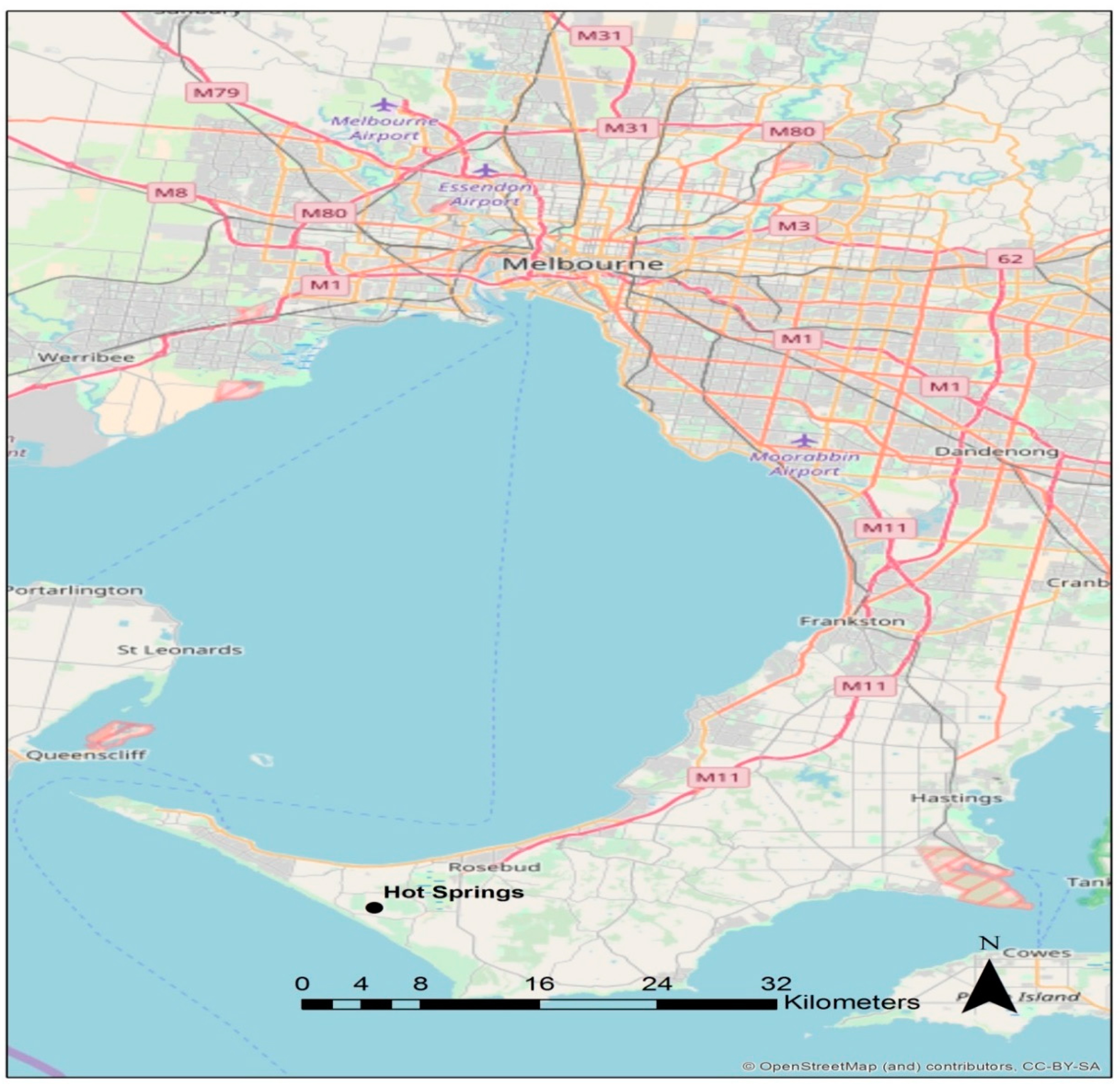
2.1. Hot Spring Hydrology and Sampling

Peninsula Hot Springs (PHS) in Victoria, Australia (Figure 1) is Australia’s largest geothermal bathing facility located at 38°24′32″ S, 144°50′12″ E. The Hot spring obtains its groundwater from various lithological formations. The aquifer is lies in a band of older volcanics between the Selwyn Fault and Postulated Fault. A coal seam is immediately above the older volcanics [40].
The groundwater aquifer is approximately 44 million years old, confined, fractured and composed of transitional older volcanic basalt. The basalt aquifer contains phenocrysts with a matrix of plagioclase, olivine, apatite, and opaque oxides [40]. The aquifer is confined by the overlying Fyansford formation and the potentiometric surface of the aquifer reaches 10 metres below the surface. The fracture of the basalt most likely occurred 39 million years ago when the block containing the aquifer slipped down compared to the block on the east of the Selwyn Fault.
Water samples were collected on 10 October 2016 from three different locations within PHS. A total of 3 L of water were filtered on site in triplicate using Millipore membrane filters (pore size, 0.22 µm). The sampling sites were all located within the PHS facilities and are as follows: (1) borehole, (2) chlorinated, (3) cold and (4) post-bathing water. Briefly the water was filtered through sterile filter units placed on a manifold using an electric pump for nucleic acid extractions.

2.2. Physicochemical Data

All samples (borehole, chlorinated, cold and post bathing) plus a post-filter water sample were analysed by an accredited laboratory (ALS Ltd., Melbourne, Australia) using standard operating protocols for several physicochemical parameters such as pH, dissolved oxygen (DO), salinity, turbidity, redox potential, total chlorine and concentration of metals among others (Table 1).

2.3. Total and Faecal Coliforms Determination

The quantification of total and faecal coliforms was performed with the Colilert and Enterolert systems (IDEXX) [41] in duplicate for the following samples: borehole, cold and post bathing following the manufacturer’s instructions.

2.4. Next Generation Sequencing (NGS) (16S rRNA) and Analysis

Sequencing and analysis. Total genomic DNA was extracted from 0.22-µm filters using a MoBio Power Soil DNA extraction kit according to the manufacturer’s instructions (MoBio Laboratories Inc., Carlsbad, CA, USA).
Three replicates of DNA from each sample were pooled and subjected to library preparation using Illumina Nextera® XT Index Kits (Illumina, San Diego, CA, USA) with Illumina’s primer set recommendation following the company’s instructions. The set of primers were Forward Primer = 5’ TCGTCGGCAGCGTCAGATGTGTATAAGAGACAGCCTACGGGNGGCWGCAG and Reverse Primer = 5’ GTCTCGTGGGCTCGGAGATGTGTATAAGAGACAGGACTACHVGGGTATCTAATCC. The DNA from the library was quantified using Qubit® 2.0 Fluorometer (Life Technologies, Carlsbad, CA, USA) and 2100 Bioanalyzer (Agilent Technologies, Santa Clara, CA, USA). The samples were pooled and run in a MiSeq platform (Illumina, San Diego, CA, USA). The obtained sequences were analysed using the 16S Metagenomics app in the Illumina Basespace and Quantitative Insights Into Microbial Ecology (QIIME) package available in the lllumina Basespace. Analysis of the data was performed with the Megan software. The number of reads and percentage of quality filtering was satisfactory for all samples. For example, for sample Postbath3 there were 64,413 reads (95% reads passing quality filtering).

2.5. Statistical Analysis

An Operational Taxonomic Units (OTU) genus-level table was imported in Primer 7 software, (Auckland, New Zealand); the data was standardized and transformed using square root. Shannon diversity and richness indices were also calculated using Primer 7 software as described in the software manual. The standardized and transformed data were subjected to resemblance analysis using S17 Bray Curtis similarity resemblance measure and cluster analysis applied to generate plot dendrograms using the group average cluster mode. In addition, PCA (Figure 2) and Non-metric multidimensional scaling (NMDS) were performed using Primer 7 [42] (Figure 3).
Data from this study was subjected to XLSTAt (2014) for Principal component analysis with a Pearson (n) coefficient.

3. Results and Discussion

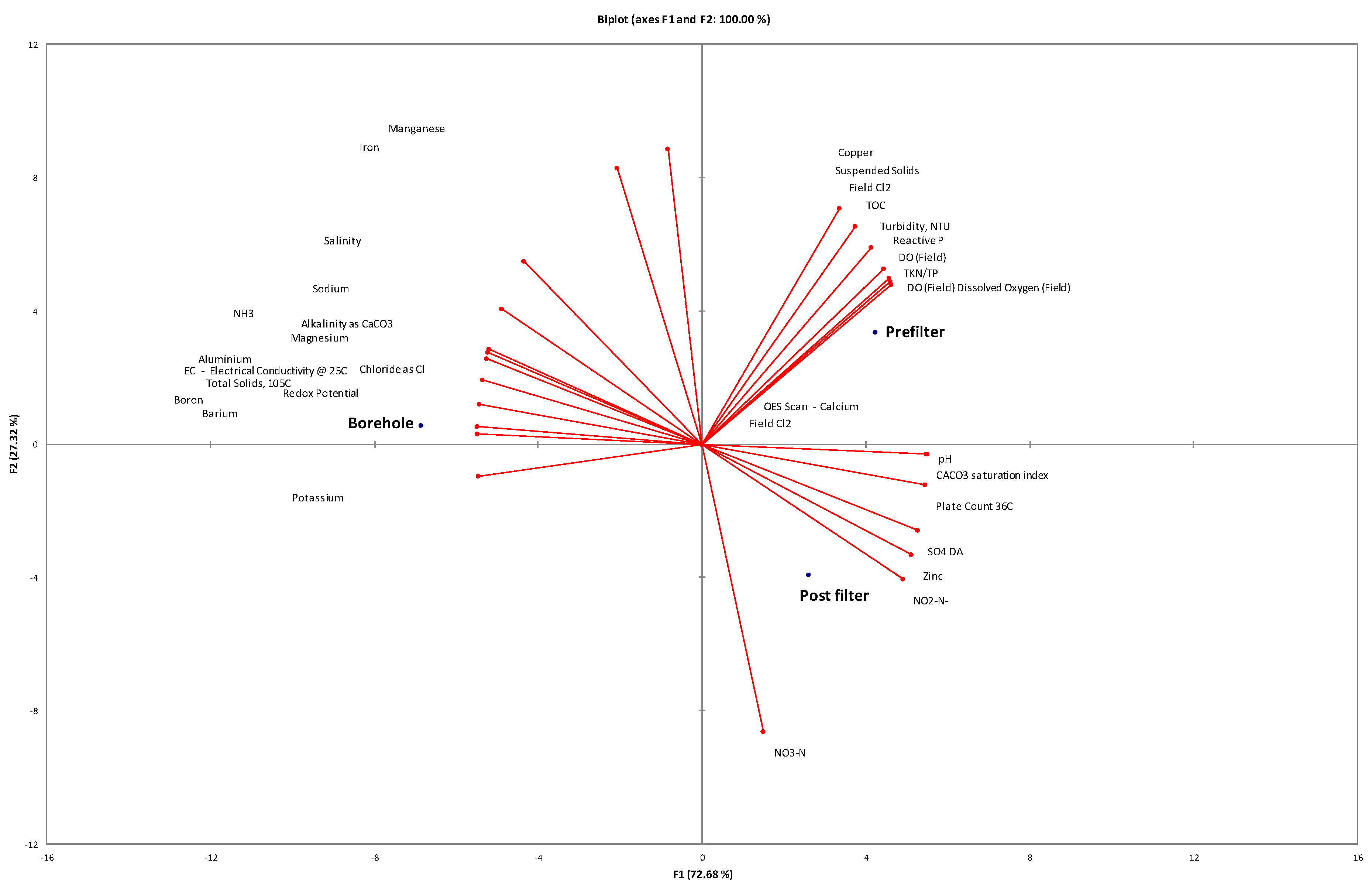
3.1. Physicochemical and Microbial Characteristics

As expected, significant differences in temperature and dissolved oxygen concentration occurred when geothermal borehole water was used for bathing. The geothermal water comes out at 44 °C and subsequently cools through the bathing process to 24 °C. Dissolved oxygen (DO) concentration increased from 14.4% in the borehole to 90.8% in the pre-filter water and decreases to 39% again in the post-filter water. The pH fluctuated from 6.9 in the borehole to 7.8 in the Spa facilities (the pre-filter water).
Examination of the microbial load in the water samples was assessed using heterotrophic plate counts, as it remains the methodology used for reporting against legislative guidelines for microbial loads. As expected, the number of colony forming units (cfu) increased from 1800 cfu/mL in the borehole to >10,000 cfu/mL in the pre- and post-filter water (Table 1). Principal component analysis (PCA) (Figure 2) confirmed that each of the waters (borehole, pre- and post-filter) was physico-chemically different, largely due to exposure to the bathers and well as changing environmental conditions. Such significant changes in water would be expected to lead to different microbial communities as observed in Figure 3.

3.2. Coliforms

No coliforms or E. coli were observed in the samples until after bathers had used the geothermal water (Table 1). Even so, the numbers were very low (2.3 cfu/100 mL) and within the permissible limits of the World Health Organization (WHO) that classifies the water quality used for recreation as poor when the 95th percentile intestinal enterococci/ 100 mL are in the range of 201–500 and very poor when they are >500 [43]. These results are similar to previous measurements performed with the same method on the waters by an independent laboratory (data not shown). The presence of coliforms is used as indicator organisms and their presence after human activity suggests that other introduced organisms may also be present, changing the ‘bathing biome’.

3.3. Microbial Diversity of the Hot Spring

Next generation sequencing was used in this study to characterise the microbial community in the borehole water and microbial community dynamics throughout the balneotherapy process, with the aim of elucidating the original water microbiome (borehole) and those microbiomes once the water reaches the surface and is cooled, after it is chlorinated (‘bathing biome’) and once it has been used by the bathers (post-bathing).
Such characterization allows us to know the original microbiome in an Australian hot spring and compare it to similar hot springs throughout the world, as well as evaluate the presence of pathogens in this system and observe the changes in microbial diversity driven by physicochemical changes (temperature, dissolved oxygen, chlorine addition and pH). The result of the Bray Curtis similarity and NMDS revealed that the microbial diversity in each of the waters is distinct and indeed affected by the physicochemical factors (Figure 3).
The bacterial community from the borehole (44 °C) was dominated by Thiobacillus followed by Sphingobium and Agrobacterium (Figure 4). Thiobacillus-like bacteria were previously reported to be isolated from Icelandic thermal areas [44] and other strains related to Thiobacillus hydrothermalis have been found on hot springs microbial mats also in Iceland [45]. Other thermotolerant bacteria such as Acidithiobacillus caldus have been reported in geothermal pools on the Caribbean island of Montserrat [46], in a Mexican geothermal field [47] and in mud from a sulphurous-bromine-iodine Italian spa complex [37]. Members of the Sphingobium have also been found in hot springs in Taiwan [48] and China [33], while similar sequences (PK34) to Agrobacterium rhizogenes have been found in a Thai hot spring [49].
The fourth predominant group in the borehole water was the Sulfuricurvum. This genus has also been found in different hot springs around the world such as Peru [50], Armenia [51], Svalbard, Norway [52] and the USA [53]. This finding indicates the presence of sulphur oxidisers (chemolithoautotrophs).
The specific physicochemical properties of geothermal water differ according to local conditions such as temperature, which is generally higher than the one recorded at the PHS (44 °C) and it has been observed that species richness and diversity is strongly correlated to temperature [39]. The temperatures of water sampled from geothermal boreholes mentioned above were in the following ranges: 65 to 90 °C in Iceland [45], 33 to 48 °C in Montserrat (pH range of 1.6 to 3.0) [46] and 35 to 90 °C in Mexico (pH: 2–4.8) [47]. The temperature of spring water varies even more beyond these locations; 60 to 65 °C in Tibet [35], 45 to 99 °C in China, 71.9 to 90.7 °C in Yellowstone [31], 60 to 95 °C in Pakistan [32] and 48 to 58 °C in India [34]. Other hot springs in Australia (the great Artesian basin, which is a deep geothermal aquifer) are dominated by hydrogen-oxidizing thermophiles such as Hydrogenobacter and Hydrogenophilus; however, these waters are at 64 °C, pH 8 and have redox values of −48 mV [36].
Hot springs can be generally categorized in two groups: acid springs and those neutral or slightly alkaline springs [39]. PHS falls in the neutral group. We aimed to compare the microbial diversity in PHS to others around the world with similar temperature (44 to 50 °C) (regardless of pH). The Volcanic Taupo hot spring in New Zealand, the Forest springs in California and the hot spring in Los Azufres, Mexico, have similar temperatures to the PHS. Proteobacteria is the common phylum among the four hot springs (Figure 5). Firmicutes is common to PHS, Taupo and Forest springs while Chlorobi is common to PHS, Taupo and los Azufres.
Although some phyla are common among the different hot springs, it is also noticeable that most phyla present in the PHS are unique and have not been identified in spa water from other samples. This suggests a certain uniqueness of the microbial community in the samples examined in this study.

3.4. Microbial Diversity Changes in the PHS Facilities

Prior to being used for bathing, the geothermal water at PHS is treated with chlorine to a final concentration of 0.2 mg L−1. Chlorination is recommended by the World Health Organization (WHO) [43] and the Australian guidelines for managing risks in recreational waters [54]. Chlorine is the most widely used water disinfectant. It has been used since 1845 to control the cholera outbreak in London and is still being used worldwide by most governments. It prevents Pseudomonas dermatitis outbreaks if the pH is 7.2–7.8 and the residual chlorine levels are between 2–5 ppm [55].
Moreover, chlorination of the water is recommended since there have been several outbreaks of gastrointestinal disease in recreational waters in Australia [56] and the US [55]. The etiological agents in those outbreaks have been Shigella sp., E. coli O157:H7, Leptospira sp., Giardia lamblia, Cryptosporidium parvum, Norwalk-like viruses and Adenovirus 3.
Causes of the outbreaks are varied and include poor treatment, low chlorine levels or even resistant microorganisms such as Cryptosporidium and Giardia which are relatively resistant to disinfection by chlorine [55].
Recent studies have proposed the use of nanomaterials (Ag and TiO2 nanoparticles, graphene oxide, ZnO), photocatalysis and advanced filtration as alternatives to chlorine [5,57]. This is because the use of chlorine may produce some disinfection by products (DBP) such as trihalomethanes, haloacetic acids and halo nitromethanes, chloramines and chlorophenols among others [58]. Moreover, the ‘untouchability’ concept [5,57] of the natural waters has been proposed. Such a concept states that the biological and physico-chemical components of natural spa pools should remain untreated because each of them plays a specific role in the unique properties of the water and their claimed therapeutic effects. NGS has been proposed to classify the waters and their properties, which in turn should aid in the formulation of strategies to manage hygiene [4,6,59,60] and a database for spa microbiota has been developed and can be accessed at mfATLAS.it [6].
The total chlorine concentration increases from 0 in the borehole to 0.81 mg/L in the pre-filter water to below 0.05 mg/L in the post-filter water (Table 1). The presence of chlorine is likely to have a significant effect on the microbial community. The pre-filter water with a final chlorine concentration of 0.2 mg L−1 was found to be dominated by Sphingobium, Parvibaculum and Achromobacter, followed by Pseudomonas and Halothiobacillus (Figure 4).
As mentioned earlier, Sphingobium lutaoense was isolated from a coastal hot spring in Taiwan [48]; this species is of biotechnological interest as a zeaxanthin producer which is a powerful antioxidant [61]. Some sequences retrieved from the Tengchong geothermal fields in Southwest China were also similar to Sphingomodales [33]. Parvibaculum strains have been found in Tunisian hot springs [62] and in the travertine at a crystal geyser in Utah, USA [63]; Parvibaculum lavamentivorans has been shown to metabolize alkylbenzenesulfonates [64]. Members related to Achromobacter xylosidans strains have been found in the Thailand hot spring Bor Khluen [49]. Achromobacter has also been linked to the degradation of PCBs, hydrocarbons, BTEX and crude oil. A purple sulphur bacteria sequence obtained from microbial mats in Greenland hot springs showed distant relation to a (non-photosynthetic) Halothiobacillus strain [65]. Additionally, members related to this group have been found in Japanese hot springs, one novel obligate chemolithoautotrophic, sulphur oxidizing strain member of the Gammaproteobacteria and related to the Halothiobacillus genera was isolated from a Japanese hot spring in the Fukushima prefecture [66], while others were the dominant group in a bacterial mat at the Naruko hot spring in the Miyagi prefecture [67]. Moreover, other Halothiobacillus-related clones have been reported from a sulphide-rich spring in Oklahoma, US [68].
Clearly the chlorine treatment and/or the change in temperature result in a significant reduction in Thiobacillus dominance as seen in the borehole water, favouring the Sphingobium and Parvibaculum as well as other groups of bacteria that may be resistant to the final chlorine concentration.
The Thiobacillus population were reduced from more than 50% of the total population in the borehole to below 5% after the chlorine treatment, while the Sphingobium slightly increase to almost 10% of the total population (Figure 4). Achromobacter seems favoured by the chlorine/temperature treatment to become one of the dominant groups in the treated water.
Before disposal, the chlorine-treated water is cooled to ambient temperature (24 °C) and the microbial community observed in samples from this water was very similar to the chlorine-treated water; as expected, Sphingobium and Parvibaculum persist as dominant groups but members of the Azospira were also predominant. Members of the Azospira have been found in a South African hot spring [69]. Some Azospira have been found capable of selenite reduction to elemental selenium [70]. Another group also observed in the cold water were the Sphingosinicella. This genus belongs to the Sphingomonadaceae family and the species Microcystinivorans was first described in 2006 [71]. In summary, further cooling to ambient temperature does not seem to affect the microbial diversity.
We also analysed samples of the water following bathing (32 °C) and found that the microbial community changes significantly, being dominated by Pseudomonas although Sphingobium was also observed (Figure 4). This was an interesting finding and there are a number of possible explanations including anthropogenic origin (i.e., visit of a Pseudomonas-carrying/-infected bathers) or the presence of Pseudomonas where this water is stored; Pseudomonas have also been reported in other thermal baths in Budapest [72]. Further spatial-temporal samples may be necessary to pinpoint the origin of the Pseudomonas. However, we can conclude that this post-bathing water should not be used for recreational purposes before the filtering process or before it undergoes another treatment since some of the risks when exposed to Pseudomonas strains include P. otitis [73]. The filtering process of the post-bathing water that is carried out on the site seems to aid in removing potential pathogens since the borehole samples were not dominated by Pseudomonas, despite the fact that the filtered post-bathing water is recharged into the borehole.
In summary, the microbial diversity in the PHS seems to be unique when compared to other hot springs within the same temperature range, regardless of pH. The disinfection treatment with chlorine impacts the microbial diversity by decreasing the number of Thiobacillus and favouring Achromobacter. More importantly, no pathogens were observed in any samples other than the post-bathing samples, which showed some evidence of E coli and Pseudomonas. Since the post-bathing water is later used for the bore recharge, it is recommended to continue disinfecting the water with chlorine or a novel strategy (nanoparticles) and the filtering process before discharging water underground.

Author Contributions

Conceptualization, A.S.B. and M.C.; Methodology, A.S.B., M.C. and A.A.-M. Software, E.S.; Validation, A.A.-M., E.S. and A.S.B.; Formal Analysis, A.A.-M., E.S. and A.S.B.; Investigation, A.A.-M.; Resources, A.S.B.; Data Curation, A.A.-M.; Writing—Original Draft Preparation, A.A.-M.; Writing—Review & Editing, A.A.-M., A.S.B., E.S., N.M. and M.C.; Project Administration, A.A.-M. and A.S.B.; Funding Acquisition, A.S.B. and M.C. All authors have read and agreed to the published version of the manuscript.

Funding

This research received no external funding.

Acknowledgments

The authors acknowledge the support of the staff at PHS for their assistance in sampling, specially Anthony Cenacchi.

Conflicts of Interest

The authors declare no conflict of interest.

References

  1. Karagülle, M.; Karagülle, M.Z. Effectiveness of balneotherapy and spa therapy for the treatment of chronic low back pain: A review on latest evidence. Clin. Rheumatol. 2014, 34, 207–214. [Google Scholar] [CrossRef]
  2. Peroni, A.; Gisondi, P.; Zanoni, M.; Girolomoni, G. Balneotherapy for chronic plaque psoriasis at Comano spa in Trentino, Italy. Dermatol. Ther. 2008, 21, S31–S38. [Google Scholar] [CrossRef] [PubMed]
  3. Miseta, R.; Palatinszky, M.; Makk, J.; Márialigeti, K.; Borsodi, A.K. Phylogenetic Diversity of Bacterial Communities Associated with Sulfurous Karstic Well Waters of a Hungarian Spa. Geomicrobiol. J. 2012, 29, 101–113. [Google Scholar] [CrossRef]
  4. Valeriani, F.; Crognale, S.; Protano, C.; Gianfranceschi, G.; Orsini, M.; Vitali, M.; Romano Spica, V.J. Metagenomic analysis of bacterial community in a travertine depositing hot spring. New Microbiol. 2018, 41, 126–135. [Google Scholar] [PubMed]
  5. Valeriani, F.; Margarucci, L.M.; Spica, V.R. Recreational Use of Spa Thermal Waters: Criticisms and Perspectives for Innovative Treatments. Int. J. Environ. Res. Public Health 2018, 15, 2675. [Google Scholar] [CrossRef] [Green Version]
  6. Valeriani, F.; Protano, C.; Gianfranceschi, G.; Leoni, E.; Galasso, V.; Mucci, N.; Vitali, M.; Romano-Spica, V. Microflora Thermarum Atlas project: Biodiversity in thermal spring waters and natural SPA pools. Water Supply 2017, 18, 1472–1483. [Google Scholar] [CrossRef]
  7. Adongo, C.A.; Amuquandoh, F.E.; Amenumey, E. Modelling spa-goers’ choices of therapeutic activities. J. Hosp. Tour. Manag. 2017, 31, 105–113. [Google Scholar] [CrossRef]
  8. Csirmaz, É.; Pető, K. International Trends in Recreational and Wellness Tourism. Procedia Econ. Financ. 2015, 32, 755–762. [Google Scholar] [CrossRef] [Green Version]
  9. Loureiro, S.M.C.; Almeida, M.; Rita, P. The effect of atmospheric cues and involvement on pleasure and relaxation: The spa hotel context. Int. J. Hosp. Manag. 2013, 35, 35–43. [Google Scholar] [CrossRef]
  10. McCarthy, J. Wellbeing. Global Spa & Wellness Trends. Psychology of Spas and Wellbeing. 2017. Available online: http://psychologyofwellbeing.com/201701/2017-global-spa-wellness-trends.html (accessed on 12 June 2020).
  11. Gutenbrunner, C.; Sukenik, S.; Bender, T.; Karagülle, Z.; Bálint, G.P.; Balint, P.V. Hydrotherapy, balneotherapy, and spa treatment in pain management. Rheumatol. Int. 2004, 25, 220–224. [Google Scholar] [CrossRef]
  12. Evcik, D.; Kızılay, B.; Gökçen, E. The effects of balneotherapy on fibromyalgia patients. Rheumatol. Int. 2002, 22, 56–59. [Google Scholar] [CrossRef] [PubMed]
  13. Matz, H.; Orion, E.; Wolf, R. Balneotherapy in dermatology. Dermatol. Ther. 2003, 16, 132–140. [Google Scholar] [CrossRef] [PubMed] [Green Version]
  14. Corral, M.M.; Galindo, E.; Ontiveros, C.; Muñoz, J.A.D. Hydrogeochemical areas as background for specific mineral and thermal waters of Spain. Environ. Earth Sci. 2014, 73, 2683–2697. [Google Scholar] [CrossRef]
  15. Bouvier, C. Balneotherapy in Europe—The current situation. Ann. Phys. Rehabil. Med. 2014, 57, e159. [Google Scholar] [CrossRef] [Green Version]
  16. Baumgartner, M.; Yapi, A.; Stetter, K.O.; Gröbner-Ferreira, R. Cultivation and properties of Echinamoeba thermarum n. sp., an extremely thermophilic amoeba thriving in hot springs. Extremophiles 2003, 7, 267–274. [Google Scholar] [CrossRef] [PubMed]
  17. Sheehan, K.B.; Fagg, J.A.; Ferris, M.J.; Henson, J.M. PCR Detection and Analysis of the Free-Living Amoeba Naegleria in Hot Springs in Yellowstone and Grand Teton National Parks. Appl. Environ. Microbiol. 2003, 69, 5914–5918. [Google Scholar] [CrossRef] [PubMed] [Green Version]
  18. Badirzadeh, A.; Niyyati, M.; Babaei, Z.; Amini, H.; Badirzadeh, H.; Rezaeian, M. Isolation of Free-Living Amoebae from Sarein Hot Springs in Ardebil Province, Iran. Iran. J. Parasitol. 2011, 6, 1–8. [Google Scholar] [PubMed]
  19. Mohamed, Z.A. Toxic cyanobacteria and cyanotoxins in public hot springs in Saudi Arabia. Toxicon 2008, 51, 17–27. [Google Scholar] [CrossRef]
  20. Kanatani, J.-I.; Isobe, J.; Norimoto, S.; Kimata, K.; Mitsui, C.; Amemura-Maekawa, J.; Kura, F.; Sata, T.; Watahiki, M. Prevalence of Legionella species isolated from shower water in public bath facilities in Toyama Prefecture, Japan. J. Infect. Chemother. 2017, 23, 265–270. [Google Scholar] [CrossRef]
  21. Kuroki, T.; Amemura-Maekawa, J.; Ohya, H.; Furukawa, I.; Suzuki, M.; Masaoka, T.; Aikawa, K.; Hibi, K.; Morita, M.; Lee, K.-I.; et al. Outbreak of Legionnaire’s Disease Caused by Legionella pneumophila Serogroups 1 and 13. Emerg. Infect. Dis. 2017, 23, 349–351. [Google Scholar] [CrossRef] [Green Version]
  22. Sheehan, K.B.; Henson, J.M.; Ferris, M.J. Legionella Species Diversity in an Acidic Biofilm Community in Yellowstone National Park. Appl. Environ. Microbiol. 2005, 71, 507–511. [Google Scholar] [CrossRef] [PubMed] [Green Version]
  23. Huang, W.-C.; Tsai, H.-C.; Tao, C.-W.; Chen, J.-S.; Shih, Y.-J.; Kao, P.-M.; Huang, T.-Y.; Hsu, B.-M. Approach to determine the diversity of Legionella species by nested PCR-DGGE in aquatic environments. PLoS ONE 2017, 12, e0170992. [Google Scholar] [CrossRef] [PubMed] [Green Version]
  24. Armstrong, T.W.; Haas, C. Legionnaires’ disease: Evaluation of a quantitative microbial risk assessment model. J. Water Health 2008, 6, 149–166. [Google Scholar] [CrossRef] [Green Version]
  25. Żbikowska, E.; Walczak, M.; Krawiec, A. Distribution of Legionella pneumophila bacteria and Naegleria and Hartmannella amoebae in thermal saline baths used in balneotherapy. Parasitol. Res. 2012, 112, 77–83. [Google Scholar] [CrossRef] [PubMed] [Green Version]
  26. Nakagawa, T.; Fukui, M. Molecular Characterization of Community Structures and Sulfur Metabolism within Microbial Streamers in Japanese Hot Springs. Appl. Environ. Microbiol. 2003, 69, 7044–7057. [Google Scholar] [CrossRef] [Green Version]
  27. Kublanov, I.; Perevalova, A.A.; Slobodkina, G.B.; Lebedinsky, A.V.; Bidzhieva, S.K.; Kolganova, T.V.; Kaliberda, E.N.; Rumsh, L.D.; Haertlï, T.; Bonch-Osmolovskaya, E.A. Biodiversity of Thermophilic Prokaryotes with Hydrolytic Activities in Hot Springs of Uzon Caldera, Kamchatka (Russia). Appl. Environ. Microbiol. 2008, 75, 286–291. [Google Scholar] [CrossRef] [PubMed] [Green Version]
  28. Ferris, M.J.; Ward, D.M. Seasonal distributions of dominant 16S rRNA-defined populations in a hot spring microbial mat examined by denaturing gradient gel electrophoresis. Appl. Environ. Microbiol. 1997, 63, 1375–1381. [Google Scholar] [CrossRef] [PubMed] [Green Version]
  29. Ferris, M.J.; Muyzer, G.; Ward, D.M. Denaturing gradient gel electrophoresis profiles of 16S rRNA-defined populations inhabiting a hot spring microbial mat community. Appl. Environ. Microbiol. 1996, 62, 340–346. [Google Scholar] [CrossRef] [PubMed] [Green Version]
  30. Ward, D.M.; Ferris, M.J.; Nold, S.C.; Bateson, M.M. A Natural View of Microbial Biodiversity within Hot Spring Cyanobacterial Mat Communities. Microbiol. Mol. Biol. Rev. 1998, 62, 1353–1370. [Google Scholar] [CrossRef] [PubMed] [Green Version]
  31. Colman, D.R.; Feyhl-Buska, J.; Robinson, K.J.; Fecteau, K.M.; Xu, H.; Shock, E.L.; Boyd, E.S. Ecological differentiation in planktonic and sediment-associated chemotrophic microbial populations in Yellowstone hot springs. FEMS Microbiol. Ecol. 2016, 92, 137. [Google Scholar] [CrossRef]
  32. Amin, A.; Ahmed, I.; Salam, N.; Kim, B.-Y.; Singh, D.; Zhi, X.-Y.; Xiao, M.; Li, W.-J. Diversity and Distribution of Thermophilic Bacteria in Hot Springs of Pakistan. Microb. Ecol. 2017, 74, 116–127. [Google Scholar] [CrossRef] [PubMed]
  33. Li, J.; Peng, X.; Zhang, L.; Jiang, L.; Chen, S. Linking microbial community structure to S, N and Fe biogeochemical cycling in the hot springs at the Tengchong geothermal fields, Southwest China. Geomicrobiol. J. 2015, 33, 135–150. [Google Scholar] [CrossRef]
  34. Sahoo, R.K.; Gaur, M.; Das, A.; Singh, A.; Kumar, M.; Subudhi, E. Comparative analysis of 16S rRNA gene Illumina sequence for microbial community structure in diverse unexplored hot springs of Odisha, India. Geomicrobiol. J. 2016, 34, 567–576. [Google Scholar] [CrossRef]
  35. Lau, M.C.Y.; Aitchison, J.C.; Pointing, S.B. Bacterial community composition in thermophilic microbial mats from five hot springs in central Tibet. Extremophiles 2008, 13, 139–149. [Google Scholar] [CrossRef] [PubMed]
  36. Kimura, H.; Sugihara, M.; Yamamoto, H.; Patel, B.; Kato, K.; Hanada, S. Microbial community in a geothermal aquifer associated with the subsurface of the Great Artesian Basin, Australia. Extremophiles 2005, 9, 407–414. [Google Scholar] [CrossRef]
  37. Paduano, S.; Valeriani, F.; Spica, V.R.; Bargellini, A.; Borella, P.; Marchesi, I. Microbial biodiversity of thermal water and mud in an Italian spa by metagenomics: A pilot study. Water Supply 2017, 18, 1456–1465. [Google Scholar] [CrossRef]
  38. Valeriani, F.; Gianfranceschi, G.; Spica, V.R. The microbiota as a candidate biomarker for SPA pools and SPA thermal spring stability after seismic events. Environ. Int. 2020, 137, 105595. [Google Scholar] [CrossRef]
  39. Sharp, C.E.; Brady, A.L.; Sharp, G.H.; Grasby, S.; Stott, M.B.; Dunfield, P.F. Humboldt’s spa: Microbial diversity is controlled by temperature in geothermal environments. ISME J. 2014, 8, 1166–1174. [Google Scholar] [CrossRef] [Green Version]
  40. Birch, W.D. Geology of Victoria; Geological Society of Australia: Victoria, Australia, 2003. [Google Scholar]
  41. Edberg, S.C.; Allen, M.J.; Smith, D.B.; Kriz, N.J. Enumeration of total coliforms and Escherichia coli from source water by the defined substrate technology. Appl. Environ. Microbiol. 1990, 56, 366–369. [Google Scholar] [CrossRef] [Green Version]
  42. Clarke, K.; Gorley, R.J.P. PRIMER v7: User Manual/Tutorial, 3rd ed.; Primer-E Ltd.: Devon, UK, 2015. [Google Scholar]
  43. World Health Organization. Guidelines for Safe Recreational Water Environments: Coastal and Fresh Waters; World Health Organization: Geneva, Switzerland, 2003. [Google Scholar]
  44. Le Roux, N.W.; Wakerley, D.S.; Hunt, S.D. Thermophilic Thiobacillus-type Bacteria from Icelandic Thermal Areas. J. Gen. Microbiol. 1977, 100, 197–201. [Google Scholar] [CrossRef] [Green Version]
  45. Skirnisdottir, S.; Hreggvidsson, G.O.; Hjörleifsdottir, S.; Marteinsson, V.; Petursdottir, S.K.; Holst, O.; Kristjansson, J.K.; Hjörleifsdottir, S. Influence of Sulfide and Temperature on Species Composition and Community Structure of Hot Spring Microbial Mats. Appl. Environ. Microbiol. 2000, 66, 2835–2841. [Google Scholar] [CrossRef] [PubMed] [Green Version]
  46. Burton, N.P.; Norris, P.R. Microbiology of acidic, geothermal springs of Montserrat: Environmental rDNA analysis. Extremophiles 2000, 4, 315–320. [Google Scholar] [CrossRef] [PubMed]
  47. Brito, E.M.S.; Villegas-Negrete, N.; Sotelo-González, I.A.; Caretta, C.A.; Goñi-Urriza, M.; Gassie, C.; Hakil, F.; Colin, Y.; Duran, R.; Gutierrez-Corona, F.; et al. Microbial diversity in Los Azufres geothermal field (Michoacán, Mexico) and isolation of representative sulfate and sulfur reducers. Extremophiles 2014, 18, 385–398. [Google Scholar] [CrossRef] [PubMed]
  48. Kämpfer, P.; Arun, A.B.; Young, C.-C.; Busse, H.-J.; Kassmannhuber, J.; Rossello-Mora, R.; Geueke, B.; Rekha, P.; Chen, W.-M. Sphingomicrobium lutaoense gen. nov., sp. nov., isolated from a coastal hot spring. Int. J. Syst. Evol. Microbiol. 2012, 62, 1326–1330. [Google Scholar] [CrossRef] [PubMed] [Green Version]
  49. Kanokratana, P.; Chanapan, S.; Pootanakit, K.; Eurwilaichitr, L. Diversity and abundance ofBacteria andArchaea in the Bor Khlueng Hot Spring in Thailand. J. Basic Microbiol. 2004, 44, 430–444. [Google Scholar] [CrossRef] [PubMed]
  50. Paul, S.; Cortez, Y.; Vera, N.; Villena, G.K.; Gutierrez-Correa, M. Metagenomic Analysis of Microbial Communities in the Soil-mousse Surrounding of an Amazonian Geothermal Spring in Peru. Br. Biotechnol. J. 2016, 15, 1–11. [Google Scholar] [CrossRef]
  51. Hedlund, B.P.; Dodsworth, J.A.; Cole, J.K.; Panosyan, H. An integrated study reveals diverse methanogens, Thaumarchaeota, and yet-uncultivated archaeal lineages in Armenian hot springs. Antonie Van Leeuwenh. 2013, 104, 71–82. [Google Scholar] [CrossRef] [PubMed]
  52. Reigstad, L.J.; Jørgensen, S.L.; Lauritzen, S.-E.; Schleper, C.; Urich, T. Sulfur-Oxidizing Chemolithotrophic Proteobacteria Dominate the Microbiota in High Arctic Thermal Springs on Svalbard. Astrobiology 2011, 11, 665–678. [Google Scholar] [CrossRef] [PubMed]
  53. Rossmassler, K.; Engel, A.S.; Twing, K.I.; Hanson, T.E.; Campbell, B.J. Drivers of epsilonproteobacterial community composition in sulfidic caves and springs. FEMS Microbiol. Ecol. 2011, 79, 421–432. [Google Scholar] [CrossRef] [Green Version]
  54. NHMRC. The Guidelines for Managing Risks in Recreational Water; NHMRC publications: Canberra, Australia, 2008. Available online: http://www.nhmrc.gov.au/guidelines/publications/eh38 (accessed on 12 June 2020).
  55. Calderon, R.L.; Craun, G.F. Outbreaks associated with recreational water in the United States. Int. J. Environ. Health Res. 2005, 15, 243–262. [Google Scholar] [CrossRef]
  56. Dale, K.D.; Kirk, M.D.; Sinclair, M.; Hall, R.; Leder, K. Reported waterborne outbreaks of gastrointestinal disease in Australia are predominantly associated with recreational exposure. Aust. N. Z. J. Public Health 2010, 34, 527–530. [Google Scholar] [CrossRef]
  57. Margarucci, L.M.; Spica, V.R.; Gianfranceschi, G.; Valeriani, F. Untouchability of natural spa waters: Perspectives for treatments within a personalized water safety plan. Environ. Int. 2019, 133, 105095. [Google Scholar] [CrossRef] [PubMed]
  58. Water, S.; World Health Organization. Guidelines for Safe Recreational Water Environments; Volume 2, Swimming Pools and Similar Environments; World Health Organization: Geneva, Switzerland, 2006. [Google Scholar]
  59. Fortney, N.W.; He, S.; Converse, B.J.; Boyd, E.S.; Roden, E.E. Investigating the Composition and Metabolic Potential of Microbial Communities in Chocolate Pots Hot Springs. Front. Microbiol. 2018, 9, 2075. [Google Scholar] [CrossRef] [PubMed]
  60. Giampaoli, S.; Valeriani, F.; Spica, V.R. Thermal water for recreational use: Overview of international standards. Ig. Sanita Pubb. 2013, 68, 863–873. [Google Scholar]
  61. Asker, D.; Awad, T.S.; Beppu, T.; Ueda, K. Novel Zeaxanthin-Producing Bacteria Isolated from a Radioactive Hot Spring Water. In Advanced Structural Safety Studies; Springer Science and Business Media LLC.: Berlin/Heidelberg, Germany, 2012; Volume 892, pp. 99–131. [Google Scholar]
  62. Sayeh, R.; Birrien, J.L.; Alain, K.; Barbier, G.; Hamdi, M.; Prieur, D. Microbial diversity in Tunisian geothermal springs as detected by molecular and culture-based approaches. Extremophiles 2010, 14, 501–514. [Google Scholar] [CrossRef]
  63. Takashima, C.; Okumura, T.; Nishida, S.; Shimamoto, T.; Koike, H.; Kano, A.; Reitner, J.; Quéric, N.-V.; Arp, G. Microbial Control on Lamina Formation in a Travertine of Crystal Geyser, Utah. In Earthquakes and Water; Springer Science and Business Media LLC.: Berlin/Heidelberg, Germany, 2010; Volume 131, pp. 123–133. [Google Scholar]
  64. Schleheck, D.; Tindall, B.J.; Rossello-Mora, R.; Cook, A.M. Parvibaculum lavamentivorans gen. nov., sp. nov., a novel heterotroph that initiates catabolism of linear alkylbenzenesulfonate. Int. J. Syst. Evol. Microbiol. 2004, 54, 1489–1497. [Google Scholar] [CrossRef] [Green Version]
  65. Roeselers, G.; Norris, T.B.; Castenholz, R.W.; Rysgaard, S.; Glud, R.N.; Kühl, M.; Muyzer, G. Diversity of phototrophic bacteria in microbial mats from Arctic hot springs (Greenland). Environ. Microbiol. 2007, 9, 26–38. [Google Scholar] [CrossRef]
  66. Mori, K.; Suzuki, K.-I. Thiofaba tepidiphila gen. nov., sp. nov., a novel obligately chemolithoautotrophic, sulfur-oxidizing bacterium of the Gammaproteobacteria isolated from a hot spring. Int. J. Syst. Evol. Microbiol. 2008, 58, 1885–1891. [Google Scholar] [CrossRef] [Green Version]
  67. Naraoka, H.; Uehara, T.; Hanada, S.; Kakegawa, T. δ13C–δD distribution of lipid biomarkers in a bacterial mat from a hot spring in Miyagi Prefecture, NE Japan. Org. Geochem. 2010, 41, 398–403. [Google Scholar] [CrossRef]
  68. Elshahed, M.S.; Senko, J.M.; Najar, F.Z.; Kenton, S.M.; Roe, B.A.; Dewers, T.A.; Spear, J.R.; Krumholz, L.R. Bacterial Diversity and Sulfur Cycling in a Mesophilic Sulfide-Rich Spring. Appl. Environ. Microbiol. 2003, 69, 5609–5621. [Google Scholar] [CrossRef] [Green Version]
  69. Tekere, M. Metagenomic analysis of bacterial diversity of Siloam hot water spring, Limpopo, South Africa. Afr. J. Biotechnol. 2011, 10, 18005–18012. [Google Scholar] [CrossRef] [Green Version]
  70. Ghalib, A.K.; Yasin, M.; Faisal, M. Characterization and Metal Detoxification Potential of Moderately Thermophilic Bacillus cereus from Geothermal Springs of Himalaya. Braz. Arch. Biol. Technol. 2014, 57, 554–560. [Google Scholar] [CrossRef] [Green Version]
  71. Maruyama, T.; Park, H.-D.; Ozawa, K.; Tanaka, Y.; Sumino, T.; Hamana, K.; Hiraishi, A.; Kato, K. Sphingosinicella microcystinivorans gen. nov., sp. nov., a microcystin-degrading bacterium. Int. J. Syst. Evol. Microbiol. 2006, 56, 85–89. [Google Scholar] [CrossRef]
  72. Szuróczki, S.; Kéki, Z.; Káli, S.; Lippai, A.; Márialigeti, K.; Tóth, E. Microbiological investigations on the water of a thermal bath at Budapest. Acta Microbiol. Immunol. Hung. 2016, 63, 229–241. [Google Scholar] [CrossRef] [PubMed] [Green Version]
  73. Weingarten, M.A. Otitis externa due to Pseudomonas in swimming pool bathers. J. R. Coll. Gen. Pr. 1977, 27, 359–360. [Google Scholar]
Figure 1. Location of the sampling site in Victoria, Australia, Coordinates: 38°24′32″ S, 144°50′12″ E.
Water 12 01705 g001
Figure 2. Principal component analysis of three types of water: Borehole, Pre- and Post-filter. Measurements for carbonate and hydroxide alkalinity as CaCO3, Arsenic, Beryllium, Cadmium, Chromium, Cobalt, Lead, Mercury, Nickel and Vanadium were below the detection limit for the three types of water.
Water 12 01705 g002
Figure 3. (A) Bray Curtis similarity resemblance measure applied to samples from PHS. (B) Non-metric multidimensional scaling (NMDS) was performed using Primer 7.
Water 12 01705 g003
Figure 4. Microbial diversity (genus) in waters of the Peninsula hot springs. Borehole (Bore1–3) (44 °C), Chlorine treated (CL 1–3) and Post-bathing (Used 1–3) (percentage). Triplicates are shown.
Water 12 01705 g004
Figure 5. Percentage of Phyla in Peninsula hot springs Australia (borehole); Forest springs California, Taupo springs NZ and los Azufres, Mexico. Phylum common in the four hot springs is the Proteobacteria. Range of temperature in the four hot springs: 35 to 48 °C. The pH is different in each.
Water 12 01705 g005
Table 1. Physicochemical data measured at Peninsula hot springs from the borehole, pre- and post-filter.
AnalyteUnitsBoreholePre-Filter (Chlorine, Cold and Post Bathing)Post Filter
Total coliforms (n = 2)cfu/mL <1<1 (cold); 2.3 (post bathing) -
Faecal coliforms (n = 2)cfu/mL<1<1 (cold); 2.3 (post bathing)-
E. coli (n = 2)cfu/mL <1<1-
Temperature °C442424
Dissolved Oxygen (Field)mg/L0.97.03.0
Dissolved Oxygen Calc (Field)%14.490.839.0
Free Chlorine (Field)mg/L 0.27<0.05
Total Chlorine (Field)mg/L 0.81<0.05
Field Monochloramine as Cl2mg/L <0.05<0.05
pHUnits6.97.87.7
Phosphorus, reactive as Pmg P/L0.140.210.16
Phosphorus, total as Pmg P/L0.200.260.22
Total Organic Carbonmg/L1.62.61.7
Suspended Solidsmg/L<23<2
Total Solids, 105 °Cmg/L320030003000
Electrical Conductivity at 25 °CuS/cm560053005300
Salinity, calculatedmg/L370036003500
Turbidity, NTUNTU<0.10.50.1
Redox Potential against CalomelmV200180180
Chloride, as Clmg/L150014001400
Sulphate, as SO4mg/L152021
Alkalinity Total Alkalinity as CaCO3mg CaCO3/L730690680
Calcium carbonate saturation index (no units) 0.121.00.90
Ammonia, as Nmg N/L2.00.70.4
Nitrate, as Nmg N/L0.04<0.010.33
Nitrite, as Nmg N/L<0.010.020.03
Aluminiummg/L0.220.02<0.01
Arsenicmg/L<0.001<0.001<0.001
Bariummg/L0.630.540.55
Berylliummg/L<0.001<0.001<0.001
Boronmg/L1.10.980.99
Cadmiummg/L<0.0002<0.0002<0.0002
Chromiummg/L<0.001<0.001<0.001
Cobaltmg/L<0.001<0.001<0.001
Coppermg/L<0.0010.001<0.001
Iron mg/L0.030.03<0.01
Leadmg/L<0.001<0.001<0.001
Manganesemg/L0.0720.0860.029
Mercurymg/L<0.0001<0.0001<0.0001
Nickel mg/L<0.001<0.001<0.001
Vanadiummg/L<0.001<0.001<0.001
Zincmg/L0.0010.0040.005
Calciummg/L120120120
Magnesiummg/L979291
Potassiummg/L686465
Sodiummg/L880840820
Plate Count 36 °Corgs/mL1800>10,000>10,000

