Review

Potential Health Risks Linked to Emerging Contaminants in Major Rivers and Treated Waters

1
Department of Biological Sciences, Marshall University, Huntington, WV 25755, USA
2
Joan C. Edwards School of Medicine, Marshall University, Huntington, WV 25755, USA
3
Department of Biological Sciences, College of Science, Cell Differentiation and Development Center, Marshall University, Huntington, WV 25755, USA
*
Author to whom correspondence should be addressed.
Water 2019, 11(12), 2615; https://doi.org/10.3390/w11122615
Submission received: 30 October 2019 / Revised: 3 December 2019 / Accepted: 4 December 2019 / Published: 11 December 2019

Abstract

:
The presence of endocrine-disrupting chemicals (EDCs) in our local waterways is becoming an increasing threat to the surrounding population. These compounds and their degradation products (found in pesticides, herbicides, and plastic waste) are known to interfere with a range of biological functions from reproduction to differentiation. To better understand these effects, we used an in silico ontological pathway analysis to identify the genes affected by the most commonly detected EDCs in large river water supplies, which we grouped together based on four common functions: Organismal injuries, cell death, cancer, and behavior. In addition to EDCs, we included the opioid buprenorphine in our study, as this similar ecological threat has become increasingly detected in river water supplies. Through the identification of the pleiotropic biological effects associated with both the acute and chronic exposure to EDCs and opioids in local water supplies, our results highlight a serious health threat worthy of additional investigations with a potential emphasis on the effects linked to increased DNA damage.

1. Introduction

Outline: endocrine-disruption chemical (EDC) effects are not solely mediated by competing for hormone receptors in cells, but may also be caused by an increase in reactive oxygen species, leading to DNA damage. There is also a potential epigenetic effect linking to changes in levels of DNA methylation
The quality of tap and drinking water is an important issue affecting most countries worldwide. Contamination and its downstream effect on human health can vary widely between regions. In most industrialized countries, a significant portion of the tap water used for daily consumption comes from local waterways. Industrialization results in greater populations which necessitates larger water sources, increasing the likelihood that various chemical contaminants will be present. Concerns about water quality were initially triggered in the 1990s due to the presence of pharmaceuticals such as antibiotics at detectable, biologically significant concentrations [1]. In addition, the requirement for increased food production and the control of insect populations has led to the augmented use of herbicides and pesticides. Simultaneously, urban development has coincided with an increased use of detergents and plastic materials, leading to by-product dissipation in local water supplies. Of specific concern among these emerging contaminants are endocrine disrupting chemicals (EDCs), compounds which interfere with hormone metabolism in the body (National Institutes of Environmental Health Science: Endocrine disruptors fact sheet; for review, see [2] and opioids [3]). These polyphenolic chemical structures found in waste, such as plastic containers, detergents, herbicides, and pesticides, can mimic hormones and cause major disturbances (even at low concentrations) in cellular homeostasis, functionality, and differentiation [4,5]. For example, links between EDCs and elevated cancer risks have clearly been established [6]. In addition, exposure to EDCs has been demonstrated to negatively impact the immune response [7], and compounding health factors, such as obesity, can exacerbate the severity of this reaction [8]. Chronic exposure to EDCs in local waterways can also have environmental consequences, including the feminization of several species of crustacea, fish, and other vertebrates [9]. Importantly, EDCs have been recognized to be capable of disrupting neuroendocrine processes, modifying the patterns of expression and production in neurotransmitters, resulting in the alteration of physiological and behavioral responses [10,11]. Another factor which may complicate the analysis of the results of studies involving EDCs’ effects on organisms relates to the genetic variability in individuals which, in turn, can lead to different biological responses to environmental exposures [12,13]. Although not yet fully understood, the molecular reactions to EDC exposure have recently been linked with epigenetic changes, potentially affecting cells in a genome-wide manner [14].
To examine the potential biological effects of EDCs contaminating major waterways, we selected commonly used polyphenolic hormone mimetics utilized as herbicides, insecticides, and detergent as our representative examples (these products have been shown to be present in the Ohio River and other major water sources, see Table 1 for detected concentrations and references). Data on EDC concentrations in surface water, rivers, sediments, and tap water have been recorded for extended periods of time. This data set is summarized in Table 1. Note that the range of concentrations reported may be influenced by the location of collection of the samples. The proximity of chemical or pharmaceutical plants, or even hospitals, may significantly affect the values reported (for example, see the values reported for buprenorphine in Table 1). Also, note that the values reported do not reflect toxicity, but simply the concentration of the various compounds investigated.
We selected atrazine as an herbicidal EDC example, as it is arguably the most commonly used agricultural herbicide. It has been detected in tap water at levels above 3 ppb in 19 U.S. states (https://www.ewg.org/tapwater). As for insecticides and pesticides, we decided to focus on Chlorpyrifos (http://www.health.state.mn.us/divs/eh/risk/guidance/gw/chlorpyinfo.pdf. [15,16]) and Endosulfan, two widely-used EDCs which have both been detected in major U.S. waterways [17,18] and tap water (http://md.water.usgs.gov/nawqa; [18]).
Bisphenol A was chosen as a known organic wastewater EDC contaminant detected in various large waterways world-wide, reaching median concentrations ranging from 0.016 to 0.5 µg/L in reported European and U.S. studies [19]. It is also present at detectable levels in drinking water from Asia, Europe and North America [20,21]. To complete our basic investigation, we included a very common EDC detergent, known as Nonyl Phenol, commonly detected in water and accumulating in sediments due to its poor solubility [22]. Despite standard tap water purification procedures, Nonyl Phenol has been detected as a contaminant in tap water [23].
Recently, a significant increase in the use of opioids, as both a therapeutic agent and recreational drug, has raised concerns that they will soon emerge as a major contaminant in local water treatment facilities [24], Among addiction treatment options for opiate-abusing patients, buprenorphine has become a popular alternative to methadone as a maintenance/weaning agent, particularly in the Appalachian region [25]. As a result, renewed efforts to monitor local water treatment plants for buprenorphine have been initiated. Highlighting a need for a more rigorous monitoring system, a recent study in France has revealed the presence of hot spots in water treatment plants containing buprenorphine levels capable of generating the biological responses known to affect brain function and development [26,27].
In order to further understand the pleiotropic levels of disruption caused by EDC and buprenorphine exposure, we undertook a bioinformatics in silico gene ontology investigation focused on the various cellular functions and genetic pathways that are affected by these emerging contaminants detected in a variety of water sources worldwide [1]. Our goal for this analysis was to identify and evaluate the potential biological effects and modes of action of each contaminant investigated. By finding the common trends among these affected functions and pathways, we gained valuable insight on the potential mechanisms of action of these emerging contaminants and outlined the alarming possibility that additive and/or synergistic properties could enhance the deleterious effects resulting from acute or long-term co-exposure from multiple sources.

2. Material and Methods

Ingenuity Pathway Analysis (IPA®, Ingenuity Pathway Analysis, Qiagen Corp, Germantown, MD, USA): The identification of genes affected by the emerging contaminants was accomplished using the Ingenuity Pathway Analysis software. Information related to each individual emerging contaminant was entered into the software, resulting in a list of associated genes generated by the Ingenuity Pathways Knowledge Base. The cellular network and localization of these genes were then algorithmically elucidated using the IPA® mapping tools, allowing for the visualization of the various interactive disease and functions nodes affected by each individual emerging contaminant. Matching tables were compiled using the “BioProfiler” function of the IPA® mapping tool, and the display of our analysis was focused on the gene’s symbols, synonyms(s), NCBI Entrez gene names, cellular location, and nature/function (shown as “Family” in the tables). All of the compounds were analyzed for their roles, links, and functions related to the following IPA® selection “Families”: Organismal injury, cell death, organismal survival, cancer, and behavior (which were the top five functions outlined using the default ontologic/metabolomic analytical “BioProfiler” as a basis for our specific analysis settings). The tables and figures presented are based on our analysis and display all of the data generated by the above-mentioned algorithm. The absence of data for any specific “Family” indicates a lack of IPA-available information about the particular compound analyzed. All data were obtained without statistical bias or any favorable weight assigned. For each “Family”, the tables were compiled to present all the genes affected by a specific compound (see below for a more detailed algorithm and Tables in Supplementary Data).
Enter name of Chemical investigated Select IPA Analysis Tool: Path Designer, Biomarker filters: Path Explorer General Filter: Interactions Direct and Indirect; data Source: All; Species: All; All Tissues and Cell Lines: All; Relation Types: All; Node Types: All; Diseases: All; Biofluids: All; Biomarkers: All; Mutations: All. Select Option: Gens and Chemicals: Enter chemical name, run search. Select Analysis Display in BioProfiler results. Select Display Option: Display by protein cellular location.

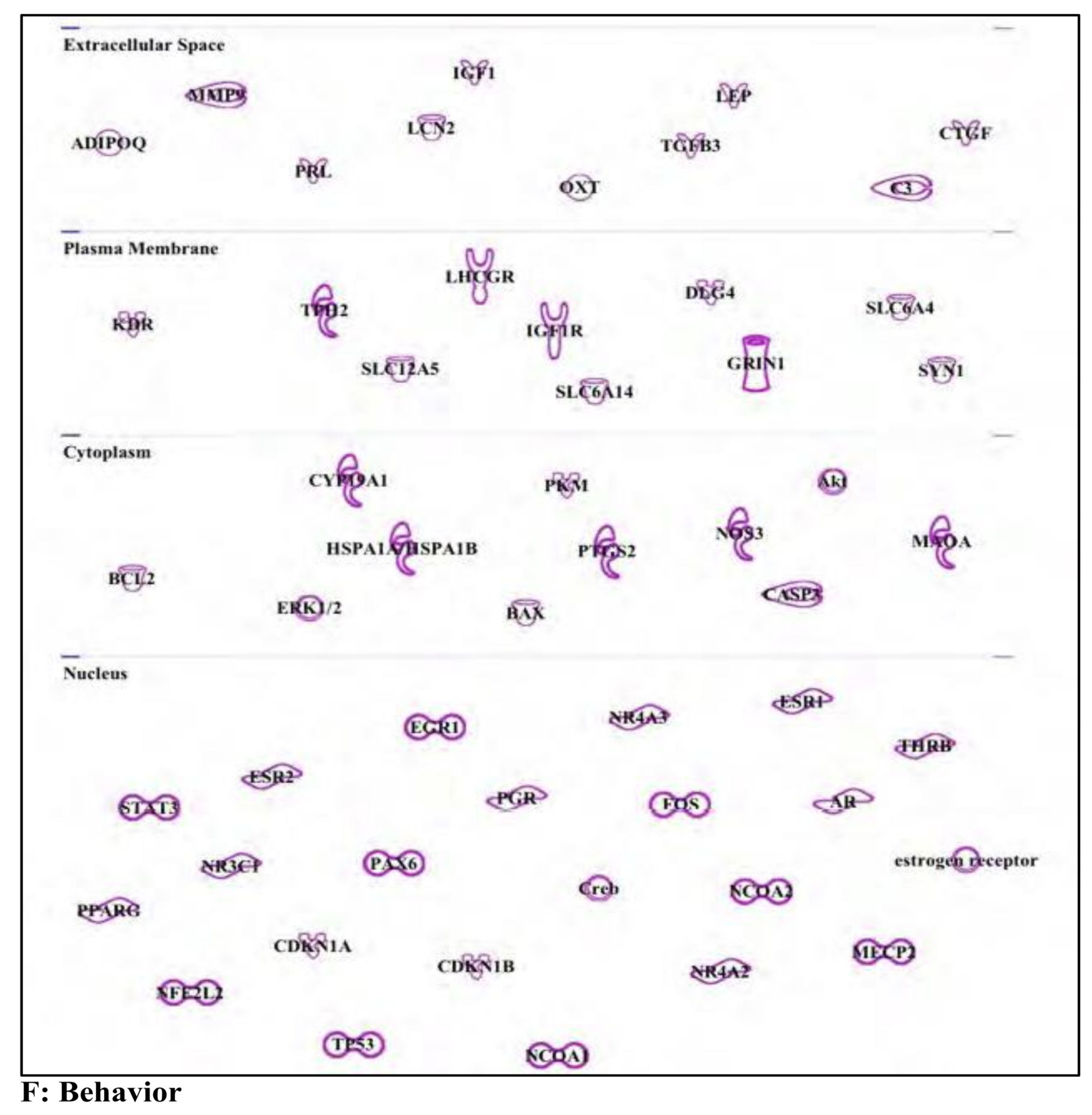
3. Results and Discussion

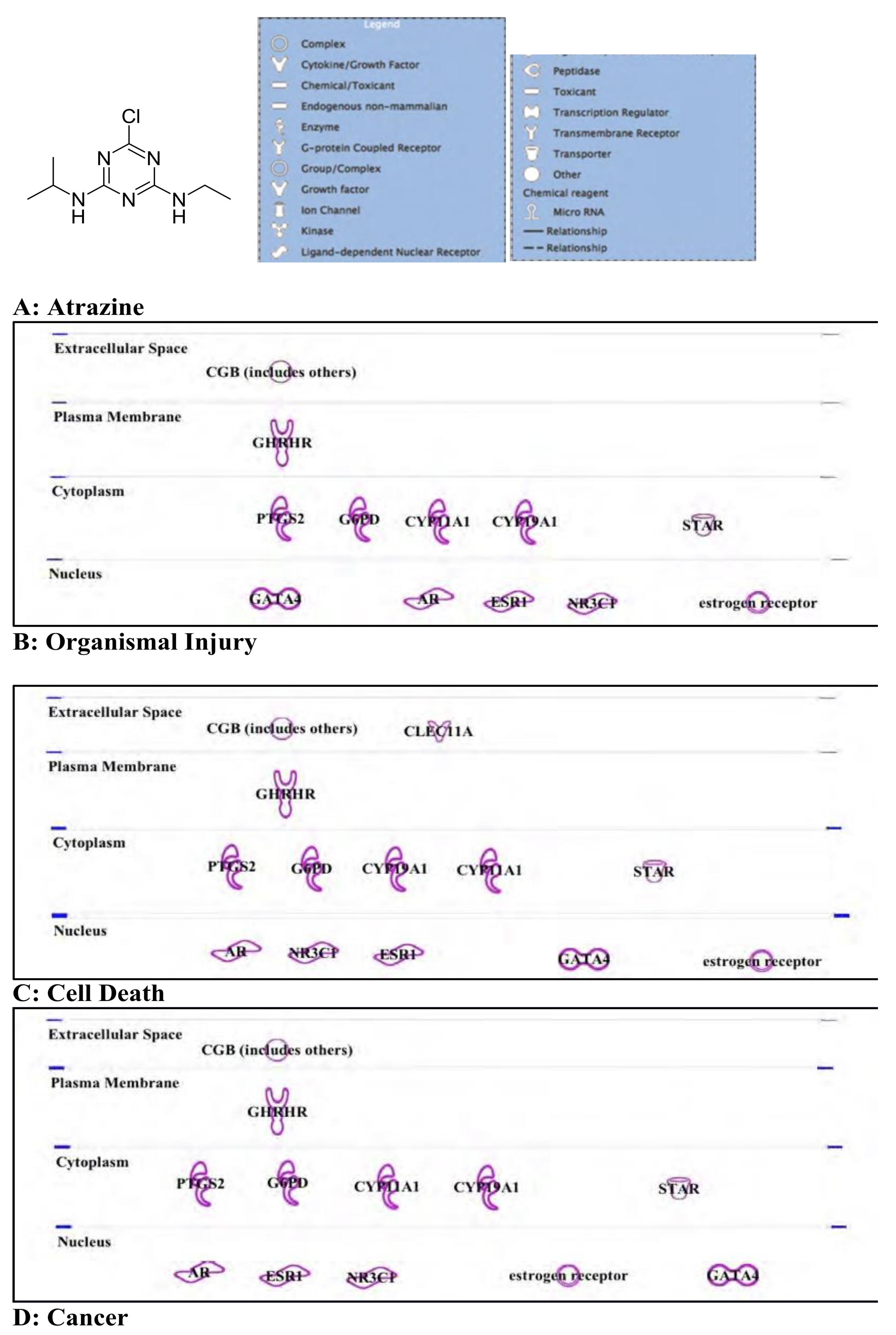
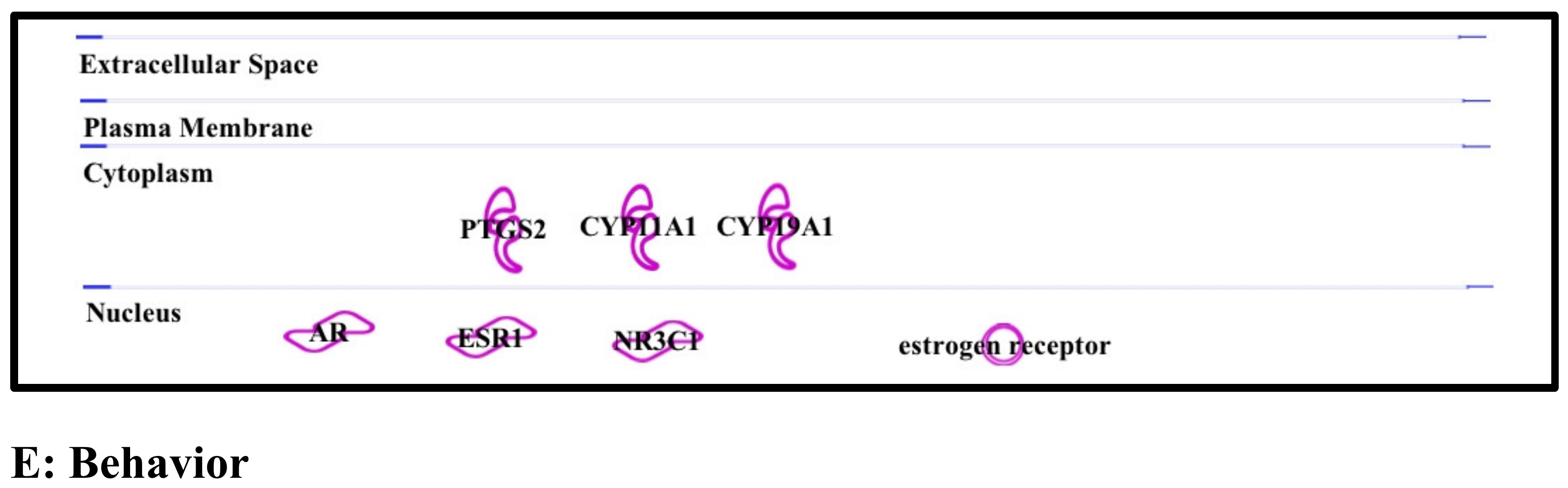
3.1. Atrazine: Standard Herbicide, Potential Carcinogen, and Environmental Immune Disruptor

Atrazine is an herbicide belonging to the triazine family (see Figure 1A for structure) that has been widely used by agriculture workers in the USA for decades and has long been suspected to have multiple deleterious effects on both invertebrates and vertebrates. Based on this suspicion, it was banned in 2004 by the European Union, as the levels in ground water were exceeding the legal limits considered safe for the environment (See European Commission Decision C (2004) 731 [28]). Despite this fact, in 2003, the Environmental Protection Agency (EPA) estimated that “cumulative exposures to these pesticides (atrazine and simazine) through food and drinking water are safe and meet the rigorous human health standards set forth in the Food Quality Protection Act (FQPA).” (http://www.epa.gov/pesticides/reregistration/atrazine/atrazine_update.htm#atrazine). This statement was regarded as being highly controversial, as recent publications have indicated that chronic exposure to the presence of atrazine and/or degradations products is likely to have serious negative effects on human health [29]. To further investigate these potential effects of atrazine exposure, we performed an in silico gene ontology analysis using the Ingenuity Pathway Analysis (IPA) software package, focusing on identifying how atrazine affects cellular functions, such as organismal injuries, cell death, cancer and behavior.
(A) Organismal injury: As was expected, based on its chemical structure, atrazine has an effect on the expression and cellular pathways of genes involved in hormonal responses. The testosterone, estrone, estrogen, and estradiol pathways are all prime targets in atrazine-exposed cells (see Figure 1B and Supplementary Materials Table S1). Increased levels of expression were noted for specific hormone receptors, such as the androgen receptor (AR), glucocorticoid receptor (GR), growth hormone-releasing hormone receptor (GHRHR), and the estrogen receptor (ER). The cytochrome p450 family genes CYP11A1 and CYP19A1, known to be involved in drug metabolism and detoxification, were found to be affected by atrazine exposure as well [30].
(B) Cell death: Many of the pathways and receptors associated with organismal injury are also involved in atrazine-mediated cell death (Figure 1C and Supplementary Materials Table S2), in addition to the growth factor CLEC11A, a member of the C-Type lectin superfamily-3 [31], and known to be involved in apoptosis [32]. Based on our analysis, most of the genes that we identified to be involved in the atrazine exposure response have been shown to be linked with triggering apoptosis in fish [33], amphibians [34], and mice [35,36]. This effect can be mediated by changes in access to membrane receptors, such as the glucocorticoid receptor (GR, [37,38]), a process which has been linked with the gene NR3C1, coding for the glucocorticoid nuclear receptor variant 1 ([39], see Figure 1C). The intra-cellular response involves detoxification through cytochrome p450 genes, such as CYP11A1 and CYP19A.1, Interestingly, these genes are also known as the key regulators in cytotoxicity and apoptosis [30,40].
(C) Cancer: Again, the genes affected by atrazine exposure manifest their effects through responses linked to hormone receptor pathways, involving various genes from the cytochrome p450 family genes [30,41] (Figure 1D and Supplementary Materials Table S3). The dysregulation of the aromatase CYP19, mediated by the steroidogenic factor 1 (SF-1), leads to increased risks of developing prostate and breast cancers in human [42,43]. The general mechanism of action of atrazine is considered to involve oxidative stress and the production of reactive oxygen species (ROS) and hydroxyl radicals, processes which are known to contribute to increased DNA damage [41]. An increased DNA damage response in human breast epithelial cells (MCF-10A) has recently been reported in response to atrazine exposure-mediated DNA double-strand breaks [44]. The atrazine-mediated increase in the expression of GATA4, a transcription factor involved in DNA damage response, confirms this connection [45]. GATA4 may also indirectly cause an increase in inflammation [46]. As an increasing amount of evidence suggests a role for atrazine as an oncogenesis trigger in breast cancer, one may envision that atrazine may soon be officially considered as a potential carcinogen, based on its xenoestrogen properties as well as its ability to induce DNA double-strand breaks.
(D) Behavior: The effects attributed to an exposure to atrazine and behavior changes are also mediated by hormone receptors and cytochrome p450 family genes [30,41] (Figure 1E and Supplementary Materials Table S4). An exposure to low doses of atrazine has been shown to affect the behaviors of young male mice [47,48]. The reproductive behaviors of male Drosophila have also been shown to be affected by atrazine exposure [49]. Atrazine-mediated mis-regulation of acetylcholinesterase was recently shown to affect defensive behavior in zebrafish [50].
As we investigated the effects of atrazine on various biological systems and functions, our results indicate that this specific herbicide harbors a strong carcinogenic potential, as has been long suspected [51]. As a hormone mimic, the main identified targets of atrazine are hormone receptors, some of which (AR, GR, and ER) are strongly associated with breast [52,53] and/or prostate cancers [29]. These receptors are also involved in the processes of growth and development, therefore long-term exposure should be considered potentially hazardous for developing aquatic and semi-aquatic animals (amphibians, fish, etc.) [54], as well as mammals exposed to contaminated water [55,56]. In addition, atrazine has also been recently associated with disruptions of immune evasion, a mechanism involved in regulating tumor formation, progression, and evasion [57,58], and future studies are likely to uncover additional effects mediated by atrazine exposure involving the immune response and their connection with oncogenesis. Deficiencies in processes such as tumor evasion prevent one’s immune system from recognizing and disposing of malignant tumors. Importantly, recent studies have also linked exposure to atrazine with increased DNA damage in fish [59] and mammals [60], including humans [44,61]. As previously mentioned, this aspect of atrazine toxicity is likely to be connected with the increase in the formation of ROS caused by the reduced expression of cytochrome p450 family genes, hence leading to increased DNA damage. This effect on impeding DNA damage might give a clue to the pluripotent effects of atrazine, as the increased presence of ROS will promote DNA single and double-strand breaks, causing mutations and chromosomal instability.

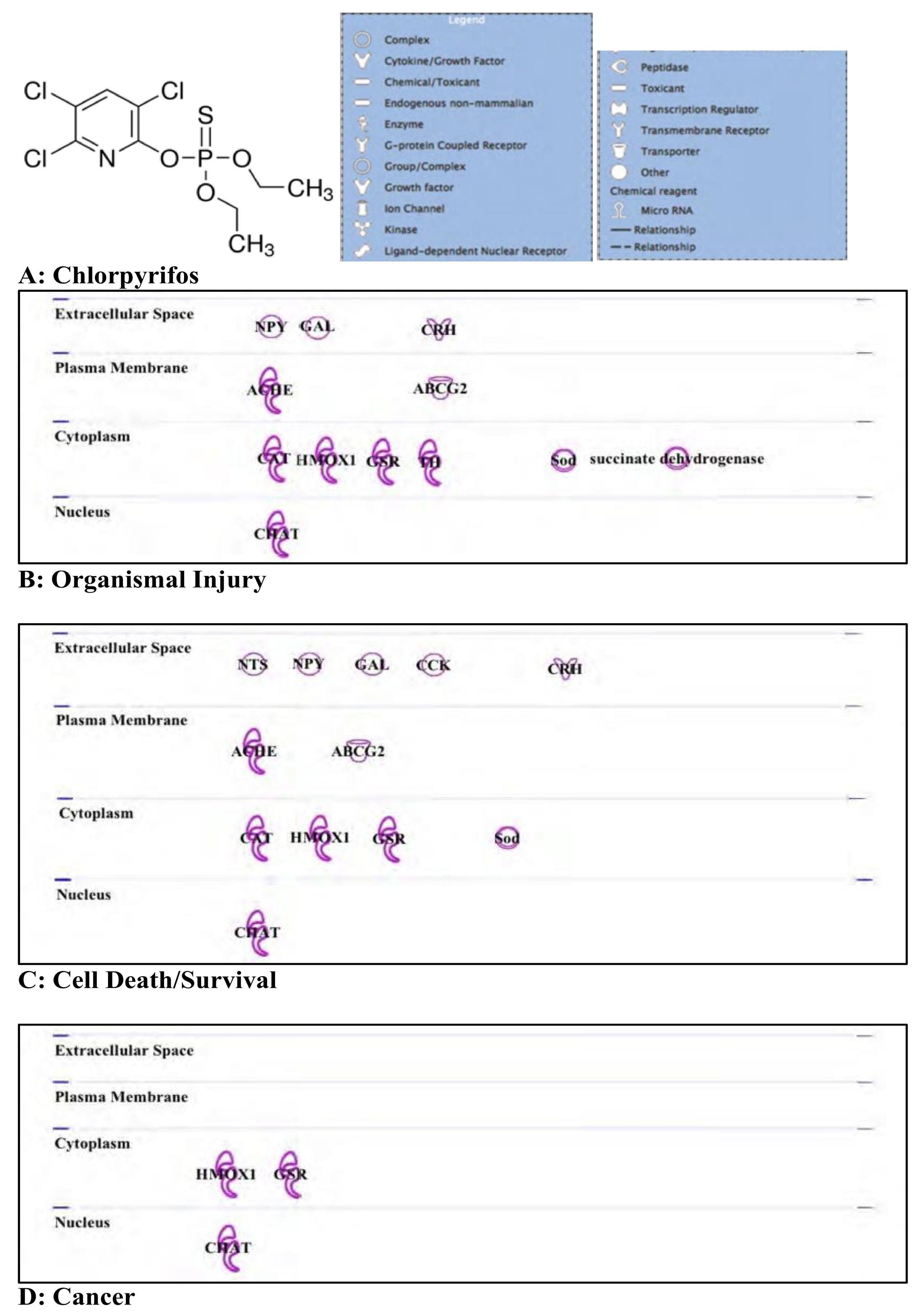
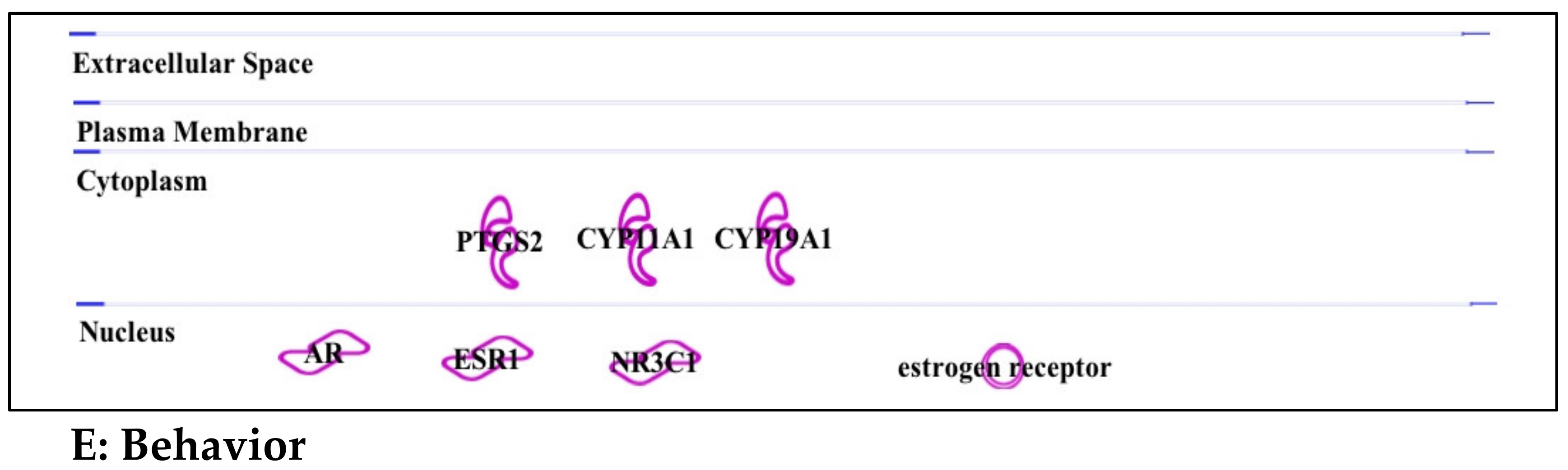
3.2. Chlorpyrifos: Organophosphates and Their Neural and Hepatic Toxicities

The use of organophosphate insecticides is widespread in agriculture, despite being a frequent source of poisoning around the world. In 2002, there were an estimated 3,000,000 cases of organophosphate poisoning globally, which resulted in 300,000 deaths [62]. The United States has had far fewer cases of lethal organophosphate poisoning reported compared to other developing nations. This lower number might be partially due to better access to the main antidotes to chlorpyrifos poisoning—atropine and pralidoxime. Nonetheless, organophosphates such as chlorpyrifos (for structure, see Figure 2A) are still found in commercially available household insecticides, which increases the average individual’s risk of exposure to these harmful compounds.
Although the modern use of organophosphates is generally limited to insecticides, these compounds have also been used historically in chemical warfare due to their inherent lethality in humans. While unfortunate, this has resulted in a wealth of anthropocentric research data not available when compared to with many other insecticides. Examples of organophosphates include physostigmine, which is naturally found in the Calabar bean and has been used for centuries in West African witch trials [63], and sarin gas, first used during World War II and a known contributor to the high rate of insecticide-mediated suicides in modern Asia [64]. On a molecular level, these deadly chemicals exert their effects on the brain through the inhibition of the critical enzyme cholinesterase (an enzyme involved in the degradation of the neurotransmitter acetylcholine).
Much of the early research on organophosphate toxicity failed to definitively link exposure to any long-term effects in humans [65]. However, decades of investigation have followed and led to a change of opinion, as several neuropsychiatric conditions have become strongly associated with organophosphate exposure [66]. Broadly speaking, long-term toxicity results in similar symptoms to those seen in acute exposure, and a single severe episode of acute poisoning may lead to chronic effects long after functional cholinesterase levels are restored [67]. Adding to the current knowledge, our study has linked organophosphate exposure to changes in gene expression involved in organismal injury, cell death, cancer, and behavior (Figure 2B–E).
(A) Organismal injury: Chlorpyrifos exposure increases the expression of many genes associated with stress (Figure 2B and Supplementary Materials Table S5). These include CRH (corticotropin-releasing hormone), an important regulator in the hypothalamic–pituitary axis; GAL (galanin), which may be neuroprotective; and ABCG2, a multi-drug resistance transporter gene. The choline acetyltransferase-encoding gene ChAT was affected, which is not surprising considering the direct mechanism of action of chlorpyrifos is through the inhibition of the cholinesterase enzyme. Other gene targets of chlorpyrifos exposure include succinate dehydrogenase, one of the key enzymes in the citric acid cycle and electron transport chain and Sod, a superoxide dismutase gene which is important in apoptosis, and is linked with DNA damage.
(B) Cell death/survival: The cell death- and survival-associated genes targeted by chlorpyrifos are similar to those involved in organismal injury (Figure 2C and Supplementary Materials Table S6). Examples include Sod, ChAT, ABCG2, CRH, NPY, and GA. In addition, a heme-oxygenase encoding gene known as HMOX1 is affected by chlorpyrifos exposure. Overall, the profile of cell death and organismal injury targeted by chlorpyrifos activity demonstrates that the toxicity of this chemical affects not only the existing cholinesterase enzyme, but also the genes which are key factors involved in cell protection and metabolism.
(C) Cancer: Only three cancer-related genes were identified as being altered by chlorpyrifos exposure, and all three were common to the other categories delineated in this paper: ChAT, GSR, and HMOX 1 (see sections A, B, and D, Figure 2D and Supplementary Materials Table S7). GSR encodes glutathione disulfide reductase, a highly conserved gene which is important for preventing oxidative stress in humans. This function is highly relevant in cancer, as the conversion of GSSG (oxidated Glutathion) to GSH (Glutathion) promotes increased DNA synthesis, which aids the growth and development of tumors [68].
(D) Behavior: The main behavior-related genes identified in this ontological study were ESR1, NR3C1, and genes involved in the conversion of fatty acids or cholesterol to hormones. These include PTGS2, CYP11A1, and CYP19A1, which encodes aromatase (Figure 2E and Supplementary Materials Table S8). These findings suggest that chlorpyrifos exposure is closely associated with the production of sex steroids and the resultant signaling pathways, via the estrogen receptor-encoding genes ESR1 and NR3C1.
As was expected in the case for atrazine, based on its chemical structure, chlorpyrifos is a strong disruptor of hormonal responses. However, its biological effects are more targeted towards the processes involving cellular detoxification and glutathione (GSH) [68]. Although the majority of the data available were generated using the planktonic crustacean Daphnia as a model system, several reports suggest that its effects follow a similar mode of action in humans [69,70], also potentially mediated by GSH availability. Recent studies of chlorpyrifos exposure also give indications that it may alter proper brain function and development by impairing the cortical axon functions in rats [71], as well as proper differentiation of neural stem cells into neuronal and glial cell phenotypes [72]. The links between chlorpyrifos toxicity with cancer are less obvious than that observed with atrazine, as they are apparently mediated by defective detoxification genes, such as GSR. This might be a secondary effect, possibly related more to a potential red-ox imbalance (oxidative stress in general) than a direct carcinogenic effect. As a potential link to this oxidative stress, DNA damage associated with chrlorpyrifos exposure has been reported in various mammalian tissue, most notably in the brain [73].

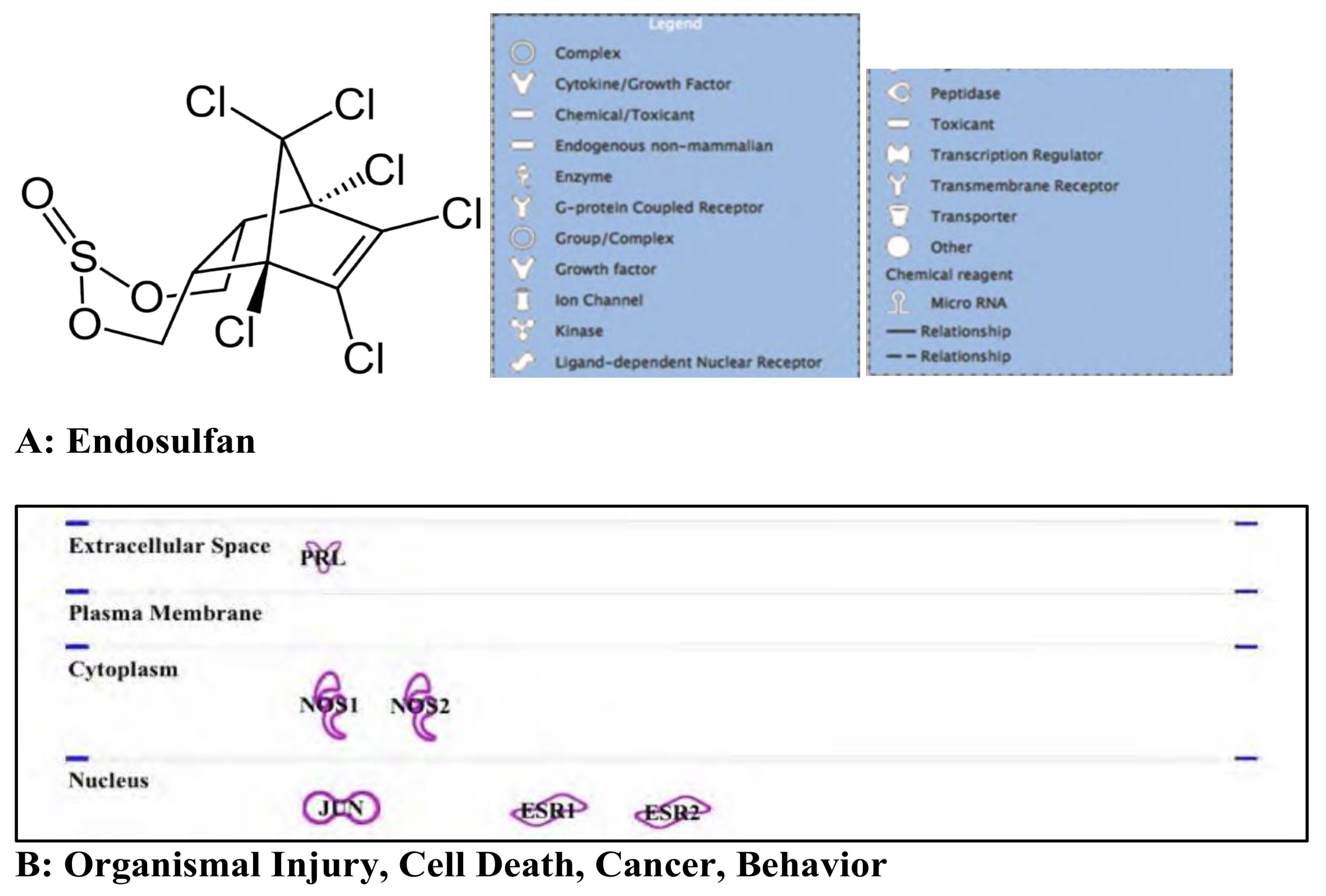
3.3. Endosulfan: Pesticide

Endosulfan is an organochlorine cyclodiene pesticide (see Figure 3A for structure) considered to be highly toxic because of its endocrine effects and high potential for bioaccumulation (EPA toxicity Class I, for additional details see https://www.epa.gov/sites/production/files/2014-07/documents/chapter7_revised_final_0714.pdf). Based on our analysis, all changes in organismal injuries, cell death, cancer and behavior a similar sub-set of genes, which are generally associated with endosulfan’s ability to be recognized as a hormone mimetic substance (Figure 3B and Supplementary Table S9). Most reports on the toxicity of endosulfan are based on its biological effects on aquatic organisms [74]. In contrast, less information is available on its effects on humans, although initial reports, from as early as 1982, described its potential bioaccumulation in humans as well as several other non-target species (for more details, see [75]). Aquatic species appear to be more sensitive to the bioaccumulation of endosulfan, therefore experiencing a higher toxicity [74,75,76]. According to studies performed on various animal models and as seen with the previously-mentioned EDCs and based on its chemical structure, endosulfan can predictably disrupt hormonal responses in both fish and mammals (including humans) [77]. Our ontological study results identified a similar set of genes affected by endosulfan exposure in all the investigated cellular-function scenarios. These deleterious effects can result in the triggering of developmental issues in multiple cell types including, but not limited to, the reproductive tracts [78]. Additionally, two important genes involved in cell proliferation, the estrogen receptor ESR1 and ESR2, are strongly affected by endosulfan exposure [11]. Another report associates endosulfan toxicity with prolactin (PRL) expression [79], an effect which may also be mediated by changes in expression of the nitric oxide synthase genes NOS1 and NOS2, altering the normal functions of the pituitary glands [80]. In addition, exposure to endosulfan has been linked to an increased incidence of various types of cancers, with cell types involved in sexual development, primarily breast, as primary targets [78,79,80,81,82]. Other tissues or cell types are also targeted, such as in colon cancer, in which jun/AP-1 is the main pathway affected by endosulfan exposure known to date [83]. As a possible linkage to explain its activity in cancer cells, endosulfan has been identified as an apoptotic agent [84]. The affected cell types by this mode of action cover a wide spectrum, from human T-cells [84] and peripheral mononuclear cells [85] to umbilical, embryonic, and placental cells [86].
As revealed in our chlorpyrifos analysis, the bulk of evidence for endosulfan toxicity uncovered in our ontologic study was based on experiments performed using aquatic species. Nonetheless, a small number of mammalian (mostly rat) studies on endosulfan exist providing valuable information pertaining to human health. Exposure to endosulfan in young male rats interferes with normal development of the mammary glands [87], an expected result for EDC toxicity. Recent work has also linked endosulfan exposure to deficiencies in the brain and behavioral functions [88]. Aiming to determine its general mechanism of action, additional studies have indicated that endosulfan appears to be associated with an apparent increase in non-sequence-specific DNA damage (for review, see Sebastian and Raghavan, 2017 [89]). This might explain how endosulfan can affect a large number of genes involved in a variety of cellular functions instead of having more defined and specific genetic targets. This potential mechanism of action involving genomic instability raises the possibility that endosulfan displays carcinogenic properties.

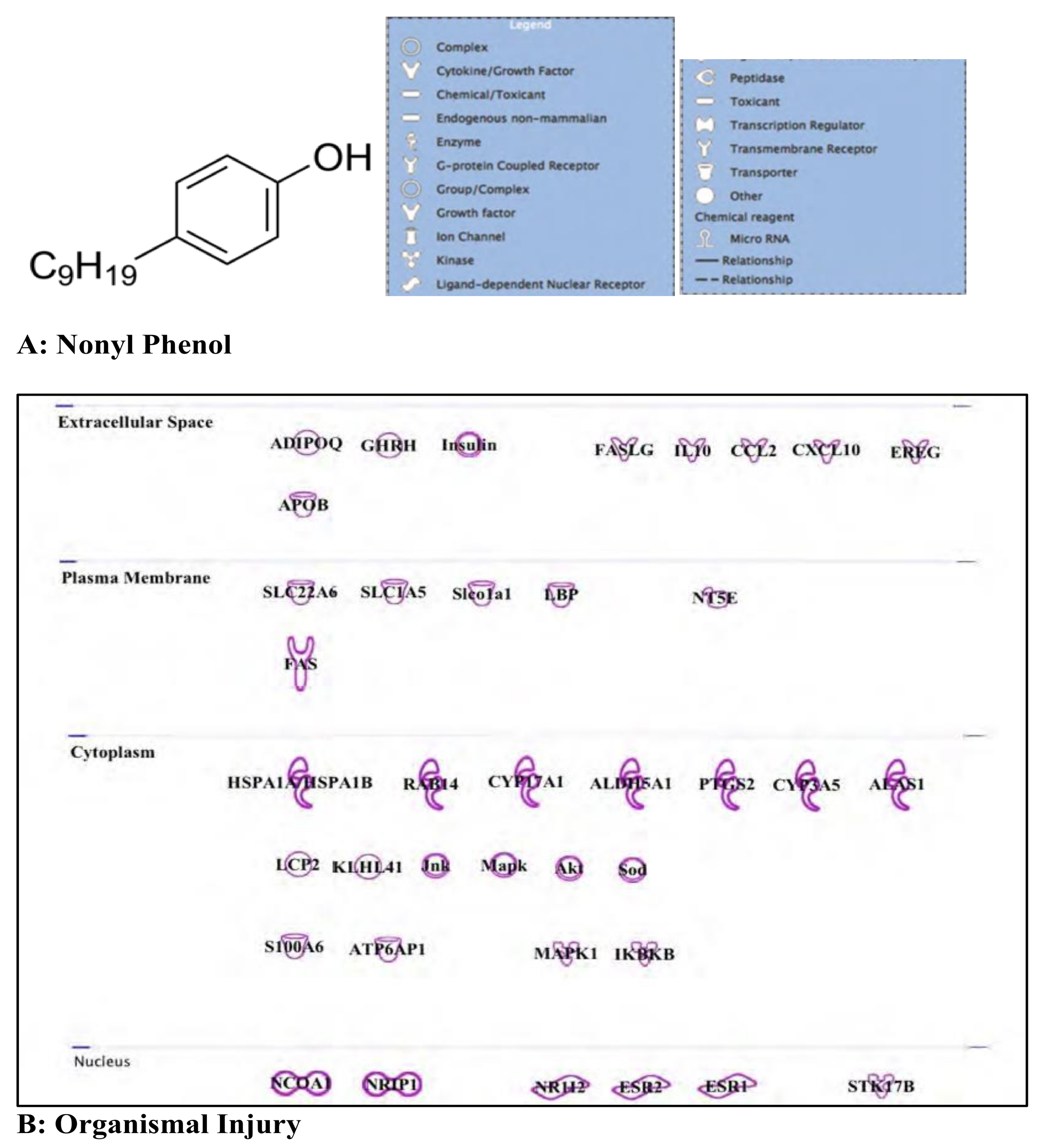
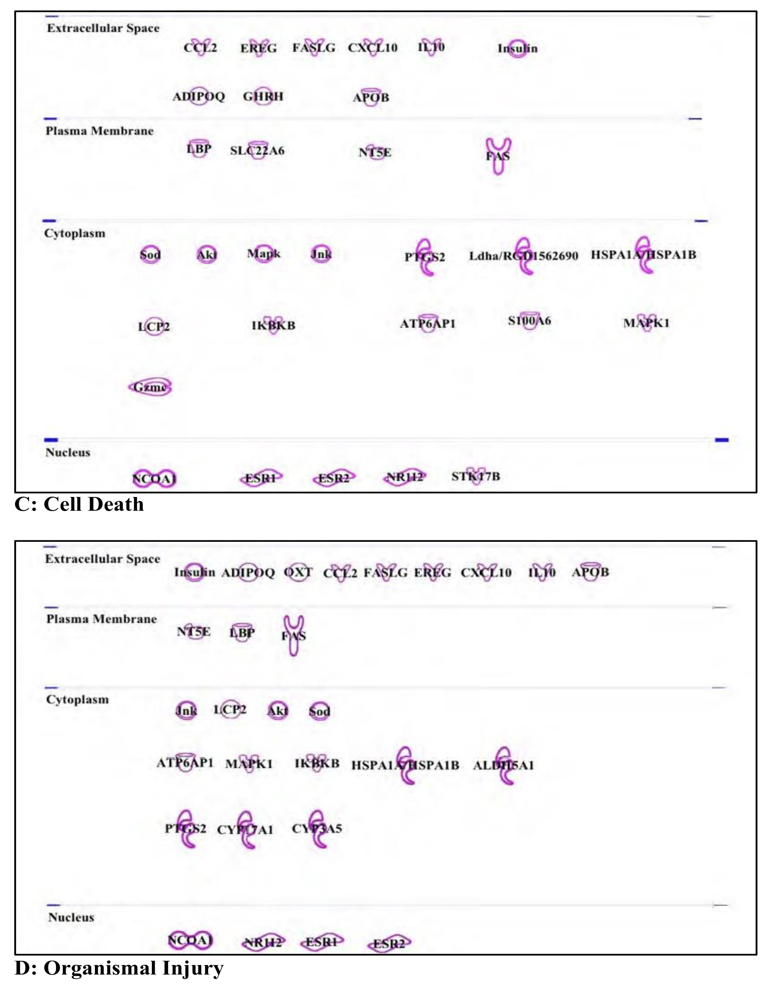
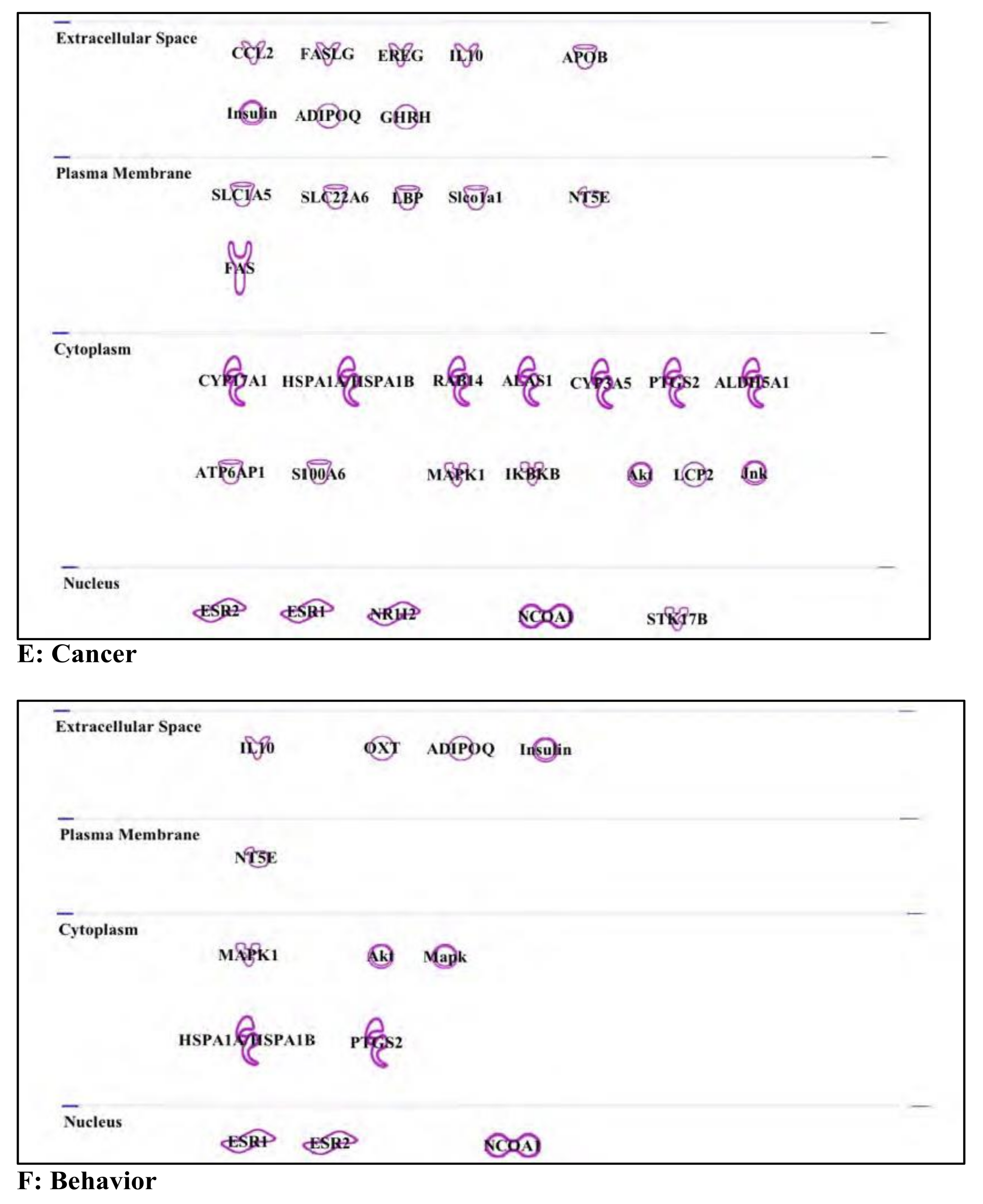
3.4. Nonyl Phenol and Nonyl Phenol Ethoxylates: Endocrine Disruptor

Nonyl phenol, or 4-nonyl phenol (NP), is a non-ionic surfactant (see Figure 4A for structure) commonly derivatized to generate Nonyl phenol ethoxylates (NPE) which are used as emulsifiers, detergents, and dispersing agents [90]. Since 2000, NP and NPE have been highly regulated in European Union countries because of their inherent toxicities [91] (PARCOM 92/8, 2000; Directive 2000/60/EC, 2000; [92] Directive 2003/53/EC, 2003).
As was shown in planarians and numerous other non-vertebrates, organismal survival and ability to respond to injury can be affected by NP exposure, similarly to what has been described for other EDCs [93] (for examples, see [94,95]). In zebrafish, long-term exposure was shown to be a factor in survival by reducing the reproduction rate [96]. Also, the survival and injury response may be reduced because of the ability of NP to form adducts with DNA which leads to possible increases in mutation rate [97].
(A) Cell death and organismal injury/survival: Exposure to NP has been reported to induce apoptosis in various cell types, including sexually related cells, neurons, and neural stem cells [98]. As NP acts as a hormone mimic, the effect on sexual organs was expected; however, the apoptotic induction in neurons and neuronal stem cells was more surprising. Nonetheless, the consequences of such exposure during gestation and/or early brain development may be a very serious issue that has not yet received full attention. These observed effects can be linked to hormone receptors mediated by the signal transduction triggered by several types of surface receptors such as the FAS receptor (also referred to as the “apoptosis antigen”) and the anion transporter SCL22A6 ([99] (see Figure 4B–D and Supplementary Materials Tables S10–S12).
(B) Cancer: As an EDC, the health risk related to exposure to NP and/or NPE stems from their ability to act as a hormone mimic [100]. Studies have linked NP with breast cancer, as it can mimic 17B-oestradiol and compete for the binding site of oestrogen receptors ESR1, ESR2, and the related steroid receptor co-activator-1 (SRC-1/NCOA1) [101] (see Figure 4E and Supplementary Materials Table S13). Also, a linkage with androgen receptors has also been reported. This suggests a potential involvement of NP exposure with prostate cancer [102]. This mechanism may not involve a direct interaction, but instead may be mediated by cross-talks with other hormone receptor(s). As there is very little information connecting NP with other types of cancers, it is possible that its effect is exerted in a compound manner with other EDCs or emerging water contaminants [90].
(C) Behavior: In addition to a propensity for promoting breast cancer, NP can also alter the neuroendocrine system [90,103,104]. This interference may be associated, directly or indirectly, with behavioral modifications, such as motility changes (swimming in fish), or social behaviors [103,105,106]. Interestingly, these modifications have been observed over two generations, suggesting a potentially epigenetic mechanism of transmission [107]. As was the case for the cancer-associated effects, hormone receptors (ESR1/2, NCOA1, see Figure 4F) may play a significant role in these behavioral changes.
Overall, as with most EDCs, NP can act on a plethora of genes, resulting in alterations of multiple functions. The effects are most often mediated by changes in hormone receptor activity. Interestingly, and similarly to endosulfan, NP exposure was shown, in human keratinocytes, to be linked with DNA damage. This mechanism involved multiple factors, including ATM (ataxia-telangiectasia mutated), the tumor suppressor p53, and the histone H2A.X (a marker of DNA double-strand breaks). The same study linked NP to apoptosis, mediated by activation of poly(ADP-ribose) polymerase (PARP) and caspase 3 [108]. Although not clearly outlined by our bioinformatics analysis, very recent literature has connected NP with liver toxicity in mammals, including humans, as its pro-inflammatory properties negatively affects liver cells [109].

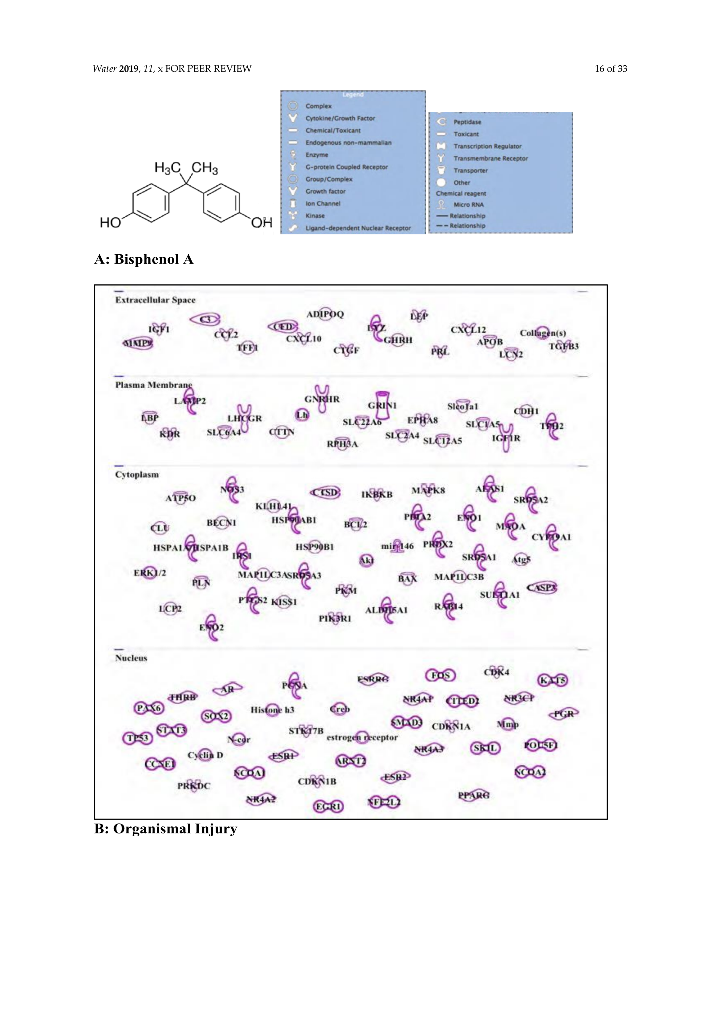
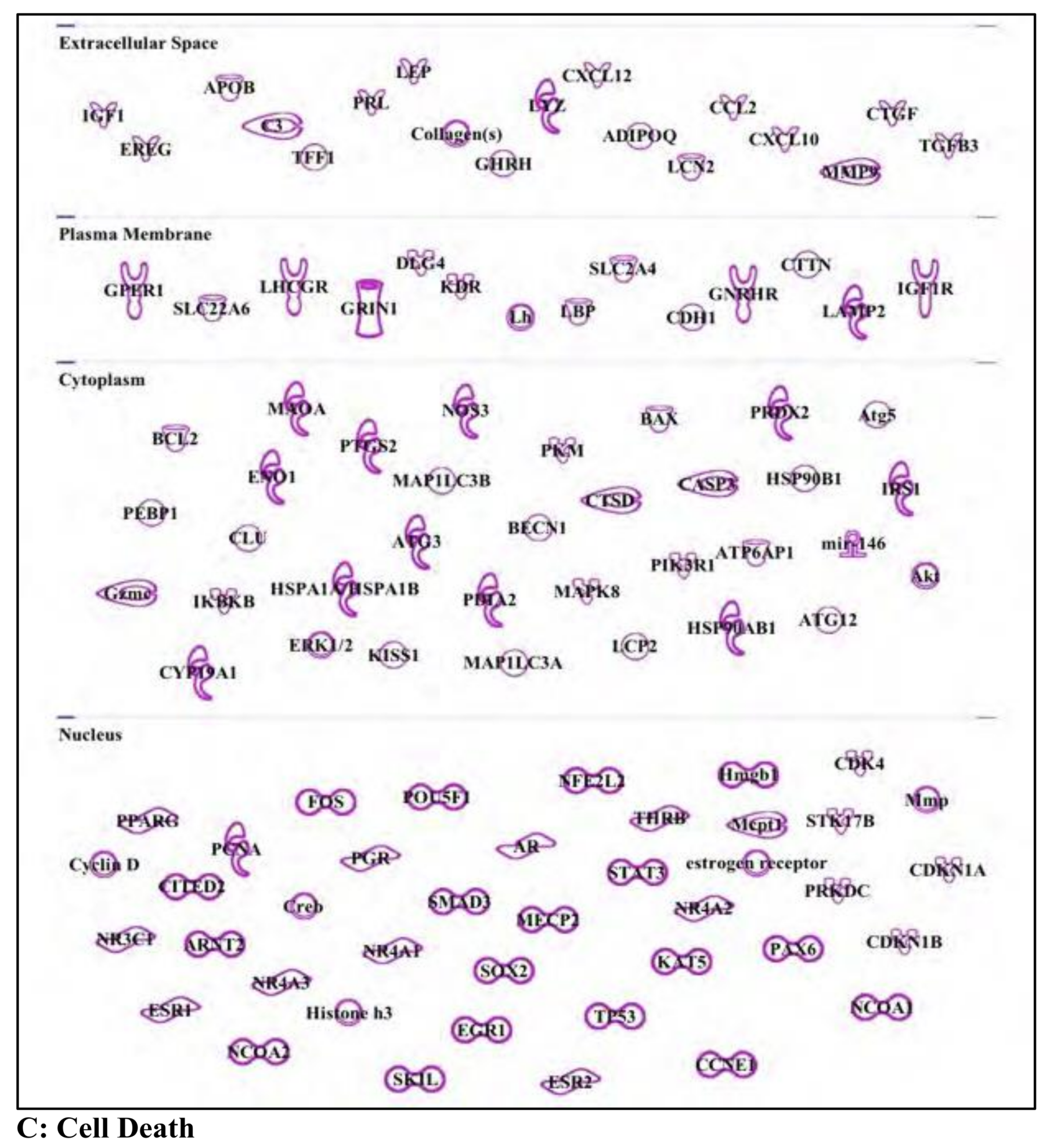
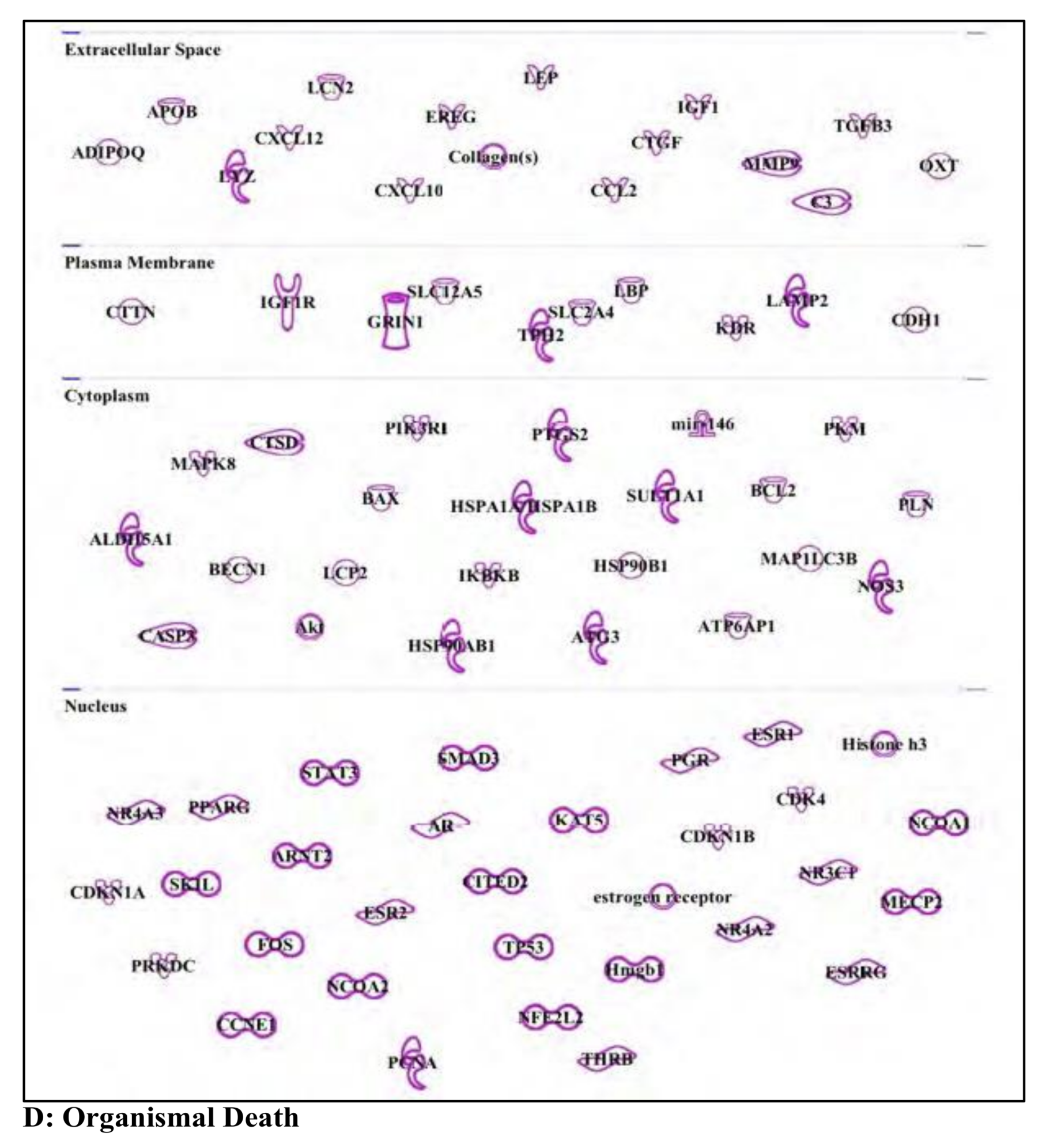
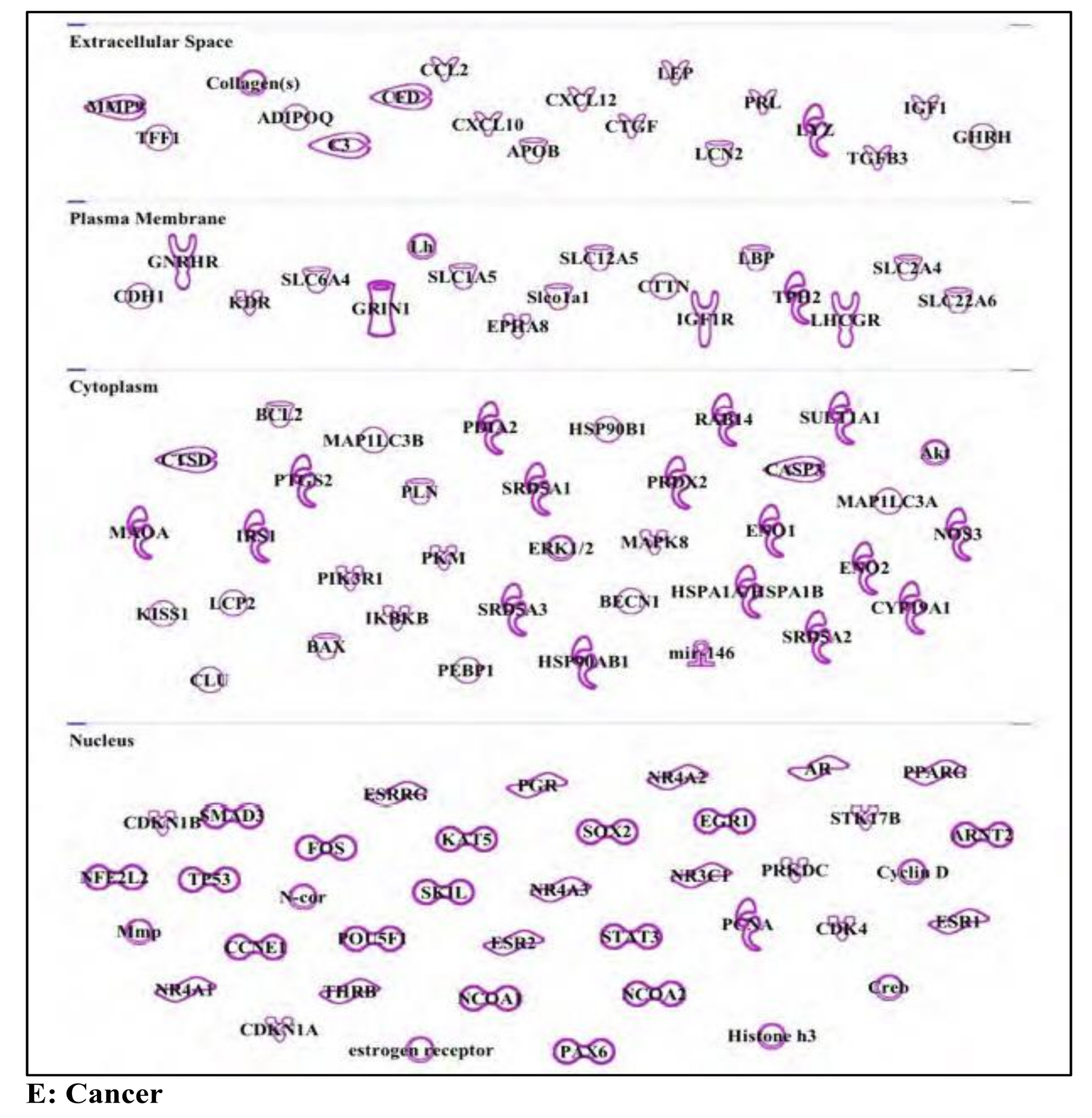
3.5. Bisphenol A: Endocrine Disruptor

The rise of safety concerns related to chemicals released from consumer polycarbonate plastics has led to a concomitant increase in the study of their potential toxic effects. Bisphenol A (BPA) is perhaps the most widely recognized plastic-derived toxin (for structure, see Figure 5A). Although most of the major studies regarding the potential toxic effects of BPA used rats as an animal model, it has since been found that primates metabolize BPA into an inactive form more efficiently than rodents, suggesting that humans may be less susceptible to its toxic effects than initially believed [110]. Fortunately, despite the uncertainty on the extent of BPA exposure to human toxicity, industries have ceased using BPA in the manufacture of consumer plastics based on an amendment of FDA restrictions ([111] Fed. Reg. 41,899; 78 Fed. Reg. 41,840).
Although it appears that current and future plastics may be largely free of BPA, due to the non-biodegradable nature of plastics, there is still concern regarding less direct routes of exposure. In 2011, 19 landfills had their leachate tested for potential contaminants and 95% of these samples tested positive for BPA [112]. It was also detected at levels known to negatively affect some invertebrate species in untreated and treated wastewater, as well as in river water downstream from a paper factory [113]. In a recent study to determine the possibility of food-borne exposure toxicity, researchers grew vegetable plants using BPA-contaminated water and found that ingestion of contaminated agriculture products would result in exposure to physiologically significant doses of BPA known to cause observable developmental changes experimentally [114]. Thus, despite the absence of BPA in modern plastics, former industrial practices may continue to have a negative impact on human health through soil, water, and agriculture contamination.
The health risks associated with BPA exposure are primarily related to endocrine disruption, as its main mechanism of action involves binding to estrogen receptors (ER), thus acting as a weak estrogen [115,116]. A few of the major health research foci regarding the effect of BPA’s endocrine disrupting activities are obesity, reproductive system dysfunction, immune dysfunction, and cancers of the breast, prostate, and uterus [117]. The ability of BPA to disrupt the endocrine system may also interfere with progesterone receptor expression in human endometrial tissue [116], leading to an increased body mass and decreased glucose tolerance in males [118], negatively impacting male fertility [119], and accelerated growth during childhood [120]. In addition to these effects, as an EDC, BPA also targets genes involved in organismal injury, cell death, cancer, and behavior Figure 5B–F.
(A) Organismal injury: BPA can affect an organism’s ability to heal and respond to injury by the disruption and alteration of the endocrine system (Figure 5B and Supplementary Materials Table S14). Chronic BPA exposure has also been shown to reduce cardiac remodeling in mice [120], and is toxic to bone mesenchymal stem cells [116]. In addition to affecting these systems, exposure to BPA also has an impact on the Sertoli cells of the testes, [121] as well as on ovarian cells [81].
(B) Cell/organismal death: In addition to overt organismal injury, BPA exposure can lead to apoptosis of spermatogenic cells [122]. In fact, many of the most toxic effects of BPA are observed in cells of the reproductive system (Figure 5C,D and Supplementary Materials Tables S15 and S16). To this end, BPA has been shown to negatively impact embryo and oocyte quality in mice by increasing apoptosis [81,116]. Additionally, BPA exposure can result in the induction of apoptosis in mouse pancreatic islet cells [116].
(C) Cancer: BPA exposure is specifically associated with breast cancers (Figure 5E and Supplementary Materials Table S17), through the increased expression of HOXB9, a gene important in mammary gland development known for its potential oncogenic properties [121]. This effect seems to be related to its ability to mimic or activate similar pathways as those mediated by endogenous estrogens. In addition, BPA exposure can also increase the expression of two matrix metalloproteinases, MMP2 and MMP9 [111], the functions of which are linked with enhanced breast cancer migration and invasion.
(D) Behavior: Sharing similar mechanisms with endogenous estrogens, it is not surprising that BPA has behavioral effects (Figure 5F and Supplementary Materials Table S18). A systematic review on BPA exposure in Canadian school-age children revealed that early-life and prenatal exposure was associated with increased behavioral disorders, such as anxiety, depression, hyperactivity, inattention, and conduct problems [115]. Additionally, children taking psychotropic medications were found to be more likely to be susceptible to BPA exposure than those not taking such medications [120].
BPA is probably the most notorious EDC and has been known for several years to be a cause of cellular toxicity. As mentioned earlier, the recent banning of its use will hopefully lead to a decrease in its abundance in major river basins, as well as in tap/drinking water. Studies involving BPA degradation during chemical remediation using Persulfate have indicated that, despite the efficient degradation of BPA itself, its degradation products have genotoxic effects of their own [123,124]. This highlights the fact that the risks associated with prior BPA use are still a serious concern for human health. Appearing to be a common theme for EDC exposure, BPA’s mode of action is also likely correlated with increased oxidative stress, which would contribute to a BPA-associated increase in DNA damage in mammals [125].

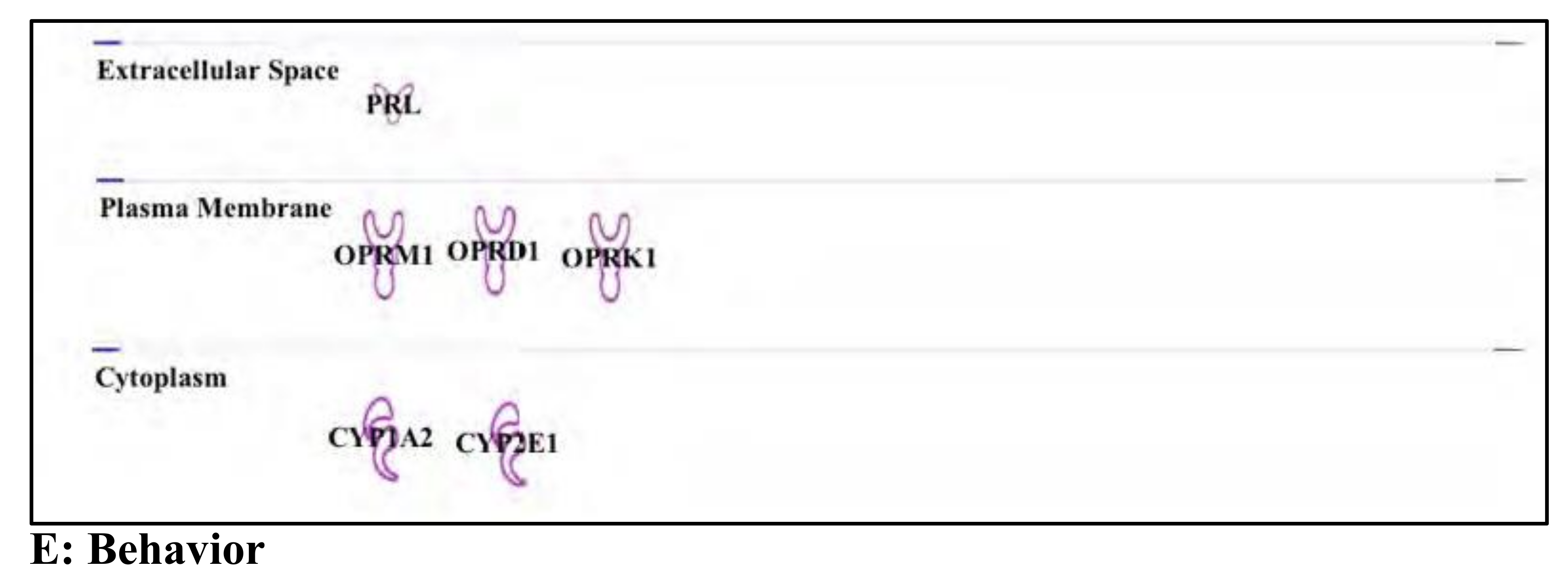
3.6. Buprenorphine: Opioid

Buprenorphine is now considered to be a substance of interest as a potential emerging water contaminant (for structure, see Figure 6A), based on its increased usage and prevalence as a treatment option for opioid addiction. Pregnant women abusing opioids present an additional problem compared to other drug abusers, as supporting these women in overcoming their addiction must be balanced with the serious concerns in protecting the fetus from the dangers of withdrawal. To palliate the negative effects of opioid abuse, one of the preferred agents of maintenance therapy in these pregnant patients is the opioid derivative buprenorphine, as it has a higher potential to reduce neonate abstinence syndrome (NAS, [126]) compared to methadone. Buprenorphine is a mixed agonist-antagonist of opioid receptors—a partial agonist of µ opioid receptors (MOR), and an antagonist of κ opioid receptors (KOR) and δ opioid receptors (DOR). In addition to its primary effects, the metabolic products of buprenorphine degradation, though differing in opioid receptor selectivity, half-life, and potency, may also be considered of concern for water quality.
Despite its status as a controlled substance, there is potential that physiologically relevant amounts of buprenorphine may be contaminating drinking water. In France, for example, buprenorphine was initially detected at levels of 40 ng/L in sewage water effluent at one study site, and at levels varying from 42 ng/L to 195 ng/L in sewage water influent samples at three additional study sites [26]. An earlier study, focusing on the Paris area, also detected low levels of buprenorphine from samples of wastewater entering four treatment plants [85]. While these levels correspond to small therapeutic doses compared to those prescribed to human patients, there is the possibility of a cumulative long-term effect through chronic exposure to the contaminated drinking water. The recent increase in buprenorphine use may eventually enhance its presence in water supplies. Therefore, it is important to identify the genes which may mediate any chronic effects of low-dose and long-term buprenorphine exposure. Consequently, as with our selected EDCs, we used our IPA® analysis to identify the buprenorphine-targeted genes involved in organismal injury, cell death, cancer, and behavior (Figure 6).
(A) Organismal injury/survival and cell death: Aside from the analgesic properties of opioids, an important secondary effect is increasing cell death, particularly of cells in the nervous and immune systems (Figure 6B,C and Supplementary Materials Tables S19–S21). Consequently, opioid addicts have been found to have decreased number of circulating progenitor stem cells [116]. Intriguingly, buprenorphine seems to exert a dose-dependent effect, either promoting cell survival and differentiation or increasing cell death and apoptosis [6,119,127]. Buprenorphine exposure in neuronal cells has also been shown to lead to caspase-3 activation and efflux of cytochrome c from the mitochondria, indicative of activation of the mitochondrial pathway of apoptosis [118].
(B) Cancer: Because of the associated addiction risks, medical practice for chronic pain in the United States is moving away from the use of opioids as treatment, except in cases where the benefits outweigh the risk [128] (Figure 6D and Supplementary Materials Table S22). However, due to the decreased abuse and addiction potential associated with buprenorphine compared to other pain management drugs, it is being investigated as an attractive substitute for managing chronic pain associated with cancer. However, given the previously discussed effects of opioids on cell populations, it is important to consider all the potential effects that buprenorphine might have on cancer cell populations.
For some time, it has been known that buprenorphine has a dose-dependent effect on serum prolactin levels in human, as low doses increase serum prolactin production levels while high doses of buprenorphine decrease them. Importantly, both the positive and negative effects on serum prolactin production are blocked by the opioid receptor antagonist naloxone [111]. Aside from the behavioral implications associated with this effect, prolactin has also been implicated in breast cancer [98] as high levels of prolactin receptor (PRLR) are expressed in some breast cancers, leading to increased cancer cell invasiveness [129].
(C) Behavior: Although prolactin is most directly associated with lactation and some aspects of maternal behavior [128], the other pathways involved in prolactin release should also be considered when analyzing the effects of buprenorphine (Figure 6E and Supplementary Materials Table S23). One of the key feedback pathways involved in prolactin release is dopamine release from the hypothalamus [130], as high dopamine levels exert an inhibitory effect on the release of prolactin [131]. The fact that buprenorphine can both increase or decrease prolactin levels supports the hypothesis that it interacts with the dopaminergic systems of the brain, and, also supporting this conclusion, it has been demonstrated that buprenorphine exposure affects the dopamine receptor density in the striatum [123]. In turn, dopamine is widely implicated in the pathology of psychiatric disorders.
Though buprenorphine is not a bona fide EDC, its potential to become a major water contaminant justifies its inclusion in our study. Its presence in the tap/drinking water in large urban areas (see Table 1) suggests that buprenorphine contamination is becoming a serious issue in regions currently dealing with any type of “opioid crisis”. As opposed to the EDCs that we investigated, which seem to share a mode of action strongly involving oxidative stress and DNA damage, buprenorphine does not appear to work in a similar manner, despite a few reports which link its toxicity with increased apoptosis [132].

4. Conclusions

As has become obvious over the last few years, water quality and emerging contaminants have become a serious concern for public health. Our ontology analysis clearly demonstrates that our selected examples have a plethora of negative effects on cellular systems, spanning from micro-organisms to human. The IPA® analytical settings used for our study outlined the most commonly observed effects, based on the current available literature, and we decided to focus on the to five “families” of function most often outlined by our search. The deleterious effects related to endocrine mimicking properties are a major concern directly related to exposure to these emerging contaminants. Even very low concentrations, such as those detected in the Ohio River (see Table 1), can lead to serious behavioral changes, as exemplified by the effects of buprenorphine anxiety, depression, hyperactivity, inattention, and conduct problems, as shown by Ejaredar et al. [133]. As expected, exposure to EDCs affects endocrine functions, mostly through standard interactions with hormone receptors, acting either as activators or inhibitors. These hormonal disruptions cause increased risks of developing various types of cancer (for example, but not limited to: prostate, breast, and ovarian cancers). In addition to these oncogenic properties, exposure to EDCs in our water supply clearly affects basic cellular functions as well, such as the ability to control cell death and survival (commonly mediated through modifications of genes involved in the apoptotic pathway).
From our analysis and based on recent reports, a common theme emerges connecting EDC exposure and increased oxidative stress which eventually leads to DNA damage. All the investigated EDCs in this study share a common link with this mode of action, as evidenced by studies on various organisms, from invertebrates to humans. This increased inability to compensate for EDC-induced DNA damage may play an important role in the toxicity of these products. Therefore, the cytotoxicity of these emerging contaminants might be provoked by, or be the consequence of, an underlying genotoxicity manifesting itself in a variety of ways, affecting a litany of genes involved in nearly all aspects of cell development and viability.
A potential explanation for why so many different genes can be affected by EDC or opioid exposure is that their mode of action could possibly be associated with epigenetic changes, although this possibility currently remains understudied. DNA methylation, as well as histone post-translational modifications (PTM), are critical epigenetic elements involved in regulating gene expression (for recent review, see Corella and Ordova, 2017 [134]). and an increasing number of publications have now demonstrated a connection between EDC exposure and the deregulation of DNA methylation at specific chromosomal locations.
Such connections between epigenetic regulations and EDC exposure have been reported for atrazine, the mode of action of which appears to affect the global levels of DNA methylation [135,136] as well as that of specific histone PTMs [137]. Highlighting the negative impact of EDC exposure, these epigenetic events have even been shown to affect rat sperm cells in a transgenerational manner, changing the DNA methylation pattern for up to three generations [138].
Endosulfan has also been shown to affect the expression of specific genes in an epigenetic manner [139,140], potentially modulating the expression profile of the histone modifiers histone deacetylase (HDAC) HDAC1 and 3, as well as histone methyltransferase PRMT5 and EZH2, in breast cancer cells [140]. This results in an increase in the histone PTMs tri-methylated histone H3 Lysine 27 (H3K27me3) and di-methylation of histone H4 arginine 3 (H4R3me2), two epigenetic modifications that are known to be involved in the regulation of gene expression by promoting the recruitment of activator or repressor proteins. EDC-mediated epigenetic changes in levels of DNA methylation targeting ERα have also been linked to impaired fertility in female rats mediated by endosulfan activity [139].
Nonyl phenol exposure can have an effect both on DNA methylation and on histone PTM patterns. The changes in DNA methylation were the first reported by [107], but NP exposure was more recently found to result in the deregulation of histone PTMs such as H3 and H4 acetylation, as well as the trimethylation of histone H3 Lysine 4 (H3K4me3) [141] in dendritic [141,142] and testicular cells [143].
BPA exposure can also lead to disruptions in DNA methylation and histone acetylation patterns [144]. Also, as was shown with atrazine exposure, a potential epigenetic transgenerational effect (on insulin production in this case) has been reported in F1 and F2 mouse offspring after maternal BPA exposure [145]. Additionally, this effect may be linked with changes in germ line cells affecting the animal’s fertility (see Chianese et al., 2017 for review [146]).
Buprenorphine has also been suspected to act at an epigenetic level [147,148], as it has been linked with changes in gene expression of the methyl-DNA binding protein MeCP2, an important regulator of brain development, as well as several histone PTMs ([131,149], Georgel’s laboratory, unpublished data).
Although these changes have not been fully characterized, a growing number of studies point towards complex regulatory mechanisms on an epigenetic level being strongly affected by novel emerging water contaminants. A systematic approach will be required to better understand all the implications which have been revealed to link EDCs and opioid water contamination with epigenetic events. Such an approach would potentially lead to a better understanding of the various, and often unrelated, cytotoxic and genotoxic effects mediated by emerging contaminants in our water supply.
The evidence collected through multiple studies, using a variety of model systems, including human cell lines, indicate that exposure to EDCs and buprenorphine, as expected, influences the expression of many genes involved in a variety of cellular steroid response events. The effects of EDCs have also been commonly linked with inflammation response, as well as changes in the expression of genes involved in the regulation of the cellular Red-ox potential (possibly linked to an increase in the generation of DNA-damaging radicals). The apparent link between EDC exposure and DNA damage was not as easily predictable when considering the chemical nature and makeup of EDCs. Such an increase in cellular DNA damage, which is non-sequence-specific in nature, may partially explain why such a variety of genes in various and seemingly unrelated cellular functions can be affected. The implied correlations from evidence which suggests the involvement of epigenetic re-mapping may also contribute to explaining the mechanisms of action for EDCs and opioids. The epigenetic changes over potentially large sections of the genome would also contribute to explaining the pleiotropic nature of the cells’ and organisms’ responses to EDC exposure.
If each emerging contaminant included in our analysis can individually affect at the level of gene expression of specific genes and contributes to DNA damage and epigenomic changes, the combinatorial effect of EDCs might prove to be additive or even synergistic in nature. This consideration would require a new and more global research strategy to be developed to investigate the holistic effects of exposure to multiple EDCs, as the combined effects of exposure to these chemicals may be more detrimental and widespread than initially anticipated. Long-term exposure studies involving the monitoring of genome stability and global epigenetic events would likely provide us with an improved view of the global scale of the cellular and organismal responses to these emerging contaminants.

Supplementary Materials

The following are available online at https://www.mdpi.com/2073-4441/11/12/2615/s1, List of tables (results from ontologic/metabolomic analysis using Ingenuity Pathways Analysis): Table S1: Atrazine and Cell Death; Table S2: Atrazine and Organismal Injury; Table S3: Atrazine and Survival; Table S4: Atrazine and Cancer; Table S5: Chloropyrifos and Organismal Injury; Table S6: Chloropyrifos and Cell Death and Survival; Table S7: Chloropyrifos and Cancer; Table S8: Chloropyrifos and Behavior; Table S9: Endosulfan; Table S10: Nonyl Phenol and Cell Death; Table S11: Nonyl Phenol and Organismal Injury; Table S12: Nonyl Phenol and Organismal Survival; Table S13: Nonyl Phenol and Cancer; Table S14: BPA and Organismal Injury; Table S15: BPA and Cell Death; Table S16: BPA and Organismal Death; Table S17: BPA and Cancer; Table S18: BPA and Behavior; Table S19: Buprenorphine and Organismal Injury; Table S20: Buprenorphine and Organismal Survival; Table S21: Buprenorphine and Cell Death; Table S22: Buprenorphine and Cancer; Table S23: Buprenorphine and Behavior.

Author Contributions

J.K. ran the IPA analysis and prepared the initial set of figures. D.D. wrote the endosulfan and atrazine section. D.C. contributed to the preparation of the final version of the Figures and Tables. R.G. contributed to the preparation of the final version of the Figures and Tables. P.T.G. wrote the Introduction and all result sections and the discussion other than “endosulfan and atrazine” (see Diane Dawley’s contribution), as well as the conclusion.

Funding

This work was supported by the National Science Foundation, Award Number: 1458952.Proposal Title: RII Track-1: Gravitational Wave Astronomy and the Appalachian Freshwater Initiative, the MU Genomics Core, Bioinformatics Core and the WV-INBRE grant (P20GM103434). The license for IPA was provided by the WV-INBRE bioinformatics core, supported by the WV-INBRE grant (P20GM103434) NIH/NIGMS.Thanks to Emily Gillepsie and J. Adam Hall for critical reading and editing.

Conflicts of Interest

The authors declare no conflict of interest.

References

  1. Cicmanec, J.L.S.D.; Gebbard, P.; Li, Y.; George, J.E.; Music, C.; Luna, G.; Tieman, G.; Hope, J.B. Measurement of Endocrine Disrupting Chemicals in West Virginia’s Waterways: Seasonal Comparisons for Agricultural, Industrial and Residential Areas. 2002. Available online: http://info.ngwa.org/GWOL/pdf/030777111.pdf (accessed on 11 December 2019).
  2. Ankley, G.E.; Francis, E.; Gray, R.; Kavlock, S.; McMaster, D.; Reese, G.; Sayles, A.; Sergeant, A.; Vallero, D. Research Plan for Endocrine Disruptors; U.S. Epa: Washington, DC, USA, 1998. [Google Scholar]
  3. Lin, A.Y.; Wang, X.H.; Lin, C.F. Impact of wastewaters and hospital effluents on the occurrence of controlled substances in surface waters. Chemosphere 2010, 81, 562–570. [Google Scholar] [CrossRef] [PubMed]
  4. Habauzit, D.; Flouriot, G.; Pakdel, F.; Saligaut, C. Effects of estrogens and endocrine-disrupting chemicals on cell differentiation-survival-proliferation in brain: contributions of neuronal cell lines. J. Toxicol. Environ. Health. Part B 2011, 14, 300–327. [Google Scholar] [CrossRef] [PubMed]
  5. Vandenberg, L.N.; Colborn, T.; Hayes, T.B.; Heindel, J.J.; Jacobs, D.R.; Lee, D.H.; Shioda, T.; Soto, A.M.; vom Saal, F.S.; Welshons, W.V.; et al. Hormones and endocrine-disrupting chemicals: low-dose effects and nonmonotonic dose responses. Endocr. Rev. 2012, 33, 378–455. [Google Scholar] [CrossRef] [PubMed]
  6. Amaro, A.A.; Esposito, A.I.; Mirisola, V.; Mehilli, A.; Rosano, C.; Noonan, D.M.; Albini, A.; Pfeffer, U.; Angelini, G. Endocrine disruptor agent nonyl phenol exerts an estrogen-like transcriptional activity on estrogen receptor positive breast cancer cells. Curr. Med. Chem. 2014, 21, 630–640. [Google Scholar] [CrossRef] [PubMed]
  7. Rogers, J.A.; Metz, L.; Yong, V.W. Review: Endocrine disrupting chemicals and immune responses: a focus on bisphenol-A and its potential mechanisms. Mol. Immunol. 2013, 53, 421–430. [Google Scholar] [CrossRef]
  8. Vom Saal, F.S.; Nagel, S.C.; Coe, B.L.; Angle, B.M.; Taylor, J.A. The estrogenic endocrine disrupting chemical bisphenol A (BPA) and obesity. Mol. Cell. Endocrinol. 2012, 354, 74–84. [Google Scholar] [CrossRef] [Green Version]
  9. Ford, A.T. Intersexuality in Crustacea: an environmental issue? Aquat. Toxicol. 2012, 108, 125–129. [Google Scholar] [CrossRef]
  10. Waye, A.; Trudeau, V.L. Neuroendocrine disruption: more than hormones are upset. J. Toxicol. Environ. Health. Part B 2011, 14, 270–291. [Google Scholar] [CrossRef] [Green Version]
  11. Leon-Olea, M.; Martyniuk, C.J.; Orlando, E.F.; Ottinger, M.A.; Rosenfeld, C.; Wolstenholme, J.; Trudeau, V.L. Current concepts in neuroendocrine disruption. Gen. Comp. Endocrinol. 2014, 203, 158–173. [Google Scholar] [CrossRef] [Green Version]
  12. Garcia-Closas, M.; Thompson, W.D.; Robins, J.M. Differential misclassification and the assessment of gene-environment interactions in case-control studies. Am. J. Epidemiol. 1998, 147, 426–433. [Google Scholar] [CrossRef]
  13. Balik-Meisner, M.; Truong, L.; Scholl, E.H.; La Du, J.K.; Tanguay, R.L.; Reif, D.M. Elucidating Gene-by-Environment Interactions Associated with Differential Susceptibility to Chemical Exposure. Environ. Health Perspect. 2018, 126, 067010. [Google Scholar] [CrossRef] [PubMed]
  14. Kundakovic, M.; Champagne, F.A. Epigenetic perspective on the developmental effects of bisphenol A. Brain Behav. Immun. 2011, 25, 1084–1093. [Google Scholar] [CrossRef] [PubMed] [Green Version]
  15. Stone, W.W.; Gilliom, R.J.; Ryberg, K.R. Pesticides in U.S. streams and rivers: Occurrence and trends during 1992–2011. Environ. Sci. Technol. 2014, 48, 11025–11030. [Google Scholar] [CrossRef] [PubMed]
  16. Ryberg, K.R.; Vecchia, A.V.; Gilliom, R.J.; Martin, J.D. Pesticide Trends in Major Rivers of the United States, 1992–2010; US Geological Survey Scientific Investigations Report; USGS: Reston, VA, USA, 2014. [Google Scholar]
  17. Fawell, J.K.; Ohanian, E.; Giddings, M.; Toft, P.; Magara, Y.; Jackson, P. Endosulfan in Drinking Water; WHO/SDE/WSH/03.04/92; World Health Organization: Geneva, Switzerland, 2004. [Google Scholar]
  18. Shivaramaiah, H.M.; Sanchez-Bayo, F.; Al-Rifai, J.; Kennedy, I.R. The fate of endosulfan in water. J. Environ. Sci. Health B 2005, 40, 711–720. [Google Scholar] [CrossRef] [PubMed]
  19. Kolpin, D.W.; Furlong, E.T.; Meyer, M.T.; Thurman, E.M.; Zaugg, S.D.; Barber, L.B.; Buxton, H.T. Pharmaceuticals, hormones, and other organic wastewater contaminants in U.S. streams, 1999–2000: A national reconnaissance. Environ. Sci. Technol. 2002, 36, 1202–1211. [Google Scholar] [CrossRef] [PubMed] [Green Version]
  20. Santhi, V.A.; Sakai, N.; Ahmad, E.D.; Mustafa, A.M. Occurrence of bisphenol A in surface water, drinking water and plasma from Malaysia with exposure assessment from consumption of drinking water. Sci. Total Environ. 2012, 427–428, 332–338. [Google Scholar] [CrossRef]
  21. Arnold, S.M.; Clark, K.E.; Staples, C.A.; Klecka, G.M.; Dimond, S.S.; Caspers, N.; Hentges, S.G. Relevance of drinking water as a source of human exposure to bisphenol A. J. Expo. Sci. Environ. Epidemiol. 2013, 23, 137–144. [Google Scholar] [CrossRef]
  22. Rice, C.P.; Schmitz-Afonso, I.; Loyo-Rosales, J.E.; Link, E.; Thoma, R.; Fay, L.; Altfater, D.; Camp, M.J. Alkylphenol and alkylphenol-ethoxylates in carp, water, and sediment from the Cuyahoga River, Ohio. Environ. Sci. Technol. 2003, 37, 3747–3754. [Google Scholar] [CrossRef]
  23. Mao, Z.; Zheng, X.F.; Zhang, Y.Q.; Tao, X.X.; Li, Y.; Wang, W. Occurrence and biodegradation of nonylphenol in the environment. Int. J. Mol. Sci. 2012, 13, 491–505. [Google Scholar] [CrossRef]
  24. Terzic, S.; Senta, I.; Ahel, M. Illicit drugs in wastewater of the city of Zagreb (Croatia)—Estimation of drug abuse in a transition country. Environ. Pollut. 2010, 158, 2686–2693. [Google Scholar] [CrossRef]
  25. Tenore, P.L. Psychotherapeutic benefits of opioid agonist therapy. J. Addict. Dis. 2008, 27, 49–65. [Google Scholar] [CrossRef]
  26. Nefau, T.; Karolak, S.; Castillo, L.; Boireau, V.; Levi, Y. Presence of illicit drugs and metabolites in influents and effluents of 25 sewage water treatment plants and map of drug consumption in France. Sci. Total. Environ. 2013, 461–462, 712–722. [Google Scholar] [CrossRef]
  27. Konijnenberg, C.; Melinder, A. Prenatal exposure to methadone and buprenorphine: A review of the potential effects on cognitive development. Child Neuropsychol. 2011, 17, 495–519. [Google Scholar] [CrossRef] [PubMed]
  28. European Commission. Commission Decision of 10 March 2004 concerning the non-inclusion of atrazine in Annex I to Council Directive 91/414/EEC and the withdrawal of authorisations for plant protection products containing this active substance. In C(2004) 731. 2004/248/EC; European Commission: Brussels, Belgium, 2004. [Google Scholar]
  29. Boffetta, P.; Adami, H.O.; Berry, S.C.; Mandel, J.S. Atrazine and cancer: A review of the epidemiologic evidence. Eur. J. Cancer Prev. 2013, 22, 169–180. [Google Scholar] [CrossRef]
  30. Guengerich, F.P. Cytochrome p450 and chemical toxicology. Chem. Res. Toxicol. 2008, 21, 70–83. [Google Scholar] [CrossRef]
  31. Reichert, K.; Menzel, R. Expression profiling of five different xenobiotics using a Caenorhabditis elegans whole genome microarray. Chemosphere 2005, 61, 229–237. [Google Scholar] [CrossRef] [PubMed]
  32. Andriani, G.A.; Almeida, V.P.; Faggioli, F.; Mauro, M.; Tsai, W.L.; Santambrogio, L.; Maslov, A.; Gadina, M.; Campisi, J.; Vijg, J.; et al. Whole Chromosome Instability induces senescence and promotes SASP. Sci. Rep. 2016, 6, 35218. [Google Scholar] [CrossRef]
  33. Liu, X.M.; Shao, J.Z.; Xiang, L.X.; Chen, X.Y. Cytotoxic effects and apoptosis induction of atrazine in a grass carp (Ctenopharyngodon idellus) cell line. Environ. Toxicol. 2006, 21, 80–89. [Google Scholar] [CrossRef]
  34. Jia, X.; Wang, D.; Gao, N.; Cao, H.; Zhang, H. Atrazine Triggers the Extrinsic Apoptosis Pathway in Lymphocytes of the Frog Pelophylax nigromaculata in Vivo. Chem. Res. Toxicol. 2015, 28, 2010–2018. [Google Scholar] [CrossRef]
  35. Zhang, X.; Wang, M.; Gao, S.; Ren, R.; Zheng, J.; Zhang, Y. Atrazine-induced apoptosis of splenocytes in BALB/C mice. BMC Med. 2011, 9, 117. [Google Scholar] [CrossRef] [Green Version]
  36. Lee, E.J.; Jang, Y.; Kang, K.; Song, D.H.; Kim, R.; Chang, H.W.; Lee, D.E.; Song, C.K.; Choi, B.; Kang, M.J.; et al. Atrazine induces endoplasmic reticulum stress-mediated apoptosis of T lymphocytes via the caspase-8-dependent pathway. Environ. Toxicol. 2016, 31, 998–1008. [Google Scholar] [CrossRef]
  37. Smith, L.K.; Shah, R.R.; Cidlowski, J.A. Glucocorticoids modulate microRNA expression and processing during lymphocyte apoptosis. J. Biol. Chem. 2010, 285, 36698–36708. [Google Scholar] [CrossRef] [PubMed] [Green Version]
  38. Kay, P.; Schlossmacher, G.; Matthews, L.; Sommer, P.; Singh, D.; White, A.; Ray, D. Loss of glucocorticoid receptor expression by DNA methylation prevents glucocorticoid induced apoptosis in human small cell lung cancer cells. PLoS ONE 2011, 6, e24839. [Google Scholar] [CrossRef] [PubMed]
  39. Foradori, C.D.; Hinds, L.R.; Quihuis, A.M.; Lacagnina, A.F.; Breckenridge, C.B.; Handa, R.J. The differential effect of atrazine on luteinizing hormone release in adrenalectomized adult female Wistar rats. Biol. Reprod. 2011, 85, 684–689. [Google Scholar] [CrossRef] [PubMed]
  40. Chen, Q.; Cederbaum, A.I. Cytotoxicity and apoptosis produced by cytochrome P450 2E1 in Hep G2 cells. Mol. Pharm. 1998, 53, 638–648. [Google Scholar] [CrossRef]
  41. Alavanja, M.C.; Ross, M.K.; Bonner, M.R. Increased cancer burden among pesticide applicators and others due to pesticide exposure. CA Cancer J. Clin. 2013, 63, 120–142. [Google Scholar] [CrossRef] [Green Version]
  42. Fan, W.; Yanase, T.; Morinaga, H.; Gondo, S.; Okabe, T.; Nomura, M.; Komatsu, T.; Morohashi, K.; Hayes, T.B.; Takayanagi, R.; et al. Atrazine-induced aromatase expression is SF-1 dependent: implications for endocrine disruption in wildlife and reproductive cancers in humans. Environ. Health Perspect. 2007, 115, 720–727. [Google Scholar] [CrossRef]
  43. Simpkins, J.W.; Swenberg, J.A.; Weiss, N.; Brusick, D.; Eldridge, J.C.; Stevens, J.T.; Handa, R.J.; Hovey, R.C.; Plant, T.M.; Pastoor, T.P.; et al. Atrazine and breast cancer: A framework assessment of the toxicological and epidemiological evidence. Toxicol. Sci. 2011, 123, 441–459. [Google Scholar] [CrossRef]
  44. Huang, P.; Yang, J.; Ning, J.; Wang, M.; Song, Q. Atrazine Triggers DNA Damage Response and Induces DNA Double-Strand Breaks in MCF-10A Cells. Int. J. Mol. Sci. 2015, 16, 14353–14368. [Google Scholar] [CrossRef] [Green Version]
  45. Midic, U.; Vincent, K.A.; VandeVoort, C.A.; Latham, K.E. Effects of long-term endocrine disrupting compound exposure on Macaca mulatta embryonic stem cells. Reprod. Toxicol. 2016, 65, 382–393. [Google Scholar] [CrossRef] [PubMed] [Green Version]
  46. Kang, C.; Xu, Q.; Martin, T.D.; Li, M.Z.; Demaria, M.; Aron, L.; Lu, T.; Yankner, B.A.; Campisi, J.; Elledge, S.J. The DNA damage response induces inflammation and senescence by inhibiting autophagy of GATA4. Science 2015, 349, aaa5612. [Google Scholar] [CrossRef] [PubMed] [Green Version]
  47. Giusi, G.; Facciolo, R.M.; Canonaco, M.; Alleva, E.; Belloni, V.; Dessi’-Fulgheri, F.; Santucci, D. The endocrine disruptor atrazine accounts for a dimorphic somatostatinergic neuronal expression pattern in mice. Toxicol. Sci. 2006, 89, 257–264. [Google Scholar] [CrossRef] [PubMed] [Green Version]
  48. Belloni, V.; Dessi-Fulgheri, F.; Zaccaroni, M.; Di Consiglio, E.; De Angelis, G.; Testai, E.; Santochirico, M.; Alleva, E.; Santucci, D. Early exposure to low doses of atrazine affects behavior in juvenile and adult CD1 mice. Toxicology 2011, 279, 19–26. [Google Scholar] [CrossRef] [PubMed]
  49. Vogel, A.; Jocque, H.; Sirot, L.K.; Fiumera, A.C. Effects of atrazine exposure on male reproductive performance in Drosophila melanogaster. J. Insect. Physiol. 2015, 72, 14–21. [Google Scholar] [CrossRef] [Green Version]
  50. Schmidel, A.J.; Assmann, K.L.; Werlang, C.C.; Bertoncello, K.T.; Francescon, F.; Rambo, C.L.; Beltrame, G.M.; Calegari, D.; Batista, C.B.; Blaser, R.E.; et al. Subchronic atrazine exposure changes defensive behaviour profile and disrupts brain acetylcholinesterase activity of zebrafish. Neurotoxicol. Teratol. 2014, 44, 62–69. [Google Scholar] [CrossRef]
  51. Jowa, L.; Howd, R. Should atrazine and related chlorotriazines be considered carcinogenic for human health risk assessment? J. Environ. Sci. Health Part C 2011, 29, 91–144. [Google Scholar] [CrossRef]
  52. Mitra, A.K.; Faruque, F.S. Breast cancer incidence and exposure to environmental chemicals in 82 counties in Mississippi. South Med. J. 2004, 97, 259–263. [Google Scholar] [CrossRef]
  53. Mitra, A.K.; Faruque, F.S.; Avis, A.L. Breast cancer and environmental risks: Where is the link? J. Environ. Health 2004, 66, 24. [Google Scholar]
  54. Van Der Kraak, G.J.; Hosmer, A.J.; Hanson, M.L.; Kloas, W.; Solomon, K.R. Effects of atrazine in fish, amphibians, and reptiles: an analysis based on quantitative weight of evidence. Crit. Rev. Toxicol. 2014, 44 Suppl. S5, 1–66. [Google Scholar] [CrossRef] [Green Version]
  55. Goodman, M.; Mandel, J.S.; DeSesso, J.M.; Scialli, A.R. Atrazine and pregnancy outcomes: A systematic review of epidemiologic evidence. Birth Defects Res. Part B 2014, 101, 215–236. [Google Scholar] [CrossRef] [PubMed] [Green Version]
  56. Kuckelkorn, J.; Redelstein, R.; Heide, T.; Kunze, J.; Maletz, S.; Waldmann, P.; Grummt, T.; Seiler, T.B.; Hollert, H. A hierarchical testing strategy for micropollutants in drinking water regarding their potential endocrine-disrupting effects-towards health-related indicator values. Environ. Sci. Pollut. Res. Int. 2017. [Google Scholar] [CrossRef] [PubMed]
  57. Pinchuk, L.M.; Lee, S.R.; Filipov, N.M. In vitro atrazine exposure affects the phenotypic and functional maturation of dendritic cells. Toxicol. Appl. Pharmacol. 2007, 223, 206–217. [Google Scholar] [CrossRef] [PubMed] [Green Version]
  58. Kravchenko, J.; Corsini, E.; Williams, M.A.; Decker, W.; Manjili, M.H.; Otsuki, T.; Singh, N.; Al-Mulla, F.; Al-Temaimi, R.; Amedei, A.; et al. Chemical compounds from anthropogenic environment and immune evasion mechanisms: potential interactions. Carcinogenesis 2015, 36 Suppl. S1, S111–S127. [Google Scholar] [CrossRef] [PubMed] [Green Version]
  59. Toughan, H.; Khalil, S.R.; El-Ghoneimy, A.A.; Awad, A.; Seddek, A.S. Effect of dietary supplementation with Spirulina platensis on Atrazine-induced oxidative stress- mediated hepatic damage and inflammation in the common carp (Cyprinus carpio L.). Ecotoxicol. Environ. Saf. 2017, 149, 135–142. [Google Scholar] [CrossRef]
  60. Yuan, B.; Liang, S.; Jin, Y.X.; Zhang, M.J.; Zhang, J.B.; Kim, N.H. Toxic effects of atrazine on porcine oocytes and possible mechanisms of action. PLoS ONE 2017, 12, e0179861. [Google Scholar] [CrossRef] [Green Version]
  61. Ruiz-Guzman, J.A.; Gomez-Corrales, P.; Cruz-Esquivel, A.; Marrugo-Negrete, J.L. Cytogenetic damage in peripheral blood lymphocytes of children exposed to pesticides in agricultural areas of the department of Cordoba, Colombia. Mutat. Res. 2017, 824, 25–31. [Google Scholar] [CrossRef]
  62. Eyer, P. The role of oximes in the management of organophosphorus pesticide poisoning. Toxicol. Rev. 2003, 22, 165–190. [Google Scholar] [CrossRef]
  63. Casida, J.E. Esterase inhibitors as pesticides. Science 1964, 146, 1011–1017. [Google Scholar] [CrossRef]
  64. Eddleston, M.; Phillips, M.R. Self poisoning with pesticides. Bmj 2004, 328, 42–44. [Google Scholar] [CrossRef] [Green Version]
  65. Namba, T. Cholinesterase inhibition by organophosphorus compounds and its clinical effects. Bull. World Health Organ. 1971, 44, 289–307. [Google Scholar] [PubMed]
  66. Jamal, G.A.; Hansen, S.; Julu, P.O. Low level exposures to organophosphorus esters may cause neurotoxicity. Toxicology 2002, 181–182, 23–33. [Google Scholar] [CrossRef] [Green Version]
  67. Lotti, M.; Moretto, A. Organophosphate-induced delayed polyneuropathy. Toxicol. Rev. 2005, 24, 37–49. [Google Scholar] [CrossRef] [PubMed]
  68. Traverso, N.; Ricciarelli, R.; Nitti, M.; Marengo, B.; Furfaro, A.L.; Pronzato, M.A.; Marinari, U.M.; Domenicotti, C. Role of glutathione in cancer progression and chemoresistance. Oxid. Med. Cell. Longev. 2013, 2013, 972913. [Google Scholar] [CrossRef] [PubMed] [Green Version]
  69. Poet, T.S.; Timchalk, C.; Hotchkiss, J.A.; Bartels, M.J. Chlorpyrifos PBPK/PD model for multiple routes of exposure. Xenobiotica 2014, 44, 868–881. [Google Scholar] [CrossRef]
  70. Foxenberg, R.J.; Ellison, C.A.; Knaak, J.B.; Ma, C.; Olson, J.R. Cytochrome P450-specific human PBPK/PD models for the organophosphorus pesticides: Chlorpyrifos and parathion. Toxicology 2011, 285, 57–66. [Google Scholar] [CrossRef]
  71. Gao, J.; Naughton, S.X.; Beck, W.D.; Hernandez, C.M.; Wu, G.; Wei, Z.; Yang, X.; Bartlett, M.G.; Terry, A.V., Jr. Chlorpyrifos and chlorpyrifos oxon impair the transport of membrane bound organelles in rat cortical axons. Neurotoxicology 2017, 62, 111–123. [Google Scholar] [CrossRef]
  72. Slotkin, T.A.; Skavicus, S.; Card, J.; Levin, E.D.; Seidler, F.J. Diverse neurotoxicants target the differentiation of embryonic neural stem cells into neuronal and glial phenotypes. Toxicology 2016, 372, 42–51. [Google Scholar] [CrossRef] [Green Version]
  73. Sultana Shaik, A.; Shaik, A.P.; Jamil, K.; Alsaeed, A.H. Evaluation of cytotoxicity and genotoxicity of pesticide mixtures on lymphocytes. Toxicol. Mech. Methods 2016, 26, 588–594. [Google Scholar] [CrossRef]
  74. Ernst, W.R.; Jonah, P.; Doe, K.; Julien, G.; Hennigar, P. Toxicity to aquatic organisms of off-target deposition of endosulfan applied by aircraft. Environ. Toxicol. Chem. 1991, 10, 103–114. [Google Scholar] [CrossRef]
  75. Naqvi, S.M.; Vaishnavi, C. Bioaccumulative potential and toxicity of endosulfan insecticide to non-target animals. Comp. Biochem. Physiol. Part C Comp. Pharmacol. Toxicol. 1993, 105, 347–361. [Google Scholar] [CrossRef]
  76. Moses, V.; Peter, J.V. Acute intentional toxicity: endosulfan and other organochlorines. Clin. Toxicol. 2010, 48, 539–544. [Google Scholar] [CrossRef] [PubMed]
  77. Chakravorty, S.L.B.; Singh, T.P. Effect of endosulfan (thiodan) on vitellogenesis and its modulation by different hormones in the vitellogenic catfish Claria batrachus. Toxicology 1992, 75, 191–198. [Google Scholar] [CrossRef]
  78. Colborn, T.; vom Saal, F.S.; Soto, A.M. Developmental effects of endocrine-disrupting chemicals in wildlife and humans. Environ. Health Perspect. 1993, 101, 378–384. [Google Scholar] [CrossRef]
  79. Rousseau, J.; Cossette, L.; Grenier, S.; Martinoli, M.G. Modulation of prolactin expression by xenoestrogens. Gen. Comp. Endocrinol. 2002, 126, 175–182. [Google Scholar] [CrossRef]
  80. Caride, A.; Lafuente, A.; Cabaleiro, T. Endosulfan effects on pituitary hormone and both nitrosative and oxidative stress in pubertal male rats. Toxicol. Lett. 2010, 197, 106–112. [Google Scholar] [CrossRef]
  81. Bradlow, H.L.; Davis, D.L.; Lin, G.; Sepkovic, D.; Tiwari, R. Effects of pesticides on the ratio of 16 alpha/2-hydroxyestrone: a biologic marker of breast cancer risk. Environ. Health Perspect. 1995, 103 Suppl. S7, 147–150. [Google Scholar] [CrossRef]
  82. Suzuki, T.; Ide, K.; Ishida, M. Response of MCF-7 human breast cancer cells to some binary mixtures of oestrogenic compounds in-vitro. J. Pharm. Pharmacol. 2001, 53, 1549–1554. [Google Scholar] [CrossRef]
  83. Hu, L.; Xia, L.; Zhou, H.; Wu, B.; Mu, Y.; Wu, Y.; Yan, J. TF/FVIIa/PAR2 promotes cell proliferation and migration via PKCalpha and ERK-dependent c-Jun/AP-1 pathway in colon cancer cell line SW620. Tumour Biol. 2013, 34, 2573–25781. [Google Scholar] [CrossRef]
  84. Kannan, K.; Holcombe, R.F.; Jain, S.K.; Alvarez-Hernandez, X.; Chervenak, R.; Wolf, R.E.; Glass, J. Evidence for the induction of apoptosis by endosulfan in a human T-cell leukemic line. Mol. Cell. Biochem. 2000, 205, 53–66. [Google Scholar] [CrossRef]
  85. Ahmed, T.; Tripathi, A.K.; Ahmed, R.S.; Das, S.; Suke, S.G.; Pathak, R.; Chakraboti, A.; Banerjee, B.D. Endosulfan-induced apoptosis and glutathione depletion in human peripheral blood mononuclear cells: Attenuation by N-acetylcysteine. J. Biochem. Mol. Toxicol. 2008, 22, 299–304. [Google Scholar] [CrossRef] [PubMed]
  86. Benachour, N.; Séralini, G.E. Glyphosate formulations induce apoptosis and necrosis in human umbilical, embryonic, and placental cells. Chem. Res. Toxicol. 2008, 22, 97–105. [Google Scholar] [CrossRef] [PubMed]
  87. Altamirano, G.A.; Delconte, M.B.; Gomez, A.L.; Alarcon, R.; Bosquiazzo, V.L.; Luque, E.H.; Munoz-de-Toro, M.; Kass, L. Early postnatal exposure to endosulfan interferes with the normal development of the male rat mammary gland. Toxicol. Lett. 2017, 281, 102–109. [Google Scholar] [CrossRef] [PubMed]
  88. Gomez-Gimenez, B.; Llansola, M.; Cabrera-Pastor, A.; Hernandez-Rabaza, V.; Agusti, A.; Felipo, V. Endosulfan and Cypermethrin Pesticide Mixture Induces Synergistic or Antagonistic Effects on Developmental Exposed Rats Depending on the Analyzed Behavioral or Neurochemical End Points. ACS Chem. Neurosci. 2017. [Google Scholar] [CrossRef] [PubMed]
  89. Sebastian, R.; Raghavan, S.C. Molecular mechanism of Endosulfan action in mammals. J. Biosci. 2017, 42, 149–153. [Google Scholar] [CrossRef] [PubMed]
  90. Soares, A.; Guieysse, B.; Jefferson, B.; Cartmell, E.; Lester, J.N. Nonylphenol in the environment: A critical review on occurrence, fate, toxicity and treatment in wastewaters. Environ. Int. 2008, 34, 1033–1049. [Google Scholar] [CrossRef] [PubMed]
  91. OSPAR Convention. Recommendation on Nonylphenol-Ethoxylates; OSPAR Convention: London, UK, 2000. [Google Scholar]
  92. Directive, EU Cement. Amending for the 26th time the Council directive 76/769/EEC relating to restrictions on the marketing and use of certain dangerous substances and preparations (nonylphenol, nonylphenol ethoxylate and cement). In Directive 2003/53/EC; Off. J. Eur. Commun. L; European Commission: Luxembourg, 2003. [Google Scholar]
  93. Li, M.H. Effects of nonylphenol on cholinesterase and carboxylesterase activities in male guppies (Poecilia reticulata). Ecotoxicol. Environ. Saf. 2008, 71, 781–786. [Google Scholar] [CrossRef]
  94. Gejlsbjerg, B.; Klinge, C.; Samsoe-Petersen, L.; Madsen, T. Toxicity of linear alkylbenzene sulfonates and nonylphenol in sludge-amended soil. Environ. Toxicol. Chem. 2001, 20, 2709–2716. [Google Scholar] [CrossRef]
  95. Park, S.Y.; Choi, J. Genotoxic effects of nonylphenol and bisphenol A exposure in aquatic biomonitoring species: Freshwater crustacean, Daphnia magna, and aquatic midge, Chironomus riparius. Bull. Environ. Contam. Toxicol. 2009, 83, 463–468. [Google Scholar] [CrossRef]
  96. Shen, W.Y.; Zhou, Z.L.; Li, X.J. Effects of long term exposure to bisphenol A and nonylphenol on the reproduction of zebrafish (Danio rerio). J. Fish. China 2007, 31, 59–64. [Google Scholar]
  97. Vazquez-Duhalt, R.; Marquez-Rocha, F.; Ponce, E.; Licea, A.F.; Viana, M.T. Nonylphenol, an integrated vision of a pollutant. Appl. Ecol. Environ. Res. 2005, 4, 1–25. [Google Scholar] [CrossRef]
  98. Aoki, M.; Kurasaki, M.; Saito, T.; Seki, S.; Hosokawa, T.; Takahashi, Y.; Fujita, H.; Iwakuma, T. Nonylphenol enhances apoptosis induced by serum deprivation in PC12 cells. Life Sci. 2004, 74, 2301–2312. [Google Scholar] [CrossRef] [PubMed]
  99. Wong, C.T.; Wais, J.; Crawford, D.A. Prenatal exposure to common environmental factors affects brain lipids and increases risk of developing autism spectrum disorders. Eur. J. Neurosci. 2015, 42, 2742–2760. [Google Scholar] [CrossRef] [PubMed]
  100. Soto, A.M.; Justicia, H.; Wray, J.W.; Sonnenschein, C. p-Nonyl-phenol: an estrogenic xenobiotic released from "modified" polystyrene. Environ. Health Perspect. 1991, 92, 167–173. [Google Scholar] [CrossRef] [PubMed]
  101. White, R.; Jobling, S.; Hoare, S.A.; Sumpter, J.P.; Parker, M.G. Environmentally persistent alkylphenolic compounds are estrogenic. Endocrinology 1994, 135, 175–182. [Google Scholar] [CrossRef]
  102. Mendes, J.A. The endocrine disrupters: a major medical challenge. Food Chem. Toxicol. 2002, 40, 781–788. [Google Scholar] [CrossRef]
  103. Ferguson, S.A.; Flynn, K.M.; Delclos, K.B.; Newbold, R.R. Maternal and offspring toxicity but few sexually dimorphic behavioral alterations result from nonylphenol exposure. Neurotoxicol. Teratol. 2000, 22, 583–591. [Google Scholar] [CrossRef]
  104. Ponzo, O.J.; Silvia, C. Evidence of reproductive disruption associated with neuroendocrine changes induced by UV-B filters, phthalates and nonylphenol during sexual maturation in rats of both gender. Toxicology 2013, 311, 41–51. [Google Scholar] [CrossRef]
  105. Negishi, T.; Kawasaki, K.; Suzaki, S.; Maeda, H.; Ishii, Y.; Kyuwa, S.; Kuroda, Y.; Yoshikawa, Y. Behavioral alterations in response to fear-provoking stimuli and tranylcypromine induced by perinatal exposure to bisphenol A and nonylphenol in male rats. Environ. Health Perspect. 2004, 112, 1159–1164. [Google Scholar] [CrossRef] [Green Version]
  106. Ferguson, S.A.; Flynn, K.M.; Delclos, K.B.; Newbold, R.R.; Gough, B.J. Effects of lifelong dietary exposure to genistein or nonylphenol on amphetamine-stimulated striatal dopamine release in male and female rats. Neurotoxicol. Teratol. 2002, 24, 37–45. [Google Scholar] [CrossRef]
  107. Nagao, T.; Wada, K.; Marumo, H.; Yoshimura, S.; Ono, H. Reproductive effects of nonylphenol in rats after gavage administration: a two-generation study. Reprod. Toxicol. 2001, 15, 293–315. [Google Scholar] [CrossRef]
  108. Kim, H.; Oh, S.; Gye, M.C.; Shin, I. Comparative toxicological evaluation of nonylphenol and nonylphenol polyethoxylates using human keratinocytes. Drug Chem. Toxicol. 2017. [Google Scholar] [CrossRef] [PubMed]
  109. Derakhshesh, N.; Movahedinia, A.; Salamat, N.; Hashemitabar, M.; Bayati, V. Using a liver cell culture from Epinephelus coioides as a model to evaluate the nonylphenol-induced oxidative stress. Mar. Pollut. Bull. 2017, 122, 243–252. [Google Scholar] [CrossRef] [PubMed]
  110. Doerge, D.R.; Twaddle, N.C.; Vanlandingham, M.; Fisher, J.W. Pharmacokinetics of bisphenol A in neonatal and adult Sprague-Dawley rats. Toxicol. Appl. Pharmacol. 2010, 247, 158–165. [Google Scholar] [CrossRef]
  111. Antherieu, S.; Ledirac, N.; Luzy, A.P.; Lenormand, P.; Caron, J.C.; Rahmani, R. Endosulfan decreases cell growth and apoptosis in human HaCaT keratinocytes: partial ROS-dependent ERK1/2 mechanism. J. Cell. Physiol. 2007, 213, 177–186. [Google Scholar] [CrossRef]
  112. Masoner, J.R.; Kolpin, D.W.; Furlong, E.T.; Cozzarelli, I.M.; Gray, J.L.; Schwab, E.A. Contaminants of emerging concern in fresh leachate from landfills in the conterminous United States. Environ. Sci. Process. Impacts 2014, 16, 2335–2354. [Google Scholar] [CrossRef]
  113. Dsikowitzky, L.; Botalova, O.; Illgut, S.; Bosowski, S.; Schwarzbauer, J. Identification of characteristic organic contaminants in wastewaters from modern paper production sites and subsequent tracing in a river. J. Hazard. Mater. 2015, 300, 254–262. [Google Scholar] [CrossRef]
  114. Lu, J.; Wu, J.; Stoffella, P.J.; Wilson, P.C. Uptake and distribution of bisphenol A and nonylphenol in vegetable crops irrigated with reclaimed water. J. Hazard. Mater. 2015, 283, 865–870. [Google Scholar] [CrossRef]
  115. Adewale, H.B.; Jefferson, W.N.; Newbold, R.R.; Patisaul, H.B. Neonatal bisphenol-a exposure alters rat reproductive development and ovarian morphology without impairing activation of gonadotropin-releasing hormone neurons. Biol. Reprod. 2009, 81, 690–699. [Google Scholar] [CrossRef] [Green Version]
  116. Aldad, T.S.; Rahmani, N.; Leranth, C.; Taylor, H.S. Bisphenol-A exposure alters endometrial progesterone receptor expression in the nonhuman primate. Fertil. Steril. 2011, 96, 175–179. [Google Scholar] [CrossRef] [PubMed] [Green Version]
  117. Heindel, J.J.; Newbold, R.R.; Bucher, J.R.; Camacho, L.; Delclos, K.B.; Lewis, S.M.; Vanlandingham, M.; Churchwell, M.I.; Twaddle, N.C.; McLellen, M.; et al. NIEHS/FDA CLARITY-BPA research program update. Reprod. Toxicol. 2015, 58, 33–44. [Google Scholar] [CrossRef] [PubMed] [Green Version]
  118. Angle, B.M.; Do, R.P.; Ponzi, D.; Stahlhut, R.W.; Drury, B.E.; Nagel, S.C.; Welshons, W.V.; Besch-Williford, C.L.; Palanza, P.; Parmigiani, S.; et al. Metabolic disruption in male mice due to fetal exposure to low but not high doses of bisphenol A (BPA): evidence for effects on body weight, food intake, adipocytes, leptin, adiponectin, insulin and glucose regulation. Reprod. Toxicol. 2013, 42, 256–268. [Google Scholar] [CrossRef] [PubMed] [Green Version]
  119. Amoroso, S.; Di Renzo, G.; Cuocolo, R.; Amantea, B.; Leo, A.; Taglialatela, M.; Annunziato, L. Evidence for a differential interaction of buprenorphine with opiate receptor subtypes controlling prolactin secretion. Eur. J. Pharmacol. 1988, 145, 257–260. [Google Scholar] [CrossRef]
  120. Ahangarpour, A.; Afshari, G.; Mard, S.A.; Khodadadi, A.; Hashemitabar, M. Preventive effects of procyanidin A2 on glucose homeostasis, pancreatic and duodenal homebox 1, and glucose transporter 2 gene expression disturbance induced by bisphenol A in male mice. J. Physiol. Pharmacol. 2016, 67, 243–252. [Google Scholar]
  121. Baker, D.R.; Barron, L.; Kasprzyk-Hordern, B. Illicit and pharmaceutical drug consumption estimated via wastewater analysis. Part A: Chemical analysis and drug use estimates. Sci. Total Environ. 2014, 487, 629–641. [Google Scholar] [CrossRef] [Green Version]
  122. Xie, M.; Bu, P.; Li, F.; Lan, S.; Wu, H.; Yuan, L.; Wang, Y. Neonatal bisphenol A exposure induces meiotic arrest and apoptosis of spermatogenic cells. Oncotarget 2016, 7, 10606–10615. [Google Scholar] [CrossRef] [Green Version]
  123. Arslan-Alaton, I.; Olmez-Hanci, T.; Dogan, M.; Ozturk, T. Zero-valent aluminum-mediated degradation of Bisphenol A in the presence of common oxidants. Water Sci. Technol. 2017, 76, 2455–2464. [Google Scholar] [CrossRef]
  124. Olmez-Hanci, T.; Arslan-Alaton, I.; Dogan, M.; Khoei, S.; Fakhri, H.; Korkmaz, G. Enhanced degradation of micropollutants by zero-valent aluminum activated persulfate: assessment of toxicity and genotoxic activity. Water Sci. Technol. 2017, 76, 3195–3204. [Google Scholar] [CrossRef]
  125. Ding, Z.M.; Jiao, X.F.; Wu, D.; Zhang, J.Y.; Chen, F.; Wang, Y.S.; Huang, C.J.; Zhang, S.X.; Li, X.; Huo, L.J. Bisphenol AF negatively affects oocyte maturation of mouse in vitro through increasing oxidative stress and DNA damage. Chem. Biol. Interact 2017, 278, 222–229. [Google Scholar] [CrossRef]
  126. Jones, H.E.; Johnson, R.E.; Jasinski, D.R.; O’Grady, K.E.; Chisholm, C.A.; Choo, R.E.; Crocetti, M.; Dudas, R.; Harrow, C.; Huestis, M.A.; et al. Buprenorphine versus methadone in the treatment of pregnant opioid-dependent patients: effects on the neonatal abstinence syndrome. Drug Alcohol Depend. 2005, 79, 1–10. [Google Scholar] [CrossRef] [PubMed]
  127. Allouche, S.; Le Marec, T.; Coquerel, A.; Noble, F.; Marie, N. Striatal dopamine D1 and D2 receptors are differentially regulated following buprenorphine or methadone treatment. Psychopharmacology 2015, 232, 1527–1533. [Google Scholar] [CrossRef] [PubMed]
  128. Annamalai, J.; Namasivayam, V. Endocrine disrupting chemicals in the atmosphere: Their effects on humans and wildlife. Environ. Int. 2015, 76, 78–97. [Google Scholar] [CrossRef] [PubMed]
  129. Pedraz-Cuesta, E.; Fredsted, J.; Jensen, H.H.; Bornebusch, A.; Nejsum, L.N.; Kragelund, B.B.; Pedersen, S.F. Prolactin Signaling Stimulates Invasion via Na(+)/H(+) Exchanger NHE1 in T47D Human Breast Cancer Cells. Mol. Endocrinol. 2016, 30, 693–708. [Google Scholar] [CrossRef] [Green Version]
  130. Fitzgerald, P.; Dinan, T.G. Prolactin and dopamine: what is the connection? A review article. J. Psychopharmacol. 2008, 22, 12–19. [Google Scholar] [CrossRef]
  131. Ausio, J. MeCP2 and the enigmatic organization of brain chromatin. Implications for depression and cocaine addiction. Clin. Epigenet. 2016, 8, 58. [Google Scholar] [CrossRef] [Green Version]
  132. Kugawa, F.; Arae, K.; Ueno, A.; Aoki, M. Buprenorphine hydrochloride induces apoptosis in NG108-15 nerve cells. Eur. J. Pharmacol. 1998, 347, 105–112. [Google Scholar] [CrossRef]
  133. Ejaredar, M.; Lee, Y.; Roberts, D.J.; Sauve, R.; Dewey, D. Bisphenol A exposure and children’s behavior: A systematic review. J. Expo. Sci. Environ. Epidemiol. 2016. [Google Scholar] [CrossRef]
  134. Corella, D.; Ordovas, J.M. Basic Concepts in Molecular Biology Related to Genetics and Epigenetics. Rev. Esp. Cardiol. 2017, 70, 744–753. [Google Scholar] [CrossRef]
  135. Gely-Pernot, A.; Hao, C.; Becker, E.; Stuparevic, I.; Kervarrec, C.; Chalmel, F.; Primig, M.; Jegou, B.; Smagulova, F. The epigenetic processes of meiosis in male mice are broadly affected by the widely used herbicide atrazine. BMC Genom. 2015, 16, 885. [Google Scholar] [CrossRef]
  136. Wirbisky-Hershberger, S.E.; Sanchez, O.F.; Horzmann, K.A.; Thanki, D.; Yuan, C.; Freeman, J.L. Atrazine exposure decreases the activity of DNMTs, global DNA methylation levels, and dnmt expression. Food Chem. Toxicol. 2017, 109, 727–734. [Google Scholar] [CrossRef] [PubMed]
  137. Hao, C.; Gely-Pernot, A.; Kervarrec, C.; Boudjema, M.; Becker, E.; Khil, P.; Tevosian, S.; Jegou, B.; Smagulova, F. Exposure to the widely used herbicide atrazine results in deregulation of global tissue-specific RNA transcription in the third generation and is associated with a global decrease of histone trimethylation in mice. Nucl. Acids Res. 2016, 44, 9784–9802. [Google Scholar] [CrossRef] [PubMed] [Green Version]
  138. McBirney, M.; King, S.E.; Pappalardo, M.; Houser, E.; Unkefer, M.; Nilsson, E.; Sadler-Riggleman, I.; Beck, D.; Winchester, P.; Skinner, M.K. Atrazine induced epigenetic transgenerational inheritance of disease, lean phenotype and sperm epimutation pathology biomarkers. PLoS ONE 2017, 12, e0184306. [Google Scholar] [CrossRef] [PubMed]
  139. Milesi, M.M.; Varayoud, J.; Ramos, J.G.; Luque, E.H. Uterine ERalpha epigenetic modifications are induced by the endocrine disruptor endosulfan in female rats with impaired fertility. Mol. Cell. Endocrinol. 2017, 454, 1–11. [Google Scholar] [CrossRef]
  140. Ghosh, K.; Chatterjee, B.; Jayaprasad, A.G.; Kanade, S.R. The persistent organochlorine pesticide endosulfan modulates multiple epigenetic regulators with oncogenic potential in MCF-7 cells. Sci. Total Environ. 2017. [Google Scholar] [CrossRef]
  141. Hung, C.H.; Yang, S.N.; Kuo, P.L.; Chu, Y.T.; Chang, H.W.; Wei, W.J.; Huang, S.K.; Jong, Y.J. Modulation of cytokine expression in human myeloid dendritic cells by environmental endocrine-disrupting chemicals involves epigenetic regulation. Environ. Health Perspect. 2010, 118, 67–72. [Google Scholar] [CrossRef] [Green Version]
  142. Hung, C.H.; Yang, S.N.; Wang, Y.F.; Liao, W.T.; Kuo, P.L.; Tsai, E.M.; Lee, C.L.; Chao, Y.S.; Yu, H.S.; Huang, S.K.; et al. Environmental alkylphenols modulate cytokine expression in plasmacytoid dendritic cells. PLoS ONE 2013, 8, e73534. [Google Scholar] [CrossRef]
  143. Ajj, H.; Chesnel, A.; Pinel, S.; Plenat, F.; Flament, S.; Dumond, H. An alkylphenol mix promotes seminoma derived cell proliferation through an ERalpha36-mediated mechanism. PLoS ONE 2013, 8, e61758. [Google Scholar] [CrossRef] [Green Version]
  144. Zheng, H.; Zhou, X.; Li, D.K.; Yang, F.; Pan, H.; Li, T.; Miao, M.; Li, R.; Yuan, W. Genome-wide alteration in DNA hydroxymethylation in the sperm from bisphenol A-exposed men. PLoS ONE 2017, 12, e0178535. [Google Scholar] [CrossRef]
  145. Bansal, A.; Rashid, C.; Xin, F.; Li, C.; Polyak, E.; Duemler, A.; van der Meer, T.; Stefaniak, M.; Wajid, S.; Doliba, N.; et al. Sex- and Dose-Specific Effects of Maternal Bisphenol A Exposure on Pancreatic Islets of First- and Second-Generation Adult Mice Offspring. Environ. Health Perspect. 2017, 125, 097022. [Google Scholar] [CrossRef]
  146. Chianese, R.; Troisi, J.; Richards, S.; Scafuro, M.; Fasano, S.; Guida, M.; Pierantoni, R.; Meccariello, R. Bisphenol A in reproduction: Epigenetic effects. Curr. Med. Chem. 2017. [Google Scholar] [CrossRef] [PubMed]
  147. McLemore, G.L.; Lewis, T.; Jones, C.H.; Gauda, E.B. Novel pharmacotherapeutic strategies for treatment of opioid-induced neonatal abstinence syndrome. Semin. Fetal Neonatal Med. 2013, 18, 35–41. [Google Scholar] [CrossRef] [PubMed] [Green Version]
  148. McCarthy, J.J.; Leamon, M.H.; Finnegan, L.P.; Fassbender, C. Opioid dependence and pregnancy: Minimizing stress on the fetal brain. Am. J. Obs. Gynecol. 2017, 216, 226–231. [Google Scholar] [CrossRef] [PubMed]
  149. Ausio, J.; Georgel, P.T. MeCP2 and CTCF: Enhancing the cross-talk of silencers. Biochem. Cell Biol. 2017, 95, 593–608. [Google Scholar] [CrossRef] [PubMed]
Figure 1. Atrazine. Panel A: Structure of atrazine and legend of symbols used for panels B to E (as provided by Ingenuity Pathway Analysis (IPA) modeling “Bioprofiler” function). Panel BE: Cellular location and names of genes affected by atrazine exposure linked to, respectively, Panel B: Cellular response to organismal injury, Panel C: Cell death, Panel D: Cancer. Panel E: Behavior. Note that all symbols and genes names and functions are described in Supplementary Materials Tables S1–S4. The data were generated using human and animal model data. They include information from in vivo and in vitro experiments.
Water 11 02615 g001aWater 11 02615 g001b
Figure 2. Chlorpyrifos. Panel A: Structure of chlorpyrifos and legend of symbols used for panels B to E (as provided by IPA modeling “Bioprofiler” function). Cellular location and names of genes affected by chlorpyrifos exposure linked to, respectively, Panel B: Cellular response to organismal injury, Panel C: Cell death/survival, Panel D: Cancer, Panel E: Behavior. Note that all symbols and genes names and functions are described in Supplementary Materials Tables S5–S8. The data were generated using human and animal model data. They include information from in vivo and in vitro experiments.
Water 11 02615 g002aWater 11 02615 g002b
Figure 3. Endosulfan. Panel A: Structure of endosulfan and legend of symbols used for panel B (as provided by IPA modeling “Bioprofiler” function). Panel B: Cellular location and names of genes affected by endosulfan exposure linked to cellular response to organismal injury, cell death, cancer, and behavior. Note that all symbols and genes names and functions are described in Supplementary Materials Table S9. The data were generated using human and animal model data. They include information from in vivo and in vitro experiments.
Water 11 02615 g003
Figure 4. Nonyl phenol. Panel A: Structure of nonyl phenol and legend of symbols used for panels B to F (as provided by IPA modeling “Bioprofiler” function). Cellular location and names of genes affected by nonyl phenol exposure linked to, respectively, Panel B: Cellular response to organismal injury, Panel C: Cell death, Panel D: Organismal survival, Panel E: Cancer, and Panel F: Behavior. Note that all symbols and genes names and functions are described in Supplementary Materials Tables S10–S13. The data were generated using human and animal model data. They include information from in vivo and in vitro experiments.
Water 11 02615 g004aWater 11 02615 g004bWater 11 02615 g004c
Figure 5. Bisphenol A. Panel A: Structure of bisphenol A (BPA) and legend of symbols used for panels B to F (as provided by IPA modeling “Bioprofiler” function). Cellular location and names of genes affected by bisphenol A exposure linked to, respectively, Panel B: Cellular response to organismal injury, Panel C: Cell death, Panel D: Organismal death, Panel E: Cancer, and Panel F: Behavior. Note that all symbols and genes names and functions are described in Supplementary Materials Tables S14–S18. The data were generated using human and animal model data. They include information from in vivo and in vitro experiments.
Water 11 02615 g005aWater 11 02615 g005bWater 11 02615 g005cWater 11 02615 g005dWater 11 02615 g005e
Figure 6. Buprenorphine. Panel A: Structure of buprenorphine and legend of symbols used for panels B to E (as provided by IPA modeling “Bioprofiler” function). Cellular location and names of genes affected by buprenorphine exposure linked to, respectively, Panel B: Cellular response to organismal injury and survival, Panel C: Cell death, Panel D: Cancer, and Panel E: Behavior. Note that all symbols and genes names and functions are described in Supplementary Materials Tables S19–S23. The data were generated using human and animal model data. They include information from in vivo and in vitro experiments.
Water 11 02615 g006aWater 11 02615 g006b
Table 1. Concentrations of emerging contaminants. Data were collected from various databases, reports, and poster presentations. µg L−1 (microgram per liter), µg K−1 (microgram per kilogram)1. ORSANCO, Ohio River, Evansville, IN (2016); 2. https://www.ewg.org/tapwater); 3. http://www.health.state.mn.us/divs/eh/risk/guidance/gw/chlorpyinfo.pdf.
EDCConcentration (ppb)µg L−1µg.K−1 (in Sediments)Presence in Tap/Drinking Water
Atrazine (ppb1)0.19 to 1.88 (Ohio River)1 Detected in 28 U.S. states2
Bis-Phenol A 0.016–0.5 [26,27,28,29] Detected in Asia, Europe, North America [26]
Chlorpyrifos0.24 (ground water, MN)30–2.828 [30] Detected in drinking water3 [30]
Endosulfan <1 (WHO), 0.020–0.11 [31] Detected in drinking water [31]
Nonyl Phenol 0.1–0.5 (Ohio Tributary) [32]
0000.1-0.0027 (Tap water, Chongqing China [28])
75–340 (Ohio Tributary) [32]Detected in drinking water [28,32]
Buprenorphine 0.042–0.195 (sewage water, Paris) France [7] Detected in waste water [7,33,34]

