Article

Microbial Community Composition Analysis in Spring Aerosols at Urban and Remote Sites over the Tibetan Plateau

1
State Key Laboratory of Cryospheric Science, Northwest Institute of Eco-environment and Resources, Chinese Academy of Sciences (CAS), Lanzhou 730000, China
2
Himalayan Environment Research Institute (HERI), Kathmandu 44602, Nepal
3
CAS Center for Excellence in Tibetan Plateau Earth Sciences, Beijing 100085, China
4
University of Chinese Academy of Sciences, Beijing 100864, China
*
Author to whom correspondence should be addressed.
Atmosphere 2020, 11(5), 527; https://doi.org/10.3390/atmos11050527
Submission received: 1 April 2020 / Revised: 9 May 2020 / Accepted: 14 May 2020 / Published: 20 May 2020

Abstract

:
This study presents features of airborne culturable bacteria and fungi from three different sites (Lanzhou; LZ; 1520 m ASL, Lhasa; LS; 3640 m ASL and Qomolangma; ZF; 4276 m ASL) representing urban (LZ and LS) and remote sites (ZF) over the Tibetan Plateau (TP). Total suspended particle (TSP) samples were collected with an air sampler (Laoying 2030, China) on a quartz filter. Community structures of bacteria and fungi were studied and compared among three different locations. The average levels of bacterial load in the outdoor air ranged from approximately 8.03 × 101 to 3.25 × 10² CFU m–3 (Colony forming unit per m3). However, the average levels of fungal loads ranged from approximately 3.88 × 100 to 1.55 × 101 CFU m−3. Bacterial load was one magnitude higher at urban sites LZ (2.06 × 10²–3.25 × 10² CFU m−3) and LS (1.96 × 10²–3.23 × 10² CFU m−3) compared to remote sites ZF (8.03 × 101–9.54 × 101 CFU m−3). Similarly, the maximum fungal load was observed in LZ (1.02 × 101–1.55 × 101 CFU m−3) followed by LS (1.03 × 101–1.49 × 101 CFU m−3) and ZF (3.88 × 100–6.26 × 100 CFU m−3). However, the maximum microbial concentration was observed on the same day of the month, corresponding to a high dust storm in Lanzhou during the sampling period. The reported isolates were identified by phylogenetic analysis of 16S rRNA genes for bacteria and ITS sequences for fungi amplified from directly extracted DNA. Bacterial isolates were mostly associated with Proteobacteria, Eurotiomycetes and Bacillus, whereas fungal isolates were mostly Aspergillus and Alternaria. Overall, this is a pioneer study that provides information about the airborne microbial concentration and composition of three sites over the TP region depending on environmental parameters. This study provided preliminary insight to carry out more advanced and targeted analyses of bioaerosol in the sites presented in the study.

1. Introduction

Microorganisms in the aerosol are often considered as passive inhabitants of the atmosphere that are scattered via airborne dust particles, desert-sand and anthropogenic particles [1]. However, modern studies propose that many atmospheric microbes are metabolically active [2,3,4], even up to altitudes of 20,000 m [3,5]. Bioaerosol particles transport through the free troposphere to-and-from the Asian continental area to other up/downwind areas greatly influence climate change, ecosystem dynamics and human health [1,6,7,8,9]. These atmospheric microbes need comprehensive investigation as it impacts human health in many ways, such as respiratory diseases [10,11,12]. The abundance and composition of airborne microbial groups vary across time and space [3,13,14,15,16]. Till to the date, the environmental conditions and factors influencing changes in microbial abundances are poorly characterized. However, several studies have reported data on microbial diversity in different conditions such as season, meteorological factors and altitude [6,17,18].
Atmospheric microbes are highly ubiquitous (density up to 103–106 cells per cubic meter of air), which are mainly emitted from soil, forest, terrestrial as well as marine environments, desert, agricultural/composting activities and anthropogenic activities [19,20]. These organisms can even survive in hostile conditions, such as desiccation, high altitude, scarcity of nutrients, ultraviolet (UV) radiation and temperature [21]. Interestingly, researchers have identified that the physical and chemical features of aerosols in the atmospheric boundary layer (ABL; from the surface to about 1–2 km high) are different from those in the free troposphere just above the ABL. Hence, it can be assumed that the microbial abundance and properties may also differ in different atmospheric layers. Bowers et al., (2012) have demonstrated the seasonal variability of airborne bacterial communities obtained in the remote site at Mt. Werner (3220 m ASL), USA [22]. Further, Tanaka et al., (2019) have reported the bacterial communities dominated by Actinobacteria, Firmicutes and Proteobacteria, while the eukaryotic communities Ascomycota, Basidiomycota and Streptophyta at a sub-urban site (23 m AMSL) in Toyama, Japan [20]. Although, past studies have not shown much significant difference in bacterial communities between the remote and sub-urban sites except for some less commonly found genus such as Agaricomycetes (Basidiomycota) at the remote site and Dothideomycetes (Ascomycota) at the suburban location. However, the findings from the study done by Tanaka et al., (2019) suggested that the bacterial and eukaryotic communities at the remote high-altitude site fluctuate more than at the sub-urban site [20] and local environmental factors influence the airborne eukaryotic community more than those on the airborne bacterial community, which are likely to be due to local vegetation type and weather condition [20,23,24].
Studies done worldwide have shown that bacterial concentrations differ among different types of outdoor milieus, with significant seasonal variations [25]. Similarly, many studies in the past have primarily recognized and presented the correlation between different weather conditions, meteorological factors and bioaerosol concentrations. The study conducted in Upper Silesia, Poland, presented the highest concentrations of airborne bacteria in outdoor air during spring [26]. Comparable result for higher bacterial concentration was observed in Colorado, USA and Montreal, Canada, during spring [16,22]. Bowers et al. (2012) accounted for the maximum average concentration of bacterial aerosol in the outdoor air during the spring season in Colorado [22]. Similarly, the annual bacterial distribution reported in the Montreal maxima in spring and autumn and minimal concentration in summer and winter [16]. This suggested the level of bacteria in the outdoor air vary with geographical region followed by seasonal change. However, the month or season cannot be regarded as the only criteria for seasonal patterns of airborne microbial variation, as many other factors influence the result. For example, the trend was opposite for the study done by Genitsaris et al. (2017) [27] who emphasized the higher concentration of bacterial aerosol in winter comparing to spring. Therefore, it is essential to explain the influence of meteorological parameters on the concentration and size distribution of bacterial bioaerosols in moderate climate zones. The present study was carried out in a typical Tibetan Plateau region, including remote and urban cities, to fill the knowledge gaps. It is challenging to collect aerosol samples in the remote Tibetan Plateau due to harsh environmental conditions and geographical patterns. Hence, the field campaign called “the second Tibetan Plateau Scientific Expedition and Research Program” was carried out during May 2019; therefore, the samples were collected during the spring period. In order to set a baseline and implement the research design, we examined the culturable bacteria, their concentration levels and community composition during a month of the spring season. This work was concentrated on culturable bacteria only because these microorganisms are very sensitive and seem to be highly influenced by a variety of meteorological factors [26]. Several studies have successfully used a culture-dependent method for analysis of bioaerosol and established a general baseline dataset about culturable microorganisms in several sites [28,29]. A culture-dependent method is a standardized method (e.g., ISO 337 methods) that are usually considered the reference analytical methods for official controls [26,28,29,30,31]. Thus, to enhance the understanding of atmospheric dynamics under different geographic and environmental conditions, a precise investigation relating to environmental factors and bioaerosols is needed.
In this study, the population of airborne bacterial and fungal communities in total suspended particle (TSP) samples were simultaneously collected and evaluated from urban and remote sites of the Tibetan Plateau (TP) region. 16S and ITS gene hypervariable were evaluated to identify the bacterial and fungal isolates, respectively. We believe that this is the first-ever study done to investigate the type and abundance of bacterial and fungal communities in aerosol samples simultaneously collected at urban and remote sites over the TP zone. Moreover, this study can increase awareness of the influence of bioaerosols on human health, as well as provide references for a better understanding of OAQ (outdoor air quality) in the remote and urban areas of the TP region.

2. Materials and Methods

2.1. Sampling Site Description

TSP samples were collected at three sites: Qomolangma (Mt. Everest) (ZF; 28.36° N, 86.95° E, 4276 m ASL) Atmospheric and Environmental Observation and Research Station, Lhasa station (LS; 29°38′ N, 91°38′ E, 3640 m ASL.) in the campus of the Institute of Tibetan Plateau Research (Lhasa branch) and Lanzhou station (LZ; 36°3′1″ N, 103°51′33″ E, 1520 m ASL) on the roof of the building No. 3 at the Northwest Institute of Eco-Environmental and Resources, Chinese Academy of Sciences (Figure 1), based on the Atmospheric Pollution and Cryospheric Change (APCC) observation network [32]. The sampling sites were denoted as LZ: Lanzhou, LS: Lhasa, ZF: Qomolangma station. The Qomolangma (Mt. Everest) region is a typical representative of the remote site over TP in terms of climate, air circulation systems and environmental characteristics [33]. This site is relatively isolated from industrial zones and cities, with a minimal local population due to its harsh environment [34]. The city of Lhasa is located in a narrow west-east valley in the southern part of the TP. The sampling site is close to one of the busiest roads in the city, Jinzhu Road. There is a considerable coal-fired power and cement factory approximately 10 km to the west of the sampling site [35]. Moreover, Lhasa is a famous tourist–historic city and leads to significant seasonal variations in traffic and religious activities [36,37]. The climate of Lhasa is characterized by a wet summer monsoon season and a dry non-monsoon season. During the monsoon season (July through September), low pressure over the TP attracts warm air masses from the Indian Ocean into the plateau. While in other seasons (non-monsoon), the large-scale atmospheric circulation patterns over the TP are mainly dominated by westerlies [38]. The Lanzhou station located at the center of the valley and can be used to reflect the atmospheric aerosol conditions in urban areas of Lanzhou. Lanzhou has a typical semi-arid continental climate, with scarce precipitation (annual average of 250–350 mm), abundant sunshine and a significant temperature difference between day and night [39]. Its annual average temperature is 10 °C. There are usually intense sandstorms in winter and spring [40], influenced by the arid areas around Lanzhou (especially along the Hexi Corridor). Lanzhou is a typical dry valley city where the diffusion and transportation process of air pollutants is more complicated than in plain cities [41].

2.2. Samples Collection

An aerosol sampler (Laoying 2030) was used to collect TSP samples onto a quartz fiber filter 90 mm in diameter with 0.22 µm pore size (Whatman™, GE Healthcare, Chicago, IL, USA). TSP refers to the entire aerosol size range (broad range of particle sizes including fine, coarse and super coarse particles) ranging in size from 0.1 µm to about 30 µm in diameter. The flow rate was adjusted to 100 L/min, for 23 h (9:00 a.m. to 8.00 a.m. on the next day). Except the samples were collected for 48 h in Qomolangma station. Before use, the filters were sterilized in a muffle furnace at 550 °C for 5 h. The sample holder was sterilized by 75% ethanol between two samplings. Eight samples were collected from 1 May–22 May 2019, in each of the stations. A control sample was collected by putting a blank filter in the sampler, with the pump shut down for 5 min. All of the samples were stored at −20 °C until subsequent analyses.

2.3. Microbiological Analysis

For microbiological analysis, total viable cell count and culture identification were performed. The quartz filter membrane sample 27 mm was suspended in a 10 mL of sterilized normal saline (0.9% w/v of NaCl), and the suspension was diluted up to 10−7 times. For the enumeration of bacterial and fungal loads, 100 µL from each suspension was inoculated on nutrient agar medium for bacteria and potato dextrose agar (PDA) plates for fungi [42,43,44,45]. The nutrient agar plates were incubated at 37 °C for 48 h and PDA plates were incubated at 25 °C for 72 h [46]. Total viable cells were estimated as colony forming units (CFU) per mL/m3. Bacterial and fungal colonies with different morphology and pigments were selected and purified several times on respective media to obtain pure cultures. The isolates were identified and characterized based on morphologic features and ribosomal RNA gene sequence analysis (16S rRNA for bacteria and ITS1 for fungal isolates).

2.4. DNA Extraction and Phylogenetic Analysis

The purified bacterial and fungal colonies that appeared on plates were first analyzed for their morphologic characteristics and subsequently harvested for genomic DNA extraction. Genomic DNA extraction kits were used according to manufacturer instructions for bacterial DNA extraction (Tiangen Biochemical Technology (Beijing) Co., Ltd.) and fungal DNA extraction (Flying Biological Engineering (Guangzhou) Co., Ltd). The obtained genomic DNA was resuspended in 70 μL TE buffer blended with RNase and its quality was checked on agarose gel 0.8% (w/v) and stored at 4 ± 0.5 °C for subsequent analysis.
Molecular identification of bacterial and fungal isolates was carried out by sequencing the 16S rRNA gene of the bacterial isolates and ITS gene of fungal isolates. The genomic DNA from bacterial isolates was used for the full-length amplification of the 16S rRNA gene by using primers 27F (5′-AGAGTTTGATCCTGGCTCAG-3′) and 1492R (5′-GGTTACCTTGTTACGACTT-3′) [47]. In the case of fungal DNA, the internal transcribed spacer 1 (ITS1) of the fungal rRNA gene was amplified by using primers ITS1F (5′-TCCGTAGGTGAACCTGCGG-3′) and ITS4R (5′-TCCTCCGCTTATTGATATGC-3′) [48].
A PCR reaction mixture of 50 μL was used consisting of 2 μL DNA, 2 μL deoxynucleotide triphosphate (dNTP), PCR buffer 5 μL, 1 μL each reverse and forward primer, 1 μL Ex Taq DNA polymerase, and 38 μL ddH2O. The PCR reaction mixture was incubated at 94 °C for 5 min, and 35 cycles of amplification were completed at 94 °C for 30 s, 58 °C for 45 s, 72 °C for 80 s. Additionally, the reaction was incubated at 72 °C for 7 min. The control reaction was run without genomic DNA to confirm the accuracy of PCR amplification and sample purity. Each DNA sample was tested three times, and the obtained PCR products were combined. The PCR products were separated by using 1% agarose gel electrophoresis and purified with Axygen nucleic acid purification kit (Axygen, Biosciences, CA, USA) [49]. The purified PCR products (using AxyPrep DNA Gel Recovery Kit) was used for sequencing using a sequencer ABI3730-XL. The sequencing was performed on MATABIO company PR China.
The obtained sequences were checked for the homology sequences through the BLAST (basic local alignment search tool) search method in the National Center for Biotechnological Information (NCBI), and the strains were identified with 97% cutoff value at the species level. For phylogenetic analysis and tree construction, related sequences of all the species were obtained from NCBI and aligned by MUSCLE through MEGA6. Further, the obtained nucleotide sequences were deposited in the NCBI database and assigned with the accession number as MN840035-MN840042 for bacterial 16srRNA sequence and MN911298–MN911313 for fungal ITS sequence.

2.5. TSP Mass and Back Trajectory Analysis

The collected TSP filter weight was measured twice before and after the sampling and the net accumulation mass for each filter was calculated as the difference between the pre and post sampling weight microbalance after equilibration at constant temperature and humidity (20 °C, 39%) for 24 h. Field blank filters will also be collected through exposure to the sampler with no air drawn [50]. A five days air mass backward trajectory analysis was conducted to determine the air mass history at sampling sites during the sampling period, using the NOAA HYSPLIT4 model [51].

3. Results

3.1. Meteorological Conditions during the Sampling Period

The meteorological parameters, including temperature and relative humidity (RH), wind direction and wind speed (WS) were obtained from http://data.cma.cn/site/index.html. The meteorological information at three different sites during the sampling period is given in Table 1. As shown in Table 1, the temperature and pressure at Lanzhou and Lhasa stations were higher than at Qomolangma station. However, RH and WS were comparably higher in Lhasa and Qomolangma station than Lanzhou station, except for some specific days, where the RH and WS fluctuate in Lhasa and Qomolangma station. It shows that the RH and WS at the remote Qomolangma site were higher than that at the urban site (Lanzhou and Lhasa) and vice versa for temperature and pressure. The weather data obtained from http://tianqihoubao.com/lishi/lanzhou/month/201904.html showed 4 days of cloud and dust dated 12th, 13th, 15th and 18th of May 2019. Wind speed and direction are important factors that influence the movement of atmospheric aerosol particles and their mass concentration. The wind rose plots (Figure 2) indicate that the higher frequency of wind arrived mainly from W and WSW at the remote site (Qomolangma), while the tendency was different for the urban site (Lanzhou and Lhasa). At the site, westerlies predominantly prevailed owing to higher wind speed frequency up to 40%. Compared to other sites, it was also observed that high wind speeds reached 15.6 m/s, but less in frequency. The maximum wind speed at Lasha measured to be 9.6 m/s. The higher wind speed was typically from the west-north-west direction at the station site, whereas less speed observed up to 25% by frequency. At Lanzhou, the predominant wind blowing from NE followed by south direction with maximum wind speed 5.4 m/s. No precipitation was observed at each station during the sampling period.
The TSP mass concentrations across the three study sites are presented in Table 1. The TSP mass in Lanzhou, Lhasa and Qomolangma ranged from 46–1530 µg m−3, 68–199 µg m−3 and 32–130 µg m−3, respectively. The statistical correlation analysis (Supplementary Tables: Tables S1–S3) revealed that the bacterial and fungal loads correlated well with WS in Lanzhou, suggesting wind speed could have influenced bioaerosol load in Lanzhou city. In contrast, in Lhasa and Qomolangma, no such significant correlations were observed between Bacterial and fungal loads with meteorological parameters inferring the influence from other factors such as agricultural or industrial impact and human activity. Both bacterial and fungal load did not show any relation with TSP mass at all sites attributing that other aerosols mostly influenced TSP mass (e.g., dust, biomass, anthropogenic factors) rather than bioaerosols. The five days air mass back-trajectories were executed for all sites during the sampling days and presented in Figure 3. The air mass coming from South Asia (especially Nepal and India) may have influenced the aerosol mass and bioaerosols as from the back-trajectories analysis at Qomolangma and Lhasa sites. Meanwhile, the air mass in Lanzhou was originated from western regions. However, long-term spatial studies are needed in the future to better understand the bioaerosol properties, sources and transport over the region.

3.2. Airborne Microbial Community Abundance

Figure 4a represents the average load of bacteria in the outdoor air in Lanzhou, Lhasa and Qomolangma region during the study period. The first observation to be made is that the average load of the total bacteria collected over the three sites are significantly correlated. The average levels of bacterial loads in the outdoor air ranged from approximately 8.03 × 101 to 3.25 × 10² CFU m−3. However, the average levels of fungal loads ranged from approximately 3.88 × 100 to 1.55 × 101 CFU m−3. The maximum bacterial loads were observed for Lanzhou (3.25 × 10² CFU m−3), followed by Lhasa (3.23 × 10² CFU m−3) and Qomolangma (9.54 × 101 CFU m−3). Similarly, the maximum fungal loads were observed for Lanzhou (1.55 × 101 CFU m−3) followed by Lhasa (1.49 × 101 CFU m−3) and Qomolangma (6.26 × 100 CFU m−3). During the sampling period, dust-storm event was recorded in meteorological data (http://tianqihoubao.com/lishi/lanzhou/month/201905.html) in Lanzhou dated on 12th, 13th, 15th and 18th of May 2019. Similarly, the microbial loads were observed to be relatively higher on almost the closest sampling day of the month (16th, 19th and 22nd of May 2019). This observation led to consider a possible linkage between the dust storm and microbial load in Lanzhou, which could have somehow influenced the microbial concentration. Although, higher microbial load was also observed on the 7th and 10th day of the sampling period, the difference in microbial population makeup at three different sites (Figure 4b) suggests that several factors such as geographic location and weather could also influence the microbial loading and composition. However, further studies with more sample size are necessary to understand the interrelation between dust storm events with microbial load, their survival and transport. The control samples collected during the sampling period were also analyzed for the total viable count and microbial load. The culture plate did not produce any visible colonies and also could not be used for DNA extraction. This could not provide any comparative analysis with microbial loads obtained from samples. In some way this suggests that sterilization and sample handling process was void of contamination, while also to be noted the possibility of low microbial trapping amid control sampling period, however, this justifies further investigation of the situation and the requirement to take action [26].

3.3. Airborne Microbial Community Composition

The three sites are quite different in terms of aerosol loadings, which could have some impact on microbial loadings as well. This was observed in this study when the microbial composition was analyzed for three sites, as shown in Figure 4b. The bacterial community was dominated by members within Firmicutes in all three sites (LZ: 80%, LS: 67.67% and ZF: 100%) followed by Proteobacteria (20%) in LZ and Actinobacteria (33.33%) in LS. Similarly, the fungal community included members belonging to Trichocomaceae (more than 50% in all the three sites), followed by Mucoraceae (33.33% in LS and 14.29% in ZF) and Thymelaeaceae (more than 10% in all three sites), whereas Pleosporaceae, Chaetomiaceae and Psathyrellaceae were dominated only in LZ and ZF (more than 10%) (Figure 4b). This observation suggests the possible variation of microbial composition based on geographic location, followed by environment parameters that indirectly impact on transport and survival of microorganisms in ambient air.
Based on the sampling and sequencing method, the culturable microorganisms obtained showed diversity and richness congruent with previous culture-independent studies in airborne bacterial communities from rural and semi-urban regions [52,53,54,55]. In a comparison of the microbial community identified at different regions worldwide, including longitude, latitude, elevation and method used for the study are elaborated in Table 2. It can be observed that Proteobacteria and Bacillus are predominant above 1100 m ASL and Bacillus, Eurotiomycetes and Zygomycetes are predominant above 3200 m ASL However, following previous findings, it cannot be generalized that the community mentioned above is specifically dominant in given regions because the microbial community composition in the aerosol is found to be unstable in regards to geographical location.
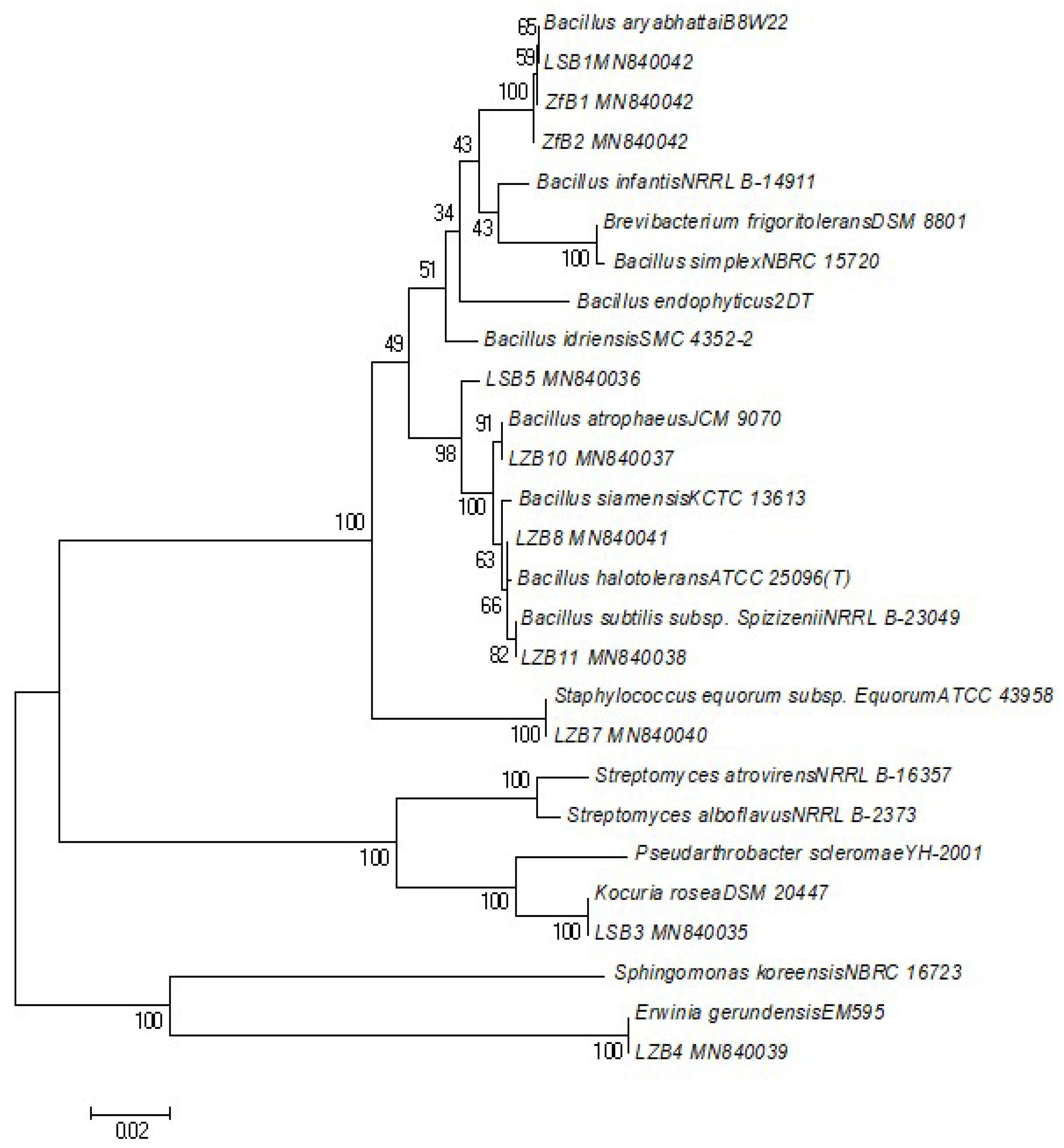
A list of bacterial cultures isolated in this study, along with information on their Gram-nature, colony characteristics, pathogenic nature, GenBank accession numbers and phylogenetic tree for 16S rRNA gene sequences, is presented in Table 3 and Figure 5. Lanzhou is found to be predominant with Bacillus during the study period of May 2019. Bacteria such as Bacillus halotolerans; ATCC 25096, Bacillus atrophaeus; JCM 9070, Bacillus subtilis subsp. Spizizeni; NRRL B-23,049 was more abundant followed by Staphylococcus equorum subsp. Equorum; ATCC 43,958 and Erwinia gerundensis; EM595. Most of the bacteria isolated in the present study were Gram-positive (more than 90%) except Erwinia gerundensis; EM595, which is Gram-negative and plant pathogen. However, the pathogenicity of other strains identified in Lanzhou was not specified yet. The pathogenic nature of species is categorized using the online tool Global Catalogue of Microorganisms (http://gcm.wfcc.info/) and ABIS Encyclopedia (http://www.tgw1916.net/ABIS/encyclopedia.html). Similarly, Lhasa and Qomolangma were also found to be predominant with strains of Bacillus. As shown in Table 3, all three regions share similar strains of bacteria. This suggests that wind direction and dust events may have played some role in microbial transfer and abundance in certain areas. Plant pathogen was predominant in all three study sites. However, one human pathogenic bacteria Kocuria rosea; DSM 20,447 was isolated in culture in the laboratory from the bioaerosol samples obtained from Lhasa.
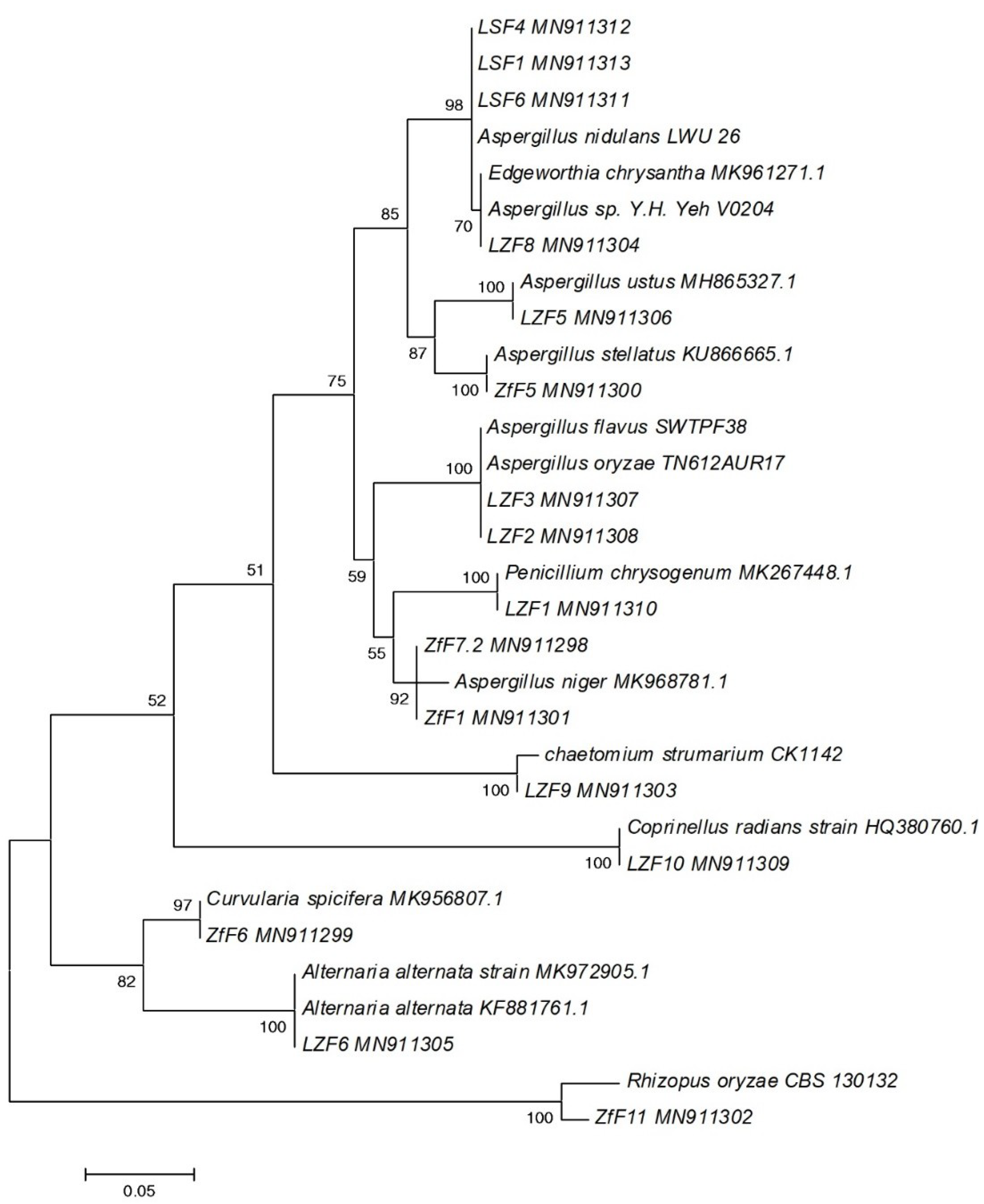
Compared to bacterial culture isolation, more varieties of fungal cultures were isolated in all three sites. Based on the previous study, this could be because of the influence of dust storm, RH or other meteorological factors [25,56]. Culture of Aspergillus flavus, Penicillium chrysogenum isolate E20399, Alternaria alternata strain SCAU-F-91, Chaetomium sp. xz11 and Coprinellus radians strain F5 was isolated from Lanzhou aerosol in which most are plant pathogen and opportunistic human pathogen Table 4. However, Lhasa and Qomolangma are found to be enriched with and shares similar fungal isolates such as Aspergillus niger, Rhizopus oryzae isolate NDA02 and Edgeworthia chrysantha strain which also belong to plant pathogen and opportunistic human pathogen Table 4. Some isolated strains such as Emericella rugulosa isolate 211, Coprinellus radians strain F5 and Edgeworthia chrysantha strain F025 possess unknown pathogenic properties and need more study on its pathogenicity. A Maximum Likelihood tree generated using the ITS gene sequence dataset in MEGA is shown in Figure 6.

4. Discussion

The microbial abundance and structure present in ambient air have been observed several times in previous studies. When compared to previous analysis it can be seen that the level of airborne bacteria detected in this study is slightly different compared to other cities in the world such as Beijing, China (Bacteria: 5.8 × 10³ CFU m−3, Fungi: 7.2 × 10³ CFU m−3), Cincinnati, USA (Fungi: 3.8 × 10³ CFU m−3), Tijuana, Moscow (Bacteria: 1.7 × 10³ CFU m−3), Seoul, South Korea (Bacteria: 3 × 10² CFU m−3, Fungi: 9 × 10² CFU m−3) [57,58,59,60]. One of the reasons could be accredited to different meteorological and environmental conditions in different regions. In contrast, the previous studies by Li et al. (2017) and Wang et al., (2010) in Northwest region of China, such as Xian (Bacteria:1.9 × 10³ CFU m−3, Fungi: 1.7 × 10³ CFU m−3) and Dunhuang (Bacteria: 3.8 × 10³ CFU m−3) were comparable with the results from the present study [54,61]. Alike Xian and Dunhuang, the present study regions, are also located in Northwestern China, which is semi-arid or arid regions. Because of the dry condition and intense solar radiation, the growth and survival of airborne microorganisms may be unfavorable [54,61]. On the other hand, Lanzhou and Lhasa being urban regions, the concentration of airborne microorganisms seem comparably higher, probably due to dust events occurred during May 2019 in Lanzhou. Previous studies have pointed out that dust can influence microbial growth [62], especially for fungal growth.
The inconsistency observed in previous studies about microbial communities in environmental samples provides some insight into the microbial physiological properties and their adaptation [63,64]. As for example, Bacillus, Proteobacteria and spores are most commonly found in the air; however, the community differs from location, season, altitude, etc. Yan et al. (2017) isolated the thermophilic sulfate-reducing non-spore forming bacterium Desulfurispora, from Beijing city of China [63]. In contrast, this strain was not isolated in this study period, which suggests the variation in strain dominance depending on geographical location. Several taxa were found to be related to specific regions, specific environment and haze levels [3,54,63,64]. However, unlike the presence of certain specific microbes in soil or rock or lake, the indication and discussion about specific and constant microbial communities in the air are not anywhere mentioned in previous studies. Hence, several factors may alter the airborne microbial community, and we can speculate variation in microbial composition in air acted upon at genetic (DNA, RNA or protein) molecular or metabolomics level.
Several previous studies have attempted to find the correlation between microbial concentration and environmental factors; nevertheless, the constant observation has not been recorded [17,52,54,63]. A study done by Yan et al. (2018) in Beijing air showed the correlated distributions of bacterial genera in relation to environmental factors are somehow in line with the current study and several other studies [63]. Bacterial genera, such as Methylophilus, Ensifer, Meiothermus and Propionibacterium were found to be positively correlated with temperature and RH at the lower concentrations of SO2 and CO. However, genera such as, Algoriphagus, Achromobacter and Brevibacillus were observed to be negatively correlated with temperature and RH at the higher concentrations of SO2 and CO. On the other hand, Gao et al. (2016) observed negative correlation of bioaerosol concentration with season and temperature in the morning time, whereas positive correlation at the day time [52]. In this study, the wind rose plot and back-trajectories analysis suggested that the wind direction was mostly from the western side. Additionally, the air mass also arrived from southwest to two sites viz—Qomolangma and Lhasa and the western part of Gansu to Lanzhou, suggesting the westerlies wind could have an effect on the bioaerosols composition during the sampling period. The data presented in the study also showed that the bacterial and fungal loads showed significant correlations with RH and temperature. A significant positive correlation of fungal concentration with WS in Lanzhou was observed, whereas bacterial concentration in Lhasa and Qomolangma showed a statistically insignificant, but negative correlation. Past studies have demonstrated that the concentration of bioaerosols differs depending on the dominant season and climatic conditions because hydrodynamic and kinetic factors primarily direct the transport of bioaerosol and their fate is reliant on the chemical composition and the meteorological factors to which they are exposed [65]. Several studies have shown that seasonality in bacterial and viral infections [66]; suggesting the risk of contracting bacterial infections will be higher in the seasons with high concentrations of both indoor and outdoor bacteria. Reports from past studies have shown that spring and fall possess higher microbial concentration compared to other seasons. A study done by Frankel et al. in 2012, showed the seasonal pattern for indoor fungi, peaking from spring to summer and declining throughout fall to winter in urban areas of Australia and Central Europe [66,67,68]. Meanwhile, outdoor air from urban areas of Europe and the USA also revealed similar patterns in bioaerosol concentration. These findings support the approach of this study of choosing spring for sampling to develop a standard and reference point for analyzing bioaerosol in remote and urban areas of the TP region.
To our knowledge, this is the first study done on the three sites of Northwest China over the TP region. The above-discussed observation showed that urbanization seems to influence the diversity and richness of airborne microbial communities. The previous study done by Wei et al. (2015) observed a comparable concentration of viable bioaerosol particles among heavily polluted areas and pristine regions [69]. This shows that both the meteorological factors, as well as the components of aerosol (TSP or PM) can act as carriers as well as supply the nutrition for microbial survival, growth and abundance. On the other hand, extreme environment, UV radiation, as well as highly concentrated environmental chemical or organic pollutants can also inactivate microbial functioning and abundance [63,70]. Thus, it would not be wrong to say that microbial survival and growth in the air is dependent on various environmental factors and is inconsistent and unpredictable. Similarly, there is another inevitable fact that microbes themselves possess unique physiology or may develop a feature that enables them to survive in the environment. For example, Methylobacillus and Tumebacillus are rich during hazy days. Moreover, small bacteria and spores can easily float in the air and increase in concentration [63,64,71]. Furthermore, it is considered that the variety of other volatile organic compounds, greenhouse gases as well as chemical composition have some role in the fungal and bacterial metabolic activities affecting their growth or survival [17], which has not been measured and considered in the study. The study also lacks the replicate sampling and long-term seasonal comparison. The other possible parameters that impact microbial composition and abundance have not been considered in the present study. Some other limitation of the study could be such as the surface area of fine and coarse particles which assists the microbial attachment and estimation of only the viable bioaerosols which are culturable. Because it has been found that only 1%–10% of microbial particles present in the air are cultivable in the laboratory provided specific growth conditions [17,62,72]. Hence, the study could be missing out on all other viable, but unculturable airborne microbes, which could be obtained by metagenomics analysis. Furthermore, only the microbial concentration (CFU/m3) itself is inadequate and does not provide an actual concentration of bacteria and fungi present in the air in a given time. In addition to this, a long-term study and replicate sample will provide a more precise comparison and accurate interpretation of the result. More detailed investigations such as metagenomics and enzymatic analysis are essential and provide future direction to dig out more information on the relation of airborne microorganisms and environmental factors in the given study site.
Despite the restrictions of the culture-based method employed in the present study, the results of this campaign may still be useful in developing control or baseline data for bacterial and fungal loads and in predicting the occurrence of the microbial community including pathogenic bioaerosols.
In addition to this, the influence of environmental parameters on microbial loads and community composition could be drawn out. It should be noted that this preliminary assessment had created basic information on microbial population variation in the three sites in the TP region. Additionally, this study provides an easy, convenient and low-cost approach as well as the genetic basis for routine analysis of airborne microbes present in ambient air.

Supplementary Materials

The following are available online at https://www.mdpi.com/2073-4433/11/5/527/s1, Table S1: Correlation matrix of the major parameters measured at Lanzhou site, China, Table S2: Correlation matrix of the major parameters measured at Lhasa site, China, Table S3: Correlation matrix of the major parameters measured at Qomolangma site, China.

Author Contributions

Conceptualization, S.K. and P.S.G.; methodology, validation, laboratory analysis, formal analysis and writing—original draft preparation, P.S.G.; field work and sample collection, P.C.; writing—review and editing, L.T., W.S., and B.A.; project administration and funding acquisition, S.K. All authors have read and agreed to the published version of the manuscript.

Funding

This research was funded by the second Tibetan Plateau Scientific Expedition and Research Program (STEP) (2019QZKK0605) and the National Natural Science Foundation of China (41630754, 41721091) and the State Key Laboratory of Cryospheric Science (SKLCS-ZZ-2018). Prakriti Sharma Ghimire is supported by a PIFI Fellowship from the Chinese Academy of Sciences (PIFI2018PC20021) and China Postdoctoral Science Funding (2019M663859).

Conflicts of Interest

The authors have declared no conflict of interest.

References

  1. Maki, T.; Hara, K.; Iwata, A.; Lee, K.C.; Kawai, K.; Kai, K.; Iwasaka, Y. Variations in airborne bacterial communities at high altitudes over the Noto Peninsula (Japan) in response to Asian dust events. Atmos. Chem. Phys. 2017, 17, 11877–11897. [Google Scholar] [CrossRef] [Green Version]
  2. Amato, P.; Ménager, M.; Sancelme, M.; Laj, P.; Mailhot, G.; Delort, A.-M. Microbial population in cloud water at the Puy de Dôme: Implications for the chemistry of clouds. Atmos. Environ. 2005, 39, 4143–4153. [Google Scholar] [CrossRef] [Green Version]
  3. Bowers, R.M.; Lauber, C.L.; Wiedinmyer, C.; Hamady, M.; Hallar, A.G.; Fall, R.; Knight, R.; Fierer, N. Characterization of airborne microbial communities at a high-elevation site and their potential to act as atmospheric ice nuclei. Appl. Environ. Microbiol. 2009, 75, 5121–5130. [Google Scholar] [CrossRef] [PubMed] [Green Version]
  4. Sattler, B.; Puxbaum, H.; Psenner, R. Bacterial growth in supercooled cloud droplets. Geophys. Res. Lett. 2001, 28, 239–242. [Google Scholar] [CrossRef]
  5. Hamady, M.; Walker, J.J.; Harris, J.K.; Gold, N.J.; Knight, R. Error-correcting barcoded primers for pyrosequencing hundreds of samples in multiplex. Nat. Methods 2008, 5, 235. [Google Scholar] [CrossRef] [PubMed]
  6. Qi, J.; Li, M.; Zhen, Y.; Wu, L. Characterization of bioaerosol bacterial communities during hazy and foggy weather in Qingdao, China. J. Ocean Univ. China 2018, 17, 516–526. [Google Scholar] [CrossRef]
  7. Romano, S.; Di Salvo, M.; Rispoli, G.; Alifano, P.; Perrone, M.R.; Talà, A. Airborne bacteria in the Central Mediterranean: Structure and role of meteorology and air mass transport. Sci. Total Environ. 2019, 697, 134020. [Google Scholar] [CrossRef]
  8. Triadó-Margarit, X.; Caliz, J.; Reche, I.; Casamayor, E.O. High similarity in bacterial bioaerosol compositions between the free troposphere and atmospheric depositions collected at high-elevation mountains. Atmos. Environ. 2019, 203, 79–86. [Google Scholar] [CrossRef]
  9. Tang, K.; Huang, Z.; Huang, J.; Maki, T.; Zhang, S.; Shimizu, A.; Ma, X.; Shi, J.; Bi, J.; Wang, G.; et al. Characterization of atmospheric bioaerosols along the transport pathway of Asian dust during the Dust-Bioaerosol 2016 Campaign. Atmos. Chem. Phys. 2018, 18, 7131. [Google Scholar] [CrossRef] [Green Version]
  10. D’amato, G. Environmental urban factors (air pollution and allergens) and the rising trends in allergic respiratory diseases. Allergy 2002, 57, 30–33. [Google Scholar] [CrossRef]
  11. Menetrez, M.; Foarde, K.; Esch, R.; Dean, T.; Betancourt, D.; Moore, S.; Yeatts, K.; Svendsen, E.R. The measurement of ambient bioaerosol exposure. Aerosol Sci. Technol. 2007, 41, 884–893. [Google Scholar] [CrossRef] [Green Version]
  12. Kumar, A.; Attri, A.K. Characterization of fungal spores in ambient particulate matter: A study from the Himalayan region. Atmos. Environ. 2016, 142, 182–193. [Google Scholar] [CrossRef]
  13. Brown, J.K.; Hovmøller, M.S. Aerial dispersal of pathogens on the global and continental scales and its impact on plant disease. Science 2002, 297, 537–541. [Google Scholar] [CrossRef] [PubMed] [Green Version]
  14. Elster, J.; Delmas, R.; Petit, J.-R.; Reháková, K. Composition of microbial communities in aerosol, snow and ice samples from remote glaciated areas (Antarctica, Alps, Andes). Biogeosci. Discuss. 2007, 4, 1779–1813. [Google Scholar] [CrossRef] [Green Version]
  15. Fierer, N.; Liu, Z.; Rodríguez-Hernández, M.; Knight, R.; Henn, M.; Hernandez, M.T. Short-term temporal variability in airborne bacterial and fungal populations. Appl. Environ. Microbiol. 2008, 74, 200–207. [Google Scholar] [CrossRef] [PubMed] [Green Version]
  16. Lighthart, B.; Shaffer, B.T. Viable bacterial aerosol particle size distributions in the midsummer atmosphere at an isolated location in the high desert chaparral. Aerobiologia 1995, 11, 19–25. [Google Scholar] [CrossRef]
  17. Agarwal, S.; Mandal, P.; Majumdar, D.; Aggarwal, S.G.; Srivastava, A. Characterization of bioaerosols and their relation with OC, EC and carbonyl VOCs at a busy roadside restaurants-cluster in New Delhi. Aerosol Air Qual. Res. 2016, 16, 3198–3211. [Google Scholar] [CrossRef] [Green Version]
  18. Du, P.; Du, R.; Ren, W.; Lu, Z.; Zhang, Y.; Fu, P. Variations of bacteria and fungi in PM2. 5 in Beijing, China. Atmos. Environ. 2018, 172, 55–64. [Google Scholar] [CrossRef]
  19. Gandolfi, I.; Bertolini, V.; Ambrosini, R.; Bestetti, G.; Franzetti, A. Unravelling the bacterial diversity in the atmosphere. Appl. Microbiol. Biotechnol. 2013, 97, 4727–4736. [Google Scholar] [CrossRef]
  20. Tanaka, D.; Sato, K.; Goto, M.; Fujiyoshi, S.; Maruyama, F.; Takato, S.; Shimada, T.; Satakotu, A.; Aoli, K.; Nakamura, S. Airborne microbial communities at high-altitude and suburban sites in Toyama, Japan suggest a new perspective for bioprospecting. Front. Bioeng. Biotechnol. 2019, 7, 12. [Google Scholar] [CrossRef]
  21. Jaenicke, R. Abundance of cellular material and proteins in the atmosphere. Science 2005, 308, 73. [Google Scholar] [CrossRef] [PubMed]
  22. Bowers, R.M.; McCubbin, I.B.; Hallar, A.G.; Fierer, N. Seasonal variability in airborne bacterial communities at a high-elevation site. Atmos. Environ. 2012, 50, 41–49. [Google Scholar] [CrossRef]
  23. Awad, A.H.A. Vegetation: A source of air fungal bio-contaminant. Aerobiologia 2005, 21, 53–61. [Google Scholar] [CrossRef]
  24. Lin, W.-R.; Wang, P.-H.; Tien, C.-J.; Chen, W.-Y.; Yu, Y.-A.; Hsu, L.-Y. Changes in airborne fungal flora along an urban to rural gradient. J. Aerosol Sci. 2018, 116, 116–123. [Google Scholar] [CrossRef]
  25. Brągoszewska, E.; Pastuszka, J.S. Influence of meteorological factors on the level and characteristics of culturable bacteria in the air in Gliwice, Upper Silesia (Poland). Aerobiologia 2018, 34, 241–255. [Google Scholar] [CrossRef] [Green Version]
  26. Brągoszewska, E.; Mainka, A.; Pastuszka, J.S. Concentration and size distribution of culturable bacteria in ambient air during spring and winter in Gliwice: A typical urban area. Atmosphere 2017, 8, 239. [Google Scholar] [CrossRef] [Green Version]
  27. Genitsaris, S.; Stefanidou, N.; Katsiapi, M.; Kormas, K.A.; Sommer, U.; Moustaka-Gouni, M. Variability of airborne bacteria in an urban Mediterranean area (Thessaloniki, Greece). Atmos. Environ. 2017, 157, 101–110. [Google Scholar] [CrossRef]
  28. Hubad, B.; Lapanje, A. The efficient method for simultaneous monitoring of the culturable as well as nonculturable airborne microorganisms. PLoS ONE 2013, 8, e82186. [Google Scholar] [CrossRef] [Green Version]
  29. Yoo, K.; Lee, T.K.; Choi, E.J.; Yang, J.; Shukla, S.K.; Hwang, S.-I.; Park, J. Molecular approaches for the detection and monitoring of microbial communities in bioaerosols: A review. J. Environ. Sci. 2017, 51, 234–247. [Google Scholar] [CrossRef]
  30. Fang, Z.; Ouyang, Z.; Zheng, H.; Wang, X. Concentration and size distribution of culturable airborne microorganisms in outdoor environments in Beijing, China. Aerosol Sci. Technol. 2008, 42, 325–334. [Google Scholar] [CrossRef] [Green Version]
  31. Tsai, F.; Macher, J.; Hung, Y. Concentrations of airborne bacteria in 100 US office buildings. Proc. Indoor Air 2002, 15, 353–359. [Google Scholar]
  32. Kang, S.; Zhang, Q.; Qian, Y.; Ji, Z.; Li, C.; Cong, Z.; You, Q.; Panday, A.K.; Rupakheti, M.; Chen, D.; et al. Linking atmospheric pollution to cryospheric change in the Third Pole region: Current progress and future prospects. Natl. Sci. Rev. 2019, 6, 796–809. [Google Scholar] [CrossRef]
  33. Cong, Z.; Kang, S.; Kawamura, K.; Liu, B.; Wan, X.; Wang, Z.; Gao, S.; Fu, P. Carbonaceous aerosols on the south edge of the Tibetan Plateau: Concentrations, seasonality and sources. Atmos. Chem. Phys. 2015, 15, 1573–1584. [Google Scholar] [CrossRef] [Green Version]
  34. Ma, Y.; Wang, Y.; Wu, R.; Hu, Z.; Yang, K.; Li, M.; Ma, W.; Zhong, L.; Sun, F.; Chen, X.; et al. Recent advances on the study of atmosphere-land interaction observations on the Tibetan Plateau. Hydrol. Earth Syst. Sci. 2009, 13, 1103–1111. [Google Scholar] [CrossRef] [Green Version]
  35. Wan, X.; Kang, S.; Xin, J.; Liu, B.; Wen, T.; Wang, P.; Wang, Y.; Cong, Z. Chemical composition of size-segregated aerosols in Lhasa city, Tibetan Plateau. Atmos. Res. 2016, 174, 142–150. [Google Scholar] [CrossRef]
  36. Cong, Z.; Kang, S.; Luo, C.; Li, Q.; Huang, J.; Gao, S.; Li, X. Trace elements and lead isotopic composition of PM10 in Lhasa, Tibet. Atmos. Environ. 2011, 45, 6210–6215. [Google Scholar] [CrossRef]
  37. Huang, J.; Kang, S.; Wang, S.; Wang, L.; Zhang, Q.; Guo, J.; Wang, K.; Zhang, G.; Tripathee, L. Wet deposition of mercury at Lhasa, the capital city of Tibet. Sci. Total Environ. 2013, 447, 123–132. [Google Scholar] [CrossRef]
  38. Guo, J.; Kang, S.; Huang, J.; Zhang, Q.; Tripathee, L.; Sillanpää, M. Seasonal variations of trace elements in precipitation at the largest city in Tibet, Lhasa. Atmos. Res. 2015, 153, 87–97. [Google Scholar] [CrossRef]
  39. Zhang, Y.; Kang, S. Characteristics of carbonaceous aerosols analyzed using a multiwavelength thermal/optical carbon analyzer: A case study in Lanzhou City. Sci. China Earth Sci. 2019, 62, 389–402. [Google Scholar] [CrossRef]
  40. Yu, X.; Zhu, B.; Fan, S.; Yin, Y.; Bu, X. Ground-based observation of aerosol optical properties in Lanzhou, China. J. Environ. Sci. 2009, 21, 1519–1524. [Google Scholar] [CrossRef]
  41. Qiang, Z. The influence of terrain and inversion layer on pollutant transfer over Lanzhou City. China Environ. Sci. 2001, 21, 230–234. [Google Scholar]
  42. Bielawska-Drózd, A.; Cieślik, P.; Bohacz, J.; Korniłłowicz-Kowalska, T.; Żakowska, D.; Bartoszcze, M.; Wlizło-Skowronek, B.; Winnicka, I.; Brytan, M.; Kubiak, L. Microbiological analysis of bioaerosols collected from Hospital Emergency Departments and ambulances. Ann. Agric. Environ. Med. 2018, 25, 274–279. [Google Scholar] [CrossRef] [PubMed] [Green Version]
  43. Dacarro, C.; Picco, A.; Grisoli, P.; Rodolfi, M. Determination of aerial microbiological contamination in scholastic sports environments. J. Appl. Microbiol. 2003, 95, 904–912. [Google Scholar] [CrossRef] [PubMed] [Green Version]
  44. Joshi, M.; Srivastava, R. Identification of indoor airborne microorganisms in residential rural houses of Uttarakhand, India. Int. J. Curr. Microbiol. Appl. Sci. 2013, 2, 146–152. [Google Scholar]
  45. Srivastava, A.; Singh, M.; Jain, V. Identification and characterization of size-segregated bioaerosols at Jawaharlal Nehru University, New Delhi. Nat. Hazards 2012, 60, 485–499. [Google Scholar] [CrossRef]
  46. Madsen, A.M.; Zervas, A.; Tendal, K.; Nielsen, J.L. Microbial diversity in bioaerosol samples causing ODTS compared to reference bioaerosol samples as measured using Illumina sequencing and MALDI-TOF. Environ. Res. 2015, 140, 255–267. [Google Scholar] [CrossRef] [Green Version]
  47. Piterina, A.V.; Bartlett, J.; Pembroke, J.T. Molecular analysis of bacterial community DNA in sludge undergoing autothermal thermophilic aerobic digestion (ATAD): Pitfalls and improved methodology to enhance diversity recovery. Diversity 2010, 2, 505–526. [Google Scholar] [CrossRef] [Green Version]
  48. White, T.J.; Bruns, T.; Lee, S.; Taylor, J. Amplification and direct sequencing of fungal ribosomal RNA genes for phylogenetics. PCR Protoc. A Guide Methods Appl. 1990, 18, 315–322. [Google Scholar]
  49. Lan, P.T.N.; Hayashi, H.; Sakamoto, M.; Benno, Y. Phylogenetic analysis of cecal microbiota in chicken by the use of 16S rDNA clone libraries. Microbiol. Immunol. 2002, 46, 371–382. [Google Scholar] [CrossRef]
  50. Tripathee, L.; Kang, S.; Rupakheti, D.; Cong, Z.; Zhang, Q.; Huang, J. Chemical characteristics of soluble aerosols over the central Himalayas: Insights into spatiotemporal variations and sources. Environ. Sci. Pollut. Res. Int. 2017, 24, 24454–24472. [Google Scholar] [CrossRef]
  51. Draxler, R.; Stunder, B.; Rolph, G.; Stein, A.; Taylor, A. HYSPLIT4 User’s Guide Version 4-Last Revision: February 2016; HYSPLIT Air Resources Laboratory: College Park, MD, USA, 2016. [Google Scholar]
  52. Gao, M.; Yan, X.; Qiu, T.; Han, M.; Wang, X. Variation of correlations between factors and culturable airborne bacteria and fungi. Atmos. Environ. 2016, 128, 10–19. [Google Scholar] [CrossRef]
  53. González-Rocha, G.; Muñoz-Cartes, G.; Canales-Aguirre, C.B.; Lima, C.A.; Domínguez-Yévenes, M.; Bello-Toledo, H.; Hernandez, C.E. Diversity structure of culturable bacteria isolated from the Fildes Peninsula (King George Island, Antarctica): A phylogenetic analysis perspective. PLoS ONE 2017, 12, e0179390. [Google Scholar] [CrossRef] [Green Version]
  54. Li, Y.; Lu, R.; Li, W.; Xie, Z.; Song, Y. Concentrations and size distributions of viable bioaerosols under various weather conditions in a typical semi-arid city of Northwest China. J. Aerosol Sci. 2017, 106, 83–92. [Google Scholar] [CrossRef]
  55. Liu, H.-M.; Lin, Y.-H.; Tsai, M.-Y.; Lin, W.-H. Occurrence and characterization of culturable bacteria and fungi in metalworking environments. Aerobiologia 2010, 26, 339–350. [Google Scholar] [CrossRef]
  56. Dannemiller, K.C.; Weschler, C.J.; Peccia, J. Fungal and bacterial growth in floor dust at elevated relative humidity levels. Indoor Air 2017, 27, 354–363. [Google Scholar] [CrossRef] [PubMed]
  57. Gao, M.; Qiu, T.; Jia, R.; Han, M.; Song, Y.; Wang, X. Concentration and size distribution of viable bioaerosols during non-haze and haze days in Beijing. Environ. Sci. Pollut. Res. 2015, 22, 4359–4368. [Google Scholar] [CrossRef]
  58. Heo, K.J.; Kim, H.B.; Lee, B.U. Concentration of environmental fungal and bacterial bioaerosols during the monsoon season. J. Aerosol Sci. 2014, 77, 31–37. [Google Scholar] [CrossRef]
  59. Hurtado, L.; Rodríguez, G.; López, J.; Castillo, J.; Molina, L.; Zavala, M.; Quintana, P.J. Characterization of atmospheric bioaerosols at 9 sites in Tijuana, Mexico. Atmos. Environ. 2014, 96, 430–436. [Google Scholar] [CrossRef]
  60. Lee, T.; Grinshpun, S.A.; Martuzevicius, D.; Adhikari, A.; Crawford, C.M.; Reponen, T. Culturability and concentration of indoor and outdoor airborne fungi in six single-family homes. Atmos. Environ. 2006, 40, 2902–2910. [Google Scholar] [CrossRef] [Green Version]
  61. Wang, W.; Ma, Y.; Ma, X.; Wu, F.; Ma, X.; An, L.; Feng, H. Seasonal variations of airborne bacteria in the Mogao Grottoes, Dunhuang, China. Int. Biodeterior. Biodegr. 2010, 64, 309–315. [Google Scholar] [CrossRef]
  62. Ghimire, P.S.; Tripathee, L.; Chen, P.; Kang, S. Linking the conventional and emerging detection techniques for ambient bioaerosols: A review. Rev. Environ. Sci. Biotechnol. 2019, 18, 495–523. [Google Scholar] [CrossRef]
  63. Yan, D.; Zhang, T.; Su, J.; Zhao, L.L.; Wang, H.; Fang, X.M.; Zhang, Y.-Q.; Liu, H.-Y.; Yu, L.-Y. Structural variation in the bacterial community associated with airborne particulate matter in Beijing, China, during hazy and nonhazy days. Appl. Environ. Microbiol. 2018, 84, e00004–e00018. [Google Scholar] [CrossRef] [Green Version]
  64. Yan, D.; Zhang, T.; Su, J.; Zhao, L.L.; Wang, H.; Fang, X.M.; Zhang, Y.-Q.; Liu, H.-Y.; Yu, L.-Y. Diversity and composition of airborne fungal community associated with particulate matters in Beijing during haze and non-haze days. Front. Microbiol. 2016, 7, 487. [Google Scholar] [CrossRef] [PubMed] [Green Version]
  65. Mouli, P.; Mohan, S.; Reddy, S. Assessment of microbial(bacteria) Concentrations of ambient air at semi-arid urban region: Influence of meteorological factors. Appl. Ecol. Environ. Res. 2005, 3, 139–149. [Google Scholar] [CrossRef]
  66. Frankel, M.; Bekö, G.; Timm, M.; Gustavsen, S.; Hansen, E.W.; Madsen, A.M. Seasonal variations of indoor microbial exposures and their relation to temperature, relative humidity, and air exchange rate. Appl. Environ. Microbiol. 2012, 78, 8289–8297. [Google Scholar] [CrossRef] [PubMed] [Green Version]
  67. Garrett, M.H.; Hooper, B.M.; Cole, F.M.; Hooper, M.A. Airborne fungal spores in 80 homes in the Latrobe Valley, Australia: Levels, seasonality and indoor-outdoor relationship. Aerobiologia 1997, 13, 121–126. [Google Scholar] [CrossRef]
  68. Haas, D.; Habib, J.; Galler, H.; Buzina, W.; Schlacher, R.; Marth, E.; Reinthaler, F. Assessment of indoor air in Austrian apartments with and without visible mold growth. Atmos. Environ. 2007, 41, 5192–5201. [Google Scholar] [CrossRef]
  69. Wei, K.; Zheng, Y.; Li, J.; Shen, F.; Zou, Z.; Fan, H.; Li, X.; Wu, C.Y.; Yao, M. Microbial aerosol characteristics in highly polluted and near-pristine environments featuring different climatic conditions. Sci. Bull. 2015, 60, 1439–1447. [Google Scholar] [CrossRef] [Green Version]
  70. Vaïtilingom, M.; Deguillaume, L.; Vinatier, V.; Sancelme, M.; Amato, P.; Chaumerliac, N.; Delort, A.M. Potential impact of microbial activity on the oxidant capacity and organic carbon budget in clouds. Proc. Natl. Acad. Sci. USA 2013, 110, 559–564. [Google Scholar] [CrossRef] [Green Version]
  71. Sun, Y.; Jiang, Q.; Wang, Z.; Fu, P.; Li, J.; Yang, T.; Yin, Y. Investigation of the sources and evolution processes of severe haze pollution in Beijing in January 2013. J. Geophys. Res. Atmos. 2014, 119, 4380–4398. [Google Scholar] [CrossRef]
  72. Tong, Y.; Lighthart, B. The annual bacterial particle concentration and size distribution in the ambient atmosphere in a rural area of the Willamette Valley, Oregon. Aerosol Sci. Technol. 2000, 32, 393–403. [Google Scholar] [CrossRef]
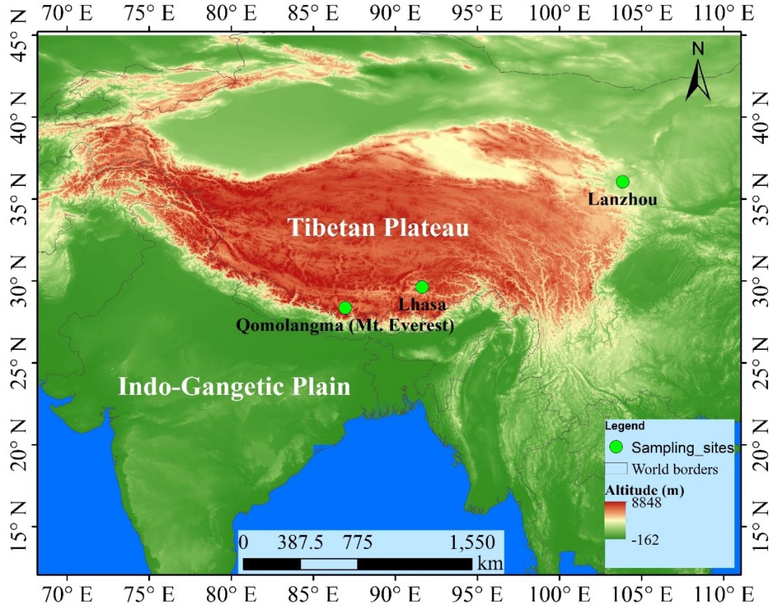
Figure 1. Geographical map for three sites presented in the study.
Atmosphere 11 00527 g001
Figure 2. Windrose plot for three sites presented in the study. Windrose plot showing the frequency of wind speed and wind direction at three sites during the study period.
Atmosphere 11 00527 g002
Figure 3. HYSPLIT backward-trajectory analysis for three sites Lanzhou (LZ), Lhasa (LS), and Qomolangma (ZF) during the sampling period.
Atmosphere 11 00527 g003
Figure 4. Concentrations and composition of bioaerosol present in the outdoor air in Lanzhou, Lhasa and Qomolangma site during May 2019. (a) average concentrations of bacterial and fungal aerosol collected in the outdoor air in Lanzhou, Lhasa and Qomolangma region during the analyzed period; (b) The composition of bacterial and fungal communities in the aerosol samples from Lanzhou, Lhasa and Qomolangma region during the study period.
Atmosphere 11 00527 g004
Figure 5. Phylogenetic analysis of the bacterial strain isolated from bioaerosol samples from three different regions. Based on related sequences of all the species were obtained from NCBI and aligned by MUSCLE through MEGA6. Evolutionary distances were computed using the maximum composite likelihood method. The obtained nucleotide sequences were deposited in the NCBI database and assigned with the accession number as MN840035-MN840042 for bacterial 16srRNA sequence.
Atmosphere 11 00527 g005
Figure 6. Phylogenetic analysis of the fungal strain isolated from bioaerosol samples from three different regions. Based on related sequences of all the species were obtained from NCBI and aligned by MUSCLE through MEGA6. Evolutionary distances were computed using the maximum composite likelihood method. The obtained nucleotide sequences were deposited in the NCBI database and assigned with the accession number as MN911298–MN911313 for fungal ITS sequence.
Atmosphere 11 00527 g006
Table 1. Summary of meteorological information of three sites Lanzhou (LZ), Lhasa (LS) and Qomolangma (ZF) and chemical composition of bioaerosol during the sampling period (May 2019).
Temperature (°C)Pressure (hPa)RHU (%)Wind Speed (m/s)Wind DirectionTSP (µg m−3)
DateLZLSZFLZLSZFLZLSZFLZLSZFLZLSZFLZLSZF
5/1/201921.122.715.8831.1661.9605.532.989.949.93.55.610.9SWWNWWSW58.01129.2690.35
5/4/201925.119.418.6832.3660.9603.822.932.918.94.44.96.7EENEW72.0596.4940.39
5/7/20192125.515.6834.8658603.531.999.999.94.75.910.3NESSWW1530.54170.7761.49
5/10/201918.124.218.4833.4658.260536.959.969.94.59.211.8NEWW1521.63153.8672.15
5/13/201923.31912.7834.5658.960419.918.915.94.59.68.9ENEWNWW673.59199.3130.36
5/16/201925.72016.7830.7660.9604.923.921.912.95.45.110.3NNESEWSW60.9968.7232.43
5/19/201918.722.216.2839.7659.3604.821.916.999.95.45.38.5NWNWW225.84104.3633.93
5/22/201928.118.616.8833.8663.1605.769.925.989.93.15.115.6SENEWSW40.84116.68130.05
Table 2. Comparison of the microbial community of viable bioaerosols in different regions worldwide.
LocationLatitude (°N) and Longitude (°E)Elevation (AMSL)SiteMethod Used for StudyMicrobial CommunityReferences
Toyama Prefecture, Japan36°41′54′′ N,
137°11′13′′ E
23 mSuburbanPolycarbonate filters, Illumina sequencingAlpha-Beta-Gammaproteobacteria, Acidimicrobia, Planctomycetia, Bacillus, Solibacteres, Flavobacteria[20]
Huairou, Beijing, China40°24′29″ N,
116°40′ 28″ E
40–60 mPeri-urbanQuartz filters, Illumina sequencingStreotophyta, Bacillus, Clostridium, Kocuria, Staphylococcus, MethylobacteriumSarcinomyces, Trichothecium, Acromonium, Chaetomium, Aspergillus, Penicillium[18]
New Delhi city, India28°12’ N–28°53’ N, 77°50’ E–77°23’ E218 mUrbanQuartz filters, automated DNA sequencingBacillus, AcenitobactorAspergillus, Cladosporium, Alternaria, Fusarium, Penicillium, Trichoderma, Mucor[17]
Jawali, India31°2’ N–32°5’ N
75°0’ E–77°45’ E
600 mRuralQuartz filters, Light microscopyBasidiospora, Ascospora, Fusarium, Ganoderma, Alternaria, Curvularia[12]
Erenhot43.668, 111.953957 mUrbanPolycarbonate filters, Illumina sequencingChloroacidobacter, Saprospirae, Actinobacteria, Alphaproteobacteria, BacilliAgaricomycetes, Dothideomycetes, Sordariomycetes, Eurotiomycetes[9]
Qingdao, China36°16′ N, 120° 50′ E1133 mUrbanSix stage cascade impactor, DGGE band sequencingAlphaproteobacteria, Betaproteobacteria, Bacillus[6]
Lanzhou36°3′1″ N, 103°51′33″ E1520 mUrbanQuartz filters, Illumina sequencingProteobacteria, BacillusEurotiomycetes, Malvales, Dothideomycete, Sordariales, AgaricalesThis study
Salento’s peninsula, Italy40.3° N; 18.1° E1895 mSuburbanPTFE filters, Illumina sequencingProteobacteria, Cyanobacteria, Actinobacteria, Pseudomonas, Enterobacter, Vibrio, Streptomyces[7]
Mt. Jodo, Japan36°34′00′′ N, 137°36′21′′ E2839 mHigh altitude, pristinePolycarbonate filters, Illumina sequencingAlpha/Beta, Gammaproteobacteria, Acidimicrobia, Planctomycetia, Bacillus, Solibacteres, Flavobacteria[20]
Sierra Nevada, Spain37°03’ N, 3°23’ W2896 mHigh altitude, Rocky and meadowsPassive automatic samplerAcidobacteria, Actinobacteria, Bacteroidetes, Cyanobacteria[8]
Noto Peninsula, JapanUchinada
(36°67 N, 136°64 E) to Hakui
(36°92 N, 136°76 E)
500–3000 mCoastalPolycarbonate filters, Illumina sequencingCyanobacteria, Actinobacteria, Bacillus, Alpha, Beta, Gammaproteobacteria[1]
Mt. Werner, Colorado, USA40.45° N, 106.73° W3200 mHigh altitude, pristineCellulose nitrate filters, Sanger sequencingAcidobacteria, Actinobacteria, Bacteroidetes, Cyanobacteria, Bacillus, Eurotiomycetes, Sordariomycetes, Dothideomycete[3]
Lhasa29°38′ N, 91°38′ E3640 mHigh altitude, Peri-urbanQuartz filters, Illumina sequencingBacillus, Kocuria, Zygomycetes, Eurotiomycetes, MalvalesThis study
Qomolangma28.36°N, 86.95°E4276 mHigh altitude, PristineQuartz filters, Illumina sequencingBacillus, Eurotiomycetes, Malvales, Dothideomycete, Agaricales, ZygomycetesThis study
Table 3. National Center for Biotechnological Information (NCBI) BLAST search results for each of the identified bacterial strains.
Sample NameStrainMatched Accession NumberSimilarity%Length (bp)PathogenesisGram StainColony CharacteristicsGC Content%
LZB4Erwinia gerundensisKJ004603.199.391331Plant pathogenGram-negative, rod shapedYellowish, circular56.12
LZB7Staphylococcus equorumMN229550.11001393Produce cheese and meat order, May inhibit Listeria’s growthGram-positive cocciOpaque, white entire margin50.54
LZB8Bacillus halotoleransMK517597.199.931390UnknownGram-positive bacteria, rod shapedOpaque, smooth, creamy colored54.82
LZB10Bacillus atrophaeusNR_024689.199.861410UnknownGram-positive bacteria, rod shapedOpaque, smooth, creamy colored55.04
LZB11Bacillus subtilisNR_116187.199.861408UnknownGram-positive bacteria, rod shapedDull surface, thick/opaque, creamy colored, wrinkled (sometimes)54.9
LSB1Bacillus aryabhattaiNR_115953.199.791415UnknownGram-positive bacteria, rod shapedOpaque, smooth, creamy colored53.57
LSB3Kocuria roseaNR_044871.199.851370Infections in immunocompromised patientsGram-positive cocciPinkish, smooth, shiny, circular57.23
LSB5Bacillus altitudinisNR_042337.11001382Plant soft-rot causing pathogenGram-positive bacteria, rod shapedWhite, regular margin54.99
ZFB1Bacillus aryabhattaiMK860027.11001398UnknownGram-positive bacteria, rod shapedOpaque, smooth, creamy colored53.58
ZFB2Bacillus aryabhattaiNR_115953.199.791414UnknownGram-positive bacteria, rod shapedOpaque, smooth, creamy colored53.54
Table 4. NCBI BLAST search results for each of the identified fungal strains.
Sample NameStrainMatched Accession NumberSimilarity%Length (bp)PathogenesisColony CharacteristicsGC Content (%)
LZF1Penicillium chrysogenumMK267448.1100563Rare human pathogen, human allergen, source of antibioticsBlue to blue–green conidia and the mold exudes a yellow pigment57.02
LZF2Aspergillus flavusMG575511.1100564Plant pathogen, opportunistic human and animal pathogen, causing aspergillosis in immunocompromised individualsPowdery masses of yellow-green spores on the upper surface and reddish-gold on the lower surface58.33
LZF3Aspergillus flavusMG991646.1100570Plant pathogen, opportunistic human and animal pathogen, causing aspergillosis in immunocompromised individualsPowdery masses of yellow-green spores on the upper surface and reddish-gold on the lower surface58.07
LZF4Edgeworthia chrysanthaMK961271.1100543Not known. Possess anti-inflammatory and analgesic activityPossesses pain brush type flowering58.93
LZF5Aspergillus ustusMH865327.1100551Human pathogen causing onychomycosis and otitis media, rarely found to cause endocarditis, pneumonia, disseminated disease, opportunistic pathogen in immunocompromisedDull brown with a purplish to gray brown or dark brown colonies58.08
LZF6Alternaria alternataMH865327.1100549Opportunistic pathogen causing leaf spots, rots and blights on many plant partsBlack to olivaceous-black or greyish and are suede-like to floccose46.27
LZF8Aspergillus sp.MH141246.199.82550Most commonly human, animal and plant pathogen, cause disease on many grain crops and some variants synthesize mycotoxins and aflatoxinsPowdery masses of yellow-green spores on the upper surface and yellowish on the lower surface59.09
LZF9Chaetomium sp.KJ935022.199.82544Human allergens and opportunistic agents of ungual mycosis and neurological infections. Source of cellulose degrading enzymesCottony and white in color initially. Mature colonies become gray to olive in color57.17
LZF10Coprinellus radiansHQ380760.1100664UnknownScattered yellowish-orange mat49.4
LSF2Rhizopus oryzaeMH865594.1100582Opportunistic pathogen of humans causing mucormycosis. It is also used economically in the production of the enzymes, glucoamylase and lipaseColonies are white initially, becoming brownish with age40.55
LSF3Rhizopus oryzaeMH865576.1100602Opportunistic pathogen of humans causing mucormycosis. It is also used economically in the production of the enzymes, glucoamylase and lipaseColonies are white initially, becoming brownish with age40.86
LSF6Emericella dentataMH032749.1100527UnknownColonies are white and fluffy initially59.58
LSF1Emericella rugulosaEU289912.199.82541UnknownWhite to blackish sparse colony59.7
LSF4Edgeworthia chrysanthaMK806488.1100539Not known. Possess anti-inflammatory and analgesic activityPossesses pain brush type flowering59.55
LSF8Aspergillus nigerMK256745.1100573Black mold of onions and ornamental plants, peanuts and grapes. Serious lung disease, aspergillosis in human. Produce important enzymes.Granular to cottony, velvety or powdery; usually white at first and black at age.58.46
ZFF2Aspergillus nigerMK258199.1100577Black mold of onions and ornamental plants, peanuts and grapes. Serious lung disease, aspergillosis in human. Produce important enzymesGranular to cottony, velvety or powdery; usually white at first and black at age.58.06
ZFF6Curvularia spiciferaMK956807.1100543Facultative pathogen or beneficial partner of many plant speciesWhite to pinkish gray wooly colonies46.96
ZFF11Rhizopus oryzaeMK742815.1100501Opportunistic pathogen of humans causing mucormycosis. It is also used economically in the production of the enzymes, glucoamylase and lipaseColonies are white initially, becoming brownish with age38.92
ZFF7.2Aspergillus tubingensisMF186869.1100578Involved in food spoilage of fruits and wheat and industrial fermentation and a rare human pathogen.Granular to cottony, velvety or powdery white-black colonies57.96
ZFF1Aspergillus nigerMK256745.1100572Black mold of onions and ornamental plants, peanuts and grapes. Serious lung disease, aspergillosis in humans. Produce important enzymesGranular to cottony, velvety or powdery; usually white at first and black at age.58.57
ZFF4Edgeworthia chrysanthaMK961271.1100540Not known. Possess anti-inflammatory and analgesic activityPossesses pain brush type flowering59.44
ZFF5Aspergillus stellatusKU866665.199.82541UnknownColonies are initially while and later smooth orange to reddish brown58.04