Article

Release of Highly Active Ice Nucleating Biological Particles Associated with Rain

1
Faculty of Science and Technology, Keio University, 3-14-1, Hiyoshi Kohoku-ku, Yokohama 223-8522, Japan
2
College of Science and Engineering, Kanazawa University, Kanazawa 920-1192, Japan
3
Graduate School of Natural Science and Technology, Kanazawa University, Kanazawa 920-1192, Japan
4
Institute of Nature and Environmental Technology, Kanazawa University, Kanazawa 920-1192, Japan
*
Authors to whom correspondence should be addressed.
Atmosphere 2019, 10(10), 605; https://doi.org/10.3390/atmos10100605
Submission received: 11 September 2019 / Revised: 1 October 2019 / Accepted: 3 October 2019 / Published: 8 October 2019

Abstract

:
Biological particles may play an important role in the climate system by efficiently acting as ice nucleating particles (INPs) at a higher temperature range (e.g., above −20 °C where representative INPs such as mineral dust remain inactive), but there is an obvious lack of direct evidence that these particles serve in this manner. Here, we collected ambient particles under different weather conditions for identifying INPs that are active above −22 °C. The abundance of such efficient INPs increased during or following rainfall events. The extensive characterization of individual particles by three different analyses (particle morphology and composition, heat sensitivity of ice nucleation activities, and biological fingerprinting by DNA staining) revealed that efficient INPs have distinctly biological characteristics, which differ significantly from more abundant, representative, and relatively less active INPs, such as mineral dust. Additionally, by combining the heat-sensitivity experiments and DNA staining techniques, efficient INPs were found to contain heat-sensitive biomaterials and biological cells. Our findings provide direct evidence that biological particles are preferentially released into the atmosphere during rainfall events and act as important atmospheric INPs at higher temperature ranges (warmer than −22 °C), where typical INPs remain inactive.

1. Introduction

Ice nucleation (IN) in clouds has a substantial impact on Earth’s climate by altering the radiative balance and precipitation processes in the atmosphere [1,2,3,4]. If the cloud droplets were composed of pure water, they would remain in the liquid phase until the temperature declined to as low as −38 °C, at which point they would crystalize via homogeneous nucleation [5,6,7]. However, ice crystals are present at much higher temperatures in actual clouds (i.e., in the mixed phase). This is due to the fact that ice nucleating particles (INPs) act as heterogeneous nuclei to initiate crystallization [3,8].
The abundance of ice crystals in such mixed-phase clouds leads to the modulation of precipitation and cloud life [9]. Therefore, a better understanding of the INPs involved in the formation of mixed-phase clouds is crucial for precisely predicting climate change and the water cycle on Earth’s surface, yet the total numbers of INPs in the temperature range relevant for the formation of mixed-phase clouds are believed to account only for approximately 1 out of every 105–106 ambient particles and supercooled droplets in the free troposphere [10,11,12]. Such scarcity makes the experimental characterization of atmospheric INPs extremely challenging, and IN in clouds is far less well-constrained than the activation of liquid-phase clouds by condensation [13]. Thus, understanding the IN processes better is crucial for the precise determination of ice-crystal concentrations, and, thus, the indirect climate effect.
A number of field and laboratory studies have reported that mineral dust particles are the dominant atmospheric INPs under conditions relevant for the mixed-phase cloud formation [14,15,16,17,18,19,20]. However, simulations of IN in the atmosphere have suggested the contribution of other types of INPs, as atmospheric mineral dust particles alone could not explain the observed INPs, especially at higher temperatures [9,15,21].
Other studies have indicated that biological particles, such as bacteria, spores, pollen, marine organic matter (OM) from the sea surface micro-layer, and the macromolecules extracted from those particles, have the potential to form ice at relatively high temperatures [22,23,24,25,26]. In particular, active microbes, such as Pseudomonas syringae, are known to have a high IN activity by producing IN-active proteins [27]. As these biological substances can form ice crystals at higher temperatures than the freezing temperature (e.g., −20 °C) of the representative INPs, they contribute not only to the initial formation of ice crystals in the cloud, but also the growth of ice crystal number concentration through the secondary ice crystal formation processes [2]. Although these biological INPs may only comprise a minor fraction of all INPs on a global scale, they could play a critical role in the evolution of clouds and associated precipitation [10,28,29].
Meteorological variables, such as precipitation and humidity, have been reported to influence the emissions of biological particles from plants [30,31]. Leaf surfaces are considered to be a habitat for representative IN-active microbes, such as P. syringae and Xanthomonas campestris [23]. For example, wet conditions turn habitats into better environments for microbial multiplication, and raindrops can aerosolize microbes on the leaf surface by impaction [30].
Several previous observations have shown that high concentrations of biological particles are associated with increased atmospheric INPs during precipitation, especially over semi-arid forest areas, the Amazon, and agricultural areas [31,32]. Additionally, Huffman et al., detected IN-active microbes in atmospheric particles by using DNA analysis. Based on their findings, it was noted that there is possibly a positive feedback loop between biological particles and INPs, wherein biological INPs promote precipitation, which, in turn, leads to the repeated release of biological particles [29,33,34,35]. However, the evidence in support of such theories is based on indirect findings that the INPs and biological particles in the atmosphere during rain follow similar concentration variations, despite the large difference in the number concentrations between the atmospheric INPs and biological particles.
Therefore, there is an obvious lack of more direct evidence that atmospheric biological particles indeed act as INPs. For that, it is necessary to precisely extract and identify individual INPs as a function of freezing temperature, followed by a detailed characterization to investigate whether there is any form of biological material involved. In this regard, although state-of-the-art online instruments, such as continuous-flow diffusion chambers, allow for the detection of INPs from ambient aerosols, the biological characterization of individual ice residues has not been readily performed, due to the scarcity of the INPs and the limitations in measurement techniques. The online methods typically cannot measure INPs at temperatures above −15 °C and with super-micron particle size, which is a range where biological particles might be dominating the INP population [34].
In order to complement for the above mentioned constraints related to the temperature range and particle size of the online methods, the droplet freezing method has been widely used to quantitatively assess the IN activities of atmospheric aerosols [15,36,37]. However, this approach cannot identify the exact particles that actually act as INPs, due to the fact that it is applicable only to a bulk of particle samples suspended in droplets. In order to complement these conventionally available approaches, we used the individual droplet freezing method (IDFM) in this study to identify individual INPs active at relatively high temperatures among the ambient particles collected under various weather conditions. This was followed by the application of three different types of individual particle analyses in order to provide the most direct insights into the IN activities of biological particles.

2. Sampling and Analysis

2.1. Identification of INPs by the IDFM

In order to accurately identify INPs among atmospheric particles under various weather conditions, we collected a total of 143 atmospheric particle samples at the Kanazawa University campus, Japan (36.54° N, 136.70° E; 149 m a.s.l.; Figure S1) from April to May and October to December of 2018. The sampling took place on a roof of an external passage connecting two buildings at approximately 10 m above ground. The sampling site was not in the vicinity of any agricultural fields but was surrounded by forested mountain slopes, which included patches of afforested cedar woods and natural hardwood forests. Samples were collected separately 31 times over 20 different days, depending on the weather conditions.
Atmospheric particles were collected using an impactor with a 50% cutoff diameter of 1.1 µm at a flow rate of 1.0 L min−1. A Si wafer substrate with a hydrophobic coating (Soft99, Glaco Corp., Osaka, Japan) was selected as the impaction plate. The sampling periods varied between 10 and 90 s (e.g., corresponding sampling volumes were 0.17–1.5 L), depending on the particle concentrations (particle diameter Dp > 1 µm) monitored simultaneously by an optical particle counter (HHPC-6, ARTI/Met One Instruments, Inc., USA) in order to preserve the original particle mixing state (i.e., by minimizing particle overlap and aggregation on the substrate). For the meteorological variables, we referred to the reports released by the Kanazawa local meteorological office, located approximately 8 km northwest of the sampling site.
The method of identifying INPs by the IDFM is detailed elsewhere [38]. In brief, the atmospheric individual particles deposited on the substrate first undergo droplet activation in the water supersaturated condition. As the temperature of the substrate is lowered to −30 °C, individual INPs can then be clearly identified under an optical microscope by the rapid growth of ice crystals. Finally, after heating the substrate to −5 °C (which is above the initial dew point), the nuclei are exposed again as a result of the evaporation and/or sublimation of water. Subsequently, various detailed individual particle analyses can be applied to the actual INPs.
The limitation of this technique is that the identification of both the water condensation and the IN by individual particles relies on the optical images, and, thus, only super-micron particles are detectable. This is the main reason why we confined our samples to the coarse particle mode. We have to admit that the IDFM is not the perfect method that can resolve all technical issues encountered in the IN experiments. The biggest advantage, by far, is that one can keep track and clearly identify which single particle was actually nucleating ice. In return, the quantitative comparisons of the IN concentration as a function of temperature with previous studies potentially bring some bias. Despite the uncertainties in quantitatively deriving IN activities by the IDFM, however, the IN active site densities of silver iodide and standard mineral particles such as Arizona Test Dust and kaolinite were consistent within an order of magnitude difference compared to those found in the literature [38].
In our previous study, we reported that mineral dust particles accounted for the majority of INPs acting at or above −30 °C, and no clear involvement of biological particles was found [38]. However, that study was conducted under unique atmospheric conditions, when a plume of Asian dust prevailed over the sampling location. Due to the predominance of dust particles, and the lower temperature setting used for identifying the INPs, the more efficient INPs active even at warm-temperature mixed-phase cloud conditions may have been overlooked. Therefore, in this study, the temperature control protocol of the IDFM was optimized to be more sensitive to INPs active at higher temperatures than the representative mineral dust.
The major differences in the temperature control used here from that in our previous study were the application of a slower cooling rate (10 °C/min) and the fact that the temperature was kept constant for ~10 s at −22 °C before it was further lowered to −30 °C to avoid miscounting INPs activated at warmer temperatures. This temperature interval at −22 °C was chosen based on the fact that the first set (accounting for ~1%) of the relatively IN-active Arizona Test Dust particles were reported to nucleate at or below this temperature both in previous studies [38] and in our system. Therefore, particles that form ice crystals at −22 °C or above in this method were identified as more efficient INPs than representative INPs (i.e., mineral dust). It was also confirmed that the modified temperature protocol did not affect the freezing onset temperatures of standard mineral dust particles in the lower temperature range. Additionally, the freezing onset temperature of highly IN-active silver iodide particles was confirmed to be −7 °C [39], demonstrating that the IDFM is also sensitive to INPs active at higher temperatures.

2.2. Individual Particle Analyses

The INPs identified using the IDFM were divided into groups to be characterized by three different analytical methods. The first group was subjected to morphological and chemical characterization. The OM and inorganic matter contained in the INPs were assigned based on the molecular bond structures obtained via micro-Raman spectroscopy (Nanofinder HE, Tokyo Instruments, Tokyo, Japan). The same particles were located and analyzed for their morphology and elemental compositions using scanning electron microscopy (SEM, S3000N, Hitachi, Tokyo, Japan) coupled with energy-dispersive X-ray spectroscopy (EDX, EMAX-500, Horiba, Kyoto, Japan).
The second group of INPs was tested for heat sensitivity. Previous studies have suggested that biological INPs tend to be heat-sensitive [22,25,40,41], and that the freezing temperature of droplets containing atmospheric particles decreases, as the IN activity of OM becomes inactive after heating [33,34,42]. The IN activities of several microbial genera have been shown to become inactive following heat treatment to at least 105 °C [43,44,45]. Therefore, in this study, the identified INPs were heated at 150 °C for 10 min on a hot plate, and the IN experiment was then repeated on the same particles to see if any change in the freezing temperatures occurred after heating. The coupled heat treatment and IDFM thus enabled identification of the biological INPs on an individual particle-basis.
The third group of INPs was tested for the presence of biological cells by staining them with a fluorescent dye. The change in fluorescence intensities following SYBR® Gold (Invitrogen Co., Carlsbad, CA, USA) staining was investigated under a fluorescence microscope [46]. The SYBR® Gold stain specifically binds with the single- and double-stranded DNA or RNA within organisms and increases their fluorescence intensities by more than 1000 times. As a result, as little as 25 pg of DNA can be detected with extreme sensitivity. In order to investigate whether the INPs identified by the IDFM contained or were composed of biological cells, fluorescence intensities of the same INPs before and after staining were compared.
To minimize the mobility of particles on the substrate by the dye solution, an SYBR® Gold stain solution (0.001 wt.%) was sprayed over the samples after INP identification by an atomizer for 3 min. The particles on the substrate were stained by incubating them in the dark at room temperature for 20 min. The particles before and after staining were observed under a fluorescence microscope, and the difference in their fluorescence intensities was then analyzed using ImageJ 1.51 software (https://imagej.nih.gov/ij/). Images were taken under identical exposure conditions before and after the staining, and the relative change in the fluorescence intensities were obtained by taking the difference of the average brightness of all pixels within the particles. It must be noted that, unlike the heat sensitivity experiments, in principle, this staining method is not sensitive to biological materials that do not contain DNA or RNA.

3. Results and Discussions

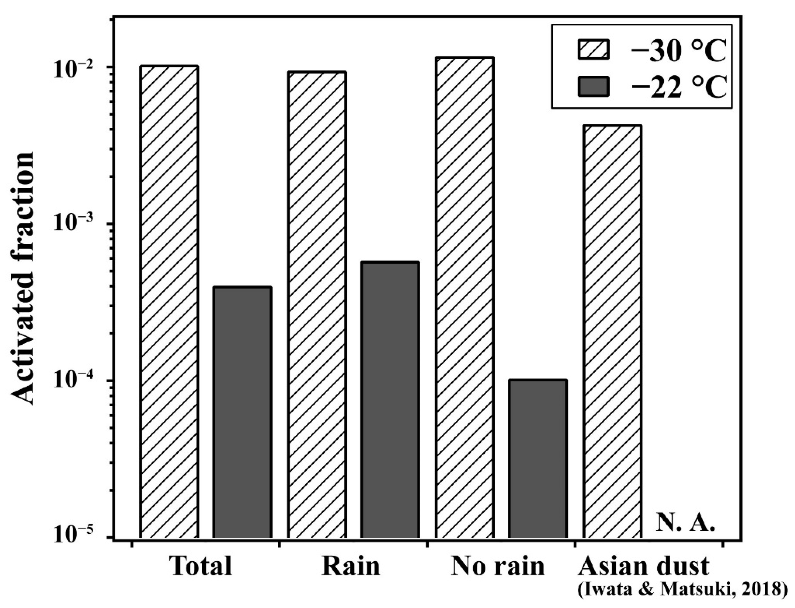
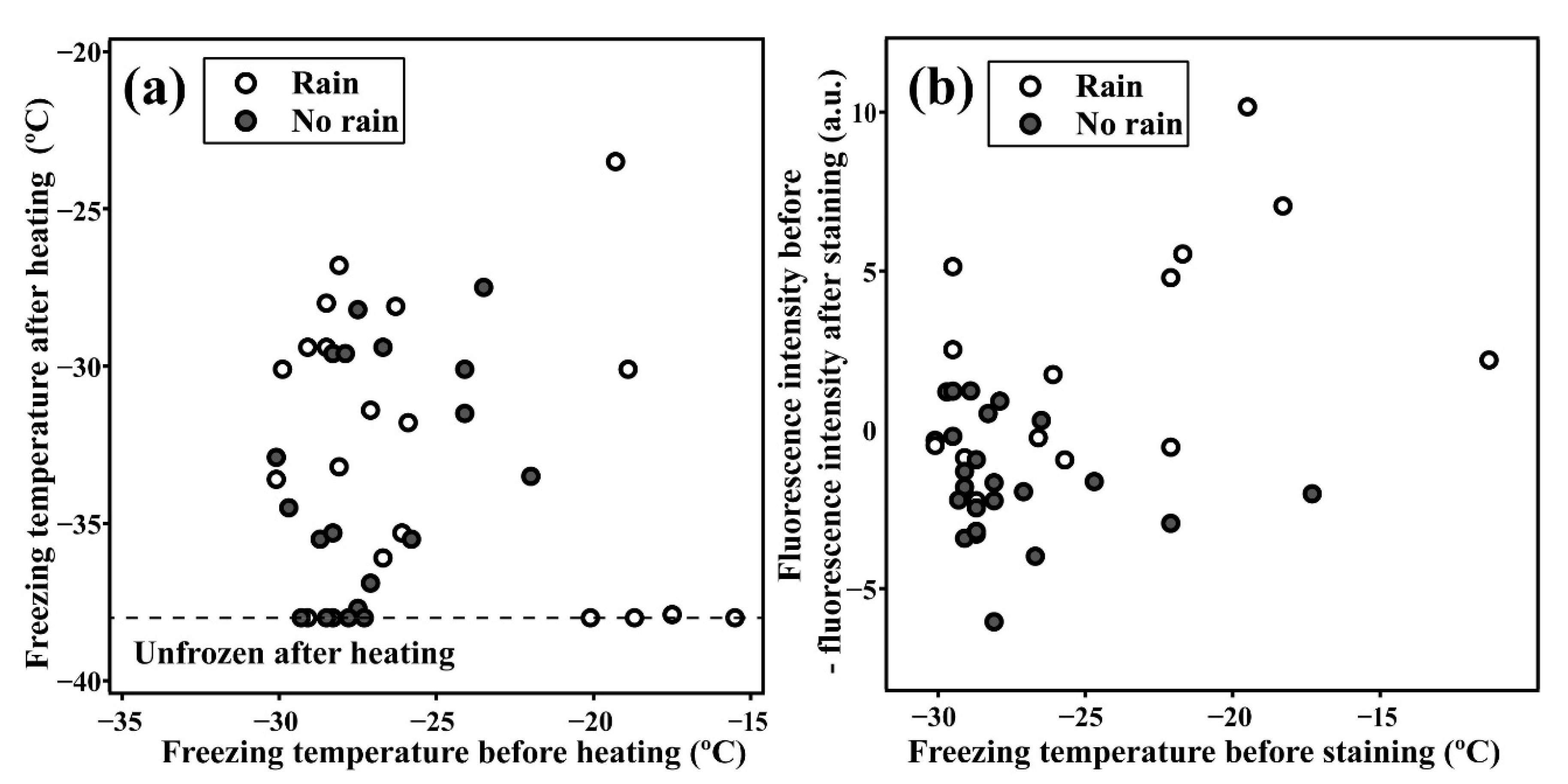
3.1. Relationship between Highly Active INPs and Weather Conditions

In this study, we observed a total of 51,917 particles by analyzing optical microscopic images, among which 538 were identified as INPs. Additionally, 21 INPs were found to be particularly IN-active and formed ice crystals even above −22 °C (INPT > −22). The highest freezing temperature recorded for one of the INPs was at −11.3 °C. It must be emphasized that this is a surprisingly high freezing temperature recorded for an ambient individual particle. It was surprising that INPs active at these high temperatures were captured in this study, given the relatively small number of particles counted, as these INPs were usually considered extremely rare (10−5–10−6 particles/L [3,47]). Additionally, no particles were found to nucleate ice crystals at temperatures above −25 °C in our previous study [38], although sampling was conducted during periods with elevated concentrations of mineral dust particles (i.e., representative INPs) during Asian dust episodes in February and April 2016.
Activated fractions, defined as the number of INPs/total observed particles for DP > 1.1 µm particles, at −30 °C and −22 °C are shown in Table S1. Despite some differences in the sampling periods, the activated fractions for DP > 1.1 µm at −30 °C fell within a relatively narrow range, between 2.3 × 10⁻³ and 33.2 × 10⁻³, and were roughly consistent with the INP concentrations observed in our previous study [38]. However, interestingly, the activated fractions were slightly higher than in our previous study (i.e., during Asian dust episodes), indicating that the elevated concentration of mineral dust did not necessarily result in higher activated fractions among coarse particles.
The INPT > −22 samples were identified in 10 periods. Based on the results of previous observations [29,34,35,40], we also focused on the association of INPs with different meteorological conditions, specifically, those in the presence or absence of precipitation. Here, the sampling periods during which precipitation was detected within 1 h before sampling were defined as rain samples, and the total activated fractions of these samples are shown in Figure 1 and compared to those sampled when there was no rain. The rain samples were also associated with higher relative humidity (RH > 70%) and notably wet ground surfaces near the sampling location. The activated fractions at −30 °C remained almost the same and were not affected by the rain. Meanwhile, most of the INPT > −22 (19 out of 21 INPT > −22) were identified in the samples collected during periods of rain.
The total activated fraction at −22 °C during rainfall events was ~6 times higher than for the samples collected without rain. In particular, the activated fractions in the rain samples were found to be significantly higher than those of the non-rain samples for temperatures above −25 °C (Figure S2). These increases in the fraction in the temperature region above −25 °C are consistent with the results of several laboratory experiments and observations, suggesting the contribution of biogenic particles [47,48,49]. These results are consistent with those of previous studies in that INP concentrations increase with rain [29,32], and supports the possibility that the proportion of INPs in atmospheric particles is increased by rain. Our results further suggested that the increase in the INP concentrations with rain occurred not only in large-scale forested regions, such as the Amazon, but also on a much smaller scale, local forested sites in Japan.
The abundance of efficient INPT > −22 was not necessarily correlated with the rainfall or precipitation intensity during sampling, but rather, they were always found during the periods with high relative humidity (RH > 70%), which included periods when the ground surface remained wet due to preceding rains. Therefore, as far as the sampling site in this study is concerned (a forested mountain site not greatly impacted by afforestation), the emissions of these efficient INPs into the atmosphere may be associated with the high humidity caused by rain rather than mechanical processes involving raindrops. Our previous study was conducted at the same site, and our findings also suggested that biological particles may be responsible for the increased concentration of INPs [33]. However, this was inferred mainly from the heat sensitivity of bulk aerosol samples. In this study, we verified the biological contribution to INPs on an individual particle-basis.

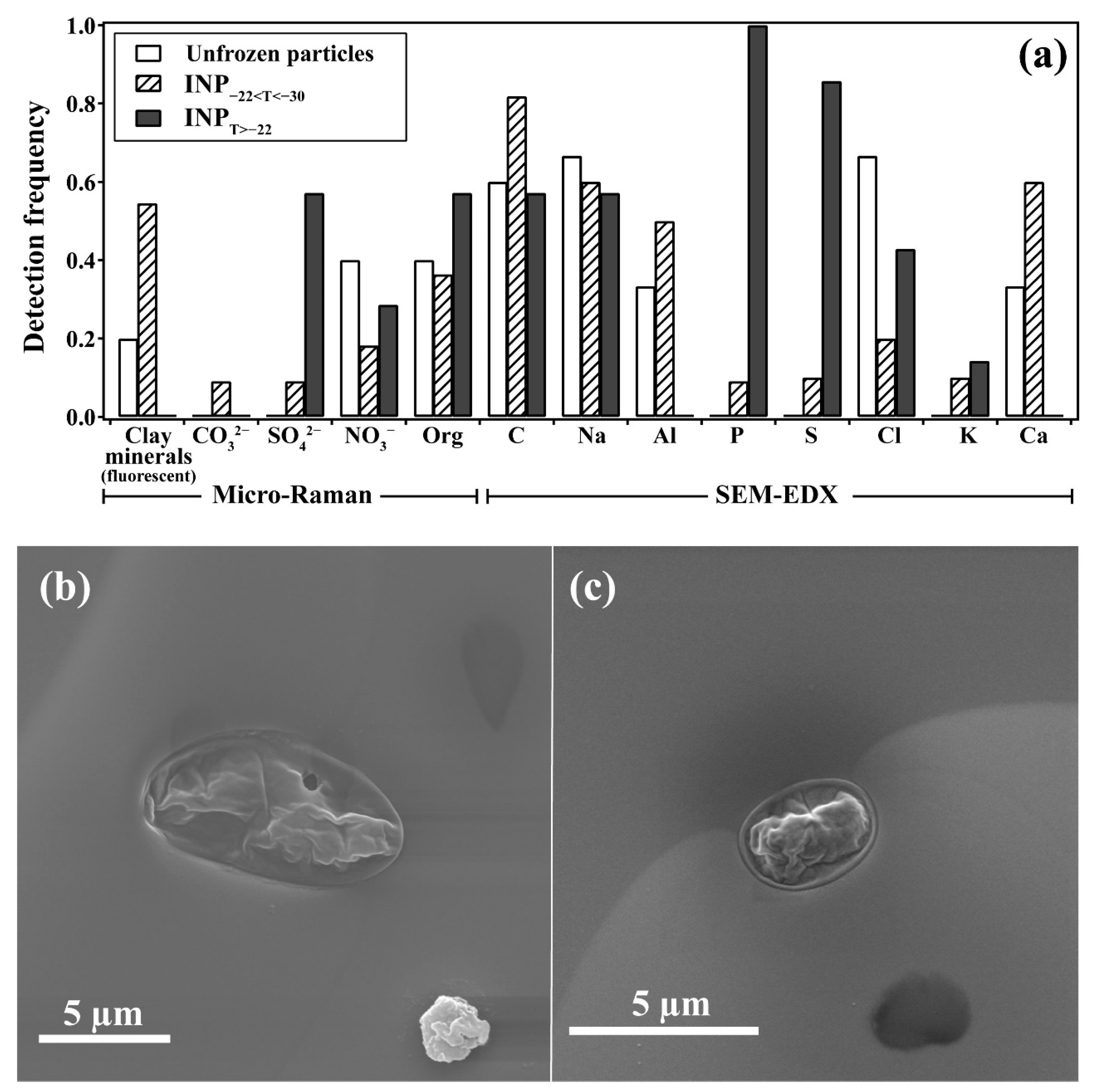
3.2. Biological Fingerprints of Efficient INPs

In order to characterize the particles identified as INPT > −22, every group of seven INPT > −22 was observed by using three different methods. The first group was subjected to morphological and chemical characterization by micro-Raman spectroscopy followed by SEM-EDX. We also measured 11 INPs that were only active between −22 °C to −30 °C (INP−22 < T < −30), as well as five inactive particles for reference. The peaks found in the Raman spectra were assigned to the clay minerals, carbonates, sulfates, nitrates, and OM contained in individual particles (Figure S3a) [38,50,51,52,53]. The elements of Na, Mg, Al, S, K, Cl, Ca, Ti, and Fe contained in the particles were semi-quantitatively analyzed by EDX (Figure S3b). Due to the overlap of C, Si, and P peaks from the sampling substrate, these elements could not be included in the analysis. However, particles showing significantly higher peak intensities of these elements than those in the background substrate were defined as particles containing those elements.
As a result of micro-Raman analysis (Figure 2a), more than half of the INP−22 < T < −30 showed fluorescence in the Raman spectra. Such fluorescence has been associated with clay minerals contained within particles [54,55]. In fact, most of these particles were classified as irregularly shaped mineral dust particles containing Al and Mg by the following EDX analysis. Meanwhile, all seven INPT > −22 exhibited no characteristic fluorescence of cray minerals by micro-Raman analysis, nor aluminum peaks that can generally be associated with mineral dust by EDX (Figure 2a). Additionally, these efficient INPs showed higher detection frequencies of organics and sulfates than either the inactive particles or INP−22 < T < −30; indeed, all of the INPT > −22 were found with significantly higher peaks of phosphorus than the background, as shown in Figure S3.
Atmospheric biological particles consist largely of OM by nature; therefore, they are often identified as particles containing elements, such as carbon and phosphorus, by EDX [56]. In fact, the electron microscope images of a few of the INPT > −22 (two out of seven particles) showed smooth oval outlines, which clearly differed from the representative inorganic and coarse aerosol particles, such as mineral dust or sea salt (Figure 2b,c). Their size, morphology, and composition strongly suggested that they were a sort of airborne microbe (e.g., spores). One particle that formed ice above −22 °C was also suggested to be a microbe from its size and morphology, which resembled those of spores (Figure 2b,c). From these results, the INP−22 < T < −30 were found to be predominantly mineral dust particles, consistent with previous studies. Meanwhile, the more efficient INPs were most likely to be biological particles in an external mixture (i.e., not attached to mineral dust particles).
The second group of seven INPT > −22 was tested for their heat sensitivity by comparing the freezing temperatures before and after heat treatment. Additionally, the relatively inefficient 32 INP−22 < T < −30 at temperatures above −22 °C were also tested for their heat sensitivity for comparison (Figure 3). As shown in Figure 3a, the INP−22 < T < −30 showed the least change in their IN activity following heat treatment, and the temperature difference was less than 12 °C (Figure 3a). Recall that the chemical analyses by micro-Raman and EDX showed that even the INP−22 < T < 30 contained OM (Figure 2a). The inactivation of IN activity of Oms contained in these particles by heat treatment might be responsible for the observed decrease in the onset temperature [57]. However, the majority (approximately 85%) of INP−22 < T < −30 were still active above the homogeneous freezing temperature (−38 °C) even after the heat treatment. In contrast, the majority (approximately 60%) of the INPT > −22 showed a complete loss of heterogeneous nucleation activity and showed significantly stronger heat sensitivity after heating. Therefore, these highly active INPs should not contain heat-tolerant INPs, such as mineral dust, but should rather be biological substances, such as proteins and lipids, which become inactive at least at 150 °C [43,44,45].
Finally, the changes in the fluorescence intensities of individual particles following SYBR® Gold staining were observed for the efficient INPT > −22, and compared to the relatively inefficient INP−22 < T < −30. The results of this nucleic acid staining indicated that the inefficient INPs showed almost no change in their fluorescence intensities following staining (Figure 3b). In contrast, the more efficient INPs, especially those collected during rainfall events, were found to show notable increases in their fluorescence intensities after staining.
In general, the nucleic acid staining protocol using SYBR® Gold dye involves the pretreatment of microorganisms by aldehyde solution to facilitate nucleic acid staining [58]. However, in this study, the pretreatment was omitted to avoid the risk of mobilizing and losing the very few INPs on the substrate. It must be noted that there is a chance that the current staining method may miscount biological particles that contain nucleic acid, but at least this will not create any positive bias (i.e., miss-identification of abiotic particles as biological ones). The change in fluorescence intensities observed in this study may be considered to reflect the lower limit of the detection of nucleic acid-containing particles. However, the applicability of this method was validated using laboratory-generated particles containing the genus Bacillus. Moreover, the characteristic increase in the fluorescence intensities of the efficient INPs is a clear indication that they contain nucleic acid in a high frequency. It is also an indication that the highly active INPs are enriched in biological components, including cells themselves, in this case.
The results from the all three analytical methods indicated that particles that nucleate ice crystals at temperatures below −22 °C were dominated by mineral dust. This is consistent with the classic view of INPs, which has been shown in previous studies [8,10,15,38]. Additionally, we found a very small number of particles that nucleated ice crystals at much higher temperatures, and they have now been shown to have characteristics that are distinct from the INPs active at lower temperatures; they can be termed as biological particles. Based on the multitude of individual particle characterizations, our results provide a direct fingerprint of biological particles as important atmospheric INPs, especially at higher temperatures, in which typical INPs, such as mineral dust particles, remain inactive.

4. Conclusions

In order to investigate the relation between INPs acting at higher temperatures and atmospheric conditions, we identified individual INPs out of the atmospheric particles collected under rainy and non-rainy conditions. To directly elucidate the contribution of biological particles as important sources of efficient INPs, the collected INPs were characterized using three different individual particle analyses: (i) particle composition (both molecular and elemental) and morphology, (ii) heat sensitivity of IN activity, and (iii) fluorescence observations by DNA staining.
We observed higher activated fractions of particles that formed ice crystals (by immersion freezing) during sampling periods with rain than those with no rain, specifically in the higher temperature range (>−22 °C). While the INPs acting at lower temperatures (<−22 °C) were suggested to be dominantly comprised of mineral dust particles, we found a small number of particles that act more efficiently as INPs at higher temperatures. These particles showed different chemical and physical properties from typical atmospheric INPs (e.g., mineral dust), and were further demonstrated to be mainly composed of biological substances. Due to the combined technical limitations of INP identification by IDFM (only concerns coarse particles in a small sampling volume) and the extreme scarcity of the atmospheric INPT > −22, we must note that our results still involve statistical uncertainty in terms or their abundance and atmospheric concentrations. However, it must also be emphasized that the current results provide direct evidence that (i) biological aerosols play important roles as efficient INPs, especially under high-temperature conditions and (ii) that these particles are released into the atmosphere preferentially during rainfall events.
In this study, we could not pinpoint the exact source or species involved in the release of biological INPs, nor the mechanisms of their release associated with precipitation. Though such specificity was beyond the scope of this study, we nevertheless attempted to narrow the potential sources based on circumstantial findings. For example, bacterial particles with high IN activity, such as P. syringae, have been proposed to be released into the atmosphere by the impact of raindrops on the surface of leaves [59]. However, as mentioned previously, the abundance of efficient INP T > −22 was not necessarily correlated with precipitation intensity during sampling. Therefore, the emissions of biological INPs into the atmosphere may be associated with biological processes (e.g., the release of fungal spores triggered by high relative humidity) rather than mechanical processes involving raindrops. However, further study is needed to fully reveal the emissions source.
Whatever the release mechanism may be, the results of this study imply that the terrestrial ecosystem has been actively involved in the development of the water cycle in the surface environment, at least in the region in concern and in similar climate zones in the mid-latitudes. The remaining concern may be that the current result is based on the particles collected close to the ground, which may not be directly applicable to the aerosol behavior at higher altitudes where cloud freezing actually takes place. It is beyond the scope of this paper, but to what extent (both in terms of time and space) such biological particles released near the ground as a response to rainfall would travel and be dispersed in the atmosphere remains a future task. Generally speaking, rainfall takes place in an area of convergence (hence updraft), and particles emitted from the ground surface may have a higher chance of being mixed vertically in a hydrostatically unstable condition.
Supposing that these particles were effectively dispersed at high altitudes in the atmosphere, there may potentially be a positive feedback loop between precipitation and biological INPs, wherein precipitation (and, thus, high humidity) triggers the release of biological particles by the ecosystem, which then leads to the dispersion of efficient INPs that are eventually incorporated into clouds and act to initiate ice crystal formation among super-cooled droplets, in turn triggering precipitation (cold rain) and so forth. The possible involvement of the ecosystem in the water cycle may have played an important role in the expansion of these habitats. Therefore, we suggest that deforestation, land use changes, and the associated degradation of biodiversity might have a significant influence on the abundance of atmospheric INPs, and, thus, on the microphysics and dynamics of clouds and precipitation in these regions, thereby affecting the regional and global climate.

Supplementary Materials

The following are available online at https://www.mdpi.com/2073-4433/10/10/605/s1, Figure S1: Sampling location of the atmospheric particles (Kanazawa University, 36.54° N, 136.70° E; 149 m a.s.l.). Figure S2: Activated fractions for DP > 1.1 µm particles as a function of temperature. Figure S3: Raman (a) and EDX (b) spectra of the representative identified INPs. Table S1: Summary of the activated fractions and the weather condition during sampling periods. Table S2: Summary of the chemical characterization of INPs based on the micro-Raman and EDX analyses.

Author Contributions

Conceptualization, A.I. and A.M.; Data curation, A.I. and M.I.; Formal analysis, A.I., M.I., M.H., N.T. and R.K.; Funding acquisition, A.M.; Investigation, M.I., M.H., N.T. and R.K.; Methodology, A.I., M.H., T.M., N.T. and R.K.; Project administration, A.M.; Supervision, T.M. and A.M.; Validation, A.I. and M.I.; Writing—original draft, A.I.; Writing—review & editing, A.I., T.M. and A.M.

Funding

This study was supported by the Japan Society for the Promotion of Science (JSPS) Funding Program for Next Generation World-Leading Researchers (grant no. GR045).

Acknowledgments

We thank Tomoyuki Mizukami and Noriko Hasebe (Kanazawa University) and Norikatsu Akizawa (Kyoto University) for providing experimental equipment. We gratefully acknowledge Hirosi Tsurumaru (Tokyo Metropolitan Research Institute for Environmental Protection) and Risa Maki for providing valuable comments for detecting biological particles. We also acknowledge the Faculty of Science and Technology, Keio University for supplementary observations of particles by electronic microscope. The meteorological data were obtained from the Japanese Meteorological Agency (http://www.data.jma.go.jp/gmd/risk/obsdl/index.php).

Conflicts of Interest

The authors declare no conflict of interest.

References

  1. Lohmann, U.; Feichter, J. Global indirect aerosol effects: A review. Atmos. Chem. Phys. 2005, 5, 715–737. [Google Scholar] [CrossRef]
  2. Pruppacher, H.R.; Klett, J.D. Microphysics of Clouds and Precipitation, 2nd ed.; Springer: Amsterdam, The Netherlands, 1998. [Google Scholar]
  3. Kanji, Z.A.; Ladino, L.A.; Wex, H.; Boose, Y.; Burkert-Kohn, M.; Cziczo, D.J.; Krämer, M. Overview of Ice Nucleating Particles. Meteorol. Monogr. 2017, 58. [Google Scholar] [CrossRef]
  4. Cantrell, W.; Heymsfield, A. Production of ice in tropospheric clouds: A review. Bull. Am. Meteorol. Soc. 2005, 86, 795–807. [Google Scholar] [CrossRef]
  5. Demott, P.J.; Rogers, D.C. Freezing nucleation rates of dilute solution droplets measured between −30° and −40 °C in laboratory simulations of natural clouds. J. Atmos. Sci. 1990, 47, 1056–1064. [Google Scholar] [CrossRef]
  6. Koop, T.; Luo, B.; Tsias, A.; Peter, T. Water activity as the determinant for homogeneous ice nucleation in aqueous solutions. Nature 2000, 406, 611–614. [Google Scholar] [CrossRef]
  7. Murray, B.J.; Broadley, S.L.; Wilson, T.W.; Bull, S.J.; Wills, R.H.; Christenson, H.K.; Murray, E.J. Kinetics of the homogeneous freezing of water. Phys. Chem. Chem. Phys. 2010, 12, 10380–10387. [Google Scholar] [CrossRef] [PubMed]
  8. Murray, B.J.; O’Sullivan, D.; Atkinson, J.D.; Webb, M.E. Ice nucleation by particles immersed in supercooled cloud droplets. Chem. Soc. Rev. 2012, 41, 6519. [Google Scholar] [CrossRef] [PubMed]
  9. DeMott, P.; Prenni, A.J.; Liu, X.; Kreidenweis, S.M.; Petters, M.D.; Twohy, C.H.; Richardson, M.S.; Eidhammer, T.; Rogers, D.C. Predicting global atmospheric ice nuclei distributions and their impacts on climate. Proc. Natl. Acad. Sci. USA 2010, 107, 11217–11222. [Google Scholar] [CrossRef] [Green Version]
  10. Pratt, K.A.; Demott, P.J.; French, J.R.; Wang, Z.; Westphal, D.L.; Heymsfield, A.J.; Twohy, C.H.; Prenni, A.J.; Prather, K.A. In situ detection of biological particles in cloud ice-crystals. Nat. Geosci. 2009, 2, 398–401. [Google Scholar] [CrossRef]
  11. Prenni, A.J.; DeMott, P.J.; Sullivan, A.P.; Sullivan, R.C.; Kreidenweis, S.M.; Rogers, D.C. Biomass burning as a potential source for atmospheric ice nuclei: Western wildfires and prescribed burns. Geophys. Res. Lett. 2012, 39. [Google Scholar] [CrossRef] [Green Version]
  12. Ardon-Dryer, K.; Levin, Z. Ground-based measurements of immersion freezing in the eastern Mediterranean. Atmos. Chem. Phys. 2014, 14, 5217–5231. [Google Scholar] [CrossRef] [Green Version]
  13. Rosenfeld, D.; Lohmann, U.; Raga, G.B.; O’Dowd, C.D.; Kulmala, M.; Fuzzi, S.; Reissell, A.; Andreae, M.O. Flood or Drought: How Do Aerosols Affect Precipitation? Science 2008, 321, 1309–1313. [Google Scholar] [CrossRef] [PubMed] [Green Version]
  14. Niemand, M.; Möhler, O.; Vogel, H.; Vogel, B.; Hoose, C.; Connolly, P.; Klein, H.; Bingemer, H.; DeMott, P.; Skrotzki, J.; et al. A Particle-Surface-Area-Based Parameterization of Immersion Freezing on Desert Dust Particles. J. Atmos. Sci. 2012, 69, 3077–3092. [Google Scholar] [CrossRef]
  15. Atkinson, J.D.; Murray, B.J.; Woodhouse, M.T.; Whale, T.F.; Baustian, K.J.; Carslaw, K.S.; Dobbie, S.; O’Sullivan, D.; Malkin, T.L. The importance of feldspar for ice nucleation by mineral dust in mixed-phase clouds. Nature 2013, 498, 355–358. [Google Scholar] [CrossRef] [PubMed]
  16. Worringen, A.; Kandler, K.; Benker, N.; Dirsch, T.; Mertes, S.; Schenk, L.; Kästner, U.; Frank, F.; Nillius, B.; Bundke, U.; et al. Single-particle characterization of ice-nucleating particles and ice particle residuals sampled by three different techniques. Atmos. Chem. Phys. 2015, 15, 4161–4178. [Google Scholar] [CrossRef] [Green Version]
  17. Kamphus, M.; Ettner-Mahl, M.; Klimach, T.; Drewnick, F.; Keller, L.; Cziczo, D.J.; Mertes, S.; Borrmann, S.; Curtius, J. Chemical composition of ambient aerosol, ice residues and cloud droplet residues in mixed-phase clouds: Single particle analysis during the Cloud and Aerosol Characterization Experiment (CLACE 6). Atmos. Chem. Phys. 2010, 10, 8077–8095. [Google Scholar] [CrossRef]
  18. Boose, Y.; Sierau, B.; García, M.I.; Rodríguez, S.; Alastuey, A.; Linke, C.; Schnaiter, M.; Kupiszewski, P.; Kanji, Z.A.; Lohmann, U. Ice nucleating particles in the Saharan Air Layer. Atmos. Chem. Phys. 2016, 16, 9067–9087. [Google Scholar] [CrossRef] [Green Version]
  19. Harrison, A.D.; Whale, T.F.; Carpenter, M.A.; Holden, M.A.; Neve, L.; O’Sullivan, D.; Temprado, J.V.; Murray, B.J. Not all feldspars are equal: A survey of ice nucleating properties across the feldspar group of minerals. Atmos. Chem. Phys. 2016, 16, 10927–10940. [Google Scholar] [CrossRef]
  20. Isono, K.; Komabayasi, M.; Ono, A. The Nature and the Origin of Ice Nuclei in the Atmosphere. J. Meteorol. Soc. Japan. Ser. II 1959, 37, 211–233. [Google Scholar] [CrossRef]
  21. Vergara-Temprado, J.; Murray, B.J.; Wilson, T.W.; O’Sullivan, D.; Browse, J.; Pringle, K.J.; Ardon-Dryer, K.; Bertram, A.K.; Burrows, S.M.; Ceburnis, D.; et al. Contribution of feldspar and marine organic aerosols to global ice nucleating particle concentrations. Atmos. Chem. Phys. 2017, 17, 3637–3658. [Google Scholar] [CrossRef] [Green Version]
  22. Pummer, B.G.; Bauer, H.; Bernardi, J.; Bleicher, S.; Grothe, H. Suspendable macromolecules are responsible for ice nucleation activity of birch and conifer pollen. Atmos. Chem. Phys. 2012, 12, 2541–2550. [Google Scholar] [CrossRef] [Green Version]
  23. Morris, C.E.; Georgakopoulos, D.G.; Sands, D.C. Ice nucleation active bacteria and their potential role in precipitation. J. Phys. IV 2004, 121, 87–103. [Google Scholar] [CrossRef]
  24. Morris, C.E.; Sands, D.C.; Glaux, C.; Samsatly, J.; Asaad, S.; Moukahel, A.R.; Gonçalves, F.L.T.; Bigg, E.K. Urediospores of rust fungi are ice nucleation active at >−10 °C and harbor ice nucleation active bacteria. Atmos. Chem. Phys. 2013, 13, 4223–4233. [Google Scholar] [CrossRef]
  25. Wilson, T.W.; Ladino, L.A.; Alpert, P.A.; Breckels, M.N.; Brooks, I.M.; Browse, J.; Burrows, S.M.; Carslaw, K.S.; Huffman, J.A.; Judd, C.; et al. A marine biogenic source of atmospheric ice-nucleating particles. Nature 2015, 525, 234–238. [Google Scholar] [CrossRef] [PubMed] [Green Version]
  26. Pummer, B.G.; Budke, C.; Augustin-Bauditz, S.; Niedermeier, D.; Felgitsch, L.; Kampf, C.J.; Huber, R.G.; Liedl, K.R.; Loerting, T.; Moschen, T.; et al. Ice nucleation by water-soluble macromolecules. Atmos. Chem. Phys. 2015, 15, 4077–4091. [Google Scholar] [CrossRef] [Green Version]
  27. Maki, L.R.; Galyan, E.L.; Chang-Chien, M.M.; Caldwell, D.R. Ice nucleation induced by pseudomonas syringae. Appl. Microbiol. 1974, 28, 456–459. [Google Scholar] [PubMed]
  28. Möhler, O.; DeMott, P.J.; Vali, G.; Levin, Z. Microbiology and atmospheric processes: The role of biological particles in cloud physics. Biogeosciences 2007, 4, 1059–1071. [Google Scholar] [CrossRef]
  29. Huffman, J.A.; Prenni, A.J.; DeMott, P.J.; Pöhlker, C.; Mason, R.H.; Robinson, N.H.; Fröhlich-Nowoisky, J.; Tobo, Y.; Desprès, V.R.; Garcia, E.; et al. High concentrations of biological aerosol particles and ice nuclei during and after rain. Atmos. Chem. Phys. 2013, 13, 6151–6164. [Google Scholar] [CrossRef] [Green Version]
  30. Jones, A.M.; Harrison, R.M. The effects of meteorological factors on atmospheric bioaerosol concentrations—A review. Sci. Total Environ. 2004, 326, 151–180. [Google Scholar] [CrossRef]
  31. Wright, T.P.; Hader, J.D.; McMeeking, G.R.; Petters, M.D. High relative humidity as a trigger for widespread release of ice nuclei. Aerosol. Sci. Technol. 2014, 48. [Google Scholar] [CrossRef]
  32. Tobo, Y.; Prenni, A.J.; Demott, P.J.; Huffman, J.A.; McCluskey, C.S.; Tian, G.; Pöhlker, C.; Pöschl, U.; Kreidenweis, S.M. Biological aerosol particles as a key determinant of ice nuclei populations in a forest ecosystem. J. Geophys. Res. Atmos. 2013, 118, 10100–10110. [Google Scholar] [CrossRef]
  33. Hara, K.; Maki, T.; Kobayashi, F.; Kakikawa, M.; Wada, M.; Matsuki, A. Variations of ice nuclei concentration induced by rain and snowfall within a local forested site in Japan. Atmos. Environ. 2016, 127, 1–5. [Google Scholar] [CrossRef]
  34. Garcia, E.; Hill, T.C.J.; Prenni, A.J.; DeMott, P.J.; Franc, G.D.; Kreidenweis, S.M. Biogenic ice nuclei in boundary layer air over two U.S. high plains agricultural regions. J. Geophys. Res. Atmos. 2012, 117, 1–12. [Google Scholar] [CrossRef]
  35. Pöhlker, C.; Wiedemann, K.T.; Sinha, B.; Shiraiwa, M.; Gunthe, S.S.; Smith, M.; Su, H.; Artaxo, P.; Chen, Q.; Cheng, Y.; et al. Biogenic Potassium Salt Particles as Seeds for Secondary Organic Aerosol in the Amazon. Science 2012, 337, 1075–1078. [Google Scholar] [CrossRef] [Green Version]
  36. Tobo, Y. An improved approach for measuring immersion freezing in large droplets over a wide temperature range. Sci. Rep. 2016, 6, 1–9. [Google Scholar] [CrossRef] [PubMed]
  37. Whale, T.F.; Murray, B.J.; O’Sullivan, D.; Wilson, T.W.; Umo, N.S.; Baustian, K.J.; Atkinson, J.D.; Workneh, D.A.; Morris, G.J. A technique for quantifying heterogeneous ice nucleation in microlitre supercooled water droplets. Atmos. Meas. Tech. 2015, 8, 2437–2447. [Google Scholar] [CrossRef] [Green Version]
  38. Iwata, A.; Matsuki, A. Characterization of individual ice residual particles by the single droplet freezing method: A case study in the Asian dust outflow region. Atmos. Chem. Phys. 2018, 18, 1785–1804. [Google Scholar] [CrossRef]
  39. Marcolli, C.; Nagare, B.; Welti, A.; Lohmann, U. Ice nucleation efficiency of AgI: Review and new insights. Atmos. Chem. Phys. 2016, 16, 8915–8937. [Google Scholar] [CrossRef]
  40. Hara, K.; Maki, T.; Kakikawa, M.; Kobayashi, F.; Matsuki, A. Effects of different temperature treatments on biological ice nuclei in snow samples. Atmos. Environ. 2016, 140, 415–419. [Google Scholar] [CrossRef]
  41. Christner, B.C.; Morris, C.E.; Foreman, C.M.; Cai, R.; Sands, D.C. Ubiquity of Biological Ice Nucleators in Snowfall. Science 2008, 319, 1214. [Google Scholar] [CrossRef] [PubMed]
  42. Joly, M.; Amato, P.; Deguillaume, L.; Monier, M.; Hoose, C.; Delort, A.-M. Quantification of ice nuclei active at near 0 °C temperatures in low-altitude clouds at the Puy de Dôme atmospheric station. Atmos. Chem. Phys. 2014, 14, 8185–8195. [Google Scholar] [CrossRef]
  43. Pouleur, S.; Richard, C.; Martin, J.; Antoun, H. Ice Nucleation Activity in Fusarium acuminatum and Fusarium avenaceum. Appl. Environ. Microbiol. 1992, 58, 2960–2964. [Google Scholar] [PubMed]
  44. Fröhlich-Nowoisky, J.; Hill, T.C.J.; Pummer, B.G.; Yordanova, P.; Franc, G.D.; Pöschl, U. Ice nucleation activity in the widespread soil fungus Mortierella alpina. Biogeosciences 2015, 12, 1057–1071. [Google Scholar] [CrossRef] [Green Version]
  45. Hill, T.C.J.; Moffett, B.F.; DeMott, P.J.; Georgakopoulos, D.G.; Stump, W.L.; Franc, G.D. Measurement of Ice Nucleation-Active Bacteria on Plants and in Precipitation by Quantitative PCR. Appl. Environ. Microbiol. 2014, 80, 1256–1267. [Google Scholar] [CrossRef] [PubMed] [Green Version]
  46. Lu, J.-R.; Hodson, R.E.; Binder, B.J.; Chen, F.; Liu, Y. Application of Digital Image Analysis and Flow Cytometry To Enumerate Marine Viruses Stained with SYBR Gold. Appl. Environ. Microbiol. 2002, 67, 539–545. [Google Scholar]
  47. Petters, M.D.; Wright, T.P. Revisiting ice nucleation from precipitation samples. Geophys. Res. Lett. 2015, 42, 8758–8766. [Google Scholar] [CrossRef]
  48. Hill, T.C.J.; Demott, P.J.; Tobo, Y.; Fröhlich-Nowoisky, J.; Moffett, B.F.; Franc, G.D.; Kreidenweis, S.M. Sources of organic ice nucleating particles in soils. Atmos. Chem. Phys. 2016, 16, 7195–7211. [Google Scholar] [CrossRef] [Green Version]
  49. O’Sullivan, D.; Adams, M.P.; Tarn, M.D.; Harrison, A.D.; Vergara-Temprado, J.; Porter, G.C.E.; Holden, M.A.; Sanchez-Marroquin, A.; Carotenuto, F.; Whale, T.F.; et al. Contributions of biogenic material to the atmospheric ice-nucleating particle population in North Western Europe. Sci. Rep. 2018, 8, 1–9. [Google Scholar] [CrossRef]
  50. Ivleva, N.P.; Messerer, A.; Yang, X.; Niessner, R.; Pöschl, U. Raman microspectroscopic analysis of changes in the chemical structure and reactivity of soot in a diesel exhaust aftertreatment model system. Environ. Sci. Technol. 2007, 41, 3702–3707. [Google Scholar] [CrossRef]
  51. Baustian, K.J.; Cziczo, D.J.; Wise, M.E.; Pratt, K.A.; Kulkarni, G.; Hallar, A.G.; Tolbert, M.A. Importance of aerosol composition, mixing state, and morphology for heterogeneous ice nucleation: A combined field and laboratory approach. J. Geophys. Res. Atmos. 2012, 117. [Google Scholar] [CrossRef] [Green Version]
  52. Laskina, O.; Young, M.A.; Kleiber, P.D.; Grassian, V.H. Infrared extinction spectroscopy and micro-Raman spectroscopy of select components of mineral dust mixed with organic compounds. J. Geophys. Res. Atmos. 2013, 118, 6593–6606. [Google Scholar] [CrossRef]
  53. Freedman, M.A.; Hasenkopf, C.A.; Beaver, M.R.; Tolbert, M.A. Optical properties of internally mixed aerosol particles composed of dicarboxylic acids and ammonium sulfate. J. Phys. Chem. A 2009, 113, 13584–13592. [Google Scholar] [CrossRef] [PubMed]
  54. Sobanska, S.; Hwang, H.; Choël, M.; Jung, H.J.; Eom, H.J.; Kim, H.; Barbillat, J.; Ro, C.U. Investigation of the chemical mixing state of individual asian dust particles by the combined use of electron probe X-ray microanalysis and raman microspectrometry. Anal. Chem. 2012, 84, 3145–3154. [Google Scholar] [CrossRef] [PubMed]
  55. Jung, H.J.; Eom, H.J.; Kang, H.W.; Moreau, M.; Sobanska, S.; Ro, C.U. Combined use of quantitative ED-EPMA, Raman microspectrometry, and ATR-FTIR imaging techniques for the analysis of individual particles. Analyst 2014, 139, 3949–3960. [Google Scholar] [CrossRef]
  56. Matthias-Maser, S.; Obolkin, V.; Khodzer, T.; Jaenicke, R. Seasonal variation of primary biological aerosol particles in the remote continental region of Lake Baikal/Siberia. Atmos. Environ. 2000, 34, 3805–3811. [Google Scholar] [CrossRef]
  57. Conen, F.; Morris, C.E.; Leifeld, J.; Yakutin, M.V.; Alewell, C. Biological residues define the ice nucleation properties of soil dust. Atmos. Chem. Phys. 2011, 11, 9643–9648. [Google Scholar] [CrossRef] [Green Version]
  58. Wu, C.F.; Lai, C.H.; Chu, H.J.; Lin, W.H. Evaluating and mapping of spatial air ion quality patterns in a residential garden using a geostatistic method. Int. J. Environ. Res. Public Health 2011, 8, 2304–2319. [Google Scholar] [CrossRef] [PubMed]
  59. Morris, C.E.; Conen, F.; Huffman, J.A.; Phillips, V.; Pöschl, U.; Sands, D.C. Bioprecipitation: A feedback cycle linking Earth history, ecosystem dynamics and land use through biological ice nucleators in the atmosphere. Glob. Chang. Biol. 2014, 20, 341–351. [Google Scholar] [CrossRef] [PubMed]
Figure 1. Ice nucleation (IN) activated fractions of samples with rain and without rain. These fractions yield the ratio of ice-nucleating particles (INPs) identified at respective temperatures (striped bar: −30 °C, solid bar: −22 °C) relative to the total number of particles observed by the individual droplet freezing method (IDFM).
Atmosphere 10 00605 g001
Figure 2. Summary of chemical and morphological characterization by micro-Raman analysis and scanning electron microscopy-energy-dispersive X-ray spectroscopy (SEM-EDX). The shading indicates INPs that formed ice crystals in individual particle chemical analysis: (a) above −22 °C (striped bar) and above −30 °C (solid gray bar); unfrozen particles are denoted by the white bars. Detection frequencies (a) were accounted for if the particle contained atomic ratios of elements (excluding C and P) exceeding 15% in semi-quantitative EDX analyses. Significantly higher peak intensities were found for carbon and phosphorus relative to the background substrate (Table S2). (b,c) Electron microscopic images showing the morphology of an efficient INP collected during a rainfall event. These INPs formed ice crystals at (b) −20.5 °C and (c) −20.3 °C using the IDFM.
Atmosphere 10 00605 g002
Figure 3. (a) Comparison of freezing temperatures of INPs before and after heating. (b) Differences between the fluorescence intensities before and after staining using SYBR® Gold relative to the initial freezing temperature of the INPs (i.e., before treatment). Samples collected during rainfall events are denoted by open circles; solid gray circles denote samples collected in the absence of rain. In case the INPs did not show any nucleation in the second test, in heat treatment experiments (a), their freezing temperature was assumed to be −38 °C in order to obtain the temperature following the heat treatment.
Atmosphere 10 00605 g003

