Review

PRC2 Diversity in Neuronal Differentiation and Developmental Disorders

1
Perelman Center for Cellular and Molecular Therapeutics and Center for Brain Research in Development, Genetics and Engineering, Children’s Hospital of Philadelphia, Philadelphia, PA 19104, USA
2
Department of Pathology and Laboratory Medicine and Genetics and Epigenetics Graduate Group, Perelman School of Medicine, University of Pennsylvania, Philadelphia, PA 19104, USA
*
Author to whom correspondence should be addressed.
Genes 2025, 16(10), 1191; https://doi.org/10.3390/genes16101191
Submission received: 16 July 2025 / Revised: 22 September 2025 / Accepted: 2 October 2025 / Published: 13 October 2025
(This article belongs to the Special Issue The Genetic and Epigenetic Basis of Neurodevelopmental Disorders)

Abstract

Advances in genetic studies have not only improved the diagnosis and treatment of neurodevelopmental disorders but also uncovered human-specific aspects of nervous system development. The generation of neuronal diversity in the human brain relies on tightly regulated epigenetic mechanisms, with Polycomb Repressive Complex 2 (PRC2) emerging as a key player. In this review, we first summarize foundational studies that established the role of PRC2 in the epigenetic maintenance of transcriptional silencing. We then highlight recent insights into the increasing evolutionary complexity of PRC2 subcomplexes, their roles in neurodevelopment, and their contribution to human developmental disorders.

1. Introduction

Neuronal differentiation, similar to other differentiation processes, entails the progressive commitment of neural precursor cells, followed by the generation of newborn neuronal subtypes and their functional maturation [1]. Two major events define this process: the restriction of differentiation potential and the acquisition of neuronal type identity and function. While changes in gene expression govern the establishment of neuronal identity and function, the commitment to specific neural lineages and the preservation of their identity requires the maintenance of established gene expression patterns for long periods [2,3]. A classic mechanism to preserve the repression of genes associated with alternate cell lineages involves the trimethylation of lysine 27 on histone H3 (H3K27me3) of chromatin.
Originally discovered in Drosophila melanogaster as gatekeepers of body segmentation, Polycomb Group (PcG) proteins are central to H3K27me3 deposition and the long-term maintenance of gene repression [4]. To execute their function, PcG proteins assemble into Polycomb Repressive Complex 1 and 2 (PRC1 and PRC2). While the core components and function are evolutionarily conserved, PcG subcomplexes have diversified extensively in their composition, chromatin-targeting mechanisms, and biological functions, particularly in more complex organisms. For example, while Drosophila has two PRC1 and one PRC2 complexes, mammals have at least six PRC1 and two PRC2 variants [5]. Emerging evidence suggests that this diversification enables context-specific genome regulation, essential for the expansion of cellular heterogeneity. Accordingly, distinct PRC1/2 subunits in mammals show cell type-specific expression [6,7,8] or division of catalytic activities [9]. However, despite the extensive literature defining the molecular function and expression pattern of each complex, the mechanisms leading to their functional specificity across cell types are not well understood.
In this review, we present a comprehensive discussion aimed at unraveling and interpreting our current understanding of the diversification of the Polycomb complexes, with a particular emphasis on PRC2 and neuronal cell type diversity. We first briefly review historic events that uncovered the basics of PcG- and H3K27me3-mediated epigenetic maintenance of gene repression. We then provide an update on the evolutionary diversification of PRC2 core and accessory subunits and their molecular roles. Finally, we discuss emerging evidence supporting context-specific functions of PRC2 subcomplexes from the perspective of neural development and developmental disorders.

2. The Discovery of Polycomb Group (PcG) Proteins as Epigenetic Guardians of Gene Repression

Nearly a century ago, Pamela Lewis coined the term “Polycomb” to describe the phenotype of mutant Drosophila melanogaster flies exhibiting extra sex combs [10]. In subsequent work, Ed Lewis noticed that Polycomb (Pc) mutant flies resembled flies ectopically expressing Hox genes, a group of homeotic genes that provide antero-posterior identity to body segments [11]. Like Hox mutants, Pc flies showed body parts misplaced along the antero-posterior axis. This groundbreaking observation suggested that the product of the Pc gene was necessary to repress Hox genes [11]. The discovery of additional genes whose mutations caused similar body transformations led to defining them as PcG genes [12]. Among these, extra sex combs (esc), a founding PcG member with Pc, became of particular importance given that fly embryos lacking both maternal and zygotic esc displayed ectopic Hox gene expression only after gastrulation. This phenomenon revealed that ESC was required for maintaining, but not for establishing, Hox gene repression [13]. Furthermore, the phenotypic comparisons with Pc mutants suggested a mechanism where ESC would be required prior to PC for maintaining the correct Hox gene expression and body segmentation [13,14]. These studies provided the first mechanistic evidence of PcG-mediated maintenance of Hox repression and positioned PcG proteins as key components of the epigenetic cellular memory system.

3. Diversification of PRC2 Composition

The end of the 20th century was marked by the emergence of epigenetics as a field focused on studying mechanisms that preserve gene expression programs over time and cell divisions [15,16,17]. Chromatin, the multimeric complex formed by repeating units of DNA wrapped around an octamer of histone proteins, was increasingly recognized as a central platform for the inheritance of gene activity information not encoded in the genome [18]. As PC and other PcG proteins were being found to be associated with chromatin through then-pioneering chromatin immunoprecipitation assays [19,20] and in vitro histone binding experiments [21], it became clear that chromatin played a role in how PcG proteins maintain gene repression patterns established by transcription factors early in development. However, it was not until the discovery of three key pieces of evidence that the PcG-mediated repression mechanism emerged. First, purification of PcG proteins from fly embryos revealed two distinct complexes, one containing PC as a core member and designated PRC1 [22], and the other, PRC2, formed by the interaction of ESC and another PcG protein known as Enhancer of Zeste (E(Z)) [23,24]. Second, PRC1 binding to chromatin was found to depend on E(Z), despite the lack of physical interaction between them [25,26]. Finally, the identification of histone methyltransferase activity in E(Z) led to what is now recognized as the classical PcG-mediated epigenetic silencing mechanism [27,28,29]. Under this classical model, E(Z) and its mammalian homolog EZH2 catalyze H3K27me3. This histone covalent modification is recognized by the chromodomain of PC (or mammalian homologs, CBX) and recruits PRC1 to chromatin [27]. The sequential recruitment of PRC2 and PRC1 to chromatin triggers structural changes that contribute to maintaining the repression of targeted genes [30].
The classical model of PcG recruitment and repressive domain maintenance prevailed for over a decade, but recent discoveries have revealed a far more complex mechanism. This complexity was first highlighted by the finding that the core PRC1 subunits, RING1A/B, catalyze mono-ubiquitination of histone H2A at lysine 119 (H2AK119ub) [31,32], prompting a revision of the classical view. Just like PRC1 binding to PRC2-catalyzed H3K27me3, PRC2 can also bind to PRC1-catalyzed H2AK119ub [33,34]. These reciprocal interactions generate a positive feedback loop that propagates PRC1 and PRC2 recruitment and sustains repressive chromatin domains across space and time [35,36,37]. Emerging data further indicate that the interplay and activity of PRC1 and PRC2 vary depending on their subunit composition and spatiotemporal context [38,39,40,41,42]. Although, together, these findings illustrate that PcG-mediated repression is orchestrated by multiple, interconnected layers of regulation, the silencing mechanism remains incompletely understood. Recruitment can be influenced by chromatin modifications as well as interactions with RNA molecules, while mechanisms contributing to repression range from chromatin compaction to interference with transcription factors and the RNA polymerase. Since the complexity of these mechanisms, particularly those involving PRC1 variants and their diverse functions, have been extensively reviewed elsewhere [5,30,43,44], here we focus on PRC2 variants, emphasizing insights from developmental models and genetic neurodevelopmental disorders that reveal potential divergences.
The core PRC2 in mammals is composed of four proteins: EED, SUZ12, RBBP4 or RBBP7, and either EZH1 or EZH2. In addition, selective interactions with different accessory proteins define at least two major PRC2 subcomplexes with distinct activities: PRC2.1, which is formed by the interaction of the core with PHF1, MTF2, or PHF19, together with either PALI1, PALI2, or EPOP; and PRC2.2, which includes JARID2 and AEBP2 instead [40,41,42] (Figure 1). Evidence indicates that PRC2.1 facilitates the recruitment of the complex to unmethylated CpG reach islands (CGIs) devoid of activating transcription factor motifs for the establishment of H3K27me3 repressive domains in developmental gene promoters [45,46]. Accordingly, deletion of Mtf2 in mouse embryonic stem cells significantly impairs de novo H3K27me3 deposition and spreading [45]. In contrast, the ability of JARID2 and AEBP2 to bind to H2AK119ub enables the allosteric activation of PRC2.2 to propagate repressive domains at loci previously targeted by PRC1 [35,38,39]. The latest recruitment sequence is particularly important for PRC2 targeting to inactive X chromosomes [47], but also for maintaining Polycomb repressive domains at developmental genes, as demonstrated by loss of PRC2.2 and H3K27me3, and reactivation of target genes, upon the depletion of PRC1 ubiquitin-transferase activity [36,37]. Despite their functional differences, however, the coordination of PRC2.1 and PRC2.2 with PRC1 is critical for the spatiotemporal coordination of developmental gene expression programs during cell fate specification and differentiation.
Homologs of most accessory proteins defining PRC2.1 and PRC2.2 in mammals have also been identified in Drosophila [48,49] (Figure 1), suggesting that ‘division of labor’ across PRC2 subcomplexes is an essential feature conserved throughout evolution. However, the presence of many more paralogs for most PRC2 subunits in mammals raises outstanding questions that remain unknown. For example, are different paralogs within PRC2.1 and PRC2.2 interchangeable or do they further expand the number of functionally distinct PRC2 subcomplexes? Is the evolutionary diversification of PRC2 subcomplexes associated with the expanded cellular diversity in more complex organisms? Current studies, largely focused on proliferating cancer cells or pluripotent stem cell models, fail to capture the full spectrum of cellular diversity and stochiometric variation in PRC2 subcomplexes. As a result, the functional contributions of PRC2 subcomplexes, especially those that are underrepresented in commonly used models, is largely unexplored. Emerging research in human developmental models and the growing number of neurodevelopmental disorders (NDDs) caused by pathogenic variants in genes encoding core and accessory PRC2 subunits offer powerful opportunities to shed light on how PRC2 subcomplexes contribute to cellular diversity.

4. Context-Specific Functions of PRC2: Lessons from Development and Neurodevelopmental Disorders

NDDs are a group of conditions characterized by a spectrum of motor, language, cognitive, and social behavioral defects, often associated with risk variants in genes expressed during early mid-gestation of human brain development [50,51]. This developmental window coincides with a shift from proliferative to neurogenic divisions of cortical neural stem cells (e.g., radial glia (RG)), generating intermediate neural progenitors with progressively restricted lineage potential that give rise to different types of excitatory and inhibitory neurons [1]. A rapidly growing number of studies, enabled by human pluripotent stem cell (hPSC)-derived brain organoid technologies, suggest that disrupted or uncoordinated neuronal subtype generation is common to NDD pathogenesis [52,53]. Chromatin plays a critical role coordinating neuronal subtype generation by determining how accessible developmental genes are to external cues. Indeed, a recent study demonstrated that transient inhibition of specific chromatin-modifying programs in neural progenitor cultures can override the intrinsic barriers that establish the long maturation pace of human neurons [54]. In line with these studies, variants in genes encoding chromatin modifiers are among the most common genetic causes of NDDs [51,55,56,57].
Mutations in core and accessory PRC2 subunits are linked to diverse NDDs [6,58,59] (Figure 1 and Table 1). However, clinical features vary depending on the mutated subunit, suggesting context-specific functions rather than being interchangeable. The co-evolution of expanded PRC2 subcomplexes alongside increasing cellular diversity also supports this idea. For example, only focusing on the central nervous system, the adult Drosophila brain contains ~140,000 neurons of 10,000 types [60], whereas an adult human brain has ~100 billion neurons and likely millions of types [54,61]. In parallel, PRC2 subcomplexes have increased from 2 in Drosophila to potentially 12 in humans.
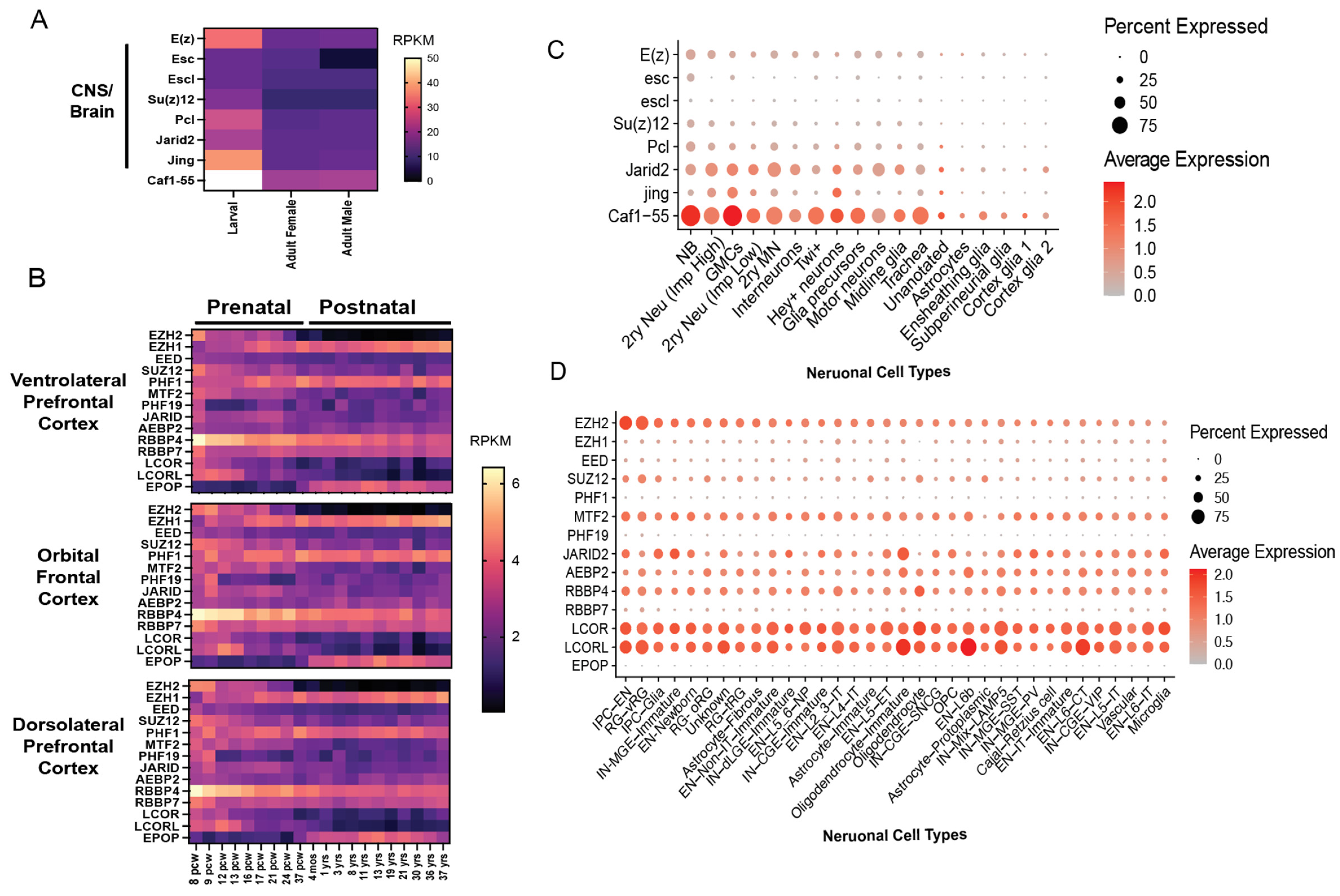
Consistent with a role for PRC2 diversification in supporting cellular complexity during evolution, PRC2 subunit expression differs across neural types and developmental stages, particularly in humans, compared with flies. As illustrated by the analysis of publicly available expression datasets (Figure 2A–C), in Drosophila, the expression of most PRC2 subunits peaks in the larval central nervous system (CNS) (Figure 2A). Similarly, in humans, the expression of EZH2 and most core and accessory subunits are high prenatally and decline after birth in the cerebral cortex. In contrast, EPOP is enriched postnatally, while EZH1 and PHF1 increase at 16–20 postconception weeks, coinciding with the onset of cortical neurogenesis, when neural stem cells transition from proliferative to neurogenic divisions (Figure 2B), and remain constant afterward.
Although bulk PRC2 subunit expression patterns are similar across different regions of the human cerebral cortex (Figure 2B), single-cell RNA sequencing data reveals neural type-specific differences, which are more pronounced in the human second trimester cortex [62] than in Drosophila third-instar larval ventral nerve cord (VNC) [63] (Figure 2C,D). In Drosophila, all PRC2 subunit expression is highest in proliferating neuroblasts (NBs) (Figure 2C). By contrast, in the developing human brain, expression patterns are more variable (Figure 2D). EZH2 and SUZ12 are enriched in neural precursor cells, including radial glia (RG) and intermediate neural progenitor cells (IPCs), compared with postmitotic neurons. EZH1 and EED show relatively uniform expression across proliferating and postmitotic neural populations, with slightly higher levels in excitatory neurons of layer 2 and 3 and select interneurons. At this developmental stage, PRC2.1 subunits MTF2 and LCOR/LCORL (which encode PAL1/2) show a variable expression across different neural types but predominate over EPOP and PHF1/19, which may be more prominent in postnatal populations, as suggested by the bulk RNA seq data in Figure 2B. PRC2.2 subunits AEBP2 and JARID2 also display variable cell type expression, though not always proportional to each other, implying the existence of possible PRC2 alternative subcomplexes or independent roles of these subunits. While the stoichiometry of PRC2 subcomplexes across developmental stages and neural cell types remains to be defined at protein level, these findings highlight unique aspects of PRC2 function in humans. Such differences align with the distinct clinical phenotypes arising from mutations in individual PRC2 subunits discussed below.

4.1. EZH2, EED and SUZ12 Associated Overgrowth with Intellectual Disability (OGID) Syndromes

While the role of somatic PRC2 dysfunction in cancer is well established [10], the connection between germline mutations in PRC2 subunits and NDDs only began to emerge in 2012, with the discovery of de novo EZH2 variants as one of the first causes of an OGID syndrome [64,65]. The syndrome had long been clinically recognized as Weaver syndrome (WVS, OMIM #277590) by its characteristic prenatal overgrowth persisting postnatally as tall stature and advanced bone age, varying degrees of intellectual disability, psychomotor delay, and a distinctive facial appearance including macrocephaly, eyelids that slant down, and large or low-set ears [66]. Most individuals with WVS carry heterozygous missense mutations that show reduced histone methyltransferase activity and H3K27me3 levels when tested in vitro or in cells [67,68,69]. Notably, few of these mutations have also been detected as somatically acquired mutations in cancers (e.g., EZH2 NM_004456: p.D664V, p.R684C and p.Y733*), and consistently, tumors, such as neuroblastoma, have been detected in some WVS patients [65,69].
A smaller subset of WVS patients harbor heterozygous truncating mutations located in the last exon, or partial or full gene deletions consistent with haploinsufficiency as the underlaying genetic mechanism [58]. However, recent work shows that certain WVS-associated EZH2 variants abolish nearly all H3K27me3 when expressed in Ezh2 heterozygous-null mouse ESC, indicating a dominant negative effect. Notably, some of these dominant variants (e.g., p.E745K, p.R684C and p.Y733*) have been identified in a subset of WVS showing polymicrogyria (i.e., excessive cortical folding) on brain imaging, suggesting they may alter specific EZH2 functions impacting cortical neuron migration [69,70,71,72]. Furthermore, a gain-of-function variant (p.A738T) that increases catalytic activity has also been identified in a patient with WVS facial features but growth restriction instead of overgrowth [73]. These findings underscore the importance of functionally assessing genetic variants, not only to elucidate mechanisms of inter-individual differences in clinical manifestations and severity, but also to inform therapeutic strategies.
Although no effective treatment currently exists for WVS, the large repertoire of pharmacological compounds developed to target chromatin regulators involved in cancer provides potential therapeutic avenues for managing progressive symptoms in WVS, including postnatal overgrowth. However, while EZH2 inhibitors may benefit patients with gain-of-function mutations, individuals with typical WVS caused by loss-of-function or dominant-negative EZH2 variants may require alternative approaches, such as inhibitors of H3K27me3 demethylases KDM6A and KDM6B. Indeed, treating a mouse model of WVS expressing the EZH2 p.R684C allele with GSK-J4, a dual KDM6A/B inhibitor, reverses osteogenic defects that lead to overgrowth [74]. Beyond pharmacological strategies, a recent proof-of-concept study has also explored CRISPR-based correction of EZH2 mutations in humanized Ezh2 mouse cells [75], highlighting the future potential of genetic medicine for WVS.
As EZH2 methyltransferase activity depends on the assembly into the PRC2 core complex with EED and SUZ12, it is not surprising to find children with OGID carrying mutations in EED and SUZ12, although these are rarer than WVS-associated EZH2 mutations [76,77]. De novo mutations in EED cause Cohen–Gibson syndrome (COGIS, OMIM #617561), which tends to be more severe than WVS. Of the COGIS patients reported to date, 100% show overgrowth and intellectual disability, compared with 93% and 85%, respectively, in WVS [76]. The majority of COGIS patients carry EED missense mutations, the functional consequences of which are challenging to predict. However, emerging data indicates that these mutations reduce PRC2 activity [9,78]. Indeed, a recent study noticed that COGIS-associated EED (NM_003797) mutations p.R236T and p.R302S/G alter amino acids implicated in the interaction with the histidine at position 129 of EZH2, and demonstrated that disrupting this interaction with pharmacologic compounds or even with the EZH2 p.H129C variant inhibits PRC2 activity [9].
Germline heterozygous SUZ12 variants cause Imagawa–Matsumoto syndrome (IMMAS, OMIM#618786), which presents with less severe overgrowth and intellectual disability than WVS and COGIS [76]. The predominance of truncating mutations that generate premature stop codons suggests that pathogenic variants cause partial or full SUZ12 loss-of-function. Nevertheless, several missense mutations have also been identified, most clustering in the VEFS-box region of SUZ12 [76]. This region is critical for PRC2 assembly and the stabilization of catalytically active EZH2 conformation [79]. The inability of the SUZ12 (NM_015355) p.E610V missense variant to restore H3K27me3 levels in SUZ12 knockdown cells [78] further supports haploinsufficiency or dominant-negative effect as the genetic mechanisms of IMMAS and, more broadly, PRC2-associated OGID syndromes.
Numerous studies in the past decades have focused on elucidating the role of EZH2, EED, and SUZ12 in development, even before their link with OGIDs. In most species, complete deletion of core PRC2 subunits and loss of H3K27me3 results in gastrulation defects that terminate embryonic development [80,81]. Therefore, animal models have been limited to tissue- or cell type-specific depletions, heterozygous-null alleles, or pathogenic variant expression. These strategies revealed, early on, that deletion of Ezh2 from forebrain cortical neural stem cells, generated by crossing mice carrying floxed Ezh2 allele with Emx1-Cre, advances the transition to basal neural progenitors and the timing of differentiation to upper layer neurons [82]. Unlike WVS patients, however, these mice exhibit thinner cerebral cortices and do not show cognitive behavioral defects or macrocephaly. Likewise, according to the Mouse Phenotyping Consortium, heterozygous deletion of Ezh2 does not cause overgrowth or behavioral defects in mice (https://www.mousephenotype.org/data/genes/MGI:107940 (accessed on 10 September 2025), while Suz12 haploinsufficiency causes brain malformations that are more severe than those observed in IMMAS [83]. In contrast, WVS mouse models that have been generated using CRISPR-mediated genome editing (EZH2 p.V626M and p.R684C) show overgrowth, but neurobehavioral defects and structural brain abnormalities have not been reported [68,74], similar to Eed heterozygous-null mice [84]. These data are consistent with genetic mechanisms that differ from haploinsufficiency as the cause of PRC2-associated overgrowth or a specie-specific role of PRC2 in development, particularly of the nervous system.
In mouse and human embryonic stem cells (ESCs), PRC2 targets many development regulator genes, including NEUROG1/2, GATA4, OLIG1 transcription factors or members of the WNT and BMP signaling molecules [85,86,87,88]. These genes are lowly expressed or repressed in ESCs but poised for activation in a cell type-specific manner upon differentiation [85]. Comparison of mouse and human ESCs studies, however, also supports that developmental PRC2 function is evolutionarily distinct and context dependent.
In mouse ESCs, loss of PRC2 through Eed deletion derepresses developmental genes marked by H3K27me3 at their promoters, though pluripotency and self-renewal remain intact [85,89]. Likewise, upon neural induction in culture, PRC2-deficient ESCs fail to repress genes that specify non-neural lineages [85,90]. A different scenario applies to anterior neural development genes marked by PRC2 at poised enhancers instead of at promoters. While these genes remain inactive in Eed-null mouse ESCs, they also fail to activate upon neural induction due to a role of PRC2 in facilitating enhancer and promoter interaction necessary for RNA Polymerase II recruitment [91]. These data indicate that depending on the genomic element, PRC2 function is different, though required for a coordinated transition of gene expression programs during development [91]. Accordingly, a recent work has shown that, although mouse ESCs can self-renew and differentiate into all three germ layers in vitro, upon depletion of H3K27me3 using EZH2 inhibitors and dominant negative EZH2 variants, they fail to maintain lineage-specific expression programs, eventually leading to unstable differentiation states and cell death [92].
In contrast to mouse ESCs, human ESCs rely more heavily on PRC2. Loss of PRC2 not only derepresses developmental target genes but also compromises ESC self-renewal, driving spontaneous differentiation toward meso-endoderm fates [86,93,94]. These species-specific differences may reflect the distinct developmental states of cultured ESCs. While mouse ESCs are maintained in naïve state, human ESCs are generally primed. Naïve ESCs have broader differentiation potential than primed ESCs, and H3K27me3 levels are low and rarely enriched at developmental gene promoters [95,96]. Consequently, recent studies show that naïve human ESCs, similar to mouse ESCs, tolerate PRC2 loss better, although defects emerge in trophoblast genes and differentiation [86,93,94,96].
Collectively, these findings reveal fundamental differences in how PRC2 maintains transcriptional repression programs across species and developmental stages and underscore the importance of human-specific models to study the mechanisms of OGID syndromes.

4.2. EZH1 Gain- and Loss-of-Function Variants in Overlapping Neurodevelopmental Syndromes

The main difference between Drosophila and mammalian PRC2 lies in the evolutionary duplication of the catalytic subunit, E(z): in mammals, PRC2 can be formed by either EZH2 or its paralog, EZH1 (Figure 1). Despite being the first E(z) homolog to be cloned, EZH1 has traditionally been considered the minor catalytic subunit, mainly due to its weaker histone lysine methyltransferase (HMT) activity. Indeed, its catalytic activity was only recognized six years after that of EZH2, following the development of more sensitive HMT assays [97,98,99]. Compared to EZH2, EZH1 is about ten times less catalytically active and primarily mono-methylates H3K27 rather than tri-methylating it [99]. Accordingly, in proliferating cells, EZH1 can sustain H3K27me1 after EZH2 depletion, but only partially maintains H3K27me2 and H3K27me3, whereas EZH2 alone can preserve all three H3K27 methylation states even without EZH1 [9,98,100]. However, although EZH2 has a dominant role in H3K27 methylation, EZH1 binds nucleosomes and compacts chromatin more strongly, owing to a unique patch of basic amino acids in its central disordered loop [101]. How this EZH1-mediated chromatin compaction contributes to developmental PRC2 function remains unclear, but one possibility is that it helps preserve repressed Polycomb domains in postmitotic cells, where EZH2 expression is minimal. Alternatively, EZH1 may facilitate the recently reported interactions between poised enhancers and cognate promoters for RNA polymerase recruitment and activation upon differentiation [91]. Indeed, evidence indicates that EZH1 is required for the transcriptional activation of muscle and blood differentiation genes [7,8].
Although EZH1 has historically remained in a secondary plane due to its apparent low molecular and disease relevance, emerging evidence challenges this view. In 2019, a genetic analysis of recessive intellectual disabilities (IDs) uncovered a homozygous truncating EZH1 variant (NM_001991: p.Leu80Serfs*6) as the candidate cause of ID in two siblings born from healthy consanguineous parents [102]. Subsequent identification of ten additional patients from five independent families, all harboring biallelic truncating variants, confirmed EZH1 deficiency as the cause of a novel NDD [6]. In the same study, we also reported heterozygous missense EZH1 variants (at least some arising de novo) in nine patients presenting a similar NDD characterized by mild to severe language, motor, and cognitive developmental delays, intellectual disability, and atypical facial features, including protruding forehead, deep-set eyes, and flattened mid-face [6]. Surprisingly, in vitro histone methyltransferase assays and H3K27me3 analyses in cells revealed that at least two missense variants (p.A678G and p.Q731E) increase catalytic activity of EZH1, indicating that both EZH1 gain- and loss-of-function can lead to comparable NDD phenotypes. Notably, the lack of overgrowth as a defining characteristic of these patients, and recently reported sisters with recessive EZH1 variants and NDD [103], distinguish EZH1-associated NDDs from other PRC2-related OGID syndromes, suggesting distinct biological roles for EZH1 and EZH2.
In line with a distinct function, Ezh1 deletion in mice and zebrafishes has a minimal impact on embryonic development, in contrast to PRC2-Ezh2 deletion [104,105]. Furthermore, Ezh1-deficient mice show only modest transcriptional changes in adult medium spiny neurons [104], though they exhibit decreased prepulse inhibition (https://www.mousephenotype.org/data/genes/MGI:1097695 (accessed on 10 September 2025)), a behavioral phenotype commonly linked to neuropsychiatric conditions. In human ESC-derived brain organoids, EZH1 loss delays neurogenesis, and EZH1 gain-of-function leads to premature generation of SATB2+ cortical neurons, suggesting asynchronous development as a common neuropathological mechanism.
Some of the differences between EZH2 and EZH1 may stem from their distinct spatiotemporal expression patterns. For instance, EZH1 is ubiquitously expressed and becomes the predominant paralog as EZH2, and to some extent SUZ12 and EED, decline during development, when progenitors differentiate, including into neurons (Figure 2B,D). This developmental switch suggests that EZH1 may assume specialized roles once cells exit the cell cycle. One possibility is that EZH1 assembles into PRC2 complexes with a composition or activity distinct from EZH2. Adding to this complexity, PRC2 has the ability to dimerize [101,106], potentially expanding the possible functional PRC2 configurations depending on the relative abundance of PRC2–EZH1 and PRC2–EZH2 across cell types. Such variability may allow PRC2 to fine-tune chromatin regulation in a context-dependent manner, particularly in tissues with high cellular heterogeneity such as the brain. Therefore, defining how EZH1 contributes to this diversity and what roles it plays in postmitotic cells, such as neurons, represents an important area for future investigation.

4.3. JARID2-Associated Neurodevelopmental Syndrome

Among PRC2 accessory subunits, JARID2 is the only one associated with an NDD. Heterozygous copy number variants (CNVs) and point mutations in JARID2 cause a clinically distinct syndrome characterized by variable developmental and intellectual disability with mild dysmorphic facies (DIDDF, OMIM #620098), such as high anterior hairline, broad forehead, deep set eyes, pressed nasal bridge, and full lips [59,107,108,109]. Other commonly reported clinical manifestations are hypotonia, autistic features, and aggressive behavior. Out of the 26 affected individuals reported to date, 10 have truncating variants, 11 carry large deletions involving at least one exon, 1 patient has a tandem duplication covering exons 1–7, and 4 have missense variants. While most of these mutations arose de novo, at least one case of inheritance from a mildly affected father to his son has been reported [59]. Given the prevalence of deletions and truncating JARID2 variants, haploinsufficiency is the most likely genetic mechanisms of the disease. Consistently, loss-of-function variants are underrepresented in the ~800,000 genomes reported in gnomAD v4.1.0, leading to a pLI = 1 and pLoF o/e = 0.13 that support JARID2 haploinsufficiency as pathogenic. Nonetheless, functional studies, particularly of missense variants carried in patients with apparently milder phenotypes, may reveal alternative genetic mechanisms in the future.
Like PRC2-EZH2 core subunits, JARID2 is highly expressed during early development and declines as cells differentiate [110,111]. Consistently, JARID2 is essential for embryogenesis and tissue homeostasis [112]. Deletion of jarid2 in Xenopus leads to gastrulation arrest, and knockout mouse embryos die at embryonic day 10.5–15.5 due to neural tube, heart, and hematopoietic defects that vary in severity depending on the genetic background of the mouse strain [113,114,115]. Data from mouse ESCs also support that in vitro differentiation is Jarid2-dosage sensitive given that homozygous and heterozygous Jarid2 knockout mouse ESCs show delayed differentiation to the three germ layer derivatives, including neural lineages [116,117,118,119].
In contrast, little is known about the function of JARID2 in human neural development. A recent genomic characterization revealed that the widely used KOLF2.1J human induced pluripotent stem cell (iPSC) line harbors CNVs, including one leading to JARID2 haploinsufficiency [120]. However, global pluripotency and neuronal differentiation appear largely preserved, although derived neurons show reduced excitatory postsynaptic activity [121]. Of note, the lack of overgrowth and macrocephaly in DIDDF patients suggest that JARID2 and/or JARID2-containing PRC2 subcomplexes have different molecular or cell type-specific functions compared to canonical PRC2-EZH2 complexes. Therefore, generating human models carrying patient mutations and proper controls will be critical to elucidating how JARID2 diversifies PRC2 functions and shapes human development.

4.4. PRC2 Subunits Not Yet Implicated in Human Neurodevelopmental Disease

Other accessory subunits, including the PCL homologues PHF1, MTF2, and PHF19, as well as PALI1/2 and EPOP, which define PRC2.1, and the PRC2.2 subunit AEBP2, have not yet been linked to neurodevelopmental disorders. Nevertheless, studies with developmental models suggest that each of these factors may play distinct roles in development. For example, Phf1- and Phf19-deficient mice are viable and lack overt neurobehavioral abnormalities (https://www.mousephenotype.org/data/genes/MGI:98647 (accessed on 15 September 2025) and https://www.mousephenotype.org/data/genes/MGI:1921266 (accessed on 10 September 2025)), whereas Mtf2 deficient mice, which die before weaning age, show posterior skeletal defects consistent with anteroposterior body pattern transformations due to Hox gene dysregulation typical of PcG proteins [122]. In addition, Lcorl deletion in mice causes increased aggression, hyperactivity, and pre-weaning lethality with incomplete penetrance (https://www.mousephenotype.org/data/genes/MGI:2651932 (accessed on 10 September 2025)).
Similarly to Jarid2, Aebp2 deficiency leads to embryonic lethality and skeletal abnormalities in mice [123,124]. However, heterozygous Aebp2-null mice often exhibit features of abnormal neural crest development, including enlarged colons and hypopigmentation reminiscent of Hirschsprung disease and Waardenburg syndrome [124]. The lack of overgrowth in these mice is reminiscent of individuals with JARID2-associated DIDDF, further suggesting that the PRC2.2 subcomplex provides specialized functions to PRC2. In agreement with this, a recent study showed that genetically dissociating PRC2.2 in human PSCs increases genomic PRC2.1 occupancy [38]. Additionally, because PRC2.1 has higher affinity for chromatin binding, H3K27me3 levels at target genes were also increased, leading to functional consequences that are in opposition to PcG protein deletion [38].
Taken together, these findings suggest that individual PRC2 subcomplexes contribute unique regulatory functions, fine-tuning PRC2 activity in both temporal and cell type-specific manners. It remains to be determined whether these regulatory functions are intrinsic to each subcomplex or subunit or instead emerge from the relative abundance of different subcomplexes across cellular and genomic contexts. Addressing these questions will be key to understanding how PRC2 achieves functional diversity as cellular complexity increases.

5. Concluding Remarks

Advances in clinical genetics have not only improved the diagnosis of PRC2-associated NDDs but also uncovered human-specific aspects of PRC2 function. Distinct clinical features across PRC2-related syndromes and developmental models, together with spatiotemporal differences in subunit expression, point to cell type-specific roles likely shaped by variation in complex composition, an area that remains largely unexplored. In particular, the structure and function of PRC2 complexes in postmitotic cells and differentiating tissues, such as neurons in the human brain, are poorly understood. Integrating clinical genetic data with emerging human developmental models and increasingly sensitive proteomic approaches offers a powerful opportunity to elucidate how PRC2 diversity contributes to cellular specialization and functional complexity in humans.

Author Contributions

Formal analysis: J.A., T.R., and N.A.; Writing—original draft: J.A. and N.A.; Writing—review and editing: J.A., T.R., and N.A. All authors have read and agreed to the published version of the manuscript.

Funding

This work was supported by the United States National Institutes of Health T32GM156697 (to J.A.), NIH/NINDS R01NS119699, NIH/NICHD R21HD107592, and the Eagles Autism Foundation Pilot grant (to N.A.).

Institutional Review Board Statement

Not applicable.

Informed Consent Statement

Not applicable.

Data Availability Statement

The human brain gene expression data used in this study were obtained from the BrainSpan Atlas of the Developing Human Brain (https://www.brainspan.org/rnaseq/search/index.html (accessed on February 2025)). The Drosophila expression data were obtained from FlyBase (https://flybase.org/) under the FlyAtlas2 Anatomy RNA-seq dataset (accessed on August 2025). Dot plots showing scRNAseq expression of PcG genes across neuronal cell types in the Drosophila third-instar larval ventral nerve cord (VNC) were generated from the analysis of GEO: GSE235231 [63] and in human second trimester cortex from the analysis of the sn-multiomic dataset, available in the CELLxGENE archive (https://cellxgene.cziscience.com/collections/ad2149fc-19c5-41de-8cfe-44710fbada73 (accessed on August 2025)) [62].

Conflicts of Interest

The authors declare no conflicts of interest.

References

  1. Silbereis, J.C.; Pochareddy, S.; Zhu, Y.; Li, M.; Sestan, N. The Cellular and Molecular Landscapes of the Developing Human Central Nervous System. Neuron 2016, 89, 248–268. [Google Scholar] [CrossRef]
  2. Yoon, K.J.; Vissers, C.; Ming, G.L.; Song, H. Epigenetics and epitranscriptomics in temporal patterning of cortical neural progenitor competence. J. Cell Biol. 2018, 217, 1901–1914. [Google Scholar] [CrossRef]
  3. Hirabayashi, Y.; Gotoh, Y. Epigenetic control of neural precursor cell fate during development. Nat. Rev. Neurosci. 2010, 11, 377–388. [Google Scholar] [CrossRef]
  4. Schuettengruber, B.; Bourbon, H.M.; Di Croce, L.; Cavalli, G. Genome Regulation by Polycomb and Trithorax: 70 Years and Counting. Cell 2017, 171, 34–57. [Google Scholar] [CrossRef] [PubMed]
  5. Kim, J.J.; Kingston, R.E. Context-specific Polycomb mechanisms in development. Nat. Rev. Genet. 2022, 23, 680–695. [Google Scholar] [CrossRef] [PubMed]
  6. Gracia-Diaz, C.; Zhou, Y.; Yang, Q.; Maroofian, R.; Espana-Bonilla, P.; Lee, C.H.; Zhang, S.; Padilla, N.; Fueyo, R.; Waxman, E.A.; et al. Gain and loss of function variants in EZH1 disrupt neurogenesis and cause dominant and recessive neurodevelopmental disorders. Nat. Commun. 2023, 14, 4109. [Google Scholar] [CrossRef] [PubMed]
  7. Xu, J.; Shao, Z.; Li, D.; Xie, H.; Kim, W.; Huang, J.; Taylor, J.E.; Pinello, L.; Glass, K.; Jaffe, J.D.; et al. Developmental control of polycomb subunit composition by GATA factors mediates a switch to non-canonical functions. Mol. Cell 2015, 57, 304–316. [Google Scholar] [CrossRef]
  8. Mousavi, K.; Zare, H.; Wang, A.H.; Sartorelli, V. Polycomb protein Ezh1 promotes RNA polymerase II elongation. Mol. Cell 2012, 45, 255–262. [Google Scholar] [CrossRef]
  9. Lee, C.H.; Holder, M.; Grau, D.; Saldana-Meyer, R.; Yu, J.R.; Ganai, R.A.; Zhang, J.; Wang, M.; LeRoy, G.; Dobenecker, M.W.; et al. Distinct Stimulatory Mechanisms Regulate the Catalytic Activity of Polycomb Repressive Complex 2. Mol. Cell 2018, 70, 435–448.e5. [Google Scholar] [CrossRef]
  10. Lewis, P.; Mislove, R. New mutants report. Drosoph. Inf. Serv. 1947, 21, 69. [Google Scholar]
  11. Lewis, E.B. A gene complex controlling segmentation in Drosophila. Nature 1978, 276, 565–570. [Google Scholar] [CrossRef] [PubMed]
  12. Jürgens, G. A group of genes controlling the spatial expression of the bithorax complex in Drosophila. Nature 1985, 316, 153–155. [Google Scholar] [CrossRef]
  13. Struhl, G.; Akam, M. Altered distributions of Ultrabithorax transcripts in extra sex combs mutant embryos of Drosophila. EMBO J. 1985, 4, 3259–3264. [Google Scholar] [CrossRef]
  14. Struhl, G. A gene product required for correct initiation of segmental determination in Drosophila. Nature 1981, 293, 36–41. [Google Scholar] [CrossRef] [PubMed]
  15. Riggs, A.D. X inactivation, differentiation, and DNA methylation. Cytogenet. Cell Genet. 1975, 14, 9–25. [Google Scholar] [CrossRef]
  16. Holliday, R.; Pugh, J.E. DNA modification mechanisms and gene activity during development. Science 1975, 187, 226–232. [Google Scholar] [CrossRef]
  17. Nanney, D.L. Epigenetic Control Systems. Proc. Natl. Acad. Sci. USA 1958, 44, 712–717. [Google Scholar] [CrossRef]
  18. Holliday, R. Epigenetics: An overview. Dev. Genet. 1994, 15, 453–457. [Google Scholar] [CrossRef]
  19. Strutt, H.; Cavalli, G.; Paro, R. Co-localization of Polycomb protein and GAGA factor on regulatory elements responsible for the maintenance of homeotic gene expression. EMBO J. 1997, 16, 3621–3632. [Google Scholar] [CrossRef]
  20. Orlando, V.; Paro, R. Mapping polycomb-repressed domains in the bithorax complex using in vivo formaldehyde cross-linked chromatin. Cell 1993, 75, 1187–1198. [Google Scholar] [CrossRef] [PubMed]
  21. Breiling, A.; Edgar, B.; Simona, F.; Becker, B.P.; Paro, R. The Drosophila Polycomb Protein Interacts with Nucleosomal Core Particles In Vitro via Its Repression Domain. Mol. Cell. Biol. 1999, 19, 8451–8460. [Google Scholar] [CrossRef]
  22. Shao, Z.; Raible, F.; Mollaaghababa, R.; Guyon, J.R.; Wu, C.-t.; Bender, W.; Kingston, R.E. Stabilization of Chromatin Structure by PRC1, a Polycomb Complex. Cell 1999, 98, 37–46. [Google Scholar] [CrossRef] [PubMed]
  23. Jones, C.A.; Ng, J.; Peterson, A.J.; Morgan, K.; Simon, J.; Jones, R.S. The Drosophila esc and E(z) Proteins Are Direct Partners in Polycomb Group-Mediated Repression. Mol. Cell. Biol. 1998, 18, 2825–2834. [Google Scholar] [CrossRef] [PubMed]
  24. Tie, F.; Furuyama, T.; Harte, P.J. The Drosophila Polycomb Group proteins ESC and E (Z) bind directly to each other and co-localize at multiple chromosomal sites. Development 1998, 125, 3483–3496. [Google Scholar] [CrossRef] [PubMed]
  25. Platero, J.S.; Sharp, E.J.; Adler, P.N.; Eissenberg, J.C. In vivo assay for protein-protein interactions using Drosophila chromosomes. Chromosoma 1996, 104, 393–404. [Google Scholar] [CrossRef]
  26. Rastelli, L.; Chan, C.S.; Pirrotta, V. Related chromosome binding sites for zeste, suppressors of zeste and Polycomb group proteins in Drosophila and their dependence on Enhancer of zeste function. EMBO J. 1993, 12, 1513–1522. [Google Scholar] [CrossRef]
  27. Cao, R.; Wang, L.; Wang, H.; Xia, L.; Erdjument-Bromage, H.; Tempst, P.; Jones, R.S.; Zhang, Y. Role of histone H3 lysine 27 methylation in Polycomb-group silencing. Science 2002, 298, 1039–1043. [Google Scholar] [CrossRef]
  28. Czermin, B.; Melfi, R.; McCabe, D.; Seitz, V.; Imhof, A.; Pirrotta, V. Drosophila enhancer of Zeste/ESC complexes have a histone H3 methyltransferase activity that marks chromosomal Polycomb sites. Cell 2002, 111, 185–196. [Google Scholar] [CrossRef]
  29. Kuzmichev, A.; Nishioka, K.; Erdjument-Bromage, H.; Tempst, P.; Reinberg, D. Histone methyltransferase activity associated with a human multiprotein complex containing the Enhancer of Zeste protein. Genes. Dev. 2002, 16, 2893–2905. [Google Scholar] [CrossRef]
  30. Uckelmann, M.; Davidovich, C. Chromatin compaction by Polycomb group proteins revisited. Curr. Opin. Struct. Biol. 2024, 86, 102806. [Google Scholar] [CrossRef]
  31. de Napoles, M.; Mermoud, J.E.; Wakao, R.; Tang, Y.A.; Endoh, M.; Appanah, R.; Nesterova, T.B.; Silva, J.; Otte, A.P.; Vidal, M.; et al. Polycomb group proteins Ring1A/B link ubiquitylation of histone H2A to heritable gene silencing and X inactivation. Dev. Cell 2004, 7, 663–676. [Google Scholar] [CrossRef]
  32. Wang, H.; Wang, L.; Erdjument-Bromage, H.; Vidal, M.; Tempst, P.; Jones, R.S.; Zhang, Y. Role of histone H2A ubiquitination in Polycomb silencing. Nature 2004, 431, 873–878. [Google Scholar] [CrossRef]
  33. Kasinath, V.; Beck, C.; Sauer, P.; Poepsel, S.; Kosmatka, J.; Faini, M.; Toso, D.; Aebersold, R.; Nogales, E. JARID2 and AEBP2 regulate PRC2 in the presence of H2AK119ub1 and other histone modifications. Science 2021, 371, 362. [Google Scholar] [CrossRef]
  34. Cooper, S.; Grijzenhout, A.; Underwood, E.; Ancelin, K.; Zhang, T.; Nesterova, T.B.; Anil-Kirmizitas, B.; Bassett, A.; Kooistra, S.M.; Agger, K.; et al. Jarid2 binds mono-ubiquitylated H2A lysine 119 to mediate crosstalk between Polycomb complexes PRC1 and PRC2. Nat. Commun. 2016, 7, 13661. [Google Scholar] [CrossRef] [PubMed]
  35. Kalb, R.; Latwiel, S.; Baymaz, H.I.; Jansen, P.W.; Muller, C.W.; Vermeulen, M.; Muller, J. Histone H2A monoubiquitination promotes histone H3 methylation in Polycomb repression. Nat. Struct. Mol. Biol. 2014, 21, 569–571. [Google Scholar] [CrossRef]
  36. Tamburri, S.; Lavarone, E.; Fernandez-Perez, D.; Conway, E.; Zanotti, M.; Manganaro, D.; Pasini, D. Histone H2AK119 Mono-Ubiquitination Is Essential for Polycomb-Mediated Transcriptional Repression. Mol. Cell 2020, 77, 840–856. [Google Scholar] [CrossRef]
  37. Blackledge, N.P.; Fursova, N.A.; Kelley, J.R.; Huseyin, M.K.; Feldmann, A.; Klose, R.J. PRC1 Catalytic Activity Is Central to Polycomb System Function. Mol. Cell 2020, 77, 857–874.e9. [Google Scholar] [CrossRef] [PubMed]
  38. Youmans, D.T.; Gooding, A.R.; Dowell, R.D.; Cech, T.R. Competition between PRC2.1 and 2.2 subcomplexes regulates PRC2 chromatin occupancy in human stem cells. Mol. Cell 2021, 81, 488–501.e9. [Google Scholar] [CrossRef]
  39. Glancy, E.; Wang, C.; Tuck, E.; Healy, E.; Amato, S.; Neikes, H.K.; Mariani, A.; Mucha, M.; Vermeulen, M.; Pasini, D.; et al. PRC2.1- and PRC2.2-specific accessory proteins drive recruitment of different forms of canonical PRC1. Mol. Cell 2023, 83, 1393–1411.e7. [Google Scholar] [CrossRef] [PubMed]
  40. Hauri, S.; Comoglio, F.; Seimiya, M.; Gerstung, M.; Glatter, T.; Hansen, K.; Aebersold, R.; Paro, R.; Gstaiger, M.; Beisel, C. A High-Density Map for Navigating the Human Polycomb Complexome. Cell Rep. 2016, 17, 583–595. [Google Scholar] [CrossRef]
  41. Alekseyenko, A.A.; Gorchakov, A.A.; Kharchenko, P.V.; Kuroda, M.I. Reciprocal interactions of human C10orf12 and C17orf96 with PRC2 revealed by BioTAP-XL cross-linking and affinity purification. Proc. Natl. Acad. Sci. USA 2014, 111, 2488–2493. [Google Scholar] [CrossRef] [PubMed]
  42. Zhang, Z.; Jones, A.; Sun, C.W.; Li, C.; Chang, C.W.; Joo, H.Y.; Dai, Q.; Mysliwiec, M.R.; Wu, L.C.; Guo, Y.; et al. PRC2 complexes with JARID2, MTF2, and esPRC2p48 in ES cells to modulate ES cell pluripotency and somatic cell reprogramming. Stem Cells 2011, 29, 229–240. [Google Scholar] [CrossRef]
  43. Ryan, C.W.; Peirent, E.R.; Regan, S.L.; Guxholli, A.; Bielas, S.L. H2A monoubiquitination: Insights from human genetics and animal models. Hum. Genet. 2024, 143, 511–527. [Google Scholar] [CrossRef]
  44. Guo, Y.; Zhao, S.; Wang, G.G. Polycomb Gene Silencing Mechanisms: PRC2 Chromatin Targeting, H3K27me3 ‘Readout’, and Phase Separation-Based Compaction. Trends Genet. 2021, 37, 547–565. [Google Scholar] [CrossRef]
  45. Oksuz, O.; Narendra, V.; Lee, C.H.; Descostes, N.; LeRoy, G.; Raviram, R.; Blumenberg, L.; Karch, K.; Rocha, P.P.; Garcia, B.A.; et al. Capturing the Onset of PRC2-Mediated Repressive Domain Formation. Mol. Cell 2018, 70, 1149–1162.e5. [Google Scholar] [CrossRef] [PubMed]
  46. Mendenhall, E.M.; Koche, R.P.; Truong, T.; Zhou, V.W.; Issac, B.; Chi, A.S.; Ku, M.; Bernstein, B.E. GC-rich sequence elements recruit PRC2 in mammalian ES cells. PLoS Genet. 2010, 6, e1001244. [Google Scholar] [CrossRef] [PubMed]
  47. Zylicz, J.J.; Bousard, A.; Zumer, K.; Dossin, F.; Mohammad, E.; da Rocha, S.T.; Schwalb, B.; Syx, L.; Dingli, F.; Loew, D.; et al. The Implication of Early Chromatin Changes in X Chromosome Inactivation. Cell 2019, 176, 182–197.e3. [Google Scholar] [CrossRef]
  48. Nekrasov, M.; Klymenko, T.; Fraterman, S.; Papp, B.; Oktaba, K.; Kocher, T.; Cohen, A.; Stunnenberg, H.G.; Wilm, M.; Muller, J. Pcl-PRC2 is needed to generate high levels of H3-K27 trimethylation at Polycomb target genes. EMBO J. 2007, 26, 4078–4088. [Google Scholar] [CrossRef]
  49. Herz, H.M.; Mohan, M.; Garrett, A.S.; Miller, C.; Casto, D.; Zhang, Y.; Seidel, C.; Haug, J.S.; Florens, L.; Washburn, M.P.; et al. Polycomb repressive complex 2-dependent and -independent functions of Jarid2 in transcriptional regulation in Drosophila. Mol. Cell Biol. 2012, 32, 1683–1693. [Google Scholar] [CrossRef]
  50. Wen, C.; Margolis, M.; Dai, R.; Zhang, P.; Przytycki, P.F.; Vo, D.D.; Bhattacharya, A.; Matoba, N.; Tang, M.; Jiao, C.; et al. Cross-ancestry atlas of gene, isoform, and splicing regulation in the developing human brain. Science 2024, 384, eadh0829. [Google Scholar] [CrossRef] [PubMed]
  51. Satterstrom, F.K.; Kosmicki, J.A.; Wang, J.; Breen, M.S.; De Rubeis, S.; An, J.Y.; Peng, M.; Collins, R.; Grove, J.; Klei, L.; et al. Large-Scale Exome Sequencing Study Implicates Both Developmental and Functional Changes in the Neurobiology of Autism. Cell 2020, 180, 568–584.e23. [Google Scholar] [CrossRef]
  52. Paulsen, B.; Velasco, S.; Kedaigle, A.J.; Pigoni, M.; Quadrato, G.; Deo, A.J.; Adiconis, X.; Uzquiano, A.; Sartore, R.; Yang, S.M.; et al. Autism genes converge on asynchronous development of shared neuron classes. Nature 2022, 602, 268–273. [Google Scholar] [CrossRef]
  53. Villa, C.E.; Cheroni, C.; Dotter, C.P.; Lopez-Tobon, A.; Oliveira, B.; Sacco, R.; Yahya, A.C.; Morandell, J.; Gabriele, M.; Tavakoli, M.R.; et al. CHD8 haploinsufficiency links autism to transient alterations in excitatory and inhibitory trajectories. Cell Rep. 2022, 39, 110615. [Google Scholar] [CrossRef]
  54. Ciceri, G.; Baggiolini, A.; Cho, H.S.; Kshirsagar, M.; Benito-Kwiecinski, S.; Walsh, R.M.; Aromolaran, K.A.; Gonzalez-Hernandez, A.J.; Munguba, H.; Koo, S.Y.; et al. An epigenetic barrier sets the timing of human neuronal maturation. Nature 2024, 626, 881–890. [Google Scholar] [CrossRef]
  55. De Rubeis, S.; He, X.; Goldberg, A.P.; Poultney, C.S.; Samocha, K.; Cicek, A.E.; Kou, Y.; Liu, L.; Fromer, M.; Walker, S.; et al. Synaptic, transcriptional and chromatin genes disrupted in autism. Nature 2014, 515, 209–215. [Google Scholar] [CrossRef] [PubMed]
  56. McCarthy, S.E.; Gillis, J.; Kramer, M.; Lihm, J.; Yoon, S.; Berstein, Y.; Mistry, M.; Pavlidis, P.; Solomon, R.; Ghiban, E.; et al. De novo mutations in schizophrenia implicate chromatin remodeling and support a genetic overlap with autism and intellectual disability. Mol. Psychiatry 2014, 19, 652–658. [Google Scholar] [CrossRef] [PubMed]
  57. Kochinke, K.; Zweier, C.; Nijhof, B.; Fenckova, M.; Cizek, P.; Honti, F.; Keerthikumar, S.; Oortveld, M.A.; Kleefstra, T.; Kramer, J.M.; et al. Systematic Phenomics Analysis Deconvolutes Genes Mutated in Intellectual Disability into Biologically Coherent Modules. Am. J. Hum. Genet. 2016, 98, 149–164. [Google Scholar] [CrossRef]
  58. Cyrus, S.; Burkardt, D.; Weaver, D.D.; Gibson, W.T. PRC2-complex related dysfunction in overgrowth syndromes: A review of EZH2, EED, and SUZ12 and their syndromic phenotypes. Am. J. Med. Genet. Part C Semin. Med. Genet. 2019, 181, 519–531. [Google Scholar] [CrossRef]
  59. Verberne, E.A.; Goh, S.; England, J.; van Ginkel, M.; Rafael-Croes, L.; Maas, S.; Polstra, A.; Zarate, Y.A.; Bosanko, K.A.; Pechter, K.B.; et al. JARID2 haploinsufficiency is associated with a clinically distinct neurodevelopmental syndrome. Genet. Med. 2021, 23, 374–383. [Google Scholar] [CrossRef] [PubMed]
  60. Schlegel, P.; Yin, Y.; Bates, A.S.; Dorkenwald, S.; Eichler, K.; Brooks, P.; Han, D.S.; Gkantia, M.; Dos Santos, M.; Munnelly, E.J.; et al. Whole-brain annotation and multi-connectome cell typing of Drosophila. Nature 2024, 634, 139–152. [Google Scholar] [CrossRef]
  61. Azevedo, F.A.; Carvalho, L.R.; Grinberg, L.T.; Farfel, J.M.; Ferretti, R.E.; Leite, R.E.; Jacob Filho, W.; Lent, R.; Herculano-Houzel, S. Equal numbers of neuronal and nonneuronal cells make the human brain an isometrically scaled-up primate brain. J. Comp. Neurol. 2009, 513, 532–541. [Google Scholar] [CrossRef] [PubMed]
  62. Wang, L.; Wang, C.; Moriano, J.A.; Chen, S.; Zuo, G.; Cebrian-Silla, A.; Zhang, S.; Mukhtar, T.; Wang, S.; Song, M.; et al. Molecular and cellular dynamics of the developing human neocortex. Nature 2025, 1–10. [Google Scholar] [CrossRef]
  63. Nguyen, T.H.; Vicidomini, R.; Choudhury, S.D.; Han, T.H.; Maric, D.; Brody, T.; Serpe, M. scRNA-seq data from the larval Drosophila ventral cord provides a resource for studying motor systems function and development. Dev. Cell 2024, 59, 1210–1230.e9. [Google Scholar] [CrossRef]
  64. Gibson, W.T.; Hood, R.L.; Zhan, S.H.; Bulman, D.E.; Fejes, A.P.; Moore, R.; Mungall, A.J.; Eydoux, P.; Babul-Hirji, R.; An, J.; et al. Mutations in EZH2 cause Weaver syndrome. Am. J. Hum. Genet. 2012, 90, 110–118. [Google Scholar] [CrossRef]
  65. Tatton-Brown, K.; Hanks, S.; Ruark, E.; Zachariou, A.; Duarte Sdel, V.; Ramsay, E.; Snape, K.; Murray, A.; Perdeaux, E.R.; Seal, S.; et al. Germline mutations in the oncogene EZH2 cause Weaver syndrome and increased human height. Oncotarget 2011, 2, 1127–1133. [Google Scholar] [CrossRef]
  66. Weaver, D.D.; Graham, C.B.; Thomas, I.T.; Smith, D.W. A new overgrowth syndrome with accelerated skeletal maturation, unusual facies, and camptodactyly. J. Pediatr. 1974, 84, 547–552. [Google Scholar] [CrossRef]
  67. Deevy, O.; Li, J.; Monger, C.; Matra, F.; Tuck, E.; Davies, M.; Badonyi, M.; Boyce, M.; Doyle, E.J.; Hokamp, K.; et al. Dominant-negative effects of Weaver syndrome-associated EZH2 variants. Genes. Dev. 2025. [Google Scholar] [CrossRef]
  68. Lui, J.C.; Barnes, K.M.; Dong, L.; Yue, S.; Graber, E.; Rapaport, R.; Dauber, A.; Nilsson, O.; Baron, J. Ezh2 Mutations Found in the Weaver Overgrowth Syndrome Cause a Partial Loss of H3K27 Histone Methyltransferase Activity. J. Clin. Endocrinol. Metab. 2018, 103, 1470–1478. [Google Scholar] [CrossRef] [PubMed]
  69. Cohen, A.S.; Yap, D.B.; Lewis, M.E.; Chijiwa, C.; Ramos-Arroyo, M.A.; Tkachenko, N.; Milano, V.; Fradin, M.; McKinnon, M.L.; Townsend, K.N.; et al. Weaver Syndrome-Associated EZH2 Protein Variants Show Impaired Histone Methyltransferase Function In Vitro. Hum. Mutat. 2016, 37, 301–307. [Google Scholar] [CrossRef]
  70. Tatton-Brown, K.; Murray, A.; Hanks, S.; Douglas, J.; Armstrong, R.; Banka, S.; Bird, L.M.; Clericuzio, C.L.; Cormier-Daire, V.; Cushing, T.; et al. Weaver syndrome and EZH2 mutations: Clarifying the clinical phenotype. Am. J. Med. Genet. A 2013, 161, 2972–2980. [Google Scholar] [CrossRef] [PubMed]
  71. Al-Salem, A.; Alshammari, M.J.; Hassan, H.; Alazami, A.M.; Alkuraya, F.S. Weaver syndrome and defective cortical development: A rare association. Am. J. Med. Genet. A 2013, 161, 225–227. [Google Scholar] [CrossRef]
  72. Ren, L.; Jiang, L.; Jiang, X.; Wang, H.; Li, Y.; Ren, X. Clinical and genetic characterization of Weaver syndrome: A case report of an EZH2 mutation and review of the literature. Medicine 2025, 104, e44080. [Google Scholar] [CrossRef]
  73. Choufani, S.; Gibson, W.T.; Turinsky, A.L.; Chung, B.H.Y.; Wang, T.; Garg, K.; Vitriolo, A.; Cohen, A.S.A.; Cyrus, S.; Goodman, S.; et al. DNA Methylation Signature for EZH2 Functionally Classifies Sequence Variants in Three PRC2 Complex Genes. Am. J. Hum. Genet. 2020, 106, 596–610. [Google Scholar] [CrossRef]
  74. Gao, C.W.; Lin, W.; Riddle, R.C.; Kushwaha, P.; Boukas, L.; Bjornsson, H.T.; Hansen, K.D.; Fahrner, J.A. A mouse model of Weaver syndrome displays overgrowth and excess osteogenesis reversible with KDM6A/6B inhibition. JCI Insight 2024, 9, e173392. [Google Scholar] [CrossRef]
  75. Gibson, W.T.; Lengyell, T.C.; Korecki, A.J.; Janssen, S.M.; Adair, B.A.; Gamu, D.; Lorincz, M.C.; Simpson, E.M. Minimally Humanized Ezh2 Exon-18 Mouse Cell Lines Validate Preclinical CRISPR/Cas9 Approach to Treat Weaver Syndrome. Hum. Gene Ther. 2025, 36, 618–627. [Google Scholar] [CrossRef]
  76. Imagawa, E.; Seyama, R.; Aoi, H.; Uchiyama, Y.; Marcarini, B.G.; Furquim, I.; Honjo, R.S.; Bertola, D.R.; Kim, C.A.; Matsumoto, N. Imagawa-Matsumoto syndrome: SUZ12-related overgrowth disorder. Clin. Genet. 2023, 103, 383–391. [Google Scholar] [CrossRef] [PubMed]
  77. Griffiths, S.; Loveday, C.; Zachariou, A.; Behan, L.A.; Chandler, K.; Cole, T.; D’Arrigo, S.; Dieckmann, A.; Foster, A.; Gibney, J.; et al. EED and EZH2 constitutive variants: A study to expand the Cohen-Gibson syndrome phenotype and contrast it with Weaver syndrome. Am. J. Med. Genet. A 2019, 179, 588–594. [Google Scholar] [CrossRef] [PubMed]
  78. Imagawa, E.; Higashimoto, K.; Sakai, Y.; Numakura, C.; Okamoto, N.; Matsunaga, S.; Ryo, A.; Sato, Y.; Sanefuji, M.; Ihara, K.; et al. Mutations in genes encoding polycomb repressive complex 2 subunits cause Weaver syndrome. Hum. Mutat. 2017, 38, 637–648. [Google Scholar] [CrossRef]
  79. Jiao, L.; Liu, X. Structural basis of histone H3K27 trimethylation by an active polycomb repressive complex 2. Science 2015, 350, aac4383. [Google Scholar] [CrossRef] [PubMed]
  80. Pasini, D.; Bracken, A.P.; Jensen, M.R.; Lazzerini Denchi, E.; Helin, K. Suz12 is essential for mouse development and for EZH2 histone methyltransferase activity. EMBO J. 2004, 23, 4061–4071. [Google Scholar] [CrossRef]
  81. O’Carroll, D.; Erhardt, S.; Pagani, M.; Barton, S.C.; Surani, M.A.; Jenuwein, T. The polycomb-group gene Ezh2 is required for early mouse development. Mol. Cell Biol. 2001, 21, 4330–4336. [Google Scholar] [CrossRef] [PubMed]
  82. Pereira, J.D.; Sansom, S.N.; Smith, J.; Dobenecker, M.W.; Tarakhovsky, A.; Livesey, F.J. Ezh2, the histone methyltransferase of PRC2, regulates the balance between self-renewal and differentiation in the cerebral cortex. Proc. Natl. Acad. Sci. USA 2010, 107, 15957–15962. [Google Scholar] [CrossRef]
  83. Miro, X.; Zhou, X.; Boretius, S.; Michaelis, T.; Kubisch, C.; Alvarez-Bolado, G.; Gruss, P. Haploinsufficiency of the murine polycomb gene Suz12 results in diverse malformations of the brain and neural tube. Dis. Model. Mech. 2009, 2, 412–418. [Google Scholar] [CrossRef]
  84. Prokopuk, L.; Stringer, J.M.; White, C.R.; Vossen, R.; White, S.J.; Cohen, A.S.A.; Gibson, W.T.; Western, P.S. Loss of maternal EED results in postnatal overgrowth. Clin. Epigenet. 2018, 10, 95. [Google Scholar] [CrossRef]
  85. Boyer, L.A.; Plath, K.; Zeitlinger, J.; Brambrink, T.; Medeiros, L.A.; Lee, T.I.; Levine, S.S.; Wernig, M.; Tajonar, A.; Ray, M.K.; et al. Polycomb complexes repress developmental regulators in murine embryonic stem cells. Nature 2006, 441, 349–353. [Google Scholar] [CrossRef]
  86. Lee, T.I.; Jenner, R.G.; Boyer, L.A.; Guenther, M.G.; Levine, S.S.; Kumar, R.M.; Chevalier, B.; Johnstone, S.E.; Cole, M.F.; Isono, K.; et al. Control of developmental regulators by Polycomb in human embryonic stem cells. Cell 2006, 125, 301–313. [Google Scholar] [CrossRef]
  87. Schwartz, Y.B.; Kahn, T.G.; Nix, D.A.; Li, X.Y.; Bourgon, R.; Biggin, M.; Pirrotta, V. Genome-wide analysis of Polycomb targets in Drosophila melanogaster. Nat. Genet. 2006, 38, 700–705. [Google Scholar] [CrossRef]
  88. Tolhuis, B.; de Wit, E.; Muijrers, I.; Teunissen, H.; Talhout, W.; van Steensel, B.; van Lohuizen, M. Genome-wide profiling of PRC1 and PRC2 Polycomb chromatin binding in Drosophila melanogaster. Nat. Genet. 2006, 38, 694–699. [Google Scholar] [CrossRef]
  89. Chamberlain, S.J.; Yee, D.; Magnuson, T. Polycomb repressive complex 2 is dispensable for maintenance of embryonic stem cell pluripotency. Stem Cells 2008, 26, 1496–1505. [Google Scholar] [CrossRef] [PubMed]
  90. Thornton, S.R.; Butty, V.L.; Levine, S.S.; Boyer, L.A. Polycomb Repressive Complex 2 regulates lineage fidelity during embryonic stem cell differentiation. PLoS ONE 2014, 9, e110498. [Google Scholar] [CrossRef] [PubMed]
  91. Cruz-Molina, S.; Respuela, P.; Tebartz, C.; Kolovos, P.; Nikolic, M.; Fueyo, R.; van Ijcken, W.F.J.; Grosveld, F.; Frommolt, P.; Bazzi, H.; et al. PRC2 Facilitates the Regulatory Topology Required for Poised Enhancer Function during Pluripotent Stem Cell Differentiation. Cell Stem Cell 2017, 20, 689–705.e9. [Google Scholar] [CrossRef]
  92. Miller, S.A.; Damle, M.; Kim, J.; Kingston, R.E. Full methylation of H3K27 by PRC2 is dispensable for initial embryoid body formation but required to maintain differentiated cell identity. Development 2021, 148, dev196329. [Google Scholar] [CrossRef]
  93. Collinson, A.; Collier, A.J.; Morgan, N.P.; Sienerth, A.R.; Chandra, T.; Andrews, S.; Rugg-Gunn, P.J. Deletion of the Polycomb-Group Protein EZH2 Leads to Compromised Self-Renewal and Differentiation Defects in Human Embryonic Stem Cells. Cell Rep. 2016, 17, 2700–2714. [Google Scholar] [CrossRef]
  94. Shan, Y.; Liang, Z.; Xing, Q.; Zhang, T.; Wang, B.; Tian, S.; Huang, W.; Zhang, Y.; Yao, J.; Zhu, Y.; et al. PRC2 specifies ectoderm lineages and maintains pluripotency in primed but not naive ESCs. Nat. Commun. 2017, 8, 672. [Google Scholar] [CrossRef]
  95. van Mierlo, G.; Dirks, R.A.M.; De Clerck, L.; Brinkman, A.B.; Huth, M.; Kloet, S.L.; Saksouk, N.; Kroeze, L.I.; Willems, S.; Farlik, M.; et al. Integrative Proteomic Profiling Reveals PRC2-Dependent Epigenetic Crosstalk Maintains Ground-State Pluripotency. Cell Stem Cell 2019, 24, 123–137.e8. [Google Scholar] [CrossRef] [PubMed]
  96. Zijlmans, D.W.; Talon, I.; Verhelst, S.; Bendall, A.; Van Nerum, K.; Javali, A.; Malcolm, A.A.; van Knippenberg, S.; Biggins, L.; To, S.K.; et al. Integrated multi-omics reveal polycomb repressive complex 2 restricts human trophoblast induction. Nat. Cell Biol. 2022, 24, 858–871. [Google Scholar] [CrossRef] [PubMed]
  97. Abel, K.J.; Brody, L.C.; Valdes, J.M.; Erdos, M.R.; McKinley, D.R.; Castilla, L.H.; Merajver, S.D.; Couch, F.J.; Friedman, L.S.; Ostermeyer, E.A.; et al. Characterization of EZH1, a human homolog of Drosophila Enhancer of zeste near BRCA1. Genomics 1996, 37, 161–171. [Google Scholar] [CrossRef]
  98. Shen, X.; Liu, Y.; Hsu, Y.J.; Fujiwara, Y.; Kim, J.; Mao, X.; Yuan, G.C.; Orkin, S.H. EZH1 mediates methylation on histone H3 lysine 27 and complements EZH2 in maintaining stem cell identity and executing pluripotency. Mol. Cell 2008, 32, 491–502. [Google Scholar] [CrossRef]
  99. Margueron, R.; Li, G.; Sarma, K.; Blais, A.; Zavadil, J.; Woodcock, C.L.; Dynlacht, B.D.; Reinberg, D. Ezh1 and Ezh2 maintain repressive chromatin through different mechanisms. Mol. Cell 2008, 32, 503–518. [Google Scholar] [CrossRef] [PubMed]
  100. Margueron, R.; Justin, N.; Ohno, K.; Sharpe, M.L.; Son, J.; Drury, W.J., 3rd; Voigt, P.; Martin, S.R.; Taylor, W.R.; De Marco, V.; et al. Role of the polycomb protein EED in the propagation of repressive histone marks. Nature 2009, 461, 762–767. [Google Scholar] [CrossRef]
  101. Grau, D.; Zhang, Y.; Lee, C.H.; Valencia-Sanchez, M.; Zhang, J.; Wang, M.; Holder, M.; Svetlov, V.; Tan, D.; Nudler, E.; et al. Structures of monomeric and dimeric PRC2:EZH1 reveal flexible modules involved in chromatin compaction. Nat. Commun. 2021, 12, 714. [Google Scholar] [CrossRef]
  102. Hu, H.; Kahrizi, K.; Musante, L.; Fattahi, Z.; Herwig, R.; Hosseini, M.; Oppitz, C.; Abedini, S.S.; Suckow, V.; Larti, F.; et al. Genetics of intellectual disability in consanguineous families. Mol. Psychiatry 2019, 24, 1027–1039. [Google Scholar] [CrossRef]
  103. Okamoto, N.; Yoshida, S.; Ogitani, A.; Etani, Y.; Yanagi, K.; Kaname, T. Biallelic loss-of-function variants of EZH1 cause a novel developmental disorder with central precocious puberty. Am. J. Med. Genet. A 2024, 194, e63726. [Google Scholar] [CrossRef]
  104. von Schimmelmann, M.; Feinberg, P.A.; Sullivan, J.M.; Ku, S.M.; Badimon, A.; Duff, M.K.; Wang, Z.; Lachmann, A.; Dewell, S.; Ma’ayan, A.; et al. Polycomb repressive complex 2 (PRC2) silences genes responsible for neurodegeneration. Nat. Neurosci. 2016, 19, 1321–1330. [Google Scholar] [CrossRef]
  105. Volkel, P.; Bary, A.; Raby, L.; Chapart, A.; Dupret, B.; Le Bourhis, X.; Angrand, P.O. Ezh1 arises from Ezh2 gene duplication but its function is not required for zebrafish development. Sci. Rep. 2019, 9, 4319. [Google Scholar] [CrossRef] [PubMed]
  106. Chen, S.; Jiao, L.; Liu, X.; Yang, X.; Liu, X. A Dimeric Structural Scaffold for PRC2-PCL Targeting to CpG Island Chromatin. Mol. Cell 2020, 77, 1265–1278.e7. [Google Scholar] [CrossRef] [PubMed]
  107. Viitasalo, L.; Kettunen, K.; Kankainen, M.; Niemela, E.H.; Kiiski, K. A novel partial de novo duplication of JARID2 gene causing a neurodevelopmental phenotype. Mol. Genet. Genom. Med. 2022, 10, e2037. [Google Scholar] [CrossRef]
  108. Cadieux-Dion, M.; Farrow, E.; Thiffault, I.; Cohen, A.S.A.; Welsh, H.; Bartik, L.; Schwager, C.; Engleman, K.; Zhou, D.; Zhang, L.; et al. Phenotypic expansion and variable expressivity in individuals with JARID2-related intellectual disability: A case series. Clin. Genet. 2022, 102, 136–141. [Google Scholar] [CrossRef]
  109. Baroy, T.; Misceo, D.; Stromme, P.; Stray-Pedersen, A.; Holmgren, A.; Rodningen, O.K.; Blomhoff, A.; Helle, J.R.; Stormyr, A.; Tvedt, B.; et al. Haploinsufficiency of two histone modifier genes on 6p22.3, ATXN1 and JARID2, is associated with intellectual disability. Orphanet J. Rare Dis. 2013, 8, 3. [Google Scholar] [CrossRef]
  110. Miller, J.A.; Ding, S.L.; Sunkin, S.M.; Smith, K.A.; Ng, L.; Szafer, A.; Ebbert, A.; Riley, Z.L.; Royall, J.J.; Aiona, K.; et al. Transcriptional landscape of the prenatal human brain. Nature 2014, 508, 199–206. [Google Scholar] [CrossRef] [PubMed]
  111. Tian, R.; Gachechiladze, M.A.; Ludwig, C.H.; Laurie, M.T.; Hong, J.Y.; Nathaniel, D.; Prabhu, A.V.; Fernandopulle, M.S.; Patel, R.; Abshari, M.; et al. CRISPR Interference-Based Platform for Multimodal Genetic Screens in Human iPSC-Derived Neurons. Neuron 2019, 104, 239–255.e2. [Google Scholar] [CrossRef]
  112. Yu, J.R.; Lee, C.H.; Oksuz, O.; Stafford, J.M.; Reinberg, D. PRC2 is high maintenance. Genes Dev. 2019, 33, 903–935. [Google Scholar] [CrossRef] [PubMed]
  113. Takeuchi, T.; Yamazaki, Y.; Katoh-Fukui, Y.; Tsuchiya, R.; Kondo, S.; Motoyama, J.; Higashinakagawa, T. Gene trap capture of a novel mouse gene, jumonji, required for neural tube formation. Genes Dev. 1995, 9, 1211–1222. [Google Scholar] [CrossRef] [PubMed]
  114. Takeuchi, T.; Watanabe, Y.; Takano-Shimizu, T.; Kondo, S. Roles of jumonji and jumonji family genes in chromatin regulation and development. Dev. Dyn. 2006, 235, 2449–2459. [Google Scholar] [CrossRef]
  115. Peng, J.C.; Valouev, A.; Swigut, T.; Zhang, J.; Zhao, Y.; Sidow, A.; Wysocka, J. Jarid2/Jumonji coordinates control of PRC2 enzymatic activity and target gene occupancy in pluripotent cells. Cell 2009, 139, 1290–1302. [Google Scholar] [CrossRef]
  116. Shen, X.; Kim, W.; Fujiwara, Y.; Simon, M.D.; Liu, Y.; Mysliwiec, M.R.; Yuan, G.C.; Lee, Y.; Orkin, S.H. Jumonji modulates polycomb activity and self-renewal versus differentiation of stem cells. Cell 2009, 139, 1303–1314. [Google Scholar] [CrossRef]
  117. Landeira, D.; Sauer, S.; Poot, R.; Dvorkina, M.; Mazzarella, L.; Jorgensen, H.F.; Pereira, C.F.; Leleu, M.; Piccolo, F.M.; Spivakov, M.; et al. Jarid2 is a PRC2 component in embryonic stem cells required for multi-lineage differentiation and recruitment of PRC1 and RNA Polymerase II to developmental regulators. Nat. Cell Biol. 2010, 12, 618–624. [Google Scholar] [CrossRef]
  118. Pasini, D.; Cloos, P.A.; Walfridsson, J.; Olsson, L.; Bukowski, J.P.; Johansen, J.V.; Bak, M.; Tommerup, N.; Rappsilber, J.; Helin, K. JARID2 regulates binding of the Polycomb repressive complex 2 to target genes in ES cells. Nature 2010, 464, 306–310. [Google Scholar] [CrossRef]
  119. Petracovici, A.; Bonasio, R. Distinct PRC2 subunits regulate maintenance and establishment of Polycomb repression during differentiation. Mol. Cell 2021, 81, 2625–2639.e5. [Google Scholar] [CrossRef]
  120. Gracia-Diaz, C.; Perdomo, J.E.; Khan, M.E.; Roule, T.; Disanza, B.L.; Cajka, G.G.; Lei, S.; Gagne, A.L.; Maguire, J.A.; Shalem, O.; et al. KOLF2.1J iPSCs carry CNVs associated with neurodevelopmental disorders. Cell Stem Cell 2024, 31, 288–289. [Google Scholar] [CrossRef]
  121. Pantazis, C.B.; Yang, A.; Lara, E.; McDonough, J.A.; Blauwendraat, C.; Peng, L.; Oguro, H.; Kanaujiya, J.; Zou, J.; Sebesta, D.; et al. A reference human induced pluripotent stem cell line for large-scale collaborative studies. Cell Stem Cell 2022, 29, 1685–1702.e2. [Google Scholar] [CrossRef] [PubMed]
  122. Li, X.; Isono, K.; Yamada, D.; Endo, T.A.; Endoh, M.; Shinga, J.; Mizutani-Koseki, Y.; Otte, A.P.; Casanova, M.; Kitamura, H.; et al. Mammalian polycomb-like Pcl2/Mtf2 is a novel regulatory component of PRC2 that can differentially modulate polycomb activity both at the HOX gene cluster and at Cdkn2a genes. Mol. Cell Biol. 2011, 31, 351–364. [Google Scholar] [CrossRef] [PubMed]
  123. Grijzenhout, A.; Godwin, J.; Koseki, H.; Gdula, M.R.; Szumska, D.; McGouran, J.F.; Bhattacharya, S.; Kessler, B.M.; Brockdorff, N.; Cooper, S. Functional analysis of AEBP2, a PRC2 Polycomb protein, reveals a Trithorax phenotype in embryonic development and in ESCs. Development 2016, 143, 2716–2723. [Google Scholar] [CrossRef] [PubMed]
  124. Kim, H.; Kang, K.; Ekram, M.B.; Roh, T.Y.; Kim, J. Aebp2 as an epigenetic regulator for neural crest cells. PLoS ONE 2011, 6, e25174. [Google Scholar] [CrossRef]
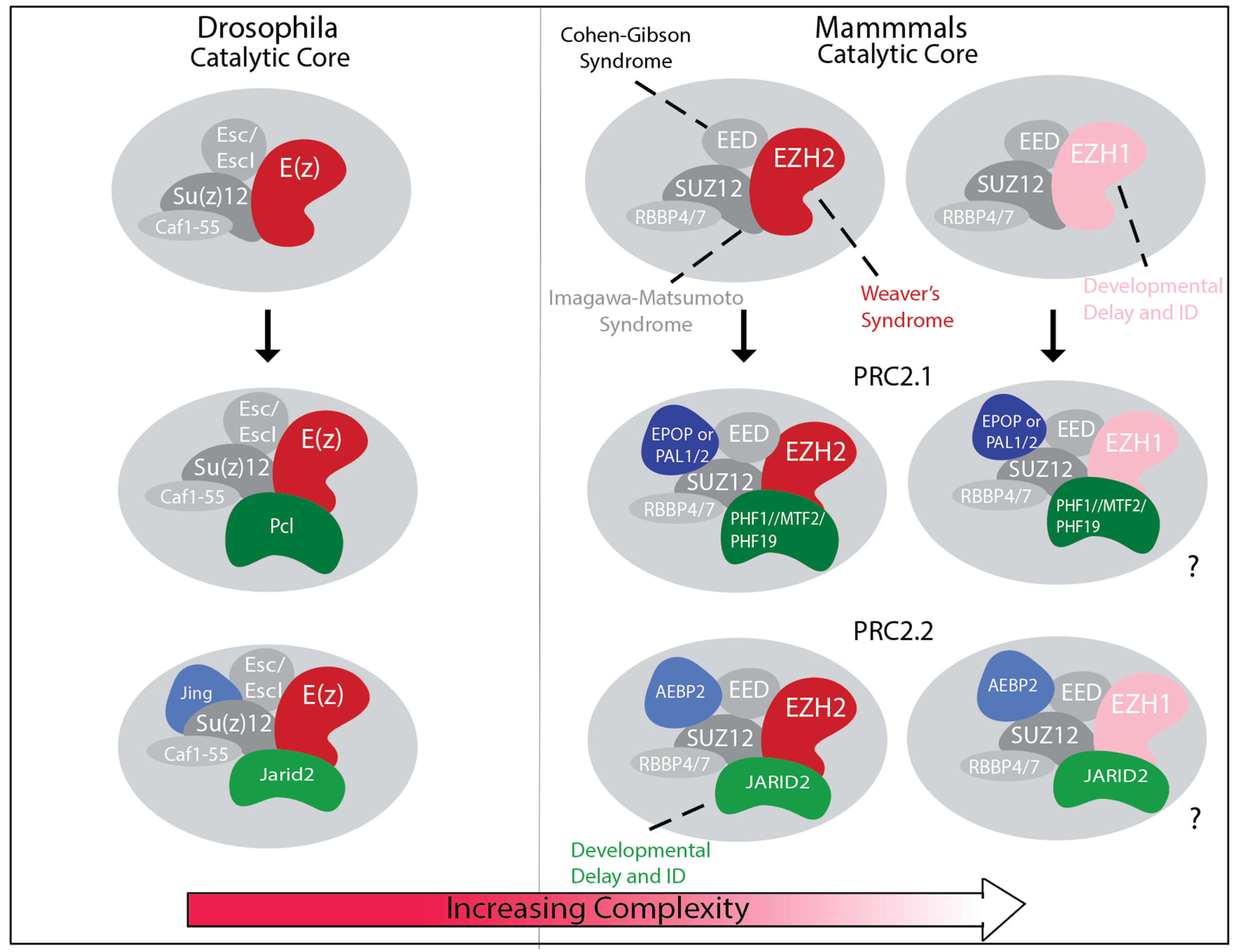
Figure 1. Graphical representation of PRC2 core and accessory subunits depicting increased number of PRC2 subcomplexes as cellular diversity increases from Drosophila to mammals. (Left) Drosophila PRC2 consists of the core subunits Enhancer of Zeste (E(Z)), Extra Sex Combs (ESC) or Extra Sex Combs like (ESCL), Suppressor of Zeste-12 (SU(Z)12), and CAF1-55, and either the accessory subunit Polycomb-like (PCL) or JING and JARID2. (Right) In mammals, the core PRC2 is formed by either EZH1 or EZH2 (E(Z) homologs), Embryonic Ectoderm Development (EED) (ESC homolog), Suppressor of Zeste 12 Protein Homolog (SUZ12) (SU(Z)12 homolog), and RBBP4 or 7 (CAF1-55 homologs). In addition, the interaction with either one of the three homologs of Drosophila PCL protein, PHF1, MTF2, or PHF19, and either PALI1, PALI2 (PRC2 Associated LCOR Isoform paralogs), or EPOP (Elongin BC complex and the PRC2-associated Protein) forms PRC2.1 subcomplexes, whereas the interaction with Adipocyte Enhancer-Binding Protein 2 (AEBP2) and Jumonji, AT-rich interaction domain 2 (JARID2) creates PRC2.2 subcomplexes. “?” indicates the uncertainty about PRC2.1 and PRC2.2 composed with EZH1, due to limited supporting literature. Mutations in nearly all core subunits (EZH1, EZH2, EED, and SUZ12) and an accessory subunit (JARID2), as indicated in the figure, are associated with human neurodevelopmental disorders.
Genes 16 01191 g001
Figure 2. Expression of PRC2 subunits in Drosophila and Human developing and adult nervous system. (A) Heatmap displaying the expression levels (RPKM) of Polycomb Group (PcG) genes in the central nervous system of third-instar (L3) larvae and the brain of adult males and females of Drosophila. Data was obtained from FlyAtlas2 Anatomical Expression Data. (B) Heatmaps displaying the expression levels (RPKM) of mammalian PRC2 core and accessory subunits during prenatal and postnatal brain development across different regions. Data were obtained from mRNA-sequencing datasets in BrainSpan. The average expression value was calculated for each time point in each brain region. (C) Dot plot showing scRNAseq expression of PcG genes across neuronal cell types in the Drosophila third-instar larval ventral nerve cord (VNC). (D) Dot plot showing scRNAseq expression of PRC2 genes across neuronal cell types in second-trimester developing human neocortex. Dot size indicates the percentage of cells expressing each gene, and color the average expression level. Dot plots were generated using the Seurat R package (v4.3.0) with the DotPlot function, based on author-defined cell type annotations and normalized RNA data from the original study. Note that LCOR and LCORL are the genes encoding PALI1 and PALI2. Abbreviations: NB, neuroblast; GMC, ganglion mother cell; Imp, insulin-like growth factor 2 mRNA-binding protein; RG, radial glia (vRG, ventricular RG; tRG, truncated RG; oRG, outer RG); OPC, oligodendrocyte precursor cell; IPC, intermediate progenitor cell (IPC–EN, excitatory neuron progenitor; IPC–Glia, glial progenitor); IN, interneuron (dLGE, dorsal lateral ganglionic eminence; MGE, medial ganglionic eminence; CGE, caudal ganglionic eminence; SST, somatostatin; PV, parvalbumin; VIP, vasoactive intestinal peptide; SNCG, synuclein gamma; EN, excitatory neuron (IT, intratelencephalic; ET, extratelencephalic; CT, corticothalamic); NP, near-progenitor.
Genes 16 01191 g002
Table 1. Summary of PRC2 component function and mutant phenotype in Drosophila and mammals. N/A: no association.
Drosophila
ComplexSubunitMolecular FunctionMutant Phenotype
PRC2E(z)Catalyzes H3K27 methylationImproper body segmentation; additional sex combs; lethality
PRC2Esc/EsclFacilitates histone methyltransferase activity Improper body segmentation; additional sex combs; lethality
PRC2Su(z)12Facilitates histone methyltransferase activityImproper body segmentation; additional sex combs; lethality
PRC2Caf1-55Enhances PRC2 activityLethality
PRC2.1PclEnhances the generation of H3K27me3 at target lociImproper body segmentation; additional sex combs; lethality
PRC2.2JingModulates PRC2 methyltransferase activityHomeotic transformations; Tracheal and nervous system defects; lethality
PRC2.2Jarid2Modulates PRC2 methyltransferase activityImproper body segmentation; lethality
Humans
ComplexSubunitMolecular FunctionNeurodevelopmental Disease
PRC2.1/PRC2.2EZH2Catalyzes H3K27 methylationWeaver syndrome: overgrowth, moderate intellectual disability (in about 80% of patients), distinctive facial appearance with macrocephaly and occasionally polymicrogyria
PRC2.1/PRC2.2EZH1Catalyzes H3K27 methylationNDD with developmental delay, mild-to severe intellectual disability and atypical facial features
PRC2.1/PRC2.2EEDFacilitates and enhances H3K27 methylation and propagationCohen–Gibson Syndrome: Overgrowth, mild-to-severe intellectual disability (in 100% of patients), distinctive facial appearance with macrocephaly
PRC2.1/PRC2.2SUZ12Facilitates H3K27 methylation and stabilizes PRC2 complexes and interactions Imagawa–Matsumoto syndrome: overgrowth, mild intellectual disability (in ~50% of patients).
PRC2.1/PRC2.2RBBP4/7Facilitates PRC2 recruitment to target lociN/A
PRC2.1PCL1/PHF1Enhances H3K27 methylation and promotes recruitment to target lociN/A
PRC2.1PCL2/MTF2Promotes recruitment to target loci and maintains H3K27 methylation N/A
PRC2.1PCL2/PHF19Enhances H3K27 methylation and promotes recruitment to target lociN/A
PRC2.1EPOPModulates and promotes PRC2 methyltransferase activityN/A
PRC2.1PAL1/2Modulates and promotes PRC2 methyltransferase activityN/A
PRC2.2JARID2Facilitates recruitment of PRC2 to target loci and enhances deposition of H3K27 methylationDevelopmental delay and intellectual disability with dysmorphic facies.
PRC2.2AEBP2Stabilizes PRC2 and helps stimulate H3K27 methylation activityN/A
Disclaimer/Publisher’s Note: The statements, opinions and data contained in all publications are solely those of the individual author(s) and contributor(s) and not of MDPI and/or the editor(s). MDPI and/or the editor(s) disclaim responsibility for any injury to people or property resulting from any ideas, methods, instructions or products referred to in the content.