Article

Epilepsy Associated Gene, Pcdh7, Is Dispensable for Brain Development in Mice

Department of Neuroscience, Feinberg School of Medicine, Northwestern University, Chicago, IL 60614, USA
*
Author to whom correspondence should be addressed.
Genes 2025, 16(8), 985; https://doi.org/10.3390/genes16080985
Submission received: 7 July 2025 / Revised: 16 August 2025 / Accepted: 20 August 2025 / Published: 21 August 2025
(This article belongs to the Special Issue The Genetic and Epigenetic Basis of Neurodevelopmental Disorders)

Abstract

Background/Objectives: Protocadherin 7 (Pcdh7) belongs to the protocadherin family, the largest subgroup of cell adhesion molecules. Members of this family are highly expressed in the brain, where they serve fundamental roles in many neurodevelopmental processes, including axon guidance, dendrite self-avoidance, and synaptic formation. PCDH7 has been strongly associated with epilepsy in multiple genome-wide association studies (GWAS), as well as with schizophrenia, PTSD, and childhood aggression. Despite these associations, the specific contributions of PCDH7 to epileptogenesis and brain development remain largely unexplored. Most of the existing literature on PCDH7 focuses on its function during cancer progression, with only one study suggesting that PCDH7 regulates dendritic spine morphology and synaptic function via interaction with GluN1. Methods: Here, we generate, validate, and characterize a murine null Pcdh7 allele in which a large deletion was introduced by CRISPR. Results: Analysis of embryonic, postnatal, and adult brain datasets confirmed PCDH7 widespread expression. Pcdh7+/− and Pcdh7−/− mice present no gross morphological defects and normal cortical layer formation. However, a seizure susceptibility assay revealed increased latencies in Pcdh7+/− mice, but not in Pcdh7+/+ and Pcdh7−/− mice, potentially explaining the association of PCDH7 with epilepsy. Conclusions: This initial characterization of Pcdh7 null mice suggests that, despite its widespread expression in the CNS and involvement in human epilepsy, PCDH7 is not essential for murine brain development and thus is not a suitable animal model for understanding PCDH7 disruption in humans. However, further detailed analysis of this mouse model may reveal circuit or synaptic abnormalities in Pcdh7 null brains.

1. Introduction

Protocadherins (PCDH) are the largest subgroup within the cadherin superfamily and function as cell-recognition molecules critical for establishing and maintaining neural circuits [1,2]. The PCDH family is divided into two groups: clustered PCDHs (PCDHα, β, and γ) and non-clustered PCDHs [3]. Non-clustered PCDHs have been implicated in neurodevelopmental processes and disorders when their function is compromised [1,2,4]. Specifically, PCDH7, -10, -12, -17, -18, and -20 mediate axon growth and extension [5,6,7,8,9,10,11,12], and PCDH7, -8, and -10 are implicated in dendritic spine and synapse density [13,14,15,16]. Moreover, PCDH12 and -19 are involved in neural proliferation and migration [17,18], while PCDH19 and -20 are involved in neuronal sorting/positioning [6,17,19]. Pathogenic variants in non-clustered PCDHs have been linked to a wide range of neurodevelopmental and psychiatric disorders. For example, PCDH19-related epilepsy is caused by heterozygous loss-of-function mutations in the X-linked gene, PCDH19 [20,21]. PCDH12 variants have been associated with schizophrenia [22], and bi-allelic pathogenic variants lead to seizures, microcephaly, and white matter abnormalities [9,23,24,25,26]. PCDH8, -9, and -10 de novo variants have been linked to autism [16,27,28], while variants in PCDH17 confer a risk for mood disorders [29].
Although much of the research on PCDH7 pathogenic variants has primarily focused on its role in cancer [30,31], genome-wide association studies (GWAS) have linked PCDH7 to various neurological disorders, including epilepsy [32,33], schizophrenia [34], PTSD [34], and childhood aggression [35]. PCDH7 is downregulated by MeCP2 and upregulated in the brains of Mecp2 KO mice [36], suggesting a connection between PCDH7 and the neurodevelopmental disorder Rett syndrome. Furthermore, recent findings suggest that haploinsufficiency of PCDH7 might contribute to the mental developmental delay observed in patients with proximal 4p deletion syndrome [37]. Additionally, PCDH7 is located in the excitatory synaptic cleft [38] and interacts with the NMDA receptor subunit GluN1 [14]. These insights collectively highlight a possible role for PCDH7 in both brain development and synaptic function.
Pcdh7 exhibits a region-specific expression pattern within the murine developing cerebral cortex [38]; however, the consequences of pcdh7 loss during brain development have yet to be investigated. The only report of a Pcdh7 null mouse focused on bone homeostasis [14], omitting any examination of brain morphology. Most research investigating the role of pcdh7 during neurodevelopment has been performed in Xenopus or zebrafish using morpholinos. These studies have shown that pcdh7 regulates the histogenesis of the embryonic ectoderm [1], neural tube formation [39], and axon growth [40] in Xenopus morphants. Morpholino knockdown in zebrafish also results in impaired neural stem cell differentiation and disrupted neural architecture during embryonic brain development. In contrast, pcdh7 null zebrafish embryos did not exhibit any phenotypic defects [41]. While these findings support a role for pcdh7 during brain development, the commonly observed discrepancies between the mutant and morphant phenotypes [11,42] complicate the interpretation of these results.
Here, we generated, validated, and characterized a murine Pcdh7 null allele containing a large CRISPR-mediated deletion. Analysis of embryonic, postnatal, and adult brain datasets confirmed PCDH7 widespread expression. Examination of brain morphology and cortical layer formation showed no abnormalities in both Pcdh7+/− and Pcdh7−/− mice. However, while Pcdh7+/− mice exhibited increased latencies to seizures, this was not observed in Pcdh7−/− nor Pcdh7+/+ mice, potentially explaining the association of PCDH7 with epilepsy. This initial characterization of the Pcdh7 mouse model reveals that, despite widespread expression in the CNS and its association with human epilepsy, Pcdh7 function is not essential for murine brain development.

2. Materials and Methods

2.1. Mice

All experiments were approved by the Northwestern University Animal Care and Use Committee in accordance with the NIH Guide for the Care and Use of Laboratory Animals and the Animal Welfare Act. Animals were housed on a 14-h light/10-h dark schedule, with food and water available ad libitum. C57BL/6J mice were used in all experiments.

2.2. CRISPR Gene Editing

Generation of Pcdh7 null mice was carried out by using the CRISPR/Cas9 system. Briefly, we selected four gRNAs using the CRISPOR program [43] based on specificity scores, location, and predicted cleavage efficiency to minimize off-target effects. The four gRNAs were synthetized using the IDT AltR system, and the high-fidelity Cas9 protein was used. All four gRNAs in conjunction with the Cas9 protein were electroporated into fertilized murine eggs (zygotes). The injected embryos were then transferred into a pseudo-pregnant female mouse. Once pups were born, screening for gene editing events was performed via PCR amplification of the Pcdh7 exon 1 locus followed by Sanger sequencing. The full Pcdh7 exon1 locus was amplified using Pcdh7 primer 2F: 5′-TCCCCTCCCTCTCGTTTCTT-3′ and Pcdh7 primer 1R: 5′-ACACTACCTTTCCCTTAGATTCCA-3′. Pcdh7 WT amplicon = 3560 bp. PCR products were separated on a 1% agarose gel. Smaller amplicons suggested deletions; thus, the bands were excised and gel-purified using the Qiagen gel purification kit (Qiagen, Germantown, MD, USA). Purified PCR products were submitted for Sanger sequencing. After sequencing, two surviving founders were identified. Male 9659 (which carried 2 mutant alleles, including the 1145 bp deletion), and Male 9660, which also carried 2 mutant alleles, including a large 2243 bp deletion. Multiple alleles in a single founder are commonly found when mutations are generated directly in the embryo, and this is due to CRIPSR-mediated editing in the zygote after the first cellular division. Because of the complexity of the mutations, founders were bred with WT females to segregate the alleles. F1 mice carrying the KO allele were recovered with a 50% transmission of the KO alleles (1145 bp deletion for KO1 and the 2243 bp deletion for KO2).

2.3. Genotyping

Genotyping was performed by PCR amplification using the following primers: Forward: 5′-TCCCCTCCCTCTCGTTTCTT-3′; Reverse-1 (KO): 5′-ACGTCTCCCACTACGGTACA-3′; Reverse-2 (WT): 5′-CCCTCCCACAATGCTGTAGT-3′. PCR products were separated on a 1% agarose gel. Pcdh7 WT and KO allele products are 273 bp and 382 bp, respectively.

2.4. RNA Isolation and RT-PCR

RNA isolation was performed using Direct-zolTM RNA miniprep plus isolation kit (Zymo Research, Irvine, CA, USA), and RNA concentration was measured using a Nanodrop. cDNA was synthesized from 5 µg RNA using the Superscript III reverse transcriptase (Thermo Fisher Scientific, Waltham, MA, USA) cDNA synthesis kit. RT-PCR was performed according to the manufacturer’s instructions. PCR amplification using the following primers: Exon 1: Forward: 5′-TCCCCTCCCTCTCGTTTCTT-3′; Reverse: 5′-ACGTCTCCCACTACGGTACA-3′; Exon 2–3: Forward: 5′-TCCCCTCCCTCTCGTTTCTT-3′; Reverse: 5′-CCCTCCCACAATGCTGTAGT-3′. PCR products were separated on a 1% agarose gel. Exons 1 and 2–3 products are 173 bp and 267 bp, respectively.

2.5. Histology and Immunohistochemistry

Brains were post-fixed in PFA 4% overnight, cryoprotected in 30% sucrose until sunk, and snap frozen embedded in OCT. 25 µm coronal sections were obtained using a cryostat (Leica CM1860, Leica Biosystems, Deer Park, IL, USA). For histological measurements, both cortical and CA1 hippocampal thicknesses were assessed across multiple serial sections and averaged to obtain a representative thickness for each individual mouse brain. For immunohistochemistry, slides were incubated overnight at 4 °C with the primary antibody, then with a fluorescently labeled secondary antibody (Invitrogen, Waltham, MA, USA) for 2 h at room temperature (RT), and counterstained with Hoechst 33342 (2 µg/mL, Thermo Fisher Scientific, Waltham, MA, USA). Fluorescent signal was detected using an inverted microscope (BZ-X710 Fluorescence Microscope, Keyence, Itasca, IL, USA). Primary antibodies include anti-Satb2 (1:200, Abcam, Waltham, MA, USA) and anti-NeuN (1:500, Thermo Fisher Scientific, Waltham, MA, USA).

2.6. Flurothyl Seizure Induction

Flurothyl seizure induction was performed as previously described [44,45]. Briefly, the mice were individually placed in a sealed induction chamber. Flurothyl (2,2,2-trifluroethyl ether, Sigma-Aldrich, St. Louis, MO, USA) was introduced by a syringe pump (Genie Touch, Kent Scientific, Torrington, CT, USA) at a constant rate of 20 μL/min. Following flurothyl induction, reviewers blinded to the genotype recorded the latency to the first myoclonic (MJ) and generalized tonic-clonic seizure (GTCS). At the onset of GTCS, the mouse was immediately placed in a holding cage for recovery. All mice were euthanized at the end of the experimental session.

2.7. Western Blot Analysis

Immediately following the flurothyl protocol, brain tissue was weighed and lysed in 1X RIPA buffer (Cell Signaling Technology, Danvers, MA, USA), supplemented with protease inhibitor cocktail (Thermo Fisher Scientific, Waltham, MA, USA) at a 1:50 ratio (weight: volume). Protein concentration was determined by BCA assay (Thermo Fisher Scientific, Waltham, MA, USA). Samples were denatured in Laemmli buffer (Bio-Rad, Hercules, CA, USA) by boiling for 5 min at 95 °C. Proteins (30 µg) were resolved on 10% acrylamide gels by SDS-PAGE and transferred onto PVDF membranes. Membranes were blocked in Intercept® (TBS) blocking buffer (LICORbio, Lincoln, NE, USA), and primary antibodies were incubated in the same buffer supplemented with 0.2% Tween20 at 4 °C overnight. Primary antibodies were anti-GFAP (1:5000, Invitrogen, Waltham, MA, USA) and anti-α-Tubulin (1:10,000, Abcam, Waltham, MA, USA). Infrared (IR) secondary antibodies were incubated in Intercept® (TBS) blocking buffer (LICORbio, Lincoln, NE, USA) supplemented with 0.2% Tween20 and 0.01% SDS at a 1:10,000 dilution for 1 h at RT. IR fluorescent blots were imaged with the Odyssey® Fc imaging system (software version 1.0.37, LICORbio, Lincoln, NE, USA). Image acquisition and quantification were performed with Image Studio (version 5.2.5, LICORbio, Lincoln, NE, USA).

2.8. Statistical Analysis

Statistical analyses were performed using GraphPad Prism 10 (version 10.4.0, GraphPad Software, Boston, MA, USA). Results are presented as mean ± standard deviation, unless mentioned otherwise. One-way analysis of variance (ANOVA) followed by Tukey’s post hoc multiple comparisons test was used after verification that normality and homoscedasticity criteria were met. A p-value < 0.05 was considered statistically significant.

3. Results

3.1. Pcdh7 Is Widely Expressed in Mice and Human Brain During Development

Most studies on PCDH7 have focused on its role in cancer, despite reports that, like most PCDHs, it is highly expressed in the nervous system [1,14]. Thus, we first conducted a comprehensive analysis of both murine and human embryonic, postnatal, and adult brain datasets to better understand PCDH7 expression patterns [46,47]. We found that PCDH7 is expressed throughout brain development, with higher expression after birth in both humans and mice (Figure 1A,B). This data is in line with previous studies reporting Pcdh7 expression in the rodent developing brain [1,41,48]. In contrast, PCDH7 expression is higher in prenatal timepoints, decreasing after birth in the cerebellum of both humans and mice (Figure 1A,B). PCDH7 is expressed in multiple regions of both the mouse and human central nervous system (CNS), with the highest expression observed in the cortex, followed by the hippocampus, basal ganglia, and thalamus, with the least expression in the cerebellum (Figure 1C,D). Finally, relative PCDH7 expression is higher in the human brain than in the mouse brain (Figure 1C,D).

3.2. Generation and Validation of a Pcdh7 Null Mouse

The Pcdh7 gene contains four exons, with exon 1 encoding most of the protein, including the extracellular and transmembrane domains (Figure 2A). Because Pcdh7 has several potential ATG start sites, a complete disruption of exon 1 is necessary for the generation of a Pcdh7 null mouse model. Thus, we targeted 4 regions of exon 1 with multiple gRNAs to obtain a CRISPR cleavage disruption. We selected four gRNAs using the CRISPOR program [43] based on specificity scores, location, and predicted cleavage efficiency. Their position in exon 1 and the resulting deletions are depicted in Figure 2B. The four gRNAs were electroporated into the murine zygotes to disrupt Pcdh7 exon 1. IDT AltR system gRNAs and the high-fidelity Cas9 protein were electroporated into the zygotes to ensure that the CRISPR complex was transient and reduce possible off-target effects. Screening for gene editing events was performed via PCR amplification of the Pcdh7 exon 1 locus followed by Sanger sequencing. PCR protocols and primer sets were optimized to amplify the full Pcdh7 exon 1 locus. Two founder male mice were then identified: KO1, harboring the 1145 bp deletion, and KO2, with the 2243 bp deletion. The disruption of the Pcdh7 gene was validated by the PCR using genomic DNA (Figure 2C), and the reduction of Pcdh7 expression was confirmed by RT-PCR using cDNA (Figure 2D).
Founders were set up in breeding trios with WT females. F1 mice carrying the KO allele were identified after successful pairing. KO1 F1 consisted of two litters, 20 mice (8 males, 12 females), with a 50% transmission of the 1145 bp deletion as expected. KO2 F1 consisted of three litters for a total of 25 mice (14 males, 11 females) with 12 of 25 mice carrying the 2243 bp deletion. F1 and subsequent Pcdh7+/− breeding pairs generated Pcdh7+/+, Pcdh7+/−, and Pcdh7−/− mice at the expected frequencies (Table 1). Adult mice from all genotypes presented with similar body sizes and no overt health issues.

3.3. Pcdh7+/− but Not Pcdh7−/− Mice Present with Increased Seizure Latencies

PCDH7 has been strongly linked to epilepsy [32]; however, we did not observe spontaneous seizures in any mice from all genotypes at any developmental time point. Next, we induced generalized seizures using the GABAA receptor function inhibitor flurothyl (Figure 3A). Mice exposed to flurothyl express a series of characteristic seizure behaviors, beginning with brief myoclonic jerks (MJ), which progress to generalized seizures (GS) and eventually culminate in generalized tonic-clonic seizures (GTCS). The average latencies to MJ and the first GS were comparable among genotypes (122 ± 14 s, 137 ± 28 s, and 140 ± 14 s to first MJ in WT, Pcdh7+/−, and Pcdh7−/− mice, respectively; 177 ± 19 s, 196 ± 27 s, and 187 ± 25 s to first GS MJ in WT, Pcdh7+/−, and Pcdh7−/− mice, respectively). However, the average latency to GTCS was significantly increased in heterozygous mice (306 ± 64 s) when compared with their WT littermates (234 ± 48 s) (Figure 3B), and no differences were observed in the number of jerks regardless of genotype (Figure 3C). Finally, flurothyl induced astrocyte activation, as observed by GFAP expression in Western blots of brain lysates from all genotypes.

3.4. Pcdh7+/− and Pcdh7−/− Mice Exhibit No Gross Brain Abnormalities

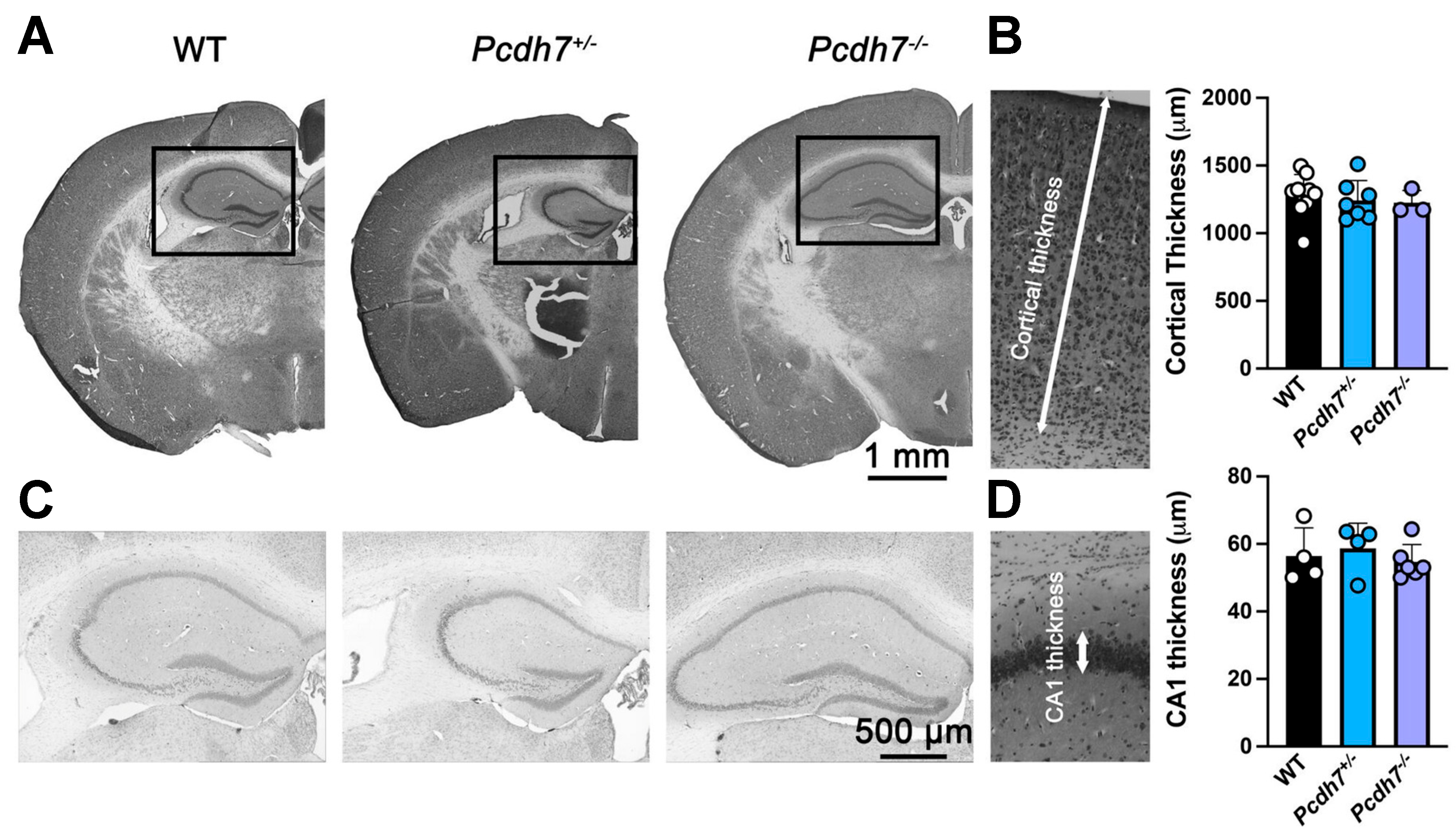
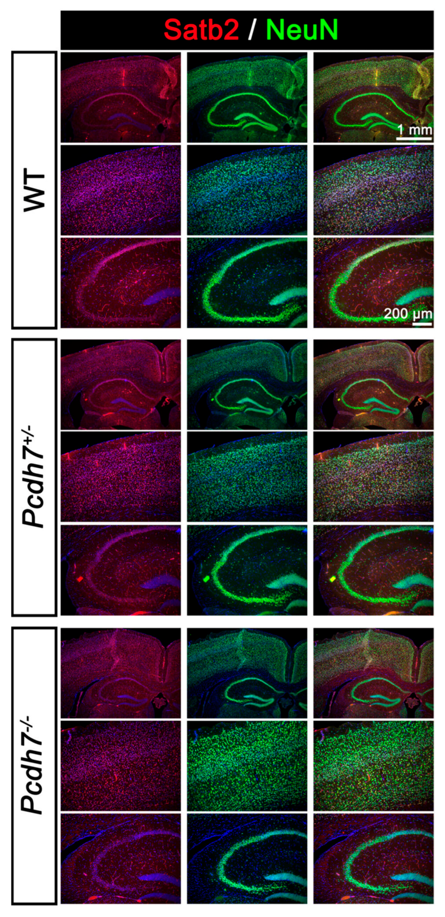
Morpholino knockdown of pcdh7 in Xenopus and zebrafish embryos results in CNS defects. We therefore examined gross brain morphology in our newly generated Pcdh7 mouse model. Histological analysis of WT, Pcdh7+/−, and Pcdh7−/− brains showed no gross morphological abnormalities. As Pcdh7 is highly expressed in the cerebral cortex of both humans and mice (Figure 1), we measured the cortical and hippocampus CA1 thickness (Figure 4). We also analyzed cortical layer formation using immunostaining with the pan-neuronal marker NeuN and the pyramidal neuron marker Satb2 (Figure 5). NeuN is found in the nucleus of mature neurons, and it was used to visualize neuronal density and potential neuronal loss due to the lack of Pcdh7. Satb2 is required during cortical development for the cell fate specification of callosal projection neurons, and it is expressed in excitatory neurons in the adult. No differences between Pcdh7+/− and Pcdh7−/− brains and Pcdh7+/+ controls were observed (Figure 4 and Figure 5), suggesting that loss of Pcdh7 in mice does not overtly alter brain structure.

4. Discussion

Pathogenic variants in protocadherin genes have been implicated in several neurological diseases. Specifically, PCDH7 has been associated with multiple neurological disorders, including epilepsy [32,34,35,36,37,49]. Pcdh7 is predominantly expressed in the developing murine brain, in both excitatory and inhibitory neurons, with an enrichment in synapses [38]. Our data analysis also confirms differential PCDH7 expression throughout development, with higher levels after birth in both human and mouse nervous systems, supporting the role of PCDH7 in synapse formation and function. We observed robust Pcdh7 expression, particularly in brain regions implicated in epilepsy, such as the hippocampus and cortex, consistent with previous studies in rodents [1,41,48,50]. Animal models lacking PCDHs have revealed important functions in various aspects of neurodevelopment, including axon outgrowth [5,12], dendrite branching [51,52], dendritic spine density [15], and synaptic formation and function [53]. Thus, to start exploring the impact of loss of pcdh7 during brain development, in general, and in epilepsy, in particular, we generated a Pcdh7 null mouse model.
Previous studies have reported that disruption of pcdh7 causes neural defects during brain development in zebrafish embryos [41]. However, these defects are only observed using morpholino knockdown and were not present in zebrafish with a genetic mutation of pcdh7 [41]. These phenotypic differences may reflect a lack of genetic compensation in response to acute morpholino knockdown, as previously reported [54,55]. Nevertheless, the lack of brain abnormalities in our newly generated Pcdh7+/+, Pcdh7+/−, and Pcdh7−/− mice is consistent with pcdh7−/− zebrafish embryos and with other protocadherin null mouse models of disease. Of particular interest are the Pcdh19 null mice, which have been reported to be healthy, fertile, and with no gross brain defects [56]. In contrast, PCDH19 patients develop infantile seizures with variable cognitive defects and cortical dysplasia [17,21]. Another example is the Pcdh12 null mice, also reported to be viable and show no morphological abnormalities [57], while the patients carrying bi-allelic pathogenic variants present with seizures, microcephaly, and white matter abnormalities [9,23,24,25,26]. The absence of gross brain defects in our Pcdh7 null mouse model may present functional redundancy of Pcdh7 that was within expectations.
The association of PCDH7 with genetic generalized epilepsy [32,33] prompted us to investigate whether our Pcdh7 mouse model exhibits seizures. While we did not observe spontaneous seizures in any genotype, Pcdh7+/− mice, but not Pcdh7+/+ and Pcdh7−/− mice, presented with increased latency to seizures induced by flurothyl. The Pcdh7+/− genotype is the one carried by patients identified with epilepsy through GWAS. Although the mechanisms by which PCDH variants lead to neurological disorders are not fully understood, insights from the well-studied PCDH19 suggest that its unique X-linked inheritance pattern may disrupt cellular adhesion affinities, an occurrence potentially exacerbated by X-inactivation [17]. Unlike PCDH19, which is located on the X chromosome, PCDH7 and other members of the PCDH family reside on autosomes. Nonetheless, evidence indicates that these proteins can undergo random monoallelic expression [58]. This may cause symptoms in individuals with heterozygous variants due to an imbalance in PCDH expression, potentially leading to similar disrupted adhesion affinities to those seen in PCDH19-related epilepsy. In cases where all cells either express only the WT PCDH or none at all, no symptoms occur. This phenomenon is exemplified by hemizygous males with PCDH19 mutations, who typically do not exhibit symptoms associated with PCDH19-related epilepsy [21]. Therefore, our findings may provide an explanation for the association of PCDH7 with epilepsy in humans.
Most of the genes known to cause or contribute to epilepsy can be traced to the synapse, including PCDH7, which is located in the excitatory synaptic cleft [38], and interacts with the NMDA receptor subunit GluN1 [14]. Interestingly, cell-autonomous knockdown of PCDH7 did not affect the NMDA receptor current [14]. Whether the NMDA receptor current is affected by PCDH7 in all components of the circuit, and not only in the postsynaptic cell, remains an open line for future investigation.
In summary, our characterization of Pcdh7 null mice suggests that, although Pcdh7 is widely expressed in the CNS, it is not required for murine brain development and thus not a suitable animal model for understanding PCDH7 disruption in humans. However, given the increased latency to seizures in Pcdh7 heterozygous mice and the identification of multiple roles for Pcdh7 in neurodevelopmental processes in vitro, further detailed analysis of this mouse model may reveal circuit or synaptic abnormalities in Pcdh7 null brains.

Author Contributions

Conceptualization, A.G.-G. and J.R.; Experimentation and data analysis, J.R., D.D., X.V., X.G.-M. and O.S.; writing—original draft preparation, A.G.-G. and J.R.; writing—editing, X.G.-M. and X.V.; figures, J.R. and X.G.-M.; funding acquisition, A.G.-G. All authors have read and agreed to the published version of the manuscript.

Funding

This research was funded by the NIH, National Institute of Neurological Disorders and Stroke, grant number R00 NS089943 to A.G.-G.

Institutional Review Board Statement

The animal study protocol was approved by the Northwestern University, Feinberg School of Medicine Institutional Animal Care and Use Committee (protocol code IS00005193, date of approval 7 June 2023).

Informed Consent Statement

Not applicable.

Data Availability Statement

PCDH7 expression profiles were obtained from The Evo-devo mammalian organs project (https://apps.kaessmannlab.org/evodevoapp, accessed on 7 May 2025) and The Human Protein Atlas (https://www.proteinatlas.org, accessed on 7 May 2025).

Acknowledgments

The genetically engineered mice were generated with the assistance of the Northwestern University Transgenic and Targeted Mutagenesis Laboratory. Comparative histopathology and molecular phenotyping services were provided by the Mouse Histology and Phenotyping Laboratory (MHPL, RRID:SCR_017870) of Northwestern University, which is supported by the National Cancer Institute (NCI) P30-CA060553 grant awarded to the Robert H. Lurie Comprehensive Cancer Center.

Conflicts of Interest

The authors declare no conflicts of interest.

Abbreviations

The following abbreviations are used in this manuscript:
PCDH7Protocadherin 7
CRISPRClustered Regularly Interspaced Short Palindromic Repeats
HETHeterozygous
KOKnock-out
MJMyoclonic jerk
GTCSGeneralized tonic-clonic seizure
GSGeneralized seizure
gRNAGuide RNA

References

  1. Kim, S.Y.; Mo, J.W.; Han, S.; Choi, S.Y.; Han, S.B.; Moon, B.H.; Rhyu, I.J.; Sun, W.; Kim, H. The expression of non-clustered protocadherins in adult rat hippocampal formation and the connecting brain regions. Neuroscience 2010, 170, 189–199. [Google Scholar] [CrossRef]
  2. Kim, S.Y.; Yasuda, S.; Tanaka, H.; Yamagata, K.; Kim, H. Non-clustered protocadherin. Cell Adhes. Migr. 2011, 5, 97–105. [Google Scholar] [CrossRef] [PubMed]
  3. Morishita, H.; Yagi, T. Protocadherin family: Diversity, structure, and function. Curr. Opin. Cell Biol. 2007, 19, 584–592. [Google Scholar] [CrossRef] [PubMed]
  4. Kim, Y.; Sung, J.Y.; Ceglia, I.; Lee, K.W.; Ahn, J.H.; Halford, J.M.; Kim, A.M.; Kwak, S.P.; Park, J.B.; Ho Ryu, S.; et al. Phosphorylation of WAVE1 regulates actin polymerization and dendritic spine morphology. Nature 2006, 442, 814–817. [Google Scholar] [CrossRef] [PubMed]
  5. Uemura, M.; Nakao, S.; Suzuki, S.T.; Takeichi, M.; Hirano, S. OL-Protocadherin is essential for growth of striatal axons and thalamocortical projections. Nat. Neurosci. 2007, 10, 1151–1159. [Google Scholar] [CrossRef]
  6. Oishi, K.; Nakagawa, N.; Tachikawa, K.; Sasaki, S.; Aramaki, M.; Hirano, S.; Yamamoto, N.; Yoshimura, Y.; Nakajima, K. Identity of neocortical layer 4 neurons is specified through correct positioning into the cortex. eLife 2016, 5. [Google Scholar] [CrossRef]
  7. Hayashi, S.; Inoue, Y.; Kiyonari, H.; Abe, T.; Misaki, K.; Moriguchi, H.; Tanaka, Y.; Takeichi, M. Protocadherin-17 mediates collective axon extension by recruiting actin regulator complexes to interaxonal contacts. Dev. Cell 2014, 30, 673–687. [Google Scholar] [CrossRef]
  8. Biswas, S.; Emond, M.R.; Duy, P.Q.; Hao le, T.; Beattie, C.E.; Jontes, J.D. Protocadherin-18b interacts with Nap1 to control motor axon growth and arborization in zebrafish. Mol. Biol. Cell 2014, 25, 633–642. [Google Scholar] [CrossRef]
  9. Guemez-Gamboa, A.; Caglayan, A.O.; Stanley, V.; Gregor, A.; Zaki, M.S.; Saleem, S.N.; Musaev, D.; McEvoy-Venneri, J.; Belandres, D.; Akizu, N.; et al. Loss of Protocadherin-12 Leads to Diencephalic-Mesencephalic Junction Dysplasia Syndrome. Ann. Neurol. 2018, 84, 638–647. [Google Scholar] [CrossRef]
  10. Hoshina, N.; Tanimura, A.; Yamasaki, M.; Inoue, T.; Fukabori, R.; Kuroda, T.; Yokoyama, K.; Tezuka, T.; Sagara, H.; Hirano, S.; et al. Protocadherin 17 regulates presynaptic assembly in topographic corticobasal Ganglia circuits. Neuron 2013, 78, 839–854. [Google Scholar] [CrossRef]
  11. Leung, L.C.; Harris, W.A.; Holt, C.E.; Piper, M. NF-Protocadherin Regulates Retinal Ganglion Cell Axon Behaviour in the Developing Visual System. PLoS ONE 2015, 10, e0141290. [Google Scholar] [CrossRef]
  12. Piper, M.; Dwivedy, A.; Leung, L.; Bradley, R.S.; Holt, C.E. NF-protocadherin and TAF1 regulate retinal axon initiation and elongation in vivo. J. Neurosci. 2008, 28, 100–105. [Google Scholar] [CrossRef] [PubMed]
  13. Keeler, A.B.; Molumby, M.J.; Weiner, J.A. Protocadherins branch out: Multiple roles in dendrite development. Cell Adhes. Migr. 2015, 9, 214–226. [Google Scholar] [CrossRef] [PubMed]
  14. Wang, Y.; Kerrisk Campbell, M.; Tom, I.; Foreman, O.; Hanson, J.E.; Sheng, M. PCDH7 interacts with GluN1 and regulates dendritic spine morphology and synaptic function. Sci. Rep. 2020, 10, 10951. [Google Scholar] [CrossRef] [PubMed]
  15. Yasuda, S.; Tanaka, H.; Sugiura, H.; Okamura, K.; Sakaguchi, T.; Tran, U.; Takemiya, T.; Mizoguchi, A.; Yagita, Y.; Sakurai, T.; et al. Activity-induced protocadherin arcadlin regulates dendritic spine number by triggering N-cadherin endocytosis via TAO2beta and p38 MAP kinases. Neuron 2007, 56, 456–471. [Google Scholar] [CrossRef]
  16. Tsai, N.P.; Wilkerson, J.R.; Guo, W.; Maksimova, M.A.; DeMartino, G.N.; Cowan, C.W.; Huber, K.M. Multiple autism-linked genes mediate synapse elimination via proteasomal degradation of a synaptic scaffold PSD-95. Cell 2012, 151, 1581–1594. [Google Scholar] [CrossRef]
  17. Pederick, D.T.; Richards, K.L.; Piltz, S.G.; Kumar, R.; Mincheva-Tasheva, S.; Mandelstam, S.A.; Dale, R.C.; Scheffer, I.E.; Gecz, J.; Petrou, S.; et al. Abnormal Cell Sorting Underlies the Unique X-Linked Inheritance of PCDH19 Epilepsy. Neuron 2018, 97, 59–66.e5. [Google Scholar] [CrossRef]
  18. Rakotomamonjy, J.; Rylaarsdam, L.; Fares-Taie, L.; McDermott, S.; Davies, D.; Yang, G.; Fagbemi, F.; Epstein, M.; Fairbanks-Santana, M.; Rozet, J.M.; et al. PCDH12 loss results in premature neuronal differentiation and impeded migration in a cortical organoid model. Cell Rep. 2023, 42, 112845. [Google Scholar] [CrossRef]
  19. Niu, W.; Deng, L.; Mojica-Perez, S.P.; Tidball, A.M.; Sudyk, R.; Stokes, K.; Parent, J.M. Abnormal cell sorting and altered early neurogenesis in a human cortical organoid model of Protocadherin-19 clustering epilepsy. Front. Cell Neurosci. 2024, 18, 1339345. [Google Scholar] [CrossRef]
  20. Trivisano, M.; Pietrafusa, N.; Terracciano, A.; Marini, C.; Mei, D.; Darra, F.; Accorsi, P.; Battaglia, D.; Caffi, L.; Canevini, M.P.; et al. Defining the electroclinical phenotype and outcome of PCDH19-related epilepsy: A multicenter study. Epilepsia 2018, 59, 2260–2271. [Google Scholar] [CrossRef]
  21. Dibbens, L.M.; Tarpey, P.S.; Hynes, K.; Bayly, M.A.; Scheffer, I.E.; Smith, R.; Bomar, J.; Sutton, E.; Vandeleur, L.; Shoubridge, C.; et al. X-linked protocadherin 19 mutations cause female-limited epilepsy and cognitive impairment. Nat. Genet. 2008, 40, 776–781. [Google Scholar] [CrossRef]
  22. Gregorio, S.P.; Sallet, P.C.; Do, K.A.; Lin, E.; Gattaz, W.F.; Dias-Neto, E. Polymorphisms in genes involved in neurodevelopment may be associated with altered brain morphology in schizophrenia: Preliminary evidence. Psychiatry Res. 2009, 165, 1–9. [Google Scholar] [CrossRef] [PubMed]
  23. Aran, A.; Rosenfeld, N.; Jaron, R.; Renbaum, P.; Zuckerman, S.; Fridman, H.; Zeligson, S.; Segel, R.; Kohn, Y.; Kamal, L.; et al. Loss of function of PCDH12 underlies recessive microcephaly mimicking intrauterine infection. Neurology 2016, 86, 2016–2024. [Google Scholar] [CrossRef]
  24. Nicolas, G.; Sanchez-Contreras, M.; Ramos, E.M.; Lemos, R.R.; Ferreira, J.; Moura, D.; Sobrido, M.J.; Richard, A.C.; Lopez, A.R.; Legati, A.; et al. Brain calcifications and PCDH12 variants. Neurol. Genet. 2017, 3, e166. [Google Scholar] [CrossRef] [PubMed]
  25. Suzuki-Muromoto, S.; Wakusawa, K.; Miyabayashi, T.; Sato, R.; Okubo, Y.; Endo, W.; Inui, T.; Togashi, N.; Kato, A.; Oba, H.; et al. A case of new PCDH12 gene variants presented as dyskinetic cerebral palsy with epilepsy. J. Hum. Genet. 2018, 63, 749–753. [Google Scholar] [CrossRef] [PubMed]
  26. Vineeth, V.S.; Das Bhowmik, A.; Balakrishnan, S.; Dalal, A.; Aggarwal, S. Homozygous PCDH12 variants result in phenotype of cerebellar ataxia, dystonia, retinopathy, and dysmorphism. J. Hum. Genet. 2019, 64, 183–189. [Google Scholar] [CrossRef]
  27. Morrow, E.M.; Yoo, S.Y.; Flavell, S.W.; Kim, T.K.; Lin, Y.; Hill, R.S.; Mukaddes, N.M.; Balkhy, S.; Gascon, G.; Hashmi, A.; et al. Identifying autism loci and genes by tracing recent shared ancestry. Science 2008, 321, 218–223. [Google Scholar] [CrossRef]
  28. Butler, M.G.; Rafi, S.K.; Hossain, W.; Stephan, D.A.; Manzardo, A.M. Whole exome sequencing in females with autism implicates novel and candidate genes. Int. J. Mol. Sci. 2015, 16, 1312–1335. [Google Scholar] [CrossRef]
  29. Chang, H.; Hoshina, N.; Zhang, C.; Ma, Y.; Cao, H.; Wang, Y.; Wu, D.D.; Bergen, S.E.; Landen, M.; Hultman, C.M.; et al. The protocadherin 17 gene affects cognition, personality, amygdala structure and function, synapse development and risk of major mood disorders. Mol. Psychiatry 2018, 23, 400–412. [Google Scholar] [CrossRef]
  30. Redies, C.; Vanhalst, K.; Roy, F. delta-Protocadherins: Unique structures and functions. Cell Mol. Life Sci. 2005, 62, 2840–2852. [Google Scholar] [CrossRef]
  31. Li, H.; Xu, H.; Guo, H.; Du, K.; Chen, D. Integrative analysis illustrates the role of PCDH7 in lung cancer development, cisplatin resistance, and immunotherapy resistance: An underlying target. Front. Pharmacol. 2023, 14, 1217213. [Google Scholar] [CrossRef]
  32. International League Against Epilepsy Consortium on Complex Epilepsies. Genetic determinants of common epilepsies: A meta-analysis of genome-wide association studies. Lancet Neurol. 2014, 13, 893–903. [Google Scholar] [CrossRef] [PubMed]
  33. International League Against Epilepsy Consortium on Complex Epilepsies. GWAS meta-analysis of over 29,000 people with epilepsy identifies 26 risk loci and subtype-specific genetic architecture. Nat. Genet. 2023, 55, 1471–1482. [Google Scholar] [CrossRef] [PubMed]
  34. Ashley-Koch, A.E.; Garrett, M.E.; Gibson, J.; Liu, Y.; Dennis, M.F.; Kimbrel, N.A.; Veterans Affairs Mid-Atlantic Mental Illness Research, Education, and Clinical Center Workgroup; Beckham, J.C.; Hauser, M.A. Genome-wide association study of posttraumatic stress disorder in a cohort of Iraq-Afghanistan era veterans. J. Affect. Disord. 2015, 184, 225–234. [Google Scholar] [CrossRef] [PubMed]
  35. Ip, H.F.; van der Laan, C.M.; Krapohl, E.M.L.; Brikell, I.; Sanchez-Mora, C.; Nolte, I.M.; St Pourcain, B.; Bolhuis, K.; Palviainen, T.; Zafarmand, H.; et al. Genetic association study of childhood aggression across raters, instruments, and age. Transl. Psychiatry 2021, 11, 413. [Google Scholar] [CrossRef] [PubMed]
  36. Miyake, K.; Hirasawa, T.; Soutome, M.; Itoh, M.; Goto, Y.; Endoh, K.; Takahashi, K.; Kudo, S.; Nakagawa, T.; Yokoi, S.; et al. The protocadherins, PCDHB1 and PCDH7, are regulated by MeCP2 in neuronal cells and brain tissues: Implication for pathogenesis of Rett syndrome. BMC Neurosci. 2011, 12, 81. [Google Scholar] [CrossRef]
  37. Di, J.; Yenwongfai, L.; Rieger, H.T.; Zhang, S.; Wei, S. Familial 4p Interstitial Deletion Provides New Insights and Candidate Genes Underlying This Rare Condition. Genes 2023, 14, 635. [Google Scholar] [CrossRef]
  38. Loh, K.H.; Stawski, P.S.; Draycott, A.S.; Udeshi, N.D.; Lehrman, E.K.; Wilton, D.K.; Svinkina, T.; Deerinck, T.J.; Ellisman, M.H.; Stevens, B.; et al. Proteomic Analysis of Unbounded Cellular Compartments: Synaptic Clefts. Cell 2016, 166, 1295–1307 e1221. [Google Scholar] [CrossRef]
  39. Rashid, D.; Newell, K.; Shama, L.; Bradley, R. A requirement for NF-protocadherin and TAF1/Set in cell adhesion and neural tube formation. Dev. Biol. 2006, 291, 170–181. [Google Scholar] [CrossRef]
  40. Heggem, M.A.; Bradley, R.S. The cytoplasmic domain of Xenopus NF-protocadherin interacts with TAF1/set. Dev. Cell 2003, 4, 419–429. [Google Scholar] [CrossRef]
  41. Xiao, Y.; Hu, M.; Lin, Q.; Zhang, T.; Li, S.; Shu, L.; Song, X.; Xu, X.; Meng, W.; Li, X.; et al. Dopey2 and Pcdh7 orchestrate the development of embryonic neural stem cells/ progenitors in zebrafish. iScience 2023, 26, 106273. [Google Scholar] [CrossRef]
  42. Leung, L.C.; Urbancic, V.; Baudet, M.L.; Dwivedy, A.; Bayley, T.G.; Lee, A.C.; Harris, W.A.; Holt, C.E. Coupling of NF-protocadherin signaling to axon guidance by cue-induced translation. Nat. Neurosci. 2013, 16, 166–173. [Google Scholar] [CrossRef] [PubMed]
  43. Concordet, J.P.; Haeussler, M. CRISPOR: Intuitive guide selection for CRISPR/Cas9 genome editing experiments and screens. Nucleic Acids Res. 2018, 46, W242–W245. [Google Scholar] [CrossRef] [PubMed]
  44. Martin, M.S.; Tang, B.; Papale, L.A.; Yu, F.H.; Catterall, W.A.; Escayg, A. The voltage-gated sodium channel Scn8a is a genetic modifier of severe myoclonic epilepsy of infancy. Hum. Mol. Genet. 2007, 16, 2892–2899. [Google Scholar] [CrossRef] [PubMed]
  45. Rakotomamonjy, J.; Sabetfakhri, N.P.; McDermott, S.L.; Guemez-Gamboa, A. Characterization of seizure susceptibility in Pcdh19 mice. Epilepsia 2020, 61, 2313–2320. [Google Scholar] [CrossRef]
  46. Cardoso-Moreira, M.; Halbert, J.; Valloton, D.; Velten, B.; Chen, C.; Shao, Y.; Liechti, A.; Ascencao, K.; Rummel, C.; Ovchinnikova, S.; et al. Gene expression across mammalian organ development. Nature 2019, 571, 505–509. [Google Scholar] [CrossRef]
  47. Sjostedt, E.; Zhong, W.; Fagerberg, L.; Karlsson, M.; Mitsios, N.; Adori, C.; Oksvold, P.; Edfors, F.; Limiszewska, A.; Hikmet, F.; et al. An atlas of the protein-coding genes in the human, pig, and mouse brain. Science 2020, 367, eaay5947. [Google Scholar] [CrossRef]
  48. Kim, S.Y.; Chung, H.S.; Sun, W.; Kim, H. Spatiotemporal expression pattern of non-clustered protocadherin family members in the developing rat brain. Neuroscience 2007, 147, 996–1021. [Google Scholar] [CrossRef]
  49. Yu, H.; Yan, H.; Wang, L.; Li, J.; Tan, L.; Deng, W.; Chen, Q.; Yang, G.; Zhang, F.; Lu, T.; et al. Five novel loci associated with antipsychotic treatment response in patients with schizophrenia: A genome-wide association study. Lancet Psychiatry 2018, 5, 327–338. [Google Scholar] [CrossRef]
  50. Yoshida, K.; Yoshitomo-Nakagawa, K.; Seki, N.; Sasaki, M.; Sugano, S. Cloning, expression analysis, and chromosomal localization of BH-protocadherin (PCDH7), a novel member of the cadherin superfamily. Genomics 1998, 49, 458–461. [Google Scholar] [CrossRef]
  51. Garrett, A.M.; Schreiner, D.; Lobas, M.A.; Weiner, J.A. gamma-protocadherins control cortical dendrite arborization by regulating the activity of a FAK/PKC/MARCKS signaling pathway. Neuron 2012, 74, 269–276. [Google Scholar] [CrossRef]
  52. Lefebvre, J.L.; Kostadinov, D.; Chen, W.V.; Maniatis, T.; Sanes, J.R. Protocadherins mediate dendritic self-avoidance in the mammalian nervous system. Nature 2012, 488, 517–521. [Google Scholar] [CrossRef]
  53. Garrett, A.M.; Weiner, J.A. Control of CNS synapse development by gamma-protocadherin-mediated astrocyte-neuron contact. J. Neurosci. 2009, 29, 11723–11731. [Google Scholar] [CrossRef] [PubMed]
  54. Rossi, A.; Kontarakis, Z.; Gerri, C.; Nolte, H.; Holper, S.; Kruger, M.; Stainier, D.Y. Genetic compensation induced by deleterious mutations but not gene knockdowns. Nature 2015, 524, 230–233. [Google Scholar] [CrossRef] [PubMed]
  55. Kok, F.O.; Shin, M.; Ni, C.W.; Gupta, A.; Grosse, A.S.; van Impel, A.; Kirchmaier, B.C.; Peterson-Maduro, J.; Kourkoulis, G.; Male, I.; et al. Reverse genetic screening reveals poor correlation between morpholino-induced and mutant phenotypes in zebrafish. Dev. Cell 2015, 32, 97–108. [Google Scholar] [CrossRef] [PubMed]
  56. Pederick, D.T.; Homan, C.C.; Jaehne, E.J.; Piltz, S.G.; Haines, B.P.; Baune, B.T.; Jolly, L.A.; Hughes, J.N.; Gecz, J.; Thomas, P.Q. Pcdh19 Loss-of-Function Increases Neuronal Migration In Vitro but is Dispensable for Brain Development in Mice. Sci. Rep. 2016, 6, 26765. [Google Scholar] [CrossRef]
  57. Rampon, C.; Prandini, M.H.; Bouillot, S.; Pointu, H.; Tillet, E.; Frank, R.; Vernet, M.; Huber, P. Protocadherin 12 (VE-cadherin 2) is expressed in endothelial, trophoblast, and mesangial cells. Exp. Cell Res. 2005, 302, 48–60. [Google Scholar] [CrossRef]
  58. Esumi, S.; Kakazu, N.; Taguchi, Y.; Hirayama, T.; Sasaki, A.; Hirabayashi, T.; Koide, T.; Kitsukawa, T.; Hamada, S.; Yagi, T. Monoallelic yet combinatorial expression of variable exons of the protocadherin-alpha gene cluster in single neurons. Nat. Genet. 2005, 37, 171–176. [Google Scholar] [CrossRef]
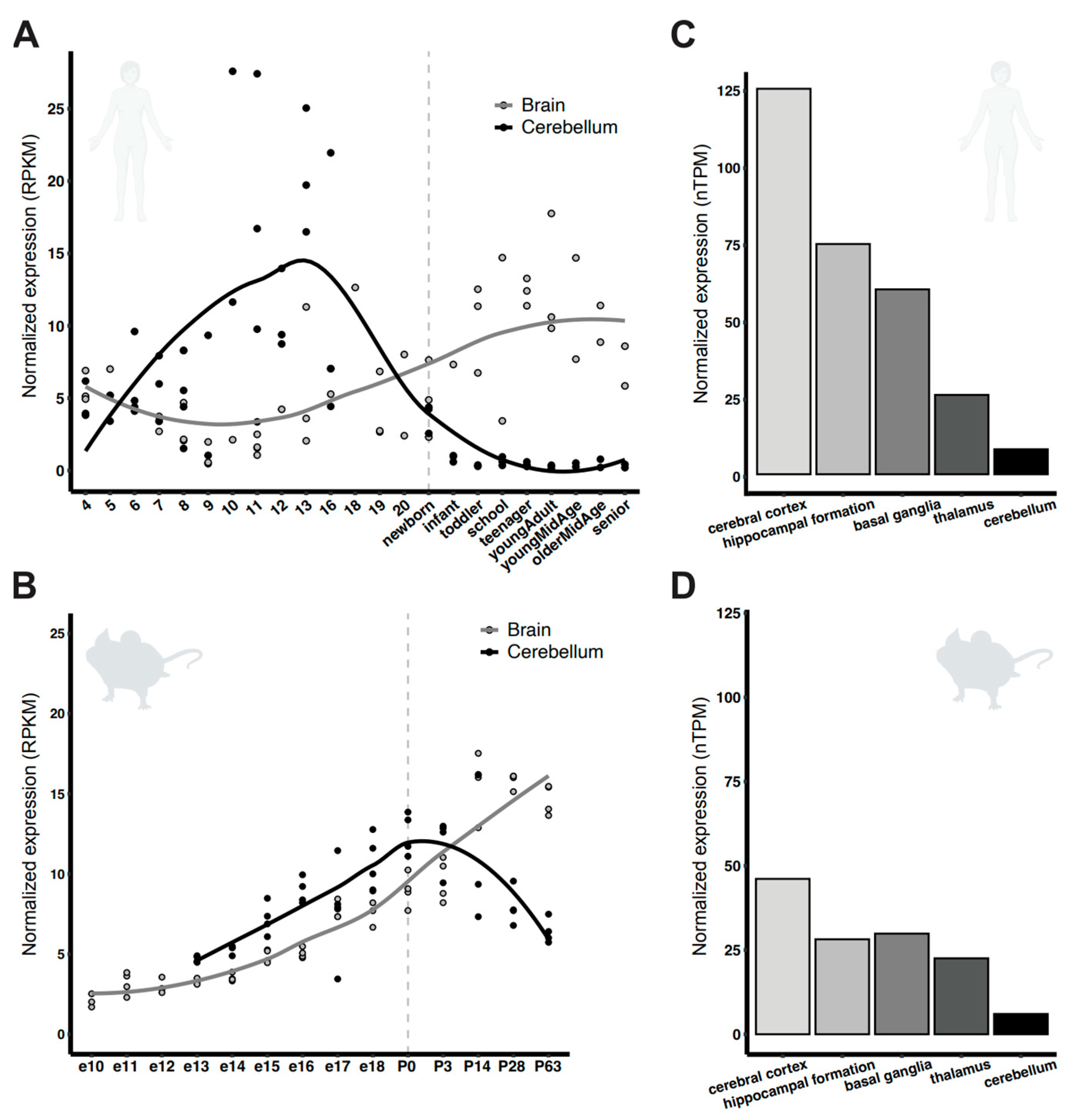
Figure 1. PCDH7 expression in human and murine brain. Normalized expression across developmental stages in (A) human, and (B) mouse. The vertical line separates prenatal and postnatal stages. Data was adapted from [46]. Normalized expression across the adult brain regions in (C) human and (D) mouse. Data was adapted from [47].
Genes 16 00985 g001
Figure 2. Generation of the Pcdh7 null mouse model. Schematic representation of the WT mouse Pchd7 genomic locus (A) and the sgRNA target sites for the generation of two Pcdh7 null mouse strains (B). Conventional PCR analysis of genomic DNA from the 1145-bp-deletion Pcdh7 mice, Pcdh7+/+ WT band, Pcdh7+/− WT and KO bands, Pcdh7−/− KO band (C). Confirmation of Pcdh7 loss of expression by RT-PCR amplifying a 173-bp cDNA fragment in exon 1 and a 267-bp fragment spanning exons 2 and 3 (D).
Genes 16 00985 g002
Figure 3. Seizure susceptibility in the Pcdh7 null mouse model. Schematic representation of the experimental setup of the flurothyl-induced seizure assay (A). Latencies to first myoclonic jerk (MJ), first generalized seizure (GS), characterized by transient loss of balance, and general tonic-clonic seizure (GTCS), characterized by loss of balance, wild running, and hopping behavior, were recorded (B), as well as the number of myoclonic jerks exhibited before the occurrence of the first GS (C). Western blot analyses of GFAP and α-Tubulin expression in control (D) or flurothyled (E) brain lysates. Data are shown as mean ± SD. n = 5–10 mice/genotype. * p = 0.0200, One-way Anova followed by Tukey’s multiple comparison test.
Genes 16 00985 g003
Figure 4. Histological analysis of the Pcdh7 mouse model. Cresyl violet staining was performed on coronal sections of adult Pcdh7 mice (A). The graph represents the thickness measurement of the lateral cortex (B). Hippocampal formation from insets shown in (A,C). The graph represents the thickness measurement of the hippocampus CA1 regions (D). No significant differences were observed between genotypes. Data are shown as mean + SD. Each data point represents a measurement in one coronal section. n = 3 mice/genotype.
Genes 16 00985 g004
Figure 5. Immunostaining of coronal sections of adult Pcdh7 mice. Hippocampal formation is shown in the insets. Satb2 (red), NeuN (green), and DAPI (blue). No differences were observed between genotypes.
Genes 16 00985 g005
Table 1. Breeding Pcdh7+/− mice generated litters with the expected genotype and sex frequencies.
FemalesMalesTotal
Numbers334073
%4555100
WTPcdh7+/−Pcdh7−/−Total
Numbers15381770
%215424100
WTPcdh7+/−Pcdh7−/−Total
NumbersFemales715830
Males823940
%Females235027100
Males205823100
Disclaimer/Publisher’s Note: The statements, opinions and data contained in all publications are solely those of the individual author(s) and contributor(s) and not of MDPI and/or the editor(s). MDPI and/or the editor(s) disclaim responsibility for any injury to people or property resulting from any ideas, methods, instructions or products referred to in the content.