Article

Novel Pathogenic Variants Leading to Sporadic Amyotrophic Lateral Sclerosis in Greek Patients

by
Ouliana Ivantsik
1,
Anne John
2,
Kyriaki Kydonopoulou
3,
Konstantinos Mitropoulos
4,
Spyridon Gerou
3,
Bassam R. Ali
2,5 and
George P. Patrinos
1,2,5,6,7,*
1
Laboratory of Pharmacogenomics and Individualized Therapy, Division of Pharmacology and Biosciences, Department of Pharmacy, School of Health Sciences, University of Patras, 26504 Rion, Greece
2
Department of Genetics and Genomics, College of Medicine and Health Sciences, United Arab Emirates University, Al-Ain P.O. Box 15551, United Arab Emirates
3
ANALYSI Biomedical Laboratories S.A., 54623 Thessaloniki, Greece
4
Department of Histology and Embryology, School of Medicine, National and Kapodistrian University of Athens, 10679 Athens, Greece
5
ASPIRE Abu Dhabi Precision Medicine Ρesearch Institute, Al-Ain P.O. Box 15551, United Arab Emirates
6
Zayed Center for Health Sciences, United Arab Emirates University, Al-Ain P.O. Box 15551, United Arab Emirates
7
Clinical Bioinformatics Unit, Department of Pathology, Faculty of Medicine and Health Sciences, Erasmus University Medical Center, 3000 CA Rotterdam, The Netherlands
*
Author to whom correspondence should be addressed.
Genes 2024, 15(3), 309; https://doi.org/10.3390/genes15030309
Submission received: 7 February 2024 / Revised: 22 February 2024 / Accepted: 26 February 2024 / Published: 28 February 2024
(This article belongs to the Special Issue Genetics of Multifactorial Diseases)

Abstract

:
Amyotrophic lateral sclerosis (ALS) is a rapidly progressive disease that affects motor neurons, leading to paralysis and death usually 3–5 years after the onset of symptoms. The investigation of both sporadic and familial ALS highlighted four main genes that contribute to the pathogenesis of the disease: SOD1, FUS, TARDBP and C9orf72. This study aims to provide a comprehensive investigation of genetic variants found in SOD1, FUS and TARDBP genes in Greek sporadic ALS (sALS) cases. Our sequencing analysis of the coding regions of the abovementioned genes that include the majority of the variants that lead to ALS in 32 sALS patients and 3 healthy relatives revealed 6 variants in SOD1, 19 variants in FUS and 37 variants in TARDBP, of which the SOD1 p.D90A and the FUS c.*356G>A (rs886051940) variants have been previously associated with ALS, while two novel nonsense pathogenic variants were also identified, namely FUS p.R241* and TDP-43 p.Y214*. Our study contributes to the worldwide effort toward clarifying the genetic basis of sALS to better understand the disease’s molecular pathology.

1. Introduction

Amyotrophic lateral sclerosis (ALS), also known as Lou Gehrig’s syndrome, is the third most common neurodegenerative disorder after Alzheimer’s and Parkinson’s [1]. More recent studies report an incidence between 0.6 and 3.8 per 100,000 person-years [2,3,4,5,6,7,8,9], with studies in Europe reporting 2.1–3.8 per 100,000 person-years [2,3,4,8]. The main feature of the disease is the progressive paralysis of the motor neurons. Symptoms include a focal onset of weakness, which gradually progresses to affect all the limbs and bulbar muscles, and hyperreflexia [10,11]. The disease progresses rapidly, leading to paralysis and death from respiratory failure, usually 3 to 5 years after the onset of symptoms. However, to date, there is no cure, or any presymptomatic testing available [12].
Most ALS cases (about 90–95%) are sporadic (sALS) and seem to be influenced by environmental factors but also largely by genetics, while 5–10% are familial (fALS) and mostly show an autosomal dominant pattern of inheritance [13]. The two forms have common symptoms and progression processes. After studying both familial and sporadic cases, more than 20 genes have emerged as being involved in the occurrence of ALS [14]. The four most commonly mutated genes in ALS are SOD1, FUS, TARDBP and C9orf72.
The first gene associated with the disease is SOD1, which encodes the enzyme superoxide dismutase (SOD1). The enzyme catalyzes the conversion of harmful superoxide radicals to nitric oxide (NO) in the superoxide anion form (ONOO-) [15,16,17]. The main mechanism proposed is that pathogenic variants in this gene can disrupt this free radical elimination process, causing their accumulation in nerve cells and death [18]. Recently, the development of animal models with a disruption in SOD1 highlighted other mechanisms, including excitotoxicity, oxidative stress, mitochondrial dysfunctions and non-cell autonomous toxicity [19]. More than 170 SOD1 variants have been reported to date, affecting approximately 20% of fALS and 1% of sALS cases [20]. Most genomic variants are missense and span the entire coding sequence.
Another gene that is strongly associated with ALS is FUS, also known as translocated in liposarcoma (TLS), which encodes a protein involved in RNA processing. More than 170 genomic variants have been identified, most of them missense [21], and are associated with approximately 5% of fALS [22] and 0.3% of sALS cases [23]. It has been reported that patients with FUS variants have an earlier onset of symptoms than other forms of the disease, usually before the age of 45 years [24,25]. Pathogenic variants in this gene have been found to correlate with all the cellular functions that are disrupted in degenerating motor neurons [26].
In 2006, a study showed that many sALS patients have an accumulation of ubiquitinated TDP-43 protein in pathological cytosolic inclusions [27]. TDP-43 is a 43 kDa protein encoded by the TARDBP gene, which is involved in RNA-related processes, namely mRNA metabolism, transcription, and splicing and transport, as well as microRNA biosynthesis [28]. To date, more than 40 genomic variants in the TARDBP gene have been documented, almost all of them missense. Pathogenic variants in the TARDBP gene are responsible for ~5% of fALS [27] and 1% of sALS cases [29].
An important landmark study in understanding the genetic basis of ALS is the discovery of the GGGGCC hexanucleotide repeat expansion in the C9orf72 gene, which appears to be the most common genetic cause of the disease in Europe and North America [30,31], representing 45–50% of fALS and 5–10% of sALS cases [32]. The physiological function of C9orf72 remains unknown.
The genetic basis of ALS in the Greek population has been poorly studied. The first study was published in 2012, where the frequency of the expansion of the hexanucleotide GGGGCC in the C9orf72 gene was investigated in 146 patients with ALS [33]. In 2017, another study performed a whole-genome sequencing analysis of Greek patients and highlighted the association of variants in the FTO gene with sALS [34]. Here, we attempt to provide a comprehensive molecular genetic analysis of variants in the SOD1, FUS and TARDBP genes in Greek sALS patients, providing further insights on the molecular genetic spectrum of ALS in Greece.

2. Materials and Methods

2.1. Study Population

sALS patients and healthy relatives, mostly referred to from the Greek ALS patients Association and the ALS Find-a-Cure Association, were recruited from June 2010 until June 2023 at the Laboratory of Pharmacogenomics and Individualized Therapy, Department of Pharmacy, University of Patras School of Health Sciences, Greece. The study was approved by the University of Patras Bioethics Committee. All participants signed an informed consent for blood sample storage, DNA extraction and analysis. In total, the study included 32 sALS patients diagnosed according to the El Escorial criteria [35] and 3 healthy relatives (Supplementary Tables S2 and S3).

2.2. Genetic Testing

DNA was extracted from whole blood samples using standard laboratory procedures [34]. The concentration and quality of the DNA assessed using Nanodrop 2000 (Thermo Fisher Scientific, Waltham, MA, USA) and 50 ng of genomic DNA was used, through Polymerase Chain Reaction (PCR), to amplify 17 DNA fragments [4 fragments including exons 2–5 of SOD1 (NM_000454.5, hg_38), 7 fragments including exons 2, 3, 5, 6, 12–15 and the 3′UTR of FUS (NM_004960.4, hg_38) and 6 fragments including exons 1–6 of the TARDBP gene (NM_007375.4, hg_38)]. The selection of these segments for analysis was based on the fact that these are the exons in which more disease-associated variants have been identified [21,36]. The amplification conditions and primer sequences are available upon request. Subsequently, PCR-amplified samples were Sanger sequenced and analyzed using the BioEdit Sequence Alignment Editor [37].

2.3. In Silico Analysis

For the in silico prediction of the variants’ pathogenicity we used the MutationTaster2021 [38], while missense variants were also assessed via PolyPhen-2 [39] and SIFT [40]. Known variants were also searched for in the ClinVar [41] and ClinGen [42] databases.

3. Results

3.1. SOD1

We sequenced exons 2–5 of the SOD1 gene and found six genetic variants in three patients (Table 1). Only one of them had an assigned rsID and was classified as benign/likely benign in ClinVar and ClinGen. Three of them were silent, while the others were missense. Five of the variants were cut during the post-translational modifications, and only one (c.349A>C, p.D11A) remained in the mature functional protein. MutationTaster assessed all the variants as disease causing. PolyPhen-2 tool and SIFT assessed as “probably damaging” and “affect protein function”, respectively the missense variants (c.251T>G p.C57W score = 1; c.292A>T p.H71L; score = 0.990) that are not participating in the final functional protein, while the only variant that remains was characterized as “benign” and “tolerated” (c.349A>C p.D90A) (Table 1 and Table S1).

3.2. FUS

The sequencing of the selected exons of the FUS gene revealed several genetic variations in Greek sALS patients (Table 2). Most of the variants (n = 8) were located in the 3′ UTR, following exon 6 (n = 6), exon 3 (n = 4) and exon 2 (n = 1). We found no variants in exons 5, 12, 13, 14 or 15. Regarding variants in coding sequence, eight of them were missense, one was silent and one was nonsense. RsIDs were found for 7 of 19 variants. We searched those in ClinVar and ClinGen but found reports only for three: rs80301724 and rs151073460 classified as benign/likely benign and rs886051940 as uncertain significance. The most common variant was c.223C>A, a silent variant, found in 10 patients; 5 were heterozygous and 5 homozygous. Other common variants were c.806C>T, a missense variant found in five heterozygous patients; c.*356G>A in 3′ UTR, found in five heterozygous patients; c.626A>C, a missense variant found in three heterozygous patients; c.760C>G, a silent variant found in two heterozygous patients; and c.800A>T, a nonsense variant found in two heterozygous patients.
We were able to assess all but two variants using MutationTaster (c.*370A>T, c.*406G>A). Eleven variants were characterized as disease causing, while six, as polymorphisms. The eight missense variants were assessed via PolyPhen-2 and SIFT. SIFT characterized all variants as affecting protein function. PolyPhen-2 characterized only four of them as “probably damaging” (c.162G>T p.S28I score = 0.868; c.184G>C p.Q35H score = 0.998; c.221G>T p.G48C score = 0.999; c.830G>T p.G251C score = 1; Table 2 and Table S1).
We also sequenced the first-degree relatives of patients ALS-58, ALS-66 and ALS-72. Patient ALS-58 had no variants in the FUS gene, while his healthy son (ALS-59) had a missense variant (c.264A>G p.N63S) (Supplementary Tables S1 and S2). Therefore, ALS-59, a healthy individual, inherited the variant from his healthy mother, so it is probably a non-harmful variant in the FUS gene (Figure 1a). Patient ALS-66 had two missense variants in the FUS gene (c.184G>C p.Q35H; c.221G>T p.G48C), both characterized as probably damaging by in silico tools. His healthy daughter (ALS-67) had no variants in the FUS gene (Figure 1b, Supplementary Tables S1 and S2), a finding that suggests that c.184G>C (p.Q35H) and c.221G>T (p.G48C) may play causative roles in developing ALS. Patient ALS-72 had no variants in the FUS gene, but his son had two variants in the 3′ UTR of the FUS gene (c.*356G>A; c.*446G>A). Since his father did not have these variants, we assume that he inherited them from his healthy mother (Figure 1c).

3.3. TARDBP

The sequencing of TARDBP exons revealed 37 genetic variations (Table 3), scattered throughout the whole coding sequence. In total, 22 of the variants were missense, 10 were silent, 4 variants were found in the UTRs, and we also identified 1 nonsense variant causing the preliminary termination of translation in amino acid 214. A total of 11 variants were found in exon 6, containing the 3′ UTR; 10 variants in exon 5 (n = 10); 7 variants in exon 2; 6 variants in exon 3; 2 variants in exon 1, containing the 5′ UTR; and 1 variant in exon 4. Only five variants had assigned rsIDs (Table 3). ClinVar and ClinGen report the silent variant rs61730366 as benign/likely benign, while the missense variant rs1643653768 as likely pathogenic or uncertain significance.
The most common variants found were the missense c.274G>A p.E57K and the silent variant c.1134G>A found in eight patients. Nine of the missense variants were characterized as possibly damaging by all in silico tools: c.295G>A (p.D64N; score = 0.570), c.295G>T (p.D64Y; score = 0.904), c.490G>T (p.V129F; score = 0.494), c.500T>G (p.V132G; score = 0.943), c.741G>T (p.Q212H; score = 0.865), c.1001G>A (p.G299E; score = 0.953), c.1180C>A (p.Q326R; score = 0.654), c.1322C>T (p.S406F; score = 0.922) and c.1328C>T (p.S408F; score = 0.472). Also, PolyPhen-2 characterized five variants as “probably damaging”: c.304T>G (p.W67G; score = 0.996), c.594G>C (p.Q163H; score = 0.997), c.686T>G (p.F193L; score = 0.988), c.781A>G (p.R226G; score = 1) and c.1326G>T (p.K407N; score = 0.998) (Table 3 and Table S3).
Comparing genetic variants in patient–healthy relative pairs, we saw that healthy individual ALS-59 carried three out of six variants from his father (ALS-58): a silent variant (c.714G>A) and two missense variants (c.674G>A, c.781A>G) (Figure 2a). Based on this, we assume that the combination of these variants does not lead to ALS. Moreover, the genetic variant c.1134G>A, which was identified in the ALS-58 patient but not in his healthy son, was also identified in seven more sALS patients (Table 3). Healthy individual ALS-67 had no variants in TARDBP, while her father had four: c.227G>C (p.R41P), c.274G>A (p.E57K), c.295G>A (p.D64N) and c.304T>G (p.W67G) (Figure 2b). Also, ALS-71 had zero variants in TARDBP, but his father carried the c.274G>A variant (Figure 2c). We saw that the c.274G>A variant was found in ALS-66, ALS-72 and seven more sALS cases, but at none of the healthy offsprings, which implies that it may be a variant related to the overall sALS phenotype. Genetic variants in the 295th nucleotide were also common in patients (c.295G>A/T), but not in healthy relatives.

4. Discussion

The aim of this study was to investigate the genetic variations in Greek ALS patients, through molecular genetic analysis in the SOD1, FUS and TARDBP genes. Our sequencing analysis revealed a number of genetic variants that may contribute to ALS pathogenesis. The most significant were the missense variant c.349A>C (p.D90A) in the SOD1 gene, a nonsense variant c.800A>T (p.R241*) in the FUS gene, a nonsense variant c.744C>G (p.Y214*) in the TARDBP gene and the c.1134G>A and c.274G>A (p.E57K) variants, which were reported only in patients and not in healthy first-degree relatives.
Regarding SOD1, we found six variants, half of them silent and the others missense. Five of the variants were on the part of the protein that is cut during post-translational modification, so they probably do not affect the function of the mature protein. On the other hand, all in silico analysis tools assessed the two missense variants (c.251T>G and c.292A>T) as “probably damaging”, so these may affect the protein’s folding, consequently obstructing proper post-translational modifications. Despite the fact that there have been no reported mutational hotspots in SOD1, we found three variants in the 248–254 region of the coding sequence (56–58 amino acids), suggesting that this may be a mutational hotspot. This region does not participate in the functional protein, but another study has reported a causative variant in amino acid 59 (p.S59S) [43]. One patient was homozygous in the c.349A>C missense variant. This was the only variant found that alters the mature protein’s sequence. This variant has been repeatedly reported before in Non-Hispanic White Americans sALS cases [44]; Caucasian, Canadian sALS and fALS cases [45], European Italian fALS cases [43]; and Asian, Iranian fALS cases [46]. PolyPhen-2 and SIFT characterized this variant as benign, but MutationTaster characterized it as disease causing. There is a family report where the c.349A>C heterozygous status was associated with ALS symptoms and related death [47]. Considering the fact that this is the sixth report highlighting the c.349A>C missense variant in ALS patients, we suggest that it is probably one of the genetic causes of both sporadic and familial ALS. Also, it would be interesting to investigate if the two missense variants that were characterized as possibly damaging affect the proper protein maturation.
In the FUS gene, we report 19 genetic variants. Most of them were located in 3′ UTR. Genomic variants in the 3′ UTR of the FUS gene have been repeatedly associated with ALS [48,49,50,51,52,53,54], but only two of the variants we found have been previously reported in ALS cases, specifically c.*41G>A (rs80301724) and c.*356G>A (rs886051940) [50,51,54,55]. The c.*41G>A (rs80301724) variant is a common variant that has been excluded as causative for ALS, since it has been proven to have the same frequency in patients and healthy controls [50,51,54]. Moreover, ClinVar and ClinGen report the variant as benign/likely benign. The rs886051940 variant found in five patients, showing a high frequency in the Greek sALS population, has been previously associated with ALS pathogenesis [55]. It is interesting that healthy individual ALS-71 also had the rs886051940 variant alongside another 3′ UTR variant, both of which he probably inherited from his healthy mother. This finding indicates that rs886051940 may be a causative variant in ALS with reduced penetrability. Based on ClinVar and ClinGen, the variant is classified as uncertain significance. The suggested mechanism on how variants in the 3′ UTR of FUS can cause ALS is that these variants drastically increase the FUS protein expression in the patients’ fibroblasts [54]. So, the overexpression of FUS seems to be toxic for nerve cells.
We also found 11 genetic variations in exons 2, 3 and 6: 8 missense, 2 silent and 1 nonsense variant. It is very interesting that most studies are referring to variants located in exons 14–15 [21], while in our study, we found none in these exons. This suggests that the genetic basis of the Greek sALS patients has a unique pattern, consistent with our previous findings. The most common variant was c.223C>A, found in 10 patients, half of them homozygous. This substitution does not alter the protein sequence, so it is interesting to investigate if it is associated with ALS or is just a common variant in the Greek population, probably as a result of a founder effect. The other silent variant (c.760C>G, rs151073460) has been previously reported in Parkinson’s disease patients; this means that despite the fact that it does not alter the protein’s sequence, it may still disrupt protein function [56]. Although ClinVar reported the variant as benign/likely benign, MutationTaster assessed it as disease causing. SIFT characterized all missense variants as probably damaging, but PolyPhen-2 characterized only four out of eight variants with this term. The other missense variants that were characterized as benign were also common in more than one sample, which contributes to the assumption that they are not harmful, but common Greek variants.
A very important finding is a novel nonsense variant (c.800A>T) that causes the early termination of translation in codon 241. This results in a truncated protein with less than half of the amino acids. The consequence of having a FUS protein that is shorter than the wild type has been studied in a mouse model with a truncated FUS mutant protein (amino acids 1–359). The mice developed ALS symptoms, but at the end stage, they showed a greater than 50% preservation of spinal motor neurons [25,57,58,59,60], suggesting that a truncated protein may be even partially sufficient for neuron function. Although, the variant we found leads to a significantly shorter protein, it may have more severe consequences in motor neurons.
TARDBP was the gene with the most genetic variants found in our study. Variants in this gene are common in sALS cases [29]. Although previous studies have identified variants mostly in exon 6 [61], we found variants that span the whole coding sequence. Only one of the variants we found has been recorded before in ALS cases (c.995G>T p.G297V) [62,63]. This is the first study recording variants in UTR; specifically, we found two variants in 5′ UTR (c.5T>C; c.1347G>A) and two in 3′ UTR (c.1350A>G; c.24C>G). As mentioned previously, the region most strongly associated with pathogenic variants that lead to ALS in FUS is 5′ UTR, as it leads to overexpression, which is toxic for nerve cells, but there is no corresponding reference for variants in TARDBP’s UTRs.
The two most common variants we found in TARDBP are c.1134G>A, which is a silent variant, and c.274G>A p.E57K, a missense variant characterized as benign via PolyPhen-2. Both of them have no or minimal influence on the protein, but they were found only in patients and not in healthy first-degree relatives. Moreover, the only variant we found that has previously been reported as associated with ALS and that both ClinVar and ClinGen classified it as both likely pathogenic and uncertain significance (c.995G>T p.G297V rs1643653768) [62,63] was also characterized as benign using the PolyPhen-2 tool; so, variants that do not significantly affect the amino acid sequence may have other consequences leading to pathogenesis, such as altering the gene’s expression. Another variant found only in patients and not in first-degree relatives is c.295G>A p.D64N, a missense variant characterized as possibly damaging by all in silico tools used.
The proposed pathogenetic mechanism by which variants in TARDBP lead to ALS involve the accumulation of ubiquitinated TDP-43 protein in pathological cytosolic inclusions [27]. The region of TDP-43 that interacts with UBQLN2 is amino acids 216–414 [64]. In that specific region, we found 11 genetic variants, 2 missense and probably damaging (c.781A>G p.R226G; c.1326G>T p.K407N), 5 possibly damaging [c.1001G>A (p.G299E); c.1001G>A (p.G299E); c.1180C>A (p.Q326R); c.1322C>T (p.S406F); c.1328C>T (p.S408F)] and the c.995G>T variant, which has previously been associated with ALS. This region is encoded primarily by triplets located in exon 6, where most genetic variants have been recorded [61].
Finally, we identified a nonsense variant [c.744C>G (p.Y214*)], which leads to the production of a truncated protein. Corcia and coworkers [65] also reported a truncation variant at the extreme C-terminus of the protein p.Y374*, which leads to ALS, but in our case, the preliminary termination leads to the production of a protein with half the size of the wild type.
Looking at the pattern of variants per patient (Table 1), we saw that patient ALS-30 had three variants in the FUS gene, one characterized via PolyPhen-2 as “probably damaging” [c.162G>T (p.S28I)] and two as “benign” [c.626A>C (p.M183L) and c.806C>T (p.R243C)]. Furthermore, patient ALS-69 had three variants in the 3′ UTR of FUS gene, so we assume he probably had a more severe alteration in protein expression. An even more intense accumulation of variants per patient was observed in the TARDBP gene. Patient ALS-57 had eight variants in the TARDBP gene, while ALS-58 and ALS-73 had six variants, each. The aggregation of more than one variant in the gene may have a cumulative effect, leading to a more serious disruption in protein function. Unfortunately, the clinical data of patients were not available to test this hypothesis.

Strengths and Limitations

This study had a few strengths and limitations. This is the first study investigating the genomic variation in the three most common genes associated with sALS in the Greek population. Moreover, comparing genetic variations in patients and first-degree relatives made it possible to distinguish some of the genetic variants that are probably benign. However, the study also has several limitations. The most important is the small number of patients and healthy individuals included. There are no official data about the prevalence of ALS in Greece, but considering that it has the same prevalence as in Europe, we assumed that we included around 15–20% of the expected ALS patient population. This of course facilitated from the strong collaborative ties with the ALS patient associations throughout the years. Concerning the genetic variants we found, although there is evidence that indicates that several could lead to ALS (those characterized as probably damaging, common in patients but not in healthy relatives, etc.), we cannot draw certain conclusions. One reason is that we studied a population with common ancestry and several variants may be the results of a founder effect.

5. Conclusions

The genetic basis of sALS remains unclear. Even though genetic variants in the SOD1, FUS and TARDBP genes have been associated with ALS pathogenesis, all the variants that may lead to ALS have not been recorded, and it is not clear which variants are causative and which are benign. This study provides a thorough investigation of genetic variants found in sALS Greek patients. The study confirmed the association of several variants that were previously reported as causative, and also highlighted novel variants. The most significant novel variants are the two nonsense variants found, the c.800A>T (p.R241*) in the FUS gene and the c.744C>G (p.Y214*) in the TARDBP gene. Both variants lead to a significantly shorter protein that has ever been reported. In addition to these, we suggest that variants characterized as probably damaging and those that are common in patients but not healthy relatives should be further investigated.
ALS is a rapidly progressive disease, and there is no cure and no presymptomatic screening available. It is important that more studies investigating the genetic basis of ALS should be carried out, so we could at least gather the most common causative variants and develop a screening panel for presymptomatic assessment in high-risk individuals or help to diagnose patients faster at an early stage.

Supplementary Materials

The following supporting information can be downloaded at https://www.mdpi.com/article/10.3390/genes15030309/s1: Table S1: Prediction and scores of variant pathogenicity based on computational prediction tools; Table S2: Genetic variants found in each sporadic ALS patient; Table S3: Genetic variants found in family members that do not suffer from sporadic ALS.

Author Contributions

Conceptualization, O.I. and G.P.P.; Methodology, O.I. and G.P.P.; Validation, O.I.; Formal Analysis, O.I., A.J. and K.K.; Investigation, O.I.; Resources, K.M., B.R.A., G.P.P. and O.I.; Writing—Original Draft Preparation, O.I.; Writing—Review and Editing, all authors; Visualization, O.I.; Supervision, B.R.A. and G.P.P.; Project Administration, G.P.P.; Funding Acquisition, G.P.P. All authors have read and agreed to the published version of the manuscript.

Funding

This research was funded by the Hellenic Precision Medicine Network for Neurodegenerative Diseases “https://neuropmnet.gr/?page_id=6109&lang=en; 2018SE01300001 (accessed on 7 February 2024)” and the University of Patras Research budget.

Institutional Review Board Statement

The study was conducted according to the guidelines of the Declaration of Helsinki and approved by the Ethics Committee of the University of Patras (12 July 2010).

Informed Consent Statement

Informed consent was obtained from all subjects involved in the study.

Data Availability Statement

Data are available upon request.

Conflicts of Interest

Author Korina Kydonopoulou is employed by ANALYSI S.A. He participated in Formal Analysis in the study. Author Spyridon Gerou is Managing Director and the lead physician in ANALYSI S.A. And both of them participate in Review and Editing of the manuscript. The remaining authors declare that the research was conducted in the absence of any commercial or financial relationships that could be construed as a potential conflict of interest.

References

  1. Gosset, P.; Camu, W.; Raoul, C.; Mezghrani, A. Prionoids in amyotrophic lateral sclerosis. Brain Commun. 2022, 4, fcac145. [Google Scholar] [CrossRef]
  2. Longinetti, E.; Regodón Wallin, A.; Samuelsson, K.; Press, R.; Zachau, A.; Ronnevi, L.O.; Kierkegaard, M.; Andersen, P.M.; Hillert, J.; Fang, F.; et al. The Swedish motor neuron disease quality registry. Amyotroph. Lateral Scler. Front. Degener. 2018, 19, 528–537. [Google Scholar] [CrossRef] [PubMed]
  3. Palese, F.; Sartori, A.; Verriello, L.; Ros, S.; Passadore, P.; Manganotti, P.; Barbone, F.; Pisa, F.E. Epidemiology of amyotrophic lateral sclerosis in Friuli-Venezia Giulia, North-Eastern Italy, 2002–2014: A retrospective population-based study. Amyotroph. Lateral Scler. Frontotemporal Degener. 2019, 20, 90–99. [Google Scholar] [CrossRef] [PubMed]
  4. Benjaminsen, E.; Alstadhaug, K.B.; Gulsvik, M.; Baloch, F.K.; Odeh, F. Amyotrophic lateral sclerosis in Nordland County, Norway, 2000–2015: Prevalence, incidence, and clinical features. Amyotroph. Lateral Scler. Frontotemporal Degener 2018, 19, 522–527. [Google Scholar] [CrossRef]
  5. Jun, K.Y.; Park, J.; Oh, K.W.; Kim, E.M.; Bae, J.S.; Kim, I.; Kim, S.H. Epidemiology of ALS in Korea using nationwide big data. J. Neurol. Neurosurg. Psychiatry 2019, 90, 395–403. [Google Scholar] [CrossRef] [PubMed]
  6. Turgut, N.; Varol SaraÇoglu, G.; Kat, S.; Balci, K.; GÜldiken, B.; Birgili, O.; Kabayel, L. An epidemiologic investigation of amyotrophic lateral sclerosis in Thrace, Turkey, 2006–2010. Amyotroph. Lateral Scler. Frontotemporal Degener. 2019, 20, 100–106. [Google Scholar] [CrossRef]
  7. Zhou, S.; Zhou, Y.; Qian, S.; Chang, W.; Wang, L.; Fan, D. Amyotrophic lateral sclerosis in Beijing: Epidemiologic features and prognosis from 2010 to 2015. Brain Behav. 2018, 8, e01131. [Google Scholar] [CrossRef]
  8. Leighton, D.J.; Newton, J.; Stephenson, L.J.; Colville, S.; Davenport, R.; Gorrie, G.; Morrison, I.; Swingler, R.; Chandran, S.; Pal, S.; et al. Changing epidemiology of motor neurone disease in Scotland. J. Neurol. 2019, 266, 817–825. [Google Scholar] [CrossRef]
  9. Rose, L.; McKim, D.; Leasa, D.; Nonoyama, M.; Tandon, A.; Bai, Y.Q.; Amin, R.; Katz, S.; Goldstein, R.; Gershon, A. Trends in incidence, prevalence, and mortality of neuromuscular disease in Ontario, Canada: A population-based retrospective cohort study (2003–2014). PLoS ONE 2019, 14, e0210574. [Google Scholar] [CrossRef]
  10. Swinnen, B.; Robberecht, W. The phenotypic variability of amyotrophic lateral sclerosis. Nat. Rev. Neurol. 2014, 10, 661–670. [Google Scholar] [CrossRef]
  11. Masrori, P.; Van Damme, P. Amyotrophic lateral sclerosis: A clinical review. Eur. J. Neurol. 2020, 27, 1918–1929. [Google Scholar] [CrossRef]
  12. Cirulli, E.T.; Lasseigne, B.N.; Petrovski, S.; Sapp, P.C.; Dion, P.A.; Leblond, C.S.; Couthouis, J.; Lu, Y.F.; Wang, Q.; Krueger, B.J.; et al. Exome sequencing in amyotrophic lateral sclerosis identifies risk genes and pathways. Science 2015, 347, 1436–1441. [Google Scholar] [CrossRef]
  13. Ji, A.L.; Zhang, X.; Chen, W.W.; Huang, W.J. Genetics insight into the amyotrophic lateral sclerosis/frontotemporal dementia spectrum. J. Med. Genet. 2017, 54, 145–154. [Google Scholar] [CrossRef] [PubMed]
  14. Chia, R.; Chiò, A.; Traynor, B.J. Novel genes associated with amyotrophic lateral sclerosis: Diagnostic and clinical implications. Lancet Neurol. 2018, 17, 94–102. [Google Scholar] [CrossRef]
  15. Salem, K.; McCormick, M.L.; Wendlandt, E.; Zhan, F.; Goel, A. Copper-zinc superoxide dismutase-mediated redox regulation of bortezomib resistance in multiple myeloma. Redox Biol. 2015, 4, 23–33. [Google Scholar] [CrossRef]
  16. Venkataramani, V.; Doeppner, T.R.; Willkommen, D.; Cahill, C.M.; Xin, Y.; Ye, G.; Liu, Y.; Southon, A.; Aron, A.; Au-Yeung, H.Y.; et al. Manganese causes neurotoxic iron accumulation via translational repression of amyloid precursor protein and H-Ferritin. J. Neurochem. 2018, 147, 831–848. [Google Scholar] [CrossRef] [PubMed]
  17. Majzúnová, M.; Pakanová, Z.; Kvasnička, P.; Bališ, P.; Čačányiová, S.; Dovinová, I. Age-dependent redox status in the brain stem of NO-deficient hypertensive rats. J. Biomed. Sci. 2017, 24, 72. [Google Scholar] [CrossRef]
  18. Schmitt, N.D.; Agar, J.N. Parsing disease-relevant protein modifications from epiphenomena: Perspective on the structural basis of SOD1-mediated ALS. J. Mass. Spectrom. 2017, 52, 480–491. [Google Scholar] [CrossRef] [PubMed]
  19. Peggion, C.; Scalcon, V.; Massimino, M.L.; Nies, K.; Lopreiato, R.; Rigobello, M.P.; Bertoli, A. SOD1 in ALS: Taking Stock in Pathogenic Mechanisms and the Role of Glial and Muscle Cells. Antioxidants 2022, 11, 614. [Google Scholar] [CrossRef]
  20. Abel, O.; Powell, J.F.; Andersen, P.M.; Al-Chalabi, A. ALSoD: A user-friendly online bioinformatics tool for amyotrophic lateral sclerosis genetics. Hum. Mutat. 2012, 33, 1345–1351. [Google Scholar] [CrossRef]
  21. Xiao, X.; Li, M.; Ye, Z.; He, X.; Wei, J.; Zha, Y. FUS gene mutation in amyotrophic lateral sclerosis: A new case report and systematic review. Amyotroph. Lateral Scler. Frontotemporal Degener. 2024, 25, 1–15. [Google Scholar] [CrossRef] [PubMed]
  22. Yan, J.; Deng, H.X.; Siddique, N.; Fecto, F.; Chen, W.; Yang, Y.; Liu, E.; Donkervoort, S.; Zheng, J.G.; Shi, Y.; et al. Frameshift and novel mutations in FUS in familial amyotrophic lateral sclerosis and ALS/dementia. Neurology 2010, 75, 807–814. [Google Scholar] [CrossRef] [PubMed]
  23. Grassano, M.; Brodini, G.; De Marco, G.; Casale, F.; Fuda, G.; Salamone, P.; Brunetti, M.; Sbaiz, L.; Gallone, S.; Cugnasco, P.; et al. Phenotype analysis of fused in sarcoma mutations in amyotrophic lateral sclerosis. Neurol. Genet. 2022, 8, e200011. [Google Scholar] [CrossRef] [PubMed]
  24. Bäumer, D.; Hilton, D.; Paine, S.M.; Turner, M.R.; Lowe, J.; Talbot, K.; Ansorge, O. Juvenile ALS with basophilic inclusions is a FUS proteinopathy with FUS mutations. Neurology 2010, 75, 611–618. [Google Scholar] [CrossRef] [PubMed]
  25. Huang, C.; Zhou, H.; Tong, J.; Chen, H.; Liu, Y.J.; Wang, D.; Wei, X.; Xia, X.G. FUS transgenic rats develop the phenotypes of amyotrophic lateral sclerosis and frontotemporal lobar degeneration. PLoS Genet. 2011, 7, e1002011. [Google Scholar] [CrossRef]
  26. Assoni, A.F.; Foijer, F.; Zatz, M. Amyotrophic lateral sclerosis, FUS and Protein synthesis defects. Stem Cell Rev. Rep. 2023, 19, 625–638. [Google Scholar] [CrossRef]
  27. Neumann, M.; Sampathu, D.M.; Kwong, L.K.; Truax, A.C.; Micsenyi, M.C.; Chou, T.T.; Bruce, J.; Schuck, T.; Grossman, M.; Clark, C.M.; et al. Ubiquitinated TDP-43 in frontotemporal lobar degeneration and amyotrophic lateral sclerosis. Science 2006, 314, 130–133. [Google Scholar] [CrossRef]
  28. Buratti, E.; Baralle, F.E. The multiple roles of TDP-43 in pre-mRNA processing and gene expression regulation. RNA Biol. 2010, 7, 420–429. [Google Scholar] [CrossRef]
  29. Kabashi, E.; Valdmanis, P.N.; Dion, P.; Spiegelman, D.; McConkey, B.J.; Vande Velde, C.; Bouchard, J.P.; Lacomblez, L.; Pochigaeva, K.; Salachas, F. TARDBP mutations in individuals with sporadic and familial amyotrophic lateral sclerosis. Nat. Genet. 2008, 40, 572–574. [Google Scholar] [CrossRef]
  30. DeJesus-Hernandez, M.; Mackenzie, I.R.; Boeve, B.F.; Boxer, A.L.; Baker, M.; Rutherford, N.J.; Nicholson, A.M.; Finch, N.A.; Flynn, H.; Adamson, J.; et al. Expanded GGGGCC hexanucleotide repeat in noncoding region of C9ORF72 causes chromosome 9p-linked FTD and ALS. Neuron 2011, 72, 245–256. [Google Scholar] [CrossRef]
  31. Renton, A.E.; Majounie, E.; Waite, A.; Simón-Sánchez, J.; Rollinson, S.; Gibbs, J.R.; Schymick, J.C.; Laaksovirta, H.; van Swieten, J.C.; Myllykangas, L.; et al. A hexanucleotide repeat expansion in C9ORF72 is the cause of chromosome 9p21-linked ALS–FTD. Neuron 2011, 72, 257–268. [Google Scholar] [CrossRef]
  32. Ghasemi, M.; Brown, R.H., Jr. Genetics of amyotrophic lateral sclerosis. Cold Spring Harb. Perspect. Med. 2018, 8, a024125. [Google Scholar] [CrossRef]
  33. Mok, K.Y.; Koutsis, G.; Schottlaender, L.V.; Polke, J.; Panas, M.; Houlden, H. High frequency of the expanded C9ORF72 hexanucleotide repeat in familial and sporadic Greek ALS patients. Neurobiol. Aging 2012, 33, 1851.e1–1851.e15. [Google Scholar] [CrossRef]
  34. Mitropoulos, K.; Merkouri Papadima, E.; Xiromerisiou, G.; Balasopoulou, A.; Charalampidou, K.; Galani, V.; Zafeiri, K.V.; Dardiotis, E.; Ralli, S.; Deretzi, G.; et al. Genomic variants in the FTO gene are associated with sporadic amyotrophic lateral sclerosis in Greek patients. Hum. Genom. 2017, 11, 30. [Google Scholar] [CrossRef]
  35. Brooks, B.R.; Miller, R.G.; Swash, M.; Munsat, T.L. World Federation of Neurology Research Group on Motor Neuron Diseases. El Escorial revisited: Revised criteria for the diagnosis of amyotrophic lateral sclerosis. Amyotroph. Lateral Scler. Other Mot. Neuron Disord. 2000, 1, 293–299. [Google Scholar] [CrossRef]
  36. Chen, Y.P.; Yu, S.H.; Wei, Q.Q.; Cao, B.; Gu, X.J.; Chen, X.P.; Song, W.; Zhao, B.; Wu, Y.; Sun, M.M. Role of genetics in amyotrophic lateral sclerosis: A large cohort study in Chinese mainland population. J. Med. Genet. 2020, 59, 840–849. [Google Scholar] [CrossRef]
  37. Hall, T.A. BioEdit: A user-friendly biological sequence alignment editor and analysis program for Windows 95/98/NT. Nucl. Acids. Symp. Ser. 1999, 41, 95–98. [Google Scholar]
  38. Schwarz, J.M.; Cooper, D.N.; Schuelke, M.; Seelow, D. MutationTaster2: Mutation prediction for the deep-sequencing age. Nat. Methods 2014, 11, 361–362. [Google Scholar] [CrossRef] [PubMed]
  39. Adzhubei, I.A.; Schmidt, S.; Peshkin, L.; Ramensky, V.E.; Gerasimova, A.; Bork, P.; Kondrashov, A.S.; Sunyaev, S.R. A method and server for predicting damaging missense mutations. Nat. Methods 2010, 7, 248–249. [Google Scholar] [CrossRef] [PubMed]
  40. Ng, P.C.; Henikoff, S. Predicting deleterious amino acid substitutions. Genome Res. 2001, 11, 863–874. [Google Scholar] [CrossRef] [PubMed]
  41. Landrum, M.J.; Chitipiralla, S.; Brown, G.R.; Chen, C.; Gu, B.; Hart, J.; Hoffman, D.; Jang, W.; Kaur, K.; Liu, C.; et al. ClinVar: Improvements to accessing data. Nucleic Acids Res. 2020, 48, D835–D844. [Google Scholar] [CrossRef]
  42. Schmidt, R.J.; Steeves, M.; Bayrak-Toydemir, P.; Benson, K.A.; Coe, B.P.; Conlin, L.K.; Ganapathi, M.; Garcia, J.; Gollob, M.H.; Jobanputra, V.; et al. Recommendations for risk allele evidence curation, classification, and reporting from the ClinGen Low Penetrance/Risk Allele Working Group. Genet. Med. 2023, 26, 101036. [Google Scholar] [CrossRef]
  43. Battistini, S.; Giannini, F.; Greco, G.; Bibbò, G.; Ferrera, L.; Marini, V.; Causarano, R.; Casula, M.; Lando, G.; Patrosso, M.C.; et al. SOD1 mutations in amyotrophic lateral sclerosis. Results from a multicenter Italian study. J. Neurol. 2005, 252, 782–788. [Google Scholar] [CrossRef]
  44. Deng, H.X.; Hentati, A.; Tainer, J.A.; Iqbal, Z.; Cayabyab, A.; Hung, W.Y.; Getzoff, E.D.; Hu, P.; Herzfeldt, B.; Roos, R.P. Amyotrophic lateral sclerosis and structural defects in Cu,Zn superoxide dismutase. Science 1993, 261, 1047–1051. [Google Scholar] [CrossRef]
  45. Eisen, A.; Mezei, M.M.; Stewart, H.G.; Fabros, M.; Gibson, G.; Andersen, P.M. SOD1 gene mutations in ALS patients from British Columbia, Canada: Clinical features, neurophysiology and ethical issues in management. Amyotroph. Lateral Scler. 2008, 9, 108–119. [Google Scholar] [CrossRef]
  46. Alavi, A.; Nafissi, S.; Rohani, M.; Zamani, B.; Sedighi, B.; Shamshiri, H.; Fan, J.B.; Ronaghi, M.; Elahi, E. Genetic analysis and SOD1 mutation screening in Iranian amyotrophic lateral sclerosis patients. Neurobiol. Aging 2013, 34, 1516.e1–1516.e8. [Google Scholar] [CrossRef]
  47. Andersen, P.M.; Sims, K.B.; Xin, W.W.; Kiely, R.; O’Neill, G.; Ravits, J.; Pioro, E.; Harati, Y.; Brower, R.D.; Levine, J.S.; et al. Sixteen novel mutations in the Cu/Zn superoxide dismutase gene in amyotrophic lateral sclerosis: A decade of discoveries, defects and disputes. Amyotroph. Lateral Scler. Other Motor Neuron Disord. 2003, 4, 62–73. [Google Scholar] [CrossRef] [PubMed]
  48. Yokoi, S.; Udagawa, T.; Fujioka, Y.; Honda, D.; Okado, H.; Watanabe, H.; Katsuno, M.; Ishigaki, S.; Sobue, G. 3′UTR length-dependent control of synGAP isoform α2 mRNA by FUS and ELAV-like proteins promotes dendritic spine maturation and cognitive function. Cell Rep. 2017, 20, 3071–3084. [Google Scholar] [CrossRef] [PubMed]
  49. Zou, Z.Y.; Peng, Y.; Feng, X.H.; Wang, X.N.; Sun, Q.; Liu, M.S.; Li, X.G.; Cui, L.Y. Screening of the FUS gene in familial and sporadic amyotrophic lateral sclerosis patients of Chinese origin. Eur. J. Neurol. 2012, 19, 977–983. [Google Scholar] [CrossRef]
  50. Ticozzi, N.; Silani, V.; LeClerc, A.L.; Keagle, P.; Gellera, C.; Ratti, A.; Taroni, F.; Kwiatkowski, T.J., Jr.; McKenna-Yasek, D.M.; Sapp, P.C.; et al. Analysis of FUS gene mutation in familial amyotrophic lateral sclerosis within an Italian cohort. Neurology 2009, 73, 1180–1185. [Google Scholar] [CrossRef] [PubMed]
  51. Groen, E.J.; van Es, M.A.; van Vught, P.W.; Spliet, W.G.; van Engelen-Lee, J.; de Visser, M.; Wokke, J.H.; Schelhaas, H.J.; Ophoff, R.A.; Fumoto, K.; et al. FUS mutations in familial amyotrophic lateral sclerosis in the Netherlands. Arch. Neurol. 2010, 67, 224–230. [Google Scholar] [CrossRef]
  52. Dini Modigliani, S.; Morlando, M.; Errichelli, L.; Sabatelli, M.; Bozzoni, I. An ALS-associated mutation in the FUS 3′-UTR disrupts a microRNA–FUS regulatory circuitry. Nat. Commun. 2014, 5, 4335. [Google Scholar] [CrossRef]
  53. Drepper, C.; Herrmann, T.; Wessig, C.; Beck, M.; Sendtner, M. C-terminal FUS/TLS mutations in familial and sporadic ALS in Germany. Neurobiol. Aging 2011, 32, 548.e1–548.e4. [Google Scholar] [CrossRef]
  54. Sabatelli, M.; Moncada, A.; Conte, A.; Lattante, S.; Marangi, G.; Luigetti, M.; Lucchini, M.; Mirabella, M.; Romano, A.; Del Grande, A.; et al. Mutations in the 3′ untranslated region of FUS causing FUS overexpression are associated with amyotrophic lateral sclerosis. Hum. Mol. Genet. 2013, 22, 4748–4755. [Google Scholar] [CrossRef]
  55. Richards, S.; Aziz, N.; Bale, S.; Bick, D.; Das, S.; Gastier-Foster, J.; Grody, W.W.; Hegde, M.; Lyon, E.; Spector, E.; et al. Standards and guidelines for the interpretation of sequence variants: A joint consensus recommendation of the American College of Medical Genetics and Genomics and the Association for Molecular Pathology. Genet. Med. 2015, 17, 405–424. [Google Scholar] [CrossRef]
  56. Labbé, C.; Rayaprolu, S.; Soto-Ortolaza, A.; Ogaki, K.; Uitti, R.J.; Wszolek, Z.K.; Ross, O.A. Investigating FUS variation in Parkinson’s disease. Park. Relat. Disord. 2014, 20 (Suppl. 1), S147–S149. [Google Scholar] [CrossRef] [PubMed]
  57. Qiu, H.; Lee, S.; Shang, Y.; Wang, W.Y.; Au, K.F.; Kamiya, S.; Barmada, S.J.; Finkbeiner, S.; Lui, H.; Carlton, C.E.; et al. ALS-associated mutation FUS-R521C causes DNA damage and RNA splicing defects. J. Clin. Investig. 2014, 124, 981–999. [Google Scholar] [CrossRef]
  58. Sephton, C.F.; Tang, A.A.; Kulkarni, A.; West, J.; Brooks, M.; Stubblefield, J.J.; Liu, Y.; Zhang, M.Q.; Green, C.B.; Huber, K.M.; et al. Activity-dependent FUS dysregulation disrupts synaptic homeostasis. Proc. Natl. Acad. Sci. USA 2014, 111, E4769–E4778. [Google Scholar] [CrossRef] [PubMed]
  59. Sharma, A.; Lyashchenko, A.K.; Lu, L.; Nasrabady, S.E.; Elmaleh, M.; Mendelsohn, M.; Nemes, A.; Tapia, J.C.; Mentis, G.Z.; Shneider, N.A. ALS-associated mutant FUS induces selective motor neuron degeneration through toxic gain of function. Nat. Commun. 2016, 7, 10465. [Google Scholar] [CrossRef]
  60. Shelkovnikova, T.A.; Peters, O.M.; Deykin, A.V.; Connor-Robson, N.; Robinson, H.; Ustyugov, A.A.; Bachurin, S.O.; Ermolkevich, T.G.; Goldman, I.L.; Sadchikova, E.R.; et al. Fused in sarcoma (FUS) protein lacking nuclear localization signal (NLS) and major RNA binding motifs triggers proteinopathy and severe motor phenotype in transgenic mice. J. Biol. Chem. 2013, 288, 25266–25274. [Google Scholar] [CrossRef] [PubMed]
  61. Lombardi, M.; Corrado, L.; Piola, B.; Comi, C.; Cantello, R.; D’Alfonso, S.; Mazzini, L.; De Marchi, F. Variability in Clinical Phenotype in TARDBP Mutations: Amyotrophic Lateral Sclerosis Case Description and Literature Review. Genes 2023, 14, 2039. [Google Scholar] [CrossRef] [PubMed]
  62. Corrado, L.; Ratti, A.; Gellera, C.; Buratti, E.; Castellotti, B.; Carlomagno, Y.; Ticozzi, N.; Mazzini, L.; Testa, L.; Taroni, F.; et al. High frequency of TARDBP gene mutations in Italian patients with amyotrophic lateral sclerosis. Hum. Mutat. 2009, 30, 688–694. [Google Scholar] [CrossRef]
  63. Lattante, S.; Sabatelli, M.; Bisogni, G.; Marangi, G.; Doronzio, P.N.; Martello, F.; Renzi, A.G.; Del Giudice, E.; Leon, A.; Cimbolli, P.; et al. Evaluating the contribution of the gene TARDBP in Italian patients with amyotrophic lateral sclerosis. Eur. J. Neurol. 2023, 30, 1246–1255. [Google Scholar] [CrossRef]
  64. Saxton, A.D.; Kraemer, B.C. Human Ubiquilin 2 and TDP-43 copathology drives neurodegeneration in transgenic Caenorhabditis elegans. G3 2021, 11, jkab158. [Google Scholar] [CrossRef] [PubMed]
  65. Corcia, P.; Valdmanis, P.; Millecamps, S.; Lionnet, C.; Blasco, H.; Mouzat, K.; Daoud, H.; Belzil, V.; Morales, R.; Pageot, N.; et al. Phenotype and genotype analysis in amyotrophic lateral sclerosis with TARDBP gene mutations. Neurology 2012, 78, 1519–1526. [Google Scholar] [CrossRef] [PubMed]
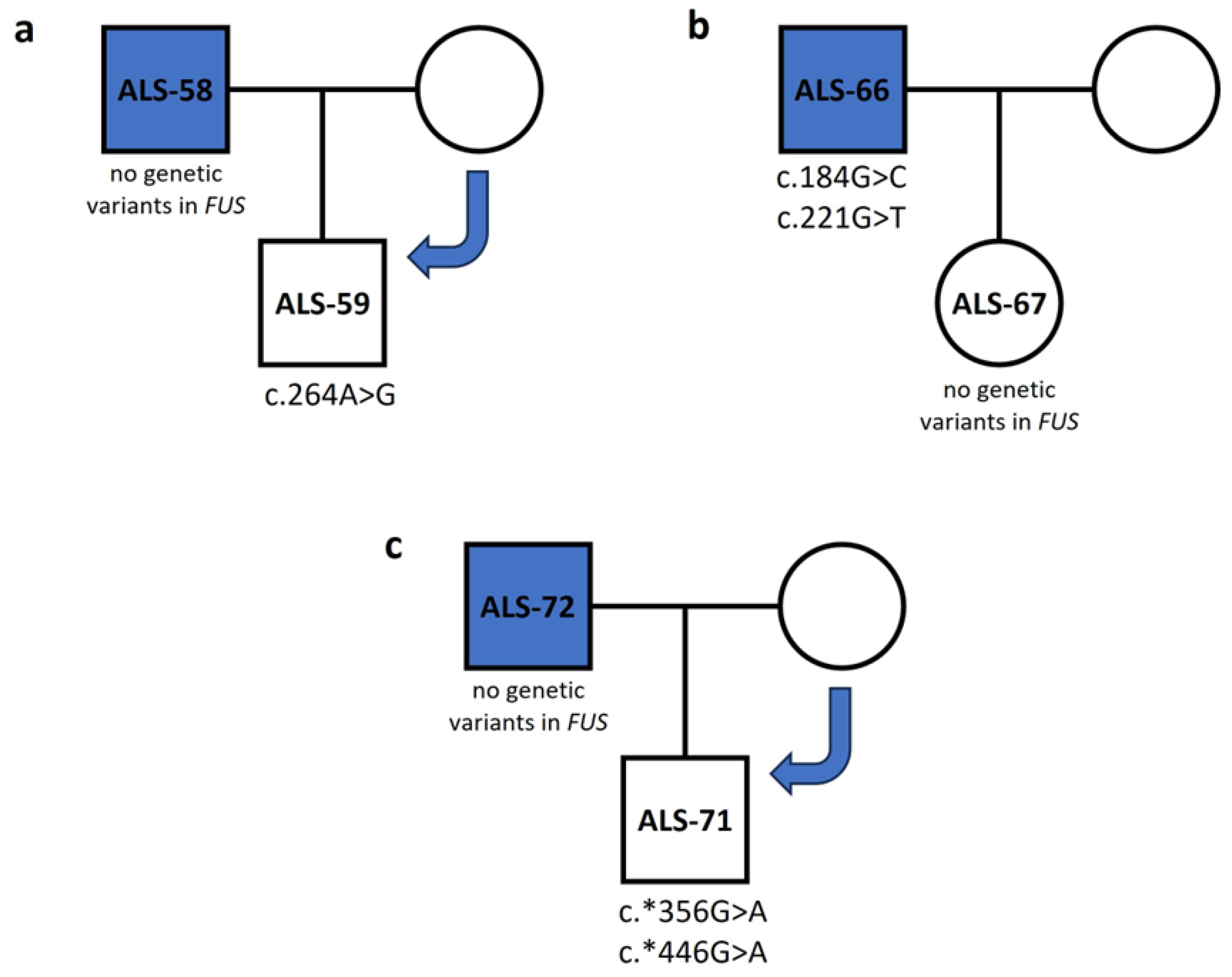
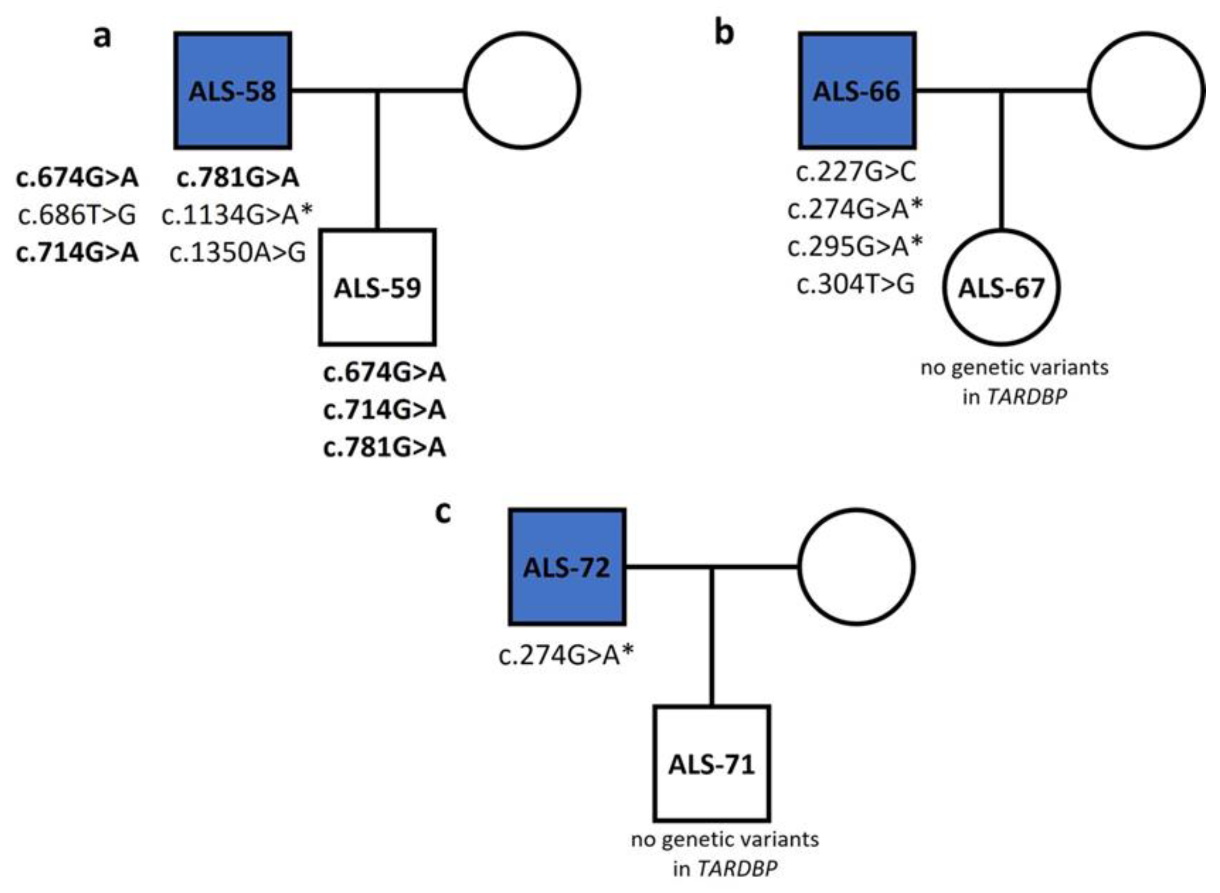
Figure 1. Inheritance tree diagrams of three patient–healthy relative pairs show the inheritance pattern of FUS variants. (a) Variant c.264A>G was found in healthy individual ALS-59, but not patient ALS-58. (b) Variants c.184G>C and c.221G>T were found in patient ALS-66 but not in their healthy daughter ALS-67. (c) Variants c.*356G>A and c.*446G>A were found in healthy individual ALS-71, but not in patient ALS-72.
Genes 15 00309 g001
Figure 2. Inheritance tree diagrams of three patient–healthy relative pairs show the inheritance pattern of TARDBP variants. Bold indicates variants inherited from an ALS patient to healthy offspring. (*) indicate variants that are common in patients but not in healthy individuals.
Genes 15 00309 g002
Table 1. Genetic variants in the SOD1 gene found in Greek sALS patients (variants previously associated with ALS are indicated in bold letters).
cDNAExonrsIDAmino Acid ChangeFunctional ProteinType of VariantMutationTasterPolyPhen-2SIFTSamples
(Genotype)
c.248C>G3--Not includedSilentDisease causing--ALS-12 (het)
c.251T>G3-p.C57WNot includedMissenseDisease causingProbably damagingAffects protein functionALS-12 (het)
c.254C>A3rs549580868-Not includedSilentDisease causing--ALS-12 (het)
c.292A>T3-p.H71LNot includedMissenseDisease causingProbably damagingAffects protein functionALS-22 (het)
c.308T>C3--Not includedSilentDisease causing--ALS-22 (het)
c.349A>C4-p.D90Ap.D11AMissenseDisease causingBenignToleratedALS-39 (hom)
Table 2. Genetic variants in the FUS gene found in Greek sALS patients (variants previously associated with ALS are indicated in bold letters).
cDNAExonProteinType of VariantrsIDMutationTasterPolyPhen-2SIFTSamples (Genotype)
c.101C>A2p.Q8KMissense-Disease causingBenignAffects protein functionALS-58 (het)
c.162G>T3p.S28IMissense-Disease causingProbably damagingAffects protein functionALS-30 (het)
c.184G>C3p.Q35HMissensers772271532Disease causingProbably damagingAffects protein functionALS-66 (het)
c.221G>T3p.G48CMissense-Disease causingProbably damagingAffects protein functionALS-66 (het)
c.223C>A3-Silent-Polymorphism--ALS-9 (het), ALS-12 (hom), ALS-13 (hom), ALS-14 (hom), ALS-21 (hom), ALS-24 (het), ALS-30 (het), ALS-38 (hom), ALS-40 (het), ALS-68 (het)
c.626A>C6p.M183LMissensers762914131Disease causingBenignAffects protein functionALS-30 (het), ALS-38 (het), ALS-57 (het)
c.759G>T6p.G227VMissense-Disease causingBenignAffects protein functionALS-70 (het)
c.760C>G6-Silentrs151073460Disease causing--ALS-10 (het), ALS-30 (het)
c.800A>T6p.R241*Nonsense-Disease causing--ALS-38 (het), ALS-40 (het)
c.806C>T6p.R243CMissensers1165095258Disease causingBenignAffects protein functionALS-2 (het), ALS-10 (het), ALS-22 (het), ALS-25 (het), ALS-30 (het)
c.830G>T6p.G251CMissense-Disease causingProbably damagingAffects protein functionALS-65 (het)
c.*41G>A3′ UTR--rs80301724Disease causing--ALS-13 (het)
c.*81C>T3′ UTR--rs768544815Polymorphism--ALS-73 (het)
c.*306T>C3′ UTR---Polymorphism--ALS-63 (het)
c.*354A>T3′ UTR---Polymorphism--ALS-70 (het)
c.*356G>A3′ UTR--rs886051940Polymorphism--ALS-60 (het), ALS-61 (het), ALS-64 (het), ALS-65 (het), ALS-69 (het)
c.*362T>G3′ UTR---Polymorphism--ALS-73 (het)
c.*370A>T3′ UTR------ALS-69 (het)
c.*406G>A3′ UTR------ALS-69 (het)
Table 3. Genetic variants in the TARDBP gene found in Greek sALS patients (variants previously associated with ALS are indicated in bold letters).
cDNAExonProteinType of VariantrsIDMutationTasterPolyPhen-2SIFTSamples (Genotype)
c.5T>C1 (5′ UTR)---Disease causing--ALS-6 (het)
c.24C>G1 (5′ UTR)--rs965172966Polymorphism--ALS-2 (het), ALS-6 (het), ALS-13 (hom)
c.227G>C2p.R41PMissense-Disease causingBenignAffects protein functionALS-66 (het)
c.274G>A2p.E57KMissense-Disease causingBenignAffects protein functionALS-30 (het), ALS-40 (het), ALS-41 (het), ALS-66 (het), ALS-68 (het), ALS-69 (het), ALS-72 (het), ALS-73 (het)
c.295G>A2p.D64NMissense-Disease causingPossibly damagingAffects protein functionALS-41 (het), ALS-66 (het)
c.295G>T2p.D64YMissense-Disease causingPossibly damagingAffects protein functionALS-57 (het)
c.300T>C2-Silentrs61730366Disease causing--ALS-63 (het)
c.303C>G2-Silent-Disease causing--ALS-57 (het)
c.304T>G2p.W67GMissense-Disease causingProbably damagingAffects protein functionALS-66 (het)
c.363G>A3-Silent-Disease causing--ALS-73 (het)
c.405G>A3-Silent-Disease causing--ALS-73 (het)
c.468G>T3p.E121DMissense-PolymorphismBenignToleratedALS-73 (het)
c.487G>A3p.E128KMissense-Disease causingPossibly damagingToleratedALS-73 (het)
c.490G>T3p.V129FMissense-Disease causingPossibly damagingAffects protein functionALS-73 (het)
c.500T>G3p.V132GMissensers766116483Disease causingPossibly damagingAffects protein functionALS-25 (het)
c.594G>C4p.Q163HMissense-Disease causingProbably damagingAffects protein functionALS-41 (het)
c.674G>A5p.R190KMissense-Disease causingBenignToleratedALS-14 (het), ALS-58 (het)
c.686T>G5p.F193LMissense-Disease causingProbably damagingAffects protein functionALS-58 (het)
c.703G>A5p.D200NMissense-Disease causingPossibly damagingToleratedALS-57 (het)
c.714G>A5-Silentrs1333943256Disease causing--ALS-58 (het)
c.715G>A5p.D204NMissense-Disease causingBenignToleratedALS-57 (het)
c.741G>T5p.Q212HMissense-Disease causingPossibly damagingAffects protein functionALS-14 (het)
c.744C>G5p.Y214*Nonsense-Disease causing--ALS-57 (het)
c.777A>T5-Silent-Disease causing--ALS-57 (het)
c.781A>G5p.R226GMissense-Disease causingProbably damagingAffects protein functionALS-57 (het), ALS-58 (het)
c.801A>T5-Silent-Disease causing--ALS-57 (het)
c.972T>A6-Silent-Disease causing--ALS-70 (het)
c.995G>T6p.G297VMissensers1643653768Disease causingBenignAffects protein functionALS-63 (het)
c.1001G>A6p.G299EMissense-Disease causingPossibly damagingAffects protein functionALS-70 (het)
c.1134G>A6-Silent-Disease causing--ALS-9 (het), ALS-13 (het), ALS-23 (het), ALS-25 (het), ALS-38 (het), ALS-58 (het), ALS-68 (het), ALS-73 (het)
c.1180C>A6p.Q326RMissense-Disease causingPossibly damagingAffects protein functionALS-70 (het)
c.1182G>A6-Silent-Disease causing--ALS-70 (het)
c.1322C>T6p.S406FMissense-Disease causingPossibly damagingAffects protein functionALS-21 (het)
c.1326G>T6p.K407NMissense-Disease causingProbably damagingAffects protein functionALS-70 (het)
c.1328C>T6p.S408FMissense-Disease causingPossibly damagingAffects protein functionALS-21 (het)
c.1347G>A6 (3′ UTR)-- Disease causing--ALS-68 (het)
c.1350A>G6 (3′ UTR)-- Polymorphism--ALS-58 (het)
Disclaimer/Publisher’s Note: The statements, opinions and data contained in all publications are solely those of the individual author(s) and contributor(s) and not of MDPI and/or the editor(s). MDPI and/or the editor(s) disclaim responsibility for any injury to people or property resulting from any ideas, methods, instructions or products referred to in the content.

