Article

Implementation of Public Funded Genome Sequencing in Evaluation of Fetal Structural Anomalies

1
Department of Obstetrics and Gynecology, Tuen Mun Hospital, Hong Kong SAR, China
2
Department of Obstetrics & Gynaecology, Prince of Wales Hospital, Hong Kong SAR, China
3
Department of Obstetrics & Gynaecology, Queen Elizabeth Hospital, Hong Kong SAR, China
4
Department of Obstetrics & Gynaecology, Pamela Youde Nethersole Eastern Hospital, Hong Kong SAR, China
5
Department of Obstetrics & Gynaecology, Queen Mary Hospital, Hong Kong SAR, China
6
Department of Obstetrics & Gynaecology, United Christian Hospital, Hong Kong SAR, China
7
Department of Obstetrics & Gynaecology, Princess Margaret Hospital, Hong Kong SAR, China
8
Department of Pathology, Hong Kong Children’s Hospital, Hong Kong SAR, China
9
Department of Radiology, Hong Kong Children’s Hospital, Hong Kong SAR, China
10
Department of Paediatrics, Prince of Wales Hospital, Hong Kong SAR, China
11
Department of Paediatrics and Adolescent Medicine, Li Ka Shing Faculty of Medicine, The University of Hong Kong, Hong Kong SAR, China
12
Clinical Genetics Service Unit, Hong Kong Children’s Hospital, Hong Kong SAR, China
13
Prenatal Genetic Diagnosis Centre, Department of Obstetrics & Gynaecology, Chinese University of Hong Kong, Hong Kong SAR, China
14
Prenatal Diagnostic Laboratory, Tsan Yuk Hospital, Hong Kong SAR, China
15
Department of Obstetrics & Gynaecology, Kwong Wah Hospital, Hong Kong SAR, China
*
Authors to whom correspondence should be addressed.
Genes 2022, 13(11), 2088; https://doi.org/10.3390/genes13112088
Submission received: 25 October 2022 / Revised: 6 November 2022 / Accepted: 7 November 2022 / Published: 10 November 2022
(This article belongs to the Special Issue Novel Insights into Prenatal Genetic Testing)

Abstract

:
With the advancements in prenatal diagnostics, genome sequencing is now incorporated into clinical use to maximize the diagnostic yield following uninformative conventional tests (karyotype and chromosomal microarray analysis). Hong Kong started publicly funded prenatal genomic sequencing as a sequential test in the investigation of fetal structural anomalies in April 2021. The objective of the study was to evaluate the clinical performance and usefulness of this new service over one year. We established a web-based multidisciplinary team to facilitate case selection among the expert members. We retrospectively analyzed the fetal phenotypes, test results, turnaround time and clinical impact in the first 15 whole exome sequencing and 14 whole genome sequencing. Overall, the molecular diagnostic rate was 37.9% (11/29). De novo autosomal dominant disorders accounted for 72.7% (8/11), inherited autosomal recessive disorders for 18.2% (2/11), and inherited X-linked disorders for 9.1% (1/11). The median turnaround time for ongoing pregnancy was 19.5 days (range, 13–31 days). Our study showed an overall clinical impact of 55.2% (16/29), which influenced reproductive decision-making in four cases, guided perinatal management in two cases and helped future family planning in ten cases. In conclusion, our findings support the important role of genome sequencing services in the prenatal diagnosis of fetal structural anomalies in a population setting. It is important to adopt a multidisciplinary team approach to support the comprehensive genetic service.

1. Introduction

Fetal structural anomalies are common and affect about 2–3% of pregnancies [1]. Babies born with congenital anomalies can have significant morbidity and mortality [2]. The underling etiologies are complex. In the past, many epidemiologic studies and research have been performed to investigate the causes leading to congenital malformations [3]. With the recent advances in massive parallel sequencing technology, genetic disorders have been revealed to have an important contribution to congenital anomalies [4]. Two large prospective studies using whole exome sequencing to evaluate pregnancies with unselected fetal structural anomalies on prenatal ultrasound and negative karyotype and chromosomal microarray analysis (CMA) reported a diagnostic genetic variant rate of 8.5–10% [5,6]. Subsequently, a number of similar cohort studies and meta-analyses of these studies were published, confirming their diagnostic value [7,8,9].
Therefore, a 2022 updated position statement published by the International Society for Prenatal Diagnosis (ISPD) recommends the prenatal genome-wide sequencing is beneficial for (1) a current pregnancy with a fetus with a single major anomaly or with multiple organ system anomalies and non-diagnostic CMA or no CMA result, suggesting of a possible genetic etiology by clinical genetic expert review; (2) a prior undiagnosed fetus (or child) affected with a major single or multiple anomalies and the recurrence of similar unexplained anomalies in the current pregnancy [10]. The American College of Medical Genetics and Genomics (ACMG) also supports the consideration of its use when a diagnosis cannot be obtained using routine prenatal methods in a fetus with one or more significant anomalies [11]. Currently, prenatal exome sequencing is the main approach used for genome-wide sequencing in clinical practice. Genome sequencing, which has potential in the detection of variants outside the protein-coding exome and other forms of structural variations, is now emerging [12,13,14,15,16].
The prenatal genetic diagnosis can provide parents with more information about the health and prognosis of their fetus, including the functional anomalies and developmental outcomes apart from the physical anomaly. This information is important for informed decision-making in reproductive choice, pregnancy management, postnatal care and expanded reproductive options in subsequent pregnancies [17]. Several studies were published demonstrating the usefulness of prenatal exome sequencing in clinical management that exceeded the diagnostic yield [18,19,20,21,22,23,24]. However, the optimal use of this comprehensive molecular diagnostic technology in public clinical service is lacking.
In April 2021, the Hong Kong Hospital Authority launched a publicly funded next-generation sequencing (whole exome sequencing (WES) and whole genome sequencing (WGS)) as a sequential test in the investigation of fetal structural anomalies in all pregnant women. The aim of the new program was to enhance our prenatal diagnosis for special prenatal cases with the presence of fetal ultrasound anomalies after receiving a negative finding by conventional tests (CMA and karyotyping). The objective of the current study was to evaluate the one- year clinical performance as well as the clinical utility of WES/WGS in public funded service to identify the genetic diagnosis for undiagnosed fetal structural anomalies.

2. Material and Methods

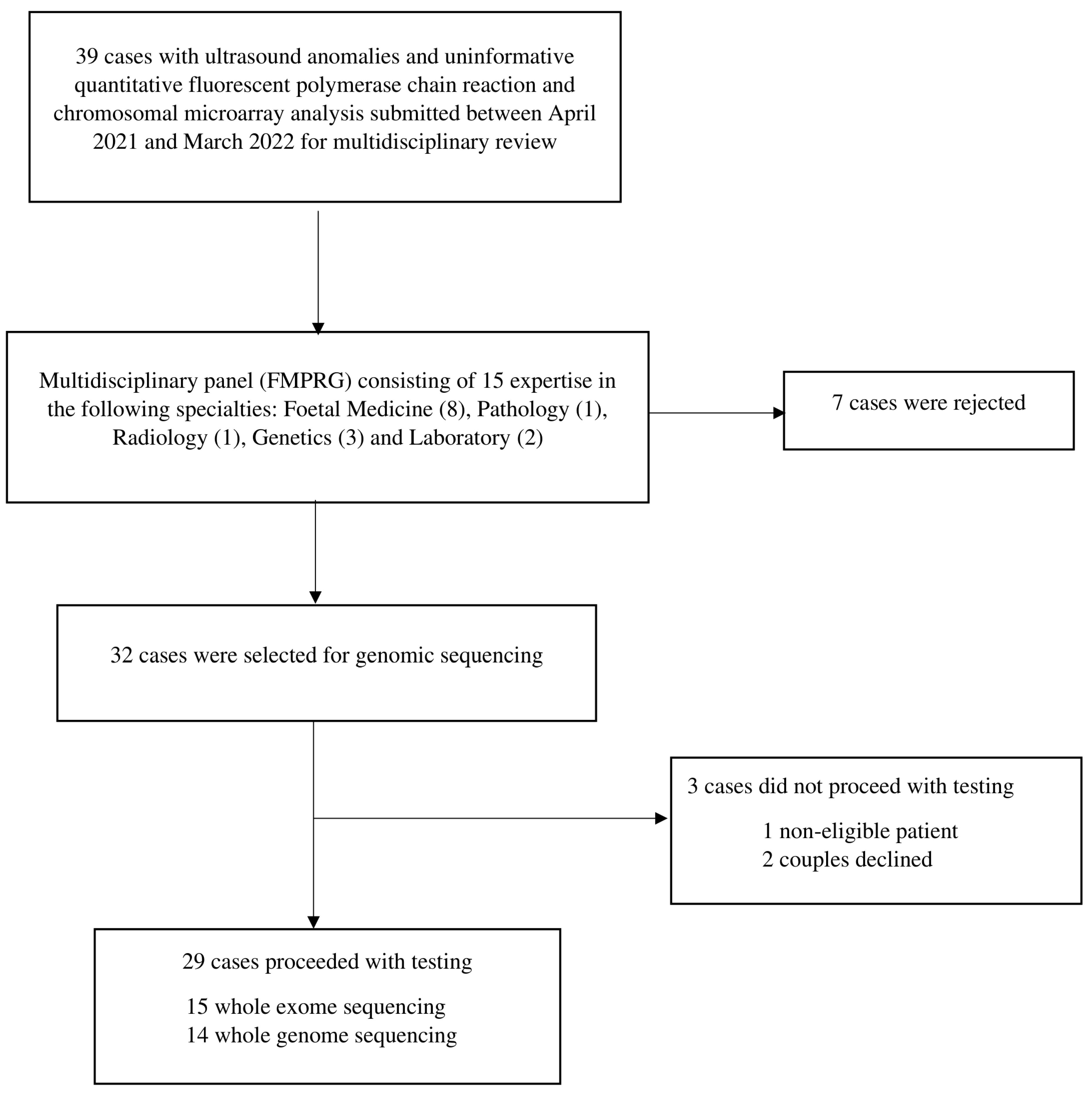
2.1. Study Population and Selection

This was a retrospective review of all women who had undergone publicly funded genomic sequencing for investigation of ultrasound fetal abnormalities between April 2021 and March 2022. The study was approved by the Central Institutional Review Board of the Hospital Authority (reference: PAED-2022-013).
In 2021, the Hong Kong population size was about 7,000,000, with 23,485 deliveries among our eight public hospitals with obstetric services (Kwong Wah Hospital, Queen Elizabeth Hospital, Queen Mary Hospital, Pamela Youde Nethersole Eastern Hospital, Princess Margaret Hospital, Prince of Wales Hospital, Tuen Mun Hospital and United Christian Hospital) under the Hospital Authority, Hong Kong SAR Government. Each of these eight hospitals had a prenatal diagnostic clinic that provided prenatal genetic screening and diagnosis services to women residing nearby. For pregnant women with fetal ultrasound abnormalities confirmed by the maternal-fetal medicine specialists in the clinic, they were offered invasive prenatal diagnostic testing. The standard workflow consisted of quantitative fluorescent polymerase chain reaction (QF-PCR) for rapid common aneuploidies detection, and if QF-PCR showed normal results, CMA was performed to look for copy number variations.
Since April 2021, all pregnant women were eligible for publicly funded whole exome or genome sequencing during pregnancy or after a fetal demise or pregnancy termination when they had one or more fetal malformations with uninformative QF-PCR and CMA likely to have an underlying genetic etiology, evaluated by a multidisciplinary team. The multidisciplinary team, with the abbreviation FMPRG, was made up of 15 experts in the following specialties: Foetal Medicine (8), Pathology (1), Radiology (1), Genetics (3) and the Laboratory (2). Detailed prenatal phenotyping with ultrasound was done by fetal-medicine specialists in prenatal diagnostic clinics. Additional fetal assessment by fetal magnetic resonance imaging (MRI) was performed in some cases, depending on the available expertise in different hospitals. The upper gestational limit for termination of pregnancy in Hong Kong is 24 weeks. Further relevant information on the terminated or demised fetuses was gained at postmortem examination if couples agreed. The case summary with anonymous clinical information and radiological and pathological data/images were prepared by case managers in different units and put in the web-based platform via the webmaster for discussion among the panel members. This online platform, which was established by the Department of Pathology of Hong Kong Children’s Hospital, could facilitate data sharing and multidisciplinary discussion. For any case submission, the web master notified all panel members via email. The testing criteria of the National Health Service (England) fetal whole exome service was used as a reference for our panel case selection. Patients were considered eligible for exome or genome sequencing if their fetus had a single major anomaly or multiple organ system anomalies on ultrasound suggestive of a possible genetic etiology after a multidisciplinary review with more than 7 members voting for the test before a pre-set deadline. The selected cases would have the genomic sequencing carried out by one of the two laboratories: the Prenatal Diagnostic Laboratory at Tsan Yuk Hospital using trios WES and the Prenatal Genetic Diagnosis Centre at the Chinese University of Hong Kong using trios WGS (PMID: 35441720). Each laboratory received fetal samples from the obstetric units in its catchment area. Written informed consent was obtained from the parents after pre-test counseling with verbal and written information given by the fetal-medicine specialists. The information provided included test limitations, possible test results (such as negative, positive, and uncertain genetic variants) and their implications. Incidental findings, ACMG 59 secondary findings and carrier status for autosomal recessive conditions were also included in the pre-test consent procedure for WGS, with an opt-out option. The genomic sequencing was performed on DNA extracted from amniocytes, chorionic villi, placental tissue, cord blood, or skin biopsy. Parental peripheral blood samples were obtained for trio analysis. Maternal cell contamination was excluded in the fetal DNA using QF-PCR data of the fetus and the mother. The turnaround time was proposed to be 4 calendar weeks for ongoing pregnancies and 4 calendar weeks after further investigation results are available (e.g., postmortem report, babygram) for fetal demised/terminated cases.

2.2. WES Procedure and Variant Interpretation

Genomic DNA was subjected to exome enrichment using double-stranded DNA capture baits from Twist Comprehensive Exome kit (Twist Bioscience, San Francisco, CA, USA), which targets approximately 36.8 Mb of the human coding exome regions. The generated library was sequenced on Illumina NextSeq 500 (Illumina, San Diego, CA, USA) to obtain at least 30× coverage depth for >98% of the targeted bases. Sequencing data were analyzed using the Congenica® analytical pipeline, which included read alignment to reference genome (GRCh37), variant calling (single nucleotide and small deletion/insertion variants), and variant annotation. Variant selection and prioritization were evaluated based on provided clinical information, suspected mode of inheritance and other laboratory data. Targeted regions were defined as coding exons +/− 5 bp. Only variants in the targeted region with clear gene-phenotype evidence from Genomics England PanelApp and OMIM and a minor allele frequency (MAF) < 1% reported in the Genome Aggregation Database were investigated. Disease-causing variants reported in ClinVar, DECIPHER, and Mastermind were also considered. Pathogenicity interpretation of genetic variants was based on the ACMG variant classification guidelines. Only variants in genes related to the patient’s phenotype were reported.

2.3. WGS Procedure and Variant Interpretation

Genome sequencing was based on a PCR-free library constructed with genomic DNA extracted from the sample. Paired-end sequencing was performed on a next-generation sequencing platform (NovaSeq, HiSeq X Ten or MGISEQ-2000). A minimal 30-fold read depth was generated for each case unless otherwise indicated. Sequencing reads were aligned to the reference genome (GRCh37/hg19) using the Burrows-Wheeler Alignment tool (PMID: 19451168). This test comprehensively identified genomic variants, including single-nucleotide variants (SNVs), small insertions and deletions (InDels), copy-number variants (CNVs), structural rearrangements (SVs), and absence of heterozygosity (AOHs). SNV/InDels calling was carried out by HaplotypeCaller (GATK) and annotated by ANNOVAR (PMID: 20601685) with different public reference databases. SNV/InDels interpretation was performed by referencing the medical literature and online databases according to the guidelines of the ACMG. For CNV analysis, reads were classified into sliding windows (50 kb with 5 kb increment) in terms of the aligned coordinates. After GC correction and population-based normalization, CNV detection was performed by an in-house pipeline (Increment Rate of Coverage), and interpretation was carried out by referencing the medical literature and online databases according to the guidelines of ACMG. For analysis of structural variants, uniquely aligned read-pairs were processed for identifying the chimeric read-pairs supporting a potential translocation, inversion, insertion or complex rearrangement. For AOH detection, genotyping was carried out by analysis of the mpileup file (Samtools). Regions (>5 Mb) with a decreased rate of heterozygous SNVs and an increased rate of homozygous SNVs were detected as regions with AOH. Clinically significant SNVs/InDels, CNVs, SVs and regions with AOHs related to patients’ clinical phenotype were reported. In addition, incidental findings, ACMG 59 secondary findings and carrier status for autosomal recessive conditions were reported if parents’ consent was obtained.

2.4. Post-Test Follow-Up

The post-test counseling was carried out by trained genetic health professionals such as maternal-fetal medicine specialists with genetic expertise from the respective prenatal diagnostic clinic and/or clinical geneticists from two university hospitals (Queen Mary Hospital and Prince of Wales Hospital), Hong Kong SAR Department of Health or Hong Kong Children’s Hospital. This included the disclosure of genetic results, explanation of the molecular diagnosis if found, further possible genetic tests, pregnancy management options, neonatal care and implication for other family members. The parents were also informed about recurrence risk and options for future family planning, including prenatal or pre-implantation genetic testing. The follow-up information, including pregnancy outcomes, was put back into the platform for educational and research purposes.

2.5. Study Data Collection and Analysis

We collected data from the case summaries in the online platform and laboratory databases. We studied the clinical phenotypes, the diagnostic yield, test turnaround times and the clinical impacts of the genetic results. The clinical phenotypes included prenatal phenotypes seen on fetal imaging (i.e., ultrasound and MRI) and postnatal phenotypes or postmortem reports. The diagnostic yield was defined as the percentage of the number of cases with pathogenic/likely pathogenic variants identified via WES/WGS considered to have caused the fetal structural anomaly from the total number of selected cases in a year. We also collected the number of variants of uncertain significance (VUS) as well as incidental and secondary findings for those who consented to reporting. Turnaround time was measured as days between the time that the test was ordered with patient consent received by the laboratory until the return of the final written diagnostic test report. For terminated cases, the turnaround time was counted from when the postmortem report was available. We also assessed the impact of this new clinical service on clinical management by sending out a survey to case managers to collect data about reproductive choice, clinical decision-making, pregnancy management and outcome, and neonatal treatment. Descriptive statistical tests were performed using SPSS (Windows version 23; IBM Corp, Armonk, NY, USA).

3. Results

3.1. Cohort Characteristics

From April 2021 to March 2022, 39 patients with fetuses that had structural anomalies were reviewed by the multidisciplinary team, and 32 fetuses were selected for the high probability of monogenic disorders (Figure 1). Three patients were excluded because two patients declined further genetic testing after counseling, and one patient was not entitled to the public funded test due to being a non-local citizen. The overview characteristics of 29 patients are included in Table 1. The majority were Chinese patients (89.7%), and 62.1% were tertiary-educated. Almost all were married (96.6%) with two consanguineous relationships and had planned pregnancies (79.3%); 65.5% of them were nulliparous. Three patients had conceived through assisted reproductive technologies. There were 28 singletons and one twin pregnancy. The median maternal age was 33 years (range, 25–39 years). Two had a pregnancy history affected by a genetic condition, including a chromosome 6 abnormality and alpha thalassemia major. One had a family history of Down’s syndrome (second- and third-degree relatives).
The prenatal phenotypic information of all cases was obtained from a prenatal ultrasound scan. In addition, five fetuses were investigated with fetal MRI, which supported the ultrasound finding of brain anomalies. The ultrasound structural abnormalities included multisystem anomalies (11/29, 37.9%), single system anomalies (11/29, 37.9%), non-immune hydrops fetalis (6/29, 20.7%) and fetal growth restriction (1/29, 3.4%).
Six patients (20.7%) had genomic sequencing before 24 weeks of pregnancy, two (6.9%) during ongoing pregnancy after 24 weeks, one (3.4%) after intrauterine death and 20 (69.0%) after the termination of pregnancy. All patients and their partners were sequenced as complete parental-fetal trios. WES was performed on 15 trios, and WGS was performed on 14 trios. The median gestational age for sequencing requests during ongoing pregnancy was 19+5 weeks (range, 15–32+1 weeks), and the median turnaround time was 19.5 days (range, 13–31 days). For the remaining 21 pregnancies with termination or stillbirth, the median turnaround time was 23 days (range: 14–56 days).

3.2. Overview of Diagnostic Yield and Prenatal Ultrasound Identified Abnormalities

Among the selected cases with WES/WGS performed, diagnostic genetic variants (pathogenic/likely pathogenic) associated with prenatal phenotypes were identified in 11 cases (11/29, 37.9%). Autosomal dominant disorders accounted for 72.7% (8/11), autosomal recessive disorders for 18.2% (2/11), and X-linked disorders for 9.1% (1/11). All autosomal dominant disorders were caused by de novo variants, and all autosomal recessive and X-linked disorders were caused by inherited variants. The twelve contributing diagnostic variants included three nonsense, four missense and five frameshift variants. VUS were detected in five cases (5/29, 17.2%), and there were incidental findings in two cases (2/14, 14.3%) and secondary findings in three cases (3/14, 21.4%). Two novel genetic variations predicted as pathogenic in NEB and KMT2D were discovered. The former was a nonsense variant, and the latter was a frameshift variant associated with a premature stop codon downstream. Both were likely to result in protein loss resulting from a loss-of-function allele.
Of the structural anomalies detected on prenatal ultrasound, the diagnostic yield was 100% (1/1) in fetuses with isolated skeletal abnormalities, 57.1% (4/7) in fetuses with isolated brain abnormalities, 36.4% (4/11) in fetuses with multiple organ systems (more than one fetal organ system abnormality) and 33.3% (2/6) in fetal hydrops. No molecular diagnosis was made in cases with isolated renal malformations, isolated facial malformations, and intrauterine growth restriction.

3.3. Overview of Pregnancy Outcome and Clinical Impact (Table 2 and Table 3)

3.3.1. Pregnancy Outcome

Among the eight patients with WES/WGS ordered during ongoing pregnancy, one pregnancy ended in miscarriage at 18 weeks, one pregnancy was terminated, and the remaining six pregnancies resulted in a live birth, including one case of monochorionic diamniotic twin pregnancy continued the pregnancy after selective feticide.

3.3.2. Clinical Impact- Helped Reproductive Decision-Making

Of the six patients with WES/WGS performed to help decision-making regarding continuation or termination of pregnancy before 24 weeks of gestation, one received a definite molecular diagnosis and ended up in miscarriage (case ES6); one received VUS result and continued the pregnancy (case ES7); four received negative results (cases GS8, GS9, GS10 and GS11) in which one underwent pregnancy termination based on ultrasound finding (case GS10). In case ES6, the fetus had multiple anomalies and hydrops fetalis. The parents opted to wait for the WES result. This case ended in a miscarriage after 2 weeks before the availability of the genetic result. WES detected a de novo pathogenic variant in the HRAS gene confirming Costello syndrome (OMIM 218040). In case ES7, cystic hygroma and hydrops fetalis were identified. A maternal inherited VUS was detected in the LZTR1 gene associated with both autosomal dominant Noonan syndrome 10 (OMIM 616564) and autosomal recessive Noonan syndrome 2 (OMIM 605275). The mother attended a genetic clinic where she was not found to have any features of Noonan syndrome. Since the public variant databases had conflicting interpretations of its pathogenicity and no prenatal phenotype of fetal hydrops was reported in the literature, it was classified as VUS. The fetus had resolution of hydrops at 22 weeks of gestation. The parents decided to carry on with the pregnancy. The baby was born alive at 38 weeks with clinical features of Noonan syndrome and was referred to the genetic clinic for assessment and follow-up. This genetic finding facilitated the parents in making a reproductive choice and the subsequent neonatal management. Case GS11 was a monochorionic diamniotic twin pregnancy with cystic hygroma and hydrops detected in one of the fetuses in the first trimester. Chorionic villus sampling was performed. After normal CMA and karyotyping results, the parents decided selective feticide by radiofrequency ablation to minimize the potential neurological damage to the normal co-twin. Subsequent negative WGS results supported the parental decision to continue the pregnancy of the phenotypically normal co-twin. The baby was born alive at 41 weeks and in good condition. In case GS10, a megacystis, bilateral dilated renal pelvises, umbilical cord cyst and oligohydramnios were identified. Vesicoamniotic shunting was performed. After the negative WES result, the parents decided to terminate the pregnancy because of the poor prognosis. Overall, the uncertain or negative genetic result contributed to the decision to continue the pregnancy in four cases.

3.3.3. Clinical Impact- Guided Perinatal Management

In two patients with WGS performed to guide perinatal management, one with inherited molecular diagnosis had implications in neonatal management and future reproductive counseling (case GS1), and one with an uncertain result had an adjustment in pregnancy management (case GS6). In case GS1, hydrocephalus, periventricular nodular heterotopia, mega cisterna magna and partial dysgenesis of the corpus callosum were detected at 28 weeks of gestation. WGS was performed on the umbilical cord blood sample saved at birth. A maternal inherited pathogenic variant was found in the FLNA gene, which was associated with X-linked periventricular nodular heterotopia (OMIM 300049). A paternal inherited HBA gene deletion of the alpha-thalassemia trait was also detected. Postnatal MRI brain of the female newborn confirmed the prenatal ultrasound findings. The infant had normal growth and development at 8 months old. The family was referred to the genetic clinic for further work-up of family members and counseling on reproductive planning for future pregnancy. In case GS6, aortic stenosis, aortopulmonary window, malformation of cortical development and early onset fetal growth restriction were identified at 21 weeks of gestation. A maternal inherited VUS was detected in the MAP2K2 gene, which was associated with the autosomal dominant cranio-facio-cutaneous syndrome (OMIM 115150). The mother was phenotypically normal. In the absence of pathogenic findings, fetal growth was closely monitored. Subsequent fetal MRI brain showed no structural abnormalities. The pregnancy ended in live birth at 35+2 weeks of gestation when abnormal fetal Doppler occurred. At birth, the baby was admitted to the neonatal intensive care unit because of uncontrolled heart failure, with surgical ligation of the patent ductus arteriosus. Aortic stenosis and secundum atrial septal defect were noted. At 13 months old, the child showed no dysmorphic features but gross motor delay.

3.3.4. Clinical Impact: Aided Future Family Planning

Of the 21 patients with WES/WGS performed to aid future family planning, they had significant fetal structural anomalies resulting in termination of pregnancy and intrauterine fetal demise before the testing request. Postnatal phenotyping by external examination was available in 19 cases, while the final full autopsy performed by pathologists was available in 11 cases to support the prenatal structural findings. Nine pregnancies were found to be harboring pathogenic/likely pathogenic variants that contributed to the phenotype observed on prenatal imaging and/or autopsy. Seven of them had de novo mutations (RARB, PPP1R12A, NIPBL, COL1A1, SMARCB1, RERE, KMT2D). The genetic finding provided the underlying genetic cause of the fetal anomalies and supported counseling the parents about low recurrence risk for their future pregnancy. The remaining two fetuses had mutation variants (NEB, ASPM) of an autosomal recessive disorder transmitted from their parents. The diagnosis assisted specific counseling in the future recurrence risk of one in four, and the parents could use the result to pursue preimplantation genetic diagnosis in a future pregnancy. Three patients received inherited VUS results (cases GS3, GS4 and GS7) and two of them also received inherited incidental findings (cases GS3 and GS7), which might have implications for future pregnancies. In case GS4, semilobar holoprosencephaly, bilateral microphthalmia, absent left lens, hypotelorism, bilateral cleft lip and cleft palate, low thoracic scoliosis, suspected hemivertebra and bilateral club foot were detected by ultrasound at 13+6 weeks. A paternal inherited VUS in the GLI2 gene was also detected in addition to a likely pathogenic variant in the RERE gene. Defects in the GLI2 gene can cause autosomal dominant holoprosencephaly (OMIM 610829) and Culler-Jones syndrome (OMIM 615849). Further work-up for the husband, as well as family cascade screening of the husband’s family, could help to delineate the significance of the VUS, which was suggested by the clinical geneticist. In case GS3, agenesis of the corpus callosum and small cerebellum was detected by ultrasound at 21+3 weeks. One likely pathogenic paternally inherited variant in the LAMC3 gene, together with another maternal inherited VUS in-trans arrangement affecting the same gene, was identified by WGS. Bi-allelic LAMC3 mutations can cause autosomal recessive occipital cortical malformations (OMIM 614115). However, the fetal anomalies seen on ultrasound were already explained by another pathogenic variant in the SMARCB1 gene. Therefore, the pathogenicity of the VUS in the LAMC3 gene was still uncertain. In case GS7, hydrops fetalis and bilateral pleural effusion were found at 20 weeks of gestation. The parents were related as first-degree cousins, and this was their first pregnancy. WGS detected two incidental findings, two VUS and the absence of a heterozygosity region. The two incidental findings were biparentally inherited likely pathogenic compound homozygous variant (nonsense variant in the KPTN gene) and maternal inherited likely pathogenic heterozygous variant (nonsense variant in the TCF12 gene). The former was associated with autosomal recessive intellectual developmental disorder 41 (OMIM 615637), and the latter was associated with autosomal dominant craniosynostosis 3 (OMIM 615314). The two VUS were paternally inherited missense variants in the COL1A1 gene related to autosomal dominant forms of osteogenesis imperfecta (OMIM 166210) and paternally inherited nonsense variant in the COL10A1 gene related to autosomal dominant metaphyseal chondrodysplasia Schmid type (OMIM 156500). The absence of heterozygosity was also identified in eight chromosomes with approximately 173 Mb in size, which would raise the possibility of autosomal recessive disorders. Both parents were phenotypically normal. They were seen in the genetic clinic for counseling on family planning. In case GS2, skeletal abnormality in the fetus was associated with the pathogenic variant in the COL1A1 gene. WGS also discovered two secondary findings in the father. One was a heterozygous pathogenic variant in the LDLR gene associated with autosomal dominant familial hypercholesterolemia (OMIM 143890). The other was a heterozygous pathogenic variant in the G6PC gene associated with autosomal recessive glycogen storage disease Ia (OMIM 232200). The family was counseled by the clinical geneticist to have family screening and regular medical check-ups of lipid profiles. Thus, the sequencing had an implication on reproductive genetic counseling in 10 cases.
Table 2. Results of whole exome sequencing.
CaseGestational Age at Test OrderPrenatal PhenotypeGeneFetal Sequencing ResultACMG ClassificationMolecular DiagnosisInheritanceNovel VariantClinical ImpactPregnancy OutcomePost-Mortem Result/Postnatal PhenotypeIncidental FindingSecondary Finding
ES1Post-TOPUSG:
absent bilateral lens, microphthalmia, left diaphragmatic hernia, overlapping fingers over bilateral hands
RARBNM_000965.3:c.1210C>T p.(Gln404Ter), het, de novoLikely pathogenicSyndromic microphthalmia-12 Autosomal dominantNoInformation provision TOPFused eyelids, absent lens of the left eye, left diaphragmatic hernia with herniation of the left lobe of the liver, the stomach, part of the small intestine and spleen into the left hemithoraxNRNR
ES2Post-TOPUSG:
holoprosencephaly
PPP1R12ANM_002480.2:c.609delp.(His204Thrfs*12), het, de novoPathogenicGenitourinary and/or brain malformation syndromeAutosomal dominantNoInformation provisionTOPGross examination showed no gross abnormality; declined autopsyNRNR
ES3Post-TOPUSG:
micrognathia, overriding aorta, ventricular septal defect, pulmonary stenosis
NIPBLNM_015384.4:c.2642dup p.(Ser882Ilefs*10), het, de novoPathogenicCornelia de Lange syndrome 1Autosomal dominantNoInformation provisionTOPMandibular hypoplasia, cleft palate, ventricular septal defect, overriding aorta, mild pulmonary stenosis, and Meckel’s diverticulum noted in the ileumNRNR
ES4Post-TOPUSG:
cystic hygroma, micrognathia, club feet, hydrops fetalis
NEBNM_001164508.2:c.8712G>A p.(Trp2904Ter), het, mat


NM_001164508.2:c.133_146delTCAGAAACTTCCAA p.(Ser45ThrfsTer48), het, pat
Likely pathogenic


Likely pathogenic
Arthrogryposis multiplex congenita 6


Nemaline myopathy 2
Autosomal recessiveYes


No
Familial diagnosis,
family planning and counseling
TOPGross examination found low-set ears, micrognathia, and club feet with abnormal posture; declined an autopsyNRNR
ES5Post-TOPUSG:
microcephaly, sloping forehead, flat occiput

MRI:
microcephaly, hypoplasia of bilateral frontal lobes and frontal horns, abnormal sulcation pattern with underdeveloped Sylvian fissure, suspected bilateral thalamic fusion and partial agenesis/dysgenesis of the corpus callosum
ASPMNM_018136.4:c.349C>T, p.(Arg117*), homo, matpatPathogenicPrimary microcephaly 5Autosomal recessiveNoFamilial diagnosis,
family planning and counseling
TOPGross examination found microcephaly, sloping forehead and flat occiput; declined fetal autopsyNRNR
ES616+1 weeksUSG:
invisible cerebellum with dilated cisterna magna, cystic hygroma, upper and lower limbs in flexion posture, rocker bottom feet, hydrops fetalis
HRASNM_005343.2:c.182G>A p.(Gln61Arg), het, de novoPathogenicCostello syndromeAutosomal dominantNoInformation provisionMiscarriage at 18 weeksLimited internal assessment because of extensive autolysisNRNR
ES717+4 weeksUSG:
cystic hygroma, hydrops fetalis
LZTR1NM_006767.3:c.848G>A, p.(Arg283Gln), het, matVariant of uncertain significanceNoonan syndromeAutosomal dominantNoReproductive decision, familial diagnosis,
family planning and counseling, and neonatal management and follow-up
Livebirth Delivery at 38 weeks, at 2 months old, with clinical features of Noonan syndromeNRNR
ES8Post-TOPUSG:
occipital encephalocele, hypoplastic left heart,
bilateral enlarged cystic kidneys, upper limbs and lower limbs short and bowed, feet with polydactyl
--Negative----TOPEncephalocele, hypoplastic thymus, hypoplastic of left heart, polycystic change of bilateral kidneys, feet with polydactylNRNR
ES9Post-TOPUSG:
dysplastic mitral valve, atrioventricular septal defect, smallish distal aorta, persistent left superior vena cava, absent right kidney, ureterocele, single umbilical artery, persistent right umbilical vein
--Negative ----TOPGross examination found a 3 mm skin tag around the anus, no anal opening and pre-axial polydactyly (extra thumb) over left hand; declined fetal autopsyNRNR
ES10Post-TOPUSG:
small cerebellum, absent cavum septum pellucidum, small corpus callosum, mild hypertelorism
--Negative ----TOPMild hypertelorism, low-set ears and the marked autolytic change limited the assessment of internal structure of the brainNRNR
ES11Post-TOPUSG:
Heterotaxy syndrome with dextrocardia and central liver, right atrial isomerism, unbalanced atrioventricular septal defect, double outlet right ventricle, pulmonary stenosis, infracardiac total anomalous pulmonary venous connection, bilateral superior vena cava
--Negative ----TOPSevere hypertelorism with prominent supra-orbital and infraorbital ridges, short nose, carp-sharped mouth, heterotaxy syndrome with dextrocardia and central liver, complex congenital heart disease with hypoplastic left ventricle, atrioventricular septal defect, bilateral superior vena cava, dominant right ventricle with small pulmonary artery, both lungs with 3 lobes and accentuation of other fissuresNRNR
ES12Post-IUDUSG:
early onset intrauterine growth restriction, oligohydramnios, placentomegaly, single umbilical artery
--Negative----Intrauterine death at 27+4 weeksWeight 0.59 kg, low-set ears and receding jawNRNR
ES13Post-TOPUSG:
bilateral hydrocephalus, small and abnormal cerebellum and deficient vermis, severe micrognathia, bilateral fixed equinovarus with pes cavus, left lower lobe congenital pulmonary airway malformations
--Negative ----TOPDeclined fetal autopsyNRNR
ES14Post-TOPUSG:
hydrops fetalis
--Negative----TOPHydrops fetalisNRNR
ES15Post-TOPUSG:
alobar holoprosencephaly, depressed facial profile, no obvious nose seen, no proboscis, single orbit, hypoplastic left heart, left hand abnormal with second finger shorter and abnormal posture
--Negative ----TOPGross examination found a flat facial profile, absent nose, and shortened second finger of the left hand; declined fetal autopsyNRNR
Abbreviation: ACMG, American College of Medical Genetics and Genomics; Het, heterozygous; Homo, homozygous; Mat, maternal inherited; Matpat, biparental inherited; MRI, magnetic resonance imaging; NR, not reported; Pat, paternal inherited; TOP, termination of pregnancy; USG, ultrasound; * terminating.
Table 3. Results of whole genome sequencing.
CaseGestational Age at Test OrderPrenatal PhenotypeGeneFetal Sequencing ResultACMG ClassificationMolecular DiagnosisInheritanceNovel VariantClinical ImpactPregnancy OutcomePost-Mortem Result/Postnatal PhenotypeIncidental FindingSecondary Finding
GS132+1 weeksUSG:
hydrocephalus, periventricular nodular heterotopia, mega cisterna magna, partial dysgenesis of the corpus callosum

MRI:
diffuse bilateral periventricular grey matter heterotropia, mega cisterna magna, normal corpus callosum
FLNANM_001456.3 exon39 c.6368_6369del p.S2123fs, hemi, matLikely pathogenicPeriventricular nodular heterotopiaX-linkedNoFamilial diagnosis,
family planning and counselling, neonatal management and follow-up
LivebirthDelivery at 37+1 for IUGR, postdelivery MRI showed mild hydrocephalus, periventricular nodular heterotopia, prominent extra-axial cerebrospinal fluid spaces, mega cisterna magna, thinned corpus callosum; at 8 months old, normal growth and development-Fetus and father: heterozygous deletion on 16p13.3, including HBA1 and HBA2
(carrier of α thalassemia-1)
GS2Post-TOPUSG:
hypomineralized cranial bone above the base of the skull, short and angulated right femur
COL1A1NM_000088.3 exon33 c.2299G>A p.G767S, het, de novoPathogenicOsteogenesis imperfectaAutosomal dominantNoInformation provision, familial diagnosis,
family planning and counselling
TOPDeclined fetal autopsy-Father:
heterozygous pathogenic variant, c.1747C>T(p.H583Y) in the LDLR gene (autosomal dominant familial hypercholesterolemia);
carrier of pathogenic variant G6PC c.248G>A (autosomal recessive glycogen storage disease Ia)
GS3Post-TOPUSG:
agenesis of the corpus callosum, small cerebellum

MRI:
agenesis of the corpus callosum, small cerebellum
SMARCB1


LAMC3
NM_003073.5 exon9 c.1130G>A p.R377H, het, de novo


NM_006059.4 exon12
c.1963C>T p.R655W, het, mat
Pathogenic


Variant of uncertain significance
Coffin-Siris syndrome


Malformations of cortical development
Autosomal dominant


Autosomal recessive
No


No
Information provision, familial diagnosis,
family planning and counseling
TOPGross examination found bilateral club foot with short big toes, bilateral clenched hands, low-set ears; declined fetal autopsy

Placental pathologic finding of single umbilical artery
LAMC3
NM_006059.4 exon18 c.3140G>A p.W1047X, het, pat
(likely pathogenic, autosomal recessive malformations of cortical development)
-
GS4Post-TOPUSG:
semilobar holoprosencephaly, bilateral microphthalmia, left lens absent, hypotelorism, bilateral cleft lip and cleft palate, low thoracic scoliosis, suspected hemivertebra, bilateral club foot
RERE


GLI2
NM_012102.4 exon19 c.3385C>G p.Q1129E, het, de novo


NM_005270.5 intron13 c.2293+3G>A, het, pat
Likely pathogenic


Variant of uncertain significance
Neurodevelopmental disorder with or without anomalies of the brain, eye, or heart

Holoprosencephaly 9
Autosomal dominant


Autosomal dominant
NoInformation provision, familial diagnosis,
family planning and counseling
TOPGross examination found bilateral microphthalmia, hypotelorism, bilateral cleft lips and cleft palate, micrognathia, ambiguous genitalia, and left clubfoot; declined fetal autopsy--
GS5Post-TOPUSG:
Tetralogy of Fallot, persistent left superior vena cava, atrioventricular septal defect, horseshoe kidneys, single umbilical artery
KMT2DNM_003482.4 exon11/55 c.1899dup p.Pro634AlafsTer7, het, de novoPathogenicKabuki syndromeAutosomal dominantYesInformation provisionTOPTetralogy of Fallot with atrioventricular septal defect, horseshoe kidneys with fusion at the lower pole, single umbilical artery--
GS629 weeksUSG:
bilateral Sylvian fissure angle severely delayed, asymmetrical diagonal echogenic lines outside the ventricles, suggestive of neuronal migration disorder, aortic stenosis, aortopulmonary window, early onset symmetrical IUGR, oligohydramnios

MRI:
no structural brain abnormalities
MAP2K2NM_030662.3 exon1 c.3G>A p.M1I, het, matVariant of uncertain significanceCranio-facio-cutaneous syndromeAutosomal dominantNoFamilial diagnosis,
family planning and counselling, neonatal management and follow-up
LivebirthDelivery at 35+2 weeks for abnormal Doppler and IUGR, patent ductus arteriosus with ligation for uncontrolled heart failure on day 20, bovine aortic arch, moderate aortic stenosis, secundum atrial septal defect, right renal stone;
at 13 months old, gross motor delay on physiotherapy training, closed atrial septal defect, resolving nephrocalcinosis
--
GS7Post-TOPUSG:
hydrops fetalis
COL1A1


COL10A1
NM_000088.3 exon47
c.3469G>A p.G1157S, het, pat

COL10A1 NM_000493.4 exon3c.688C>T p.Q230X, het, pat
Variant of uncertain significance

Variant of uncertain significance
Osteogenesis imperfecta


Metaphyseal chondrodysplasia, Schmid type
Autosomal dominant


Autosomal dominant
No


No
Familial diagnosis,
family planning and counselling
TOPGross examination found hydropic abortus; declined fetal autopsyKPTN
NM_007059.4 intron4 c.450-2A > G, hom, matpat
(likely pathogenic, intellectual developmental disorder, autosomal recessive 41)

TCF12
NM_207036.2 exon11
c.956C>G p.S319X, het, mat
(likely pathogenic, autosomal dominant, craniosynostosis 3)

172.8 Mb of the absence of heterozygosity region
-
GS821+4 weeksUSG:
micrognathia
--Negative---Reproductive decisionLivebirthDelivery at 39+5 weeks, normal newborn examination-Fetus and father: heterozygous deletion on 16p13.3, including HBA1 and HBA2
(carrier of α-thalassemia-1)
GS920+3 weeksUSG:
megacystis, bilateral dilated renal pelvises
--Negative --Reproductive decisionLivebirth #Delivery at 39+3 weeks, at 5 months old,
bilateral grade 4 vesicoureteral reflux, posterior urethral valve with bilateral hydronephrosis
--
GS1019+2 weeksUSG:
megacystis, bilateral dilated renal pelvises, umbilical cord cyst
oligohydramnios
--Negative --Reproductive decisionTOPTermination of pregnancy in the private sector, postmortem report not available--
GS11Post- selective TOP
20+2 weeks
USG:
monochorionic, diamniotic twin, with one hydrops with cystic hygroma
--Negative --Reproductive decisionSelective feticide;
co-twin livebirth
Delivery of remaining twin at 41 weeks, normal newborn examination--
GS12Post-TOPUSG:
cerebellar hypoplasia, cardiomegaly, right ventricular hypertrophy, pericardial effusion
--Negative ---TOPCerebellar hypoplasia, increased right cardiac ventricular wall thickness, atrial septal defect, pulmonary hypoplasia
GS13Post-TOPUSG:
cerebellar hypoplasia, left ventriculomegaly
--Negative ---TOPHypertelorism, left ventriculomegaly, inconclusive of cerebellar hypoplasia, right thumb hypoplasia
GS14Post-TOPUSG:
bilateral borderline ventriculomegaly, agenesis of the corpus callosum

MRI:
complete agenesis of the corpus callosum
--Negative ---TOPAgenesis of corpus callosum
Abbreviation: ACMG, American College of Medical Genetics and Genomics; Hemi, hemizygous; Het, heterozygous; IUGR, intrauterine growth restriction; Mat, maternal inherited; MRI, magnetic resonance imaging; Pat, paternal inherited; TOP, termination of pregnancy; USG, ultrasound. # Chromosomal microarray analysis revealed a paternally inherited variant of uncertain significance (copy number gain of chromosome 18q11.1-q11.2). The final karyotyping result was supernumerary ring chromosome 18 syndrome inherited from the father, who also had a history of ureteroplasty and reimplantation of the ureter into the bladder as a teenager.

3.4. Personnel Involving in Pre-Test and Post-Test Genetic Counselling

The majority of cases received pre-test genetic counseling conducted by a maternal-fetal medicine specialist or obstetrician with genetic expertise (27/29, 93.1%). In post-test genetic counseling, clinical geneticists were involved in 10 cases (10/29, 34.5%), including all cases with uncertain results, cases with incidental/secondary findings, cases with inherited causative variants except for one case in which the parents returned to their own country and could not attend the post-test counseling.

4. Discussion

Our first-year experience of application of advanced sequencing technologies in public healthcare service for prenatal diagnosis of fetal structural anomalies achieved a diagnostic yield of 37.9%. Our diagnostic yield is similar to the recent review of worldwide studies [7]. The latest systematic review and meta-analysis of 66 cohort studies with WES/WGS on prenatal samples for fetal anomalies, including 4350 anomalous fetuses from 2010 to 2021, showed the pooled incremental yield of genomic sequencing after normal karyotype/CMA was 31% (95% confidence interval 26–36%) [7]. In addition, subgroup analysis found a significantly higher incremental yield with selected cases for more likely monogenic disease compared to unselected cases (42% vs. 15%). Locally, a multidisciplinary team comprising 15 health care professionals in different specialties was organized to help with case selection in this public funded service. The setup of a web-based platform allows convenient access to anonymous medical data of submitted cases, including ultrasound images, promotes sharing of information and creates more interaction and communication among the multidisciplinary team members. We select the cases with reference to the updated eligibility criteria from overseas programs (e.g., National Health Service (England) [25]) and professional society recommendations [10,11]. With the emergence of more and more literature about prenatal presentations of genetic disorders, the selection criteria are also widened to include a fetus with increased nuchal translucency (≥6.5 mm) plus another anomaly and isolated non-immune fetal hydrops [25,26,27,28,29]. Besides, the diagnostic yield may also vary widely across different structural abnormalities, as previous studies reported higher yields in multisystem anomalies, isolated skeletal anomalies and isolated brain anomalies [7]. Despite our relatively small sample size, these phenomena were also observed in our highly selected cohort.
We retrospectively assessed the impact of prenatal genomic sequencing on clinical management in our service review. Overall, the clinical impact of receiving a genomic sequencing result was observed in 55.2% (16/29) of the cases, which influenced reproductive decision-making in four cases, guided perinatal management in two cases and helped future family planning in ten cases. Although the majority of our cases were post-abortion pregnancies, knowing the definitive genetic diagnosis for a fetus with ultrasound abnormalities assisted in better recurrence risk assessment in 10 families. Detection of de novo variants may alleviate the guilty feeling for the parents and provide reassurance of low recurrence risk in their subsequent pregnancies, although it is not negligible because there may be a possibility of germline mosaicism and other genetic cause that cannot be totally excluded. On the other hand, the detection of inherited variants can alter reproductive planning and allow prenatal testing or in-vitro fertilization with preimplantation genetic diagnosis in future pregnancies. Molecular genetic test also allows more accurate prognostic prediction, which is very important for the parental decision-making process about pregnancy continuation. A significant impact was observed in our four cases, and the parents continued the pregnancy after knowing no causal variants were identified, and most monogenetic disorders were excluded. In addition, definite genetic results can guide obstetric and neonatal management. For example, the non-diagnostic sequencing report in case GS6 helped adjustment the antenatal surveillance and the time of delivery of the severe early-onset growth-restricted fetus at later gestation until abnormal ultrasound Doppler occurred. Another example, case GS1, was a female fetus with a structural brain malformation due to an inherited pathogenic variant of the FLNA gene. It is known as periventricular nodular heterotopia, which is a neuronal migration disorder with an X-linked dominant mode of inheritance [30]. Heterozygous females can be asymptomatic and have normal intelligence, but some may suffer from seizures and various cardiovascular manifestations. Hemizygous males tend to die in utero. The mother and her first daughter were assessed by a clinical geneticist, and her newborn was then under pediatric follow-up. Therefore, a prenatal genetic diagnosis can trigger the cascade screening in relatives of probands, shorten the postnatal diagnostic odyssey and facilitate personalized medical treatment and follow-up for the baby after birth without delay. Furthermore, other studies also demonstrated the identification of a genetic disorder in the prenatal period could have additional benefits that helped reduction of maternal morbidity by avoiding operative delivery for a fetus with a poor prognosis and improved the baby’s quality of life by withholding futile and painful life-sustaining procedures and arranging palliative care [21,22,24]. The broadening of prenatal genetic diagnostic capabilities can also help in the selection process for fetal surgery and open the potential target for in-utero treatment [31]. The ultimate goal of genetic diagnosis is to prevent and reduce morbidity and mortality.
The development of high-throughput next-generation sequencing allows the large-scale and rapid assessment of entire genomes. However, it introduces challenges with respect to the interpretation of genetic variation and communication of the uncertain genetic findings to the family [32]. In our cohort with the application of WES and WGS technologies, six VUS were found in 5 fetuses, yielding an overall VUS detection rate of 17.2% (5/29), 6.7% (1/15) for WES and 28.6% (4/14) for WGS. A systematic review by Pratt et al. reported VUS rate of exome sequencing for fetuses with structural anomalies ranged from 3.9% to 20% [33]. Exome sequencing examines only the protein-coding regions (exons), which constitute 1%–2% of the genome but harbor more than 85% of all disease-causing mutations, whereas whole genome sequencing examines the whole genome. Potentially WGS can increase the sensitivity of diagnostic variant detection because of comprehensive DNA coverage, including the non-protein-coding regions (introns) and structural variant/copy number variants; however, it could lead to increased costs, increased frequency of VUS and decreased interpretability. In our study, the VUS rate of our WGS cohort was higher compared with our WES cohort. Genomic sequencing is a phenotype-driven test [11]. The interpretation and classification of genetic variants is a complex procedure that requires bioinformatics support to weigh evidence from prediction tools, population frequency, co-occurrence, segregation, and functional studies [34]. In the prenatal setting, the interpretation is more complicated as we lack publicly available prenatal ultrasound-genotype databases. The causal relationship can be difficult to delineate because of possible coincidental findings. In addition, the accuracy and completeness of prenatal phenotyping are limited by prenatal ultrasound resolution and scanning techniques; the prenatal features are sometimes non-specific and may change with gestation. Further fetal imaging strategies, such as fetal MRI, can play an important adjunctive role in confirming or excluding abnormal findings detected by ultrasound, especially for brain abnormalities. The benefits of its use were demonstrated in our cases GS1, GS3, GS6 and GS14. For terminated fetuses or deceased fetuses/neonates with prenatal identified structural abnormality, detailed postmortem examination can supplement additional information on subtle dysmorphic abnormalities not visualized in prenatal imaging, for example, in our cases ES11 and ES12. All can maximize and refine the phenotypic information to improve genetic variant interpretation. Our case ES7 illustrated the difficulties that can occur in interpreting genotype-phenotype causality during the prenatal period. The hydropic fetus harbored a maternally inherited variant in the LZTR1 gene, which mutated gene is associated with autosomal dominant Noonan syndrome. After discussion among the laboratory scientists, geneticists and fetal-medicine specialists, the variant was reported as VUS because of conflicting data from public variant databases and a lack of prenatal phenotypic information about this variant. The mother was apparently normal, and the family received detailed post-test counseling with a clinical geneticist. Finally, the pregnancy was continued, and her baby was born with features of Noonan syndrome and a subsequent pediatric assessment was arranged. Therefore, the variant inherited from a normal parent may provide false reassurance to the parents and healthcare providers. But genetic diseases can have a wide spectrum of clinical presentations and variable outcomes due to incomplete penetrance and variable expressivity. Hence, it is important the additional postnatal phenotypic information of the proband can be returned to the laboratories as well as to public databases for the re-analysis of its pathogenicity. Re-classification of genetic variants to benign or pathogenic can occur over time as more experience and scientific evidence on new genotype-phenotype correlation evolves. Two studies reported the reclassification of variants in their cohorts after birth and even a few years later [22,35]. This future revision of data and data sharing will be beneficial to the parents as they may request prenatal or preimplantation genetic diagnosis to avoid recurrence, as well as counseling for future parents encountering the same genetic variant. After all, the uncertain finding is unavoidable, and it can create significant parental anxiety as well as difficulties in information processing and decision-making. Therefore, it is essential to prepare them psychologically by telling them about the possibility of finding and reporting VUS before taking the test [36].
Compared with single-gene and panel tests, genome-wide DNA sequencing increases the chance of reaching a definite genetic diagnosis; however, it also increases the possibility of identifying incidental and secondary genetic findings in a fetus and/or parents. In our review, we found three incidental findings in two fetuses, a heterozygous likely pathogenic variant in LAMC3 with a heterozygous VUS in the same gene in case GS3; two homozygous biallelic likely pathogenic variants in the KPTN gene and a heterozygous likely pathogenic variant in the TCF12 gene together with the absence of heterozygosity in case GS7. These pathogenic variants were all inherited from the parents and are associated with neurodevelopmental disorders having incomplete penetrance. Some deleterious recessive genes may also be hidden in the loss of heterozygosity chromosomal region. In view of the possible implications for reproductive planning, these findings were reported to the parents as they opted to receive incidental findings in the pre-test consent process. The father of case GS2 also received secondary findings about his carrier status of two genetic conditions. Currently, there is no universal consensus on reporting incidental and secondary findings which are unrelated to the indication for sequencing in a prenatal setting. The disclosure is debatable as it carries potential clinical and ethical challenges apart from medical benefits to the family [37]. Some of the clinical challenges described included workload in counseling, diagnosis confirmation, screening, and interventions, together with raised parental anxiety and social stigmatization. The ethical issues included pregnancy termination based on the secondary findings and the unborn child’s autonomy. Some guidelines issued by professional bodies recommend parents should know the reporting policy and have the option to opt-out of revealing this information in the informed consent process after consideration of the possible implication to them and others in the family after positive results [10,11]. Parents should also be aware of the additional findings of non-paternity or consanguinity.
Furthermore, all parents undergoing sequencing are recommended to receive post-test counseling conducted by individuals with relevant genetic expertise, irrespective of the result findings [10,11]. All our cases with uncertain, positive and incidental/secondary genetic findings received post-test counseling provided by clinical geneticists who could help in the provision of accurate information based on current knowledge and non-directive supportive counseling tailored to the genetic literacy of each individual. It is of utmost importance as the reproductive decision is based on the understanding of the information given as well as personal values. Apart from counseling on genetic etiology, fetal structural anomalies have to be managed after birth by different parties, such as neontologists, pediatricians, cardiologists, and surgeons. For example, our cases GS9 and GS10 having fetal urinary tract abnormality would be highly beneficial by talking to a urologist about the child’s long-term renal prognosis, the options of renal support and urological interventions for potential urinary obstruction and vesicoureteral reflux. Therefore, multidisciplinary counseling by knowledgeable health professionals on treatment options and prognosis covering all aspects of their baby’s physical anomaly should be offered, preferably supplemented by written, visual and web-based resources [38]. This would help reduce parental anxiety. Moreover, psychological stress after fetal diagnoses should be addressed, and emotional support should be provided throughout the pregnancy or termination procedure [39,40].
Among our eight ongoing pregnancies undergoing the genomic sequencing, the median turnaround time of the genetic report was 19.5 days (average, 20.4 days; range, 13–31 days), which was similar to the meta-analysis of 15 studies by Mellis et al. reported the median turnaround time for exome sequencing of 20 days (range, 4–141 days) [7]. A faster turnaround time for prenatal diagnosis is important because of the reproductive decision within the local legal time limit of pregnancy termination and the influence on immediate neonatal management. This would depend on the technical workflow, DNA sequencing approaches, data interpretation pipeline, reporting policy and complexity of the genetic findings. Trio analysis can help fasten the bioinformatics analysis [31]. Importantly, the pre-test counseling should address the expected timeframe of the result’s return to the local testing laboratory.
A major limitation of this study is the small sample size. This is a balance between careful case selection based on professional society guidelines, resource allocation and the local laboratory capability. Despite this limitation, our findings constitute a good estimate of genome-wide sequencing yield on prenatal diagnosis for fetal anomalies. Since this clinical service is ongoing, a larger sample size with follow-up information would be available in a future review. The VUS rate and unsolved cases would be helped by future data re-analysis. Another limitation is the different sequencing techniques and reporting policies used by two local prenatal laboratories. Prenatal exome sequencing is predominantly used in clinical practice and research. Prenatal genome sequencing is an emerging technology, and there are limited studies on its prenatal application. With its potential to detect kinds of genetic variation, genome sequencing may replace chromosomal microarray and exome sequencing in the future. Although our review is not a direct comparative study between WES and WGS, it would give us some insight into the clinical benefits and limitations of different sequencing approaches. On the other hand, the widespread use of genome-wide sequencing technology would carry resource implications in overall health economics as well as socio- ethical concerns. The challenges would be further heightened if it is used in non-invasive approaches or moved into the commercial market. It is imperative for the usage of prenatal genomic testing to be well-regulated to ensure good clinical ethics. Future prospective studies are required to evaluate the psychological impact of the molecular diagnosis on the parents and the cost-effective analysis of next-generation sequencing applications in prenatal diagnostics.
To our knowledge, this is the first evaluation study to look at the public prenatal sequencing service in real clinical practice in Hong Kong. We demonstrated the feasibility as well as the clinical utility of the new prenatal WES/WGS service to identify the genetic diagnosis for fetal structural anomalies after normal conventional tests. In addition, two novel genetic variants were discovered. The new genetic findings can help to understand human fetal development and reveal the unrecognized prenatal phenotypes associated with monogenetic diseases. Our service is under the public-funded framework with a stringent selection process and clear inclusion criteria so that everyone can access the free clinical service fairly without deprivation associated with poverty.

5. Conclusions

Our review supports the important role of genome-wide sequencing services in the prenatal diagnosis of fetal structural anomalies in a population setting. Involving a multidisciplinary team approach in case selection, variant interpretation, counseling on genetic aspects and perinatal management, and the provision of future care to the unborn child are essential to support a comprehensive genetic service.

Author Contributions

Conceptualization: P.L.S., A.S.Y.H., T.W.L.M., W.S., A.P.W.H., C.W.K., T.K.L., A.N.C.K., E.Y.L.K., S.C.C., B.H.Y.C., H.M.L., K.W.C., A.S.Y.K. and W.C.L.; data curation: P.L.S., A.S.Y.H., T.W.L.M., W.S., A.P.W.H., C.W.K., T.K.L., K.W.C., A.S.Y.K. and W.C.L.; formal analysis: P.L.S., H.M.L., K.W.C. and A.S.Y.K.; Funding acquisition: K.W.C., A.S.Y.K. and W.C.L.; writing—original draft preparation: P.L.S.; writing—review and editing: P.L.S., A.S.Y.H., T.W.L.M., W.S., A.P.W.H., C.W.K., T.K.L., A.N.C.K., E.Y.L.K., S.C.C., B.H.Y.C., H.M.L., K.W.C., A.S.Y.K. and W.C.L. All authors have read and agreed to the published version of the manuscript.

Funding

The funding of public genome sequencing service in the evaluation of fetal structural anomalies was provided under an annual plan submitted via the Central Committee (Genetic Services), Hospital Authority, Hong Kong SAR.

Institutional Review Board Statement

The study was approved by the Central Institutional Review Board of Hospital Authority (reference: PAED-2022-013).

Informed Consent Statement

Informed consent was waived because of the retrospective nature of the study, and the analysis used anonymous clinical data.

Data Availability Statement

The datasets used and analyzed during the current study are available from the corresponding author upon reasonable request.

Acknowledgments

The authors would like to gratefully acknowledge Wai Fu Ng, who pioneered the multidisciplinary group (FMPRG), and the technical staff from the Department of Pathology, Hong Kong Children’s Hospital, who set up and manage the web-based platform for discussion and education. The authors would also like to thank the laboratory staff at the Prenatal Diagnostic Laboratory at Tsan Yuk Hospital and the Prenatal Genetic Diagnosis Centre at the Chinese University of Hong Kong for assisting with prenatal diagnosis.

Conflicts of Interest

The authors declare no conflict of interest.

References

  1. Rydberg, C.; Tunón, K. Detection of fetal abnormalities by second-trimester ultrasound screening in a non-selected population. Acta Obstet. Gynecol. Scand. 2017, 96, 176–182. [Google Scholar] [CrossRef] [PubMed] [Green Version]
  2. Liu, L.; Oza, S.; Hogan, D.; Perin, J.; Rudan, I.; Lawn, J.E.; Cousens, S.; Mathers, C.; Black, R.E. Global, regional, and national causes of child mortality in 2000–13, with projections to inform post-2015 priorities: An updated systematic analysis. Lancet 2015, 385, 430–440. [Google Scholar] [CrossRef]
  3. Carmichael, S.L. Birth defects epidemiology. Eur. J. Med. Genet. 2014, 57, 355–358. [Google Scholar] [CrossRef] [PubMed]
  4. Toufaily, M.H.; Westgate, M.N.; Lin, A.E.; Holmes, L.B. Causes of Congenital Malformations. Birth Defects Res. 2018, 110, 87–91. [Google Scholar] [CrossRef]
  5. Lord, J.; McMullan, D.J.; Eberhardt, R.Y.; Rinck, G.; Hamilton, S.J.; Quinlan-Jones, E.; Prigmore, E.; Keelagher, R.; Best, S.K.; Carey, G.K.; et al. Prenatal Assessment of Genomes and Exomes Consortium. Prenatal exome sequencing analysis in fetal structural anomalies detected by ultrasonography (PAGE): A cohort study. Lancet 2019, 393, 747–757. [Google Scholar] [CrossRef] [Green Version]
  6. Petrovski, S.; Aggarwal, V.; Giordano, J.L.; Stosic, M.; Wou, K.; Bier, L.; Spiegel, E.; Brennan, K.; Stong, N.; Jobanputra, V.; et al. Whole-exome sequencing in the evaluation of fetal structural anomalies: A prospective cohort study. Lancet 2019, 393, 758–767. [Google Scholar] [CrossRef]
  7. Mellis, R.; Oprych, K.; Scotchman, E.; Hill, M.; Chitty, L.S. Diagnostic yield of exome sequencing for prenatal diagnosis of fetal structural anomalies: A systematic review and meta-analysis. Prenat. Diagn. 2022, 42, 662–685. [Google Scholar] [CrossRef]
  8. Pauta, M.; Martinez-Portilla, R.J.; Borrell, A. Diagnostic yield of exome sequencing in fetuses with multisystem malformations: Systematic review and meta-analysis. Ultrasound Obstet. Gynecol. 2022, 59, 715–722. [Google Scholar] [CrossRef]
  9. Mastromoro, G.; Guadagnolo, D.; Khaleghi Hashemian, N.; Marchionni, E.; Traversa, A.; Pizzuti, A. Molecular Approaches in Fetal Malformations, Dynamic Anomalies and Soft Markers: Diagnostic Rates and Challenges-Systematic Review of the Literature and Meta-Analysis. Diagnostics 2022, 12, 575. [Google Scholar] [CrossRef]
  10. Van den Veyver, I.B.; Chandler, N.; Wilkins-Haug, L.E.; Wapner, R.J.; Chitty, L.S.; ISPD Board of Directors. International Society for Prenatal Diagnosis Updated Position Statement on the use of genome-wide sequencing for prenatal diagnosis. Prenat. Diagn. 2022, 42, 796–803. [Google Scholar] [CrossRef]
  11. Monaghan, K.G.; Leach, N.T.; Pekarek, D.; Prasad, P.; Rose, N.C.; ACMG Professional Practice and Guidelines Committee. The use of fetal exome sequencing in prenatal diagnosis: A points to consider document of the American College of Medical Genetics and Genomics (ACMG). Genet. Med. 2020, 22, 675–680. [Google Scholar] [CrossRef] [Green Version]
  12. Wang, Y.; Greenfeld, E.; Watkins, N.; Belesiotis, P.; Zaidi, S.H.; Marshall, C.; Thiruvahindrapuram, B.; Shannon, P.; Roifman, M.; Chong, K.; et al. Diagnostic yield of genome sequencing for prenatal diagnosis of fetal structural anomalies. Prenat. Diagn. 2022, 42, 822–830. [Google Scholar] [CrossRef] [PubMed]
  13. Zhou, J.; Yang, Z.; Sun, J.; Liu, L.; Zhou, X.; Liu, F.; Xing, Y.; Cui, S.; Xiong, S.; Liu, X.; et al. Whole Genome Sequencing in the Evaluation of Fetal Structural Anomalies: A Parallel Test with Chromosomal Microarray Plus Whole Exome Sequencing. Genes 2021, 12, 376. [Google Scholar] [CrossRef] [PubMed]
  14. Choy, K.W.; Wang, H.; Shi, M.; Chen, J.; Yang, Z.; Zhang, R.; Yan, H.; Wang, Y.; Chen, S.; Chau, M.H.K.; et al. Prenatal Diagnosis of Fetuses With Increased Nuchal Translucency by Genome Sequencing Analysis. Front. Genet. 2019, 10, 761. [Google Scholar] [CrossRef] [PubMed] [Green Version]
  15. Liao, Y.; Yang, Y.; Wen, H.; Wang, B.; Zhang, T.; Li, S. Abnormal Sylvian fissure at 20-30 weeks as indicator of malformations of cortical development: Role of prenatal whole-genome sequencing. Ultrasound Obstet. Gynecol. 2022, 59, 552–555. [Google Scholar] [CrossRef]
  16. Liu, Y.; Wang, L.; Yang, Y.K.; Liang, Y.; Zhang, T.J.; Liang, N.; Yang, L.M.; Li, S.J.; Shan, D.; Wu, Q.Q. Prenatal diagnosis of fetal skeletal dysplasia using targeted next-generation sequencing: An analysis of 30 cases. Diagn. Pathol. 2019, 14, 76. [Google Scholar] [CrossRef] [Green Version]
  17. Castleman, J.S.; Wall, E.; Allen, S.; Williams, D.; Doyle, S.; Kilby, M.D. The prenatal exome-a door to prenatal diagnostics? Expert Rev. Mol. Diagn. 2021, 21, 465–474. [Google Scholar] [CrossRef]
  18. Normand, E.A.; Braxton, A.; Nassef, S.; Ward, P.A.; Vetrini, F.; He, W.; Patel, V.; Qu, C.; Westerfield, L.E.; Stover, S.; et al. Clinical exome sequencing for fetuses with ultrasound abnormalities and a suspected Mendelian disorder. Genome Med. 2018, 10, 74. [Google Scholar] [CrossRef] [Green Version]
  19. de Koning, M.A.; Hoffer, M.; Nibbeling, E.; Bijlsma, E.K.; Toirkens, M.; Adama-Scheltema, P.N.; Verweij, E.J.; Veenhof, M.B.; Santen, G.; Peeters-Scholte, C. Prenatal exome sequencing: A useful tool for the fetal neurologist. Clin. Genet. 2022, 101, 65–77. [Google Scholar] [CrossRef]
  20. Corsten-Janssen, N.; Bouman, K.; Diphoorn, J.; Scheper, A.J.; Kinds, R.; El Mecky, J.; Breet, H.; Verheij, J.; Suijkerbuijk, R.; Duin, L.K.; et al. A prospective study on rapid exome sequencing as a diagnostic test for multiple congenital anomalies on fetal ultrasound. Prenat. Diagn. 2020, 40, 1300–1309. [Google Scholar] [CrossRef]
  21. Tolusso, L.K.; Hazelton, P.; Wong, B.; Swarr, D.T. Beyond diagnostic yield: Prenatal exome sequencing results in maternal, neonatal, and familial clinical management changes. Genet. Med. 2021, 23, 909–917. [Google Scholar] [CrossRef] [PubMed]
  22. Deden, C.; Neveling, K.; Zafeiropopoulou, D.; Gilissen, C.; Pfundt, R.; Rinne, T.; de Leeuw, N.; Faas, B.; Gardeitchik, T.; Sallevelt, S.; et al. Rapid whole exome sequencing in pregnancies to identify the underlying genetic cause in fetuses with congenital anomalies detected by ultrasound imaging. Prenat. Diagn. 2020, 40, 972–983. [Google Scholar] [CrossRef] [PubMed]
  23. de Koning, M.A.; Haak, M.C.; Adama van Scheltema, P.N.; Peeters-Scholte, C.; Koopmann, T.T.; Nibbeling, E.; Aten, E.; den Hollander, N.S.; Ruivenkamp, C.; Hoffer, M.; et al. From diagnostic yield to clinical impact: A pilot study on the implementation of prenatal exome sequencing in routine care. Genet. Med. 2019, 21, 2303–2310. [Google Scholar] [CrossRef] [PubMed]
  24. Dempsey, E.; Haworth, A.; Ive, L.; Dubis, R.; Savage, H.; Serra, E.; Kenny, J.; Elmslie, F.; Greco, E.; Thilaganathan, B.; et al. A report on the impact of rapid prenatal exome sequencing on the clinical management of 52 ongoing pregnancies: A retrospective review. BJOG 2021, 128, 1012–1019. [Google Scholar] [CrossRef] [PubMed]
  25. NHS. (n.d.). NHS Choices. Retrieved 20 October 2022. Available online: https://www.england.nhs.uk/publication/national-genomic-testdirectories/25.https://www.england.nhs.uk/publication/national-genomic-test-directories/ (accessed on 25 October 2022).
  26. Sparks, T.N.; Lianoglou, B.R.; Adami, R.R.; Pluym, I.D.; Holliman, K.; Duffy, J.; Downum, S.L.; Patel, S.; Faubel, A.; Boe, N.M.; et al. Exome Sequencing for Prenatal Diagnosis in Nonimmune Hydrops Fetalis. N. Engl. J. Med. 2020, 383, 1746–1756. [Google Scholar] [CrossRef]
  27. Al-Kouatly, H.B.; Makhamreh, M.M.; Rice, S.M.; Smith, K.; Harman, C.; Quinn, A.; Valcarcel, B.N.; Firman, B.; Liu, R.; Hegde, M.; et al. High diagnosis rate for nonimmune hydrops fetalis with prenatal clinical exome from the Hydrops-Yielding Diagnostic Results of Prenatal Sequencing (HYDROPS) Study. Genet. Med. 2021, 23, 1325–1333. [Google Scholar] [CrossRef]
  28. Mone, F.; Eberhardt, R.Y.; Hurles, M.E.; Mcmullan, D.J.; Maher, E.R.; Lord, J.; Chitty, L.S.; Dempsey, E.; Homfray, T.; Giordano, J.L.; et al. Fetal hydrops and the Incremental yield of Next-generation sequencing over standard prenatal Diagnostic testing (FIND) study: Prospective cohort study and meta-analysis. Ultrasound Obstet. Gynecol. 2021, 58, 509–518. [Google Scholar] [CrossRef]
  29. Kelley, J.; McGillivray, G.; Meagher, S.; Hui, L. Increased nuchal translucency after low-risk noninvasive prenatal testing: What should we tell prospective parents? Prenat. Diagn. 2021, 41, 1305–1315. [Google Scholar] [CrossRef]
  30. Chen, M.H.; Walsh, C.A. FLNA Deficiency. In GeneReviews® [Internet]; Adam, M.P., Everman, D.B., Mirzaa, G.M., Eds.; 8 October 2002 [Updated 30 September 2021]; University of Washington: Seattle, WA, USA, 1993–2022. Available online: https://www.ncbi.nlm.nih.gov/books/NBK1213/ (accessed on 25 October 2022).
  31. Best, S.; Wou, K.; Vora, N.; Van der Veyver, I.B.; Wapner, R.; Chitty, L.S. Promises, pitfalls and practicalities of prenatal whole exome sequencing. Prenat Diagn. 2018, 38, 10–19. [Google Scholar] [CrossRef] [Green Version]
  32. Westerfield, L.; Darilek, S.; van den Veyver, I.B. Counseling Challenges with Variants of Uncertain Significance and Incidental Findings in Prenatal Genetic Screening and Diagnosis. J. Clin. Med. 2014, 3, 1018–1032. [Google Scholar] [CrossRef]
  33. Pratt, M.; Garritty, C.; Thuku, M.; Esmaeilisaraji, L.; Hamel, C.; Hartley, T.; Millar, K.; Skidmore, B.; Dougan, S.; Armour, C.M. Application of exome sequencing for prenatal diagnosis: A rapid scoping review. Genet. Med. 2020, 22, 1925–1934. [Google Scholar] [CrossRef] [PubMed]
  34. Richards, S.; Aziz, N.; Bale, S.; Bick, D.; Das, S.; Gastier-Foster, J.; Grody, W.W.; Hegde, M.; Lyon, E.; Spector, E.; et al. Standards and guidelines for the interpretation of sequence variants: A joint consensus recommendation of the American College of Medical Genetics and Genomics and the Association for Molecular Pathology. Genet. Med. 2015, 17, 405–424. [Google Scholar] [CrossRef] [Green Version]
  35. Vora, N.L.; Gilmore, K.; Brandt, A.; Gustafson, C.; Strande, N.; Ramkissoon, L.; Hardisty, E.; Foreman, A.; Wilhelmsen, K.; Owen, P.; et al. Correction: An approach to integrating exome sequencing for fetal structural anomalies into clinical practice. Genet. Med. 2020, 22, 1426. [Google Scholar] [CrossRef] [PubMed]
  36. Biesecker, B.B.; Klein, W.; Lewis, K.L.; Fisher, T.C.; Wright, M.F.; Biesecker, L.G.; Han, P.K. How do research participants perceive “uncertainty” in genome sequencing? Genet. Med. 2014, 16, 977–980. [Google Scholar] [CrossRef] [PubMed] [Green Version]
  37. Amor, D.J.; Chitty, L.S.; Van den Veyver, I.B. Current controversies in prenatal diagnosis 2: The 59 genes ACMG recommends reporting as secondary findings when sequencing postnatally should be reported when detected on fetal (and parental) sequencing. Prenat. Diagn. 2020, 40, 1508–1514. [Google Scholar] [CrossRef]
  38. Marokakis, S.; Kasparian, N.A.; Kennedy, S.E. Prenatal counselling for congenital anomalies: A systematic review. Prenat. Diagn. 2016, 36, 662–671. [Google Scholar] [CrossRef] [PubMed]
  39. Skari, H.; Malt, U.F.; Bjornland, K.; Egeland, T.; Haugen, G.; Skreden, M.; Dalholt Björk, M.; Bjornstad Ostensen, A.; Emblem, R. Prenatal diagnosis of congenital malformations and parental psychological distress--a prospective longitudinal cohort study. Prenat. Diagn. 2006, 26, 1001–1009. [Google Scholar] [CrossRef] [PubMed]
  40. Talati, A.N.; Gilmore, K.L.; Hardisty, E.E.; Lyerly, A.D.; Rini, C.; Vora, N.L. Impact of prenatal exome sequencing for fetal genetic diagnosis on maternal psychological outcomes and decisional conflict in a prospective cohort. Genet. Med. 2021, 23, 713–719. [Google Scholar] [CrossRef]
Figure 1. Flowchart of the genome-wide sequencing service.
Genes 13 02088 g001
Table 1. Basic patient characteristics, fetal phenotypic features, genetic results and pregnancy outcomes.
CharacteristicValue *
Ethnicity
Chinese26 (89.7)
South Asian3 (10.3)
Maternal age (year)33 (25–39)
Educational level
Primary0 (0)
Secondary9 (31.0)
Tertiary18 (62.1)
Unknown2 (6.9)
Married28 (96.6)
Consanguinity2 (6.9)
Family history of genetic problems1 (3.4)
Previous pregnancy/child of genetic problems2 (6.9)
Nulliparity19 (65.5)
History of pregnancy loss6 (20.7)
History of pregnancy termination4 (13.8)
Planned pregnancy23 (79.3)
Conception
Natural26 (89.7)
Assisted3 (10.3)
Number of fetuses
Singleton28 (96.6)
Twin1 (3.4)
Type of testing
Whole exome sequencing (WES)15 (51.7)
Whole genome sequencing (WGS)14 (48.3)
Trio analysis29 (100)
Invasive diagnostic procedure
No3 (10.3)
Chorionic villus sampling7 (24.1)
Amniocentesis19 (65.5)
Source of fetal DNA
Chorionic villi6 (20.7)
Amniocytes19 (65.5)
Products of conception3 (10.3)
Fetal blood/tissue1 (3.4)
Prenatal phenotype at test request
Single system11 (37.9)
- Brain malformation 7 (24.1)
- Renal malformation 2 (6.9)
- Skeletal malformation 1 (3.4)
- Facial malformation 1 (3.4)
Multiple systems11 (37.9)
Intrauterine growth restriction1 (3.4)
Hydrops fetalis6 (20.7)
Timing of test request
Before 24 weeks of pregnancy6 (20.7)
After 24 weeks of pregnancy2 (6.9)
After intrauterine death1 (3.4)
After the termination of pregnancy20 (69.0)
Report turnaround time (day) during pregnancy19.5 (13–31)
WES results (genetic variant)
Pathogenic/Likely pathogenic7 (46.7)
Variant of uncertain significance1 (6.7)
Negative8 (53.3)
WGS results (genetic variant)
Pathogenic/Likely pathogenic5 (35.7)
Variant of uncertain significance5 (35.7)
Negative7 (50.0)
Incidental finding3 (21.4)
Secondary finding4 (28.6)
Pregnancy outcome
Livebirth5 (17.2)
Intrauterine death1 (3.4)
Miscarriage1 (3.4)
Termination of pregnancy22 (75.9)
Neonatal death0 (0)
Personnel conducting pre-test counseling
Midwife0 (0)
Maternal-fetal medicine specialist/obstetrician27 (93.1)
Clinical geneticist2 (6.9)
Personnel conducting post-test counseling
Midwife1 (3.4)
Maternal-fetal medicine specialist/obstetrician17 (58.6)
Clinical geneticist10 (34.5)
Not available1 (3.4)
* Data are presented as median (range) or No. (%) of patients.
Publisher’s Note: MDPI stays neutral with regard to jurisdictional claims in published maps and institutional affiliations.

