Article

Prenatal Genetic Testing in the Era of Next Generation Sequencing: A One-Center Canadian Experience

1
Department of Medical Genetics, University of Alberta, Edmonton, AB T6G 2H7, Canada
2
Women’s and Children’s Health Research Institute, University of Alberta, Edmonton, AB T6G 1C9, Canada
3
Maternal Fetal Medicine Clinic, Royal Alexandra Hospital, Edmonton, AB T5H 3V9, Canada
4
Regional Genetics Program, Children’s Hospital of Eastern Ontario, Ottawa, ON K1H 8L1, Canada
*
Author to whom correspondence should be addressed.
Genes 2022, 13(11), 2019; https://doi.org/10.3390/genes13112019
Submission received: 1 October 2022 / Revised: 29 October 2022 / Accepted: 31 October 2022 / Published: 3 November 2022
(This article belongs to the Special Issue Novel Insights into Prenatal Genetic Testing)

Abstract

:
The introduction of next generation sequencing (NGS) technologies has revolutionized the practice of Medical Genetics, and despite initial reticence in its application to prenatal genetics (PG), it is becoming gradually routine, subject to availability. Guidance for the clinical implementation of NGS in PG, in particular whole exome sequencing (ES), has been provided by several professional societies with multiple clinical studies quoting a wide range of testing yields. ES was introduced in our tertiary care center in 2017; however, its use in relation to prenatally assessed cases has been limited to the postnatal period. In this study, we review our approach to prenatal testing including the use of microarray (CMA), and NGS technology (gene panels, ES) over a period of three years. The overall diagnostic yield was 30.4%, with 43.2% of those diagnoses being obtained through CMA, and the majority by using NGS technology (42% through gene panels and 16.6% by ES testing, respectively). Of these, 43.4% of the diagnoses were obtained during ongoing pregnancies. Seventy percent of the abnormal pregnancies tested went undiagnosed. We are providing a contemporary, one tertiary care center retrospective view of a real-life PG practice in the context of an evolving use of NGS within a Canadian public health care system that may apply to many similar jurisdictions around the world.

1. Introduction

Congenital anomalies impact approximately 2–4% of pregnancies and lead to structural or functional abnormalities [1], with severe consequences including intrauterine and newborn demise, chronic diseases and lifelong disability, posing a significant burden on the families affected and the medical system [2]. Modalities of prenatal testing that lead to diagnoses and facilitate decision making have evolved significantly over the past decade as part of the unprecedented progress registered in the field of genetics. The armamentarium in prenatal genetics (PG) includes both targeted tests (QF-PCR, gene panels) and genomic approaches at different levels of resolution (karyotype, chromosomal microarray, whole exome and whole genome sequencing). The tests used are based on internal clinical protocols (first line testing is for the common aneuploidies by screening and then diagnostic tests), but also based on fetal abnormalities identified by various imaging methods (ultrasound, echocardiogram, MRI), family history [3] and parental choices [4] and in keeping with medical systems availability. Up to 30% of diagnoses are achieved by karyotype [5], and an additional 4–6% by chromosomal microarray [6]. That leaves more than half of the fetal cases with anomalies undiagnosed [7]. The gradual introduction of next generation sequencing (NGS) over the past decade has marked a shift in the approach to genetic diagnosis either by a targeted gene panel approach that incorporates a differential diagnosis when a diagnostic handle can be identified, or by a comprehensive genomic approach when the diagnosis is non-specific or too complex through exome (ES) and, exceedingly rare at the moment, genome sequencing.
Exome sequencing does not require prior knowledge regarding the potential causative gene for an observed phenotype [4]. Therefore, it appears to be particularly well poised in a prenatal context. Its use is generally accepted as a powerful tool in pediatric [8] and adult genetics [9], and has slowly expanded to the area of prenatal diagnosis. Its initial reluctance being explained by unknown or incompletely understood prenatal phenotypes in relation to interpretation of DNA variants [10]. Guidance for the clinical implementation of NGS in PG is supported by several professional associations [4,11,12]. Over the past few years, a flurry of studies has showed a wide range of diagnostic yields anywhere between 6% [13] and 89% [14], demonstrating its overall clinical utility [15], with a pooled yield of 31% as showed by a recent meta-analysis [16]. Discrepancies in achieving a diagnosis by ES between studies are reflected in the variable inclusion criteria, study cohort size, and interpretation of results, amongst other factors [16]. Despite all supportive evidence, the reality of prenatal diagnostic practice is that its application remains inconsistent, including countries with publicly available medical systems as is the case in Canada. We have evaluated the prenatal diagnostic yields in our tertiary care center over a period of three years, since ES has been clinically available postnatally, and are describing the range of diagnoses achieved with a focus on those obtained using NGS.

2. Materials and Methods
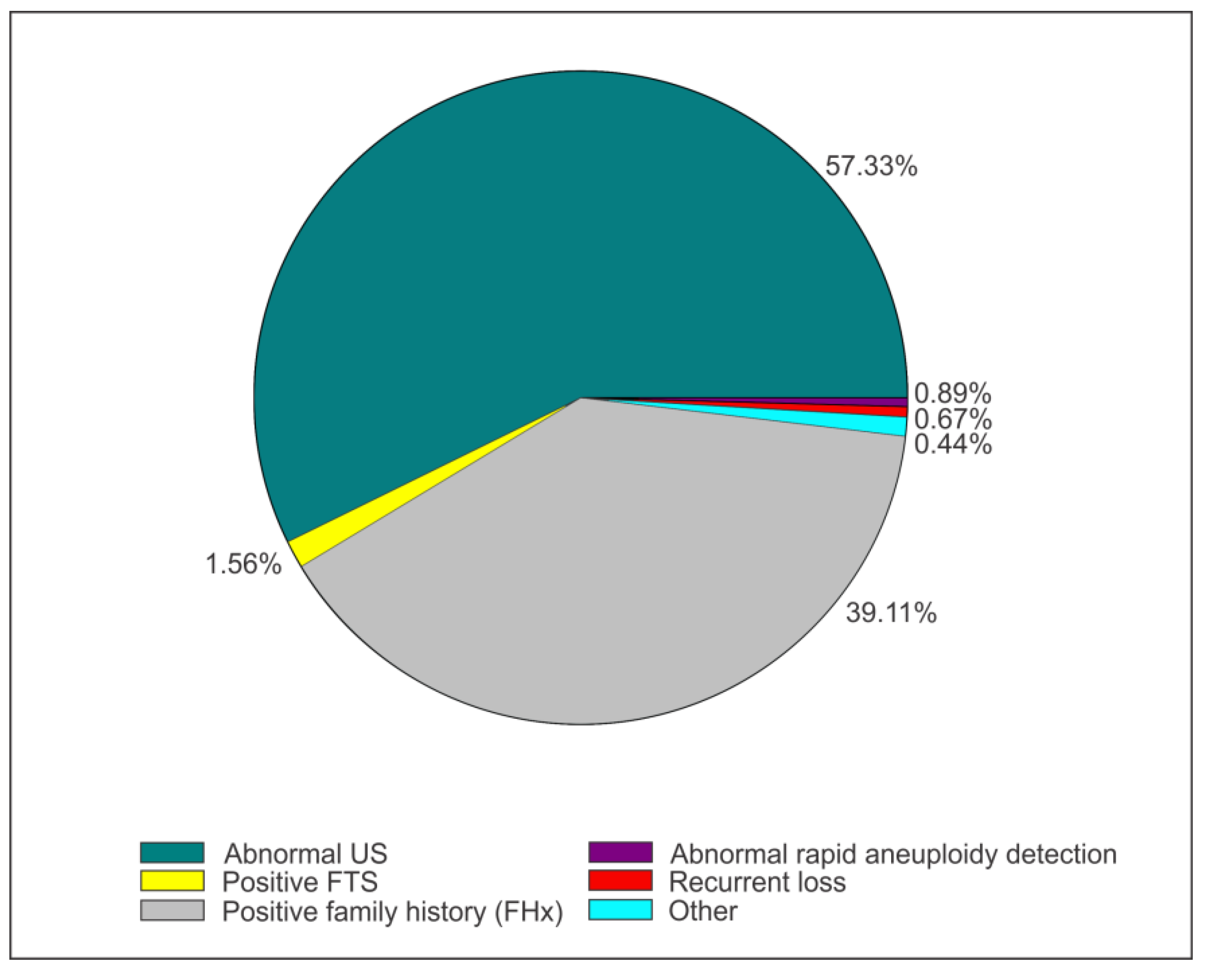
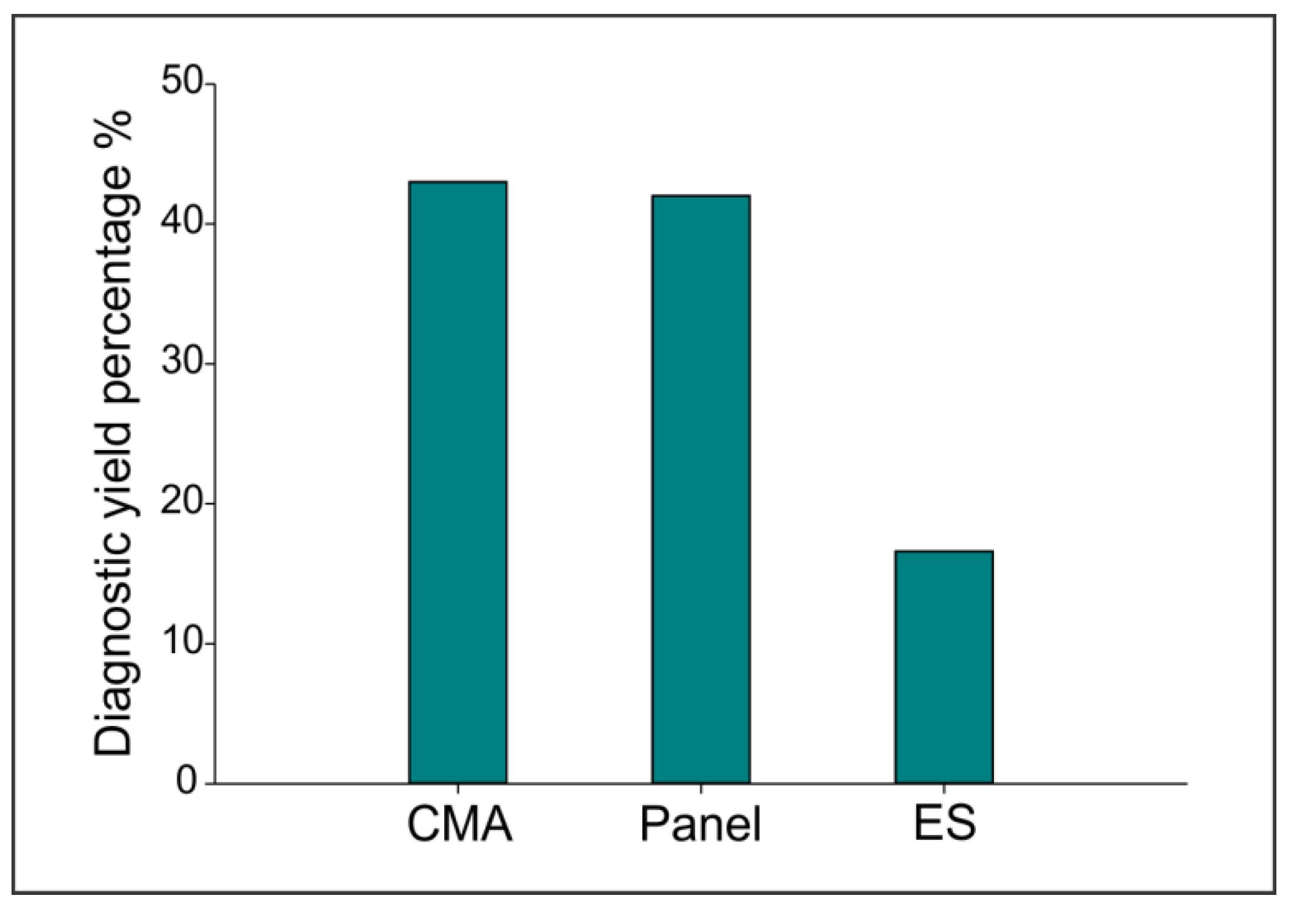
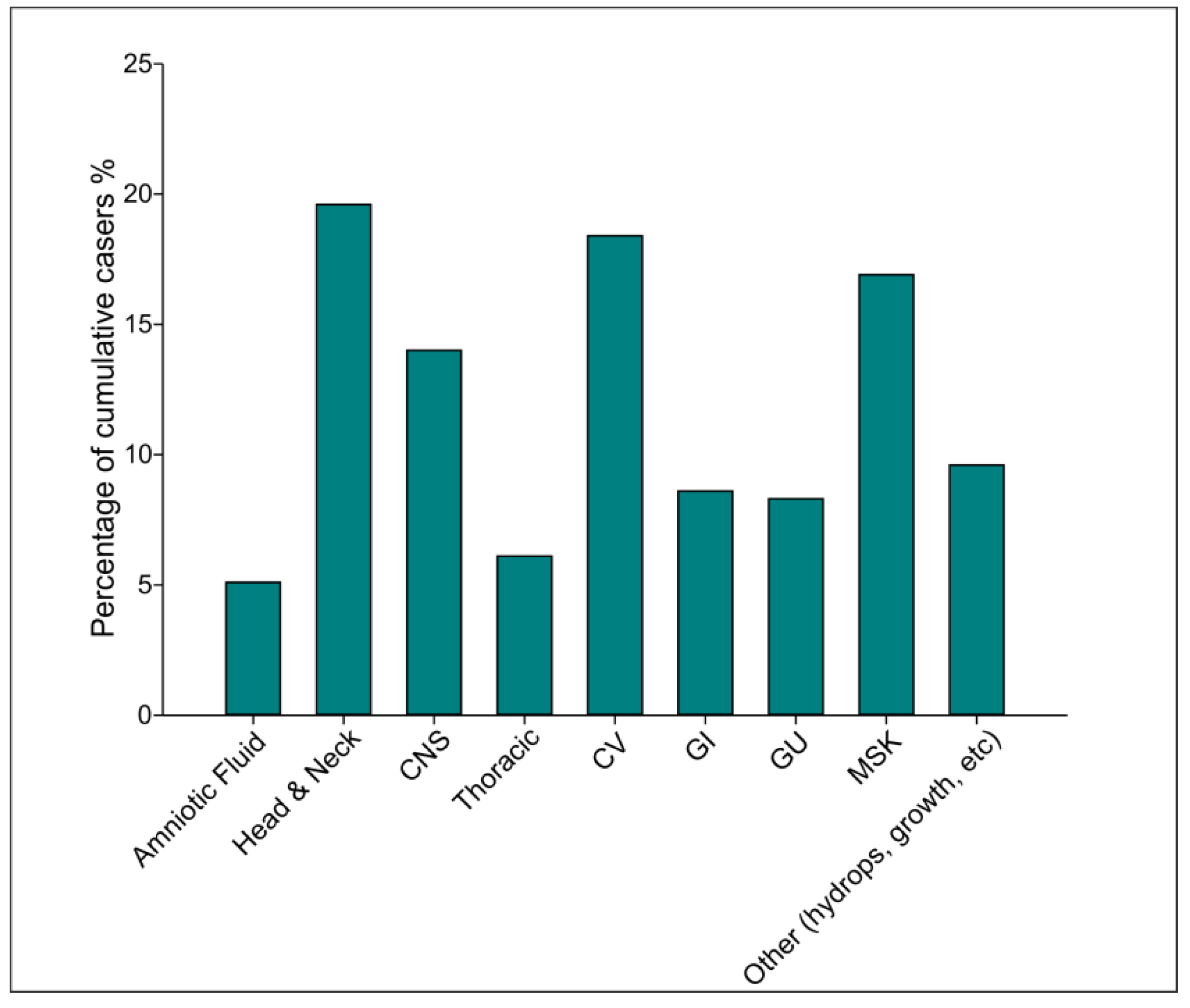
Alberta is a province in Western Canada with a population of approximately 4 million individuals and about 50,000 births a year [17]. Much of this population is centered in two major urban communities, in Calgary and Edmonton [17]. Our center, Edmonton, receives referrals from the Northern half of the province, including Metro Edmonton area (1.1 million), and neighboring provinces of Western Saskatchewan, Eastern British Columbia, Nunavut and Northwest Territories serving a total population of over 2 million people [17]. All permanent residents or Canadian citizens have access to a public health care system, and the modalities of genetic testing reviewed by this study are covered as part of publicly funded medical care [18].
We have performed a retrospective chart review of 450 consecutive cases referred and assessed during the pregnancy by geneticist evaluation between May 2017 and June 2020. The service of the geneticists was utilized on a case-by-case basis and requested by the Maternal Fetal Medicine Clinic at the Royal Alexandra Hospital in Edmonton, AB. Genetic counsellors work with MFM specialists who order first trimester screens and follow up diagnostic tests especially in the first trimester. Rapid aneuploidy detection is completed either by CVS or amniocentesis; in cases with anomalies that have normal results, or if the findings are not expected to be explained by a common aneuploidy, referral to a geneticist for further evaluation is made. Our study included a review of the modalities of genetic testing ordered by geneticists including chromosomal microarray (CMA) utilized prenatally in our center since 2015, and next generation sequencing (NGS)-based tests. If a diagnosis was not achieved through CMA or the CMA results did not explain the fetal phenotype, further testing was ordered by gene panel testing. Since May 2017, ES has become a clinically available test in our center for postnatal cases. This allowed us to use ES in fetuses including stillbirths, terminations of pregnancy, fetal demises, and newborns. The chart reviews included information regarding reason for referral, maternal age and gestational age at referral, maternal and family history, fetal and postnatal findings by imaging modalities (ultrasound, fetal echocardiogram, MRI), type of tests completed including CMA, gene panels, ES, outcomes of the pregnancy, and management implications. While all the cases reviewed were assessed during an ongoing pregnancy, tests were completed at different stages either prenatally (CMA, panels) or postnatally (CMA, panels, ES). The intricacies of real-life practice in PG, reflected in the way genetic testing has been completed following a prenatal consultation, are captured in Supplementary data, Figures S3–S5. For the results presented, variants of unknown significance (VUS) were included together with the negative results as no management decisions could be made at the time they were obtained. Data was collected in a Microsoft Excel spreadsheet and analyzed using SAS Ver9.4. All variables were reported as median (IQR) for continuous variables, and percentages (%) for categorical variables.

3. Results

Four hundred and fifty patient charts were reviewed for the mentioned timeline. The majority of cases 258 (57.3%) were referred for fetal abnormalities, followed by 176 (39.11%) of cases referred for pertinent family history that incorporates maternal and paternal positive history, previous child (PC) or previous pregnancy (PP) with abnormal findings. Other reasons for referral included a positive first trimester screen (FTS), an abnormal result on Rapid Aneuploidy Detection, or a recurrent loss (Figure 1).
The average maternal age of cases reviewed was 31 (27–35) years old, and prenatal testing was done at a median gestational age (GA) of 20 weeks. Aside from abnormal ultrasound findings, a total of 136 pregnant women received fetal echocardiography, of which 52 (38.2%; 52/136) were abnormal. Fetal magnetic resonance imaging (MRI) was completed on 51 fetuses and abnormalities were identified in 41 (80.4%; 41/51).
A total of 408 fetal records were reviewed. The majority of fetal records were positive for at least one anomaly involving amniotic fluid, head & neck, central nervous system (CNS), thorax, cardiovascular (CV), gastrointestinal (GI), genitourinary, and musculoskeletal (MSK) systems. Moreover, fetal records indicated that 290 (71.1%) cases had a single organ system involved, whereas 118 (28.9%) of the results showed the involvement of multiple systems.
294 (72.6%) of fetuses were carried through successful pregnancies, while 63 (15.6%) of pregnancies were terminated, 33 (8.1%) resulted in stillbirths, and seven (1.7%) resulted in neonatal deaths.
We were able to complete testing in a total of 250 patients in our cohort, achieving a diagnosis in 73 patients (for a total of 76 positive results, with three patients having a double diagnosis, see below for details) with an overall yield of diagnosis of 30.4% (76/250). Within the diagnoses obtained, 43.2% were represented by CMA testing (33/76), 42% were obtained through a panel (32/76), and 16.6% by ES (11/76; Figure 2) for a total yield of 56.6% by NGS technologies (43/76). Of these, 43.4% (33/76) of the diagnoses were obtained during ongoing pregnancies while the majorities were tested in the postpartum period (Figure S1).
For all the CMA performed (225), the overall positive results were obtained in 33 cases for a yield of the test of 14.6% (33/225). Of all the CMA performed 70.6% (159/225) were completed during the prenatal time with a positive diagnostic yield of 12.5% (20/159). Postnatally, a further 66 CMA were completed with a positive diagnosis in 19.6% (13/66) (Figures S3–S5).
For all the panels performed (92), the overall diagnostic yield was 34.7% (32/92) (see Table 1). Forty-seven point eight of these tests (44) were completed during ongoing pregnancies with a positive diagnosis in 29.5% (13), while the postnatally completed panel testing (48) reached a diagnosis in 39.5% (19) of the cases. Most cases (5/13; 38.4%) diagnosed prenatally were represented by skeletal dysplasias (SD), followed by Noonan syndrome (3/13; 33%), and five other diagnoses involved an overgrowth syndrome, skin disorder, one immunodeficiency, one neurological disorder, and one GI disorder (see Table 1 for details). The same trend was noticed on positive panel results obtained postnatally, with 42% of the cases being SD diagnoses, followed by 21% (4/190) cases of craniofacial disorders, and 15.7% (3/190) being represented by neurological conditions and other single diagnoses (see Table 1 for details). The decision to order a panel was based on ultrasound findings in the majority of cases diagnosed prenatally (84.6%) vs. postnatally (52.6%), and a positive family history for the remainder of cases in both settings respectively. Out of the panel diagnoses achieved prenatally, 38.4% are in living children, 23% underwent termination of pregnancy (TOP), 23% were stillbirths, and one case was a neonatal demise (7.6%). For postnatally diagnosed cases by panel, the majority, 57.9% are living, 21% were diagnosed following a TOP, and 21% were neonatal demises.
ES was completed only after the end of the pregnancy in a total of 38 cases, with a positive rate of 28.9% (11/38; Table 2) in nine patients (23.6%), and four (10.5%) had uncertain results. Three of the ES positive cases had a double diagnosis (see Section 4), the second diagnosis being achieved by ES in two cases and by CMA in one case. In all the cases, decision to perform the test was based primarily on abnormal ultrasound findings, followed by completion of a combination of negative tests (RAD, CMA, +/-panel). Almost half of the diagnoses obtained (44%) involved one system based on prenatal information, with all the positive cases being multisystemic on a further postnatal evaluation. Out of the patients with a positive diagnosis achieved through ES, more than half (55.5%; 5/90) are living, two pregnancies were tested following TOP, and two of the newborns underwent demise early postnatally (Table 2). A distribution of fetal features in cases with positive tests are illustrated in Figure 3 and Supplementary Figure S2.

4. Discussion

This retrospective chart review was performed in a Canadian publicly funded tertiary care center that has gained access to different means of prenatal diagnostic tools over time (CMA in 2015; postnatal ES in 2017). Our study evaluates and compares the diagnostic yields of three different genetic tests: CMA, and NGS-based gene panels and ES. Our review has included the referrals received for geneticist assessment during prenatal time while testing has been asynchronous (Supplementary Figures S3–S5). Thus, this study provides a transparent look at a real-world clinical PG practice scenario and how diagnoses were achieved in a clinical setting (Supplementary Figures S3–S5). The literature tendency is to focus on individual testing modalities as they become available in research or clinical settings, being either CMA or ES, or for specific phenotypes with gene panel sequencing use, but there is little literature in a more global overview of testing modalities in real-life settings.
Testing was performed in slightly more than half of the cases evaluated in our cohort (55.5%). While the majority of the tests were completed prenatally (203 vs. 152) the higher yield was achieved postnatally (41 of the positive tests vs. 35 positive tests prenatally). This may be explained by the fact that CMA has been both recommended as a first tier diagnostic tool in the evaluation of fetal structural anomalies [19] and has been routinely performed in our clinic for a longer time compared to other tests. Diagnoses achieved by CMA was higher (14.6%) than what reported in the literature, possibly due to a case selection bias favoring referrals for pregnancies with fetal imaging anomalies, as this has been consistently indicated in our cohort overall and in the proportion of cases tested with positive results. Our cohort is comprised of women with a median age below 36 and expected to have more copy number variants (CNV) than aneuploidies [20].
The highest yield of diagnoses by test type has been obtained by using panels and this reflects the nature of the test, utilized in cases with a good diagnostic handle and represented in our study disproportionately by cases with SD, confirming a previously recognized high rate of pick up by NGS in PG [14]. Our findings underline the need of maintaining panel sequencing as a tool in PG when there is a well-defined phenotype or positive family history that helps in sufficiently narrowing the differential diagnosis. This test modality limits the number of VUSs and costs less than ES which is an important consideration in a publically funded health care system. We show that our approach in utilizing ES postnatally, has led to an overall yield of 28.9%, lower than expected based on the latest evidence in the literature [16], and enhanced by the two double ES diagnoses. The highest overall diagnostic yield in our cohort was obtained by NGS technologies (gene panels and ES), the majority of the diagnostic results were obtained postnatally. That is only in part explained by the fact that WES was available postnatally, as the majority of gene panels, available in ongoing pregnancies, were also completed postnatally, in born children. In fact, while the majority of the CMA tests were completed prenatally, only half of the gene panels were done in ongoing pregnancies. It will be important to observe over time, as the public becomes familiarized with more diagnostic tools would the prenatal testing increase? Our practice is impacted by the challenging follow up with families in cases of demise and TOP that precluded some couples initially willing to stay involved to continue the diagnostic odyssey. Although our cohort was small, we were surprised to find three cases with a double diagnosis.
Case 67893001 (see Table 2) was an out of province family, and the only one that had ES during pregnancy as per the home province PG practice. The family decided to have their care continued in our center, for a fetal finding of absent ductus venosus, persistent right umbilical vein, with portosystemic shunting into the right iliac vein. The fetus was found to carry a pathogenic RASA1 variant, shared with his mother who did not have any abnormalities identified despite extensive imaging, and a de novo VUS in KMT2C gene. Postnatal evaluation showed relative macrocephaly and distinct facial features (prominent forehead, apparent hypertelorism, bilateral epicanthal folds, horizontal palpebral fissures, short nose, hypoplastic bridge, bulbous tip, anteverted nares; open mouth, Cupid bow upper lip), neonatal hypotonia, and failed hearing test. Moderate bilateral sensorineural hearing loss was confirmed and hearing aids implemented. During a zoom follow up at the age of 3 years, distinctive facial features were noticed again as well as mild gross and fine motor delays, as well as moderate speech delay. A diagnosis of Kleefstra syndrome type 2 is clinically entertained and functional studies are underway to clarify the nature of the initial VUS.
The mother of Case 67295001 was seen for a family history of spinal muscular atrophy (SMA) at 36 weeks gestation with reduced fetal movements and breech presentation. No genetic testing was completed previously. Testing was discussed but the patient went into labour before the clinicians were able to proceed. The newborn was admitted to NICU for respiratory distress and hypotonia and at examination he presented with upper limbs arthrogryposis, dorsally flexed feet, contractures at the knees and elbows, hypospadias and undescended testicles. He required feeding by nasogastric tube. A positive CMA confirmed Prader-Willi syndrome (paternal deletion) and pursuing testing for the familial history by ES identified a pathogenic variant in COL6A2 in mother and son confirming a diagnosis of Ulrich congenital muscular dystrophy.
Case 69573001 was followed during pregnancy for a single umbilical artery and increased nuchal fold since the first trimester. Craniofacial features including macrocephaly with frontal bossing, hypertelorism, and micrognathia were observed in the second trimester. In the third trimester, large for gestational age fetal measurements were recorded, and polyhydramnios developed gradually that required fluid reduction and presented the opportunity for genetic testing refused previously. QF-PCR and CMA were reported as normal. The fetus was further assessed at birth and ES was pursued. She had Chiari I malformation and squamous synostosis that was followed conservatively. Results of ES confirmed the presence of two de novo disorders: Costello syndrome, and ERF-related craniosynostosis.
The presence of ultrasound abnormalities mirrored in postnatal findings in all cases that had ES positive results may have contributed to the impetus of the clinicians pursuing further diagnosis in these cases, as well as the decision of the parents to consent for further testing. In almost half of the cases with positive ES results there was a single system involvement identified prenatally, with additional features that complemented the clinical picture postnatally. Although more information was gained about the newborn postnatally (on craniofacial features, and systems whose prenatal assessment is limited including gastrointestinal, genitourinary), the prenatally available features would have been sufficient to obtain a diagnosis should ES have been available in an ongoing pregnancy setting (Table 2). Given our testing restrictions over the time the study was conducted, we are not able to comment how many patients would have opted for more testing, including ES, if that had been available prenatally.
Fetuses tested by NGS modalities overall showed as top systems affected to be craniofacial as seen previously [21], cardiovascular, and musculoskeletal systems (Figure 3). We are confirming that between panels and ES, the majority of the diagnoses obtained in our study were due to single gene changes and not copy number variants (41 vs. 33). These findings may help guide expectations and prioritization of tests based on body system, and the justification of implementing ES in ongoing pregnancies. Although the overall number of cases in which ES was completed are small, we underline the fact that the majority of these fetuses are living, and that may be a convincing element in deciding further testing for the patients in our cohort. Exploring parental choices in our population should be an important avenue for future studies.
As positive family history was one of the primary referral reasons for many of the cases evaluated, it is of particular importance to continue exploring potential diagnoses after a negative genetic test when other individuals in the family may contribute to the understanding of a less clear prenatal phenotype. Re-analysis is becoming more acceptable as a clinical workflow step in the modern genetic clinic as it enhances diagnosis over time, especially in relation to ES [22]. We suggest that reanalysis should become routine in the prenatal clinic for all the negative or unclear results regardless of testing modality as more data are shared worldwide and available databases are improved. We suggest that it is particularly important to revisit cases in PG in order to characterize clinical phenotypes at earlier stages as more diagnoses are achieved. An additional important tool is the use of matching websites such as Matchmaker exchange [23] and similar [24] that, again, over time may contribute to the delineation of new human conditions. Such a process is under investigation for one of our ES negative cases with a finding in a candidate gene not yet confirmed to be associated with a human phenotype (Table 2).
Our retrospective chart study has a number of limitations. It did not include results from a first trimester screen, QF-PCR, or karyotype, so these diagnoses were not captured in our review as the goal was to focus on more advanced means of diagnosis either through a genomic approach (CMA, ES) or targeted testing (panels). We have not captured details regarding the ethnic background of our cohort. Our study was limited by local logistics regarding the organization of the prenatal genetics clinic not incorporating the geneticists in the maternal Fetal Medicine care provider team over the timeline targeted by the study. In addition, pregnant women represent a population in which consistent follow up for prenatal testing may prove difficult for many. However, more than half of the patients assessed underwent testing. ES was available only postnatally and applied to a presumably restricted number of cases than if offered in ongoing pregnancies. Secondary findings were not recorded for this study or the modality of performing ES (single vs. duo vs. trio approach). It is not possible to know what the uptake of ES would have been if offered in ongoing pregnancies. However, by comparing to the introduction of CMA a few years back, once available, families have been increasingly open to explore the additional diagnostic tools that may bring light on their fetus’ presentation. This is also supported by other parent’s experience with the introduction of ES elsewhere [25]. An important limitation of genetic tests includes the identification of VUSs which adds complexity to interpretation of results, to counseling and do not offer further avenues in the clinical setting. As general understanding of the genetic basis of genetic disorders improves, previous negative or VUS results can be reexamined with a more critical eye not only regarding ES but also CMA and gene panels.
Our results show that with the strategy of genetic testing used during the period of this study, two thirds of the tested cases remained without a diagnosis. Our study has not included diagnoses reached by QF-PCR and karyotype, and this may have enhanced the overall testing yield observed. Based on current literature, it would be expected that by utilizing all the genetic testing modalities (QF-PCR, karyotype, CMA, panels, ES) up to two thirds of the pregnancies will receive a diagnosis [16]. While parental decision is paramount to performing testing, and a level of unpredictability regarding decisions in a highly emotionally charged clinical environment like PG remains, it is opportune to ask how the implementation of ES in ongoing pregnancies may change parental optics and uptake of the test overtime in our setting. A prospective study with ES employed in ongoing pregnancies may answer that question.

5. Conclusions

Improved access to prenatal testing in the form of CMA, gene panel testing, and ES has vastly expanded the possibilities of reaching prenatal diagnoses in a timely fashion. With the introduction of ES in ongoing pregnancies as a routine test, this diagnostic potential will be further enhanced. CMA and panel sequencing may continue to play an important role in prenatal diagnosis, the latter especially in well-characterized phenotypes (e.g., skeletal dysplasias), or when access to ES is lacking. Single anomalies should not be a deterrent for the use on ES in the prenatal setting. Embedding prenatal genetic professionals in the Maternal Fetal Medicine clinics, sustained access to thorough imaging fetal evaluations, and collection of detailed family histories remain quintessential in prenatal genetics of a contemporary prenatal care center.
Based on the results of this study, we conclude that access to genetic testing including ES in ongoing pregnancies in a public health care system will contribute directly to increasing the yield of prenatal diagnoses, empower families and health care providers involved in the difficult path of pregnancy decision making to achieve best patient-centered pregnancy management.

Supplementary Materials

The following supporting information can be downloaded at: https://www.mdpi.com/article/10.3390/genes13112019/s1. Figure S1: Summary of test results for cases who had CMA prenatal, CMA postnatal, Panels prenatal, Panels postnatal, and WES postnatal; Figure S2: Distribution of fetal features by systems in relation to positive NGS-based tests (prenatal and postnatal gene panels and postnatal WES); Figure S3: Clinical workflow for testing for patients who had prenatal CMA; Figure S4. Clinical workflow for patients who had CMA either postnatally or both prenatally and postnatally; Figure S5. Clinical workflow for patient who did not have CMA or had it postnatally

Author Contributions

Conceptualization, O.C.; methodology, O.C. and S.R. software, S.R. and A.A.; validation, S.R. and A.A.; formal analysis S.R. and A.A.; investigation, O.C., M.S., J.D., M.L., M.J.M., K.Y.N., A.J.E., J.L. and P.K.; resources, D.Z., M.K. and A.A.; data curation, M.K.; writing—original draft preparation, A.A., O.C. and D.Z.; writing—review and editing, O.C., A.A., K.Y.N., M.J.M., M.S. and A.J.E.; visualization, D.Z. and A.A.; supervision, O.C. All authors have read and agreed to the published version of the manuscript.

Funding

This research received no external funding.

Institutional Review Board Statement

The study was conducted in accordance with the Declaration of Helsinki, and approved by the Institutional Review Board of the University of Alberta, protocol code 00104196, date of approval 25 September 2020. This study has obtained approval from the Northern Alberta Clinical Trials and Research Centre, ID#OA55731, date of approval 25 November 2020.

Informed Consent Statement

Patient consent was waived as approved by authorities cited above as patient identifiers were removed during the data collection.

Data Availability Statement

Not applicable.

Conflicts of Interest

The authors declare no conflict of interest.

Abbreviations

CMAChromosomal microarray
CNSCentral nervous system
CVCardiovascular
ESExome sequencing
FHxPositive family history
FTSFirst trimester screen
GAGestational age
GIGastrointestinal
MRIMagnetic resonance imaging
MSKMusculoskeletal
NGSNext generation sequencing
PPPrevious pregnancy
PCPrevious child
QF-PCRQuantitative Fluorescent-Polymerase chain reaction
WESWhole exome sequencing
VUSVariants of unknown significance
?Variants of unknown significance

References

  1. Bardi, F.; Bergman, J.E.H.; Siemensma-Mühlenberg, N.; Elvan-Taşpınar, A.; de Walle, H.E.K.; Bakker, M.K. Prenatal diagnosis and pregnancy outcome of major structural anomalies detectable in the first trimester: A population-based cohort study in the Netherlands. Paediatr. Perinat. Epidemiol. 2022, 36, 804–814. [Google Scholar] [CrossRef] [PubMed]
  2. Dolk, H.; Loane, M.; Garne, E. The prevalence of congenital anomalies in Europe. Adv. Exp. Med. Biol. 2010, 686, 349–364. [Google Scholar] [CrossRef] [PubMed]
  3. Greenbaum, G.; Rubin, A.; Templeton, A.R.; Rosenberg, N.A. Network-based hierarchical population structure analysis for large genomic data sets. Genome. Res. 2019, 29, 2020–2033. [Google Scholar] [CrossRef]
  4. Monaghan, K.G.; Leach, N.T.; Pekarek, D.; Prasad, P.; Rose, N.C. The use of fetal exome sequencing in prenatal diagnosis: A points to consider document of the American College of Medical Genetics and Genomics (ACMG). Genet. Med. 2020, 22, 675–680. [Google Scholar] [CrossRef] [Green Version]
  5. Zhang, B.; Lu, B.Y.; Yu, B.; Zheng, F.X.; Zhou, Q.; Chen, Y.P.; Zhang, X.Q. Noninvasive prenatal screening for fetal common sex chromosome aneuploidies from maternal blood. J. Int. Med. Res. 2017, 45, 621–630. [Google Scholar] [CrossRef] [PubMed]
  6. Hillman, S.C.; McMullan, D.J.; Hall, G.; Togneri, F.S.; James, N.; Maher, E.J.; Meller, C.H.; Williams, D.; Wapner, R.J.; Maher, E.R.; et al. Use of prenatal chromosomal microarray: Prospective cohort study and systematic review and meta-analysis. Ultrasound Obs. Gynecol. 2013, 41, 610–620. [Google Scholar] [CrossRef]
  7. Wapner, R.J.; Martin, C.L.; Levy, B.; Ballif, B.C.; Eng, C.M.; Zachary, J.M.; Savage, M.; Platt, L.D.; Saltzman, D.; Grobman, W.A.; et al. Chromosomal microarray versus karyotyping for prenatal diagnosis. N. Engl. J. Med. 2012, 367, 2175–2184. [Google Scholar] [CrossRef] [Green Version]
  8. Yang, Y.; Muzny, D.M.; Reid, J.G.; Bainbridge, M.N.; Willis, A.; Ward, P.A.; Braxton, A.; Beuten, J.; Xia, F.; Niu, Z.; et al. Clinical whole-exome sequencing for the diagnosis of mendelian disorders. N. Engl. J. Med. 2013, 369, 1502–1511. [Google Scholar] [CrossRef] [Green Version]
  9. Sawyer, S.L.; Hartley, T.; Dyment, D.A.; Beaulieu, C.L.; Schwartzentruber, J.; Smith, A.; Bedford, H.M.; Bernard, G.; Bernier, F.P.; Brais, B.; et al. Utility of whole-exome sequencing for those near the end of the diagnostic odyssey: Time to address gaps in care. Clin. Genet. 2016, 89, 275–284. [Google Scholar] [CrossRef] [Green Version]
  10. Mone, F.; McMullan, D.; Williams, D.; Chitty, L.; Maher, E.; Kilby, M. Evidence to Support the Clinical Utility of Prenatal Exome Sequencing in Evaluation of the Fetus with Congenital Anomalies. BJOG Int. J. Obstet. Amp. Gynaecol. 2021, 128, e39–e50. [Google Scholar] [CrossRef]
  11. Lazier, J.; Hartley, T.; Brock, J.A.; Caluseriu, O.; Chitayat, D.; Laberge, A.M.; Langlois, S.; Lauzon, J.; Nelson, T.N.; Parboosingh, J.; et al. Clinical application of fetal genome-wide sequencing during pregnancy: Position statement of the Canadian College of Medical Geneticists. J. Med. Genet. 2021, 59, 931–937. [Google Scholar] [CrossRef] [PubMed]
  12. Van den Veyver, I.B.; Chandler, N.; Wilkins-Haug, L.E.; Wapner, R.J.; Chitty, L.S. International Society for Prenatal Diagnosis Updated Position Statement on the use of genome-wide sequencing for prenatal diagnosis. Prenat. Diagn. 2022, 42, 796–803. [Google Scholar] [CrossRef] [PubMed]
  13. Lord, J.; McMullan, D.J.; Eberhardt, R.Y.; Rinck, G.; Hamilton, S.J.; Quinlan-Jones, E.; Prigmore, E.; Keelagher, R.; Best, S.K.; Carey, G.K.; et al. Prenatal exome sequencing analysis in fetal structural anomalies detected by ultrasonography (PAGE): A cohort study. Lancet 2019, 393, 747–757. [Google Scholar] [CrossRef] [Green Version]
  14. Han, J.; Yang, Y.D.; He, Y.; Liu, W.J.; Zhen, L.; Pan, M.; Yang, X.; Zhang, V.W.; Liao, C.; Li, D.Z. Rapid prenatal diagnosis of skeletal dysplasia using medical trio exome sequencing: Benefit for prenatal counseling and pregnancy management. Prenat. Diagn. 2020, 40, 577–584. [Google Scholar] [CrossRef]
  15. Tolusso, L.K.; Hazelton, P.; Wong, B.; Swarr, D.T. Beyond diagnostic yield: Prenatal exome sequencing results in maternal, neonatal, and familial clinical management changes. Genet. Med. 2021, 23, 909–917. [Google Scholar] [CrossRef] [PubMed]
  16. Mellis, R.; Oprych, K.; Scotchman, E.; Hill, M.; Chitty, L.S. Diagnostic yield of exome sequencing for prenatal diagnosis of fetal structural anomalies: A systematic review and meta-analysis. Prenat. Diagn. 2022, 42, 662–685. [Google Scholar] [CrossRef]
  17. Government of Alberta. Population Statistics. Available online: https://www.alberta.ca/population-statistics.aspx#population-projections (accessed on 16 September 2022).
  18. Government of Alberta. Alberta Health Care Insurance Plan (AHCIP). Available online: https://www.alberta.ca/ahcip.aspx (accessed on 16 September 2022).
  19. Committee Opinion No. 682: Microarrays and Next-Generation Sequencing Technology: The Use of Advanced Genetic Diagnostic Tools in Obstetrics and Gynecology. Obs. Gynecol. 2016, 128, e262–e268. [CrossRef]
  20. Srebniak, M.I.; Joosten, M.; Knapen, M.; Arends, L.R.; Polak, M.; van Veen, S.; Go, A.; Van Opstal, D. Frequency of submicroscopic chromosomal aberrations in pregnancies without increased risk for structural chromosomal aberrations: Systematic review and meta-analysis. Ultrasound Obs. Gynecol. 2018, 51, 445–452. [Google Scholar] [CrossRef] [Green Version]
  21. Normand, E.A.; Braxton, A.; Nassef, S.; Ward, P.A.; Vetrini, F.; He, W.; Patel, V.; Qu, C.; Westerfield, L.E.; Stover, S.; et al. Clinical exome sequencing for fetuses with ultrasound abnormalities and a suspected Mendelian disorder. Genome. Med. 2018, 10, 74. [Google Scholar] [CrossRef] [Green Version]
  22. Liu, P.; Meng, L.; Normand, E.A.; Xia, F.; Song, X.; Ghazi, A.; Rosenfeld, J.; Magoulas, P.L.; Braxton, A.; Ward, P.; et al. Reanalysis of Clinical Exome Sequencing Data. N. Engl. J. Med. 2019, 380, 2478–2480. [Google Scholar] [CrossRef]
  23. Sobreira, N.L.M.; Arachchi, H.; Buske, O.J.; Chong, J.X.; Hutton, B.; Foreman, J.; Schiettecatte, F.; Groza, T.; Jacobsen, J.O.B.; Haendel, M.A.; et al. Matchmaker Exchange. Curr. Protoc. Hum. Genet. 2017, 95, 9.31.1–9.31.15. [Google Scholar] [CrossRef]
  24. Au, P.Y.B.; You, J.; Caluseriu, O.; Schwartzentruber, J.; Majewski, J.; Bernier, F.P.; Ferguson, M.; Valle, D.; Parboosingh, J.S.; Sobreira, N.; et al. GeneMatcher aids in the identification of a new malformation syndrome with intellectual disability, unique facial dysmorphisms, and skeletal and connective tissue abnormalities caused by de novo variants in HNRNPK. Hum. Mutat. 2015, 36, 1009–1014. [Google Scholar] [CrossRef] [Green Version]
  25. de Koning, M.A.; Haak, M.C.; Adama van Scheltema, P.N.; Peeters-Scholte, C.; Koopmann, T.T.; Nibbeling, E.A.R.; Aten, E.; den Hollander, N.S.; Ruivenkamp, C.A.L.; Hoffer, M.J.V.; et al. From diagnostic yield to clinical impact: A pilot study on the implementation of prenatal exome sequencing in routine care. Genet. Med. 2019, 21, 2303–2310. [Google Scholar] [CrossRef]
Figure 1. A pie chart representing the proportion of reasons for referral to our clinic between May 2017 and June 2020 with fetal structural abnormalities (57.33%) and positive family history (39.11%) as the common reasons, followed by positive first trimester screen (1.56%), abnormal rapid aneuploidy detection (0.89%), Recurrent Loss (0.67%) and other reasons (0.44%).
Genes 13 02019 g001
Figure 2. CMA and gene panel testing has the highest diagnostic yield in our cohort. A bar graph of CMA, gene panel and ES diagnostic yield percentages in our cohort.
Genes 13 02019 g002
Figure 3. Cumulative distribution of fetal features in cases with positive NGS-based test results. A bar graph of the distribution of fetal features of cumulative cases who had prenatal and postnatal panel, and postnatal ES tests.
Genes 13 02019 g003
Table 1. Prenatal assessed cases. Data of both prenatal and postnatal panel testing were achieved.
a. Prenatally Assessed Cases for Which Pre- and Postnatal Positive Results of Gene Panels Were Obtained *.
Stud-y IDReason for ReferralAnomalies
Identified
Prenatal Genetic
Testing
Pregnancy OutcomesPostnatal Genetic
Testing
DiagnosisGeneManagement Implications (RR%)
Prenatal DetectionPostnatal Detection
Prenatal testing
67573AbN US99(-) RAD, CMA
(+) SD panel
TOPnilHypochondrogenesisCOL2A1<1%
66234AbN US99(-) RAD, CMA
(+) SD panel
StillbirthnilOpsismodysplasiaINPPL1<1%
66328AbN US3, 4, 5, 6, 73, 4, 5, 6, 7, 11(-) RAD, CMA
(+) Noonan panel
TOPnilNoonan syndromePTPN11<1%
66330AbN US1, 5, 91, 5, 9(-) RAD, CMA
(+) Arthrogryposis panel
Nn demisenilNemaline myopathyNEB25%
66883AbN US7, 83, 7, 8(-) RAD, CMA
(+) BW panel
LivingnilBeckwith-Wiedemann
syndrome
KvDMR2 loss of
methylation
<1%
66944FHxnil11(-) RAD, CMA
(+) SCID panel
LivingnilSCIDIL2RG25%
67196AbN US31, 3, 10(-) RAD, CMA
(+) Noonan panel
LivingnilNoonan syndrome (MIM163950)PTPN11<1%
67478FHx 10(-) CMA
(+) Targeted panel
LivingnilEBS (MIM131800)KRT1450%
67497AbN US99(-) RAD, CMA
(+) SD panel
LivingnilHypophosphatasiaALPL25%
67797AbN US1, 2, 61, 3, 5, 6, 7(-) RAD, CMA
(+) Noonan panel
StillbirthnilNoonan syndromePTPN11<1%
67981AbN US3, 43, 4(-) RAD, CMA
(+) Brain malformation panel
TOPnilL1CAM-related disordersL1CAM25%
68743AbN US77(-) RAD, CMA
(+) Congenital diarrhea panel
LivingnilCongenital diarrheaSLC26A325%
58884AbN US2, 5, 95, 9(-) RAD, CMA
(+) SD panel
stillbirthnilThanatophoric dysplasiaFGFR3<1%
Postnatal testing
67259AbN US93, 9nilTOP(-) CMA
(+) SD panel
OI2COL2A1<1%
68120AbN US4, 84, 8nilTOP(-) CMA
(+) Ciliopathy Panel
Joubert SyndromeRPGRIP1L25%
68445FHx99nilTOP(-) CMA
(+) SD panel
Thanatophoric dysplasiaFGFR3<1%
69205AbN US4, 93, 4, 8, 9nilInfantile demise(-) CMA
(+) Brain
malformation panel
Zellweger syndromePEX125%
66125AbN US2, 61, 2, 3, 4, 6, 9(-) RAD, CMALiving(+) Craniosynostosis panelAcromelic fronto-nasal
dysostosis
ZSWIM6<1%
67274AbN US6, 71, 3, 4, 6, 7(-) RAD, CMANn demise (+) Aicardi-Goutieres panelAicardi-Goutieres syndromeRNASEH2C25%
67502AbN US31, 3, 9(-) RAD, CMALiving(+) Stickler syndrome panelStickler syndromeCOL11A150%
7009FHxnil nilLiving(+) MH panelMalignant HyperthermiaRYR150%
17890FHxnil1, 3nilLiving(+) CDL panelCornelia de Lange syndromeNIPBL50%
63327AbN US3, 93, 9nilNn demise(+) Craniosynostosis panelBeare-Stevenson syndromeFGFR2<1%
64860FHxnil1, 3, 9nilLiving(+) OI panelBruck SyndromeFKBP1025%
66134FHxnil11nilLiving(+) DSPD panelDelta Storage pool diseaseRUNX150%
66206FHxnil1, 9nilLiving(+) Hposhosphatemia panelHypophosphatemiaPHEX50%
66428AbN US63, 6nilLiving(+) Brain malfomation panelTuberous SclerosisTSC11%
67255AbN US91, 3, 9nilNn
demise
(+) SD panelCampomelic dysplasiaSOX9<1%
67739AbN US1, 3, 91, 3, 9(-) RADTOP(+) SD panelThanatophoric dysplasiaFGFR3<1%
68014FTS+1, 2, 3, 5, 73, 5, 10nilLiving(+) Vascular malformation panelRASA1-related disorderRASA1<1%
69624FHx3, 93nilLiving(+) Craniosynostosis panelSaethre-Chotzen syndromeTWIST50%
67669FHx2, 93, 4, 9nilLiving(+) Congenital muscular dystrophy panelMyotonic dystrophy type 1DMPK50%
b. Prenatally assessed cases for which pre- and postnatal variants of unknown significance (VUS) results of gene panels were obtained.
59041FTS+1, 3, 4, 6, 9 (-) RAD, CMA
(?) Panels (hydrops, brain malformations)
TOP(?) WESAR combined oxidative phosphorylation deficiency 27/Alpers-Huttenlocher syndromeCARS2 (NM_ 024537.4): c.563C>T,p.Thr188Met25%?
67291FTS+3, 81, 3, 4, 9(-) RAD, CMA
(?) Noonan Panel
LivingnilNoonan syndrome?
Lost to follow up
LZTR1 (NM_006767.3):c.2063G>A,p.(Arg688His)?
67347FHxnilnil(-) RAD, CMA
(?) Bleeding disorder panel
LivingnilBleeding disorderABCG5(NM_022436.2):c.235G>A,p.(Gly79Arg);
FREM1(NM_144966.5):c.987A>T,p)Lys329Asn);
MPL(NM_005373.2):c.655C>G,p(Gln219Glu)
?
67980AbN US66, 8, 11(-) RAD, CMA
(?) Noonan syndrome/Rasopathies
Living nilComplex syndromal presentationKAT6B(NM_0123303.3):
c.1217G>A, p.(Arg406Gln)mat
?
68077AbN US91, 9(-) RAD, CMA
(?) SD Panel
Living nilPseudohypoparathyroidismGNAS(NM_000516.4):c.349G>A,p.(Val117Met),mat50%?
67042AbN US4, 64, 6(-) RAD, CMALiving(-) CMA
(?) Brain malformation panel
Joubert syndromeTUBA8(NM_018943.2):c.1118G>A,p.(Arg373Gln), het
VLDLR(NM003383.3):c.836G>A,p(Arg279Gln),het
?
66890FHx3, 93, 9nilTOP(-) CMA
(?) Arthrogryposis panel
Beals syndromeFBN2(NM_001999.3):c.8363C>T,p.(Ala2788Val), het?
67846AbN US2, 88nilStillbirth(-) CMA
(?) CAKUT Panel
Bilateral Renal agenesisFREM2(NM_207361.6):c.9038C>T,p.(Thr3013Met), het ?
68247AbN US3, 8, 93, 8, 9(-) RADTOP(-) CMA
(?) Arthrogryposis panel
Rothmund-Thmpson
syndrome
RECQL4(NM_004260.3): c.551G>A,p.(Gly184Asp), hom25%?
66794AbN US1, 2 (-) RAD, CMAInfantile demise(?) Hydrops PanelFeingold syndromeMYCN(NM_005378.4):c.718G>A,p.(Gly240Ser)?
66824AbN US1, 2, 9 nilTOP(-) CMA, (?) Hydrops PanelDesbuquois dysplasiaCANT1(NM_138793.3):c.872C>T,p(Thr291Met), het?
* Legend for corresponding anomalies: 1 = Growth Restriction or Syndromic (e.g., hydrops) anomaly, 2 = Abnormal Amniotic Fluid, 3 = Craniofacial or neck anomaly, 4 = Central Nervous system anomaly, 5 = Thoracic anomaly, 6 = Cardiovascular anomaly, 7 = Gastrointestinal anomaly, 8 = Genitourinary anomaly, 9 = Musculoskeletal anomaly, 10 = Skin and adnexa.
Table 2. Prenatal whole exome sequencing cases.
a. Prenatal Cases in Which a Diagnosis Was Achieved via Whole Exome Sequencing
Study IDReason for
Referral
Anomalies Identified Prenatal Genetic TestingPregnancy
Outcome
Postnatal
Testing
DiagnosisGenetic FindingManagement
Implications
Prenatal DetectionPostnatal Detection
64702001AbN US3, 4, 6, 7, 83, 4, 6, 7, 8nilTOP(-) RAD
(-) CMA
(+) WES
Developmental delay with or without dysmorphic facies and autism
(MIM618454) AD
TRRAP (NM_003496.3), c.3127G>A, p.(Ala1043Thr), heterozygous, de novoRR < 1%
71280001AbN US3, 4, 63, 4, 6, 7, 8(-) RAD, CMA, PanelTOP(+) WESNeurooculocardio-genitourinary syndrome (MIM618652), ADWDR37 (NM_014023.3), c.389C>T, p.(Thr130Ile),
heterozygous, de novo
RR < 1%
71354001AbN US5, 63, 5, 6, 7, 8, 9(-) RAD, CMA, PanelNeonatal Demise(+) WESOsteopathia striata with cranial sclerosis (MIM 300373), XLAMER1 (NM_152424.4), c.19G>&, p.Glu7 *; hemizygous, maternalRR25%
67893001AbN US66(-) RAD, CMA in Alberta
(+) WES in another province
Livingnil1. RASA1-related disorder (MIM 608354), AD
2. Kleefstra sdr. type 2
1.RASA1 (NM_002890.2), c.2131C>T,p.Arg711X;
heterozygous,
maternal
2.KMT2C (NM_170606.2), c.9773A>C, p.His3258Pro
heterozygous, de novo
Surveillance
50% RR
66175001AbN US33, 9(-) RAD, CMALiving(-) CLP Panel
(+) WES
Hartsfield syndrome (MIM615465), ADFGFR1 (NM_023110.2), c.1474G>A,p.(Val492Met),
heterozygous, de novo
Anticipatory management
RR < 1%
67295001AbN US93, 9nilLiving(+) CMA
(+) WES
1. Prader-Willi Syndrome (MIM176270), imprinting
2. Ullrich Muscular Dystrophy (MIM254090), AD
1.Del 15q11, paternal
2.COL6A2(NM_001849.3),
c.848G > A,p.Gly283Glu, heterozygous, maternal
Treatment
Anticipatory management
RR 50%
69829001AbN US1, 4, 5, 91, 3, 4, 5, 9nilLiving(-) CMA
(+) WES
Escobar Syndrome (MIM265000), ARCHRNG (NM_005199.4), c.202C>T, p.Arg68 *;
c.459dup, p.Val154Serfs*24, combined heterozygous, parents carriers
Anticipatory management
RR25%
69573001AbN US1, 2, 31, 2, 3, 6(-) RAD, CMALiving(+) WES1. Costello Syndrome (MIM218040),AD
2. ERF-related disorder (MIM600775),AD
1.HRAS (NM_005343.3), c.34G>A, p.(Gly12Ser), heterozygous, de novo
2.ERF (NM_006494.3), c.891_892del, p.(Gly299Argfs * 9), heterozygous, de novo
Anticipatory management
RR < 1%
68143001AbN US63, 6, 7(-) RAD, CMADemise at 4 mo(-) Panel
(+) WES
Zhu-Tokita-Takenouchi-Kim syndrome (ZTTKS),ADSON (NM_032195.2), c.5867G>A, p.(Arg1956His),
heterozygous, de novo
RR < 1%
b. Prenatal Cases in which WES identified a Variant of Unknown Significance
67663001AbN US33, 4nilLiving(+) CMA
(-) CLP Panel
(?) WES
1. 15q13 dup syndrome;
2. Intellectual
Developmental disorder, auto-somal dominant
52
1. Paternal
2. ASH1L (NM_018489.2): c.614A>C,p.(Lys205Thr), pat
50%
68424001FHx2, 6, 7, 82, 3, 6, 7, 8, 9nilTOP(-) RAD,
(-) CMA
(?) WES
CDX2 variant, gene not associated with human phenotypeCDX (NM_001265.4): c.592C>T,p.(Arg198Trp), de novo<1%
67572001AbN US4, 93, 4, 9nilTOP(-) CMA
(?) WES
RYR3 variants, not associated with human phenotypeRYR3 (NM_001036.4): c.90T>A,p.(His30Gln), pat
C,11730G>C,p.Leu391Phe),mat
25%
69260001AbN US6, 7, 96, 7, 9(-) RAD, CMA, PanelTOP(?) WESMandibulofacial dysostosis, Guyon Almeida type (MIM610536)EFTUD2(NM_004247.3):c.658C>T,p.(Arg220Cys),
de novo
<1%
* Legend for corresponding anomalies, 1 = Growth Restriction or Syndromic (e.g., hydrops) anomaly, 2 = Abnormal Amniotic Fluid, 3 = Craniofacial or neck anomaly, 4 = Central Nervous system anomaly, 5 = Thoracic anomaly, 6 = Cardiovascular anomaly, 7 = Gastrointestinal anomaly, 8 = Genitourinary anomaly, 9 = Musculoskeletal anomaly.
Publisher’s Note: MDPI stays neutral with regard to jurisdictional claims in published maps and institutional affiliations.

