Article

Bacterial Community Diversity and Bacterial Interaction Network in Eight Mosquito Species

by
Herculano da Silva
,
Tatiane M. P. Oliveira
and
Maria Anice M. Sallum
*
Department of Epidemiology, School of Public Health, University of São Paulo, São Paulo 01246-904, SP, Brazil
*
Author to whom correspondence should be addressed.
Genes 2022, 13(11), 2052; https://doi.org/10.3390/genes13112052
Submission received: 18 September 2022 / Revised: 28 October 2022 / Accepted: 1 November 2022 / Published: 7 November 2022
(This article belongs to the Special Issue Evolutionary Genetics and Phylogenetics of Mosquito Species)

Abstract

:
Mosquitoes (Diptera: Culicidae) are found widely throughout the world. Several species can transmit pathogens to humans and other vertebrates. Mosquitoes harbor great amounts of bacteria, fungi, and viruses. The bacterial composition of the microbiota of these invertebrates is associated with several factors, such as larval habitat, environment, and species. Yet little is known about bacterial interaction networks in mosquitoes. This study investigates the bacterial communities of eight species of Culicidae collected in Vale do Ribeira (Southeastern São Paulo State) and verifies the bacterial interaction network in these species. Sequences of the 16S rRNA region from 111 mosquito samples were analyzed. Bacterial interaction networks were generated from Spearman correlation values. Proteobacteria was the predominant phylum in all species. Wolbachia was the predominant genus in Haemagogus leucocelaenus. Aedes scapularis, Aedes serratus, Psorophora ferox, and Haemagogus capricornii were the species that showed a greater number of bacterial interactions. Bacterial positive interactions were found in all mosquito species, whereas negative correlations were observed in Hg. leucocelaenus, Ae. scapularis, Ae. serratus, Ps. ferox, and Hg. capricornii. All bacterial interactions with Asaia and Wolbachia were negative in Aedes mosquitoes.

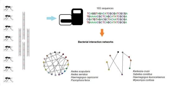
Graphical Abstract

1. Introduction

The Culicidae family includes 3608 mosquito species (Diptera), classified into 113 genera [1]. Many species are important because they feed on human and other animals’ blood, causing a disturbance, and 5% of the known species may participate in the transmission of parasites and pathogens to humans [2,3]. Mosquitoes harbour a great diversity of fungi and bacteria. The bacterial composition of the microbiota of these invertebrates depends on several factors, such as sex, larval habitat, developmental stage, environment, and species. It can also be related to microsporidian infection [4,5,6,7]. Bacteria from the microbiota can influence mosquitoes’ physiology, metabolism, and adaptation [4]. They also play a key role in protecting against pathogens [8] and developing mosquito larvae [9,10].
Recently, based on the results of an experimental investigation, Saab et al. [5] found that Anopheles gambiae and Aedes albopictus larvae reared in the same insectary room and fed on the same food source showed intraspecies and interspecies variations in the composition of the midgut microbiota, concluding that bacterial composition can be modulated by environmental variations, species factors, and complex microbial interactions.
Despite several studies focused on microbial interactions in mosquitoes, it is still unclear how these interactions may contribute to the formation of bacterial communities in mosquitoes. There are few records of natural infection of Wolbachia in Aedes aegypti [11] and anophelines [12,13]. The absence of this bacterium in most populations of these mosquito species may be due to the presence of Asaia in the native microbiome, which inhibits the vertical transmission of Wolbachia [14,15]. Serratia is present in Anopheles mosquitoes [16,17], influencing these insects’ vector capacity [18]. In field-collected Aedes mosquitoes, the prevalence of this bacterium is variable. For example, Serratia was more abundant in Ae. albopictus than in Ae. aegypti collected in Houston, TX, United States [19]; Serratia marcescens was found in Ae. albopictus collected in Sing Bur Province of Thailand, and absent in mosquitoes of this species collected in Chumphon and Yala Provinces [20]. The occurrence of Serratia in Ae. aegypti can be low because of the competitive exclusion with Cedecea [21]. Interactions between bacteria, viruses, microsporidian, fungi and Plasmodium also changes the bacterial community [6,22,23,24,25]. Specimens of Culex pipiens infected with the West Nile virus showed an increased proportion of Serratia [26]. In addition, the endosymbiotic bacteria Spiroplasma sp. PL03 and Weissela cf. viridescens depends on microsporidia infection in the mosquito gut [6], and the bacterial microbiota was distinct between specimens of An. gambiae and Anopheles funestus that were positive and negative groups for Plasmodium falciparum [22]. In addition, in the same study, authors observed that some bacterial species such as Asaia borgorensis, Burkholderia fungorum, Burkholderia cepacia and Enterobacter cloacae were present only in females that were negative for P. falciparum. Balaji et al. [27] demonstrated that Wolbachia can influence the colonization of certain bacterial taxa by competitive interactions, such as the abundance of Serratia sp. in Wolbachia-carrying mosquitoes. The 16S rRNA data have been used to verify microbial co-exclusion/co-occurrence in different organisms [19,28,29]. Relative abundance and/or presence/absence parameters are used to verify bacterial interactions and to infer bacterial networks.
The number of studies focusing on bacteria and mosquitoes is increasing. Among these studies are: understanding the acquisition and composition of the microbiota [4,20], its relationship with vector capacity [30], its importance in the modulation of pathogen development in mosquitoes [31], and how bacterial interactions can modulate the bacterial community in the mosquito [19]. Despite this increasing interest in mosquito microbial investigations and the epidemiological importance of the Vale do Ribeira region as a potential source of sylvatic arboviruses and vertebrate reservoirs, little is known about the microbiota in mosquito populations inhabiting the southeastern Atlantic tropical rain forest, São Paulo, Brazil.
This study aims to (1) investigate the microbiota present in eight species of Culicidae collected in Vale do Ribeira, and (2) verify the bacterial communities’ composition and interactions (such as co-exclusion and co-occurrence) between these communities.

2. Materials and Methods

2.1. Mosquito Collection and Identification

Adult mosquitoes were collected in a forest preserved area neighboring the town (24°47′28.8″ S, 47°54′42.6″ W) in Vale do Ribeira, Pariquera-Açu municipality, São Paulo state, Brazil. The collections were performed using an entomological net and conducted daily from 8:00 to 14:30 h from 14 December to 17 December 2021. Mosquitoes were killed with ethyl acetate (C4H8O2) and immediately preserved in silica gel. Specimens were transported at room temperature to the Laboratório de Entomologia em Saúde Pública–Sistemática Molecular and kept in these conditions until identification and processing. Specimens were morphologically identified using the identification key of Forattini [2].

2.2. Sequencing of 16S rRNA

The mosquito’s surface was rinsed in 70% ethanol and ultrapure water. The genomic DNA of each specimen was extracted separately using the Quick-DNA Fungal/Bacterial Miniprep kit (ZymoReasearch), following the manufacturer’s instructions. The V4 region of the 16S rRNA gene was amplified using isolated DNA from each specimen. Each reaction was carried out in a final volume of 20 µL containing 1 X GoTaq® Colorless Master Mix (Progema, USL), 0.3 µm of each primer (16S-V4 Forward: 5′ GTGCCAGCMGCCGCGGTAA 3′; 16S- V4 Reverse: 5′ GGACTACHVGGGTWTCTAAT 3′) [32], 8 µL of genomic DNA and ultrapure water. The PCR thermal conditions were 94 °C for 3 min, followed by 30 cycles of 94 °C for 45 s, 55 °C for 1 min, 72 °C for 1 min, and a final extension of 72 °C for 10 min. The PCR products were visualised on a 2% agarose gel stained with UniSafe Dye 0.03% (v/v) and purified with Agencourt AMPure XP magnetic beads (Beckman Coulter, Brea, CA, USA) according to the manufacturer’s recommendations. After indexing with Nextera XT Index kit (Illumina), the products were purified with magnetic beads and quantified by real-time PCR (qPCR) with the KAPA-KK4824 kit (Library Quantification kit–Illumina/Universal) following the manufacturer’s instructions. All samples were normalised to 4 nM, and an equimolar pool of DNA was prepared. Next-generation sequencing was performed on the Illumina MiSeq sequencer (Illumina, San Diego, CA, USA) using the MiSeq Reagent Micro v2 kit (300 cycles: 2 × 150 bp).

2.3. Processing of 16S Sequences and Taxonomic Attribution

FLASH v. 1.2.11 [33] was used to assemble Illumina paired-end reads with a minimum overlap of six base pairs. Quality control (denoising), taxonomic attribution, and diversity and abundance analyses were performed in QIIME2 v.2021-11 software [34].
The qiime tools import command and Casava 1.8 single-end demultiplexed format were used to import the joined sequences to QIIME2. Quality control and denoising were performed with the commands: qiime quality-filter q-score and qiime deblur denoise-16S, respectively. To taxonomic attribution was used SILVA 138 [35,36] and qiime feature-classifier classify-sklearn.

2.4. Diversity Analyses

The rarefaction curve was generated to obtain the expected number of ASVs (Amplicon Sequence Variants) in each sample for a given number of sequences, and to allow for the comparison between the richness of the samples. We used a depth value of 10,000 and qiime diversity α-rarefaction command to construct this curve.
Diversity metrics (α and β) were generated with qiime diversity core-metrics-phylogenetic, and a phylogenetic tree was constructed with qiime phylogeny align-to-tree-mafft-fasttree.
Shannon-Weaver indices (α diversity) were subject to Kruskal-Wallis followed by Dunn’s test, adjusted with Bonferroni method in RStudio v.1.4.1106 to verify whether samples from one species have greater α diversity than from other species.
Analysis of variance (PERMANOVA) statistical test was performed with β diversity data generated by Unifrac weighted and unweighted distances in QIIME 2. This statistical test makes it possible to verify whether bacterial diversity differs significantly between mosquitoes of different species.

2.5. Microbiome Composition Analysis

Microbiome composition analysis (ANCOM) was performed in QIIME 2 using ASV table, SILVA taxonomy and the commands: qiime taxa collapse, qiime composition add-pseudocount, and qiime composition ancom. This study makes it possible to verify whether any ASV is more abundant in a particular mosquito species than in another.

2.6. PCoA and Heatmap

Data of the weighted and unweighted Unifrac phylogenetic distance matrices generated in QIIME2 were used to perform Principal Coordinate Analysis (PCoA) in RStudio v.1.4.1106. PCoA images were generated with tidyverse and qiime2R packages available in RStudio. These images allow for visualizing the distance between the bacterial communities of each sample. Heatmaps were obtained in RStudio v.1.4.1106 employing data of ASV table and taxonomy. The images generated allow for verifying the abundance of each taxon per sample.

2.7. Bacterial Interaction Network

The ASV table was changed as described in Hegde et al. [19] and was then used for the bacterial interaction (co-exclusion/co-occurrence) analyses. We performed the modifications to filter the ASV table data: (1) ASVs with readings lower than 0.1% of the total number of readings from all samples were removed; (2) the remaining ASVs were combined according to the common taxonomy assignments’ lowest until genus level; (3) for each species, a relative abundance table was generated by dividing the number of sequence reads of each bacterial taxon per the total, initialing the number of sequence reads of each sample and then multiplying by 100.
After data normalization, a Spearman correlation matrix was generated. This matrix and analysis were performed for each species separately and carried out with igraph, Hmisc and Matrix packages available in RStudio. Spearman correlations with values of r ≤ 0.75 were discarded, as well as with p ≥ 0.05. The non-discarded data were used to infer a bacterial interaction network, with the blue lines corresponding to a positive correlation, whereas red lines corresponded to a negative.

3. Results

3.1. 16S Sequences Data

One-hundred-and-eleven female mosquitoes were used to obtain bacterial 16S rRNA sequences. These samples correspond to the following species: Ae. scapularis (08), Ae. serratus (06), Hg. capricornii (07), Hg. leucocelaenus (14), Ke. cruzii (15), Ps. ferox (12), Sa. conditus (30) and Wy. confusa (19) (Table S1).
A total of 9,647,379 (R1 or R2) raw reads were generated in the NGS. These reads varied between 58,143 and 123,070 in the samples (Table S1). After joining forward and reverse reads and filtering steps, 2,090,703 sequences were retained for analyses (Table S1).

3.2. Bacterial Diversity

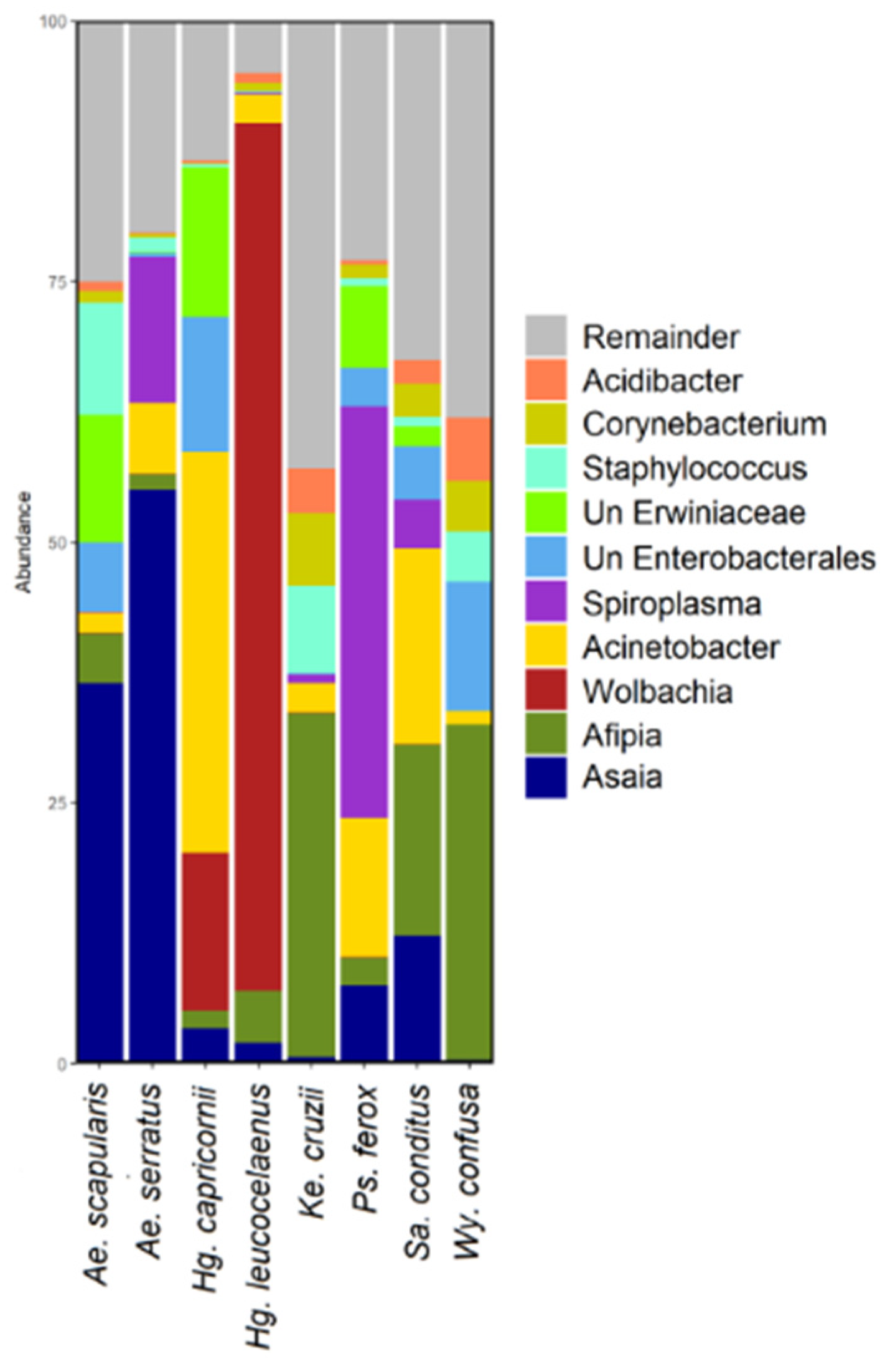
A total of 2617 ASVs were identified in the samples. Ae. scapularis showed 530 ASVs; Hg. leucocelaenus, 310; Hg. capricornii, 334; Ps. ferox, 874; Ae. serratus, 504; Sa. conditus, 1014; Wy. confusa, 623; and Ke. cruzii, 546 (Table S2). Proteobacteria was the predominant phylum, followed by Firmicutes and Actinobacteriota (Figure 1 and Figure S1). Wolbachia was the predominant genus in Hg. leucocelaenus. Afipia, Acinetobacter, and Asaia genera were abundant in most of the species analysed (Figure 2 and Figure S2).

3.3. Diversity Analysis

The number of sequences retained for the analyses was sufficient to infer the abundance of the bacterial community in each sample (Figure S3). All samples were normalised to calculate the diversity metrics. The cut-off value for normalisation was the lowest number of contigs found in the sequenced samples. This value corresponds to 8857 sequences, as shown in Table S1.
Shannon-Weaver indices ranged from 0.607 to 6.355 between samples (Table S3). These indices did not show a normal distribution (Shapiro test; W = 0.97394, p = 0.028), and then the Kruskal–Wallis test (χ2 = 24.393, p = 0.0009) followed by Dunn’s test adjusted with the Bonferroni method was performed to know which species differed in α diversity. The following groups showed significant differences in the Shannon indices: Hg. leucocelaenusKe. cruzii (p = 0.002); Hg. leucocelaenusSa. conditus (p = 0.002) and Hg. leucocelaenusWy. confusa (p = 0.026).
The β diversity was calculated for each species and visualised in the PCoAs (Figure 3). Significant differences in bacterial composition were verified with PERMANOVA analysis using unweighted and weighted Unifrac distances data (Table S4). Significant differences in both PERMANOVA analyses were found between (1) Ae. scapularis and Hg. leucocelaenus, Ke. cruzii, Sa. conditus, and Wy. confusa; (2) Ae. serratus and Hg. capricornii, Hg. leucocelaenus, Ke. cruzii, Sa. conditus, and Wy. confusa; (3) Hg. capricornii and Ke. cruzii, Sa. conditus and Wy. confusa; (4) Hg. leucocelaenus and Ke. cruzii, Sa. conditus, Wy. confusa, and Ps. ferox; (5) Ke. cruzii and Sa. conditus and Ps. ferox; (6) Ps. ferox and Sa. conditus and Wy. confusa; and (7) Sa. conditus and Wy. confusa (Table S4).

3.4. Microbiome Composition Analysis and Heatmap

In the ANCOM analysis, it was possible to verify that Wolbachia was more abundant in Hg. leucocelaenus, while Afipia and Asaia genera were more abundant in Ke. cruzii and Ae. serratus, respectively (Table S5 and Figure S4).
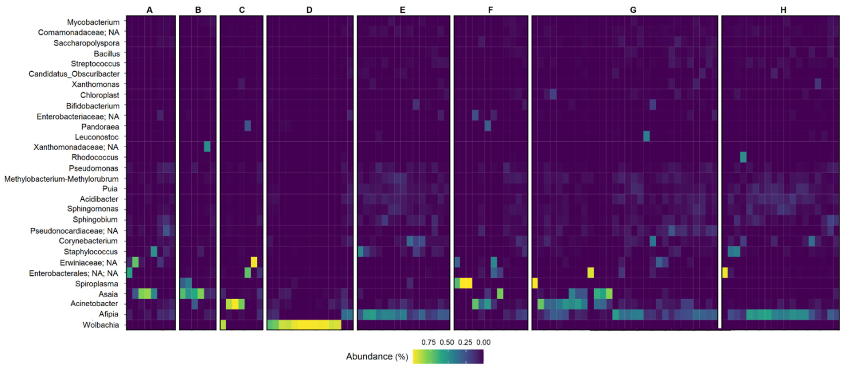
Bacteria of the Proteobacteria phylum showed the greatest number of sequences in all species (Figure S5). Figure 4 shows that a larger number of Wolbachia sequences was found in Hg. leucocelaenus, whereas Afipia was more abundant in Ke. cruzii, Sa. conditus and Wy. confusa.

3.5. Bacterial Interaction Network

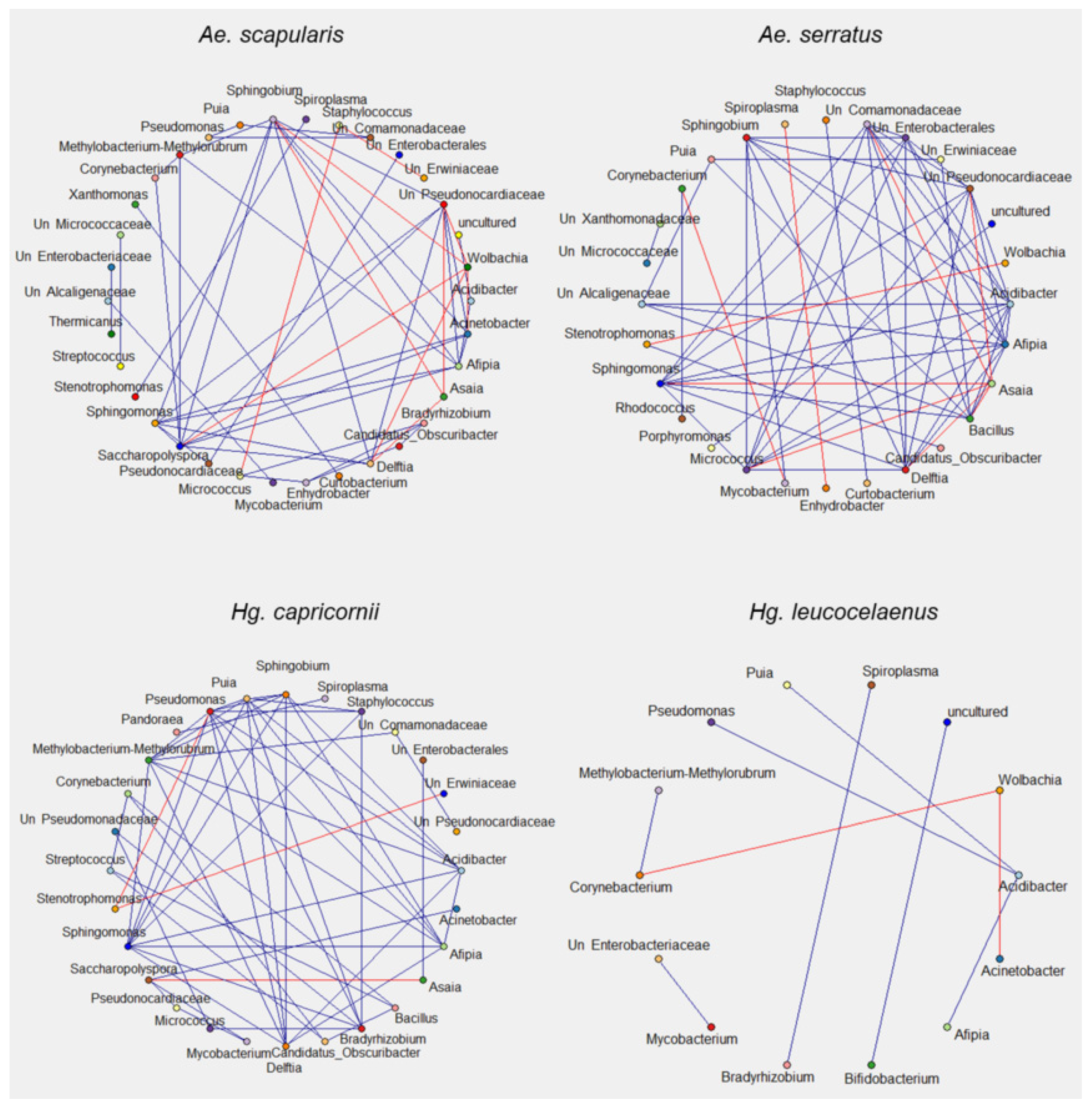
Interactions between bacterial communities in the samples were verified based on the 16S sequences. The ASV table–filtered and normalized (Table S6) of each species–was used for bacterial interaction analysis and then interaction data was used to infer bacterial interaction networks (Figure 5 and Figure 6). Bacteria found in Ke. cruzii, Sa. conditus, Hg. leucocelaenus, and Wy. confusa showed fewer interactions than those present in Ae. scapularis, Ae. serratus, Hg. capricornii, and Ps. ferox. Positive bacterial correlations were found in all species analysed, while negative correlations were observed in Hg. leucocelaenus, Ae. scapularis, Ae. serratus, Ps. ferox, and Hg. capricornii (Figure 5 and Figure 6, Table S7). The bacteria that showed more interactions were Shingobium, Unclassified Pseudonocardiaceae, Afipia, Delftia, and Shingomonas in Aedes samples; Shingomonas, Afipia, Methylobacterium-Methylobacterium, Pseudomonas, Puia, Acidibacter, and Afipia in Hg. capricornii; and Shingomonas, Methylobacterium-Methylobacterium, Acidibacter, Afipia, and Pseudomonas in Ps. ferox. All bacterial interactions with Asaia and Wolbachia were negative in the Aedes species (Figure 5).

4. Discussion

Culicidae mosquitoes have a broad geographic distribution. Although insects of this family have great epidemiological importance in the transmission of pathogens [37,38] and studies show the influence of the microbiota in this transmission [31], little has been studied about bacteria present in mosquitoes collected in Vale do Ribeira, Brazil [39]. This region is known to harbor a great diversity of vector mosquitoes, to register autochthonous malaria cases (bromeliad-malaria) [40], and to be highly vulnerable to sylvatic yellow fever [41]. Thus, this study contributes to a better understanding of the bacterial communities in eight mosquito species found in areas of the Vale do Ribeira, southeastern Atlantic tropical rain forest.
Because the mosquito species analysed in this study have great public health importance as potential vectors of arboviruses, the knowledge of bacterial communities present in these insects is essential for further investigating the bacteria-pathogen interactions and their influence on the ability of these species to be infected and transmit parasites. Ae. scapularis has a wide distribution and can be a vector for Yellow Fever Virus (YFV), Venezuelan Equine encephalitis, and Wuchereria bancrofti [42,43,44]. Ae. serratus is considered a secondary vector of both the Ilheus virus [45] and the YFV [37,38]. Hg. leucocelaenus is the primary vector of sylvatic YFV in the New World and was involved in Brazil’s greatest yellow fever outbreak between 2016 and 2018 [38]. Ke. cruzii is the primary vector of Plasmodium sp. in Atlantic Forest (bromeliad malaria) [46,47], and it was found to be naturally infected with the Zika virus [48]. Ps. ferox has already been found to be naturally infected with the Rocio virus in Vale do Ribeira region [49], with YFV in the municipality of Urupês, Ribeirão Preto region, São Paulo state [41]. Wy. confusa from Capivari-Monos EPA (São Paulo municipality) was infected with the Zika virus [48].
Many factors can modulate mosquitoes’ bacterial composition, including the mosquito species [50] and complex microbial interactions. In addition, the mosquito developmental stage, geographic location of the samples, the collection period, and adult sex can affect the bacterial composition and their interactions [5]. In this study, all specimens analysed were females collected in the same period and geographical location. Thus, the data obtained are a rich source of information, as they show the scenario of bacterial diversity and the networks of interactions that occur in a given species while in contact with nature.
Statistical tests showed a significant impact of the species on the α diversity of the mosquito, with an effect verified between Hg. leucocelaenus and Ke. cruzii and Sa. conditus and Wy. confusa. Permanova analyses showed that most mosquito species studied differed in their bacterial communities. These results corroborate other studies in the literature [5,51,52], and indicate that the species can influence the formation of bacterial composition in mosquitoes. In addition, some species are associated with distinct phytotelmata habitats, such as Ke. cruzii linked to bromeliad phytotelmata; Wy. confusa, Sabethes, and Haemagogus with phytotelmata provided by bamboo and tree holes; and other species are relations with temporary ground pools, such as Ae. scapularis, Ae. serratus and Ps. ferox. Although all specimens have been collected in the same macroregion, the larval habitats (microregions) are ecologically distinct. As bacterial communities present in immature habitats can contribute to bacterial composition in adult mosquitoes [53], the bacterial differences observed in this study can be accounted for not only by the species differences, but also by distinct larval habitats.
There are several interactions between microorganisms, such as mutualism, competition, and commensalism [28]. Increased interactions between bacterial microbiota can alter gene expression in the microbial community, the metabolism, and ecological interactions between species [54,55]. This study exploited bacterial interaction networks using the 16S sequence data from 111 specimens of eight mosquito vector species. Among the species studied, Ae. serratus showed the highest interactions between bacterial communities, followed by Hg. capricornii, Ae. scapularis, and Ps. ferox.
Afipia was found in all mosquito species, being found predominantly in Wy. confusa, Ke. cruzii, and Sa. conditus. Despite being in lower abundance in Ae. scapularis, Ae. serratus, Hg. capricornii, and Ps. ferox, in these species, Afipia interacted with at least six other bacterial genera. Afipia is a Gram-negative rod bacterium of the phylum Proteobacteria. The genus was described in 1991 [56], and one species (Afipia felix) appears to be related to cat scratch disease [57]. To date, there was no record of Afipia in mosquitoes.
Acinetobacter was found in all mosquito species studied. Species of this genus may be involved in blood digestion and parasite–vector interactions in Ae. albopictus [58]. Bacteria of the genus Delftia were found in females of Nyssorhynchus darlingi not infected with Plasmodium [59] in other Culicidae species [60]. The bacterial interaction analyses showed a negative correlation between Delftia and Asaia in Aedes mosquitoes. The negative interaction between these bacterial groups needs further investigation to verify if the co-exclusion hypothesis of these bacterial genera can occur in other Aedes mosquitoes.
Asaia is frequently found in Aedes and Anopheles mosquitoes [58,61], and it can inhibit the development of Plasmodium in female mosquitoes [62]. In addition, Asaia can diminish the longevity of infected males of Anopheles stephensi [63]. It is noteworthy that Asaia and Wolbachia showed only negative interactions with other bacterial genera in the Aedes samples analyzed in this study. Our findings corroborate the results of an investigation to verify the occurrence of reciprocal negative interference between these bacterial groups to colonise the gonads [15]. Although the results of bacterial interaction analyses did not show the mutual exclusion observed by Rossi and colleagues, the current study found differences between the number of sequences of these two genera in the Aedes species from Vale do Ribeira. Few Wolbachia sequence readings were found in Ae. scapularis and Ae. serratus, while Asaia readings were more abundant in both species. Although the Wolbachia-Asaia mutual exclusion hypothesis has been found in Hg. leucocelaenus specimens from Vale do Ribeira [39], the same effect was not verified in the females analyzed for this study, collected in the same geographical region.
Wolbachia infection in Ae. aegypti causes upregulation in the transcription of genes related to reduction–oxidation reactions and immunity. This leads to an increase of reactive oxygen species and induction of oxidative stress in the host and showing that to favor infection, the bacterium can manipulate the host’s defense system [64].
Many of the Wolbachia strains induce cytoplasmic incompatibility (CI), resulting in lethality of embryos generated from females not infected with Wolbachia or infected with a different strain present in the male [65]. It manipulates the reproduction of the host and facilitates the spread of this bacterium, since infected females have this reproductive advantage over uninfected ones. Many studies use CI as a possible method of biological control [66]. In addition, this bacterium can reduce the infection of the dengue virus in the salivary glands of Ae. albopictus [67] and to inhibit yellow fever virus replication in Ae. aegypti [68]. Thus, considering that this bacterium was abundant in samples of Hg. leucocelaenus and this species of mosquito is considered the primary vector of sylvatic yellow fever virus in the south-eastern region of Brazil [38], further studies need to be carried out to know the Wolbachia strains present in these mosquitoes, and to know if the existing strains can harm the spread of the yellow fever virus.
Serratia bacteria can modulate the vector competence of populations of Ae. aegypti for the Zika virus by interfering with mosquito salivation [69] and interfering in the vectorial capacity of other mosquito species [70]. In this study, Serratia 16S readings were found in one female of each species, Sa. conditus and Ps. ferox. The competitive exclusion between Serratia and Cedecea was verified in Ae. aegypti co-infected with these bacteria [21]. No reading of the Cedecea was recovered from the specimens of the species of the Aedini tribe from Vale do Ribeira. Consequently, the absence of Serratia in Aedes and Anopheles mosquitoes cannot be explained by the exclusion effect between Serratia and Cedecea symbionts. It is likely that other bacteria can be responsible for the competitive exclusion with Serratia.
Our findings uncovered that the analyzed species exhibited clear differences in their microbiota composition based on the α and β diversity indices. Furthermore, some bacterial interactions in this study corroborated previous findings in the literature, while other interactions will need further investigation. In addition, it will be important to verify if the interactions are observed in different spatial and temporal scales in subpopulations of a mosquito species.

5. Conclusions

Here we uncover variations in microbial composition among different Culicidae species. Wolbachia and Asaia are predominant in species of the Aedini tribe, such as Hg. leucocelaenus and Aedes mosquitoes. The bacterial interaction network was verified for each mosquito species showing that Shingobium, Unclassified Pseudonocardiaceae, Afipia, Delftia, and Shingomonas had more bacterial interactions in the Aedes specimens. Bacterial interactions with Asaia and with Wolbachia were negative in Aedes mosquitoes.

Supplementary Materials

The following supporting information can be downloaded at: https://www.mdpi.com/article/10.3390/genes13112052/s1, Figure S1: Bar graph depicting the bacterial amplicon sequence variants (ASV) composition at the highest taxonomic level (phylum) for each mosquito sample. Figure S2: Bar graph depicting the bacterial amplicon sequence variants (ASV) composition at the bottom taxonomic level (genus) for each mosquito sample. Figure S3: Rarefaction curve. Count of ASVs per given sequencing depth in each mosquito sample. Each line corresponds to a mosquito sample. Figure S4: The illustration represents ANCOM analysis. Twelve taxa were the most abundant and are marked 1 to 12 from right to left. Figure S5: Heatmap of bacterial sequences with taxonomic assignment to genus level in each mosquito sample. Each row represents a bacterial taxon and each column corresponds to a mosquito species. Abundance data are assigned colors across a gradient from yellow (higher bacterial abundance) to blue (lowest bacterial abundance). Table S1: Count of 16S rRNA raw data and contigs in each mosquito sample. Table S2: ASVs count in each sample and mosquito species. Table S3: Shannon index of each mosquito sample. Table S4: Pairwise permanova results from unweighted and weighted Unifrac distances. Table S5: ANCOM analysis. Percentile abundances of features by group. Table S6: Relative abundance of each ASV in each mosquito species. Table S7: Number of bacterial interactions per mosquito species and number of bacterial taxa related to interactions.

Author Contributions

H.d.S., M.A.M.S. conceived the study; H.d.S. and T.M.P.O. conducted the analyses and interpreted the results; H.d.S., T.M.P.O. and M.A.M.S. wrote the manuscript. All authors have read and agreed to the published version of the manuscript.

Funding

This research was funded by National Council for Scientific and Technological Development (CNPq), grant number: 301877/2016-5; São Paulo Research Foundation (Fapesp), grant number 2018/14389-0.

Institutional Review Board Statement

Not applicable.

Informed Consent Statement

Not applicable.

Data Availability Statement

Sequence data (individual fastq files) are available from the NCBI Sequence Read Archive under accession PRJNA857687 and also in the Zenodo repository (https://doi.org/10.5281/zenodo.7300402).

Conflicts of Interest

The authors declare no conflict of interest.

References

  1. Harbach, R.E. Culicidae. Mosquito Taxonomic Inventory. 2013. Available online: https://mosquito-taxonomic-inventory.myspecies.info/simpletaxonomy/term/6045 (accessed on 17 July 2022).
  2. Forattini, O.P. Culicidologia Médica: Identifcaçäo, Biologia e Epidemiologia; Editora da Universidade de São Paulo: São Paulo, Brazil, 2002; Volume 2. [Google Scholar]
  3. Guedes, M.P. Culicidae (Diptera) no Brasil: Relações entre diversidade, distribuição e enfermidades. Oecol 2012, 16, 283–296. [Google Scholar] [CrossRef]
  4. Minard, G.; Mavingui, P.; Moro, C.V. Diversity and function of bacterial microbiota in the mosquito holobiont. Parasites Vectors 2013, 6, 146. [Google Scholar] [CrossRef] [PubMed] [Green Version]
  5. Saab, S.A.; Dohna, H.Z.; Nilsson, L.; Onorati, P.; Nakhleh, J.; Terenius, O.; Osta, M.A. The environment and species affect gut bacteria composition in mosquitoes. Sci. Rep. 2020, 10, 3352. [Google Scholar] [CrossRef] [PubMed] [Green Version]
  6. Trzebny, A.; Slodkowicz-Kowalska, A.; Björkroth, J.; Dabert, M. Microsporidian Infection in Mosquitoes (Culicidae) Is Associated with Gut Microbiome Composition and Predicted Gut Microbiome Functional Content. Microb. Ecol. 2021, 1–17. [Google Scholar] [CrossRef]
  7. Coon, K.L.; Brown, M.R.; Strand, M.R. Mosquitoes host communities of bacteria that are essential for development but vary greatly between local habitats. Mol. Ecol. 2016, 25, 5806–5826. [Google Scholar] [CrossRef] [Green Version]
  8. Cirimotich, C.M.; Ramirez, J.L.; Dimopoulos, G. Native microbiota shape insect vector competence for human pathogens. Cell Host Microbe 2011, 10, 307–310. [Google Scholar] [CrossRef] [Green Version]
  9. Chouaia, B.; Rossi, P.; Epis, S.; Mosca, M.; Ricci, I.; Damiani, C.; Ulissi, U.; Crotti, E.; Daffonchio, D.; Bandi, C.; et al. Delayed larval development in Anopheles mosquitoes deprived of Asaia bacterial symbionts. BMC Microbiol. 2012, 12, S2. [Google Scholar] [CrossRef] [Green Version]
  10. Mitraka, E.; Stathopoulos, S.; Siden-Kiamos, I.; Christophides, G.K.; Louis, C. Asaia accelerates development of Anopheles gambiae. Pathog. Glob. Health 2013, 107, 305–311. [Google Scholar] [CrossRef] [Green Version]
  11. Balaji, S.; Jayachandran, S.; Prabagaran, S.R. Evidence for the natural occurrence of Wolbachia in Aedes aegypti mosquitoes. FEMS Microbiol. Lett. 2019, 366, fnz055. [Google Scholar] [CrossRef]
  12. Baldini, F.; Segata, N.; Pompon, J.; Marcenac, P.; Shaw, W.R.; Dabiré, R.K.; Diabate, A.; Levashina, E.A.; Catteruccia, F. Evidence of natural Wolbachia infections in field populations of Anopheles gambiae. Nat. Commun. 2014, 5, 3985. [Google Scholar] [CrossRef]
  13. Wong, M.L.; Liew, J.W.K.; Wong, W.K.; Pramasivan, S.; Hassan, N.M.; Sulaiman, W.Y.W.; Jeyaprakasam, N.K.; Leong, C.S.; Low, V.L.; Vythilingam, I. Natural Wolbachia infection in field-collected Anopheles and other mosquito species from Malaysia. Parasites Vectors 2020, 13, 414. [Google Scholar] [CrossRef] [PubMed]
  14. Hughes, G.L.; Dodson, B.L.; Johnson, R.M.; Murdock, C.C.; Tsujimoto, H.; Suzuki, Y.; Patt, A.A.; Cui, L.; Nossa, C.W.; Barry, R.M.; et al. Native microbiome impedes vertical transmission of Wolbachia in Anopheles mosquitoes. Proc. Natl. Acad. Sci. USA 2014, 111, 12498–12503. [Google Scholar] [CrossRef] [Green Version]
  15. Rossi, P.; Ricci, I.; Cappelli, A.; Damiani, C.; Ulissi, U.; Mancini, M.V.; Valzano, M.; Capone, A.; Epis, S.; Crotti, E.; et al. Mutual exclusion of Asaia and Wolbachia in the reproductive organs of mosquito vectors. Parasites Vectors 2015, 8, 278. [Google Scholar] [CrossRef] [PubMed] [Green Version]
  16. Dong, Y.; Manfredini, F.; Dimopoulos, G. Implication of the mosquito midgut microbiota in the defense against malaria parasites. PLoS Pathog. 2009, 5, e1000423. [Google Scholar] [CrossRef] [PubMed] [Green Version]
  17. Rocha, E.M.; Marinotti, O.; Serrão, D.M.; Correa, L.V.; Katak, R.D.M.; de Oliveira, J.C.; Muniz, V.A.; de Oliveira, M.R.; Neto, J.F.D.N.; Pessoa, M.C.F.; et al. Culturable bacteria associated with Anopheles darlingi and their paratransgenesis potential. Malar. J. 2021, 20, 40. [Google Scholar] [CrossRef]
  18. Bahia, A.C.; Dong, Y.; Blumberg, B.J.; Mlambo, G.; Tripathi, A.; BenMarzouk-Hidalgo, O.J.; Chandra, R.; Dimopoulos, G. Exploring Anopheles gut bacteria for Plasmodium blocking activity. Environ. Microbiol. 2014, 16, 2980–2994. [Google Scholar] [CrossRef] [PubMed] [Green Version]
  19. Hegde, S.; Khanipov, K.; Albayrak, L.; Golovko, G.; Pimenova, M.; Saldaña, M.; Rojas, M.M.; Hornett, E.A.; Motl, G.C.; Fredregill, C.; et al. Microbiome Interaction Networks and Community Structure From Laboratory-Reared and Field-Collected Aedes aegypti, Aedes albopictus, and Culex quinquefasciatus Mosquito Vectors. Front. Microbiol. 2018, 9, 2160. [Google Scholar] [CrossRef] [Green Version]
  20. Tuanudom, R.; Yurayart, N.; Rodkhum, C.; Tiawsirisup, S. Diversity of midgut microbiota in laboratory-colonized and field-collected Aedes albopictus (Diptera: Culicidae): A preliminary study. Heliyon 2021, 7, e08259. [Google Scholar] [CrossRef]
  21. Kozlova, E.V.; Hegde, S.; Roundy, C.M.; Golovko, G.; Saldaña, M.; Hart, C.E.; Anderson, E.R.; Hornett, E.A.; Khanipov, K.; Popov, V.L.; et al. Microbial interactions in the mosquito gut determine Serratia colonization and blood-feeding propensity. ISME J. 2021, 15, 93–108. [Google Scholar] [CrossRef]
  22. Bassene, H.; Niang, E.; Fenollar, F.; Dipankar, B.; Doucouré, S.; Ali, E.; Michelle, C.; Raoult, D.; Sokhna, C.; Mediannikov, O.; et al. 16S Metagenomic Comparison of Plasmodium falciparum-Infected and Noninfected Anopheles gambiae and Anopheles funestus Microbiota from Senegal. Am. J. Trop. Med. Hyg. 2018, 99, 1489–1498. [Google Scholar] [CrossRef]
  23. Caragata, E.P.; Tikhe, C.V.; Dimopoulos, G. Curious entanglements: Interactions between mosquitoes, their microbiota, and arboviruses. Curr. Opin. Virol. 2019, 37, 26–36. [Google Scholar] [CrossRef]
  24. Scolari, F.; Casiraghi, M.; Bonizzoni, M. Aedes spp. and Their Microbiota: A Review. Front. Microbiol. 2019, 10, 2036. [Google Scholar] [CrossRef] [PubMed] [Green Version]
  25. Gao, H.; Cui, C.; Wang, L.; Jacobs-Lorena, M.; Wang, S. Mosquito Microbiota and Implications for Disease Control. Trends Parasitol. 2020, 36, 98–111. [Google Scholar] [CrossRef] [PubMed]
  26. Zink, S.D.; Van Slyke, G.A.; Palumbo, M.J.; Kramer, L.D.; Ciota, A.T. Exposure to West Nile Virus Increases Bacterial Diversity and Immune Gene Expression in Culex pipiens. Viruses 2015, 7, 5619–5631. [Google Scholar] [CrossRef] [Green Version]
  27. Balaji, S.; Deepthi, K.; Prabagaran, S.R. Native Wolbachia influence bacterial composition in the major vector mosquito Aedes aegypti. Arch. Microbiol. 2020, 203, 5225–5240. [Google Scholar] [CrossRef] [PubMed]
  28. Faust, K.; Raes, J. Microbial interactions: From networks to models. Nat. Rev. Microbiol. 2012, 10, 538–550. [Google Scholar] [CrossRef]
  29. Faust, K.; Sathirapongsasuti, J.F.; Izard, J.; Segata, N.; Gevers, D.; Raes, J.; Huttenhower, C. Microbial co-occurrence relationships in the human microbiome. PLoS Comput. Biol. 2012, 8, e1002606. [Google Scholar] [CrossRef]
  30. Hegde, S.; Rasgon, J.L.; Hughes, G.L. The microbiome modulates arbovirus transmission in mosquitoes. Curr. Opin. Virol. 2015, 15, 97–102. [Google Scholar] [CrossRef] [Green Version]
  31. Sharma, P.; Rani, J.; Chauhan, C.; Kumari, S.; Tevatiya, S.; Das De, T.; Savargaonkar, D.; Pandey, K.C.; Dixit, R. Altered Gut Microbiota and Immunity Defines Plasmodium vivax Survival in Anopheles stephensi. Front. Immunol. 2020, 11, 609. [Google Scholar] [CrossRef]
  32. Caporaso, J.G.; Lauber, C.L.; Walters, W.A.; Berg-Lyons, D.; Lozupone, C.A.; Turnbaugh, P.J.; Fierer, N.; Knight, R. Global patterns of 16S rRNA diversity at a depth of millions of sequences per sample. Proc. Natl. Acad. Sci. USA 2011, 108, 4516–4522. [Google Scholar] [CrossRef]
  33. Magoč, T.; Salzberg, S.L. FLASH: Fast length adjustment of short reads to improve genome assemblies. Bioinform 2011, 27, 2957–2963. [Google Scholar] [CrossRef] [PubMed] [Green Version]
  34. Bolyen, E.; Rideout, J.R.; Dillon, M.R.; Bokulich, N.A.; Abnet, C.C.; Al-Ghalith, G.A.; Alexander, H.; Alm, E.J.; Arumugam, M.; Asnicar, F.; et al. Reproducible, interactive, scalable and extensible microbiome data science using QIIME 2. Nat. Biotechnol. 2019, 37, 852–857. [Google Scholar] [CrossRef] [PubMed]
  35. Quast, C.; Pruesse, E.; Yilmaz, P.; Gerken, J.; Schweer, T.; Yarza, P.; Peplies, J.; Glöckner, F.O. The SILVA ribosomal RNA gene database project: Improved data processing and web-based tools. Nucleic Acids Res. 2013, 41, D590–D596. [Google Scholar] [CrossRef]
  36. Robeson, M.S.; O’Rourke, D.R.; Kaehler, B.D.; Ziemski, M.; Dillon, M.R.; Foster, J.T.; Bokulich, N.A. RESCRIPt: Reproducible sequence taxonomy reference database management for the masses. PLoS Comput. Biol. 2020, 17, e1009581. [Google Scholar] [CrossRef]
  37. Cardoso, J.; de Almeida, M.A.; dos Santos, E.; da Fonseca, D.F.; Sallum, M.A.; Noll, C.A.; Monteiro, H.A.D.O.; Cruz, A.C.; Carvalho, V.L.; Pinto, E.V.; et al. Yellow fever virus in Haemagogus leucocelaenus and Aedes serratus mosquitoes, southern Brazil, 2008. Emerg. Infect. Dis. 2010, 16, 1918–1924. [Google Scholar] [CrossRef]
  38. Abreu, F.V.S.; Ribeiro, I.P.; Ferreira-de-Brito, A.; dos Santos, A.A.C.; de Miranda, R.M.; Bonelly, I.D.S.; Neves, M.S.A.S.; Bersot, M.I.; dos Santos, T.P.; Gomes, M.Q.; et al. Haemagogus leucocelaenus and Haemagogus janthinomys are the primary vectors in the major yellow fever outbreak in Brazil, 2016–2018. Emerg. Microbes Infect. 2019, 8, 218–231. [Google Scholar] [CrossRef] [Green Version]
  39. da Silva, H.; Oliveira, T.M.P.; Sabino, E.C.; Alonso, D.P.; Sallum, M.A.M. Bacterial diversity in Haemagogus leucocelaenus (Diptera: Culicidae) from Vale do Ribeira, São Paulo, Brazil. BMC Microbiol. 2022, 22, 161. [Google Scholar] [CrossRef]
  40. Curado, I.; Dos Santos Malafronte, R.; de Castro Duarte, A.M.; Kirchgatter, K.; Branquinho, M.S.; Galati, E.A.B. Malaria epidemiology in low-endemicity areas of the Atlantic Forest in the Vale do Ribeira, São Paulo, Brazil. Acta Trop. 2006, 100, 54–62. [Google Scholar] [CrossRef]
  41. Moreno, E.S.; Barata, R.C.B. Municipalities of higher vulnerability to Sylvatic Yellow Fever occurrence in the São Paulo State, Brazil. Rev. Inst. Med. Trop. São Paulo 2011, 53, 335–339. [Google Scholar] [CrossRef] [Green Version]
  42. Whitman, L.; Antunes, P.C.A. Studies on the capacity of various Brazilian mosquitoes, representing the Genera Psorophora, Aedes, Mansonia, and Culex, to transmit yellow fever 1. Am. J. Trop. Med. Hyg. 1937, s1-17, 803–823. [Google Scholar] [CrossRef]
  43. Couto-Lima, D.; Madec, Y.; Bersot, M.I.; Campos, S.S.; Motta, M.D.A.; Santos, F.; Vazeille, M.; Vasconcelos, P.F.D.C.; Lourenço-De-Oliveira, R.; Failloux, A.-B. Potential risk of re-emergence of urban transmission of Yellow Fever virus in Brazil facilitated by competent Aedes populations. Sci. Rep. 2017, 7, 4848. [Google Scholar] [CrossRef] [PubMed] [Green Version]
  44. Cunha, M.S.; Tubaki, R.M.; de Menezes, R.; Pereira, M.; Caleiro, G.S.; Coelho, E.; Saad, L.D.C.; Fernandes, N.C.C.D.A.; Guerra, J.M.; Nogueira, J.S.; et al. Possible non-sylvatic transmission of yellow fever between non-human primates in São Paulo city, Brazil, 2017–2018. Sci. Rep. 2020, 10, 15751. [Google Scholar] [CrossRef]
  45. Vasconcelos, P.F.C.; Travassos-da-Rosa, A.P.A.; Pinheiro, F.P.; Shope, R.E.; Degallier, N.; Travassos da Rosa, E.S. Arboviruses pathogenic for man in Brazil. In An Overview of Arbovirology in Brazil and Neighbouring Countries; Travassos-da-Rosa, A.P.A., Vasconcelos, P.F.C., Travassos-da-Rosa, J.F.S., Eds.; Instituto Evandro Chagas: Belém, Brazil, 1998; pp. 72–99. [Google Scholar]
  46. Duarte, A.M.; Pereira, D.M.; de Paula, M.B.; Fernandes, A.; Urbinatti, P.R.; Ribeiro, A.F.; Mello, M.H.S.; O Matos, M.; Mucci, L.F.; Fernandes, L.N.; et al. Natural infection in anopheline species and its implications for autochthonous malaria in the Atlantic Forest in Brazil. Parasites Vectors 2013, 6, 58. [Google Scholar] [CrossRef] [PubMed] [Green Version]
  47. Kirchgatter, K.; Tubaki, R.M.; Malafronte, R.; Alves, I.C.; Lima, G.F.M.D.C.; Guimarães, L.D.O.; Zampaulo, R.D.A.; Wunderlich, G. Anopheles (Kerteszia) cruzii (Diptera: Culicidae) in peridomiciliary area during asymptomatic malaria transmission in the Atlantic Forest: Molecular identification of blood-meal sources indicates humans as primary intermediate hosts. Rev. Inst. Med. Trop. São Paulo 2014, 56, 403–409. [Google Scholar] [CrossRef] [PubMed]
  48. Barrio-Nuevo, K.M.; Cunha, M.S.; Luchs, A.; Fernandes, A.; Rocco, I.M.; Mucci, L.F.; De Souza, R.P.; Medeiros-Sousa, A.R.; Ceretti-Junior, W.; Marrelli, M.T. Detection of Zika and dengue viruses in wild-caught mosquitoes collected during field surveillance in an environmental protection area in São Paulo, Brazil. PLoS ONE 2020, 15, e0227239. [Google Scholar] [CrossRef] [PubMed]
  49. de Lopes, O.S.; de Sacchetta, L.A.; Francy, D.B.; Jakob, W.L.; Calisher, C.H. Emergence of a new arbovirus disease in Brazil. III. Isolation of Rocio virus from Psorophora Ferox (Humboldt, 1819). Am. J. Epidemiol. 1981, 113, 122–125. [Google Scholar] [CrossRef] [PubMed]
  50. Mancini, M.V.; Damiani, C.; Accoti, A.; Tallarita, M.; Nunzi, E.; Cappelli, A.; Bozic, J.; Catanzani, R.; Rossi, P.; Valzano, M.; et al. Estimating bacteria diversity in different organs of nine species of mosquito by next generation sequencing. BMC Microbiol. 2018, 18, 126. [Google Scholar] [CrossRef] [Green Version]
  51. Tchouassi, D.P.; Muturi, E.J.; Arum, S.O.; Kim, C.-H.; Fields, C.J.; Torto, B. Host species and site of collection shape the microbiota of Rift Valley fever vectors in Kenya. PLoS Negl. Trop. Dis. 2019, 13, e0007361. [Google Scholar] [CrossRef]
  52. Pérez-Ramos, D.W.; Ramos, M.M.; Payne, K.C.; Giordano, B.V.; Caragata, E.P. Collection Time, Location, and Mosquito Species Have Distinct Impacts on the Mosquito Microbiota. Front. Trop. Dis. 2022, 3, 896289. [Google Scholar] [CrossRef]
  53. Gimonneau, G.; Tchioffo, M.T.; Abate, L.; Boissière, A.; Awono-Ambéné, P.H.; Nsango, S.E.; Christen, R.; Morlais, I. Composition of Anopheles coluzzii and Anopheles gambiae microbiota from larval to adult stages. Infect. Genet. Evol. 2014, 28, 715–724. [Google Scholar] [CrossRef]
  54. Ibberson, C.B.; Stacy, A.; Fleming, D.; Dees, J.L.; Rumbaugh, K.; Gilmore, M.S.; Whiteley, M. Co-infecting microbes dramatically alter pathogen gene essentiality during polymicrobial infection. Nat. Microbiol. 2017, 2, 17079. [Google Scholar] [CrossRef] [PubMed]
  55. Gould, A.L.; Zhang, V.; Lamberti, L.; Jones, E.W.; Obadia, B.; Korasidis, N.; Gavryushkin, A.; Carlson, J.M.; Beerenwinkel, N.; Ludington, W.B. Microbiome interactions shape host fitness. Proc. Natl. Acad. Sci. USA 2018, 115, E11951–E11960. [Google Scholar] [CrossRef] [PubMed] [Green Version]
  56. Brenner, D.J.; Hollis, D.G.; Moss, C.W.; English, C.K.; Hall, G.S.; Vincent, J.; Radosevic, J.; A Birkness, K.; Bibb, W.F.; Quinn, F.D. Proposal of Afipia gen. nov., with Afipia felis sp. nov. (formerly the cat scratch disease bacillus), Afipia clevelandensis sp. nov. (formerly the Cleveland Clinic Foundation strain), Afipia broomeae sp. nov., and three unnamed genospecies. J. Clin. Microbiol. 1991, 29, 2450–2460. [Google Scholar] [CrossRef] [Green Version]
  57. Giladi, M.; Avidor, B.; Kletter, Y.; Abulafia, S.; Slater, L.N.; Welch, D.F.; Brenner, D.J.; Steigerwalt, A.G.; Whitney, A.M.; Ephros, M. Cat scratch disease: The rare role of Afipia felis. J. Clin. Microbiol. 1998, 36, 2499–2502. [Google Scholar] [CrossRef] [PubMed] [Green Version]
  58. Minard, G.; Tran, F.H.; Raharimalala, F.N.; Hellard, E.; Ravelonandro, P.; Mavingui, P.; Moro, C.V. Prevalence, genomic and metabolic profiles of Acinetobacter and Asaia associated with field-caught Aedes albopictus from Madagascar. FEMS Microbiol. Ecol. 2013, 83, 63–73. [Google Scholar] [CrossRef] [PubMed] [Green Version]
  59. Oliveira, T.M.P.; Sanabani, S.S.; Sallum, M.A.M. Bacterial diversity associated with the abdomens of naturally Plasmodium-infected and non-infected Nyssorhynchus darlingi. BMC Microbiol. 2020, 20, 180. [Google Scholar] [CrossRef]
  60. Muturi, E.J.; Lagos-Kutz, D.; Dunlap, C.; Ramirez, J.L.; Rooney, A.P.; Hartman, G.L.; Fields, C.J.; Rendon, G.; Kim, C.-H. Mosquito microbiota cluster by host sampling location. Parasites Vectors 2018, 11, 468. [Google Scholar] [CrossRef] [Green Version]
  61. Birnberg, L.; Climent-Sanz, E.; Codoñer, F.M.; Busquets, N. Microbiota Variation Across Life Stages of European Field-Caught Anopheles atroparvus and during Laboratory Colonization: New Insights for Malaria Research. Front. Microbiol. 2021, 12, 3533. [Google Scholar] [CrossRef]
  62. Capone, A.; Ricci, I.; Damiani, C.; Short, S.M.; Cappelli, A.; Ulissi, U.; Capone, A.; Serrao, A.; Rossi, P.; Amici, A.; et al. Interactions between Asaia, Plasmodium and Anopheles: New insights into mosquito symbiosis and implications in malaria symbiotic control. Parasites Vectors 2013, 6, 182. [Google Scholar] [CrossRef] [Green Version]
  63. Mancini, M.V.; Damiani, C.; Short, S.M.; Cappelli, A.; Ulissi, U.; Capone, A.; Serrao, A.; Rossi, P.; Amici, A.; Kalogris, C.; et al. Inhibition of Asaia in Adult Mosquitoes Causes Male-Specific Mortality and Diverse Transcriptome Changes. Pathogens 2020, 9, 380. [Google Scholar] [CrossRef]
  64. Pan, X.; Zhou, G.; Wu, J.; Bian, G.; Lu, P.; Raikhel, A.S.; Xi, Z. Wolbachia induces reactive oxygen species (ROS)-dependent activation of the Toll pathway to control dengue virus in the mosquito Aedes aegypti. Proc. Natl. Acad. Sci. USA 2012, 109, E23–E31. [Google Scholar] [CrossRef] [PubMed] [Green Version]
  65. Werren, J.H.; Baldo, L.; Clark, M.E. Wolbachia: Master manipulators of invertebrate biology. Nat. Rev. Microbiol. 2008, 6, 741–751. [Google Scholar] [CrossRef] [PubMed]
  66. Iturbe-Ormaetxe, I.; Walker, T.; O’Neill, S.L. Wolbachia and the biological control of mosquito-borne disease. EMBO Rep. 2011, 12, 508–518. [Google Scholar] [CrossRef] [Green Version]
  67. Mousson, L.; Zouache, K.; Arias-Goeta, C.; Raquin, V.; Mavingui, P.; Failloux, A.B. The Native Wolbachia Symbionts Limit Transmission of Dengue Virus in Aedes albopictus. PLoS Negl. Trop. Dis. 2012, 6, e1989. [Google Scholar] [CrossRef] [PubMed]
  68. van den Hurk, A.F.; Hall-Mendelin, S.; Pyke, A.T.; Frentiu, F.D.; McElroy, K.; Day, A.; Higgs, S.; O’Neill, S.L. Impact of Wolbachia on infection with chikungunya and yellow fever viruses in the mosquito vector Aedes aegypti. PLoS Negl. Trop. Dis. 2012, 6, e1892. [Google Scholar] [CrossRef] [PubMed] [Green Version]
  69. do Nascimento, R.M.; Campolina, T.B.; Chaves, B.A.; Delgado, J.L.F.; Godoy, R.S.M.; Pimenta, P.F.P.; Secundino, N.F.C. The influence of culture-dependent native microbiota in Zika virus infection in Aedes aegypti. Parasites Vectors 2022, 15, 1–14. [Google Scholar] [CrossRef]
  70. Bai, L.; Wang, L.; Vega-Rodriguez, J.; Wang, G.; Wang, S. A gut symbiotic bacterium Serratia marcescens renders mosquito resistance to Plasmodium infection through activation of mosquito immune responses. Front Microbiol 2019, 10, 1580. [Google Scholar] [CrossRef]
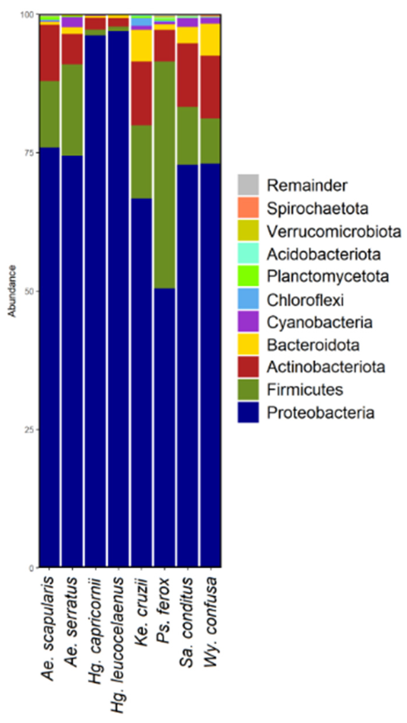
Figure 1. Bar graph depicting the bacterial amplicon sequence variants (ASV) composition at the highest taxonomic level (phylum) for each mosquito species.
Genes 13 02052 g001
Figure 2. Bar graph depicting the bacterial amplicon sequence variants (ASV) composition at the bottom taxonomic level (genus) for each mosquito species. Remainder (gray bars) corresponds to genera that had 2% or less of the total contigs analyzed.
Genes 13 02052 g002
Figure 3. Principal Coordinate Analysis (PCoA) plots β diversity differences between the mosquito species. (A) PCoA using unweighted distance data. (B) PCoA using weighted distance data.
Genes 13 02052 g003
Figure 4. Heatmap of bacterial sequences with taxonomic assignment to genus level in each mosquito sample. Each row represents a bacterial taxon and each column corresponds a mosquito sample. Relative abundance data are assigned colors across a gradient from yellow (higher bacterial abundance) to purple (lowest bacterial abundance). (A) corresponds to samples of the species Ae. scapularis; (B) corresponds to samples of the species Ae. serratus; (C) corresponds to samples of the species Hg. capricornii; (D) corresponds to samples of the species Hg. leucocelaenus; (E) corresponds to samples of the species Ke. cruzii; (F) corresponds to samples of the species Ps. ferox; (G) corresponds to samples of the species Sa. conditus and (H) corresponds to samples of the species Wy. confusa.
Genes 13 02052 g004
Figure 5. Bacterial interaction network in Ae. scapularis, Ae. serratus, Hg. capricornii and Hg. leucocelaenus. Each colored circle represents a bacterial taxon. Blue line corresponds to positive correlation between taxa. Red line represents negative correlation between taxa. Correlations were obtained by Spearman and only considered correlations with r > 0.75 and p > 0.05.
Genes 13 02052 g005
Figure 6. Bacterial interaction network in Ke. cruzii, Ps. ferox, Sa. conditus and Wy. confusa. Each colored circle represents a bacterial taxon. Blue line corresponds to positive correlation between taxa. Red line represents negative correlation between taxa. Correlations were obtained by Spearman and only considered correlations with r > 0.75 and p > 0.05.
Genes 13 02052 g006
Publisher’s Note: MDPI stays neutral with regard to jurisdictional claims in published maps and institutional affiliations.