Review

MicroRNA-221: A Fine Tuner and Potential Biomarker of Chronic Liver Injury

by
Jovana Markovic
1,2,
Amar Deep Sharma
1,2 and
Asha Balakrishnan
1,3,*
1
Department of Gastroenterology, Hepatology and Endocrinology, Hannover Medical School, 30625 Hannover, Germany
2
Unit miRNA in Liver Regeneration, REBIRTH Research Center for Translational Regenerative Medicine, Hannover Medical School, 30625 Hannover, Germany
3
Twincore Centre for Experimental and Clinical Infection Research, 30625 Hannover, Germany
*
Author to whom correspondence should be addressed.
Cells 2020, 9(8), 1767; https://doi.org/10.3390/cells9081767
Submission received: 30 March 2020 / Revised: 10 July 2020 / Accepted: 10 July 2020 / Published: 23 July 2020

Abstract

:
The last decade has witnessed significant advancements in our understanding of how small noncoding RNAs, such as microRNAs (miRNAs), regulate disease progression. One such miRNA, miR-221, has been shown to play a key role in the progression of liver fibrosis, a common feature of most liver diseases. Many reports have demonstrated the upregulation of miR-221 in liver fibrosis caused by multiple etiologies such as viral infections and nonalcoholic steatohepatitis. Inhibition of miR-221 via different strategies has shown promising results in terms of the suppression of fibrogenic gene signatures in vitro, as well as in vivo, in independent mouse models of liver fibrosis. In addition, miR-221 has also been suggested as a noninvasive serum biomarker for liver fibrosis and cirrhosis. In this review, we discuss the biology of miR-221, its significance and use as a biomarker during progression of liver fibrosis, and finally, potential and robust approaches that can be utilized to suppress liver fibrosis via inhibition of miR-221.

1. Introduction

Liver fibrosis and cirrhosis are major contributors to high mortality caused by various liver diseases. The underlying pathogenesis is the activation of quiescent hepatic stellate cells (HSCs) into activated myofibroblasts, which, in turn, secrete excessive extracellular matrix, resulting in liver fibrosis. This is accompanied by hepatocyte dysfunction, apoptosis and necrosis. Due to the presence of diverse autocrine and paracrine fibrogenic regulators, activated myofibroblasts begin to proliferate. To date, multiple molecules have been reported as regulators of HSC activation, such as growth factors including TGFβ, PDGF and FGF, potent cytokines, Toll-like receptors and interleukins. Many of these factors are secreted by hepatocytes and inflammatory cells which reside in or infiltrate into the liver due to liver injury. If it is undetected and remains unresolved, liver fibrosis may develop into cirrhosis and progress to liver cancer such as hepatocellular carcinoma (HCC) or cholangiocarcinoma (CCC). At the molecular level, deregulation of miRNAs is one of the key events in the progression of liver fibrosis and cirrhosis. MiRNAs have been shown to be key regulators of gene expression in liver development and differentiation, as well as in pathophysiological states of the liver. MiRNAs are ~22-nucleotide-long, single-stranded, small noncoding RNAs that are involved in posttranscriptional regulation of gene expression. They are considered regulators of one-third of all human genes, including those involved in fibrogenic pathways. These noncoding RNA molecules target complementary sequences in mRNA transcripts, usually in the 3′ untranslated region (3′ UTR), and prevent protein synthesis, either by inhibiting translation or inducing target degradation. Even though numerous miRNAs have been shown as key regulators of diverse liver diseases and potential biomarkers [1,2], in this review, we focus on miR-221, whose expression is not only upregulated in fibrosis, but also plays a critical role as a driver of liver fibrosis.
In this review, we discuss recent findings concerning the implication of miR-221 in liver fibrosis. First, we summarize the biogenesis and regulation of its expression. We then describe findings that show the role and potential mechanisms of miR-221 during the development of liver fibrosis, and briefly highlight the function of miR-221 in HCC. Finally, we discuss recent evidence of in vivo inhibition of miR-221 and its role as a serum marker for various liver diseases.

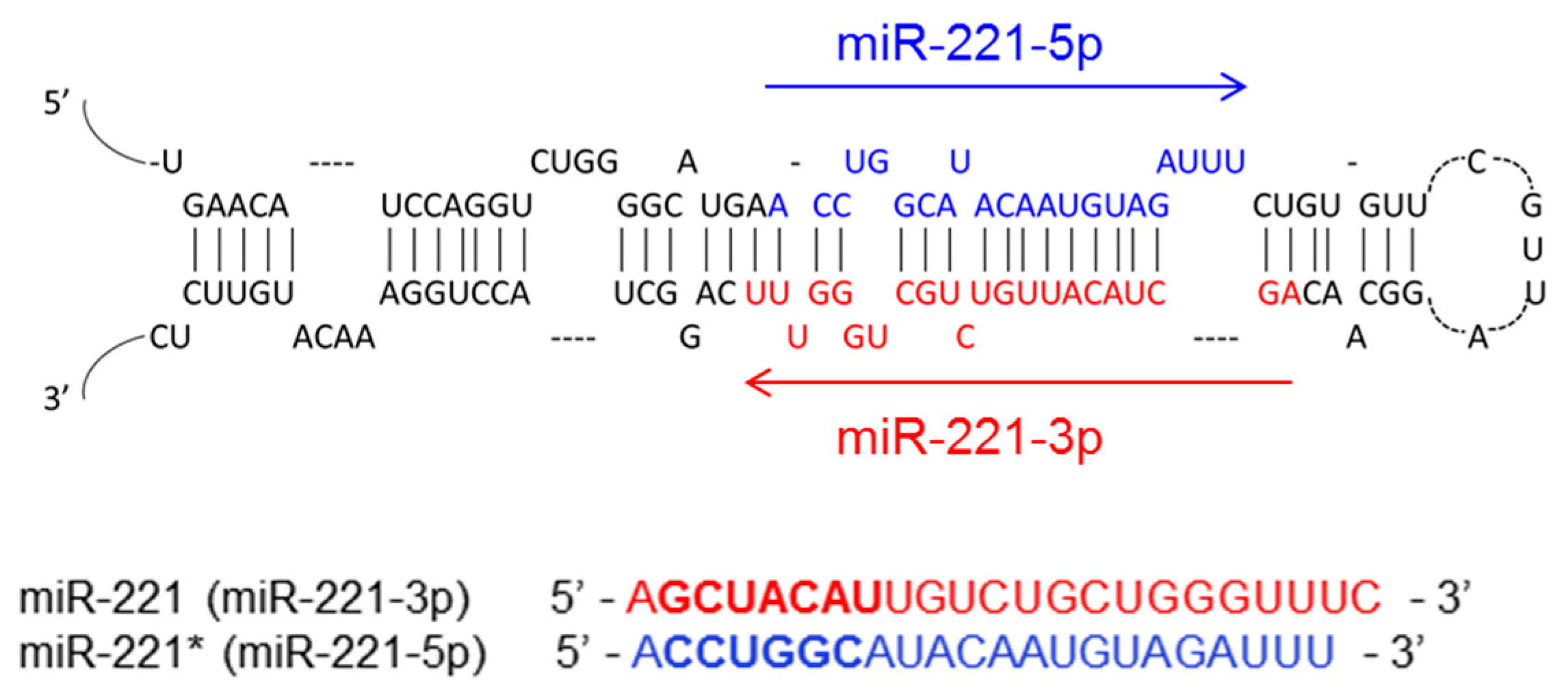
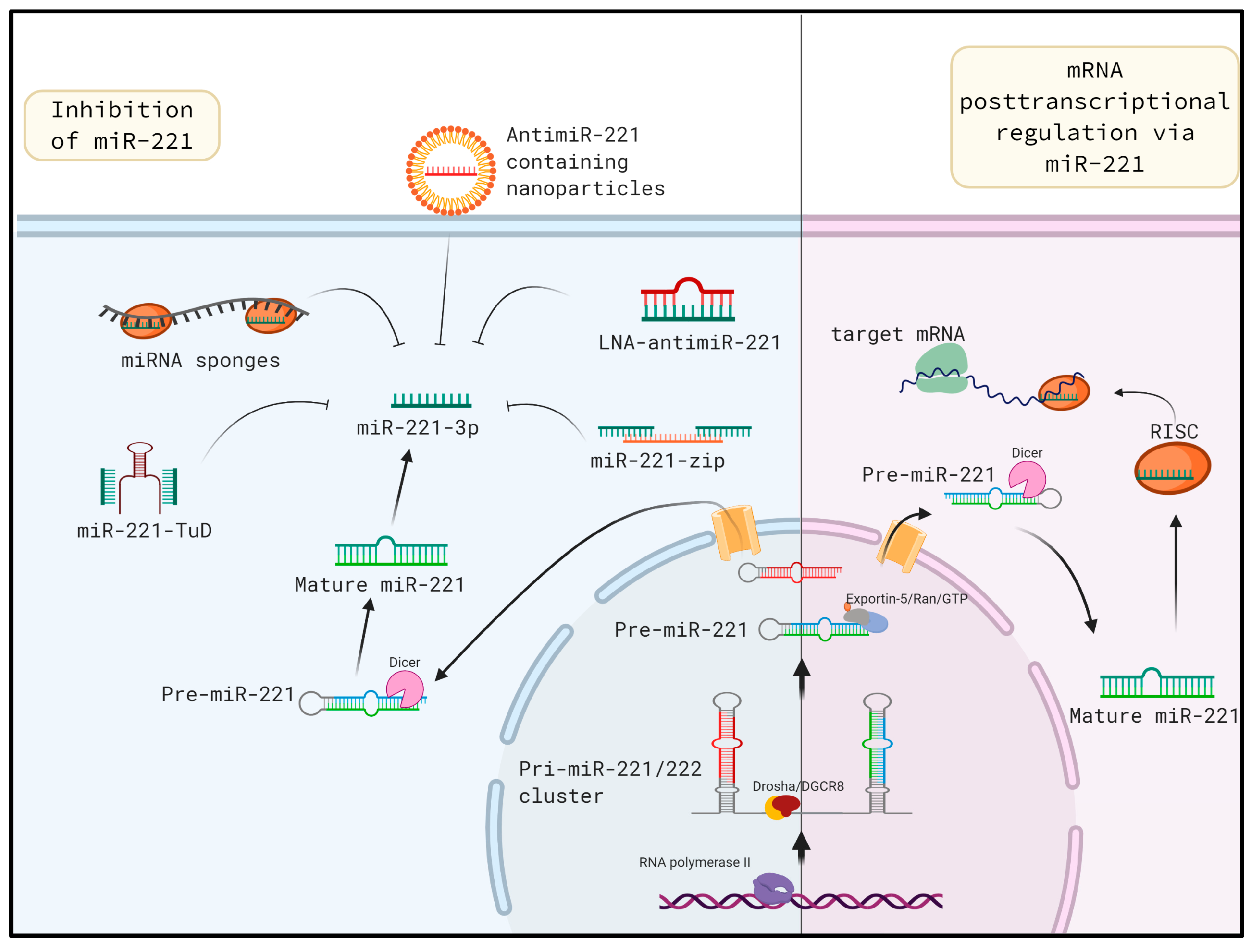
2. The Biogenesis of miR-221

MiR-221 is encoded and transcribed together with miR-222 as pri-miR-221/222. These two miRNAs are paralogous and share the same seed sequence. The promoter of pri-miR-221/222 is located upstream of miR-222, while three poly(A)-signals are located downstream of miR-221, and the miRNAs are separated by 726 bp. The pri-miR-221/222 is processed in the nucleus by the “microprocessor” complex composed of the enzyme Drosha and RNA binding protein DiGeorge syndrome critical region gene 8 (DGCR8). DGCR8 and Drosha are essential for the nuclear processing of microRNAs; the deficiency of either of them results in the global loss of miRNAs [3,4,5]. Nuclear processing of pri-miR-221/222 leads to the generation of the individual precursors, pre-miR-221 and pre-miR-222. Pre-miR-221 possesses a hairpin and is exported via the exportin5-RanGTP-shuttle system into the cytoplasm where it is further cleaved by the enzyme Dicer into the mature miR-221, whose sequence is shown in Figure 1 [5,6]. The 5′ end of pre-miR-221 gives rise to miR-221-5p, which is also known as miR-221* (star) miRNA. Similarly, the 3′ strand of pri-miR-221 generates miR-221-3p, which is interchangeably used with miR-221. This review focuses on miR-221-3p, which will henceforth be referred to as miR-221. The mature miR-221, upon loading into the RNA-induced silencing complex (RISC), regulates gene expression on a posttranscriptional level by binding to the 3′ UTR of target mRNAs (Figure 2).

3. MiR-221 is Upregulated During Liver Fibrosis and Other Chronic Liver Diseases

The indications that miR-221 is upregulated in liver fibrosis and cirrhosis were provided by pioneering studies that sought to analyze the expression of miRNAs in samples isolated from HCC patients who developed cirrhosis [7,8,9]. The elevation in miR-221 expression, among other miRNAs, was subsequently shown in a mouse model of liver fibrosis [10]. However, direct evidence of miR-221 upregulation in livers with hepatitis C virus (HCV), nonalcoholic steatohepatitis (NASH), as well as in in two independent mouse models of liver fibrosis, was provided by Kawada and colleagues [11]. In this study, the authors established a correlation between miR-221/222 expression and fibrosis in patients with HCV who had developed fibrosis. Of note, miR-221 expression was found to be elevated in fibrotic biopsies that showed high levels of collagen expression. This enhanced expression of miR-221 in fibrosis was validated in livers from patients with NASH. In concert with higher miR-221 levels in the HCV group, the authors observed increased miR-221 expression in NASH-associated fibrosis. The expression of miR-221 was examined in HSCs. Indeed, the expression of miR-221 was found to be increased in activated hepatic stellate cells compared to quiescent HSCs. Thus, this study, for the first time, demonstrated enhanced expression of miR-221 in activated HSCs during liver fibrosis.
The excessive accumulation of extracellular matrix during the development of fibrosis causes a decrease in the number of healthy and functional hepatocytes, which eventually leads to liver dysfunction. In addition to molecular changes in HSCs during fibrosis, hepatocytes also undergo dynamic changes at the transcriptomic and epigenetic levels. This includes the upregulation of miR-221 expression in hepatocytes during fibrosis, as it was recently demonstrated by us using purified hepatocytes isolated from fibrotic livers [12]. These results suggest that the upregulation of miR-221 in hepatocytes has functional relevance during fibrosis. In fact, hepatocyte-specific functional knockdown of miR-221 in vivo mitigated fibrosis in two independent mouse models of fibrosis, i.e., induced either by injecting mice with carbon-tetrachloride (CCl4) for 8 weeks, or by feeding mice with 3,5-diethoxycarbonyl-1,4-dihydrocollidine (DDC)-containing diet for 4 weeks. Mechanistically, the G-protein alpha inhibiting activity polypeptide 2 (Gnai2) posttranscriptional regulation via miR-221 causes a reduced secretion of C-C chemokine ligand 2 (CCL2), which contributes to suppression of liver fibrosis. On one hand, this study highlighted that crosstalk between hepatocytes and HSCs indeed directs fibrogenesis in an injured liver. On the other hand, these results suggested that miR-221 modulation in hepatocytes, the parenchymal cells making up over 65% of liver mass, possesses the potential for mitigation of fibrosis. Thus, both HSCs and hepatocytes show enhanced levels of miR-221 expression in response to fibrosis, and inhibition of miR-221 in both cell types can suppress fibrosis.
Subsequent studies have found upregulation of miR-221 in HCC, suggesting a correlation between high miR-221 expression and different cellular oncogenic pathways. One such pathway is Nuclear Factor kappa-light-chain-enhancer of activated B cells (NF-kB), the activation of which may be promoted by miR-221 in HCC [13]. Furthermore, in this study, the authors demonstrate that miR-221 inhibition results in reduction of metastasis, increased apoptosis, reduced tumor growth, and inactivation of the NF-kB pathway, while the re-introduction of miR-221 leads to the gain of tumor properties. Pineau et al. were able to show increased expression of miR-221 along with 11 other miRNAs in HCC tissues. Moreover, following treatment with anti-miR-221, decreased colony formation was observed in a HCC cell line, while transplantation of p53-/-myc liver progenitors with overexpressed miR-221 was shown to enhance cell proliferation and tumor growth. Further confirmation of high miR-221 levels in HCC tissues came from Li et al., who identified miR-221 as a regulator of epithelial-mesenchymal transition (EMT) in cancer cells in vitro [14]. An inhibitor of the JAK/STAT pathway, AdipoR1, was identified as a novel target, and the authors correlated tumor size, vascularity and the number of tumors with the level of miR-221. The implication of miR-221 through JAK/STAT modulation in HCC has also been shown by Huang et al. [15]. The authors confirmed previous findings where increased miR-221 levels in advanced stages of HCC influenced the invasion and migration of tumors, and additionally discovered SOCS3, an inhibitor of the JAK/STAT pathway, as a target of miR-221. They showed that SOCS3 expression is downregulated in HCC patients, as well as in cells transfected with miR-221, and that in vitro miR-221 inhibition or SOCS3 overexpression inhibits tumor growth.

4. Regulation of miRNA-221

Two transcription factors, NF-kB and c-Jun, have been shown to regulate the transcription of miR-221 by the Ciafre group [16]. The authors first demonstrated that ectopic modulation of NF-kB leads to altered miR-221 expression. Using bioinformatic prediction tools, two separate distal regions that NF-kB binds with were identified upstream of the miR-221 promoter region. Using electrophoretic mobility shift assays and chromatin immunoprecipitation experiments, the authors provided strong evidence of binding of NF-kB to the promoter of miR-221. Furthermore, the c-Jun binding site was found in the most distal enhancer region of miR-221. Subsequently, using siRNA experiments, the authors provided functional data for miR-221 regulation by both transcription factors. Importantly, both transcription factors have been shown to play critical roles in the development of liver fibrosis and HCC [17,18,19,20]. NF-kB inhibition leads to apoptosis in HSCs and reduction of extracellular matrix formation. It has also been suggested that c-Jun promotes liver fibrosis [21,22]. Therefore, upon liver damage that leads to fibrosis, NF-kB and c-Jun act as initiators of the molecular cascades that regulate apoptosis and inflammation and concomitantly increase miR-221 expression. Additionally, MET has been shown to regulate miR-221/222 expression by Garofalo et al. [23]. The authors showed that AP-1, a transcription factor from the Jun family which is involved in the c-Met pathway, binds and transcriptionally activates the miR-221/222 promoter. Subsequently, MET knockdown was demonstrated to downregulate miR-221 expression. Since miR-221 overexpression and MET pathway activation have been found in many cancers, the authors demonstrated that via both MET- and miR-221- knockdown, there is an increase in levels of tumor suppressor proteins, such as phosphatase and tensin homolog (PTEN).

5. Potential Mechanism of Fibrosis Regulation by miR-221

The function of miR-221 is cell-type dependent; it has been suggested that it acts either to promote or inhibit proliferation [24]. In the case of fibrosis, in addition to the posttranscriptional target, Gnai2 [12], miR-221 has been shown to regulate multiple other targets, and therefore, affect fibrogenesis directly or via modulation of HSC proliferation. Cyclin-dependent kinase inhibitors such as CDKN1C and CDKN1B are posttranscriptional targets of miR-221 that were discovered in liver cells [8]. The downregulation of CDKN1B enhances proliferation of HSCs [25], and the gain of CDKN1C function in HSCs inhibits their proliferation [26]. Thus, CDKN1C and CDKN1B are able to control the proliferation of HSCs, and hence, affect fibrogenesis. In addition, p53 Up-Regulated Modulator of Apoptosis (Puma) and Aryl Hydrocarbon Receptor Nuclear Translocator (Arnt), two targets of miR-221, have been shown to regulate hepatocyte apoptosis [3] and proliferation [27], respectively. Similar to miR-221-mediated Gnai2 modulation in hepatocytes, it is plausible that Puma and Arnt may affect fibrogenesis via inhibiting apoptosis of hepatocytes and enhancing proliferation. However, it remains to be examined if change in the expression of Puma and Arnt in hepatocytes influences fibrosis via HSCs or other cell types in the liver.
Suppressor of Cytokine Signaling (Socs1), another target of miR-221 [28], has been shown to regulate fibrogenesis by limiting damage to hepatocytes and the inflammatory response of macrophages [29]. Hepatocyte-specific depletion of Socs1 in mouse liver caused a higher degree of fibrosis upon CCl4 administration, as characterized by increased collagen and hydroxyproline contents and the accumulation of myofibroblasts with high levels of smooth muscle actin (α-SMA), an indicator of HSC activation. Similarly, the macrophage-specific depletion of Socs1 led to increased fibrogenesis. Both the lack of SOCS1 in hepatocytes and in macrophages resulted in increased infiltration of immune cells and high levels of CCL2, a known regulator of fibrosis [30]. It is noteworthy that two targets of miR-221, Gnai2 and Socs1, converge to support similar findings of upregulation of CCL2 as one of the driving forces of fibrosis. These findings further highlight the relevance of miR-221 and CCL2 during fibrogenesis.
miR-221 has been shown to regulate the expression of E-cadherin at the posttranscriptional level [31]. The inverse correlation of elevation of miR-221 and downregulation of E-cadherin during chronic liver diseases is frequently observed. The reduction in E-cadherin expression is one of the features of EMT. Early embryogenesis includes Type 1 EMT, while Type 2 EMT is associated with the fibrogenesis process, and Type 3 EMT is often observed during metastasis of neoplastic cells. It has also been suggested that Type 2 EMT may play a key role in the cycles of wound healing and inflammation during liver fibrosis [32,33]. Twist homolog 2 (TWIST2) [34] and Slug [31], both transcription factors known to drive EMT, have also been shown to regulate miR-221 expression. In fact, recent evidence demonstrated that miR-221 indeed regulates EMT in end-stage liver diseases [14,35]. Despite earlier reports that highlighted the relevance of EMT in liver fibrosis, subsequent reports in lineage tracing transgenic mouse models suggested that EMT only plays a limited role in fibrosis [36,37]. Therefore, it is critical to examine whether miR-221 indeed induces EMT, especially when modulated in hepatocytes, and to what extent the inhibition of EMT contributes to the attenuation of liver fibrosis by miR-221 knockdown.
Phosphatase and tensin homolog (PTEN), an inhibitor of proliferation, has been shown to be a posttranscriptional target of miR-221 [23,35]. The loss of Pten in the liver results in progressive liver fibrosis with higher expression of fibrogenic markers such as TIMP1, p75NTR, Collagen and SMA [38]. At the cellular level, isolated HSCs from Pten null livers contain lower numbers of vitamin A positive cells and increased expression of Acta2, indicating increased activation of Pten-/- HSCs compared to HSCs isolated from the wild-type livers.
TIMP3 has been shown to be an important regulator of liver fibrosis, inflammation and steatosis through the modulation of TNF-α-converting enzyme (TACE), and is therefore considered to play a key role in NASH and NAFLD development. The regulation of Timp3 mRNA expression by miR-221/222 has been shown by Jiang et al. [39]. The authors demonstrated that upon hepatocyte-specific miR-221/222 knockout, downregulation of expression of fibrogenic markers such as Col1a1, Col1a2, Col3a1 and Acta2, hepatogenic TG content and lipid droplet deposition are seen in both the methionine and choline deficient diet (MCDD)-induced NASH model and CCL4-induced liver fibrosis. Additionally, the authors detected increased expression of the miR-221/222 cluster in a NASH model, while Timp3 expression was downregulated. The inverse correlation between the expression of miR-221/222 and TIMP3 was further confirmed by Garofalo et al. [23]. The authors confirmed TIMP3 as a miR-221/222 target in HCC-derived cancer cells and showed that as a consequence of miR-221-mediated TIMP3 degradation, these cells were resistant to treatment with TNF-related, apoptosis-inducing ligand (TRAIL).
DDIT4, a regulator of the mammalian target of rapamycin (mTOR) kinase, has been shown as an important protein in tumor development due to its tumor suppressing properties. DDIT4 expression is regulated by miR-221 in vitro and in vivo [9]. The authors showed that miR-221 is overexpressed in both HCC and cirrhotic human livers, and enhances tumor growth through DDIT4 targeting and deregulation of the PI3K-PTEN-AKT-mTOR pathway. Interestingly, both DDIT4 and PTEN are shown as miR-221 targets and are separated by only 12Mb.
miR-221 has been found to increase β-catenin signaling by targeting Wnt target gene axis inhibition protein 2 (AXIN2), a protein involved in β-catenin degradation [40]. Dong et al. found increased expression of miR-221 and miR-15b in liver cancer tissues, and showed AXIN2 as a common target of both miRNAs in HCC cell lines. There was a decrease in proliferation and invasion of HCC cells upon miR-221/miR-15b knockdown or AXIN2 overexpression. Of note, Wnt signaling in hepatocytes is known to affect the expression of fibrogenic genes upon co-culture with HSCs [41].
Another important target of miR-221 is Bcl-2 modifying factor (BMF), an apoptotic activator whose expression level is in direct correlation with the caspase pathway [7]. Gramantieri et al. showed that BMF mRNA is a target of miR-221 in vitro, wherein their in vivo HCC expression is inversely correlated. Furthermore, the authors demonstrated higher expression of miR-221 and a corresponding decrease in expression of BMF in multifocal HCC compared to unifocal HCC. Accordingly, the role of miR-221 in HCC development was established, not only as a cell cycle modulator, but also as an anti-apoptotic regulator. Besides HCC, BMF has also been shown to be a miR-221 target in ovarian cancer cells [42].

6. In vivo miR-221 Inhibition: A Potential Approach for Mitigating Liver Fibrosis

6.1. Tough Decoys

The downregulation of miR-221 presents a plausible approach for the amelioration of liver fibrosis. Therefore, multiple approaches for miR-221 knockdown have been attempted. One such approach is to use tough decoys (TuD). TuD constructs contain bulge hairpin and two miRNA target sites which imperfectly pair with the target miRNA (Figure 2) [43]. TuDs have been used to efficiently downregulate multiple miRNAs including miR-221 [44]. Our laboratory adapted this method to downregulate miR-221 expression in mouse models of liver fibrosis [12]. Specifically, miR-221-TuD was cloned in an AAV expression plasmid under a hepatocyte-specific promoter, and high titer AAV vectors were prepared. In vivo knockdown of miR-221 upon AAV administration led to downregulation of fibrogenic genes such as Col1a1, Acta2 and Desmin, and reduced cell infiltration in the fibrotic area. Thus, miR-221-TuD delivered via AAV vector into a damaged liver is capable of attenuating fibrosis. MiR-221-TuD expressed from an AAV vector was tested in an additional study by Ma et al. [45]. The authors compared the miR-221-sponge, miR-221-TuD and miR-221-zip for their potential to inhibit miR-221 in vitro. After finding that miR-221-TuD and miR-221-zip were more potent, the authors then administered the inhibitors in vivo via intratumoral AAV injections into xenograft tumors which were generated from HepG2 cells in BALB/c nude mice. Upon miR-221 inhibition, a significant effect was observed, with mice having smaller tumor burdens and showing increased TRAIL-induced apoptosis.

6.2. Locked Nucleic Acids

Besides TuD, locked nucleic acids (LNA) can be used for a miRNA knockdown. LNAs are nucleic acids in which some of the ribose molecules within the nucleotides contain a methylene bridge that connects 2′ oxygen and 4′ carbon. Due to the methylene bridge, the ribose is locked in the C3’-endo or C2’-endo conformation [46]. LNAs can be designed to bind to DNA and RNA molecules. LNA-antimiRs are antisense RNAs in which usually every third nucleotide has a modified ribose moiety, and which possess a much higher hybridization affinity and stability in miRNA-LNA-antimiR duplexes than traditional probes [47]. In the miRNA-LNA-antimir duplex, miRNA function is antagonized [48]. The best-known LNA-antimiR is miravirsen (Santaris Pharma), an LNA antisense to miR-122 currently in clinical trials for the treatment of patients with hepatitis C infection [49]. MiR-122, the most abundant miRNA in the liver, has been shown to bind to the 5′ UTR of the hepatitis C virus RNA, which is conserved across all six genotypes of the virus, facilitating its replication [50]. Multiple studies have successfully used LNAs to ameliorate fibrosis in organs, including the kidney, heart and liver [51,52,53,54].
Since miR-221 is also a key regulator of NASH, Jiang et al. showed in vivo potential of LNA-antimiR-221 in reducing hepatic steatosis in the MCDD mouse model of NASH [39]. Upon in vivo administration of LNA-antimiR-221, hepatic TG content, inflammation, steatosis and hepatocyte ballooning were decreased. In concert with other studies, Jiang et al. also reported amelioration of fibrosis.

6.3. MiRNA Sponges

An additional approach for miRNA inhibition in vivo and in vitro is with miRNA sponges. MiRNA sponges contain multiple sequences that are complementary to miRNA seed sequences, resulting in miRNAs binding to the sponge instead for the target mRNA, which leads to miRNA sequestration [55]. When compared to other miRNA inhibitors, such as TuD or LNA, which require nucleotide complementarity beyond the seed sequence, sponges offer the advantage of inhibiting multiple miRNAs that share a common seed sequence. Also, miRNA sponge expressing transgenic animal models can be developed, providing lifelong inhibition of miRNAs. When it comes to clinical applications, these features are not desirable, since it is preferable to reversibly inhibit only one miRNA. Therefore, in clinical settings, oligonucleotides have been more widely used.
Moshiri et al. designed a miR-221 sponge that contains four miR-221 binding sites inserted in both the adenoviral and AAV vectors [56]. They selected a conditionally replicative oncolytic adenovirus whose replication was under the control of miR-199a, a miRNA moderately expressed in healthy livers but downregulated in HCC. They also developed a rAAV vector containing a miR-221 sponge. The inhibition potential of the miR-221 sponge was tested in vitro using HCC cell lines and assessed by the level of CDKN1B (p27), which is a known target of miR-221. The authors showed increased expression of p27 in the presence of the sponge and increased apoptosis. Even though the authors did not test it in vivo, they assumed that both approaches could be used. Naturally occurring sponges for miRNAs are long noncoding RNAs. One such RNA is CASC2, which was shown by Jin et al. to act as a sponge for miR-221 in HCC cell lines [57]. The authors predicted the binding of CASC2 with miR-221 bioinformatically and experimentally. They also demonstrated caspase-3 as a miR-221 target in HCC cells, and an inverse expression of miR-221 and CASC2, which was known as an inhibitor of cancer cell proliferation. Furthermore, it was shown that CASC2 modulated TRAIL resistance through miR-24/miR-221 and increased levels of caspase-3. Lately, nonviral systems such as nanoparticles are emerging as promising therapeutic approaches because of their low toxicity. Such a system was employed in a study by Li et al., who used nanoparticles with miR-221 mimic/inhibitor, both in vivo and in vitro, in order to track HCC development. Nanoparticle-based miR-221 inhibitors decrease cell proliferation, colony formation ability and tumor development, and result in lower serum levels of miR-221, thus representing a promising approach for HCC therapy [58]. Additionally, Cai et al. demonstrated that a synergistic effect is achieved with the usage of a combination treatment regimen of nanoparticles containing anti-miR-221 and chemotherapeutic drugs, such as sorafenib [59]. Specifically, the authors tested the combination in HCC cell lines where cell proliferation arrest was achieved. Even though their data provided initial evidence that the antimiR-221 encapsulated nanoparticles should be able to treat solid tumors, further experiments in vivo are needed for confirmation.

6.4. MiR-221 as A Serum Biomarker

To date, liver biopsy remains the gold-standard method for diagnosing liver fibrosis and end-stage liver diseases. Among noninvasive approaches, analyses of biomarkers such as circulating miRNAs hold great potential for diagnosis. Higher stability of miRNAs in the serum and plasma, as well as the fact that a change in their expression in circulation often precedes injury, make them promising candidates as biomarkers [60,61]. One of the most common causes of liver fibrosis is chronic liver damage due to HCV infection. In the serum of HCV infected patients, increased levels of miR-221 were detected by Ding et al. (Figure 3) [62]. The authors showed a correlation between increased levels of miR-221, ALT and AST. The expression of miR-221 was shown to be controlled by NF-kB, as had been previously demonstrated in other studies. Findings from this study were confirmed by Xu et al. [63]. Additionally, the authors identified SOCS1 and SOCS2 as miR-221 targets (Figure 3). HCV infection can often lead to the development of liver cirrhosis, and eventually, to liver cancer, which is the fourth most common cause of cancer related deaths worldwide. High expression of miR-221 has been demonstrated in HCC tissues and in serum. Sohn et al. showed high levels of miR-221 among other miRNAs in serum exosomes and as a circulating miRNA in patients with HCC and chronic HBV infection [64]. The authors showed that the level of miR-221 in serum exosomes could be used to distinguish between HCC and chronic hepatitis B (CHB). While analyzing circulating levels of miR-221 in the serum, the authors did not find a difference between the HCC and CHB groups. It is important to note that the authors did not correlate serum miR-221 levels with tissue expression. Furthermore, Li et al. found significantly increased levels of miR-221 in HCC serum samples [65], and showed a correlation between the level of miR-221 and HCC severity and disease outcome. The patients with high miR-221 serum levels had more advanced cirrhosis, tumor size and stage. Additionally, the survival rate was significantly lower than in patients with low miR-221 serum levels. The authors, however, implied that the combination of miR-221 and AFP serum levels provides a better diagnostic prediction than the use of each of these markers separately. Other studies have also confirmed these findings [66,67]. In contrast, in acute obstructive cholangitis (AOC), increased levels of miR-221 in patients’ sera were shown to be a positive sign of recovery [68]. In AOC, a biliary obstruction occurs, followed by an infection that leads to inflammation. There is also a regenerative response that is triggered in hepatocytes through enhanced HGF signaling. HGF is the primary ligand for the receptor tyrosine kinase and oncogene, c-Met [69], which, in turn, enhances miR-221 transcription (Figure 3). In AOC, high miR-221 is indicative of recovery. The expression of miR-221 in hepatocytes during AOC is regulated by the SOCS1–Met axis. Downregulation of SOCS1 increases the expression of miR-221. It has been shown that SOCS1 inhibits HGF signaling and attenuates HGF mediated phosphorylation of c-Met in hepatocytes. Due to the chronic inflammation which occurs in AOC, SOCS1 is upregulated. However, during regression of AOC, the authors showed that SOCS1 levels reverted to normal levels, while Met and phospho-Met levels were increased. Phosphorylated c-Met enhances miR-221 transcription, whose overexpression has been shown to accelerate hepatocyte proliferation during liver regeneration (Figure 3) [27,70,71,72]. Thus, increased miR-221 expression indicates active liver regeneration and recovery.
High levels of miR-221 in serum in HCC/HCV are correlated with advanced-stage diseases, but these same high levels in AOC are indicators of recovery. Even though the increase in miR-221 levels is triggered by different mechanisms—in HCC/HCV, its expression is mostly regulated by NFkB, while in AOC, it is accomplished via HGF—in both instances, the role of miR-221 is to accelerate cell proliferation. Therefore, the dichotomous functional relevance of elevated miR-221 levels allows for its use as a prognostic biomarker in HCC/HCV disease progression or AOC recovery monitoring.

7. Unanswered Questions

Recent research has unequivocally established miR-221 as a key regulator of liver fibrosis. However, the following questions, especially pertaining to the prerequisites to the use of miR-221 inhibitors to attenuate liver fibrosis in a clinical setting, remain:
The expression of miR-221 in nonparenchymal cells such as Kupffer cells has been shown to be higher during fibrosis than in the normal healthy liver. Whether increased levels of miR-221 in Kupffer cells contribute to progression of liver fibrosis remains to be investigated.
The knockdown of miR-221 in HSCs and hepatocytes mitigates fibrogenic gene expression. However, it remains to be seen whether combined suppression of miR-221 in hepatocytes, HSCs and other nonparenchymal cells reduces fibrosis to a greater extent.
To date, most studies have focused on the identification of miR-221 as a regulator of liver fibrosis. Given that fibrosis, if left unchecked, may develop into cirrhosis, the reversal of cirrhosis upon miR-221 inhibition must be demonstrated in order to translate the present findings regarding miR-221 into therapeutic pathways.
Likewise, the relevance of miR-221 in mouse models that develop HCC after fibrosis and cirrhosis remains to be examined. Hence, it would be extremely important to study whether the inhibition of miR-221 in those models in fibrotic or cirrhotic stages prevents progression to HCC.
Multiple studies have suggested a potential application of miR-221 as a biomarker of liver fibrosis. However, the number of patients in cohorts remains low; therefore, multicenter studies are required before miR-221 can be used as a reliable serum biomarker of liver fibrosis and cirrhosis.

8. Conclusions

Over the last few years, tremendous progress has been made towards elucidating the function of miRNAs during liver fibrosis. MiR-221 has emerged as a key regulator that is upregulated during liver fibrosis caused by various etiologies. Studies in mouse models of fibrosis have provided ample evidence that intrahepatic suppression of miR-221 holds significant potential for the mitigation of liver fibrosis. However, further research is required to specifically address the effects of miR-221 inhibition in cirrhosis, its use as a serum biomarker, and the safety, including potential side effects, of miR-221 modulation in damaged livers.

Author Contributions

A.D.S. conceptualized the manuscript. J.M. and A.B. wrote the manuscript. J.M. prepared all figures. All authors have read and agreed to the published version of the manuscript.

Funding

Authors receive grant support from the Deutsche Forschungsgemeinschaft, REBIRTH- MWK and Plus 3 Program of Boehringer Ingelheim.

Conflicts of Interest

The authors declare no conflict of interest.

References

  1. Schueller, F.; Roy, S.; Vucur, M.; Trautwein, C.; Luedde, T.; Roderburg, C. The Role of miRNAs in the Pathophysiology of Liver Diseases and Toxicity. Int. J. Mol. Sci. 2018, 19, 261. [Google Scholar] [CrossRef] [PubMed] [Green Version]
  2. Su, Q.; Kumar, V.; Sud, N.; Mahato, R.I. MicroRNAs in the pathogenesis and treatment of progressive liver injury in NAFLD and liver fibrosis. Adv. Drug Deliv. Rev. 2018, 129, 54–63. [Google Scholar] [CrossRef] [PubMed]
  3. Sharma, A.D.; Narain, N.; Händel, E.-M.; Iken, M.; Singhal, N.; Cathomen, T.; Manns, M.P.; Schöler, H.R.; Ott, M.; Cantz, T. MicroRNA-221 regulates FAS-induced fulminant liver failure. Hepatology 2011, 53, 1651–1661. [Google Scholar] [CrossRef] [PubMed]
  4. Wang, Y.; Medvid, R.; Melton, C.; Jaenisch, R.; Blelloch, R. DGCR8 is essential for microRNA biogenesis and silencing of embryonic stem cell self-renewal. Nat. Genet. 2007, 39, 380–385. [Google Scholar] [CrossRef]
  5. Denli, A.M.; Tops, B.B.J.; Plasterk, R.H.A.; Ketting, R.F.; Hannon, G.J. Processing of primary microRNAs by the Microprocessor complex. Nature 2004, 432, 231–235. [Google Scholar] [CrossRef]
  6. Lee, Y.; Ahn, C.; Han, J.; Choi, H.; Kim, J.; Yim, J.; Lee, J.; Provost, P.; Rådmark, O.; Kim, S.; et al. The nuclear RNase III Drosha initiates microRNA processing. Nature 2003, 425, 415–419. [Google Scholar] [CrossRef]
  7. Gramantieri, L.; Fornari, F.; Ferracin, M.; Veronese, A.; Sabbioni, S.; Calin, G.A.; Grazi, G.L.; Croce, C.M.; Bolondi, L.; Negrini, M. MicroRNA-221 targets Bmf in hepatocellular carcinoma and correlates with tumor multifocality. Clin. Cancer Res. 2009, 15, 5073–5081. [Google Scholar] [CrossRef] [Green Version]
  8. Fornari, F.; Gramantieri, L.; Ferracin, M.; Veronese, A.; Sabbioni, S.A.; Calin, G.; Grazi, G.L.; Giovannini, C.; Croce, C.M.; Bolondi, L.; et al. MiR-221 controls CDKN1C/p57 and CDKN1B/p27 expression in human hepatocellular carcinoma. Oncogene 2008, 27, 5651–5661. [Google Scholar] [CrossRef] [Green Version]
  9. Pineau, P.; Volinia, S.; McJunkin, K.; Marchio, A.; Battiston, C.; Terris, B.; Mazzaferro, V.; Lowe, S.; Croce, C.; Dejean, A. 579 MIR-221 Overexpression Contributes to Liver Tumorigenesis. J. Hepatol. 2010, 52, S229. [Google Scholar] [CrossRef]
  10. Roderburg, C.; Urban, G.-W.; Bettermann, K.; Vucur, M.; Zimmermann, H.; Schmidt, S.; Janssen, J.; Koppe, C.; Knolle, P.A.; Castoldi, M.; et al. Micro-RNA profiling reveals a role for miR-29 in human and murine liver fibrosis. Hepatology 2010, 53, 209–218. [Google Scholar] [CrossRef]
  11. Ogawa, T.; Enomoto, M.; Fujii, H.; Sekiya, Y.; Yoshizato, K.; Ikeda, K.; Kawada, N. MicroRNA-221/222 upregulation indicates the activation of stellate cells and the progression of liver fibrosis. Gut 2012, 61, 1600–1609. [Google Scholar] [CrossRef] [PubMed]
  12. Tsay, H.-C.; Yuan, Q.; Balakrishnan, A.; Kaiser, M.; Möbus, S.; Kozdrowska, E.; Farid, M.; Tegtmeyer, P.-K.; Borst, K.; Vondran, F.W.R.; et al. Hepatocyte-specific suppression of microRNA-221-3p mitigates liver fibrosis. J. Hepatol. 2019, 70, 722–734. [Google Scholar] [CrossRef] [PubMed] [Green Version]
  13. Liu, Z.; Wang, C.; Jiao, X.; Zhao, S.; Liu, X.; Wang, Y.; Zhang, J. miR-221 promotes growth and invasion of hepatocellular carcinoma cells by constitutive activation of NFkappaB. Am. J. Transl. Res. 2016, 8, 4764–4777. [Google Scholar]
  14. Li, T.; Li, M.; Hu, S.; Cheng, X.; Gao, Y.; Jiang, S.; Yu, Q.; Zhang, C.; Sun, P.; Xian, W.; et al. MiR-221 mediates the epithelial-mesenchymal transition of hepatocellular carcinoma by targeting AdipoR1. Int. J. Boil. Macromol. 2017, 103, 1054–1061. [Google Scholar] [CrossRef]
  15. Huang, S.; Zhou, D.; Li, Y.-X.; Ming, Z.-Y.; Li, K.-Z.; Wu, G.-B.; Chen, C.; Zhao, Y.-N. In vivo and in vitro effects of microRNA-221 on hepatocellular carcinoma development and progression through the JAK-STAT3 signaling pathway by targeting SOCS3. J. Cell. Physiol. 2018, 234, 3500–3514. [Google Scholar] [CrossRef]
  16. Galardi, S.; Mercatelli, N.; Farace, M.G.; Ciafrè, S.A. NF-kB and c-Jun induce the expression of the oncogenic miR-221 and miR-222 in prostate carcinoma and glioblastoma cells. Nucleic Acids Res. 2011, 39, 3892–3902. [Google Scholar] [CrossRef] [Green Version]
  17. Hellerbrand, C.; Jobin, C.; Iimuro, Y.; Licato, L.; Sartor, R.B.; Brenner, D.A. Inhibition of NFkappaB in activated rat hepatic stellate cells by proteasome inhibitors and an IkappaB super-repressor. Hepatology 1998, 27, 1285–1295. [Google Scholar] [CrossRef]
  18. Cui, D.; Zhang, S.; Ma, J.; Han, J.; Jiang, H. Short interfering RNA targetting NF-kappa B induces apoptosis of hepatic stellate cells and attenuates extracellular matrix production. Dig. Liver Dis. 2010, 42, 813–817. [Google Scholar] [CrossRef]
  19. Eferl, R.; Ricci, R.; Kenner, L.; Zenz, R.; David, J.P.; Rath, M.; Wagner, E.F. Liver tumor development. c-Jun antagonizes the proapoptotic activity of p53. Cell 2003, 112, 181–192. [Google Scholar]
  20. Trierweiler, C.; Hockenjos, B.; Zatloukal, K.; Thimme, R.; Blum, H.E.; Wagner, E.F.; Hasselblatt, P. The transcription factor c-JUN/AP-1 promotes HBV-related liver tumorigenesis in mice. Cell Death Differ 2016, 23, 576–582. [Google Scholar] [CrossRef] [Green Version]
  21. Schulien, I.; Hockenjos, B.; Schmitt-Graeff, A.; Perdekamp, M.G.; Follo, M.; Thimme, R.; Hasselblatt, P. The transcription factor c-Jun/AP-1 promotes liver fibrosis during non-alcoholic steatohepatitis by regulating Osteopontin expression. Cell Death Differ. 2019, 26, 1688–1699. [Google Scholar] [CrossRef] [PubMed] [Green Version]
  22. Wernig, G.; Chen, S.Y.; Cui, L.; Van Neste, C.; Tsai, J.M.; Kambham, N.; Vogel, H.; Natkunam, Y.; Gilliland, D.G.; Nolan, G.; et al. Unifying mechanism for different fibrotic diseases. Proc. Natl. Acad. Sci. USA 2017, 114, 4757–4762. [Google Scholar] [CrossRef] [PubMed] [Green Version]
  23. Garofalo, M.; Di Leva, G.; Romano, G.; Nuovo, G.; Suh, S.-S.; Ngankeu, A.; Taccioli, C.; Pichiorri, F.; Alder, H.; Secchiero, P.; et al. miR-221&222 Regulate TRAIL Resistance and Enhance Tumorigenicity through PTEN and TIMP3 Downregulation. Cancer Cell 2009, 16, 498–509. [Google Scholar] [CrossRef] [PubMed] [Green Version]
  24. Garofalo, M.; Quintavalle, C.; Romano, G.; Croce, C.M.; Condorelli, G. miR221/222 in cancer: Their role in tumor progression and response to therapy. Curr. Mol. Med. 2012, 12, 27–33. [Google Scholar] [CrossRef]
  25. Wang, B.; Li, W.; Guo, K.; Xiao, Y.; Wang, Y.; Fan, J.G. miR-181b Promotes hepatic stellate cells proliferation by targeting p27 and is elevated in the serum of cirrhosis patients. Biochem. Biophys. Res. Commun. 2012, 421, 4–8. [Google Scholar] [CrossRef]
  26. Wang, Y.G.; Xu, L.; Wang, T.; Wei, J.; Meng, W.Y.; Wang, N.; Shi, M. Givinostat inhibition of hepatic stellate cell proliferation and protein acetylation. World J. Gastroenterol. 2015, 21, 8326–8339. [Google Scholar] [CrossRef]
  27. Yuan, Q.; Loya, K.; Rani, B.; Möbus, S.; Balakrishnan, A.; Lamlé, J.; Cathomen, T.; Vogel, A.; Manns, M.P.; Ott, M.; et al. MicroRNA-221 overexpression accelerates hepatocyte proliferation during liver regeneration. Hepatology 2013, 57, 299–310. [Google Scholar] [CrossRef]
  28. Sehgal, M.; Zeremski, M.; Talal, A.H.; Ginwala, R.; Elrod, E.; Grakoui, A.; Li, Q.G.; Philip, R.; Khan, Z.K.; Jain, P. IFN-α-Induced Downregulation of miR-221 in Dendritic Cells: Implications for HCV Pathogenesis and Treatment. J. Interf. Cytokine Res. 2015, 35, 698–709. [Google Scholar] [CrossRef] [Green Version]
  29. Mafanda, E.K.; Kandhi, R.; Bobbala, D.; Khan, G.M.; Nandi, M.; Menendez, A.; Ramanathan, S.; Ilangumaran, S. Essential role of suppressor of cytokine signaling 1 (SOCS1) in hepatocytes and macrophages in the regulation of liver fibrosis. Cytokine 2019, 124, 154501. [Google Scholar] [CrossRef]
  30. Baeck, C.; Wehr, A.; Karlmark, K.R.; Heymann, F.; Vucur, M.; Gassler, N.; Huss, S.; Klussmann, S.; Eulberg, D.; Luedde, T.; et al. Pharmacological inhibition of the chemokine CCL2 (MCP-1) diminishes liver macrophage infiltration and steatohepatitis in chronic hepatic injury. Gut 2011, 61, 416–426. [Google Scholar] [CrossRef]
  31. Pan, Y.; Li, J.; Zhang, Y.; Wang, N.; Liang, H.; Liu, Y.; Zhang, C.-Y.; Zen, K.; Gu, H. Slug-upregulated miR-221 promotes breast cancer progression through suppressing E-cadherin expression. Sci. Rep. 2016, 6, 25798. [Google Scholar] [CrossRef] [PubMed] [Green Version]
  32. Kaimori, A.; Potter, J.; Kaimori, J.-Y.; Wang, C.; Mezey, E.; Koteish, A. Transforming Growth Factor-beta1 Induces an Epithelial-to-Mesenchymal Transition State in Mouse Hepatocytes in Vitro. J. Boil. Chem. 2007, 282, 22089–22101. [Google Scholar] [CrossRef] [PubMed] [Green Version]
  33. Zeisberg, M.; Yang, C.; Martino, M.; Duncan, M.B.; Rieder, F.; Tanjore, H.; Kalluri, R. Fibroblasts Derive from Hepatocytes in Liver Fibrosis via Epithelial to Mesenchymal Transition. J. Boil. Chem. 2007, 282, 23337–23347. [Google Scholar] [CrossRef] [PubMed] [Green Version]
  34. Wei, W.F.; Zhou, C.F.; Wu, X.G.; He, L.N.; Wu, L.F.; Chen, X.J.; Yan, R.M.; Zhong, M.; Yu, Y.H.; Liang, L.; et al. MicroRNA-221-3p, a TWIST2 target, promotes cervical cancer metastasis by directly targeting THBS2. Cell Death Dis. 2017, 8, 3220. [Google Scholar] [CrossRef] [PubMed] [Green Version]
  35. Li, J.; Yao, L.; Li, G.; Ma, D.; Sun, C.; Gao, S.; Zhang, P.; Gao, F. miR-221 Promotes Epithelial-Mesenchymal Transition through Targeting PTEN and Forms a Positive Feedback Loop with beta-catenin/c-Jun Signaling Pathway in Extra-Hepatic Cholangiocarcinoma. PLoS ONE 2015, 10, e0141168. [Google Scholar] [CrossRef]
  36. Taura, K.; Miura, K.; Iwaisako, K.; Österreicher, C.H.; Kodama, Y.; Penz-Österreicher, M.; Brenner, D.A. Hepatocytes do not undergo epithelial-mesenchymal transition in liver fibrosis in mice. Hepatology 2010, 51, 1027–1036. [Google Scholar] [CrossRef] [Green Version]
  37. Scholten, D.; Österreicher, C.H.; Scholten, A.; Iwaisako, K.; Gu, G.; Brenner, D.A.; Kisseleva, T. Genetic Labeling Does Not Detect Epithelial-to-Mesenchymal Transition of Cholangiocytes in Liver Fibrosis in Mice. Gastroenterology 2010, 139, 987–998. [Google Scholar] [CrossRef] [Green Version]
  38. He, L.; Gubbins, J.; Peng, Z.; Medina, V.; Fei, F.; Asahina, K.; Wang, J.; Kahn, M.; Rountree, C.B.; Stiles, B.L. Activation of hepatic stellate cell in Pten null liver injury model. Fibrogenesis Tissue Repair 2016, 9, 8. [Google Scholar] [CrossRef] [Green Version]
  39. Jiang, X.; Jiang, L.; Shan, A.; Su, Y.; Cheng, Y.; Song, D.; Ji, H.; Ning, G.; Wang, W.; Cao, Y. Targeting hepatic miR-221/222 for therapeutic intervention of nonalcoholic steatohepatitis in mice. EBioMedicine 2018, 37, 307–321. [Google Scholar] [CrossRef] [Green Version]
  40. Dong, Y.; Zhang, N.; Zhao, S.; Chen, X.; Li, F.; Tao, X. miR-221-3p and miR-15b-5p promote cell proliferation and invasion by targeting Axin2 in liver cancer. Oncol. Lett. 2019, 18, 6491–6500. [Google Scholar] [CrossRef]
  41. Wang, Q.; Chou, X.; Guan, F.; Fang, Z.; Lu, S.; Lei, J.; Li, Y.; Liu, W. Enhanced Wnt Signalling in Hepatocytes is Associated with Schistosoma japonicum Infection and Contributes to Liver Fibrosis. Sci. Rep. 2017, 7, 230. [Google Scholar] [CrossRef] [PubMed] [Green Version]
  42. Xie, X.; Huang, Y.; Chen, L.; Wang, J. miR-221 regulates proliferation and apoptosis of ovarian cancer cells by targeting BMF. Oncol. Lett. 2018, 16, 6697–6704. [Google Scholar] [CrossRef] [PubMed] [Green Version]
  43. Haraguchi, T.; Ozaki, Y.; Iba, H. Vectors expressing efficient RNA decoys achieve the long-term suppression of specific microRNA activity in mammalian cells. Nucleic Acids Res. 2009, 37, e43. [Google Scholar] [CrossRef] [PubMed]
  44. Bak, R.O.; Hollensen, A.K.; Primo, M.; Sørensen, C.D.; Mikkelsen, J.G. Potent microRNA suppression by RNA Pol II-transcribed ‘Tough Decoy’ inhibitors. RNA 2012, 19, 280–293. [Google Scholar] [CrossRef] [Green Version]
  45. Ma, S.; Sun, J.; Guo, Y.; Zhang, P.; Liu, Y.; Zheng, D.; Shi, J. Combination of AAV-TRAIL with miR-221-Zip Therapeutic Strategy Overcomes the Resistance to TRAIL Induced Apoptosis in Liver Cancer. Theranostics 2017, 7, 3228–3242. [Google Scholar] [CrossRef]
  46. Grünweller, A.; Hartmann, R.K. Locked nucleic acid oligonucleotides: The next generation of antisense agents? BioDrugs 2007, 21, 235–243. [Google Scholar] [CrossRef]
  47. Elmen, J.; Lindow, M.; Schütz, S.; Lawrence, M.; Petri, A.; Obad, S.; Lindholm, M.W.; Hedtjärn, M.; Hansen, H.F.; Berger, U.; et al. LNA-mediated microRNA silencing in non-human primates. Nature 2008, 452, 896–899. [Google Scholar] [CrossRef]
  48. Farrell, R.E. RNA Methodologies. Encycl. Mol. Cell Biol. Mol. Med. 2006. [Google Scholar] [CrossRef]
  49. Ottosen, S.; Parsley, T.B.; Yang, L.; Zeh, K.; Van Doorn, L.-J.; Van Der Veer, E.; Raney, A.K.; Hodges, M.R.; Patick, A.K. In VitroAntiviral Activity and Preclinical and Clinical Resistance Profile of Miravirsen, a Novel Anti-Hepatitis C Virus Therapeutic Targeting the Human Factor miR-122. Antimicrob. Agents Chemother. 2014, 59, 599–608. [Google Scholar] [CrossRef] [Green Version]
  50. Jopling, C.; Yi, M.; Lancaster, A.M.; Lemon, S.M.; Sarnow, P. Modulation of Hepatitis C Virus RNA Abundance by a Liver-Specific MicroRNA. Science 2005, 309, 1577–1581. [Google Scholar] [CrossRef] [Green Version]
  51. Okada, H.; Honda, M.; Campbell, J.S.; Takegoshi, K.; Sakai, Y.; Yamashita, T.; Shirasaki, T.; Takabatake, R.; Nakamura, M.; Tanaka, T.; et al. Inhibition of microRNA-214 ameliorates hepatic fibrosis and tumor incidence in platelet-derived growth factor C transgenic mice. Cancer Sci. 2015, 106, 1143–1152. [Google Scholar] [CrossRef] [PubMed] [Green Version]
  52. Luan, J.; Fu, J.; Chen, C.; Jiao, C.; Kong, W.; Zhang, Y.; Chang, Q.; Wang, Y.; Li, D.; Illei, G.G.; et al. LNA-anti-miR-150 ameliorated kidney injury of lupus nephritis by inhibiting renal fibrosis and macrophage infiltration. Arthritis Res. 2019, 21, 276–314. [Google Scholar] [CrossRef] [PubMed] [Green Version]
  53. Putta, S.; Lanting, L.; Sun, G.; Lawson, G.; Kato, M.; Natarajan, R. Inhibiting MicroRNA-192 Ameliorates Renal Fibrosis in Diabetic Nephropathy. J. Am. Soc. Nephrol. 2012, 23, 458–469. [Google Scholar] [CrossRef] [PubMed] [Green Version]
  54. Bernardo, B.C.; Nguyen, S.S.; Winbanks, C.E.; Gao, X.; Boey, E.J.H.; Tham, Y.K.; Kiriazis, H.; Ooi, J.Y.Y.; Porrello, E.R.; Igoor, S.; et al. Therapeutic silencing of miR-652 restores heart function and attenuates adverse remodeling in a setting of established pathological hypertrophy. Faseb J. 2014, 28, 5097–5110. [Google Scholar] [CrossRef] [PubMed] [Green Version]
  55. Ebert, M.S.; Neilson, J.R.A.; Sharp, P. MicroRNA sponges: Competitive inhibitors of small RNAs in mammalian cells. Nat. Methods 2007, 4, 721–726. [Google Scholar] [CrossRef]
  56. Moshiri, F.; Callegari, E.; D’Abundo, L.; Corrà, F.; Lupini, L.; Sabbioni, S.; Negrini, M. Inhibiting the oncogenic mir-221 by microRNA sponge: Toward microRNA-based therapeutics for hepatocellular carcinoma. Gastroenterol. Hepatol. Bed Bench 2014, 7, 43–54. [Google Scholar]
  57. Jin, X.; Cai, L.; Wang, C.; Deng, X.; Yi, S.; Lei, Z.; Xiao, Q.; Xu, H.; Luo, H.; Sun, J. CASC2/miR-24/miR-221 modulates the TRAIL resistance of hepatocellular carcinoma cell through caspase-8/caspase-3. Cell Death Dis. 2018, 9, 318. [Google Scholar] [CrossRef] [Green Version]
  58. Li, F.; Wang, F.; Zhu, C.; Wei, Q.; Zhang, T.; Zhou, Y.L. miR-221 suppression through nanoparticle-based miRNA delivery system for hepatocellular carcinoma therapy and its diagnosis as a potential biomarker. Int. J. Nanomed. 2018, 13, 2295–2307. [Google Scholar] [CrossRef] [Green Version]
  59. Cai, H.; Yang, Y.; Peng, F.; Liu, Y.; Fu, X.; Ji, B. Gold nanoparticles-loaded anti-miR221 enhances antitumor effect of sorafenib in hepatocellular carcinoma cells. Int. J. Med. Sci. 2019, 16, 1541–1548. [Google Scholar] [CrossRef] [Green Version]
  60. Ward, J.; Kanchagar, C.; Veksler-Lublinsky, I.; Lee, R.C.; McGill, M.R.; Jaeschke, H.; Curry, S.C.; Ambros, V. Circulating microRNA profiles in human patients with acetaminophen hepatotoxicity or ischemic hepatitis. Proc. Natl. Acad. Sci. USA 2014, 111, 12169–12174. [Google Scholar] [CrossRef] [Green Version]
  61. Yang, D.; Yuan, Q.; Balakrishnan, A.; Bantel, H.; Klusmann, J.-H.; Manns, M.P.; Ott, M.; Cantz, T.; Sharma, A.D. MicroRNA-125b-5p mimic inhibits acute liver failure. Nat. Commun. 2016, 7, 11916. [Google Scholar] [CrossRef] [PubMed] [Green Version]
  62. Ding, C.L.; Xu, G.; Ren, H.; Zhao, L.J.; Zhao, P.; Qi, Z.T.; Wang, W. HCV infection induces the upregulation of miR-221 in NF-κB dependent manner. Virus Res. 2015, 196, 135–139. [Google Scholar] [CrossRef] [PubMed]
  63. Xu, G.; Yang, F.; Ding, C.L.; Wang, J.; Zhao, P.; Wang, W.; Ren, H. MiR-221 accentuates IFN’s anti-HCV effect by downregulating SOCS1 and SOCS3. Virology 2014, 463, 343–350. [Google Scholar] [CrossRef] [PubMed] [Green Version]
  64. Sohn, W.; Kim, J.; Kang, S.H.; Yang, S.R.; Cho, J.Y.; Cho, H.C.; Shim, S.G.; Paik, Y.H. Serum exosomal microRNAs as novel biomarkers for hepatocellular carcinoma. Exp. Mol. Med. 2015, 47, e184. [Google Scholar] [CrossRef]
  65. Li, J.; Wang, Y.; Yu, W.; Chen, J.; Luo, J. Expression of serum miR-221 in human hepatocellular carcinoma and its prognostic significance. Biochem. Biophys. Res. Commun. 2011, 406, 70–73. [Google Scholar] [CrossRef]
  66. Fornari, F.; Ferracin, M.; Treré, D.; Milazzo, M.; Marinelli, S.; Galassi, M.; Venerandi, L.; Pollutri, D.; Patrizi, C.; Borghi, A.; et al. Circulating microRNAs, miR-939, miR-595, miR-519d and miR-494, Identify Cirrhotic Patients with HCC. PLoS ONE 2015, 10, e0141448. [Google Scholar] [CrossRef]
  67. Fu, X.; Wang, Q.; Chen, J.; Huang, X.; Chen, X.; Cao, L.; Tan, H.; Li, W.; Zhang, L.; Bi, J.; et al. Clinical significance of miR-221 and its inverse correlation with p27Kip1 in hepatocellular carcinoma. Mol. Boil. Rep. 2010, 38, 3029–3035. [Google Scholar] [CrossRef]
  68. Yu, J.; Zhang, W.; Qian, H.; Tang, H.; Lin, W.; Lu, B.-C. SOCS1 regulates hepatic regenerative response and provides prognostic makers for acute obstructive cholangitis. Sci. Rep. 2017, 7, 9482. [Google Scholar] [CrossRef] [Green Version]
  69. Organ, S.L.; Tsao, M.-S. An overview of the c-MET signaling pathway. Ther. Adv. Med. Oncol. 2011, 3, S7–S19. [Google Scholar] [CrossRef] [Green Version]
  70. Gui, Y.; Yeganeh, M.; Ramanathan, S.; Leblanc, C.; Pomerleau, V.; Ferbeyre, G.; Saucier, C.; Ilangumaran, S. SOCS1 controls liver regeneration by regulating HGF signaling in hepatocytes. J. Hepatol. 2011, 55, 1300–1308. [Google Scholar] [CrossRef]
  71. Yoshimura, A.; Suzuki, M.; Sakaguchi, R.; Hanada, T.; Yasukawa, H. SOCS, Inflammation, and Autoimmunity. Front. Immunol. 2012, 3, 20. [Google Scholar] [CrossRef] [PubMed] [Green Version]
  72. Khan, G.M.; Ghosh, A.; Variya, B.; Santharam, M.A.; Kandhi, R.; Ramanathan, S.; Ilangumaran, S. Hepatocyte growth control by SOCS1 and SOCS3. Cytokine 2019, 121, 154733. [Google Scholar] [CrossRef] [PubMed]
Figure 1. The sequences of miR-221 and miR-221*. Mature sequence of miR-221-5p is in blue, while miR-221-3p is shown in red. The seed sequence of the miRNA is highlighted.
Cells 09 01767 g001
Figure 2. Biogenesis of miR-221 is shown on the right side of the figure. The pri-miR-221/222 cluster is first generated and then processed into pre-miR-221 and pre-miR-222 in the nuclei. Upon translocation of pre-miR-221 from the nuclei to the cytoplasm, Dicer mediates another processing to generate miRNA duplex. One of the strands may be incorporated into RISC complex to regulate gene expression at the posttranscriptional level. On the left side of the schematic, multiple approaches of in vivo miR-221 inhibition are shown. MiR-221 can be inhibited via sponges, antimiR containing nanoparticles, TuDs, miR-zips and LNAs. Each method has been shown to be effective in miR-221 in vivo inhibition. Due to miR-221 inhibition, amelioration of liver fibrosis and attenuation of HCC development was observed.
Cells 09 01767 g002
Figure 3. The main etiologies of liver fibrosis are chronic viral hepatitis infection, NASH and cholestatic diseases. If left untreated, extensive liver fibrosis often leads to the development of HCC. At present, the most commonly used method to diagnose liver disease is biopsy, which is invasive and often time-consuming. More importantly, the biopsy is often performed at later stages, by which time the only effective treatment option is transplantation. MiR-221 has been shown as a miRNA that can serve as a serum biomarker for diagnosis, to distinguish different diseases and as a marker of recovery. (A) HCV is one of the most prevalent causes of liver fibrosis. Multiple studies have shown miR-221 upregulation in the sera of patients with HCV. Furthermore, in HCV, miR-221 expression is under the control of NF-Kb, and it targets SOCS1 and SOCS3. (B) HCC is the fourth leading cause of cancer-related deaths worldwide. High levels of miR-221 have been detected in HCC tissues and in the sera of patients with HCC. Based on the serum levels, miR-221 can be used together with AFP as a biomarker of HCC development, severity and outcome. Patients with high miR-221 had more advanced HCC and lower survival rates than those with low miR-221 serum levels. (C) In contrast, in AOC, the upregulation of miR-221 in patient’s sera is considered a sign of recovery. In AOC, hepatocyte proliferation and liver regeneration occur due to high HGF signaling. HGF is the key ligand for the receptor tyrosine kinase, c-Met, which, in turn, enhances miR-221 transcription. High levels of miR-221 in the liver repress SOCS1 levels. Since SOCS1 is an inhibitor of HGF signaling, its repression by miR-221 leads to enhanced HGF signaling and active liver regeneration and recovery.
Cells 09 01767 g003

