Expression of FGF8, FGF18, and FGFR4 in Gastroesophageal Adenocarcinomas

 , ,
, , 
Abstract
1. Introduction
2. Materials and Methods
2.1. Preliminary TCGA (The Cancer Genome Atlas) Analysis
2.2. Patient Selection
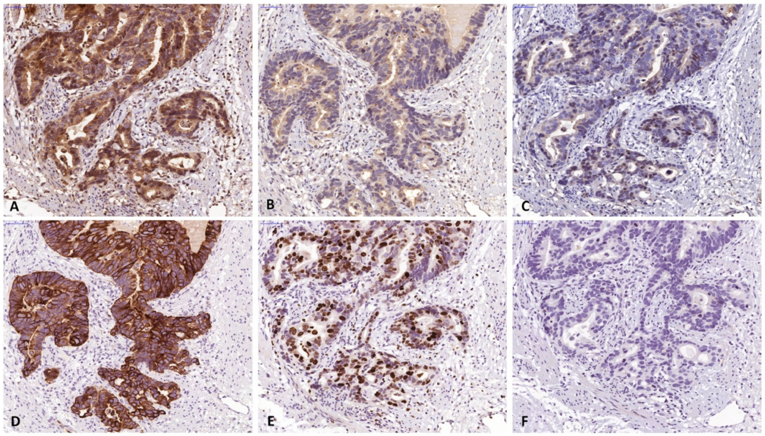
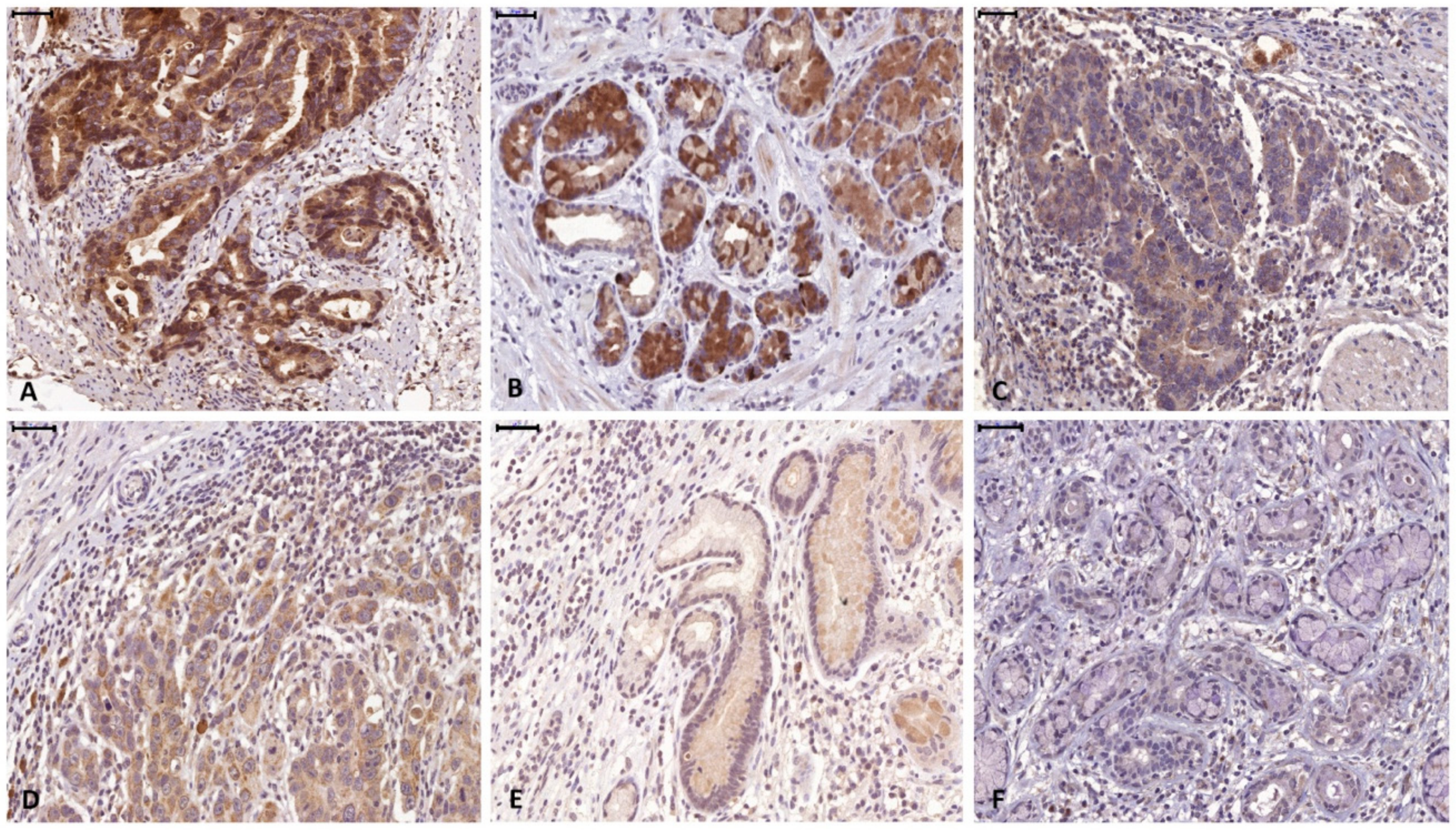
2.3. Immunohistochemistry
2.4. Statistical Analysis
3. Results
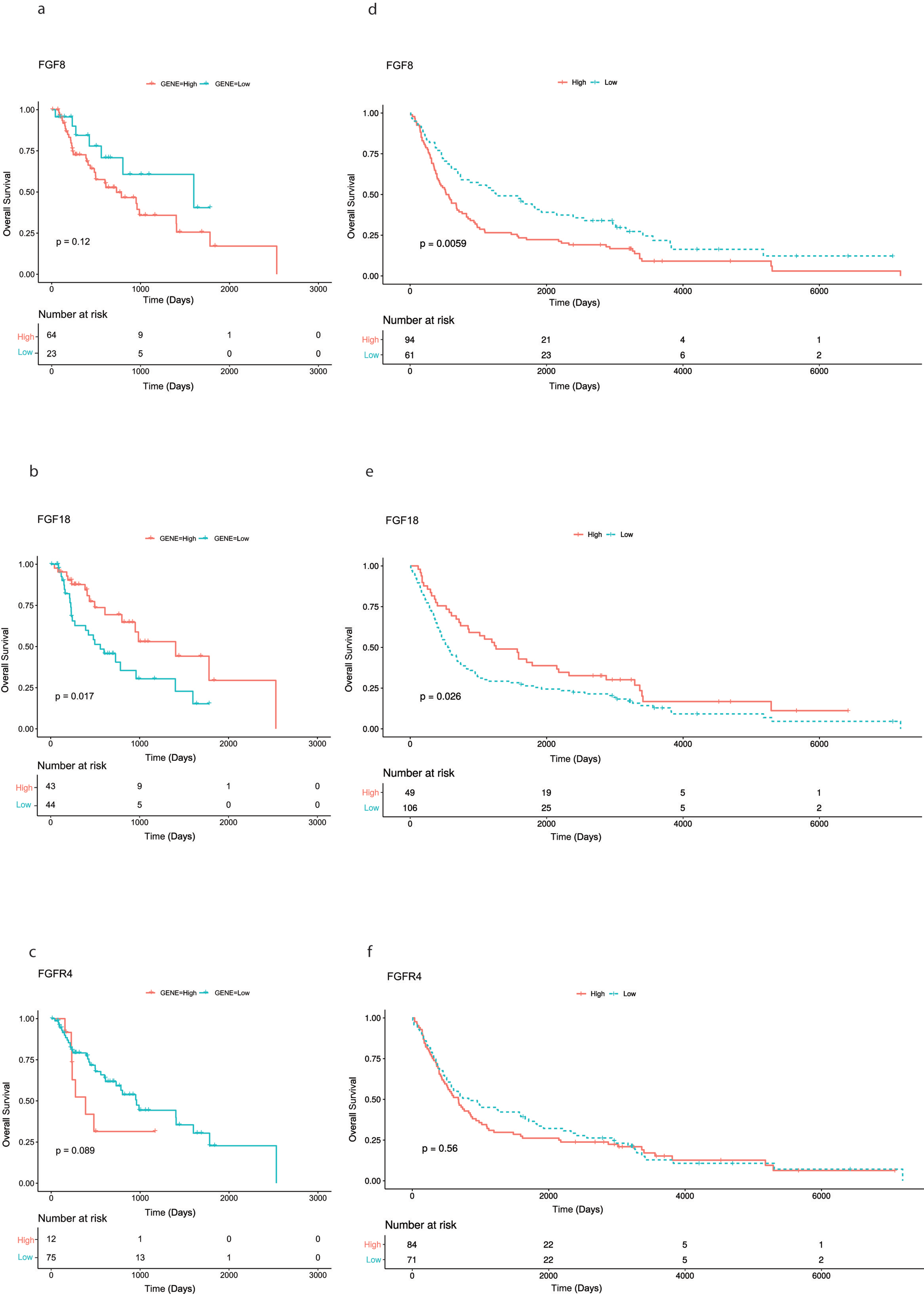
3.1. Preliminary TCGA (The Cancer Genome Atlas) Analysis
3.2. Immunohistochemical Analysis of Tumor Tissue Samples
4. Discussion
5. Conclusions
Author Contributions
Funding
Acknowledgments
Conflicts of Interest
References
- Ferlay, J.; Soerjomataram, I.; Dikshit, R.; Eser, S.; Mathers, C.; Rebelo, M.; Parkin, D.M.; Forman, D.; Bray, F. Cancer incidence and mortality worldwide: Sources, methods and major patterns in globocan 2012. Int. J. Cancer 2015, 136, E359–E386. [Google Scholar] [CrossRef] [PubMed]
- Burmeister, B.H.; Smithers, B.M.; Gebski, V.; Fitzgerald, L.; Simes, R.J.; Devitt, P.; Ackland, S.; Gotley, D.C.; Joseph, D.; Millar, J.; et al. Surgery alone versus chemoradiotherapy followed by surgery for resectable cancer of the oesophagus: A randomised controlled phase iii trial. Lancet Oncol. 2005, 6, 659–668. [Google Scholar] [CrossRef]
- Reynolds, J.V.; Muldoon, C.; Hollywood, D.; Ravi, N.; Rowley, S.; O’Byrne, K.; Kennedy, J.; Murphy, T.J. Long-term outcomes following neoadjuvant chemoradiotherapy for esophageal cancer. Ann. Surg. 2007, 245, 707–716. [Google Scholar] [CrossRef] [PubMed]
- Christein, J.D.; Hollinger, E.F.; Millikan, K.W. Prognostic factors associated with resectable carcinoma of the esophagus. Am. Surg. 2002, 68, 258–262; discussion 262–263. [Google Scholar] [PubMed]
- Gertler, R.; Stein, H.J.; Langer, R.; Nettelmann, M.; Schuster, T.; Hoefler, H.; Siewert, J.R.; Feith, M. Long-term outcome of 2920 patients with cancers of the esophagus and esophagogastric junction: Evaluation of the new union internationale contre le cancer/american joint cancer committee staging system. Ann. Surg. 2011, 253, 689–698. [Google Scholar] [CrossRef] [PubMed]
- Ku, G.Y.; Ilson, D.H. Esophagogastric cancer: Targeted agents. Cancer Treat. Rev. 2010, 36, 235–248. [Google Scholar] [CrossRef] [PubMed]
- Rassouli, F.B.; Matin, M.M.; Saeinasab, M. Cancer stem cells in human digestive tract malignancies. Tumour Biol. 2016, 37, 7–21. [Google Scholar] [CrossRef]
- Schulenburg, A.; Ulrich-Pur, H.; Thurnher, D.; Erovic, B.; Florian, S.; Sperr, W.R.; Kalhs, P.; Marian, B.; Wrba, F.; Zielinski, C.C.; et al. Neoplastic stem cells: A novel therapeutic target in clinical oncology. Cancer 2006, 107, 2512–2520. [Google Scholar] [CrossRef]
- Borah, A.; Raveendran, S.; Rochani, A.; Maekawa, T.; Kumar, D.S. Targeting self-renewal pathways in cancer stem cells: Clinical implications for cancer therapy. Oncogenesis 2015, 4, e177. [Google Scholar] [CrossRef]
- Honing, J.; Pavlov, K.V.; Mul, V.E.; Karrenbeld, A.; Meijer, C.; Faiz, Z.; Smit, J.K.; Hospers, G.A.; Burgerhof, J.G.; Kruyt, F.A.; et al. Cd44, shh and sox2 as novel biomarkers in esophageal cancer patients treated with neoadjuvant chemoradiotherapy. Radiother Oncol. 2015, 117, 152–158. [Google Scholar] [CrossRef][Green Version]
- Sui, Y.P.; Jian, X.P.; Ma, L.I.; Xu, G.Z.; Liao, H.W.; Liu, Y.P.; Wen, H.C. Prognostic value of cancer stem cell marker cd133 expression in esophageal carcinoma: A meta-analysis. Mol. Clin. Oncol. 2016, 4, 77–82. [Google Scholar] [CrossRef] [PubMed]
- Hang, D.; Dong, H.C.; Ning, T.; Dong, B.; Hou, D.L.; Xu, W.G. Prognostic value of the stem cell markers cd133 and abcg2 expression in esophageal squamous cell carcinoma. Dis. Esophagus 2012, 25, 638–644. [Google Scholar] [CrossRef] [PubMed]
- Qian, X.; Tan, C.; Wang, F.; Yang, B.; Ge, Y.; Guan, Z.; Cai, J. Esophageal cancer stem cells and implications for future therapeutics. Onco. Targets Ther. 2016, 9, 2247–2254. [Google Scholar] [PubMed]
- Heinzle, C.; Sutterluty, H.; Grusch, M.; Grasl-Kraupp, B.; Berger, W.; Marian, B. Targeting fibroblast-growth-factor-receptor-dependent signaling for cancer therapy. Expert Opin. Ther. Targets 2011, 15, 829–846. [Google Scholar] [CrossRef] [PubMed]
- Beenken, A.; Mohammadi, M. The fgf family: Biology, pathophysiology and therapy. Nat. Rev. Drug Discov. 2009, 8, 235–253. [Google Scholar] [CrossRef] [PubMed]
- Knights, V.; Cook, S.J. De-regulated fgf receptors as therapeutic targets in cancer. Pharmacol. Ther. 2010, 125, 105–117. [Google Scholar] [CrossRef]
- Powers, C.J.; McLeskey, S.W.; Wellstein, A. Fibroblast growth factors, their receptors and signaling. Endocr. Relat. Cancer 2000, 7, 165–197. [Google Scholar] [CrossRef] [PubMed]
- Kono, S.A.; Marshall, M.E.; Ware, K.E.; Heasley, L.E. The fibroblast growth factor receptor signaling pathway as a mediator of intrinsic resistance to egfr-specific tyrosine kinase inhibitors in non-small cell lung cancer. Drug Resist. Updat. 2009, 12, 95–102. [Google Scholar] [CrossRef] [PubMed]
- Motomura, K.; Hagiwara, A.; Komi-Kuramochi, A.; Hanyu, Y.; Honda, E.; Suzuki, M.; Kimura, M.; Oki, J.; Asada, M.; Sakaguchi, N.; et al. An fgf1:Fgf2 chimeric growth factor exhibits universal fgf receptor specificity, enhanced stability and augmented activity useful for epithelial proliferation and radioprotection. Biochim. Biophys. Acta 2008, 1780, 1432–1440. [Google Scholar] [CrossRef]
- Pardo, O.E.; Arcaro, A.; Salerno, G.; Raguz, S.; Downward, J.; Seckl, M.J. Fibroblast growth factor-2 induces translational regulation of bcl-xl and bcl-2 via a mek-dependent pathway: Correlation with resistance to etoposide-induced apoptosis. J. Biol. Chem. 2002, 277, 12040–12046. [Google Scholar] [CrossRef]
- Pardo, O.E.; Lesay, A.; Arcaro, A.; Lopes, R.; Ng, B.L.; Warne, P.H.; McNeish, I.A.; Tetley, T.D.; Lemoine, N.R.; Mehmet, H.; et al. Fibroblast growth factor 2-mediated translational control of iaps blocks mitochondrial release of smac/diablo and apoptosis in small cell lung cancer cells. Mol. Cell Biol. 2003, 23, 7600–7610. [Google Scholar] [CrossRef] [PubMed]
- Roidl, A.; Berger, H.J.; Kumar, S.; Bange, J.; Knyazev, P.; Ullrich, A. Resistance to chemotherapy is associated with fibroblast growth factor receptor 4 up-regulation. Clin. Cancer Res. 2009, 15, 2058–2066. [Google Scholar] [CrossRef] [PubMed]
- Koneczny, I.; Schulenburg, A.; Hudec, X.; Knofler, M.; Holzmann, K.; Piazza, G.; Reynolds, R.; Valent, P.; Marian, B. Autocrine fibroblast growth factor 18 signaling mediates wnt-dependent stimulation of cd44-positive human colorectal adenoma cells. Mol. Carcinog. 2015, 54, 789–799. [Google Scholar] [CrossRef] [PubMed]
- Sonvilla, G.; Allerstorfer, S.; Stattner, S.; Karner, J.; Klimpfinger, M.; Fischer, H.; Grasl-Kraupp, B.; Holzmann, K.; Berger, W.; Wrba, F.; et al. Fgf18 in colorectal tumour cells: Autocrine and paracrine effects. Carcinogenesis 2008, 29, 15–24. [Google Scholar] [CrossRef] [PubMed]
- Sonvilla, G.; Allerstorfer, S.; Heinzle, C.; Stattner, S.; Karner, J.; Klimpfinger, M.; Wrba, F.; Fischer, H.; Gauglhofer, C.; Spiegl-Kreinecker, S.; et al. Fibroblast growth factor receptor 3-iiic mediates colorectal cancer growth and migration. Br. J. Cancer 2010, 102, 1145–1156. [Google Scholar] [CrossRef] [PubMed]
- Bange, J.; Prechtl, D.; Cheburkin, Y.; Specht, K.; Harbeck, N.; Schmitt, M.; Knyazeva, T.; Muller, S.; Gartner, S.; Sures, I.; et al. Cancer progression and tumor cell motility are associated with the fgfr4 arg(388) allele. Cancer Res. 2002, 62, 840–847. [Google Scholar]
- Heinzle, C.; Erdem, Z.; Paur, J.; Grasl-Kraupp, B.; Holzmann, K.; Grusch, M.; Berger, W.; Marian, B. Is fibroblast growth factor receptor 4 a suitable target of cancer therapy? Curr. Pharm. Des. 2014, 20, 2881–2898. [Google Scholar] [CrossRef]
- Zhang, X.; Ibrahimi, O.A.; Olsen, S.K.; Umemori, H.; Mohammadi, M.; Ornitz, D.M. Receptor specificity of the fibroblast growth factor family. The complete mammalian fgf family. J. Biol. Chem. 2006, 281, 15694–15700. [Google Scholar] [CrossRef]
- Brewer, J.R.; Mazot, P.; Soriano, P. Genetic insights into the mechanisms of fgf signaling. Genes Dev. 2016, 30, 751–771. [Google Scholar] [CrossRef]
- Tickle, C.; Munsterberg, A. Vertebrate limb development--the early stages in chick and mouse. Curr. Opin. Genet. Dev. 2001, 11, 476–481. [Google Scholar] [CrossRef]
- Gauglhofer, C.; Sagmeister, S.; Schrottmaier, W.; Fischer, C.; Rodgarkia-Dara, C.; Mohr, T.; Stattner, S.; Bichler, C.; Kandioler, D.; Wrba, F.; et al. Up-regulation of the fibroblast growth factor 8 subfamily in human hepatocellular carcinoma for cell survival and neoangiogenesis. Hepatology 2011, 53, 854–864. [Google Scholar] [CrossRef] [PubMed]
- Mattila, M.M.; Harkonen, P.L. Role of fibroblast growth factor 8 in growth and progression of hormonal cancer. Cytokine Growth Factor Rev. 2007, 18, 257–266. [Google Scholar] [CrossRef] [PubMed]
- Harpain, F.; Ahmed, M.A.; Hudec, X.; Timelthaler, G.; Jomrich, G.; Mullauer, L.; Selzer, E.; Dorr, W.; Bergmann, M.; Holzmann, K.; et al. Fgf8 induces therapy resistance in neoadjuvantly radiated rectal cancer. J. Cancer Res. Clin. Oncol. 2019, 145, 77–86. [Google Scholar] [CrossRef] [PubMed]
- Colaprico, A.; Silva, T.C.; Olsen, C.; Garofano, L.; Cava, C.; Garolini, D.; Sabedot, T.S.; Malta, T.M.; Pagnotta, S.M.; Castiglioni, I.; et al. Tcgabiolinks: An r/bioconductor package for integrative analysis of tcga data. Nucleic Acids Res. 2016, 44, e71. [Google Scholar] [CrossRef] [PubMed]
- Kassambara, A.; Kosinski, M.; Biecek, P.; Fabian, S. Survminer: Drawing survival curves using “ggplot2”. Available online: https://rpkgs.datanovia.com/survminer/index.html (accessed on 10 June 2019).
- Chen, Y.; Lun, A.; McCarthy, D.; Robinson, M.; Phipson, B.; Hu, Y.; Zhou, X.; Robinson, M.D.; Smyth, G.K. Edger: Empirical analysis of digital gene expression data in r. Available online: https://bioconductor.org/packages/release/bioc/html/edgeR.html (accessed on 10 June 2019).
- Kanehisa, M.; Goto, S. Kegg: Kyoto encyclopedia of genes and genomes. Nucleic Acids Res. 2000, 28, 27–30. [Google Scholar] [CrossRef] [PubMed]
- Luo, W.; Brouwer, C. Pathview: An r/bioconductor package for pathway-based data integration and visualization. Bioinformatics 2013, 29, 1830–1831. [Google Scholar] [CrossRef] [PubMed]
- Mandard, A.M.; Dalibard, F.; Mandard, J.C.; Marnay, J.; Henry-Amar, M.; Petiot, J.F.; Roussel, A.; Jacob, J.H.; Segol, P.; Samama, G.; et al. Pathologic assessment of tumor regression after preoperative chemoradiotherapy of esophageal carcinoma. Clinicopathologic correlations. Cancer 1994, 73, 2680–2686. [Google Scholar] [CrossRef]
- Jomrich, G.; Maroske, F.; Stieger, J.; Preusser, M.; Ilhan-Mutlu, A.; Winkler, D.; Kristo, I.; Paireder, M.; Schoppmann, S.F. Mk2 and etv1 are prognostic factors in esophageal adenocarcinomas. J. Cancer 2018, 9, 460–468. [Google Scholar] [CrossRef]
- Ahmed, M.A.; Selzer, E.; Dorr, W.; Jomrich, G.; Harpain, F.; Silberhumer, G.R.; Mullauer, L.; Holzmann, K.; Grasl-Kraupp, B.; Grusch, M.; et al. Fibroblast growth factor receptor 4 induced resistance to radiation therapy in colorectal cancer. Oncotarget 2016, 7, 69976–69990. [Google Scholar] [CrossRef]
- R Development Core Team. R: A Language and Environment for Statistical Computing; R Foundation for Statistical Computing: Vienna, Austria, 2018; Available online: https://www.gbif.org/tool/81287/r-a-language-and-environment-for-statistical-computing (accessed on 10 June 2019).
- Therneau, T. A Package for Survival Analysis in S. Version 2.38. 2015. Available online: https://cran.r-project.org/web/packages/survival/citation.html (accessed on 10 June 2019).
- Heinzle, C.; Gsur, A.; Hunjadi, M.; Erdem, Z.; Gauglhofer, C.; Stattner, S.; Karner, J.; Klimpfinger, M.; Wrba, F.; Reti, A.; et al. Differential effects of polymorphic alleles of fgf receptor 4 on colon cancer growth and metastasis. Cancer Res. 2012, 72, 5767–5777. [Google Scholar] [CrossRef]
- Dorkin, T.J.; Robinson, M.C.; Marsh, C.; Bjartell, A.; Neal, D.E.; Leung, H.Y. Fgf8 over-expression in prostate cancer is associated with decreased patient survival and persists in androgen independent disease. Oncogene 1999, 18, 2755–2761. [Google Scholar] [CrossRef] [PubMed]
- Zhang, J.; Zhou, Y.; Huang, T.; Wu, F.; Pan, Y.; Dong, Y.; Wang, Y.; Chan, A.K.Y.; Liu, L.; Kwan, J.S.H.; et al. Fgf18, a prominent player in fgf signaling, promotes gastric tumorigenesis through autocrine manner and is negatively regulated by mir-590-5p. Oncogene 2019, 38, 33–46. [Google Scholar] [CrossRef] [PubMed]
- Shim, H.J.; Shin, M.H.; Kim, H.N.; Kim, J.H.; Hwang, J.E.; Bae, W.K.; Chung, I.J.; Cho, S.H. The prognostic significance of fgfr4 gly388 polymorphism in esophageal squamous cell carcinoma after concurrent chemoradiotherapy. Cancer Res. Treat. 2016, 48, 71–79. [Google Scholar] [CrossRef] [PubMed]
- Meyerholz, D.K.; Beck, A.P. Principles and approaches for reproducible scoring of tissue stains in research. Lab. Invest. 2018, 98, 844–855. [Google Scholar] [CrossRef]
- Meyerholz, D.K.; Beck, A.P. Fundamental concepts for semiquantitative tissue scoring in translational research. ILAR J. 2018, 59, 13–17. [Google Scholar] [CrossRef] [PubMed]
- Lin, F.; Prichard, J. (Eds.) Handbook of Practical Immunohistochemistry: Frequently Asked Questions, 2nd ed.; Springer: New York, NY, USA, 2015; p. xv. 764p. [Google Scholar]


| Factors | FGF8 | FGF18 | FGFR4 | ||||||||||||
|---|---|---|---|---|---|---|---|---|---|---|---|---|---|---|---|
| high | low/absent | p-value | High | low/absent | p-value | high | low/absent | p-value | |||||||
| Age (SD) | 65 (11) | 65 (10) | >0.05 | 66 (12) | 62 (10) | >0.05 | 66 (11) | 64 (11) | >0.05 | ||||||
| Sex | >0.05 | >0.05 | 0.008 | ||||||||||||
| Male | 75 | (48.4%) | 49 | (31.6%) | 38 | (24.5%) | 86 | (55.5%) | 74 | (47.7%) | 50 | (32.3%) | |||
| Female | 19 | (12.3%) | 12 | (7.7%) | 11 | (7.1%) | 20 | (12.9%) | 10 | (6.5%) | 21 | (13.5%) | |||
| Neoadjuvant treatment | >0.05 | >0.05 | >0.05 | ||||||||||||
| Yes | 37 | (23.9%) | 32 | (20.6%) | 26 | (16.8%) | 60 | (38.7%) | 31 | (20.0%) | 38 | (24.5%) | |||
| No | 57 | (36.8%) | 29 | (18.7%) | 23 | (14.8%) | 46 | (29.7%) | 53 | (34.2%) | 33 | (21.3%) | |||
| (y)pT | 0.003 | >0.05 | >0.05 | ||||||||||||
| 0 | 0 | (0.0%) | 3 | (1.9%) | 0 | (0.0%) | 3 | (1.9%) | 0 | (0.0%) | 3 | (1.9%) | |||
| 1 | 1 | (0.6%) | 2 | (1.3%) | 2 | (1.3%) | 1 | (0.6%) | 1 | (0.6%) | 2 | (1.3%) | |||
| 2 | 17 | (11.0%) | 23 | (14.8%) | 11 | (7.1%) | 29 | (18.7%) | 22 | (14.2%) | 18 | (11.6%) | |||
| 3 | 68 | (43.9%) | 32 | (20.6%) | 34 | (21.9%) | 66 | (42.6%) | 56 | (36.1%) | 44 | (28.4%) | |||
| 4 | 8 | (5.2%) | 1 | (0.6%) | 2 | (1.3%) | 7 | (4.5%) | 5 | (3.2%) | 4 | (2.6%) | |||
| (y)pN | >0.05 | >0.05 | >0.05 | ||||||||||||
| 0 | 17 | (11.0%) | 22 | (14.2%) | 16 | (10.3%) | 23 | (14.8%) | 18 | (11.6%) | 21 | (13.5%) | |||
| 1 | 25 | (16.1%) | 13 | (8.4%) | 13 | (8.4%) | 25 | (16.1%) | 19 | (12.3%) | 19 | (12.3%) | |||
| 2 | 24 | (15.5%) | 13 | (8.4%) | 7 | (4.5%) | 30 | (19.4%) | 25 | (16.1%) | 12 | (7.7%) | |||
| 3 | 28 | (18.1%) | 13 | (8.4%) | 13 | (8.4%) | 28 | (18.1%) | 22 | (14.2%) | 19 | (12.3%) | |||
| Tumor differentiation | >0.05 | >0.05 | >0.05 | ||||||||||||
| 0 | 0 | (0.0%) | 0 | (0.0%) | 0 | (0.0%) | 0 | (0.0%) | 0 | (0.0%) | 0 | (0.0%) | |||
| 1 | 3 | (1.9%) | 4 | (2.6%) | 1 | (0.6%) | 6 | (3.9%) | 3 | (1.9%) | 4 | (2.6%) | |||
| 2 | 29 | (18.7%) | 25 | (16.1%) | 20 | (12.9%) | 34 | (21.9%) | 26 | (16.8%) | 28 | (18.1%) | |||
| 3 | 62 | (40.0%) | 32 | (20.6%) | 28 | (18.1%) | 66 | (42.6%) | 55 | (35.5%) | 39 | (25.2%) | |||
| Lymph node ratio | >0.05 | >0.05 | >0.05 | ||||||||||||
| <0.3 | 61 | (39.4%) | 42 | (27.1%) | 35 | (22.6%) | 68 | (43.9%) | 55 | (35.5%) | 48 | (31.0%) | |||
| ≥0.3 | 33 | (21.3%) | 19 | (12.3%) | 15 | (9.7%) | 38 | (24.5%) | 29 | (18.7%) | 23 | (14.8%) | |||
| R | (0.0%) | >0.05 | >0.05 | >0.05 | |||||||||||
| 0 | 72 | (46.5%) | 51 | (32.9%) | 39 | (25.2%) | 84 | (54.2%) | 64 | (41.3%) | 59 | (38.1%) | |||
| 1 | 22 | (14.2%) | 10 | (6.5%) | 10 | (6.5%) | 22 | (14.2%) | 20 | (12.9%) | 12 | (7.7%) | |||
| UICC Staging | 0.01 | >0.05 | >0.05 | ||||||||||||
| 0 | 0 | (0.0%) | 3 | (1.9%) | 0 | (0.0%) | 3 | (1.9%) | 0 | (0.0%) | 3 | (1.9%) | |||
| I | 6 | (3.9%) | 9 | (5.8%) | 7 | (4.5%) | 8 | (5.2%) | 7 | (4.5%) | 8 | (5.2%) | |||
| II | 10 | (6.5%) | 11 | (7.1%) | 10 | (6.5%) | 11 | (7.1%) | 12 | (7.7%) | 9 | (5.8%) | |||
| III | 50 | (32.3%) | 24 | (15.5%) | 19 | (12.3%) | 55 | (35.5%) | 42 | (27.1%) | 32 | (20.6%) | |||
| IV | 28 | (18.1%) | 14 | (9.0%) | 13 | (8.4%) | 29 | (18.7%) | 23 | (14.8%) | 19 | (12.3%) | |||
| Mandard regression grade * | 0.039 | >0.05 | >0.05 | ||||||||||||
| 1 | 0 | (0.0%) | 3 | (1.9%) | 0 | (0.0%) | 3 | (1.9%) | 0 | (0.0%) | 3 | (1.9%) | |||
| 2 | 2 | (1.3%) | 1 | (0.6%) | 2 | (1.3%) | 1 | (0.6%) | 1 | (0.6%) | 2 | (1.3%) | |||
| 3 | 7 | (4.5%) | 9 | (5.8%) | 7 | (4.5%) | 9 | (5.8%) | 7 | (4.5%) | 9 | (5.8%) | |||
| 4 | 12 | (7.7%) | 14 | (9.0%) | 9 | (5.8%) | 17 | (11.0%) | 14 | (9.0%) | 12 | (7.7%) | |||
| 5 | 16 | (10.3%) | 5 | (3.2%) | 5 | (3.2%) | 16 | (10.3%) | 9 | (5.8%) | 12 | (7.7%) | |||
| Adjuvant Treatment | >0.05 | >0.05 | >0.05 | ||||||||||||
| yes | 45 | (29.0%) | 39 | (25.2%) | 24 | (15.5%) | 47 | (30.3%) | 42 | (27.1%) | 60 | (38.7%) | |||
| no | 49 | (31.6%) | 22 | (14.2%) | 25 | (16.1%) | 59 | (38.1%) | 42 | (27.1%) | 11 | (7.1%) |
| All Patients | Neoadjuvantly Treated Patients | Primarily Resected Patients | |||||||
|---|---|---|---|---|---|---|---|---|---|
| Factors | Hazard Ratio | 95% CI | p Value | Hazard Ratio | 95% CI | p Value | Hazard Ratio | 95% CI | p Value |
| FGF 8 (ref.: high) | |||||||||
| low/absent | 0.61 | (0.43–0.87) | 0.006 | 0.43 | (0.27–0.83) | 0.008 | 0.77 | (0.48–1.24) | 0.287 |
| FGF 18 (ref.: low/absent) | |||||||||
| high | 0.66 | (0.45–0.95) | 0.027 | 0.54 | 0.30–0.97 | 0.039 | 0.80 | (0.49–1.29) | 0.363 |
| FGFR 4 (ref.: high) | |||||||||
| low/absent | 0.9 | (0.64–1.27) | 0.562 | 1.05 | 0.61–1.79 | 0.871 | 0.89 | (0.56–1.40) | 0.615 |
| Age (years) | 1.00 | (0.99–1.02) | 0.887 | 0.98 | 0.96–1.01 | 0.217 | 1.01 | (0.98–1.03) | 0.341 |
| Sex (ref. Male) | |||||||||
| female | 0.87 | (0.56–1.35) | 0.529 | 1.27 | 0.62–2.63 | 0.511 | 0.89 | (0.51–1.54) | 0.672 |
| Neoadjuvant treatment (ref.: no) | |||||||||
| yes | 0.81 | (0.57–1.14) | 0.224 | / | / | / | / | / | / |
| (y)pT (ref.: T3) | |||||||||
| 0 | 0.22 | (0.03–1.60) | 0.135 | 0.22 | (0.03–1.64) | 0.139 | / | / | / |
| 1 | 0.17 | (0.02–1.22) | 0.080 | 0.16 | (0.02–1.20) | 0.075 | / | / | / |
| 2 | 0.58 | (0.39–0.86) | 0.008 | 0.63 | (0.33–1.20) | 0.160 | 0.54 | (0.31–0.91) | 0.020 |
| 4 | 2.74 | (1.37–5.50) | 0.005 | 29.63 | (5.72–153.40) | <0.001 | 1.69 | (0.72–3.99) | 0.228 |
| (y)pN (ref.: N3) | |||||||||
| 0 | 0.23 | (0.14–0.39) | <0.001 | 0.32 | 0.15–0.69 | 0.004 | 0.20 | (0.10–0.39) | <0.001 |
| 1 | 0.40 | (0.25–0.64) | <0.001 | 0.51 | 0.24–1.07 | 0.075 | 0.36 | (0.19–0.68) | 0.002 |
| 2 | 0.63 | (0.40–1.00) | 0.049 | 0.81 | 0.38–1.73 | 0.585 | 0.54 | (0.31–0.97) | 0.040 |
| Tumor differentiation (ref.: 3) | |||||||||
| 0 + 1 | 0.44 | (0.18–1.08) | 0.074 | 0.44 | 0.13–1.47 | 0.185 | 0.45 | (0.11–1.87) | 0.273 |
| 2 | 0.57 | (0.39–0.83) | 0.003 | 0.53 | 0.30–0.93 | 0.027 | 0.64 | (0.39–1.06) | 0.080 |
| Mandard regression grade * (ref.: 3 + 4) | |||||||||
| 1 + 2 | / | / | / | 1.40 | 0.49–4.02 | 0.529 | / | / | / |
| 5 | / | / | / | 2.28 | 0.76–6.86 | 0.143 | / | / | / |
| Lymph node ratio (ref.: <0.3) | |||||||||
| ≥0.3 | 1.93 | (1.35–2.77) | <0.001 | 1.62 | 0.91–2.89 | 0.100 | 2.05 | (1.29–3.25) | 0.002 |
| R (ref.: 0) | |||||||||
| 1 | 2.06 | (1.36–3.10) | <0.001 | 3.43 | 1.79–6.56 | <0.001 | 1.51 | (0.88–2.58) | 0.134 |
| UICC Staging (ref.: II + III + IV) | |||||||||
| 0 + I | 0.34 | (0.19–0.63) | <0.001 | 2.91 | 1.14–7.43 | 0.026 | 0.34 | (0.15–0.75) | 0.007° |
| Adjuvant treatment (ref.: no) | |||||||||
| yes | 1.55 | (1.10–2.18) | 0.013 | 1.33 | 0.75–2.38 | 0.330 | 1.58 | (0.99–2.50) | 0.052 |
| All Patients | Neoadjuvantly Treated Patients | Primarily Resected Patients | |||||||
|---|---|---|---|---|---|---|---|---|---|
| Factors | Hazard Ratio | 95% CI | p Value | Hazard Ratio | 95% CI | p Value | Hazard Ratio | 95% CI | p Value |
| FGF 8 (ref.: high) | |||||||||
| low/absent | 0.68 | (0.46–0.99) | 0.042 | 0.43 | (0.22–0.82) | 0.011 | 1.04 | (0.63–1.72) | 0.882 |
| FGF 18 (ref.: low/absent) | |||||||||
| high | 0.71 | (0.48–1.04) | 0.08 | 0.44 | (0.22–0.86) | 0.017 | 0.81 | (0.49–1.33) | 0.408 |
| FGFR 4 (ref.: high) | |||||||||
| low/absent | 1.04 | (0.72–1.50) | 0.834 | 1.02 | (0.58–1.81) | 0.945 | 1.03 | (0.63–1.67) | 0.908 |
© 2019 by the authors. Licensee MDPI, Basel, Switzerland. This article is an open access article distributed under the terms and conditions of the Creative Commons Attribution (CC BY) license (http://creativecommons.org/licenses/by/4.0/).
Share and Cite
Jomrich, G.; Hudec, X.; Harpain, F.; Winkler, D.; Timelthaler, G.; Mohr, T.; Marian, B.; Schoppmann, S.F. Expression of FGF8, FGF18, and FGFR4 in Gastroesophageal Adenocarcinomas. Cells 2019, 8, 1092. https://doi.org/10.3390/cells8091092
Jomrich G, Hudec X, Harpain F, Winkler D, Timelthaler G, Mohr T, Marian B, Schoppmann SF. Expression of FGF8, FGF18, and FGFR4 in Gastroesophageal Adenocarcinomas. Cells. 2019; 8(9):1092. https://doi.org/10.3390/cells8091092
Chicago/Turabian StyleJomrich, Gerd, Xenia Hudec, Felix Harpain, Daniel Winkler, Gerald Timelthaler, Thomas Mohr, Brigitte Marian, and Sebastian F. Schoppmann. 2019. "Expression of FGF8, FGF18, and FGFR4 in Gastroesophageal Adenocarcinomas" Cells 8, no. 9: 1092. https://doi.org/10.3390/cells8091092
APA StyleJomrich, G., Hudec, X., Harpain, F., Winkler, D., Timelthaler, G., Mohr, T., Marian, B., & Schoppmann, S. F. (2019). Expression of FGF8, FGF18, and FGFR4 in Gastroesophageal Adenocarcinomas. Cells, 8(9), 1092. https://doi.org/10.3390/cells8091092