Article

MapB Protein is the Essential Methionine Aminopeptidase in Mycobacterium tuberculosis

by
Miriam Vanunu
1,†,
Patrick Schall
2,†,
Tali-Haviv Reingewertz
1,
Pradip K. Chakraborti
3,
Bernhard Grimm
2 and
Daniel Barkan
1,*
1
Koret School of Veterinary Medicine, Robert H. Smith Faculty of Agriculture, Nutrition and Environment, The Hebrew University of Jerusalem, Rehovot 76100, Israel
2
Humboldt-Universität zu Berlin, Institute of Biology/Plant Physiology, Philippstr.13, Building 12, 10115 Berlin, Germany
3
Department of Biotechnology, School of Chemical and Life Sciences, Jamia Hamdard, Hamdar Nagar, New Delhi 110062, India
*
Author to whom correspondence should be addressed.
These authors contributed equally to this paper.
Cells 2019, 8(5), 393; https://doi.org/10.3390/cells8050393
Submission received: 7 April 2019 / Revised: 23 April 2019 / Accepted: 25 April 2019 / Published: 28 April 2019

Abstract

:
M. tuberculosis (Mtb), which causes tuberculosis disease, continues to be a major global health threat. Correct identification of valid drug targets is important for the development of novel therapeutics that would shorten the current 6–9 month treatment regimen and target resistant bacteria. Methionine aminopeptidases (MetAP), which remove the N-terminal methionine from newly synthesized proteins, are essential in all life forms (eukaryotes and prokaryotes). The MetAPs contribute to the cotranslational control of proteins as they determine their half life (N-terminal end rule) and facilitate further modifications such as acetylation and others. Mtb (and M. bovis) possess two MetAP isoforms, MetAP1a and MetAP1c, encoded by the mapA and mapB genes, respectively. Conflicting evidence was reported in the literature on which of the two variants is essential. To resolve this question, we performed a targeted genetic deletion of each of these two genes. We show that a deletion mutant of mapA is viable with only a weak growth defect. In contrast, we provide two lines of genetic evidence that mapB is indispensable. Furthermore, construction of double-deletion mutants as well as the introduction of point mutations into mapB resulting in proteins with partial activity showed partial, but not full, redundancy between mapB and mapA. We propose that it is MetAP1c (mapB) that is essentially required for mycobacteria and discuss potential reasons for its vitality.

1. Introduction

Mycobacterium tuberculosis-complex bacteria are a major cause of morbidity and mortality in the developing as well as the developed world [1]. Control of the disease is hindered by the emergence of highly antibiotic-resistant strains as well as by the pathogens’ ability to enter a persistent state where it tolerates conventional antibiotics even in the absence of genetic drug-resistance [2], necessitating long, complicated treatment regimens. Novel drugs are in dire need and the discovery and validation of new drug-targets are therefore of high importance.
Methionine aminopeptidase (MetAP, previously MAP) is a metallo-protease that removes the N-terminal methionine from newly synthesized proteins. This co-translational control mechanism is conserved in all life forms from bacteria to high vertebrates and plants [3]. There are two classes of this enzyme—MetAP1 and MetAP2 and humans possess them both. MetAP1, which is more prevalent in prokaryotes, is further subdivided into four subtypes—MetAP1a, b, c and d [4]. The gram negative bacteria E. coli and S. typhimurium, for example, have one copy of a MetAP1a and its function was shown to be indispensable for viability [5,6]. M. tuberculosis (like other actinobacteria) has two MetAP1 genes: MetAP1a (mapA, Rv0734 in H37Rv, Mb0755 in M. bovis) and MetAP1c (mapB, Rv2861c, Mb2886c). The main difference between them is an additional N-terminal stretch of 40 amino acids in MapB which is thought to play a role in the interaction with the ribosome [7]. The essentiality of the MetAP pathway was never formally proven in Mtb, although the outcome of studies with other organisms makes this a likely assumption. Which of the two genes in Mtb is essential or whether they are synthetically lethal are questions derived from conflicting reports: one study [8], using chemical inhibition of both enzymes, showed marked reduction of bacterial viability, however no complete lethality, possibly due to incomplete inhibition. A knock-down of each of these genes separately by expressing antisense RNA suggested that mapA (MetAP1a) was more important for viability as a knockdown of mapB (MetAP1c) using the same method had only a little effect, suggesting that mapB was less important. In contrast, analysis of a high density transposon-mutant library in Mtb [9] suggested that mapA was not essential, whereas mapB was. A targeted gene deletion experiment to clarify this conundrum was not performed. Since essential genes are an attractive target for drug development (especially genes coding for enzymes that could potentially be inhibited), it is imperative to pinpoint which of these two genes is the essential one in order to focus inhibitor development on the correct isoform [10]. We therefore intended to delete each of the two genes in order to firmly establish which is the essential, whether only the inhibition of both genes/inactivation of both isoforms leads to a lethal phenotype (synthetic lethality), or whether the MetAP pathway is completely non-essential in mycobacteria. We created a double mapA/mapB deletion mutant and continued to create point mutations of the genes affecting their activity to test the minimal requirements for viability.

2. Materials and Methods

2.1. Bacteria and Growth Conditions

We used M. tuberculosis-complex bacteria, M. bovis BCG pasteur. Bacteria were grown in standard 7H9 media or on 7H10 plates supplemented by 0.05% glycerol, 10% ADS (Albumin-Dextrose-NaCl) and tween80 (for 7H9 only). Antibiotic concentrations for mycobacteria were 50 µg/ml (hygromycin), 20 µg/ml (kanamycin and streptomycin) and 17–25 µg/ml (zeocin). ATc (Anhydrotetracycline) was added when appropriate in concentrations of 50 ng/ml.

2.2. Gene Introduction and Deletion

Plasmid introduction into mycobacteria was performed by electroporation, as previously widely described. Gene deletions were performed by the temperature-sensitive transducing mycobacteriophage technique, also previously described [11]. Briefly, the 600 bp regions flanking the gene of interest were PCR amplified, the sequence was confirmed, cloned on either side of the resistance marker on either pMSG360 (hygromycin) or pMSG360Z (Zeocin) and the resulting plasmid was digested by DraI and AflII, producing a linear piece of the knock-out construct flanked by areas homologous to the temperature-sensitive, TM4-based, phAE87. This linear fragment was electroporated into 42 °C-induced EL350 bacteria (E. coli expressing recombineering enzymes, induced at 42 °C) that also carry the phagemid of phAE87. Successful recombinant EL350 were isolated on antibiotic selective plates, the phagemid was extracted by phenol-chloroform, electroporated into M. smegmatis bacteria and plated for plaques in permissive 30 °C temperature. Plaques were picked, presence of KO construct was confirmed by PCR and it was amplified to infect target BCG bacteria at 39 °C. After infection, BCG bacteria were plated on antibiotic selective plates (with ATc when needed) and grown at 37 °C in 5% CO2 for 3–4 weeks.
The V18G and W255L mutations in mapB were introduced by a two-step PCR reaction where the primers in the first reaction contained the desired single nucleotide change. Correct sequence was confirmed by Sanger sequencing.

2.3. Expression and Purification of Recombinant MapB Proteins

Wild-type mapB and the mutant mapBV18G, W255L were cloned with a HIS-tag at their N-termini into the pET29a expression vector. The wild type protein was expressed and purified as described in "The QIAexpressionist™—A handbook for high-level expression and purification of 6xHis-tagged proteins". Hilden, Germany: Qiagen, June 2013. The recombinant MapBV18G;W255L was expressed and purified as follows: E. coli Rosetta cells, harboring the expression plasmid, were grown at 37 °C to an OD600 (Optical Density) of 0.5 and then induced by 1 mM IPTG. After Induction, cells were grown at 30 °C for 3 h before collecting the cell pellet by centrifugation at 4 °C for 15 min at 5000 g. Expression of the protein was checked by SDS-PAGE. Purification of the insoluble MapBV18G;W255L mutant from E. coli cells: Proteins were purified under denaturing conditions according to the QIAexpressionist protocol (Qiagen) using an 8 M urea buffer and a nickel-nitrilotriacetic acid agarose. After purification, the proteins were applied dropwise to a refolding buffer (400 mM Arg-HCL, 1 mM EDTA, 3 mM reduced glutathione, 0.3 mM oxidized glutathione and 0.1 M Tris, pH 8) for a series of three days at 4 °C with stirring. After three days, the refolding buffer protein mixture was dialyzed against 5 L TBS (150 mM NaCl, 50 mM Tris-HCl, pH 7.6) with SnakeSkin Dialysis Tubing (Thermo fisher) for 12 h at 4 °C. After dialysis, the protein solution was concentrated with Amicon Ultra-4 Centrifugal Filter Units (Merck-Millipore) and examined by SDS-PAGE.

2.4. Activity of the Recombinant Proteins Assessment

Activity was assayed with the fluorogenic substrate Met-MCA (Peptide Institute, Inc). The removal of the methionine was continuously measured by fluorescence (excitation wavelengths = 355 nm, emission wavelength 460 nm). Recombinant protein (0.2 µM) was incubated on ice for 10 min in the assays buffer (50 mM Tris-HCL, 1 mM DTT, 1.5 mM MgCl2, pH 7.4). The assay was started by adding of the Met-MCA (final concentration 400 µM) substrate to the assay buffer protein mixture and was performed at room temperature. The measurement was done with an F7000 (Hitachi) Flourescence Spectrophotometer.

3. Results

3.1. mapA (Rv0734, Mb0755) is Dispensible for Bacterial Viability

In order to examine the essentiality of mapA, we took an unbiased approach in which we first pre-complemented wild-type (wt) bacteria with an additional copy of mapA at the L5 phage attB site (attB), creating a mutant with two copies of the gene. To have a better ability to perform depletion studies, the complementing gene was placed under a tetracycline controlled promoter which can be induced by the addition of anhydrotetracycline (ATc), whereas withdrawal of ATc allows only for weak, if any, expression [12]. This construct was placed under kanamycin selection in the plasmid pDB116 and electroporated into BCG-Pasteur, creating the mutant mDB25 [pasteur attb:kan:mapA-ATc]. We then proceeded to deleting the native mapA gene, replacing it with a hygromycinR cassette, using a specialized transducing phage based on phAE87 (TM4) designated phDB20 [11,13]. After infection with phDB20, bacteria were plated on 7H10 plates with hygromycin and ATc. Six hygromycin resistant colonies appeared and all were picked and analyzed by PCR. A PCR using primers koA-F and koA-R (binding outside of the flanking regions used to create the deletion) would result in a 2.0 kb fragment in wt, however a 2.9 kb fragment in a mutant where successful replacement of mapA by hygR took place. All six colonies showed a 2.9 kb fragment (Figure 1a); one of them (clone 4) was arbitrarily chosen for further analysis and examined by sequencing. The flanking regions of the native mapA on both sides were found, however the mapA gene itself, except the first and last 30 bp, was replaced by the hygromycinR gene. The mapA deletion and replacement by hygromycinR was additionally shown by several digestions of the PCR product of the locus (Supplementary Figure S1). The mutant was called mDB59 [Pasteur ΔmapA:hygR, attb:kan:mapA-ATc].
To test whether mapA was essential, we first opted for transcription silencing by withdrawal of ATc, resulting in abrogation of mapA expression. We grew mDB59 in 7H9 media with or without ATc and also plated it on 7H10 plates with and without ATc. Interestingly, and in contrast to the previous reports on mapA essentiality, bacteria grew well with or without the addition of ATc—both on 7H10 plates and in 7H9 media (always after repeated washes to remove traces of ATc in media) (Figure 1b,c). This result could be explained by (i) non-essentiality of mapA, (ii) (in case mapA is essential) by an "escape" of mapA from the ATc-controlled expression, or (iii) low-level expression resulting from a leaky promotor control, allowing enough MapA expression for the survival of the bacteria. To unravel the correct possibility, it was intended to completely remove the complementing mapA sequence from the bacterial chromosome [14]. For this purpose, we used two additional plasmids: pDB19 (an empty attP-integrating plasmid, conferring resistance to zeocin) and pDB249 (the same plasmid containing the mapA gene). We separately electroporated both plasmids into mDB59 and plated them on zeocin plates. If mapA was essential, we expected to get colonies with pDB249 electroporation only as the exchange for pDB19 would create a null-mutant. Alternatively, if it was not essential, then the exchange of pDB116 for both pDB249 and pDB19 would be possible and we would get zeocin resistant colonies in both electroporations. Indeed, both electroporation yielded dozens of colonies (Figure 2a). We arbitrarily picked two colonies from each plate and examined them by PCR. The results show that all four colonies (two mDB59 + pDB19, two mDB59 + pDB249) acquired the zeocin resistance gene, with the pDB19 or pDB249 replacing the previously integrated cessette (which contained mapA). This replacement led to the loss of mapA for the two [mDB59 + pDB19] colonies and created a complete mapA null strain (Figure 2b). One of the full-deletion colonies was named mDB77 (ΔmapA;hyg, attb:zeo), whereas one of the [mDB59 + pDB249] colonies was called mDB78 (ΔmapA;hyg, attb:mapA:zeo). The growth of mDB77 (full deletion mutant) was slightly slower during the exponential phase relative to that of mDB78 (Figure 2c). However, the same final OD was reached.

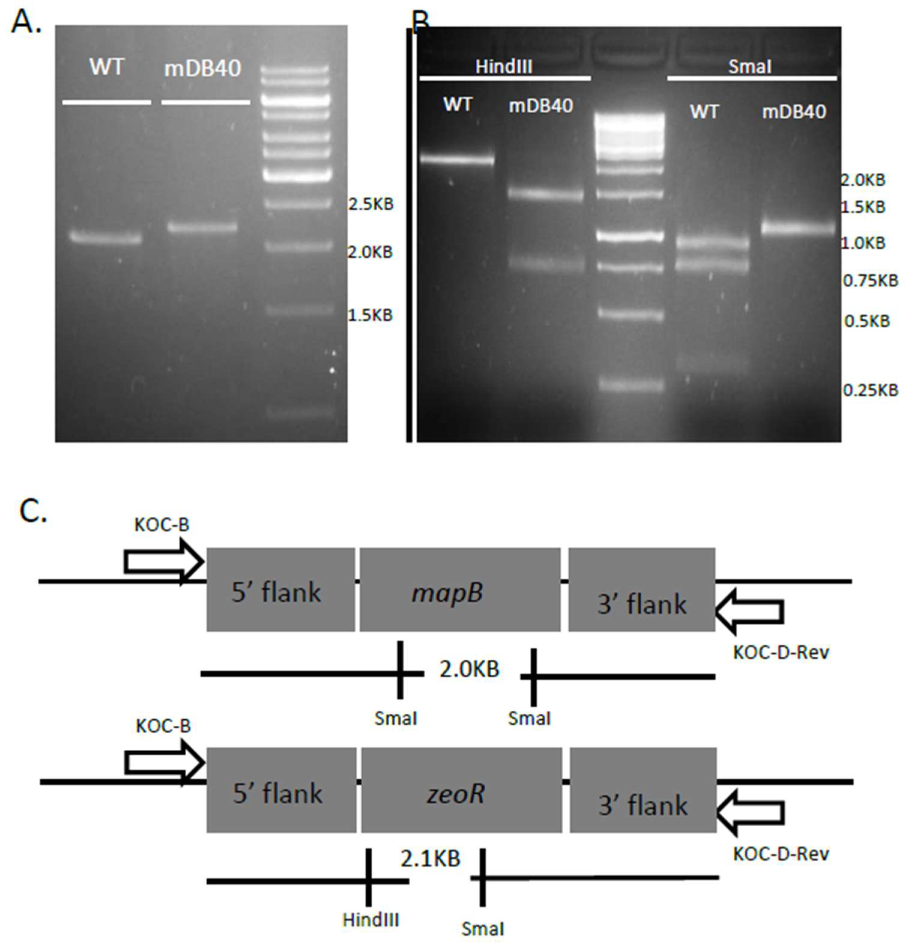
3.2. mapB (Rv 2861c, Mb2886c) is Genetically Essential for Bacterial Viability

As mapA was shown not to be essential, we opted for a similar unbiased approach towards mapB essentiality. We first pre-complemented BCG pasteur with pDB144, which is an attB integrating, kanamycin-selected plasmid, carrying a copy of mapB with its native promotor, thus creating mDB26 (BCG Pasteur attb:mapB:kan). We then proceeded to delete the native mapB gene using the phage phDB13, replacing mapB with a zeocinR cassette. After infection with the phage, bacteria were plated on 7H10 plates with zeocin, and a resistant colony was picked and analyzed in a similar manner to the mapA deletion. Because the zeocinR gene is shorter than the hygromycinR gene, the PCR product of primers flanking mapB (including 700 bases from both sides/flanks) in wt bacteria is only 100bp shorter than the product in a correct deletion mutant (Figure 3a), making a definitive proof of the deletion more difficult, and thus we sent the PCR product for sequencing. The flanking regions of mapB were present at both sides of the PCR product, however the mapB gene itself was replaced by the zeocinR gene. Also, replacement of mapB by zeocinR can be distinguished from wt by HindIII and SmaI digestions of the PCR product (Figure 3b). The correct deletion mutant was named mDB40 (ΔmapB:zeo, attb:mapB:kan). To examine if mapB was essential or not, we again attempted to remove the complementing mapB gene from the attB site by exchanging it with either one of two plasmids: an empty, hygromycin-selected pYUB412, or the same vector with wt mapB on it (pDB271). In contrast to the mapA experiment, this time we got multiple colonies only in the electroporation with mDB271 (when mapB was simply replaced by another copy), whereas electroporation with pYUB412 yielded no colonies, despite several attempts and long incubation times (Figure 4). One of the clones where mapB:kana was replaced by pDB271 (mapB:hyg) was called mDB105 (ΔmapB:zeocin; attB:mapB:hygromycin) and was used for further experiments. We also conducted a similar experiment, this time with the empty streptomycin-selected vector pDB60 and the same vector with wt mapB (pDB231). Again, whereas multiple streptomycin resistant colonies were obtained with pDB231, we did not succeed with pDB60 (Supplementary Figure S2). One of the pDB231-electroporated colonies was called mDB55 (ΔmapB:zeocin; attB:mapB:streptomycin), and was also used for further experiments.
To confirm that mapB expression was indeed essential, an additional approach was undertaken by using the newly created mDB55 (ΔmapB:zeo, attb:mapB:strep): we constructed a single-copy, kanamycin-selected, episomal plasmid (pDB261), expressing mapB. The origin of replication of this plasmid is the MF1 origin, which would be rapidly lost when its presence is not selected by an existing/included antibiotic or another essential gene [15]. The successful introduction of pDB261 into mDB55 was confirmed by PCR and the mutant was called mDB103 (ΔmapB:zeo, attb:mapB:strep, MF1:mapB:kana). Then, the mapB cassette at the attb site was exchanged with either an empty hygromycinR cassette (pYUB412), creating mDB108 (ΔmapB:zeo, attb:hyg, MF1:mapB:kana), or a similar cassette designated pDB271 containing another mapB copy, creating mDB107 (ΔmapB:zeo, attb:mapB:hyg, MF1:mapB:kana). This approach allowed us to test whether the bacterium can lose its copy of mapB if it does not have an additional copy. We grew both mDB107 and mDB108 for 12 doubling times without kanamycin with the option to lose the pDB261 plasmid in case mapB is not essential. We then plated for and examined single colonies for kanamycin resistance. As expected, ~75% of the mDB107 colonies [attb:hyg:mapB] lost their kanamycin resistance (23 colonies grew on kana + hyg, as opposed to 98 colonies on hygromycin alone) as there was no selection pressure for the retention of pDB261. In contrast, all of mDB108 [attb:hyg] mutants retained pDB261 with its kanamycin resistance (86 colonies on kana + hyg and 90 colonies on hygromycin alone), as the pDB261 plasmid was their sole source of mapB expression and MapB activity.
These results indicate that in contrast to the previous chemical inhibition report and in agreement with the transposon-mutant library analysis, mapB, and not mapA, is the essential MetAP gene in M. bovis BCG (and probably in all M. tubercu losis-Complex bacteria).

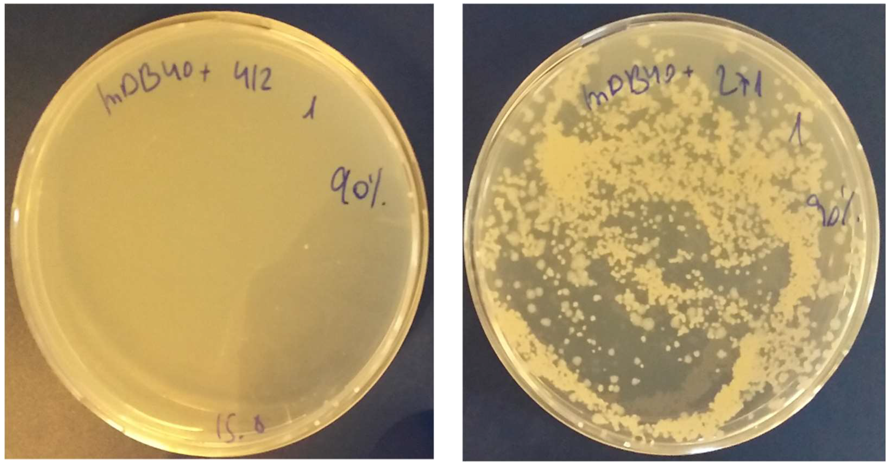
3.3. Creation of a Double Map Mutant

To further explore the role of the two map genes and to clarify whether they can compensate for each other, we decided to create a double mutant ΔmapA ΔmapB. Because the complete ΔmapB mutant is not viable, we started off with the mDB78 mutant, described earlier in this report (ΔmapA:loxP-hygromycinR-loxP; attb:zeocinR:mapA). We first "cured" mDB78 of the hygromycinR gene by introducing a kanamycin-selected, MF1-origin plasmid named pMSG381 which has the cre-recombinase on it [16]. After the loss of pMSG381 (MF1-origin plasmids are rapidly lost without the antibiotic selection pressure), the hygromycinR-cured mutant was named mDB122 (ΔmapA:loxP; attb:zeocinR:mapA). We then changed the attb cassette for an additional copy of mapB (pDB271), creating the mDB128 strain (ΔmapA:loxP; attb:hygromycinR:mapB), and used phDB13 to delete mapB from this mutant, creating mDB130 (ΔmapA:loxP; ΔmapB:zeocinR; attb:hygromycinR:mapB). The deletion of the native mapB was confirmed using the same methods as the single-deletion mapB mutant (mDB40).

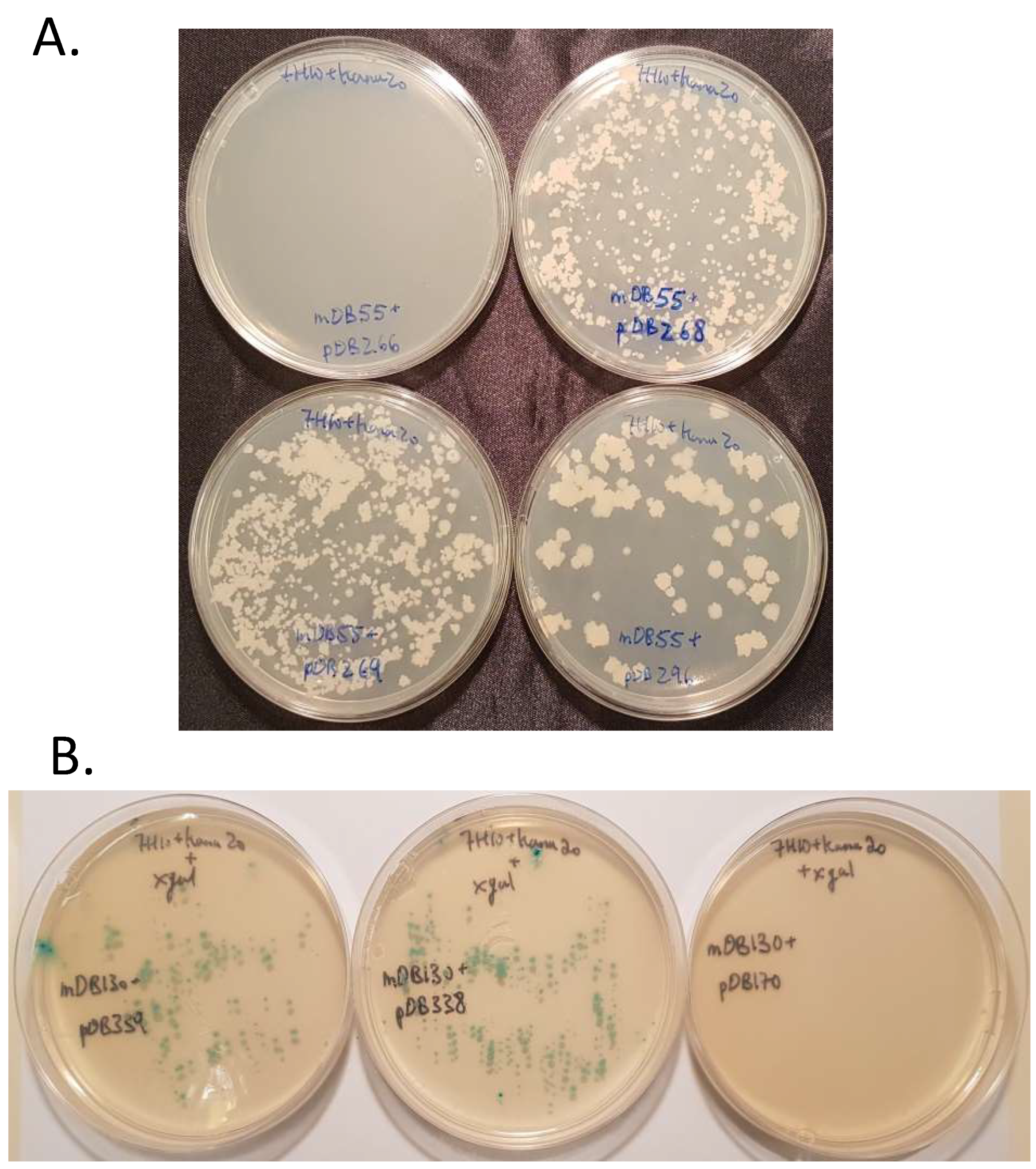
3.4. Complementation of ΔmapB Mutants with mapB with Reduced Enzymatic Activity

As we confirmed mapB is essential for M. bovis growth, we wanted to examine whether knockout mutants are rescued by expression of MAP with modified activity. It was previously reported that MapBV18G and MapBW255L were highly defective in their methionine aminopeptidase activity as the substitution W255L affects the catalytic pocket, whereas V18G affects the N-terminus extension of the protein [17]. We first designed these two mutation sequences separately and also a mapB sequence containing both point mutations: mapBV18G, mapBW255L and mapBV18G;W255L. We expressed wild-type MapB and the MapBV18G;W255L protein in E. coli, purified it and tested the activity in vitro. We found MapBV18G;W255L had approximately 8% activity of the wt protein (Figure 5 and Supplementary Table S1).
We also cloned each gene separately (mapBV18G, mapBW255L and mapBV18G;W255L) into an attp-integrating vector with kanamycin resistance. The plasmids are designated pDB268 for mapBV18G; pDB269 for mapBW255L; pDB296 for mapBV18G,W255L. We attempted to replace the wt mapB gene at the attb site of mDB55 (where it is selected by streptomycin) by each one of the three mentioned kanamycin-selected vectors. As a control, we also used an empty mapB-null vector (expecting no colonies as the gene is vital) and a vector with a wild-type mapB copy (expecting multiple kanamycin resistant colonies) and indeed got the expected results. Interestingly, mapB could easily be replaced by any of the three mutated versions, including the mapBV18G;W255L, although the recombinant mutant protein had only traces of enzymatic activity. The colonies on all plates appeared after 3–4 weeks. This suggests that these rescued strains had no substantial growth defect in comparison to the growth of control strains (Figure 6, top). Randomly-selected colonies from all three mapB point mutations were examined by mapB sequencing and the unequivocal replacement of the wt gene by the mutated one was confirmed.
To test if the bacteria could survive by means of the retained MapA activity, we performed a similar experiment with the double deletion mutant mDB130 (ΔmapA:loxP; ΔmapB:zeocin; attp:mapB:hygromycin). We attempted to replace the wt mapB gene at the attP site with the three low-activity mapB mutants, however for ease of identification of true transformants from background colonies, we used plasmids with LacZ activity (pDB338 for mapBV18G; pDB339 for mapBW255L; pDB340 for mapBV18G,W255L). Again, both mapBV18G and mapBW255L produced multiple viable colonies (Figure 6, bottom) which did not show any apparent growth defect in comparison to wt.
However, only one blue colony was detectable with the extremely low-activity MetAPB variant mapBV18G;W255L and it was designated mDB180. The colony appeared after a 12 week incubation. Preliminary analysis suggested that the clone was a true gene-replacement mutant with a mapBV18G;W255L genotype as the colony was blue when plated on Xgal, the antibiotic resistance changed from hygromycin to kanamycin and a PCR pattern was consistent with a cassette exchange (Supplementary Figure S3). However, while PCR-based sequencing the mapB gene yielded equivocal results, whole genome sequencing found the mutant to be multiploid (up to 14 copies) in regards to the lacZ, kanamycinR and mapB genes. Among the mapB gene copies, approximately half of the genes correspond to the mapBV18G,W255L genotype, while the other half of the sequences were mapBW255L, a genotype that was already shown to be fully viable. Thus, we conclude that on the background of mapA deletion, in contrast to wt background, a very-low activity MapB variant is insufficient to sustain bacterial growth. This suggested some, however not full, redundancy and mutual compensation between mapA and mapB. When mapA was present, the 8% activity of MapBV18G,W255L was sufficient for viability and normal growth, whereas bacteria without mapA could retrieve no viable colonies.

4. Discussion

As noted in the introduction, new drug targets are needed to combat tuberculosis and drug target validation is important in order to invest efforts into the development of effective drugs. Genes that code for essential enzymes are attractive targets since enzyme inhibition is a feasible drug-discovery task [18]. Therefore, correct identification of these genes/enzymes is important.
The MetAP pathway is thought to be vital in all life forms [19,20,21] and this was specifically shown in many bacteria [5,6]. The basis for the essentiality of MetAP activity is not completely clear, however it appears that the removal of the Methionine is a prerequisite for other protein modifications such as N-myristoylation (probably only existing in eukaryotes) and N-alpha acetylation [22], and affects the stability and half-life of the protein [23,24]. As a first step for drug design, it might be important to gain more clarity about the essential gene, even in a small gene family. Here we provide unambiguous genetic proof that in fact mapA is dispensable, whereas mapB is essential. Our data are supported by a transposon library analysis previously performed [9] and contradicts a chemical-inhibition assay (Olaleyle et al.) [10]. We do not have an obvious explanation as to why the carefully performed, well-designed study by Olaleye suggested other essentiality results, however this may be another example where results from chemical inhibition may differ from those obtained in genetic deletion experiments [25]. It is not excluded that MapA is indeed more robust than MapB, however the protein substrates of MapB may be more important than those of MapA (if they differ at all, a matter which is unknown). Interestingly, most microorganisms have only one map gene which is usually a MetAP1a (meaning a homologue of Mtb mapA and not mapB) [26], suggesting that mapA could be sufficient for cell viability—a suggestion refuted in M. bovis by our findings.
It is unclear why it is that mapB is essential in Mtb, considering both MapB and MapA have MetAP activity (and that of MapA was shown to be even more robust than that of MapB) [8]. There are many hypothetical explanations for the differential requirement/essentiality of the two MetAP isoforms: Interactions with other proteins, a subset of proteins only affected by MapB and not by MapA, additional as-yet unidentified roles of these enzymes or a different affinity to the ribosome [27]. It was suggested that MetAPs played an important role in methionine turnover in the cell [28]. Still, it does not explain why one isoform is essential and the other not. Also, if this is the case, then methionine supplementation could perhaps salvage a full deletion mutant—however, a previous experiment reported the opposite [28].
Also, the relation between Peptide deformylase (PDF, encoded by def, Rv0429c) and MAP genes was not investigated. PDF, which removes the formyl group from the initiating methionine before the methionine itself is removed by MAP, is considered to be essential according to the analysis of a transposon library [9] and also by chemical inhibition in eukaryotic cells [23]. Since PDF activity is an absolute prerequisite for MetAP activity, the vital importance of MetAP explains the essentiality of PDF (def). Indeed, in previous experiments, a def deletion mutant in mycobacteria could only be created on the background of an inactivating mutation in formyl tRNAfMet formyltransferase [29], making the removal of the formyl group unnecessary for the activity of MetAP.

Supplementary Materials

The following are available online at https://www.mdpi.com/2073-4409/8/5/393/s1, Figure S1: Confirmation of a mapA deletion mutant. Figure S2: Failure to completely delete mapB from the genome. Figure S3: The exchange in attb cassettes from pDB271 (mapBwt, hygR) to pDB340 (mapBV18G,W255L, lacZ + , kanamycinR) between mDB130 and mDB180. Supplementary Table S1: Plasmid table, primer table, mutant list.

Author Contributions

Performance of experiments: M.V., P.S., T.-H.R. and D.B. Conceptualization: P.K.C., B.G. and D.B. Writing the final manuscript: P.S., B.G. and D.B.

Funding

This research received no external funding.

Conflicts of Interest

The authors declare no conflict of interest.

References

  1. WHO. Global Tuberculosis Report 2016; World Health Organization: Geneva, Switzerland, 2017. [Google Scholar]
  2. Zhang, Y. The magic bullets and tuberculosis drug targets. Annu. Rev. Pharmacol. Toxicol. 2005, 45, 529–564. [Google Scholar] [CrossRef] [PubMed]
  3. Helgren, T.R.; Wangtrakuldee, P.; Staker, B.L.; Hagen, T.J. Advances in bacterial methionine aminopeptidase inhibition. Curr. Topics med. Chem. 2016, 16, 397–414. [Google Scholar] [CrossRef]
  4. Gonzales, T.; Robert-Baudouy, J. Bacterial aminopeptidases: properties and functions. FEMS Microbiol. Rev. 1996, 18, 319–344. [Google Scholar] [CrossRef]
  5. Chang, S.; McGARY, E.C.; Chang, S. Methionine aminopeptidase gene of Escherichia coli is essential for cell growth. J. Bacteriol. 1989, 171, 4071–4072. [Google Scholar] [CrossRef] [PubMed]
  6. Miller, C.G.; Kukral, A.M.; Miller, J.L.; Movva, N.R. pepM is an essential gene in Salmonella typhimurium. J. Bacteriol. 1989, 171, 5215–5217. [Google Scholar] [CrossRef] [PubMed]
  7. Addlagatta, A.; Quillin, M.L.; Omotoso, O.; Liu, J.O.; Matthews, B.W. Identification of an SH3-binding motif in a new class of methionine aminopeptidases from Mycobacterium tuberculosis suggests a mode of interaction with the ribosome. Biochemistry 2005, 44, 7166–7174. [Google Scholar] [CrossRef]
  8. Olaleye, O.; Raghunand, T.R.; Bhat, S.; He, J.; Tyagi, S.; Lamichhane, G.; Gu, P.; Zhou, J.; Zhang, Y.; Grosset, J.; et al. Methionine aminopeptidases from Mycobacterium tuberculosis as novel antimycobacterial targets. Chem. Bio. 2010, 17, 86–97. [Google Scholar] [CrossRef]
  9. Griffin, J.E.; Gawronski, J.D.; Dejesus, M.A.; Ioerger, T.R.; Akerley, B.J.; Sassetti, C.M. High-resolution phenotypic profiling defines genes essential for mycobacterial growth and cholesterol catabolism. PLoS Pathog. 2011, 7, e1002251. [Google Scholar] [CrossRef]
  10. Olaleye, O.; Raghunand, T.R.; Bhat, S.; Chong, C.; Gu, P.; Zhou, J.; Zhang, Y.; Bishai, W.R.; Liu, J.O. Characterization of clioquinol and analogues as novel inhibitors of methionine aminopeptidases from Mycobacterium tuberculosis. Tuberculosis 2011, 91, S61–S65. [Google Scholar] [CrossRef]
  11. Otero, J.; Jacobs, W.R.; Glickman, M.S. Efficient allelic exchange and transposon mutagenesis in Mycobacterium avium by specialized transduction. Appl. Environ. Microbiol. 2003, 69, 5039–5044. [Google Scholar] [CrossRef]
  12. Ehrt, S.; Guo, X.V.; Hickey, C.M.; Ryou, M.; Monteleone, M.; Riley, L.W.; Schnappinger, D. Controlling gene expression in mycobacteria with anhydrotetracycline and Tet repressor. Nucleic Acids Res. 2005, 33, e21. [Google Scholar] [CrossRef]
  13. Barkan, D.; Rao, V.; Sukenick, G.D.; Glickman, M.S. Redundant function of cmaA2 and mmaA2 in Mycobacterium tuberculosis cis cyclopropanation of oxygenated mycolates. J. Bacteriol. 2010, 192, 3661–3668. [Google Scholar] [CrossRef] [PubMed]
  14. Pashley, C.A.; Parish, T. Efficient switching of mycobacteriophage L5-based integrating plasmids in Mycobacterium tuberculosis. FEMS Microbiol. Lett. 2003, 229, 211–215. [Google Scholar] [CrossRef]
  15. Gill, W.P.; Harik, N.S.; Whiddon, M.R.; Liao, R.P.; Mittler, J.E.; Sherman, D.R. A replication clock for Mycobacterium tuberculosis. Nat. Med. 2009, 15, 211. [Google Scholar] [CrossRef]
  16. Barkan, D.; Hedhli, D.; Yan, H.G.; Huygen, K.; Glickman, M.S. Mycobacterium tuberculosis lacking all mycolic acid cyclopropanation is viable but highly attenuated and hyperinflammatory in mice. Infect. Immun. 2012, 80, 1958–1968. [Google Scholar] [CrossRef] [PubMed]
  17. Kanudia, P.; Mittal, M.; Kumaran, S.; Chakraborti, P.K. Amino-terminal extension present in the methionine aminopeptidase type 1c of Mycobacterium tuberculosis is indispensible for its activity. BMC biochem. 2011, 12, 35. [Google Scholar] [CrossRef] [PubMed]
  18. Smith, C.V.; Sharma, V.; Sacchettini, J.C. TB drug discovery: addressing issues of persistence and resistance. Tuberculosis 2004, 84, 45–55. [Google Scholar] [CrossRef]
  19. Boxem, M.; Tsai, C.W.; Zhang, Y.; Saito, R.M.; Liu, J.O. The C. elegans methionine aminopeptidase 2 analog map-2 is required for germ cell proliferation. FEBS Lett. 2004, 576, 245–250. [Google Scholar] [CrossRef] [PubMed]
  20. Sin, N.; Meng, L.; Wang, M.Q.; Wen, J.J.; Bornmann, W.G.; Crews, C.M. The anti-angiogenic agent fumagillin covalently binds and inhibits the methionine aminopeptidase, MetAP-2. Proc. Natl. Acad. Sci. USA 1997, 94, 6099–6103. [Google Scholar] [CrossRef] [Green Version]
  21. Wang, J.; Lou, P.; Henkin, J. Selective inhibition of endothelial cell proliferation by fumagillin is not due to differential expression of methionine aminopeptidases. J. Cell. Biochem. 2000, 77, 465–473. [Google Scholar] [CrossRef]
  22. Giglione, C.; Fieulaine, S.; Meinnel, T. N-terminal protein modifications: Bringing back into play the ribosome. Biochimie 2015, 114, 134–146. [Google Scholar] [CrossRef] [PubMed]
  23. Giglione, C.; Vallon, O.; Meinnel, T. Control of protein life-span by N-terminal methionine excision. EMBO J. 2003, 22, 13–23. [Google Scholar] [CrossRef] [PubMed] [Green Version]
  24. Meinnel, T.; Serero, A.; Giglione, C. Impact of the N-terminal amino acid on targeted protein degradation. Bio. Chem. 2006, 387, 839–851. [Google Scholar] [CrossRef] [PubMed]
  25. Knight, Z.A.; Shokat, K.M. Chemical genetics: where genetics and pharmacology meet. Cell 2007, 128, 425–430. [Google Scholar] [CrossRef]
  26. Reddi, R.; Arya, T.; Kishor, C.; Gumpena, R.; Ganji, R.J.; Bhukya, S.; Addlagatta, A. Selective targeting of the conserved active site cysteine of Mycobacterium tuberculosis methionine aminopeptidase with electrophilic reagents. FEBS J. 2014, 281, 4240–4248. [Google Scholar] [CrossRef] [Green Version]
  27. Sandikci, A.; Gloge, F.; Martinez, M.; Mayer, M.P.; Wade, R.; Bukau, B.; Kramer, G. Dynamic enzyme docking to the ribosome coordinates N-terminal processing with polypeptide folding. Nat. Struct. Mol. Biol. 2013, 20, 843. [Google Scholar] [CrossRef]
  28. Vetro, J.A.; Dummitt, B.; Chang, Y.-H. Methionine Aminopeptidase. In Aminopeptidases in Biology and Disease; Springer: New York, NY, USA, 2004; pp. 17–44. [Google Scholar]
  29. Teo, J.W.; Thayalan, P.; Beer, D.; Yap, A.S.; Nanjundappa, M.; Ngew, X.; Duraiswamy, J.; Liung, S.; Dartois, V.; Schreiber, M.; et al. Peptide deformylase inhibitors as potent antimycobacterial agents. Antimicrob. Agents Chemother. 2006, 50, 3665–3673. [Google Scholar] [CrossRef]
Figure 1. (A) Creation and confirmation of a mapA deletion mutant. After infection with phDB20, six hygromycin resistant colonies were analyzed by PCR. All six produced a pattern consistent with mapA deletion and colony four was named mDB59. (B,C) mDB59 is not dependent on ATc for growth. (B) mDB59 was grown with (leftmost tube) and without (middle tube) ATc (inducing the expression of mapA) in liquid. The rightmost tube is clear 7H9 media. (C) Left–with ATc, Right–no ATc. No difference in growth is apparent.
Cells 08 00393 g001
Figure 2. Complete mapA deletion mutant. (A) Electroporation of mDB59 with either pDB19 or pDB249 (exchanging mapA-Kan for null-zeocin or mapA-zeocin, respectively) both yielded multiple colonies. (B) Two random colonies from each electroporation were analyzed by PCR. All four colonies became zeo-R positive (left panel, zeo-R product is 0.35KB), confirming cassette exchange, however only mDB59 + pDB249 remains positive for mapA (Right, expected PCR product of 0.5KB). Both mDB59 + pDB19 colonies are complete mapA deletion mutants. (C) A complete mapA deletion mutant (mDB77) has a mild growth defect in 7H9 media during the exponential phase of growth as compared to mDB78, however it reaches the same final OD (done in duplicates, one of two similar experiments is shown).
Cells 08 00393 g002
Figure 3. Deletion of the native mapB. (A) A zeocin resistant colony (mDB40) was analyzed by PCR. In wt, the product size is 2KB, whereas in a correct deletion mutant it is 2.1KB. (B) The digestion pattern produced by wt or mDB40 when digested by HindIII (no effect on wt, 1.3, 0.7KB in deletion) or SmaI (0.9, 0.7, 0.3KB in wt, 1.05, 1.05 in deletion mutant) confirms the correct allelic exchange in mDB40 (see diagram in (C)).
Cells 08 00393 g003
Figure 4. mapB is essential in BCG. mDB40 [pasteur ΔmapB:zeo, attb:kana:mapB] was electroporated by either pYUB412 (attb:hyg, left) or pDB271 (attb:hyg:mapB, right). Multiple colonies arise with pDB271 where mapB is replaced by another copy of the same gene. However, pYUB412 would have yielded a null-mapB mutant. No colonies arise on that plate.
Cells 08 00393 g004
Figure 5. Enzymatic activity of recombinant M. bovis MetAP1c (MapB) and MetAP1cV18G;W225L mutant. (A) Heterologous expressed and purified wt and mutant proteins in E. coli cells displayed on a 12 % SDS-PAGE. (B) The enzymatic activity of MetAP1cWT and MetAP1cV18G;W225L proteins was assayed with 0,2 µM protein in 50 mM Tris-HCL (pH 7.5) buffer containing 400 µM fluorogenic Met-MCA substrate and 1.5 mM MgCL2.
Cells 08 00393 g005
Figure 6. (A) when MapA is present, MapB can be replaced with MapBV18G (pDB268, top right), MapBW255L (pDB269, bottom left) or MapBV18G;W255L (pDB296, bottom right), however not completely removed (pDB266, empty vector. Top left). (B) On the background of mapA deletion, MapB can still be replaced by MapBV18G (pDB338, middle plate), MapBW255L (pDB339, left plate) and not an empty vector (pDB170, right plate). All three plasmids (pDB170, 338, 339) also carry a lacZ gene for additional visual selection on top of kanamycin resistance.
Cells 08 00393 g006

