Review

Modulation of Receptor Tyrosine Kinase Activity through Alternative Splicing of Ligands and Receptors in the VEGF-A/VEGFR Axis

Institute of Biomedical & Clinical Sciences, Medical School, College of Medicine and Health, University of Exeter, Exeter EX1 2LU, UK
*
Authors to whom correspondence should be addressed.
Cells 2019, 8(4), 288; https://doi.org/10.3390/cells8040288
Submission received: 7 March 2019 / Revised: 19 March 2019 / Accepted: 22 March 2019 / Published: 28 March 2019
(This article belongs to the Special Issue Receptor Tyrosine Kinases in Health and Disease)

Abstract

:
Vascular endothelial growth factor A (VEGF-A) signaling is essential for physiological and pathological angiogenesis. Alternative splicing of the VEGF-A pre-mRNA gives rise to a pro-angiogenic family of isoforms with a differing number of amino acids (VEGF-Axxxa), as well as a family of isoforms with anti-angiogenic properties (VEGF-Axxxb). The biological functions of VEGF-A proteins are mediated by a family of cognate protein tyrosine kinase receptors, known as the VEGF receptors (VEGFRs). VEGF-A binds to both VEGFR-1, largely suggested to function as a decoy receptor, and VEGFR-2, the predominant signaling receptor. Both VEGFR-1 and VEGFR-2 can also be alternatively spliced to generate soluble isoforms (sVEGFR-1/sVEGFR-2). The disruption of the splicing of just one of these genes can result in changes to the entire VEGF-A/VEGFR signaling axis, such as the increase in VEGF-A165a relative to VEGF-A165b resulting in increased VEGFR-2 signaling and aberrant angiogenesis in cancer. Research into this signaling axis has recently focused on manipulating the splicing of these genes as a potential therapeutic avenue in disease. Therefore, further research into understanding the mechanisms by which the splicing of VEGF-A/VEGFR-1/VEGFR-2 is regulated will help in the development of drugs aimed at manipulating splicing or inhibiting specific splice isoforms in a therapeutic manner.

1. Introduction

Angiogenesis comprises the formation and maintenance of blood vessels. A variety of signaling molecules are involved in the regulation of angiogenesis, including vascular endothelial growth factor (VEGF), which is essential both for physiological and pathological angiogenesis [1]. The biological functions of VEGF proteins are mediated by a family of cognate protein tyrosine kinase receptors, known as the VEGF receptors (VEGFRs) [2]. Activation of the VEGF pathway has been implicated in a large number of disease processes ranging from cancer to autoimmunity.
There are several VEGF proteins; VEGF-A binds to and signals through VEGFR-1 (Flt-1) and VEGFR-2 (KDR/Flk-1), VEGF-B signals solely through VEGFR-1, and VEGF-C and VEGF-D have a high affinity to VEGFR-3 (Flt-4) [1,2]. In addition, there are two neuropilin receptors, which are transmembrane glycoproteins, that function in the VEGF-VEGFR axis [2]; neuropilin-1 (NRP-1), a non-kinase co-receptor for VEGFR-2, functions to enhance the binding and signaling of certain isoforms of VEGF-A. NRP-2, on the other hand, is a non-kinase co-receptor for VEGFR-3. Since VEGFR-1 and VEGFR-2 are the receptor tyrosine kinases specific for VEGF-A, this review will focus on the splice variants of these two receptors only.

2. VEGFR Splice Variants and Functions

VEGF-A binds to two tyrosine kinase VEGFRs, VEGFR-1 and VEGFR-2. There are several isoforms of these VEGFRs that arise as a result of alternative splicing of the VEGFR pre-mRNA, which can alter the protein function, as detailed below (Figure 1). Both VEGFR-1 and VEGFR-2 have seven extracellular immunoglobulin (Ig)-like domains, which consist of a tetramer of two light chains and two heavy chains linked by disulphide bonds, a single transmembrane region, and an intracellular tyrosine kinase sequence interrupted by a kinase insert domain [3]. VEGF-A binds to the extracellular domain and the kinase insert domain acts as a binding site for intracellular proteins to carry out specific signaling cascades in response to ligand binding.

2.1. VEGFR-1 Signaling

VEGFR-1 was the first receptor tyrosine kinase for VEGF-A to be identified in COS cells [4] and has since been reported to be widely expressed on many cell types; however, it has very poor tyrosine kinase activity and is not required for endothelial cell function [5]. VEGFR-1 binds VEGF-A with high affinity but there is conflicting evidence for the role of VEGFR-1 as it appears to signal differently depending on the cell type and stage of development [5]. VEGFR-1 gene expression is regulated by hypoxia in human umblical endothelial cells; the VEGFR-1 promoter contains a binding site for hypoxia inducible factor (HIF)-1α [6]. Relatively little is known about the function of VEGFR-1. Constitutive knock-out (KO) of VEGFR-1 results in embryonic lethality between embryonic days 8.5 and 9 [7]. This was later found to be the result of increased endothelial cell outgrowth and angioblast commitment, which prevented proper organization of the vascular network [8]. Previous reports have labelled VEGFR-1 as a decoy receptor, decreasing the amount of VEGF-A readily available to bind to and phosphorylate VEGFR-2 [9]. Further evidence for this is that deletion of just the intracellular kinase domain for VEGFR-1 resulted in normal vascular development in mice [9]. Therefore, VEGFR-1 is hypothesized to sequester VEGF-A, preventing it from binding to its functional receptor, VEGFR-2.

2.2. Function of sVEGFR-1

The VEGFR-1 pre-mRNA can be alternatively spliced to produce the full-length membrane-spanning receptor described above, or the truncated soluble VEGFR-1 (sVEGFR-1), which includes the seven N-terminal immunoglobulin-like extracellular domains but not the transmembrane spanning or intracellular kinase domains, thus has a specific 31-amino acid c-terminus [1]. Full-length VEGFR-1 mRNA consists of 30 exons, whereas sVEGFR-1 only contains the first 13–14 exons due to intron retention and usage of an alternative polyadenylation signal and stop codon (isoforms detailed below). sVEGFR-1 is suggested to form non-signaling complexes with VEGFR-2, thus functioning as a modulator of VEGF-A signaling [10]. Like full length VEGFR-1, sVEGFR-1 has also been shown to act as a decoy receptor; VEGFR-1 KO mice die from vasuclar overgrowth due to increased signaling of VEGF-A through VEGFR-2; however, the administration of sVEGFR-1 to VEGFR-1 KO mice partially rescues this phenotype as it reduces the levels of VEGFR-2 phosphorylation [11].
There are currently five known VEGFR-1 protein coding isoforms (reviewed in [12]) (Figure 1A). Isoform 1 is denoted by the full-length VEGFR-1. Isoform 2 is termed sVEGFR-1, which comprises the 656 N-terminal residues followed by a specific 30-amino acid C-terminus and appears to have ubiquitous expression throughout most tissues [12]. Isoform 3 is a second soluble form generated by alternative splicing downstream of exon 14, termed sVEGFR-1_i14, which has been predominantly detected in the testes and brain [12]. Isoforms 4 and 5 result from the use of a new terminal exon, termed exon 15a and 15b, which is derived from an intronic sequence. These isoforms have been found to be highly expressed in the placenta [12]. Alternative splicing of VEGFR-1 involves cis-regulatory elements in the VEGFR-1 pre-mRNA within intron 13 [13]. Hypoxia is reported to increase the expression of transmembrane VEGFR-1 [6]; however, the effect of hypoxia on sVEGFR-1 expression is not so clear. In endothelial cells, hypoxia was shown to downregulate the expression of sVEGFR-1, which was not directly attributable to HIF-1α [14]. In contrast, exposure of macrophages/monocytes to granulocyte-macrophage colony-stimulating factor (GM-CSF) under hypoxic conditions results in HIF-2α-dependent changes in sVEGFR-1 expression [15]. In cytotrophoblasts, where the sVEGFR-1_i14 isoform is most commonly expressed, hypoxia increases both sVEGFR-1_i14 and sVEGFR-1 mRNA, which is proposed to be through HIF-1α [16]. Furthermore, sVEGFR-1_i14 secretion was shown to increase under hypoxic conditions through activation of the growth arrest and DNA damage-inducible 45a (Gadd45a) factor and p38 phosphorylation [17]. Several drugs and protein factors have been shown to modulate sVEGFR-1 expression, including Jumonji domain-containing protein 6, which interacts with the splice factor U2AF65 resulting in augmented levels of sVEGFR-1 in hypoxic conditions [18]. In addition, hnRNP D and arginine methylation have also been reported to play important roles in the regulation of sVEGFR-1 mRNA alternative polyadenylation [19]. Interestingly, VEGF-A can increase the expression of sVEGFR-1 through VEGFR-2-dependent activation of protein kinase C [20].

2.3. VEGFR-2 Signaling

VEGFR-2 is the main signaling receptor for VEGF-A. It is primarily located on endothelial cells and is essential for endothelial cell biology both during development and during physiological and pathological processes in adults. Like VEGFR-1, all VEGF-A isoforms contain residues that enable them to bind to VEGFR-2 and all bind with the same affinity. However, the affinity of VEGF-A for VEGFR-2 is 10-fold lower than that for VEGFR-1 [21,22]. A constitutive KO of VEGFR-2 results in embryonic lethality on day 8.5–9.5; mice lack mature endothelial and hematopoietic cells [23]. This is similar to the phenotype observed in VEGF-A KO mice [24]. Therefore, unlike VEGFR-1, VEGFR-2 signaling is crucial for vascular development.
Proteolytic hydrolysis of membrane-bound VEGFR-2 results in the generation of soluble VEGFR-2 (sVEGFR-2) [12]. sVEGFR-2 is proposed to function as an inhibitor of angiogenesis by binding to and sequestering VEGF-A, blocking canonical VEGF-A-VEGFR-2 signaling [25,26]. A further sVEGFR-2 isoform generated by intron 13 retention has been described; as with VEGFR-1, retention of intron 13 yields a truncated transcript whose protein variant lacks the transmembrane and intracellular kinase domain of full length VEGFR-2 [27] (Figure 1B). This splice variant is reported to play a role in lymphangiogenesis by blocking VEGF-C [27]. Little is known regarding the mechanisms controlling this alternative splicing event.

3. VEGF-A Splice Variants

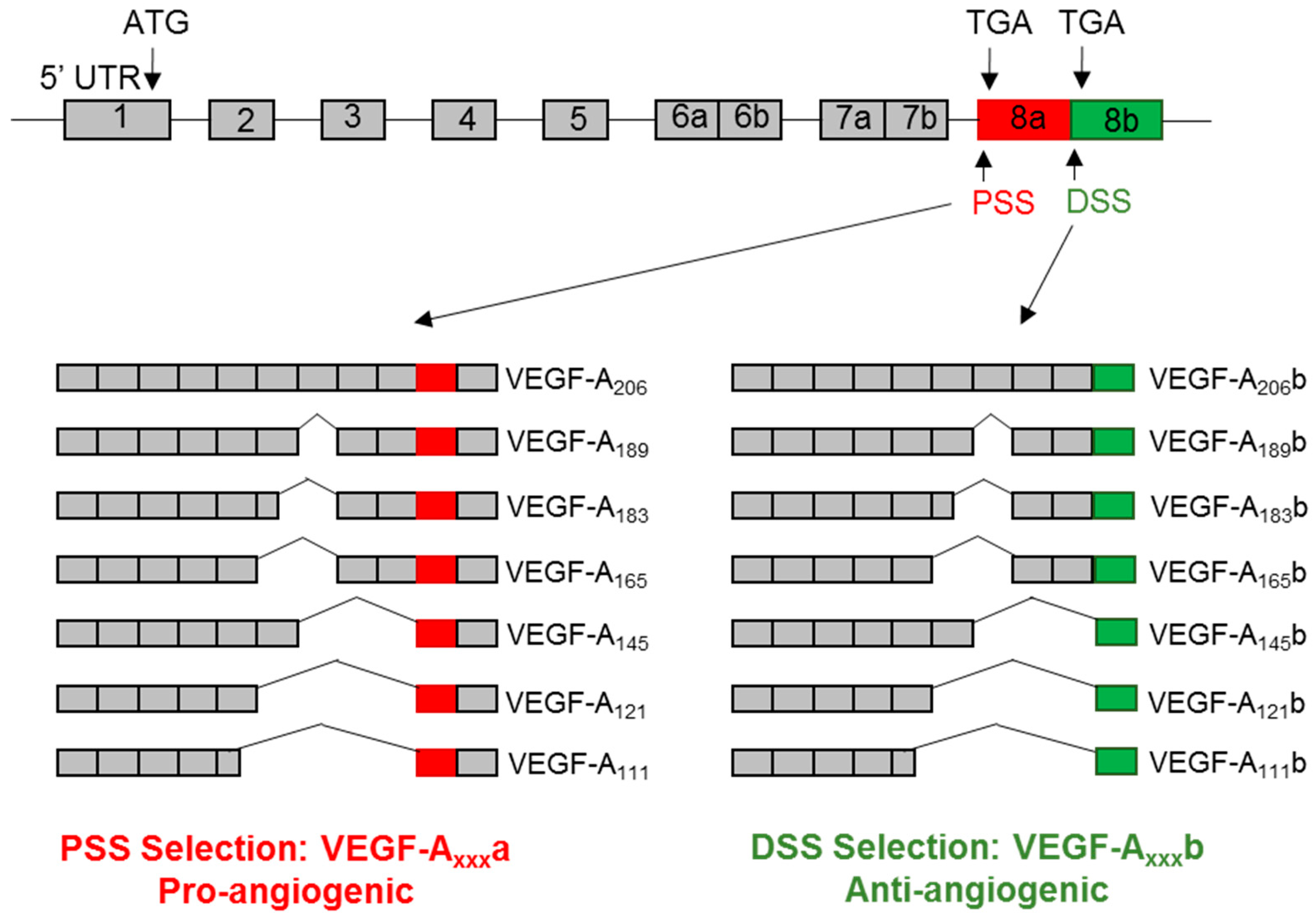
The human VEGF-A pre-mRNA consists of eight exons and seven introns. Alternative splicing of the VEGF-A pre-mRNA gives rise to a family of isoforms with a differing number of amino acids due to the exclusion/inclusion of various exons (e.g., VEGF-A121, VEGF-A165, VEGF-A189, and VEGF-A206, collectively known as VEGF-Axxxa where xxx denotes the number of amino acids) (Figure 2). Such isoforms are widely known to be pro-angiogenic, pro-permeability factors. In addition, the selection of an alternative 3′ splice site, known as the distal splice site, in exon 8 of the VEGF-A pre-mRNA results in a new family of VEGF-A isoforms, termed VEGF-Axxxb [28]. The resulting VEGF-Axxxb proteins differ in the C-terminal sequence by only six amino acids, resulting in radically different functional properties (Figure 2). In comparison to VEGF-Axxx, VEGF-Axxxb isoforms are collectively anti-angiogenic and reduce vessel permeability (anti-permeability). Sixteen isoforms of VEGF-A have been identified, including an additional isoform, VEGF-Ax, which arises from translational read-through of the VEGF-A transcript beyond the canonical stop codon (programmed translational read-through) [29].
VEGF-A splicing is predominantly regulated by a group of RNA binding proteins known as serine/arginine (SR) proteins. SRSF1, SRSF2, SRSF5, and SRSF6 have all been reported to play a role in VEGF-A alternative splicing [30]. Upon phosphorylation of multiple serine/arginine and proline/serine repeats, SR proteins are translocated from the cytoplasm to the nucleus where they bind to exonic sequence enhancers within the VEGF-A pre-mRNA, resulting in the splicing out of an exon [31]. The inclusion/exclusion of certain exons result in the different isoform properties of each VEGF-A protein. Exons 1–5 are constitutive exons; they encode a single sequence (exons 1/2), a glycosylation site (Asp74), a potential plasmin cleavage site (Arg110 and Ala111), as well as VEGFR binding residues [32,33]. Whereas exons 1–5 are present in all isoforms of VEGF-A, exons 6 and 7 are alternatively spliced. Heparin sulfate (HS) glycoproteins are present in the extracellular matrix (ECM) and can interact with both VEGF-A and VEGFRs, thus they are suggested to regulate the bioavailability of VEGF-A. Residues in exon 6a and 7 of VEGF-A are responsible for the interaction with HS [34]. VEGF-A145, VEGF-A189, and VEGF-A206 all contain exon 6a and 7 resulting in a high affinity for HS; this results in these longer isoforms being tethered to the ECM. On the other hand, VEGF-A111 and VEGF-A121 lack exon 6 and 7, so they are unable to bind HS, making them freely diffusible in the ECM and more bioavailable [35]. The most dominant isoform is VEGF-A165, which contains exon 7 but not 6. Therefore, VEGF-A165 has an intermediate bioavailability as approximately 50% remains cell- or ECM-bound [36].
Regarding exon 8 of the VEGF-A gene, selection of either the proximal or distal splice site has been reported to be dependent on the type of external stimulus; proximal splice site selection is promoted by insulin like growth factor (IGF1) and tumor necrosis factor alpha (TNFα), whereas distal splice site selection is promoted by tumor growth factor beta 1 (TGF-B1) [37]. A widely reported example of exon 8 splicing regulation involves serine/threonine-protien kinase 1 (SRPK1) and CDC-like kinase 1 (Clk-1). SRPK1 activation has been shown to phosphorylate SRSF1, resulting in proximal splice site selection and the translation of VEGF-Axxxa proteins [38]. On the other hand, Clk-1 signaling results in the phosphorylation of SRSF6, with the distal splice site being subsequently selected and VEGF-Axxxb proteins translated [37]. Other reported regulators of VEGF-A exon 8 splicing are E2F1 and SRSF2, which were both shown to increase the VEGF-Axxxb/VEGF-Axxxa ratio [39].

4. VEGFR Signaling

4.1. Role of VEGFR-1 Signlaing and sVEGFR-1 Isoforms

As mentioned previously, the role of VEGFR-1 in vasculogenesis and angiogenesis has been ascribed to VEGF-A binding, thus regulating the amount of VEGF-A available for vascular development. VEGFR-1 is widely expressed but has poor kinase activity and is not required for endothelial cell function. Further evidence for this hypothesis arose from mice with a homozygous deletion of the VEGFR-1 tyrosine kinase domain developing healthy vasculature [9]. Therefore, the primary role of VEGFR-1 in embryonic angiogenesis is restricted to its extracellular region and is independent of its tyrosine kinase activity. As sVEGFR-1 contains the extracellular domain, it also acts as a decoy receptor [40]. sVEGFR-1 is also proposed to form non-signaling complexes with VEGFR-2 [10].
A study using VEGFR-1 KO embryonic stem cells showed that sVEGFR-1 is important for the modulation of endothelial cell migration and vascular sprouting during development [41]. During vessel morphogenesis, endothelial cells are suggested to form a VEGF-A gradient via the interaction of VEGF-A with sVEGFR-1, resulting in sequestration of VEGF-A and local inactivation of VEGFR-2 signaling [42]. Therefore, sVEGFR-1 is proposed to act as a guidance molecule during vessel sprouting, i.e., inactivating VEGF-A either side of the sprout to provide a VEGF-A-rich corridor for the emerging vessel [43]. sVEGFR-1 present in the ECM is also reported to play a role in α5β1 integrin signaling regarding the cell adhesion pathway [44]; however, these signaling pathways are not related to VEGF-A and are beyond the scope of this review.
Recent studies have highlighted that VEGF-B and PIGF are able to signal through VEGFR-1, eliciting a pro-angiogenic effect independent of VEGF-A [45,46]. In addition, increased levels of sVEGFR-1 have been observed in vascular pathologies [45], indicating that VEGFR-1 may act as more than a decoy receptor/VEGFR-2 inhibitor.
The role of sVEGFR-1 in tumor development and progression has been widely reported. The expression of sVEGFR-1 has been found to be increased in many types of cancer, including glioblastoma, melanoma, breast, hepatocellular, lung, leukemia, colorectal, renal, and head and neck [47,48,49,50,51,52,53,54,55]. Increased circulating sVEGFR-1 is often correlated with poor prognosis; however, the balance between VEGF-A and sVEGFR-1 may be more important when considering the clinical outcome. For example, increased sVEGFR-1 and VEGF-A are correlated with poor prognosis in lung cancer patients [51]. On the other hand, increased VEGF-A combined with low levels of sVEGFR-1 are associated with a poor prognosis in breast cancer [56]. In addition to being a marker for tumor progression, sVEGFR-1 has also been shown to serve as a biomarker for tumor response to therapy. Using the example of bevacizumab, increased plasma levels of sVEGFR1 were reported to be inversely correlated with treatment response in breast cancer [57]. However, this appears to be dependent on the type of cancer as the sVEGFR-1 expression level was found to be decreased upon treatment of metastatic colorectal cancer [58].
Excess circulating soluble isoforms of VEGFR-1 have been shown to contribute to the pathogenesis of pre-eclampsia in pregnant women [59,60]. The sVEGFR-1_i14 isoform is presumed to be a major contributor to this condition because it is selectively expressed by placental cytotrophoblasts; the increased sequestration of platelet-derived growth factor (PIGF) and VEGF-A by excess sVEGFR-1_i14 results in endothelial dysfunction and altered neutrophil activation and migration, ultimately causing hypertension, proteinuria, and glomerular endotheliosis in patients [60,61]. Indeed, increased levels of circulating sVEGFR-1_i14 is used as a biomarker for the development of pre-eclampsia [62].
As described above in pregnant women with pre-eclampsia, increased circulating levels of sVEGFR-1 is linked to endothelial dysfunction in the glomeruli of the kidney. VEGF-A is secreted by the glomerular epithelial cells (podocytes) to signal to VEGFR-2 on the glomerular endothelial cells, a process that is tightly regulated to maintain proper functioning of the glomerular filtration barrier. Plasma levels of sVEGFR-1 are higher in patients with chronic kidney disease (CKD), which are correlated with cardiovascular disease [63,64]. On the other hand, inducible over-expression of podocyte sVEGFR-1 has been shown to be therapeutic in a model of diabetic nephropathy where excess VEGF-A expression is observed [65]. In addition, sVEGFR-1 has been reported to bind to lipid microdomains in podocytes, which can alter cell morphology and the function of the glomerular filtration barrier [66].
sVEGFR-1 has also been shown to play a role in ocular pathologies through the inhibition of VEGF-A, including the preservation of cornea avascularity [67]. In addition, reduced levels of sVEGFR-1 where observed in patients with age-related macular degeneration [68]. Regarding inflammation, increased levels of sVEGFR-1 in the blood is indicated to act as a potential new biomarker of sepsis [69], and a predictor of endothelial dysfunction/activation of coagulation in acute pancreatitis [70].
On the other hand, in mouse xenograft models of melanoma, lung cancer, fibrosarcoma, and glioblastoma, exogenous administration of sVEGFR-1 (either transfection, recombinant protein, or adenovirus infection) inhibited tumor growth and neoangiogenesis, increasing the survival rate [71,72,73,74].

4.2. VEGF-Axxxb Activation of VEGFR-1

Information on VEGFR-1 activation and signaling is sparse; however, a recent study has shown that VEGF-A165b inhibits VEGFR-1 signaling in ischemic muscle in mice, and that VEGF-A165b inhibition induces activation of VEGFR-1 [75]. Furthermore, in vitro studies showed that VEGF-A165b failed to induce the activation of VEGFR-1-Y1333, reducing VEGFR-1-STAT3 signaling [75].

4.3. Mehcanisms of VEGFR-2 Signaling

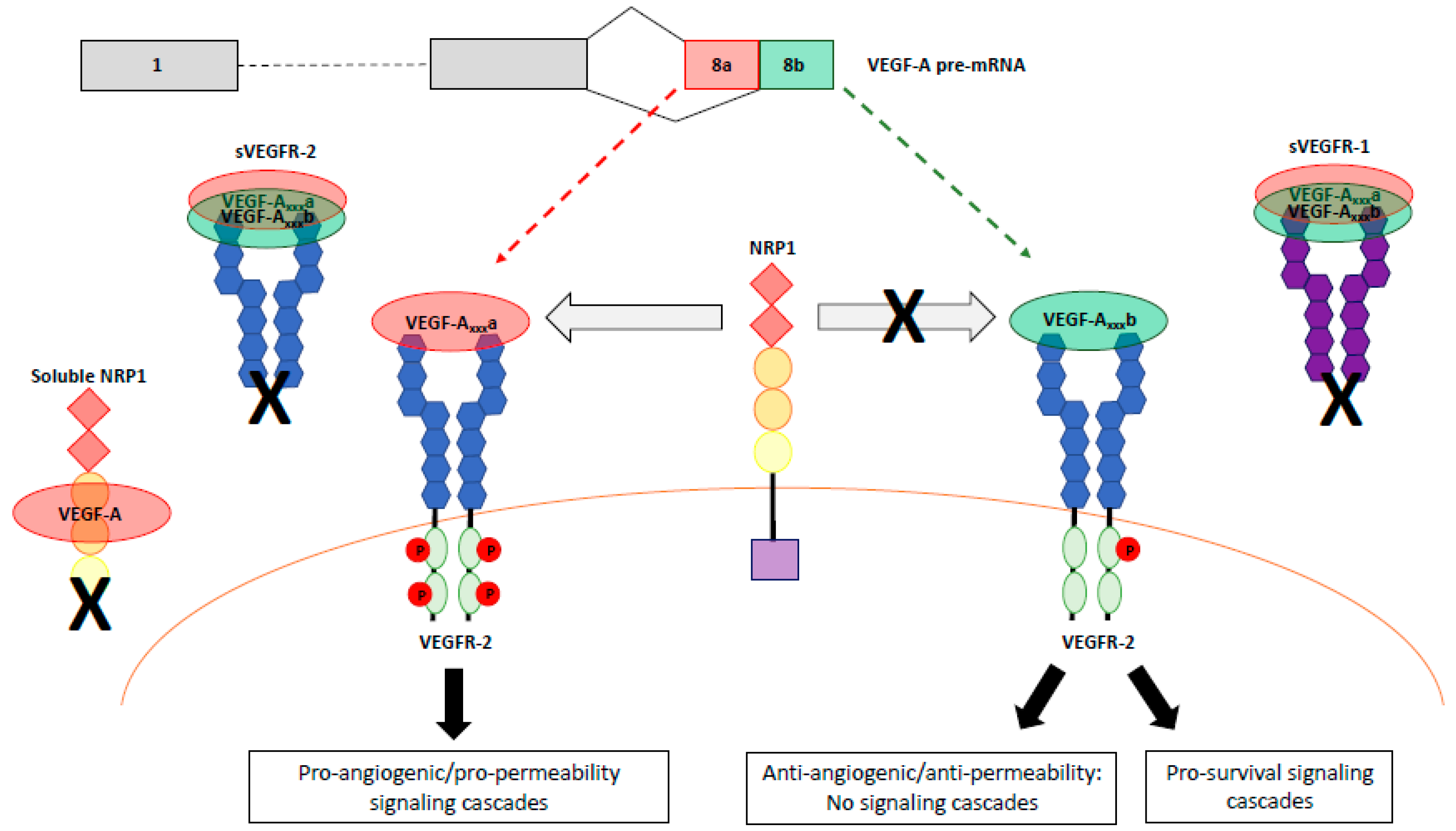
As mentioned above, all VEGF-A isoforms can bind to VEGFR-2 with similar affinity; however, different isoforms result in different activation and signaling outcomes [32] (Figure 3). Upon binding of VEGF-A to its orthosteric ligand binding site, VEGFR-2 undergoes dimerization and a conformational twist in the extracellular region results in the rotation of transmembrane helices [76,77]. Both VEGF-A165 and VEGF-A165b have been shown to result in VEGFR-2 dimerization [77]. Conformational changes in the intracellular domain of VEGFR-2 follows; ATP binds to the flexible N-lobe cleft facilitating the intrinsic kinase activity of the receptor and phosphorylation of the tyrosine residues in the C-lobe [78]. Upon phosphorylation of these tyrosine residues, certain cytoplasmic proteins bind and distinct signaling pathways are initiated, included those involved in cell survival, migration, proliferation, vasodilatation, and permeability (reviewed in [79]). The tyrosine residues include Y1054 and Y1059 in the activation loop, which are required for maximal kinase activity of VEGFR-2 [80]; Y951 in the kinase insert domain, which serves as a binding site for T cell-specific adapter molecule (TSAd) [81], and is vital for HUVEC migration in response to VEGF-A [82]; and Y1175 and Y1214 in the COOH-terminal tail. Y1175 phosphorylation mediates cell proliferation through binding of phospholipase C (PLC)-γ [83]. VEGFR-2 is dephosphorylated by protein phosphatase 1b (PTP1b) in the endoplasmic reticulum, which highlights the importance of spatiotemporal trafficking on the activation of VEGFR-2 [84,85].

4.4. VEGFR-2 Signaling in Angiogenesis

During sprouting angiogenesis, endothelial cells within existing vessels form an angiogenic sprout towards a chemotactic stimulus, such as VEGF-A. The angiogenic sprout is orientated with a leading tip cell and trailing stalk cells. The extent of sprouting in neighboring endothelial cells is regulated by delta-like ligand 4 and Notch via lateral inhibition [86]. Lumen formation occurs once two sprouts anastomose, and the new vessel is stabilized by smooth muscle cell and basement membrane deposition [87].
Cell proliferation is required for angiogenesis. VEGF-A activates VEGFR-2 and stimulates proliferation through the activation of RAS, which then activates RAF kinase to phosphorylate mitogen-activated protein kinases (MAPK/ERK) [88]. VEGFR-2 stimulates ERK activation via Y1175-dependent phosphorylation of PLC-γ, resulting in the subsequent activation of protein kinase C (PKC) [82]. Mutation of Y1175 or administration of and antibody specific to Y1175 decreased VEGF-A-dependent cell proliferation in vitro [89]. Furthermore, mutation of Y1175 in mice results in embryonic lethality on day 5–9 due to a lack of blood vessel formation [90].
Endothelial cell migration is also essential for angiogenesis. One VEGFR-2 signaling pathway that has been implicated in endothelial cell migration is initiated via the phosphorylation of Y951, which allows for the binding of T cell specific adapter protein (TSAd) [81]. Both mutation of Y951 and knock-down of TSAd are reported to inhibit VEGF-A-mediated actin reorganization, and thus migration in cultured endothelial cells; however, proliferation remained unaffected [81]. Another example of a VEGFR-2 signaling pathway involves phosphorylation of Y1175 to induce focal adhesion kinase (FAK)-mediated endothelial cell migration [91].

4.5. VEGFR-2 Signaling in Cell Survival

VEGF-A activation of VEGFR-2 is associated with increased endothelial cell survival. VEGFR-2 activates phosphoinositide 3-kinase (PI3K), which enables membrane recruitment and phosphorylation of protein kinase B (PKB/AKT) [92]. Activation of the cell survival factor AKT results in the phosphorylation of Bcl-2 associated death promoter (BAD), inhibiting the activity of pro-apoptotic factors such as Bcl-2 and caspase 9 [93].

4.6. VEGFR-2 Signaling in Permeability

VEGF-A activation of VEGFR-2 induces extravasation of proteins and leukocytes in vivo [94]. This is suggested to occur through two mechanisms: the formation of transcellular endothelial pores and the transient opening of paracellular junctions [95]. However, the exact signaling mechanisms regulating these events are not yet clear. One suggested mechanism involves VEGF-A-dependent endothelial nitric oxide synthase (eNOS) activation through PLC-γ and AKT, resulting in the activation of the pro-permeability factor nitric oxide (NO) [96,97].

4.7. Role of sVEGFR-2

The alternatively spliced sVEGFR-2 isoform has been reported to act as an endogenous VEGF-C antagonist, preventing it from binding to VEGFR-3 and consequently inhibiting lymphatic endothelial cell proliferation [27]. In addition, like sVEGFR-1, sVEGFR-2 is a natural circulating decoy receptor for VEGF, thus acting as a ligand trap [98].

4.8. VEGF-A Isoform Specific Activation of VEGFR-2

The canonical VEGF-Axxxa isoforms are widely described as pro-angiogenic, pro-permeability factors as they activate the aforementioned signaling pathways via VEGFR-2 binding and dimerization. On the other hand, VEGF-Axxxb isoforms are anti-angiogenic and anti-permeability, which is due to their effect on VEGFR-2 activation. Like VEGF-Axxxa, VEGF-Axxxb is still able to bind and dimerize VEGFR-2, but whether they result in phosphorylation of the tyrosine residues in the intracellular domain is not clear. The six-amino acid frame shift that occurs when the distal splice site is selected in the VEGF-A pre-mRNA results in the replacement of a positively charged arginine residue with neutral aspartic acid and lysine, which are predicted to decrease VEGFR-2 activation [99]. In pulmonary arterial endothelial (PAE) cells, VEGF-A165b was shown to induce VEGFR-2 activation (Y1052, Y1057) compared to untreated controls, but not to the same extent as that induced by VEGF-A165 [99]. Another report suggested that recombinant VEGF-A165b can induce Y1175 activation to almost the same extent as VEGF-A165 in HEK293-VR2 cells [100]. In addition, VEGF-A165b can induce VEGFR-2 Y1175 phosphorylation to the same extent as VEGF-A165 in endothelial cells [75]. However, anti-VEGF-A165b treatment of HUVECs and cultured visceral adipose tissue resulted in increased Y951 phosphorylation [101,102], indicating that VEGF-A165b antagonized Y951 phosphorylation. Furthermore, treatment of glomerular endothelial cells with VEGF-A165b did not result in any increases in the overall phosphorylated state of VEGFR-2 (immunoprecipitation of VEGFR-2 followed by immunoblotting with a phospho-tyrosine antibody) [103]. Taken together, these findings indicate that VEGF-A165b acts as a VEGFR-2 partial agonist/antagonist via the differential modulation of site-specific phosphorylation on VEGFR-2.
In some pathologies, VEGF-A165b expression has been shown to be down-regulated relative to VEGF-A165a. For example, in the late stages of human diabetic nephropathy when the kidney is not filtering properly, kidney VEGF-A165b levels are down-regulated relative to VEGF-A165a; however, during the early stages of diabetic nephropathy when the kidney is functioning well, the VEGF-A165b isoform is increased [104]. Therefore, VEGF-A165b may play a protective role in early nephropathy but when the expression is decreased, increased angiogenesis and permeability occur, resulting in a worse phenotype. Indeed, several studies in mouse models have shown the VEGF-A165b isoform to have reno-protective effects regarding glomerular permeability [103,104,105,106]. These protective effects are indicated to be due to VEGF-A165b decreasing the phosphorylation of VEGFR-2, which has been shown in glomerular endothelial cells [103]. Decreased levels of VEGF-A165b have also been observed in certain cancers, including colon cancer and renal cell carcinoma [28,107]. This reduction in VEGF-A165b is often accompanied by an increase in the pro-angiogenic VEGF-A165a, which contributes to angiogenesis within the tumor. Administration of VEGF-A165b, or manipulation of VEGF-A splicing to promote VEGF-A165b expression (such as with SRPK1 inhibitors), has been shown to be therapeutic in many tumor models through inhibition of VEGF-Axxxa mediated angiogenesis [108,109]. On the other hand, VEGF-A165b has also been shown to promote lung tumor progression and specific knock-down of just the VEGF-A165b isoform reduced tumor growth in lung cancer cells [110]. Thus, the role of VEGF-A165b signaling may depend on the tissue it is expressed in.
VEGF-A121a is a shorter freely diffusible VEGF-A isoform. In contrast to VEGF-A165a, VEGF-A121a has been shown to exhibit both partial and full agonist effects. On one hand, VEGF-A121a acts as a partial agonist of VEGFR-2 in both in vivo and in vitro measurements of angiogenesis and signaling, respectively [5,99], as well as slowing HUVEC proliferation and reducing sprouting in comparison to VEGF-A165a [111,112]. In contrast, VEGF-A121a-induced angiogenic sprouting ex vivo has been reported to be both comparable [33] and reduced [113] in comparison to VEGF-A165a. Similar trends are seen regarding vascular permeability [114,115,116].
VEGF-A145a and VEGF-A189a are ECM-bound isoforms that also show reduced agonistic effects on VEGFR-2 signaling in comparison to VEGF-A165a. In HUVECs, VEGF-A145a had a reduced effect on proliferation and permeability relative to VEGF-A165a, but comparable effects on migration [114]. This was indicated to be due to reduced phosphorylation of VEGFR-2 in addition to reduced activation of AKT and ERK [114]. Similarly, VEGF-A189a resulted in decreased cell survival and proliferation in BAECs, but comparable effects to VEGF-A165a on migration [117,118].

5. VEGFR Signaling Complexes

5.1. VEGFR Heterodimerization

Computational modeling has predicted VEGFR-1/2 heterodimers to comprise 10–50% of signaling VEGFR complexes, which are favored over VEGFR-1 homodimers when the VEGFR-2 abundance is higher [119]. There is evidence that suggests that VEGF-A stimulation of VEGFR-2 homodimers, VEGFR-1 homodimers, and VEGFR-1/2 heterodimers results in different efficacies of signal transduction; the pattern of Ca2+ flux was found to be unique for each type of receptor dimer in porcine aortic endothelial cells [120]. VEGF-A, VEGF-C, and VEGF-D have also been shown to induce the heterodimerization of VEGFR-2/3, which is required for certain ligand-dependent cellular responses mediated by VEGF-C and VEGF-D [121].

5.2. Roles of Neuropilins NRP1 and NRP2

Neuropilins can function as coreceptors with VEGFR-1 and VEGFR-2. There are two homologs of NRP, NRP1 and NRP2, which consist of a single transmembrane spanning domain with a small cytoplasmic domain lacking intrinsic catalytic function [122]. NRP1 was firstly suggested to bind in exon 7 of VEGF-A, which is present in isoforms such as VEGF-A165, forming a ternary complex with VEGFR-2 [112], thus primarily acting as a co-receptor for VEGFR-2. More recent studies have implicated the exon 8a-encoded arginine residue in the binding of VEGF-A to the b1 domain of NRP1 [123]. Binding of VEGF-A to NRP1 enhances VEGF-A signaling in endothelial cells with respect to migration and survival [124,125,126]. Furthermore, NRP1 is reported to be essential for VEGF-A-induced vessel sprouting and branching in angiogenesis [127]. NRP1 has also been shown to be associated with the adapter Synectin (GIPC), which is associated with the intracellular trafficking of VEGFR-2 [125]. In contrast, NRP2 acts as a co-receptor for VEGFR-3 and is therefore not involved with VEGF-A signal transduction [128]. In mice, both overexpression and disruption of NRP1 results in embryonic lethality on E12.5-13.5 due to vascular abnormalities [129]. Furthermore, siRNA [113] or antibody [112] blocking of NRP1 led to a decrease in VEGF-A165a-induced phosphorylation of VEGFR-2 in vitro.
In contrast to VEGF-A165, VEGF-A189, and VEGF-A145, fluorescent real-time ligand binding assays revealed that VEGF-A165b and VEGF-Ax are unable to bind to NRP1 as they lack the exon 7-8a-encded residues [130]. This provides further evidence for the lack of VEGFR-2 singling induced by the weak agonist VEGF-Axxxb isoforms. There is conflicting data regarding the binding of VEGF-A121a to NRP1 as it lacks exon 7, with most studies suggesting that although VEGF-A121a can bind NRP1, albeit at a lower affinity, it is unable to bridge the NRP1/VEGFR-2 complex (reviewed in [131]).

5.3. NRP1 and NRP2 Splice Variants

NRP1 exists as a full-length membrane-bound form in addition four soluble isoforms. Full-length NRP1 is comprised of 17 exons. On the other hand, two soluble splice variants, s12NRP1 and s11NRP1, are generated during pre-mRNA processing via intron read-through in the NRP1 gene, resulting in proteins that lack transmembrane and cytoplasmic domains of full-length NRP1 [132,133]. Functionally, these soluble isoforms of NRP1 were reported to bind VEGF-A165, although not VEGF-A121, thus inhibiting VEGF-A165-induced phosphorylation of VEGFR-2 in endothelial cells resulting in reduced tumor growth (anti-tumor properties) [133]. Therefore, s12NRP1 and s11NRP1 appear to act as VEGF-A165 antagonists. Two further soluble isoforms of NRP1 have also been described, sIIINRP and sIVNRP, which are proposed to have similar biological and biomechanical properties as s12NRP1 and s11NRP1 [134]. The sIIINRP1 isoform results from the deletion of exons 10 and 11, while exon 12 is still present, followed by retention of the beginning of intron 12 (28 bp). The sIVNRP1 isoform is missing exon 11, also resulting in intron 12 retention [134]. Both sIIINRP and sIVNRP have been shown to be expressed in normal and cancerous tissues and are capable of binding VEGF-A165, indicating that these two isoforms are antagonists for NRP1-mediated cellular activities [134]. The final isoform of NRP1 is NRP∆E16, which results from the skipping of exon 16 and replacement with an “AAG” Arg triple; however, this isoform does not have a functional difference to full length NRP1 [135].
NRP2 can also exist as a membrane bound or soluble form. The membrane bound form of NRP2 has two splice variants, NRP2a and NRP2b, which differ in the last 100 amino acids of the c-terminus. Therefore, these two splice variants are proposed to bind different proteins and govern different molecular pathways [132]. NRP2b has been reported to have a prometastatic role in non-small cell lung cancer, whereas NRP2a in promoting metastasis and therapy resistance [136]. However, further studies are needed to clarify the roles of each of these splice variants with respect to VEGF-A binding and signaling.

6. Regulation of Splicing as a Therapeutic Intervention

Research into the VEGF-A-VEGFR signaling axis in disease has recently taken a new direction focused on manipulating the splicing of these genes as a potential therapeutic avenue. One example of this is the regulation of the VEGF-Axxxa/VEGF-Axxxb ratio. Small molecule inhibitors of SRPK1, known as SRPIN340 and SPHINX31, have been shown to upregulate the VEGF-Axxxb isoforms relative to VEGF-Axxxa, which had a therapeutic effect in animal models of retinopathy [137,138]. Furthermore, a natural blueberry extract as also been shown to increase VEGF-A165b/VEGF-A164a in the kidneys of diabetic mice, exerting a therapeutic effect through a decrease in kidney fibrosis and permeability [139]. Regarding the VEGFRs, exogenous administration of sVEGFR-1 (either transfection, recombinant protein, or adenovirus infection) was reported to inhibit tumor growth and neoangiogenesis, increasing the survival rate in mouse xenograft models of melanoma, lung cancer, fibrosarcoma, and glioblastoma [71,72,73,74]. Therefore, further research into the regulation of VEGFR splicing is warranted to explore the potential therapeutic benefits of switching VEGFR splicing.

7. Conclusions

The VEGF-A-VEGFR axis is critical in both physiological and pathological angiogenesis and vessel permeability. The disruption of the splicing of just one of the genes involved in the VEGF-A-VEGFR axis (VEGF-A, VEGFR-1, VEGFR-2) can result in changes to the entire signaling axis, such as the increase in VEGF-A165a relative to VEGF-A165b resulting in increased VEGFR-2 signaling and aberrant angiogenesis in cancer. Further research into understanding the mechanisms by which the splicing of VEGF-A/VEGFR-1/VEGFR-2 is regulated will help in the development of drugs aimed at manipulating splicing or inhibiting specific splice isoforms in a therapeutic manner.

Author Contributions

M.S. wrote the first draft of the manuscript and reviewed intermediate versions; S.O. reviewed intermediate and the final versions.

Funding

This research was funded by grants from British Heart Foundation to SO (PG/15/53/31371), Diabetes UK to SO (17/0005668).

Conflicts of Interest

The authors declare no conflict of interest.

References

  1. Shibuya, M. Structure and function of VEGF/VEGF-receptor system involved in angiogenesis. Cell Struct. Funct. 2001, 26, 25–35. [Google Scholar] [CrossRef] [PubMed]
  2. Ferrara, N.; Gerber, H.P.; LeCouter, J. The biology of VEGF and its receptors. Nat. Med. 2003, 9, 669–676. [Google Scholar] [CrossRef]
  3. Terman, B.I.; Carrion, M.E.; Kovacs, E.; Rasmussen, B.A.; Eddy, R.L.; Shows, T.B. Identification of a new endothelial cell growth factor receptor tyrosine kinase. Oncogene 1991, 6, 1677–1683. [Google Scholar]
  4. De Vries, C.; Escobedo, J.A.; Ueno, H.; Houck, K.; Ferrara, N.; Williams, L.T. The fms-like tyrosine kinase, a receptor for vascular endothelial growth factor. Science 1992, 21, 989–991. [Google Scholar] [CrossRef]
  5. Cebe-Suarez, S.; Zehnder-Fjallman, A.; Ballmer-Hofer, K. The role of VEGF receptors in angiogenesis; complex partnerships. Cell. Mol. Life Sci. 2006, 63, 601–615. [Google Scholar] [CrossRef] [PubMed] [Green Version]
  6. Gerber, H.P.; Condorelli, F.; Park, J.; Ferrara, N. Differential transcriptional regulation of the two vascular endothelial growth factor receptor genes. Flt-1, but not Flk-1/KDR, is upregulated by hypoxia. J. Biol. Chem. 1997, 272, 23659–23667. [Google Scholar] [CrossRef]
  7. Fong, G.H.; Rossant, J.; Gertsenstein, M.; Breitman, M.L. Role of Flt-1 receptor tyrosine kinase in regulating the assembly of vascular endothelium. Nature 1995, 376, 66–70. [Google Scholar] [CrossRef]
  8. Fong, G.H.; Zhang, L.; Bryce, D.M.; Peng, J. Increased hemangioblast commitment, not vascular disorganization, is the primary defect in flt-1 knock-out mice. Development 1999, 126, 3015–3025. [Google Scholar]
  9. Hiratsuka, S.; Minowa, O.; Kuno, J.; Noda, T.; Shibuya, M. Flt-1 lacking the tyrosine domain is sufficient for normal development and angiogenesis in mice. Proc. Natl. Acad. Sci. USA 1998, 95, 9349–9354. [Google Scholar] [CrossRef] [PubMed]
  10. Kendall, R.L.; Wang, G.; Thomas, K.A. Identification of a natural soluble form of the vascular endothelial growth factor receptor, FLT-1, and its heterodimerization with KDR. Biochem. Biophys. Res. Commun. 1996, 226, 324–328. [Google Scholar] [CrossRef] [PubMed]
  11. Roberts, D.M.; Kearney, J.B.; Johnson, J.H.; Rosenberg, M.P.; Kumar, R.; Bautch, V.L. The vascular endothelial growth factor (VEGF) receptor Flt-1 (VEGFR-1) modulates Flk-1 (VEGFR-2) signaling during blood vessel formation. Am. J. Pathol. 2004, 164, 1531–1535. [Google Scholar] [CrossRef]
  12. Abou-Faycal, C.; Hatat, A.S.; Gazzeri, S.; Eymin, B. Splice variants of the RTK family: Their role in tumor progression and response to targeted therapy. Int. J. Mol. Sci. 2017, 18, 383. [Google Scholar] [CrossRef] [PubMed]
  13. Thomas, C.P.; Raikwar, N.S.; Kelley, E.A.; Liu, K.Z. Alternate processing of Flt1 transcripts is directed by conserved cis-elements within an intronic region of FLT1 that reciprocally regulates splicing and polyadenylation. Nucleic Acids Res. 2010, 38, 5130–5140. [Google Scholar] [CrossRef] [PubMed] [Green Version]
  14. Ikeda, T.; Sun, L.; Tsuruoka, N.; Ishigaki, Y.; Yoshitomi, Y.; Yoshitake, Y.; Yonekura, H. Hypoxia down-regulates sFlt-1 (sVEGFR-1) expression in human microvascular endothelial cells by a mechanism involving mRNA alternative processing. Boichem. J. 2011, 436, 399–407. [Google Scholar] [CrossRef] [Green Version]
  15. Eubank, T.D.; Roda, J.M.; Liu, H.; O’Neil, T.; Marsh, C.B. Opposing roles for HIF-1α and HIF-2α in the regulation of angiogenesis by mononuclear phagocytes. Blood 2011, 117, 323–332. [Google Scholar] [CrossRef]
  16. Thomas, R.; Kim, M.H. A HIF-1alpha-dependent autocrine feedback loop promotes survival of serum-deprived prostate cancer cells. Prostate 2009, 69, 263–275. [Google Scholar] [CrossRef] [PubMed]
  17. Xiong, Y.; Liebermann, D.A.; Tront, J.S.; Holtzman, E.J.; Huang, Y.; Hoffman, B.; Geifman-Holtzman, O. Gadd45a stress signaling regulates sFlt-1 expression in preeclampsia. J. Cell. Physiol. 2009, 220, 632–639. [Google Scholar] [CrossRef]
  18. Boeckel, J.N.; Guarani, V.; Koyanangi, M.; Roexe, T.; Lengeling, A.; Schermuly, R.T.; Gellert, P.; Braun, T.; Zeiher, A.; Dimmeler, S. Jumonji domain-containing protein 6 (Jmjd6) is required for angiogenic sprouting and regulates splicing of VEGF-receptor 1. Proc. Natl. Acad. Sci. USA 2011, 108, 3276–3281. [Google Scholar] [CrossRef] [Green Version]
  19. Ikeda, T.; Yoshitomi, Y.; Shimasaki, T.; Yamaya, H.; Kobata, T.; Ishigaki, Y.; Tomosugi, N.; Yoshitake, Y.; Yonekura, H. Regulation of soluble Flt-1 (VEGFR-1) production by hnRNP D and protein arginine methylation. Mol. Cell. Biochem. 2016, 413, 155–164. [Google Scholar] [CrossRef]
  20. Raikwar, N.S.; Liu, K.Z.; Thomas, C.P. Protein kinase C regulates FLT1 abundance and stimulates its cleavage in vascular endothelial cells with the release of a soluble PIGF/VEGF antagonist. Exp. Cell Res. 2013, 319, 2578–2587. [Google Scholar] [CrossRef] [PubMed]
  21. Fuh, G.; Li, B.; Crowley, C.; Cunningham, B.; Wells, J.A. Requirements for binding and signaling of the kinase domain receptor for vascular endothelial growth factor. J. Biol. Chem. 1998, 273, 11197–11204. [Google Scholar] [CrossRef]
  22. Shinkai, A.; Ito, M.; Anazawa, H.; Yamaguchi, S.; Shitara, K.; Shibuya, M. Mapping of the sites involved in ligand association and dissociation at the extracellular domain of the kinase insert domain-containing receptor for vascular endothelial growth factor. J. Biol. Chem. 1998, 273, 31283–31288. [Google Scholar] [CrossRef]
  23. Shalaby, F.; Rossant, J.; Yamaguchi, T.P.; Gertsenstein, M.; Wu, X.F.; Breitman, M.L.; Schuh, A.C. Failure of blood-island formation and vasculogenesis in Flk-1-deficient mice. Nature 1995, 376, 62–66. [Google Scholar] [CrossRef] [PubMed]
  24. Ferrara, N.; Carver-Moore, K.; Chen, H.; Dowd, M.; Lu, L.; O’Shea, K.S.; Powell-Braxton, L.; Hillan, K.J.; Moore, M.W. Heterozygous embryonic lethality induced by targeted inactivation of the VEGF gene. Nature 1996, 380, 439–442. [Google Scholar] [CrossRef] [PubMed]
  25. Kou, B.; Li, Y.; Zhang, L.; Zhu, G.; Wang, X.; Li, Y.; Xia, J.; Shi, Y. In vivo inhibition of tumor angiogenesis by a soluble VEGFR-2 fragment. Exp. Mol. Pathol. 2004, 76, 129–137. [Google Scholar] [CrossRef] [PubMed]
  26. Collet, G.; Lamerant-Fayel, N.; Tertil, M.; El Hafny-Rahbi, B.; Stepniewski, J.; Guichard, A.; Foucault-Collet, A.; Klimkiewicz, K.; Petoud, S.; Matejuk, A.; et al. Hypoxia-regulated overexpression of soluble VEGFR2 controls angiogenesis and inhibits tumor growth. Mol. Cancer Ther. 2014, 13, 165–178. [Google Scholar] [CrossRef] [PubMed]
  27. Albuquerque, R.J.; Hayashi, T.; Cho, W.G.; Kleinman, M.E.; Dridi, S.; Takeda, A.; Baffi, J.Z.; Yamada, K.; Kaneko, H.; Green, M.G.; et al. Alternatively spliced vascular endothelial growth factor receptor-2 is an essential endogenous inhibitor of lymphatic vessel growth. Nat. Med. 2009, 15, 1023–1030. [Google Scholar] [CrossRef] [PubMed]
  28. Bates, D.O.; Cui, T.G.; Doughty, J.M.; Winkler, M.; Sugiono, M.; Shields, J.D.; Peat, D.; Gillatt, D.; Harper, S.J. VEGF165b, an inhibitory splice variant of vascular endothelial growth factor, is down-regulated in renal cell carcinoma. Cancer Res. 2002, 62, 4123–4131. [Google Scholar] [PubMed]
  29. Eswarappa, S.M.; Potdar, A.A.; Koch, W.J.; Fan, Y.; Vasu, K.; Linder, D.; Willard, B.; Graham, L.M.; DiCorleto, P.E.; Fox, P.L. Programmed translational readthrough generates antiangiogenic VEGF-Ax. Cell 2014, 157, 1605–1618. [Google Scholar] [CrossRef] [PubMed]
  30. Guyot, M.; Pages, G. VEGF splicing and the role of VEGF splice variants: From physiological-pathological conditions to specific pre-mRNA splicing. Methods Mol. Biol. 2015, 1332, 3–23. [Google Scholar] [PubMed]
  31. Stevens, M.; Oltean, S. Modulation of VEGF-A alternative splicing as a novel treatment in chronic kidney disease. Genes 2018, 9, 98. [Google Scholar] [CrossRef] [PubMed]
  32. Woolard, J.; Bevan, H.S.; Harper, S.J.; Bates, D.O. Molecular diversity of VEGF-A as a regulator of its biological activity. Microcirculation 2009, 16, 572–592. [Google Scholar] [CrossRef] [PubMed]
  33. Holmes, D.I.; Zachary, I.C. Vascular endothelial growth factor regulates stanniocalcin-1 expression via neuropilin-1-dependent regulation of KDR and synergism with fibroblast growth factor-2. Cell Signal. 2008, 20, 569–579. [Google Scholar] [CrossRef] [PubMed]
  34. Krilleke, D.; DeErkenez, A.; Schubert, W.; Giri, I.; Robinson, G.S.; Ng, Y.S.; Shima, D.T. Molecualr mapping and functional characterization of the VEGF164 heparin-binding domain. J. Biol. Chem. 2007, 282, 28045–28056. [Google Scholar] [CrossRef]
  35. Lee, T.Y.; Folkman, J.; Javaherian, K. HSPG-binding peptide corresponding to the exon 6a-encoded domain of VEGF inhibits tumor growth by blocking angiogenesis in a murine model. PLoS ONE 2010, 5, e9945. [Google Scholar] [CrossRef]
  36. Houck, K.; Leung, D.W.; Rowland, A.M.; Winer, J.; Ferrara, N. Dual regulation of vascular endothelial growth factor bioavailability by genetic and proteolytic mechanisms. J. Biol. Chem. 1992, 267, 26031–26037. [Google Scholar]
  37. Nowak, D.G.; Woolard, J.; Amin, E.M.; Konopatskaya, O.; Saleem, M.A.; Churchill, A.J.; Ladomery, M.R.; Harper, S.J.; Bates, D.O. Expression of pro- and anti-angiogenic isoforms of VEGF is differentially regulated by splicing and growth factors. J. Cell Sci. 2008, 121, 3487–3495. [Google Scholar] [CrossRef] [Green Version]
  38. Amin, E.M.; Oltean, S.; Hua, J.; Gammons, M.V.; Hamdollah-Zadeh, M.; Welsh, G.I.; Cheung, M.K.; Ni, L.; Kase, S.; Rennel, E.S.; et al. WT1 mutants reveal SRPK1 to be a downstream angiogenesis target by altering VEGF splicing. Cancer Cell 2011, 20, 768–780. [Google Scholar] [CrossRef] [Green Version]
  39. Merdzhanova, G.; Gout, S.; Keramidas, M.; Edmond, V.; Coll, J.L.; Brambilla, C.; Brambilla, E.; Gazzeri, S.; Eymin, B. The transcription factor E2F1 and the SR protein SC35 control the ratio of pro-angiogenic verus antiangiogenic isoforms of vascular endothelial growth factor-A to inhibit noevascularization in vivo. Oncogene 2010, 29, 5392–5403. [Google Scholar] [CrossRef]
  40. Inoue, T.; Kibata, K.; Suzuki, M.; Nakamura, S.; Motoda, R.; Orita, K. Identification of a vascular endothelial growth factor (VEGF) antagonist, sFlt-1, from a human hematopoietic cell line NALM-16. FEBS Lett. 2000, 469, 14–18. [Google Scholar] [CrossRef] [Green Version]
  41. Kearney, J.B.; Kappas, N.C.; Ellerstrom, C.; DiPaola, F.W.; Bautch, V.L. The VEGF receptor flt-1 (VEGFR-1) is a positive modulator of vascular sprout formation and branching morphogenesis. Blood 2004, 103, 4527–4535. [Google Scholar] [CrossRef] [PubMed] [Green Version]
  42. Kappas, N.C.; Zeng, G.; Chappell, J.C.; Kearney, J.B.; Hazarika, S.; Kallianos, K.G.; Patterson, C.; Annex, B.H.; Bautch, V.L. The VEGF receptor Flt-1 spatially modulates Flk-1 signaling and blood vessel branching. J. Cell Biol. 2008, 181, 847–858. [Google Scholar] [CrossRef] [Green Version]
  43. Chappell, J.C.; Taylor, S.M.; Ferrara, N.; Bautch, V.L. Local guidance of emerging vessel sprouts requires soluble Flt-1. Dev. Cell 2009, 17, 377–386. [Google Scholar] [CrossRef] [PubMed]
  44. Orecchia, A.; Lacal, P.M.; Schietroma, C.; Morea, V.; Zambruno, G.; Failla, C.M. Vascualr endothelial growth factor receptor-1 is deposited in the extracellular matrix by endothelial cells and is a ligand for the alpha 5 beta integrin. J. Cell Sci. 2003, 116, 3479–3489. [Google Scholar] [CrossRef] [PubMed]
  45. Failla, C.M.; Carbo, M.; Morea, V. Positive and negative regulation of angiogenesis by soluble vascular endothelial growth factor receptor-1. Int. J. Mol. Sci. 2018, 19, 1306. [Google Scholar] [CrossRef] [PubMed]
  46. Li, X.; Tjwa, M.; Van Hove, I.; Enholm, B.; Neven, E.; Paavonen, K.; Jeltsch, M.; Juan, T.D.; Sievers, R.E.; Chorianopoulos, E.; et al. Reevaluation of the role of VEGF-B suggests a restricted role in the revascularization of the ischemic myocardium. Arterioscler. Thromb. Vasc. Biol. 2008, 28, 1614–1620. [Google Scholar] [CrossRef] [PubMed]
  47. Yamaguchi, T.; Bando, H.; Mori, T.; Takahashi, K.; Matsumoto, H.; Yasutome, M.; Weich, H.; Toi, M. Overexpression of soluble vascular endothelial growth factor receptor 1 in colorectal cancer: Association with progression and prognosis. Cancer Sci 2007, 98, 405–410. [Google Scholar] [CrossRef] [Green Version]
  48. Lamszus, K.; Ulbricht, U.; Matschke, J.; Brockmann, M.A.; Fillbrandt, R.; Westphal, M. Levels of soluble vascular endothelial growth factor (VEGF) receptor 1 in astrocytic tumors and its relation to malignancy, vascularity, and VEGF-A. Clin. Cancer Res. 2003, 9, 1399–1405. [Google Scholar]
  49. Toi, M.; Bando, H.; Ogawa, T.; Muta, M.; Hornig, C.; Weich, H.A. Significance of vascular endothelial growth factor (VEGF)/soluble VEGF receptor-1 relationship in breast cancer. Int. J. Cancer 2002, 98, 14–18. [Google Scholar] [CrossRef] [Green Version]
  50. Nagaoka, S.; Yoshida, T.; Akiyoshi, J.; Akiba, J.; Hisamoto, T.; Yoshida, Y.; Abe, M.; Koga, H.; Toirimura, T.; Ueno, T.; et al. The ratio of placenta growth factor to soluble vascular endothelial growth factor receptor-1 predicts the prognosis of hepatocellular carcinoma. Oncol. Rep. 2010, 23, 1647–1654. [Google Scholar]
  51. Ilhan, N.; Ilhan, N.; Deveci, F. Functional significance of vascular endothelial growth factor and its receptor (receptor-1) in various lung cancer types. Clin. Biochem. 2004, 37, 840–845. [Google Scholar] [CrossRef]
  52. Ruffini, F.; Failla, C.M.; Orecchia, A.; Bani, M.R.; Dorio, A.S.; Fortes, C.; Zambruno, G.; Graziani, G.; Giavazzi, R.; D’Atri, S.; et al. Expression of soluble vascular endothelial growth factor receptor-1 in cutaneous melanoma: Role in tumour progression. Br. J. Dermatol. 2011, 164, 1061–1070. [Google Scholar] [CrossRef]
  53. Wierzbowska, A.; Robak, T.; Wrzesien-Kus, A.; Krawczynska, A.; Lech-Maranda, E.; Urbanska-Rys, H. Circulating VEGF and its soluble receptors sVEGFR-1 and sVEGFR-2 in patients with acute leukemia. Eur. Cytokine Netw. 2003, 14, 149–153. [Google Scholar] [PubMed]
  54. Harris, A.L.; Reusch, P.; Barleon, B.; Hang, C.; Dobbs, N.; Marme, D. Soluble Tie2 and Flt1 extracellular domains in serum of patients with renal cancer and response to antiangiogenic therapy. Clin. Cancer Res. 2001, 7, 1992–1997. [Google Scholar]
  55. Kulapaditharom, B.; Boonkitticharoen, V.; Sritara, C. Plasma vascular endothelial growth factor dysregulation in defining aggressiveness of head and neck squamous cell carcinoma. J. Oncol. 2012, 2012, 687934. [Google Scholar] [CrossRef] [PubMed]
  56. Bando, H.; Weich, H.A.; Brokelmann, M.; Horiguchi, S.; Funata, N.; Ogawa, T.; Toi, M. Association between intratumoral free and total VEGF, soluble VEGFR-1, VEGFR-2 and prognosis in breast cancer. Br. J. Cancer 2005, 92, 553–561. [Google Scholar] [CrossRef] [PubMed] [Green Version]
  57. Tolany, S.M.; Boucher, Y.; Duda, D.G.; Martin, J.D.; Seano, G.; Ancukiewicz, M.; Barry, W.T.; Goel, S.; Lahdenrata, J.; Isakoff, S.J.; et al. Role of vascular density and normalization in response to neoadjuvant bevacizumab and chemotherapy in breast cancer patients. Proc. Natl. Acad. Sci. USA 2015, 112, 14325–14330. [Google Scholar] [CrossRef] [PubMed] [Green Version]
  58. Willett, C.G.; Duda, D.G.; di Tomaso, E.; Boucher, Y.; Ancukiewicz, M.; Sahani, D.V.; Lahdenranta, J.; Chung, D.C.; Fischman, A.J.; Lauwers, G.Y.; et al. Efficacy, safety, and biomarkers of neoadjuvant bevacizumab, radiation therapy, and fluorouracil in rectal cancer: A multidisciplinary phase II study. J. Clin. Oncol. 2009, 27, 3020–3026. [Google Scholar] [CrossRef]
  59. Maynard, S.E.; Min, J.Y.; Merchen, J.; Lim, K.H.; Li, J.; Mondal, S.; Libermann, T.A.; Morgan, J.P.; Selke, F.W.; Stillman, I.E.; et al. Excess placental soluble fms-like tyrosine kinase 1 (sFlt1) may contribute to endothelial dysfunction, hypertension, and proteinuria in preeclampsia. J. Clin. Investig. 2003, 111, 649–658. [Google Scholar] [CrossRef] [Green Version]
  60. McKeeman, G.C.; Ardill, J.E.; Caldwell, C.M.; Hunter, A.J.; McClure, N. Soluble vascular endothelial growth factor receptor-1 (sFlt-1) is increased throughout gestation in patients who have preeclampsia. Am. J. Obstet. Gynecol. 2004, 191, 1240–1246. [Google Scholar] [CrossRef] [PubMed]
  61. Krysiak, O.; Bretschneider, A.; Zhong, E.; Webb, J.; Hopp, H.; Verlohren, S.; Fuhr, N.; Lanowska, M.; Nonnenmacher, A.; Vetter, R.; et al. Soluble vascular endothelial growth factor receptor-1 (sFLT-1) mediates downregulation of FLT-1 and prevents activated neutrophils from women with preeclampsia from additional migration by VEGF. Circ. Res. 2005, 97, 1253–1261. [Google Scholar] [CrossRef]
  62. Palmer, K.R.; Tong, S.; Kaitu’u-Lino, T.J. Plcental-specific sFLT-1: Role in pre-eclamptic pathophysiology and its translational possibilities for clinical prediction and diagnosis. Mol. Hum. Reprod. 2017, 23, 69–78. [Google Scholar] [PubMed]
  63. Di Marco, G.S.; Kentrup, D.; Reuter, S.; Mayer, A.B.; Golle, L.; Tiemann, K.; Fobker, M.; Engelbertz, C.; Breithardt, G.; Brande, E.; et al. Soluble Flt-1 links microvascular disease with heart failure in CKD. Basic Res. Cardiol. 2015, 110, 30. [Google Scholar] [CrossRef] [PubMed]
  64. Di Marco, G.S.; Reuter, S.; Hillebrand, U.; Amler, S.; Konig, M.; Larger, E.; Oberleithner, H.; Brand, E.; Pavenstadt, H.; Brand, M. The soluble VEGF receptor sFlt1 contributes to endothelial dysfunction in CKD. J. Am. Soc. Npehrol. 2009, 20, 2235–2245. [Google Scholar] [CrossRef]
  65. Ku, C.H.; White, K.E.; Dei Cas, A.; Hayward, A.; Webster, Z.; Bilous, R.; Marshall, S.; Viberti, G.; Gnudi, L. Inducible overexpression of sFlt-1 in podocytes ameliorates glomerulopathy in diabetic mice. Diabetes 2008, 57, 2824–2833. [Google Scholar] [CrossRef] [PubMed]
  66. Jin, J.; Sison, K.; Li, C.; Tian, R.; Wnuk, M.; Sung, H.K.; Jeansson, M.; Zhang, C.; Tucholska, M.; Jones, N.; et al. Soluble FLT1 binds lipid microdomains in podocytes to control cell morphology and glomerular barrier function. Cell 2012, 151, 384–399. [Google Scholar] [CrossRef] [PubMed]
  67. Ambati, B.K.; Nozaki, M.; Singh, N.; Takeda, A.; Jani, P.D.; Suthar, T.; Albuquerque, R.J.; Richter, E.; Sakurai, E.; Newcomb, M.T.; et al. Corneal avascualrity is due to soluble VEGF receptor-1. Nature 2006, 443, 993–997. [Google Scholar] [CrossRef]
  68. Uehara, H.; Mamalis, C.; McFadden, M.; Taggart, M.; Stagg, B.; Passi, S.; Earle, P.; Chakravarthy, U.; Hogg, R.E.; Ambati, B.K. The reduction of serum soluble Flt-1 in patients with neovascular age-related macular degeneration. Am. J. Ophthalmol. 2015, 159, 92–100. [Google Scholar] [CrossRef] [PubMed]
  69. Shapiro, N.I.; Yano, K.; Okada, H.; Fischer, C.; Howell, M.; Spokes, K.C.; Ngo, L.; Angus, D.C.; Aird, W.C. A prospective, observational study of soluble FLT-1 and vascular endothelial growth factor in sepsis. Shock 2008, 29, 452–457. [Google Scholar] [CrossRef] [PubMed]
  70. Dumnicka, P.; Kusnierz-Cabala, B.; Sporek, M.; Mazur-Laskowska, M.; Gil, K.; Kuzniewski, M.; Ceranowicz, Z.; Warzacha, Z.; Dembinski, A.; Bonior, J.; et al. Serum concentrations of angiopoietin-2 and soluble fms-like tyrosine kinase 1 (sFlt-1) are associated with coagulopathy among patients with acute pancreatitis. Int. J. Mol. Sci. 2017, 18, 753. [Google Scholar] [CrossRef]
  71. Goldman, C.K.; Kendll, R.L.; Cabrera, G.; Soroceanu, L.; Heike, Y.; Gillespie, G.Y.; Siegal, G.P.; Mao, X.; Bett, A.J.; Huckle, W.R.; et al. Paracrine expression of a native soluble vascular endothelial growth factor receptor inhibits tumor growth, metastasis, and mortality rate. Proc. Natl. Acad. Sci. USA 1998, 95, 8795–8800. [Google Scholar] [CrossRef] [PubMed] [Green Version]
  72. Verrax, J.; Defresne, F.; Lair, F.; Vandermeulen, G.; Rath, G.; Dessy, C.; Preat, V.; Feron, O. Delivery of soluble VEGF receptor 1 (sFlt1) by gene electrotransfer as a new antiangiogenic cancer therapy. Mol. Pharm. 2011, 8, 701–708. [Google Scholar] [CrossRef] [PubMed]
  73. Takayama, K.; Ueno, H.; Nakanishi, Y.; Sakamoto, T.; Inoue, K.; Shimizu, K.; Oohashi, H.; Hara, N. Suppression of tumor angiogenesis and growth by gene transfer of a soluble form of vascular endothelial growth factor receptor into a remote organ. Cancer Res. 2000, 60, 2169–2177. [Google Scholar] [PubMed]
  74. Shiose, S.; Sakamoto, T.; Yoshikawa, H.; Hata, Y.; Kawano, Y.; Ishibashi, T.; Inomata, H.; Takayama, K.; Ueno, H. Gene transfer of a soluble receptor of VEGF inhibits the growth of experimental eyelid malignant melanoma. Investig. Ophthalmol. Vis. Sci 2000, 41, 2395–2403. [Google Scholar]
  75. Ganta, V.C.; Choi, M.; Kutateladze, A.; Annex, B.H. VEGF165b modulates endothelial VEGFR1-STAT3 signaling pathway and angiogenesis in human and experimental peripheral arterial disease. Circ. Res. 2017, 120, 282–295. [Google Scholar] [CrossRef] [PubMed]
  76. Ruch, C.; Skiniotis, G.; Steinmetz, M.O.; Walz, T.; Ballmer-Hoffer, K. Structure of a VEGF-VEGF receptor complex determined by electron microscopy. Nat. Struct. Mol. Biol. 2007, 14, 249–250. [Google Scholar] [CrossRef] [PubMed]
  77. Sarabipour, S.; Ballmer-Hofer, K.; Hristova, K. VEGFR-2 conformational switch in response to ligand binding. Elife 2016, 5, e13876. [Google Scholar] [CrossRef] [PubMed] [Green Version]
  78. Manni, S.; Kisko, K.; Schleier, T.; Missimer, J.; Ballmer-Hofer, K. Functional and structural characterization of the kinase insert domain and the carboxy terminal domain in VEGF receptor 2 activation. FASEB J. 2014, 28, 4914–4923. [Google Scholar] [CrossRef] [PubMed]
  79. Koch, S.; Tugues, S.; Li, X.; Gualandi, L.; Claesson-Welsh, L. Signal transduction by vascular endothelial growth factor receptors. Biochem. J. 2011, 437, 169–183. [Google Scholar] [CrossRef] [Green Version]
  80. Dougher, M.; Terman, B.I. Autophosphorylation of KDR in the kinase domain is required for maximal VEGF-stimulated kinase activity and receptor internalization. Oncogene 1999, 18, 1619–1627. [Google Scholar] [CrossRef] [PubMed] [Green Version]
  81. Matsumoto, T.; Bohman, S.; Dixelius, J.; Berge, T.; Dimberg, A.; Magnusson, P.; Wang, L.; Wikner, C.; Qi, J.H.; Wernstedt, C.; et al. VEGF receptor-2 Y951 signlaing and a role for the adapter molecule TSAd in tumor angiogenesis. EMBO J. 2005, 24, 2342–2353. [Google Scholar] [CrossRef] [PubMed]
  82. Zeng, H.; Sanyal, S.; Mukhopadhyay, D. Tyrosine residues 951 and 1059 of vascular endothelial growth factor receptor-2 (KDR) are essential for vascular permeability factor/vascular endothelial growth factor-induced endothelium migration and proliferation, respectively. J. Biol. Chem. 2001, 276, 32714–32719. [Google Scholar] [CrossRef] [PubMed]
  83. Takahashi, T.; Yamaguchi, S.; Chida, K.; Shibuya, M. A single autophosphorylation site on KDR/Flk-1 is essential for VEGF-A-dependent activation of PLC-gamma and DNA synthesis in vascular endothelial cells. EMBO J. 2001, 20, 2768–2778. [Google Scholar] [CrossRef]
  84. Lanahan, A.A.; Lech, D.; Dubrac, A.; Zhang, Z.W.; Eichmann, A.; Simons, M. PTP1b is a physiologic regulator of vascular endothelial growth factor signaling in endothelial cells. Circulation 2014, 130, 902–909. [Google Scholar] [CrossRef] [PubMed]
  85. Haj, F.G.; Markova, B.; Klaman, L.D.; Bohmer, F.D.; Neel, B.G. Regulation of receptor tyrosine kinase signaling by protein tyrosine phosphatase-1B. J. Biol. Chem. 2003, 278, 739–744. [Google Scholar] [CrossRef] [PubMed]
  86. Jackobsson, L.; Bentley, K.; Gerhardt, H. VEGFRs and Notch: A dynamic collaboration in vascular patterning. Biochem. Soc. Trans. 2009, 37, 1233–1236. [Google Scholar] [CrossRef] [PubMed]
  87. Fantin, A.; Vieira, J.M.; Gestri, G.; Denti, L.; Schwarz, Q.; Prykhozhij, S.; Peri, F.; Wilson, S.W.; Ruhrberg, C. Tissue macrophages act as cellular chaperones for vascular anastomosis downstream of VEGF-mediated endothelial tip induction. Blood 2010, 116, 829–840. [Google Scholar] [CrossRef] [PubMed]
  88. Meadows, K.N.; Bryant, P.; Pumiglia, K. Vascular endothelial growth factor induction of the angiogenic phenotype requires Ras activation. J. Biol. Chem. 2001, 276, 49289–49298. [Google Scholar] [CrossRef] [PubMed]
  89. Takahashi, T.; Ueno, H.; Shibuya, M. VEGF activates protein kinase C-dependent, but Ras-independent Raf-MEK-MAP kinase pathway for DNA synthesis in primary endothelial cells. Oncogene 1999, 18, 2221–2230. [Google Scholar] [CrossRef] [PubMed] [Green Version]
  90. Sakurai, Y.; Ohgimoto, K.; Kataoka, Y.; Yoshida, N.; Shibuya, M. Essential role of Flk-1 (VEGF receptor 2) tyrosine residue 1173 in vasculogenesis in mice. Proc. Natl. Acad. Sci. USA 2005, 102, 1076–1081. [Google Scholar] [CrossRef] [PubMed] [Green Version]
  91. Abu-ghazaleh, R.; Kabir, J.; Jia, H.; Lobo, M.; Zachary, I. Src mediates stimulation by vascular endothelial growth factor of the phosphorylation of focal adhesion kinase at tyrosine 861, and migration and anti-apoptosis in endothelial cells. Biochem. J. 2001, 360, 255–264. [Google Scholar] [CrossRef] [PubMed] [Green Version]
  92. Gerber, H.P.; McMurtrey, A.; Kowalski, J.; Yan, M.; Keyt, B.A.; Dixit, V.; Ferrara, N. Vascualr endothelial growth factor regulates endothelial cell survival through the phosphatidylinositol 3′-kinase/Akt signal transduction pathway. Requirement for Flk-1/KDR activation. J. Biol. Chem. 1998, 273, 30336–30343. [Google Scholar] [CrossRef] [PubMed]
  93. Cardone, M.H.; Roy, N.; Stennicke, H.R.; Salvesen, G.S.; Frank, T.F.; Stanbridge, E.; Frisch, S.; Reed, J.C. Regulation of cell death protease caspase-9 by phosphorylation. Science 1998, 282, 1318–1321. [Google Scholar] [CrossRef] [PubMed]
  94. Bates, D.O.; Harper, S.J. Regulation of vascular permeability by vascular endothelial growth factors. Vasc. Pharmacol. 2002, 39, 225–237. [Google Scholar] [CrossRef]
  95. Garrido-urbani, S.; Bradfield, P.F.; Lee, B.P.; Imhof, B.A. Vascular and epithelial junctions: A barrier for leucocyte migration. Biochem. Soc. Trans. 2008, 36, 203–211. [Google Scholar] [CrossRef] [PubMed]
  96. Fulton, D.; Gratton, J.P.; McCabe, T.J.; Fontana, J.; Fujio, Y.; Walsh, K.; Franke, T.F.; Papapetropoulos, A.; Sessa, W.C. Regulation of endothelium-derived nitric oxide production by the protein kinase Akt. Nature 1999, 399, 597–601. [Google Scholar] [CrossRef] [PubMed] [Green Version]
  97. Dimmeler, S.; Fleming, I.; Fisslthaler, B.; Hermann, C.; Busse, R.; Zeiher, A.M. Activation of nitric oxide synthase in endothelial cells in Akt-dependent phosphorylation. Nature 1999, 339, 601–605. [Google Scholar] [CrossRef] [PubMed]
  98. Ebos, J.M.; Bocci, G.; Man, S.; Thorpe, P.E.; Hicklin, D.J.; Zhou, D.; Jia, X.; Kerbel, R.S. A naturally occurring soluble form of vascular endothelial growth factor receptor 2 detected in mouse and human plasma. Mol. Cancer Res. 2003, 2, 315–326. [Google Scholar]
  99. Kawamura, H.; Li, X.; Harper, S.J.; Bates, D.O.; Claesson_Welsh, L. Vascualr endothelial growth factor (VEGF)-A165b is a weak in vitro agonist for VEGF receptor-2 due to lack of coreceptor binding and deficient regulation of kinase activity. Cancer Res. 2008, 68, 4683–4692. [Google Scholar] [CrossRef]
  100. Catena, R.; Larzabal, L.; Larrayoz, M.; Molina, E.; Hermida, J.; Agorreta, J.; Montes, R.; Ruben, P.; Montuenga, L.M.; Calvo, A. VEGF121b and VEGF165b are weakly angiogenic isoforms of VEGF-A. Mol. Cancer 2010, 9, 320. [Google Scholar] [CrossRef]
  101. Kikuchi, R.; Nakamura, K.; MacLauchlan, S.; Ngo, D.T.; Shimizu, I.; Fuster, J.J.; Katanasaka, Y.; Yoshida, S.; Qiu, Y.; Yamaguchi, T.P.; et al. An antiangiogenic isoform of VEGF-A contributes to impaired vascularization in peripheral artery disease. Nat. Med. 2014, 20, 1464–1471. [Google Scholar] [CrossRef]
  102. Ngo, D.T.; Farb, M.G.; Kikuchi, R.; Karki, S.; Tiwari, S.; Bigornia, S.J.; Bates, D.O.; LaValley, M.P.; Hamburg, N.M.; Vita, J.A.; et al. Antiangiogenic actions of vascular endothelial growth factor-A165b, an inhibitory isoform of vascular endothelial growth factor-A, in human obesity. Circulation 2014, 130, 1072–1080. [Google Scholar] [CrossRef] [PubMed]
  103. Stevens, M.; Neal, C.R.; Salmon, A.H.J.; Bayes, D.O.; Harper, S.J.; Oltean, S.O. VEGF-A165b protects against proteinuria in a mouse model with progressive depletion of all endogenous VEGF-A splice variants from the kidney. J. Physiol. 2017, 595, 6281–6298. [Google Scholar] [CrossRef] [PubMed]
  104. Oltean, O.; Qiu, Y.; Ferguson, J.K.; Stevens, M.; Neal, C.; Russell, A.; Kaura, A.; Arkill, K.P.; Harris, K.; Symonds, C.; et al. Vascular endothelial growth factor-A165b is protective and restores endothelial glycocalyx in diabetic nephropathy. J. Am. Soc. Nephrol. 2015, 26, 1889–1904. [Google Scholar] [CrossRef] [PubMed]
  105. Stevens, M.; Neal, C.R.; Salmon, A.H.J.; Bates, D.O.; Harper, S.J.; Oltean, O. Vascular endothelial growth factor-A165b restores normal glomerular water permeability in a diphtheria-toxin mouse model of glomerular injury. Nephron 2018, 139, 51–62. [Google Scholar] [CrossRef] [PubMed]
  106. Oltean, O.; Neal, C.R.; Mavrou, A.; Patel, P.; Ahad, T.; Alsop, C.; Lee, T.; Sison, K.; Qiu, Y.; Harper, S.J.; et al. VEGF165b overexpression restores normal glomerular water permeability in VEGF164-overexpressing adult mice. Am. J. Physiol. Renal. Physiol. 2012, 303, F1026–F1036. [Google Scholar] [CrossRef] [PubMed] [Green Version]
  107. Varey, A.H.; Rennel, E.S.; Qiu, Y.; Bevan, H.S.; Perrin, R.M.; Raffy, S.; Dixon, A.R.; Paraskeva, C.; Zaccheo, O.; Hassan, A.B.; et al. VEGF 165 b, an antiangiogenic VEGF-A isoform, binds and inhibits bevacizumab treatment in experimental colorectal carcinoma: Balance of pro- and antiangiogenic VEGF-A isoforms has implications for therapy. Br. J. Cancer 2008, 98, 1366–1379. [Google Scholar] [CrossRef] [PubMed]
  108. Mavrou, A.; Brakspear, K.; Hamdollah-Zadeh, M.; Damodaran, G.; Babaei-Jadidi, R.; Oxley, J.; Gillatt, D.A.; Ladomery, M.R.; Harper, S.J.; Bates, D.O.; et al. Serine-arginine protein kinase 1 (SRPK1) inhibition as a potential novel targeted therapeutic strategy in prostate cancer. Oncogene 2015, 34, 4311–4319. [Google Scholar] [CrossRef]
  109. Rennel, E.S.; Hamdollah-Zadeh, M.A.; Wheatley, E.R.; Magnussen, A.; Schuler, Y.; Kelly, S.P.; Finucane, C.; Ellison, D.; Cebe-Suarez, S.; Ballmer-Hofer, K.; et al. Recombinant human VEGF165b protein is an effective anti-cancer agent in mice. Eur. J. Cancer 2008, 44, 1883–1894. [Google Scholar] [CrossRef] [PubMed] [Green Version]
  110. Boudria, A.; Abou Faycal, C.; Jia, T.; Gout, S.; Keramidas, M.; Didier, C.; Lemaitre, N.; Manet, S.; Coll, J.L.; Toffart, A.C.; et al. VEGF165b. a splice variant of VEGF-A, promotes lung tumor progression and escape from anti-angiogenic therapies through a B1 integrin/VEGFR autocrine loop. Oncogene 2018. [Google Scholar] [CrossRef]
  111. Keyt, B.A.; Berleau, L.T.; Nguyen, H.V.; Chen, H.; Heinsohn, H.; Vandlen, R.; Ferrara, N. The carboxyl-terminal domain (111-165) of vascular endothelial growth factor is critical for its mitogenic potency. J. Biol. Chem. 1996, 271, 7788–7795. [Google Scholar] [CrossRef] [PubMed]
  112. Pan, Q.; Chathery, Y.; Wu, Y.; Rathore, N.; Tong, R.K.; Peale, F.; Bagri, A.; Tessier-Lavigne, M.; Koch, A.W.; Watts, R.J. Neuropilin-1 binds to VEGF121 and regulates endothelial cell migration and sprouting. J. Biol. Chem. 2007, 282, 24049–24056. [Google Scholar] [CrossRef] [PubMed]
  113. Fearnley, G.W.; Bruns, A.F.; Wheatcroft, S.B.; Ponnambalam, S. VEGF-A isoform-specific regulation of calcium ion flux, transcriptional activation and endothelial cell migration. Biol. Open 2015, 4, 731–742. [Google Scholar] [CrossRef] [PubMed] [Green Version]
  114. Fearnley, G.W.; Smith, G.A.; Abdul-Zani, I.; Yuldasheva, N.; Mughal, N.A.; Homer-Vanniasinkam, S.; Kearney, M.T.; Zachary, I.C.; Tomlinson, D.C.; Harrison, M.A.; et al. VEGF-A isoforms program differential VEGFR2 signal transduction, trafficking and proteolysis. Biol. Open 2016, 5, 571–583. [Google Scholar] [CrossRef] [PubMed] [Green Version]
  115. Xu, D.; Fuster, M.M.; Lawrence, R.; Esko, J.D. Heparin sulfate regulates VEGF165- and VEGF121-mediated vascular hyperpermeability. J. Biol. Chem. 2011, 286, 737–745. [Google Scholar] [CrossRef] [PubMed]
  116. Becker, P.M.; Waltenberger, J.; Yachechko, R.; Mirzapoiazova, T.; Sham, J.S.; Lee, C.G.; Elias, J.A.; Verin, A.D. Neuropilin-1 regulates vascular endothelial growth factor-mediated endothelial permeability. Circ. Res. 2005, 96, 1257–1265. [Google Scholar] [CrossRef] [PubMed]
  117. Herve, M.A.; Buteau-Lozano, H.; Mourah, S.; Calvo, F.; Perrot-Applanat, M. VEGF189 stimulates endothelial cells proliferation and migration in vu=itro and up-regulates the expression of Flk-1/KDR mRNA. Exp. Cell Res. 2005, 309, 24–31. [Google Scholar] [CrossRef] [PubMed]
  118. Yamamoto, H.; Rundqvist, H.; Branco, C.; Johnson, R.S. Autocrine VEGF isoforms differentially regulate endothelial cell behavior. Front. Cell Dev. Biol. 2016, 4, 99. [Google Scholar] [CrossRef]
  119. Mac Gabhann, F.; Popel, A.S. Dimerization of VEGF receptors and implications for signal transduction: A computational study. Biophys. Chem. 2007, 128, 125–139. [Google Scholar] [CrossRef] [PubMed]
  120. Huang, K.; Andersson, C.; Roomans, G.M.; Ito, N.; Claesson-Welsh, L. Signaling properties of VEGF receptor-1 and -2 homo- and heterodimers. Int. J. Biochem. Cell Biol. 2001, 33, 315–324. [Google Scholar] [CrossRef]
  121. Alam, A.; Herault, J.P.; Barron, P.; Favier, B.; Fons, P.; Delesque-Touchard, N.; Senegas, I.; Laboudie, P.; Bonnin, J.; Cassan, C.; et al. Heterodimerization with vascular endothelial growth factor receptor-2 (VEGFR-2) is necessary for VEGFR-3 activity. Biochem. Biophys. Res. Commun. 2004, 324, 909–915. [Google Scholar] [CrossRef] [PubMed]
  122. Fujisawa, H.; Kitsukawa, T.; Kawakami, A.; Takagi, S.; Shimizu, M.; Hirata, T. Roles of a neuronal cell-surface molecule, neuropilin, in nerve fiber fasciculation and guidance. Cell Tissue Res. 1997, 290, 465–470. [Google Scholar] [CrossRef] [PubMed]
  123. Vander Kooi, C.W.; Jusino, M.A.; Perman, B.; Neau, D.B.; Bellamy, H.D.; Leahy, D.J. Structural basis for ligand and heparin binding to neuropilin B domains. Proc. Natl. Acad. Sci. USA 2007, 104, 6152–6157. [Google Scholar] [CrossRef]
  124. Soker, S.; Takashima, S.; Miao, H.Q.; Neufeld, G.; Klagsbrun, M. Neuropilin-1 is expressed by endothelial and tumor cells as an isoform-specific receptor for vascular endothelial growth factor. Cell 1998, 92, 735–745. [Google Scholar] [CrossRef]
  125. Wang, L.; Zeng, H.; Wang, P.; Soker, S.; Mukhopadhyay, D. Neuropilin-1-mediated vascular permeability factor/vascular endothelial growth factor-dependent endothelial cell migration. J. Biol. Chem. 2003, 278, 48848–48860. [Google Scholar] [CrossRef] [PubMed]
  126. Favier, B.; Alam, A.; Barron, P.; Bonnin, J.; Laboudie, P.; Fons, P.; Mandron, M.; Herault, J.P.; Neufeld, G.; Savi, P.; et al. Neuropilin-2 interacts with VEGFR-2 and VEGFR-3 and promotes human endothelial cell survival and migration. Blood 2006, 108, 1243–1250. [Google Scholar] [CrossRef] [PubMed] [Green Version]
  127. Kawamura, H.; Li, X.; Goishi, K.; van Meeteren, L.A.; Jakobsson, L.; Cebe-Suarez, S.; Shimizu, A.; Edholm, D.; Ballmer-Hofer, K.; Kjellen, L.; et al. Neuropilin-1 in regulation of VEGF-induced activation of p38MAPK and endothelial cell organization. Blood 2008, 112, 3638–3649. [Google Scholar] [CrossRef] [PubMed] [Green Version]
  128. Gluzman-Poltorak, Z.; Cohen, T.; Shibuya, M.; Neufeld, G. Vascualr endothelial growth factor receptor-1 and neuropilin-2 form complexes. J. Biol. Chem. 2001, 276, 18688–18694. [Google Scholar] [CrossRef]
  129. Fujisawa, H.; Kitsukawa, T. Receptors for collapsing/semaphorins. Curr. Opin. Neurobiol. 1998, 8, 587–592. [Google Scholar] [CrossRef]
  130. Peach, C.J.; Mignone, V.W.; Arruda, M.A.; Alcobia, D.C.; Hill, S.J.; Kilpatrick, L.E.; Wollard, J. Molecular pharmacology of VEGF-A isoforms: Binding and signaling at VEGFR2. Int. J. Mol. Sci. 2018, 19, 1264. [Google Scholar] [CrossRef]
  131. Sarabipour, S.; Mac Gabhann, F. VEGF-A121a binding to neuropilins- A concept revisited. Cell Adhes. Migr. 2018, 12, 204–214. [Google Scholar] [CrossRef] [PubMed]
  132. Rossignol, M.; Gagnon, M.L.; Klagsburn, M. Genomic organization of human nueopilin-1 and neuropilin-2 genes: Identification and distribution of splice variants and soluble forms. Genomics 2000, 70, 211–222. [Google Scholar] [CrossRef] [PubMed]
  133. Gagnon, M.L.; Bienlenberg, D.R.; Gechtman, Z.; Miao, H.Q.; Takashima, S.; Soker, S.; Klagsbrun, M. Identification of a natural soluble neuopilin-1 that binds vascular endothelial growth factor: In vivo expression and antitumor activity. Proc. Natl. Acad. Sci. USA 2000, 97, 2573–2578. [Google Scholar] [CrossRef] [PubMed]
  134. Cackowski, F.C.; Xu, L.; Hu, B.; Cheng, S.Y. Identification of two novel alternatively spliced Neuropilin-1 isoforms. Genomics 2010, 84, 82–94. [Google Scholar] [CrossRef]
  135. Tao, Q.; Spring, S.C.; Terman, B.I. Characterization of a new alternatively spliced neuropilin-1 isoform. Angiogenesis 2003, 6, 39–45. [Google Scholar] [CrossRef] [PubMed]
  136. Germmill, R.M.; Nasarra, P.; Nair-Menon, J.; Cappuzzo, F.; Landi, L.; D’Incecco, A.; Uramoto, H.; Yoshida, T.; Haura, E.B.; Armeson, K.; et al. The neuropilin 2 isoform NRP2b uniquely supports TGFbeta-mediated progression in lung cancer. Sci. Signal. 2017, 10, eaag0528. [Google Scholar] [CrossRef] [PubMed]
  137. Gammons, M.V.; Dick, A.D.; Harper, S.J.; Bates, D.O. SRPK1 inhibition modulates VEGF splicing to reduce pathological neovascularization in a rat model of retinipathy of prematurity. Investig. Ophthalmol. Vis. Sci. 2013, 54, 5797–5806. [Google Scholar] [CrossRef] [PubMed]
  138. Batson, J.; Toop, H.D.; Redondo, C.; Babaei-Jadidi, R.; Chaikuad, A.; Wearmouth, S.F.; Gibbons, B.; Allen, C.; Tallant, C.; Zhang, J.; et al. Development of potent, selective SRPK1 inhibitors as potential topical therapeutics for neovascular eye disease. ACS Chem. Biol. 2017, 12, 825–832. [Google Scholar] [CrossRef]
  139. Stevens, M.; Neal, C.R.; Craciun, E.C.; Dronca, M.; Harper, S.J.; Oltean, S. The natural drug DIAVIT is protective in a type II mouse model of diabetic nephropathy. PLoS ONE 2019, 14, e0212910. [Google Scholar] [CrossRef] [PubMed]
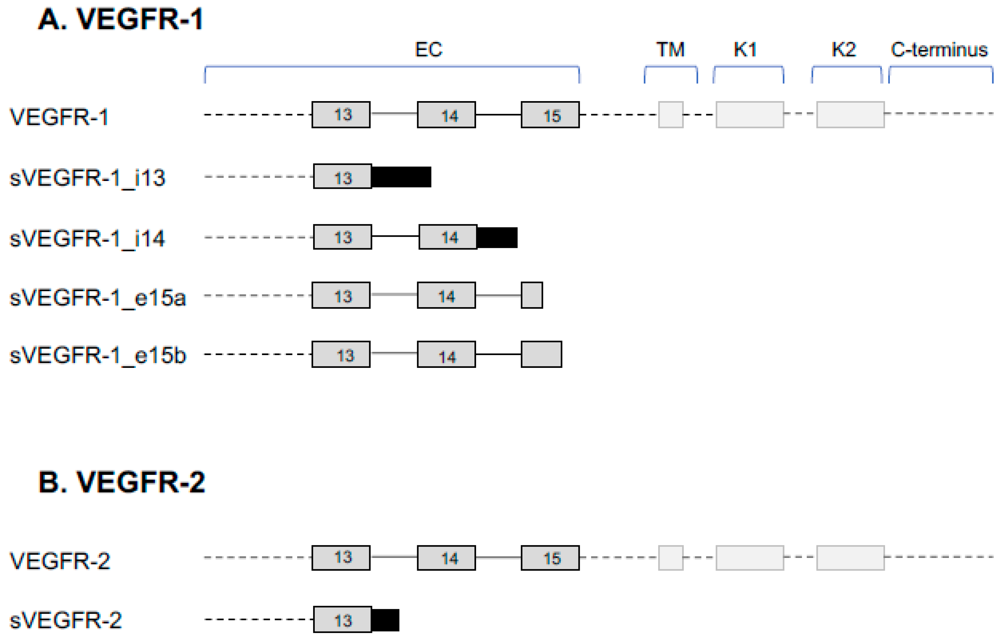
Figure 1. Alternative splice variants of VEGFR-1 and VEGFR-2. (A) Alternative splicing gives rise to five known splice variants of VEGFR-1: full length VEGFR-1, intron 13 retention (sVEGFR-1_i13), intron 14 retention (sVEGFR-1_i14), terminal exon 15a (sVEGFR-1_e15a), and terminal exon 15b (sVEGFR-1_e15b). The soluble isoforms only contain the extracellular (EC) domain and are missing the transmembrane (TM) and kinase (K1 and K2) domains. (B) Alternative splicing gives rise to two known splice variants of VEGFR-2: full length VEGFR-2 and sVEGFR-2, which result from intron 13 retention. The sVEGFR-2 only contains the EC domain.
Cells 08 00288 g001
Figure 2. Alternative splicing of VEGF-A. The VEGF-A pre-mRNA is comprised of 8 exons. Inclusion/exclusion of exons 6a,b and 7a,b gives rise to VEGF-A isoforms with differing numbers of amino acids. The use of an alternative 3′ splice site in exon 8 results in a differing c-terminal sequence of amino acids (VEGF-Axxxb isoforms). The VEGF-Axxxa family of isoforms have pro-angiogenic, pro-permeability properties whereas the VEGF-Axxxb isoforms are anti-angiogenic and anti-permeability. Figure adapted from Stevens et al. 2018.
Cells 08 00288 g002
Figure 3. VEGF-Axxxa and VEGF-Axxxb signaling through VEGFR splice variants and NRP1. Both VEGF-Axxxa and VEGF-Axxxb can bind and dimerize VEGFR-2. VEGF-Axxxa recruits NRP1, a co-receptor for VEGFR-2, which results in phosphorylation of the tyrosine kinase domains of VEGFR-2, producing pro-angiogenic and pro-permeability intracellular signaling cascades. In contrast, VEGF-Axxxb is unable to recruit NRP1, resulting in weak, transient phosphorylation of VEGFR-2 and some pro-survival signaling cascades. Soluble isoforms of NRP1, as well as sVEGFR-2 and sVEGFR-1 lack transmembrane domains and act as decoy receptors, sequestering VEGF-A.
Cells 08 00288 g003