Review

2D- and 3D-Based Intestinal Stem Cell Cultures for Personalized Medicine

The State Key Laboratory of Membrane Biology, Tsinghua-Peking Center for Life Sciences, School of Life Sciences, Tsinghua University, Beijing 100084, China
*
Author to whom correspondence should be addressed.
Cells 2018, 7(12), 225; https://doi.org/10.3390/cells7120225
Submission received: 4 October 2018 / Revised: 5 November 2018 / Accepted: 16 November 2018 / Published: 22 November 2018
(This article belongs to the Special Issue Stem Cells in Personalized Medicine)

Abstract

:
Colorectal cancer (CRC) is one of the most common cancers that have high occurrence and death in both males and females. As various factors have been found to contribute to CRC development, personalized therapies are critical for efficient treatment. To achieve this purpose, the establishment of patient-derived tumor models is critical for diagnosis and drug test. The establishment of three-dimensional (3D) organoid cultures and two-dimensional (2D) monolayer cultures of patient-derived epithelial tissues is a breakthrough for expanding living materials for later use. This review provides an overview of the different types of 2D- and 3D-based intestinal stem cell cultures, their potential benefits, and the drawbacks in personalized medicine in treatment of the intestinal disorders.

1. Introduction

The intestine is a key organ that is responsible for food digestion, nutrients absorption, and waste excretion [1,2,3], with the capacity to sustain microbial and defense pathogens, as well as to regenerate following damage by inflammation or irradiation [4]. The intestinal epithelium, which is composed of a single layer of cells and forms the crypt–villus structure, is rapidly renewed, with a complete turnover every 4–5 days [3]. The self-renewal is driven by intestinal stem cells (ISCs), which are marked by Leucine-rich-repeat-containing G-protein-coupled receptor 5 (Lgr5) and localized at the base of the crypts and intermingled with Paneth cells that provide signals to support stem cell self-renewal and maintenance [5,6,7,8,9]. Along the villus, the epithelium consists of terminally differentiated functional cells, including enterocytes, enteroendocrine cells, goblet cells, tuft cells, and other cell types. These ISCs-derived cells move up toward the tip of villus during differentiation, and eventually die at the tip of villus and are shed off into the intestinal lumen [10,11]. Although there are no villus structures in the large intestine, the architecture of the epithelium is similar to the one of the small intestine, with the ISCs at the bottom of the crypts [3]. The self-renewal and differentiation of the ISCs are tightly controlled by the coordination of multiple niche factors [2,12], such as epidermal growth factor (EGF), Wnt ligands [13,14], bone morphogenetic proteins (BMPs) [15,16,17], Notch [18,19], and others [20], as well as Hippo/YAP signaling [21].
Colorectal cancer (CRC) progresses sequentially from adenomatous polyps to advanced adenomas, carcinomas, and adenocarcinomas, and gene mutations in multiple pathways have been reported in different stages of CRC development [20,22,23]. For instance, gain-of-function mutations have been observed in the various components of the Wnt/β-catenin pathway, such as APC, CTNNB1, RNF43 and, while loss-of-function mutations are found to be associated with transforming growth factor β (TGF-β)/bone morphogenetic protein (BMP) signaling components such as SMAD2, SMAD4, TGFBR2, BMPR2, and BMPR1A. Gene mutations have also been reported in KRAS, BRAF, TP53, and DNA repair machinery. Different mutations cast complexity upon CRC treatment. In addition, ulcerative colitis (UC) and Crohn’s disease have abnormal immune responses in the colon and rectum, but whether these immune responses are a cause or a result of the disease is still under debate [24,25,26]. Although dextran sodium sulfate (DSS)-induced intestinal colitis in mice is a well-established and characterized model for acute intestinal injury and inflammation, this model, as well as the immune cells and epithelium co-culture system, have not yielded to effective therapies to treat inflammatory bowel colitis [27,28]. Furthermore, there are no specific treatments for enteric pathogen-caused illness [29,30].
Although chemotherapy and irradiation therapies are developing rapidly, gastroenterological disorders are still a major public health problem in the world due to the disease complexity. At the moment, most patients receive similar one-size-fits-all treatments. However, certain therapies work well for some patients, but they do not display promising results in others [31,32]. Therefore, personalized medicine or individualized treatments are in urgent demand to revolutionize future healthcare. To achieve this, precise patient-derived disease models are essential. Several patient-derived preclinical models have been established for intestinal disorders, including patient-derived tumor xenografts (PDX), culture systems of cancer cell lines, spheroids, and organoids and monolayers of intestinal epithelium and tumor tissues. These models have proven to be extremely valuable, not only for drug discovery and clinical trials, but also for a basic understanding of the fundamental mechanisms underlying tumorigenesis, inflammation, and interactions between the host–pathogen. This review summarizes the current 2D- or 3D-based intestinal epithelium culture systems, and discusses the advantages, caveats, potentials, and challenges, highlighting their applications in personalized medicine.

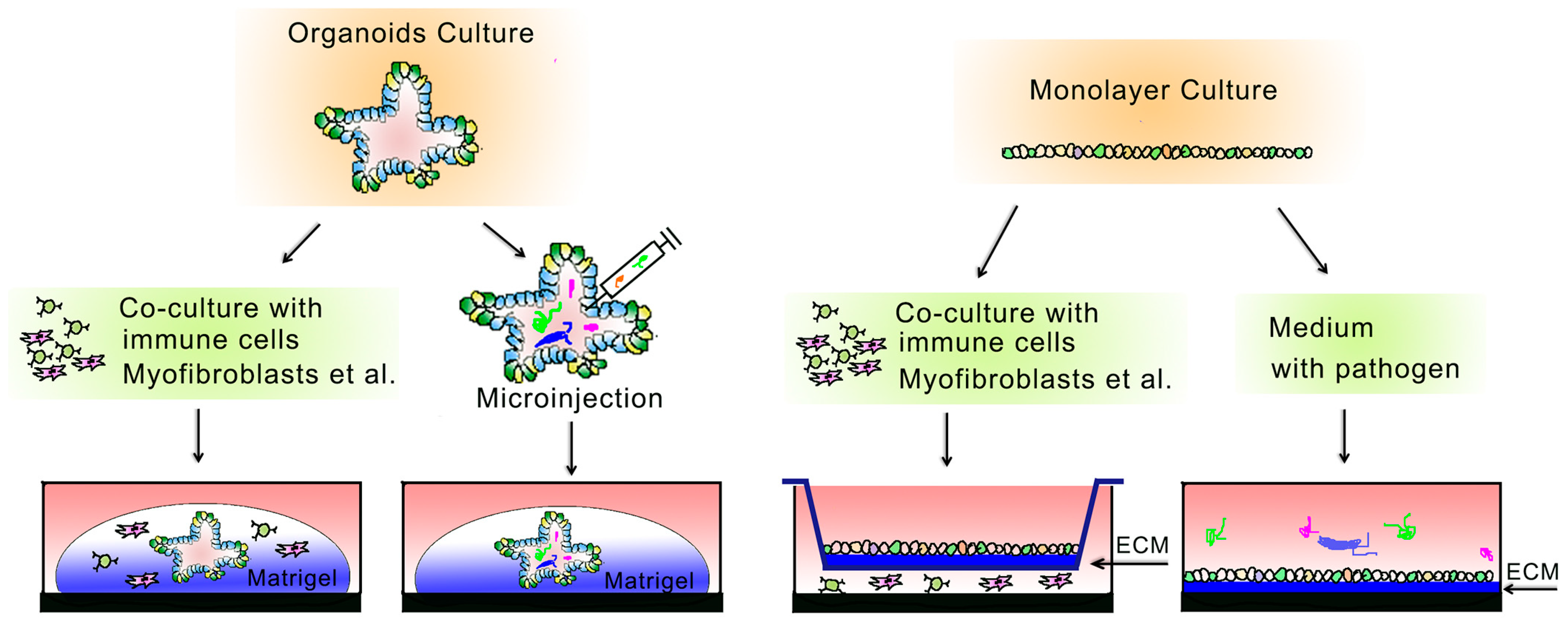
2. 3D-Based Organoid Culture System

Cell-based assays have been widely adopted in drug discovery for past decades. Historically, colon cancer cell lines, for instance HCT-116, HT-29 and SW480, have been used as the only practical option to discover drug candidates [31,32]. However, it has been demonstrated that the traditional culture system does not have sufficient clinical predictive power, due to the loss of tissue-specific architecture, the existence of gene mutations, and the lack of cell diversity in the intestine, therefore poorly recapturing tumor or inflammation conditions in patients [33]. Moreover, the necessity of adding three dimensions to the cell culture system that more closely resembles the natural environment, and its therapeutic potential, is being increasingly recognized for the future.
Scaffold-free and scaffold-based cultures are two major 3D culture systems. The scaffold-free system, like the sphere culture of the tumor cell lines in a low-attachment culture dish, suffers from similar defects as mentioned above [34]. Using droplets of a hanging drop plate, neural stem cells [35], mammary stem cells [36], corneal stem cells [37], and the embryoid bodies (EB) cultured from embryonic stem cells (ES) [38] could be cultivated in a spheroid, while it cannot be applied to the intestine. The other scaffold-based system, PDX models generated by transplanting the patient’s tumor cells to an immune-deficient mouse, could recapture some aspects of niche effects, such as the interactions of tumor cells with the surrounding stromal cells and immune cells; however, the nonmetastatic colorectal cancers fail to establish engraftments in mice [39]. A significant breakthrough was achieved by the establishment of three-dimensional (3D) organoid cultures of intestinal epithelium [40]. This culture system can rebuild the morphological, functional, and certain microenvironmental features of the intestine in vitro. The 3D organoid culture can mimic the in vivo physiology of the intestinal epithelium and it can therefore be used as a new tool for drug discovery and drug testing (Table 1).
The development of the murine intestinal organoid culture system by Clevers’ group in 2009 opened up a new era for the investigation of organ regeneration and homeostasis [41]. The successful establishment of the organoid culture system was based on the identification of the leucine-rich repeat-containing G protein-coupled receptor 5 (Lgr5) as a feasible marker of intestinal stem cells [42]. The intestinal epithelial organoids, also named “mini-gut”, can be cultured from the crypt epithelium, or even from single Lgr5+ cells [40]. Briefly, the isolated crypt epithelium or intestinal stem cells are embedded in Matrigel, a collagen- and laminin-rich extracellular matrix extract, and allowed to self-organize into a gut-like epithelial structure. The culture medium was supplemented with three growth factors: the Wnt agonist R-spondin1, the bone morphogenetic protein (BMP) antagonist Noggin, and epidermal growth factor (EGF) [40]. The 3D organotypic structures are very similar to that of normal intestinal epithelium in vivo, comprising of multiple crypt-like domains containing basal Lgr5+ cells interspersed with Paneth cells, and proliferative transient amplifying (TA) cells, enterocytes, goblet cells, and enteroendocrine cells [3,41,43]. Importantly, these cultured organoids retain their self-renewal and differentiation abilities, and their phenotype, karyotype, and gene expression profile are similar to the gut epithelium in vivo.
After the 3D culture system for murine intestinal organoids was set up, more efforts have been made to adapt this system for human tissues, especially for patient-derived colon cancers [44] (Figure 1). In addition to the three growth factors R-spondin, Noggin, and EGF, the generation of patient-derived organoids requires other factors such as Wnt3a, gastrin, prostaglandin E2, nicotinamide, the TGF-β receptor inhibitor A83-01, and the p38 inhibitor SB202190 [43].

3. 2D-Based Culture System

Although 3D-based organoid culture technology has had a great impact on the in vitro study of gut epithelium biology and its therapeutic potential, the culture system faces significant challenges. Firstly, the self-renewing organoids embedded in Matrigel could mimic some of the in vivo epithelium features, but these organoids cultured as pure epithelium lack niche cells such as stromal cells, immune cells, and vasculature [45,46,47], and are unable to fully mimic the in vivo microenvironment. Secondly, some key cell types were not found in organoids; for example, secretin+ enteroendocrine cells were missing in the 3D culture [16,48]. Thirdly, the spheroidal structure of organoids restricts luminal access of exogenous compounds and pathogens, therefore restricting physiologically relevant studies on apical transporters, metabolic enzymes, and microbiota [49,50,51,52,53,54]. They could not be used for drug screening to treat UC or Crohn’s disease. Lastly, as the Matrigel is derived from animals with undefined hydrogel matrix compositions, it has limited broad applications for the 3D organoid culture system in mechanistic and clinic-related studies. To overcome some of these limitations, therefore, researchers have focused back on 2D monolayer culture systems.
Previous works had suggested that long-term culture of primary intestinal epithelium as a planar monolayer was impossible because of rapid stem cell loss and accelerated apoptosis. Recently, several groups have successfully established 2D monolayer culture systems of intestinal epithelia that can promote ISC maintenance [51,55,56,57,58,59,60].
Although different approaches have been used to culture intestinal epithelial monolayers (Table 1), a common method involves culturing cells on an extracellular matrix-coated surface. Use of a thin coating of type I collagen and recombinant human laminin is the most common choice, which has been shown to support the rapid expansion of proliferating monolayers of mice and human intestinal epithelium containing ISCs [57,60]. Although these approaches are appealing, the technical complexity associated with matrix neutralization of the acidic collagen solution with sodium hydroxide [60] or with intestinal subepithelial myofibroblast conditioned medium [57] may restrict the broad application of such systems. Based on the transwell system, several groups have described robust monolayer culture systems, which generate cells with the apical face of epithelium towards the culture medium [51,56,59]. Furthermore, a combination of the epithelial monolayer culture on the transwell insert and the niche cell culture on the transwell plate provides an effective tool to assess the effect of niche factors from immune cells or stromal cells in the intestine (Figure 2).
However, the Bolstering Lgr5 Transformational (BLT) sandwich culture system, consisting of a bottom collagen IV coating and a collagen I gel overlay, with intestinal epithelial cells between them [59], is still complex, and this may limit its application. Furthermore, mature secretory cell markers (Lyz1, Muc2, and Chga) were absent in the system established by Tong and colleagues. Similarly, the Paneth cell marker Lyz1 was not detected in the monolayer culture described by Puzan and colleagues [56]. A lack of important cell types raises the concern of whether these systems can recapture the pathophysiological features of the intestine in normal people and patients.
Most recently, we and Thorne et al. independently established simple and economical 2D monolayer culture systems that can recapture most of the features of 3D-cultured organoids and in vivo tissue [55,58]. The intestinal crypt epithelium is placed on a thin Matrigel layer on the culture dish. To maximize the expansion of Lgr5+ ISCs, the small molecules CHIR-99021 (inhibitor of GSK3 to activate Wnt/β-catenin signaling) and LDN-193189 (BMP type I receptor inhibitor) are added in the culture medium [55,58,59]. However, different from the system by Thorne et al., our system uses blebbistatin, which could improve the survival of cultured intestinal epithelium [61], and places Matrigel with uniform thickness, which could maintain the uniform growth of epithelial cells and reduce the cost of Matrigel.
Using a transwell-based 2D system, Wang and colleagues were able to establish a columnar epithelium with villus-like structures [62]. In their culture system, irradiated 3T3-J2 cells are seeded to transwell inserts coated with 20% Matrigel as a feeder layer. Then, human ISCs are placed onto the feeder layer in a medium containing R-spondin1, Jagged-1, Noggin, and other small molecules. More importantly, by removing the apical medium, the system allowed for columnar epithelium differentiation toward villus-like structures at the air–liquid interface. They further reported the cloning and propagation of ‘’ground state’’ human intestinal stem cells (ISCGS) that had higher clonogenicity compared with organoids.
A 2D-based monolayer culture allows for better imaging observation of the dynamics of Lgr5+ stem cells and the interplay between stem cells and differentiated cells. Moreover, in contrast to the inaccessibility of the apical domain of the epithelium in organoids, exposure of the surface in the monolayer provides a suitable system to investigate the pathogen–epithelial cell interaction [63]. However, one vital limitation of the all monolayer culture systems that needs to be solved is that it cannot be easily passaged and propagated [64]. Although efficient passaging between 2D and 3D can be achieved, 2D-to-2D passaging can be accomplished with a limited number of iterations, and long-term passaging needs further optimization [16,55,57,60].

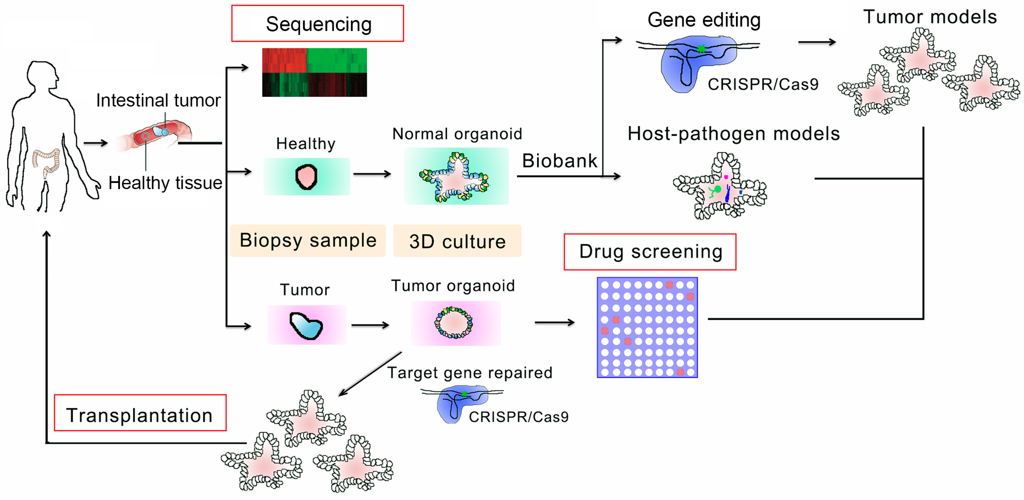
4. Applications in Personalized Therapy

Development of stem-cell-derived culture technologies raises an unprecedented opportunity to reform the health care system by transforming disease modeling to the assessment of efficacy and safety profiles, including potential drug development, the cytotoxicity evaluation of new therapeutic compounds, personalized medicine, and transplantation-based therapies for end-stage diseases [44,65,66].
As 3D organoid culture is based on patient-specific tissues and can recapture in vivo-like complexity and architecture, it has been used to investigate the mechanisms of tissue homeostasis and disease development, including colorectal cancer and ulcerative colitis [67]. Many genetic gastroenterological disorders can be modeled with patient-derived or gene-edited organoids. For instance, Jarno et al. and Mami et al. have genetically edited the genome of normal human intestinal stem cells isolated from the patients by CRISPR/Cas9 technology to target the most highly mutated colorectal cancer hotspot (APC, TP53, KRAS, and SMAD4) [68,69]. Moreover, Verissimo and colleagues reported that patient-derived tumor organoids with a CRISPR/Cas9-introduced oncogenic KRAS mutation could be used to evaluate drug efficacy in a preclinical setting [70]. Therefore, these gene-edited organoids yield a new avenue by which to screen anticancer candidates in different stages of tumorigenesis.
Another important application of intestinal organoids is to investigate the pathogenesis of various infectious diseases, and to understand the dynamics of the host–microbe interaction [29,30]. By microinjection into the organoid lumen (Figure 2), the interaction of enteric pathogens with the human intestinal epithelia have already been demonstrated, such as Vibrio cholera [71], enteroaggregative Escherichia coli (EAEC), enteropathogenic E. coli (EPEC), enterohaemorrhagic E. coli (EHEC) [72], and rotavirus [73,74]. Similarly or even better, the 2D models can be used to explore the interaction between gut microbes and the intestinal epithelium, which could not only uncover the impact of microbes on epithelium homeostasis, but also help develop an effective and personalized treatment of the intestinal disorders.
In addition to the establishment of several specific disease models, organoids can be used for high-throughput drug screens [75]. A variety of organoid-based high-throughput screen systems have been established, such as organoid-on-chip [76,77]. As there is a strong correlation between gene mutation status and the therapeutic response, known as mutation-based drug sensitivity [78], the adoption of the tumor organoids has been used in precise cancer therapy. A good example was reported by van de Wetering and colleagues, who revealed that the organoids with TP53 loss-of-function mutations displayed resistance to the MDM2 inhibitor Nutlin-3a using 20 colon tumor organoids [65]. Interestingly, an investigation of drug sensitivities of tumor-derived organoids against the library of 85 therapeutic compounds including chemotherapy and targeted therapy agents resulted in the identification of an effective treatment for each individual patient. Together, modeling specific and rare subtypes of cancer by the means of genetically engineered organoids could help to identify effective and personalized treatments [75,79].
Although much work has been done with 3D cultured organoids as disease models, drug screening, and personalized therapy, the 2D monolayer culture represents a transformative technology for personalized medicine applications that depend on drug and compound screening related to dietary and microbial metabolites. Parasites (Cryptosporidium) and microbes (Shigella, E. coli, Clostridium difficile and Salmonella typhi) can be introduced into the medium of 2D cultures directly. Using a 2D monolayer system, Wang et al. found that tannic acid could significantly inhibit intestinal epithelium growth in 2D monolayers, but not in 3D organoids, which may be due to exposure of the compound to different cell surfaces (apical side in 2D vs basolateral side in 3D) [60]. Moreover, the monolayer also provides a system for characterizing ion transport across the intestinal barrier [63]. Indeed, Kozuka et al. identified an inhibitor of potassium absorption in the murine distal colon using an epithelial monolayer culture [51]. Furthermore, the transwell-based monolayer culture is an adequate system for investigating the crosstalk between intestinal epithelial cells and niche cells (mesenchymal cells, immune cells, and myofibroblasts) as well as the enteric nervous system [56,80].

5. Challenges, Limitations, and Perspectives

As patient-derived organoids and monolayers are faithful replicas of the patient’s intestine epithelial tissues, these systems are great models that will undoubtedly facilitate diagnosis, molecule screening, drug testing, and transplantation as personalized approaches to treating intestine disorders. However, many challenges remain to meet the demands in quantity, quality, and processing robustness for commercialization and clinical trials.
For regeneration medicine, successful and efficient FDA-approved transplantation needs further improvement under culture conditions, including the animal-derived Matrigel and the high-cost of the growth factors. Several groups have used polyethylene glycol (PEG) and collagen to replace Matrigel as the supporting matrix in the culture [49,57,59,63], but the technical tediousness for handling PEG should not be neglected. Another big challenge is that niche reconstitution as the current culture system is designed for epithelial tissues. Considering the multi-functional and structurally complexity of the in vivo environment, niche cells including immune cells, stromal cells and other cell types, and vasculature, should be taken into account to better reflect the pathophysiological conditions. It is extremely important to understand the mechanism of inflammatory intestinal disorders, and ultimately to design efficient therapies to treat these untreatable diseases. For instance, the inclusion of niche cells would facilitate the screening of immunotherapy drugs and/or stromal-targeted agents. In this regard, the transwell-based 2D monolayer culture and microfluidics technology may provide some advantages of co-culture of epithelial cells and other types of cells to reconstitute the microenvironment [56,81]. Co-culture with niche cells has also been attempted in 3D systems [45], but the complex nature and high cost of 3D cultures limit their application, especially for a large-scale expansion. Recently, we have successfully established a growth factor-free culture system of murine intestinal organoids with two small molecules, which lights hope in this direction [82]. Furthermore, safe application in the clinic requires the long-term maintenance of the genome, without mutations or epigenetic changes.
With a number of major hurdles to be overcome, personalized medicine, taking advantage of the great promising potential of 3D- and 2D-based culture system, is rapidly evolving. Recently, a first-in-human trial of intestinal organoids for the treatment of inflammatory bowel disease is about to be carried out [83]. More excitingly, the patient-derived organoids have been used to test and select effective drugs for cystic fibrosis [71,84]. The 2D-based monolayer culture also holds great promises in personalized medicine. There is no doubt that 3D organoid and 2D monolayer cultures provide complementary, efficient, and clinically relevant models for personalized medicine.

Author Contributions

Y.L., and Y.-G.C. wrote the manuscript.

Funding

Research in the Y. G. Chen Lab is supported by grants from the National Key Research and Development Program of China (2017YFA0103601), and the National Natural Science Foundation of China (31330049) to YGC.

Conflicts of Interest

The authors declare no conflict of interest.

References

  1. Clevers, H. The intestinal crypt, a prototype stem cell compartment. Cell 2013, 154, 274–284. [Google Scholar] [CrossRef] [PubMed]
  2. Qi, Z.; Chen, Y.G. Regulation of intestinal stem cell fate specification. Sci. China Life Sci. 2015, 58, 570–578. [Google Scholar] [CrossRef] [PubMed] [Green Version]
  3. Barker, N. Adult intestinal stem cells: Critical drivers of epithelial homeostasis and regeneration. Nat. Rev. Mol. Cell Biol. 2014, 15, 19–33. [Google Scholar] [CrossRef] [PubMed]
  4. Nusse, Y.M.; Savage, A.K.; Marangoni, P.; Rosendahl-Huber, A.K.M.; Landman, T.A.; de Sauvage, F.J.; Locksley, R.M.; Klein, O.D. Parasitic helminths induce fetal-like reversion in the intestinal stem cell niche. Nature 2018, 559, 109–113. [Google Scholar] [CrossRef] [PubMed]
  5. Vermeulen, L.; Snippert, H.J. Stem cell dynamics in homeostasis and cancer of the intestine. Nat. Rev. Cancer 2014, 14, 468–480. [Google Scholar] [CrossRef] [PubMed]
  6. van der Flier, L.G.; Clevers, H. Stem cells, self-renewal, and differentiation in the intestinal epithelium. Annu. Rev. Physiol. 2009, 71, 241–260. [Google Scholar] [CrossRef] [PubMed]
  7. Clevers, H.C.; Bevins, C.L. Paneth cells: Maestros of the small intestinal crypts. Annu. Rev. Physiol. 2013, 75, 289–311. [Google Scholar] [CrossRef] [PubMed]
  8. Sato, T.; van Es, J.H.; Snippert, H.J.; Stange, D.E.; Vries, R.G.; van den Born, M.; Barker, N.; Shroyer, N.F.; van de Wetering, M.; Clevers, H. Paneth cells constitute the niche for Lgr5 stem cells in intestinal crypts. Nature 2011, 469, 415–418. [Google Scholar] [CrossRef] [PubMed]
  9. Rodriguez-Colman, M.J.; Schewe, M.; Meerlo, M.; Stigter, E.; Gerrits, J.; Pras-Raves, M.; Sacchetti, A.; Hornsveld, M.; Oost, K.C.; Snippert, H.J.; et al. Interplay between metabolic identities in the intestinal crypt supports stem cell function. Nature 2017, 543, 424–427. [Google Scholar] [CrossRef] [PubMed]
  10. Beumer, J.; Clevers, H. Regulation and plasticity of intestinal stem cells during homeostasis and regeneration. Development 2016, 143, 3639–3649. [Google Scholar] [CrossRef] [PubMed] [Green Version]
  11. Heath, J.P. Epithelial cell migration in the intestine. Cell. Biol. Int. 1996, 20, 139–146. [Google Scholar] [CrossRef] [PubMed]
  12. Scoville, D.H.; Sato, T.; He, X.C.; Li, L. Current view: Intestinal stem cells and signaling. Gastroenterology 2008, 134, 849–864. [Google Scholar] [CrossRef] [PubMed]
  13. Schuijers, J.; Clevers, H. Adult mammalian stem cells: The role of Wnt, Lgr5 and R-spondins. EMBO J. 2012, 31, 2685–2696. [Google Scholar] [CrossRef] [PubMed]
  14. Niehrs, C. The complex world of WNT receptor signalling. Nat. Rev. Mol. Cell Biol. 2012, 13, 767–779. [Google Scholar] [CrossRef] [PubMed]
  15. Qi, Z.; Li, Y.; Zhao, B.; Xu, C.; Liu, Y.; Li, H.; Zhang, B.; Wang, X.; Yang, X.; Xie, W.; et al. BMP restricts stemness of intestinal Lgr5(+) stem cells by directly suppressing their signature genes. Nat. Commun. 2017, 8, 13824. [Google Scholar] [CrossRef] [PubMed]
  16. Beumer, J.; Artegiani, B.; Post, Y.; Reimann, F.; Gribble, F.; Nguyen, T.N.; Zeng, H.; Van den Born, M.; Van Es, J.H.; Clevers, H. Enteroendocrine cells switch hormone expression along the crypt-to-villus BMP signalling gradient. Nat. Cell. Biol. 2018, 20, 909–916. [Google Scholar] [CrossRef] [PubMed]
  17. Wang, S.; Chen, Y.G. BMP signaling in homeostasis, transformation and inflammatory response of intestinal epithelium. Sci. China Life Sci. 2018, 61, 800–807. [Google Scholar] [CrossRef] [PubMed]
  18. Nakamura, T.; Tsuchiya, K.; Watanabe, M. Crosstalk between Wnt and Notch signaling in intestinal epithelial cell fate decision. J. Gastroenterol. 2007, 42, 705–710. [Google Scholar] [CrossRef] [PubMed]
  19. Stanger, B.Z.; Datar, R.; Murtaugh, L.C.; Melton, D.A. Direct regulation of intestinal fate by Notch. Proc. Natl. Acad. Sci. USA 2005, 102, 12443–12448. [Google Scholar] [CrossRef] [PubMed] [Green Version]
  20. Sancho, E.; Batlle, E.; Clevers, H. Signaling pathways in intestinal development and cancer. Annu. Rev. Cell. Dev. Biol. 2004, 20, 695–723. [Google Scholar] [CrossRef] [PubMed]
  21. Le Bouteiller, M.; Jensen, K.B. Hippo signalling directs intestinal fate. Nat. Cell. Biol. 2015, 17, 5–6. [Google Scholar] [CrossRef] [PubMed]
  22. Fodde, R.; Smits, R.; Clevers, H. APC, signal transduction and genetic instability in colorectal cancer. Nat. Rev. Cancer 2001, 1, 55–67. [Google Scholar] [CrossRef] [PubMed]
  23. Kedrin, D.; Gala, M.K. Genetics of the serrated pathway to colorectal cancer. Clin. Transl. Gastroenterol. 2015, 6, e84. [Google Scholar] [CrossRef] [PubMed]
  24. Rutter, M.; Saunders, B.; Wilkinson, K.; Rumbles, S.; Schofield, G.; Kamm, M.; Williams, C.; Price, A.; Talbot, I.; Forbes, A. Severity of inflammation is a risk factor for colorectal neoplasia in ulcerative colitis. Gastroenterology 2004, 126, 451–459. [Google Scholar] [CrossRef] [PubMed]
  25. Jess, T.; Loftus, E.V., Jr.; Velayos, F.S.; Harmsen, W.S.; Zinsmeister, A.R.; Smyrk, T.C.; Schleck, C.D.; Tremaine, W.J.; Melton, L.J., 3rd; Munkholm, P.; et al. Risk of intestinal cancer in inflammatory bowel disease: A population-based study from olmsted county, Minnesota. Gastroenterology 2006, 130, 1039–1046. [Google Scholar] [CrossRef] [PubMed]
  26. Gupta, R.B.; Harpaz, N.; Itzkowitz, S.; Hossain, S.; Matula, S.; Kornbluth, A.; Bodian, C.; Ullman, T. Histologic inflammation is a risk factor for progression to colorectal neoplasia in ulcerative colitis: A cohort study. Gastroenterology 2007, 133, 1099–1105; quiz 1340–1341. [Google Scholar] [CrossRef] [PubMed]
  27. Kiesler, P.; Fuss, I.J.; Strober, W. Experimental Models of Inflammatory Bowel Diseases. Cell. Mol. Gastroenterol. Hepatol. 2015, 1, 154–170. [Google Scholar] [CrossRef] [PubMed] [Green Version]
  28. Tanaka, T.; Kohno, H.; Suzuki, R.; Yamada, Y.; Sugie, S.; Mori, H. A novel inflammation-related mouse colon carcinogenesis model induced by azoxymethane and dextran sodium sulfate. Cancer Sci. 2003, 94, 965–973. [Google Scholar] [CrossRef] [PubMed] [Green Version]
  29. Abba, K.; Sinfield, R.; Hart, C.A.; Garner, P. Pathogens associated with persistent diarrhoea in children in low and middle income countries: Systematic review. BMC Infect. Dis. 2009, 9, 88. [Google Scholar] [CrossRef] [PubMed]
  30. Fletcher, S.M.; McLaws, M.L.; Ellis, J.T. Prevalence of gastrointestinal pathogens in developed and developing countries: Systematic review and meta-analysis. J. Public Health Res. 2013, 2, 42–53. [Google Scholar] [CrossRef] [PubMed]
  31. Liu, Y.; Zhang, X.; Han, C.; Wan, G.; Huang, X.; Ivan, C.; Jiang, D.; Rodriguez-Aguayo, C.; Lopez-Berestein, G.; Rao, P.H.; et al. TP53 loss creates therapeutic vulnerability in colorectal cancer. Nature 2015, 520, 697–701. [Google Scholar] [CrossRef] [PubMed] [Green Version]
  32. Yu, T.; Guo, F.; Yu, Y.; Sun, T.; Ma, D.; Han, J.; Qian, Y.; Kryczek, I.; Sun, D.; Nagarsheth, N.; et al. Fusobacterium nucleatum Promotes Chemoresistance to Colorectal Cancer by Modulating Autophagy. Cell 2017, 170, 548–563. [Google Scholar] [CrossRef] [PubMed]
  33. Wilding, J.L.; Bodmer, W.F. Cancer cell lines for drug discovery and development. Cancer Res. 2014, 74, 2377–2384. [Google Scholar] [CrossRef] [PubMed]
  34. Fang, Y.; Eglen, R.M. Three-Dimensional Cell Cultures in Drug Discovery and Development. SLAS Discov. 2017, 22, 456–472. [Google Scholar] [CrossRef] [PubMed] [Green Version]
  35. Reynolds, B.A.; Rietze, R.L. Neural stem cells and neurospheres—Re-evaluating the relationship. Nat. Methods 2005, 2, 333–336. [Google Scholar] [CrossRef] [PubMed]
  36. Dontu, G.; Abdallah, W.M.; Foley, J.M.; Jackson, K.W.; Clarke, M.F.; Kawamura, M.J.; Wicha, M.S. In vitro propagation and transcriptional profiling of human mammary stem/progenitor cells. Genes Dev. 2003, 17, 1253–1270. [Google Scholar] [CrossRef] [PubMed] [Green Version]
  37. Li, H.; Dai, Y.; Shu, J.; Yu, R.; Guo, Y.; Chen, J. Spheroid cultures promote the stemness of corneal stromal cells. Tissue Cell 2015, 47, 39–48. [Google Scholar] [CrossRef] [PubMed]
  38. Shevde, N.K.; Mael, A.A. Techniques in embryoid body formation from human pluripotent stem cells. Methods Mol. Biol. 2013, 946, 535–546. [Google Scholar] [CrossRef] [PubMed]
  39. Aboulkheyr Es, H.; Montazeri, L.; Aref, A.R.; Vosough, M.; Baharvand, H. Personalized Cancer Medicine: An Organoid Approach. Trends Biotechnol. 2018, 36, 358–371. [Google Scholar] [CrossRef] [PubMed]
  40. Sato, T.; Vries, R.G.; Snippert, H.J.; van de Wetering, M.; Barker, N.; Stange, D.E.; van Es, J.H.; Abo, A.; Kujala, P.; Peters, P.J.; et al. Single Lgr5 stem cells build crypt-villus structures in vitro without a mesenchymal niche. Nature 2009, 459, 262–265. [Google Scholar] [CrossRef] [PubMed]
  41. Sato, T.; Clevers, H. Growing self-organizing mini-guts from a single intestinal stem cell: Mechanism and applications. Science 2013, 340, 1190–1194. [Google Scholar] [CrossRef] [PubMed]
  42. Barker, N.; van Es, J.H.; Kuipers, J.; Kujala, P.; van den Born, M.; Cozijnsen, M.; Haegebarth, A.; Korving, J.; Begthel, H.; Peters, P.J.; et al. Identification of stem cells in small intestine and colon by marker gene Lgr5. Nature 2007, 449, 1003–1007. [Google Scholar] [CrossRef] [PubMed]
  43. Leushacke, M.; Barker, N. Ex vivo culture of the intestinal epithelium: Strategies and applications. Gut 2014, 63, 1345–1354. [Google Scholar] [CrossRef] [PubMed]
  44. de Sousa e Melo, F.; Kurtova, A.V.; Harnoss, J.M.; Kljavin, N.; Hoeck, J.D.; Hung, J.; Anderson, J.E.; Storm, E.E.; Modrusan, Z.; Koeppen, H.; et al. A distinct role for Lgr5(+) stem cells in primary and metastatic colon cancer. Nature 2017, 543, 676–680. [Google Scholar] [CrossRef] [PubMed]
  45. Degirmenci, B.; Valenta, T.; Dimitrieva, S.; Hausmann, G.; Basler, K. GLI1-expressing mesenchymal cells form the essential Wnt-secreting niche for colon stem cells. Nature 2018, 558, 449–453. [Google Scholar] [CrossRef] [PubMed]
  46. Shoshkes-Carmel, M.; Wang, Y.J.; Wangensteen, K.J.; Toth, B.; Kondo, A.; Massasa, E.E.; Itzkovitz, S.; Kaestner, K.H. Subepithelial telocytes are an important source of Wnts that supports intestinal crypts. Nature 2018, 557, 242–246. [Google Scholar] [CrossRef] [PubMed]
  47. Schneider, C.; O’Leary, C.E.; von Moltke, J.; Liang, H.E.; Ang, Q.Y.; Turnbaugh, P.J.; Radhakrishnan, S.; Pellizzon, M.; Ma, A.; Locksley, R.M. A Metabolite-Triggered Tuft Cell-ILC2 Circuit Drives Small Intestinal Remodeling. Cell 2018, 174, 271–284.e14. [Google Scholar] [CrossRef] [PubMed]
  48. Basak, O.; Beumer, J.; Wiebrands, K.; Seno, H.; van Oudenaarden, A.; Clevers, H. Induced Quiescence of Lgr5+ Stem Cells in Intestinal Organoids Enables Differentiation of Hormone-Producing Enteroendocrine Cells. Cell Stem Cell 2017, 20, 177–190.e4. [Google Scholar] [CrossRef] [PubMed]
  49. Wang, Y.; Kim, R.; Hinman, S.S.; Zwarycz, B.; Magness, S.T.; Allbritton, N.L. Bioengineered Systems and Designer Matrices That Recapitulate the Intestinal Stem Cell Niche. Cell. Mol. Gastroenterol. Hepatol. 2018, 5, 440–453.e1. [Google Scholar] [CrossRef] [PubMed]
  50. Beyaz, S.; Mana, M.D.; Roper, J.; Kedrin, D.; Saadatpour, A.; Hong, S.J.; Bauer-Rowe, K.E.; Xifaras, M.E.; Akkad, A.; Arias, E.; et al. High-fat diet enhances stemness and tumorigenicity of intestinal progenitors. Nature 2016, 531, 53–58. [Google Scholar] [CrossRef] [PubMed] [Green Version]
  51. Kozuka, K.; He, Y.; Koo-McCoy, S.; Kumaraswamy, P.; Nie, B.; Shaw, K.; Chan, P.; Leadbetter, M.; He, L.; Lewis, J.G.; et al. Development and Characterization of a Human and Mouse Intestinal Epithelial Cell Monolayer Platform. Stem Cell Reports 2017, 9, 1976–1990. [Google Scholar] [CrossRef] [PubMed]
  52. Dutta, D.; Clevers, H. Organoid culture systems to study host-pathogen interactions. Curr. Opin. Immunol. 2017, 48, 15–22. [Google Scholar] [CrossRef] [PubMed]
  53. Tilg, H.; Adolph, T.E.; Gerner, R.R.; Moschen, A.R. The Intestinal Microbiota in Colorectal Cancer. Cancer Cell 2018, 33, 954–964. [Google Scholar] [CrossRef] [PubMed]
  54. Jobin, C. Human Intestinal Microbiota and Colorectal Cancer: Moving Beyond Associative Studies. Gastroenterology 2017, 153, 1475–1478. [Google Scholar] [CrossRef] [PubMed]
  55. Liu, Y.; Qi, Z.; Li, X.; Du, Y.; Chen, Y.G. Monolayer culture of intestinal epithelium sustains Lgr5(+) intestinal stem cells. Cell Discov. 2018, 4, 32. [Google Scholar] [CrossRef] [PubMed]
  56. Puzan, M.; Hosic, S.; Ghio, C.; Koppes, A. Enteric Nervous System Regulation of Intestinal Stem Cell Differentiation and Epithelial Monolayer Function. Sci. Rep. 2018, 8, 6313. [Google Scholar] [CrossRef] [PubMed]
  57. Scott, A.; Rouch, J.D.; Jabaji, Z.; Khalil, H.A.; Solorzano, S.; Lewis, M.; Martin, M.G.; Stelzner, M.G.; Dunn, J.C. Long-term renewable human intestinal epithelial stem cells as monolayers: A potential for clinical use. J. Pediatr. Surg. 2016, 51, 995–1000. [Google Scholar] [CrossRef] [PubMed] [Green Version]
  58. Thorne, C.A.; Chen, I.W.; Sanman, L.E.; Cobb, M.H.; Wu, L.F.; Altschuler, S.J. Enteroid Monolayers Reveal an Autonomous WNT and BMP Circuit Controlling Intestinal Epithelial Growth and Organization. Dev. Cell 2018, 44, 624–633.e4. [Google Scholar] [CrossRef] [PubMed]
  59. Tong, Z.; Martyn, K.; Yang, A.; Yin, X.; Mead, B.E.; Joshi, N.; Sherman, N.E.; Langer, R.S.; Karp, J.M. Towards a defined ECM and small molecule based monolayer culture system for the expansion of mouse and human intestinal stem cells. Biomaterials 2018, 154, 60–73. [Google Scholar] [CrossRef] [PubMed]
  60. Wang, Y.; DiSalvo, M.; Gunasekara, D.B.; Dutton, J.; Proctor, A.; Lebhar, M.S.; Williamson, I.A.; Speer, J.; Howard, R.L.; Smiddy, N.M.; et al. Self-renewing Monolayer of Primary Colonic or Rectal Epithelial Cells. Cell. Mol. Gastroenterol. Hepatol. 2017, 4, 165–182.e7. [Google Scholar] [CrossRef] [PubMed]
  61. Zhao, B.; Qi, Z.; Li, Y.; Wang, C.; Fu, W.; Chen, Y.G. The non-muscle-myosin-II heavy chain Myh9 mediates colitis-induced epithelium injury by restricting Lgr5+ stem cells. Nat. Commun. 2015, 6, 7166. [Google Scholar] [CrossRef] [PubMed] [Green Version]
  62. Wang, X.; Yamamoto, Y.; Wilson, L.H.; Zhang, T.; Howitt, B.E.; Farrow, M.A.; Kern, F.; Ning, G.; Hong, Y.; Khor, C.C.; et al. Cloning and variation of ground state intestinal stem cells. Nature 2015, 522, 173–178. [Google Scholar] [CrossRef] [PubMed] [Green Version]
  63. Gjorevski, N.; Sachs, N.; Manfrin, A.; Giger, S.; Bragina, M.E.; Ordonez-Moran, P.; Clevers, H.; Lutolf, M.P. Designer matrices for intestinal stem cell and organoid culture. Nature 2016, 539, 560–564. [Google Scholar] [CrossRef] [PubMed]
  64. Braverman, J.; Yilmaz, O.H. From 3D Organoids back to 2D Enteroids. Dev. Cell 2018, 44, 533–534. [Google Scholar] [CrossRef] [PubMed]
  65. van de Wetering, M.; Francies, H.E.; Francis, J.M.; Bounova, G.; Iorio, F.; Pronk, A.; van Houdt, W.; van Gorp, J.; Taylor-Weiner, A.; Kester, L.; et al. Prospective derivation of a living organoid biobank of colorectal cancer patients. Cell 2015, 161, 933–945. [Google Scholar] [CrossRef] [PubMed]
  66. Praharaj, P.P.; Bhutia, S.K.; Nagrath, S.; Bitting, R.L.; Deep, G. Circulating tumor cell-derived organoids: Current challenges and promises in medical research and precision medicine. Biochim. Biophys. Acta. Rev. Cancer 2018, 1869, 117–127. [Google Scholar] [CrossRef] [PubMed]
  67. Lancaster, M.A.; Knoblich, J.A. Organogenesis in a dish: Modeling development and disease using organoid technologies. Science 2014, 345, 1247125. [Google Scholar] [CrossRef] [PubMed]
  68. Matano, M.; Date, S.; Shimokawa, M.; Takano, A.; Fujii, M.; Ohta, Y.; Watanabe, T.; Kanai, T.; Sato, T. Modeling colorectal cancer using CRISPR-Cas9-mediated engineering of human intestinal organoids. Nat. Med. 2015, 21, 256–262. [Google Scholar] [CrossRef] [PubMed]
  69. Drost, J.; van Jaarsveld, R.H.; Ponsioen, B.; Zimberlin, C.; van Boxtel, R.; Buijs, A.; Sachs, N.; Overmeer, R.M.; Offerhaus, G.J.; Begthel, H.; et al. Sequential cancer mutations in cultured human intestinal stem cells. Nature 2015, 521, 43–47. [Google Scholar] [CrossRef] [PubMed]
  70. Verissimo, C.S.; Overmeer, R.M.; Ponsioen, B.; Drost, J.; Mertens, S.; Verlaan-Klink, I.; Gerwen, B.V.; van der Ven, M.; Wetering, M.V.; Egan, D.A.; et al. Targeting mutant RAS in patient-derived colorectal cancer organoids by combinatorial drug screening. Elife 2016, 5, e18489. [Google Scholar] [CrossRef] [PubMed]
  71. Schwank, G.; Koo, B.K.; Sasselli, V.; Dekkers, J.F.; Heo, I.; Demircan, T.; Sasaki, N.; Boymans, S.; Cuppen, E.; van der Ent, C.K.; et al. Functional repair of CFTR by CRISPR/Cas9 in intestinal stem cell organoids of cystic fibrosis patients. Cell Stem Cell 2013, 13, 653–658. [Google Scholar] [CrossRef] [PubMed]
  72. VanDussen, K.L.; Marinshaw, J.M.; Shaikh, N.; Miyoshi, H.; Moon, C.; Tarr, P.I.; Ciorba, M.A.; Stappenbeck, T.S. Development of an enhanced human gastrointestinal epithelial culture system to facilitate patient-based assays. Gut 2015, 64, 911–920. [Google Scholar] [CrossRef] [PubMed]
  73. Saxena, K.; Blutt, S.E.; Ettayebi, K.; Zeng, X.L.; Broughman, J.R.; Crawford, S.E.; Karandikar, U.C.; Sastri, N.P.; Conner, M.E.; Opekun, A.R.; et al. Human Intestinal Enteroids: A New Model To Study Human Rotavirus Infection, Host Restriction, and Pathophysiology. J. Virol. 2016, 90, 43–56. [Google Scholar] [CrossRef] [PubMed]
  74. Yin, Y.; Bijvelds, M.; Dang, W.; Xu, L.; van der Eijk, A.A.; Knipping, K.; Tuysuz, N.; Dekkers, J.F.; Wang, Y.; de Jonge, J.; et al. Modeling rotavirus infection and antiviral therapy using primary intestinal organoids. Antiviral Res. 2015, 123, 120–131. [Google Scholar] [CrossRef] [PubMed]
  75. Weeber, F.; van de Wetering, M.; Hoogstraat, M.; Dijkstra, K.K.; Krijgsman, O.; Kuilman, T.; Gadellaa-van Hooijdonk, C.G.; van der Velden, D.L.; Peeper, D.S.; Cuppen, E.P.; et al. Preserved genetic diversity in organoids cultured from biopsies of human colorectal cancer metastases. Proc. Natl. Acad. Sci. USA 2015, 112, 13308–13311. [Google Scholar] [CrossRef] [PubMed] [Green Version]
  76. Kasendra, M.; Tovaglieri, A.; Sontheimer-Phelps, A.; Jalili-Firoozinezhad, S.; Bein, A.; Chalkiadaki, A.; Scholl, W.; Zhang, C.; Rickner, H.; Richmond, C.A.; et al. Development of a primary human Small Intestine-on-a-Chip using biopsy-derived organoids. Sci. Rep. 2018, 8, 2871. [Google Scholar] [CrossRef] [PubMed]
  77. May, P.; Evans, S.; Parry, L. Organoids, organs-on-chips and other systems, and microbiota. Emerg. Top. Life Sci. 2017, 1, 385–400. [Google Scholar] [CrossRef] [Green Version]
  78. Voest, E.E.; Bernards, R. DNA-Guided Precision Medicine for Cancer: A Case of Irrational Exuberance? Cancer Discov. 2016, 6, 130–132. [Google Scholar] [CrossRef] [PubMed] [Green Version]
  79. Friedman, A.A.; Letai, A.; Fisher, D.E.; Flaherty, K.T. Precision medicine for cancer with next-generation functional diagnostics. Nat. Rev. Cancer 2015, 15, 747–756. [Google Scholar] [CrossRef] [PubMed] [Green Version]
  80. Hayakawa, Y.; Sakitani, K.; Konishi, M.; Asfaha, S.; Niikura, R.; Tomita, H.; Renz, B.W.; Tailor, Y.; Macchini, M.; Middelhoff, M.; et al. Nerve Growth Factor Promotes Gastric Tumorigenesis through Aberrant Cholinergic Signaling. Cancer Cell 2017, 31, 21–34. [Google Scholar] [CrossRef] [PubMed] [Green Version]
  81. Yissachar, N.; Zhou, Y.; Ung, L.; Lai, N.Y.; Mohan, J.F.; Ehrlicher, A.; Weitz, D.A.; Kasper, D.L.; Chiu, I.M.; Mathis, D.; et al. An Intestinal Organ Culture System Uncovers a Role for the Nervous System in Microbe-Immune Crosstalk. Cell 2017, 168, 1135–1148. [Google Scholar] [CrossRef] [PubMed]
  82. Li, Y.; Liu, Y.; Liu, B.; Wang, J.; Wei, S.; Qi, Z.; Wang, S.; Fu, W.; Chen, Y.G. A growth factor-free culture system underscores the coordination between Wnt and BMP signaling in Lgr5(+) intestinal stem cell maintenance. Cell Discov. 2018, 4, 49. [Google Scholar] [CrossRef] [PubMed]
  83. Takebe, T.; Wells, J.M.; Helmrath, M.A.; Zorn, A.M. Organoid Center Strategies for Accelerating Clinical Translation. Cell Stem Cell 2018, 22, 806–809. [Google Scholar] [CrossRef] [PubMed]
  84. Dekkers, J.F.; Wiegerinck, C.L.; de Jonge, H.R.; Bronsveld, I.; Janssens, H.M.; de Winter-de Groot, K.M.; Brandsma, A.M.; de Jong, N.W.; Bijvelds, M.J.; Scholte, B.J.; et al. A functional CFTR assay using primary cystic fibrosis intestinal organoids. Nat. Med. 2013, 19, 939–945. [Google Scholar] [CrossRef] [PubMed]
Figure 1. Applications of organoid technology for drug screening and therapy in personalized medicine. Gene mutations in patients’ tissues can be identified by genome or transcriptome sequencing. Meanwhile, the biopsy materials can be cultured in Matrigel to form organoids, which can be used to establish disease models via gene editing and co-culturing with niche cells and pathogens. The modified tumor organoids can be utilized for drug screening and testing. Gene mutations in organoids can be corrected by clustered regularly interspaced short palindromic repeats/CRISPR-associated 9 (CRISPR/Cas9)-mediated gene editing for transplantation.
Cells 07 00225 g001
Figure 2. Models for intestinal epithelium-niche cells and epithelium–microbes/pathogen interactions. Both the 3D organoid culture and the transwell-based 2D monolayer culture can be used for the investigation of intestinal epithelium–niche cell interactions. To study the epithelium–microbe/pathogen interactions, microbes and pathogens can be introduced directly into the culture medium of 2D monolayer systems, while for the 3D cultures, pathogens need to be injected into the lumen of organoids to allow them to come into contact with the apical side of the epithelium.
Cells 07 00225 g002
Table 1. Characteristics of the described preclinical cancer models.
3D-Based Culture System2D-Based Culture System
SpheroidsPDXOrganoidsMonolayerAir-Liquid Interface Culture
Culture TechniqueLow-melting AgroseTransplanted into immune-deficient mouseEmbended in MatrigelCell linesPlate coated with Collagen ITranswell coated with Collagen IPlate coated with MatrigelSeeded onto 3T3-J2 cells which cultured on transwell coated with 20% Matrigel
OriginCell linesPatient-derived tumorsmIEC; hIEC; Patient-derived tumorsCell linesmIEC; hIECmIEC; hIECmIEC; hIEC; TumorshIEC; Tumors
Key FeaturesIn vivo-like complexityLack of mature secretory lineageLack of mature secretory lineage
Stemness and multipotency--
Easily passaged
ApplicationsCancer subtype modeling (gene manipulation)
Host-pathogen interaction
Co-culture
High-throughput screening
High-throughput imaging
Time consumption for modelingMediumLongMediumShortMediumMediumMediumMedium
Cost benefitsLowHighHighLowMediumMediumMediumHigh
Reference[34][34][34,35,36,39,47,58,59,63][31,32,34][47,55,59][47,54,59][47,50,53,59][57]

