5-Methylcytidine RNA Epitranscriptomics in Women’s Health and Disease: Mechanisms and Clinical Implications

 ,
,  , , and
Highlights
- 5-methylcytidine (m5C) is a dynamic RNA modification that regulates RNA stability, translation, localization, and stress responses across diverse biological systems.
- m5C dysregulation is implicated in key processes relevant to women’s health, including early embryonic development, reproductive function, pregnancy adaptation, metabolic regulation, and cancer progression.
- m5C represents a critical epitranscriptomic regulatory layer linking RNA metabolism with hormone-responsive physiology and sex-specific disease mechanisms.
- It has strong potential as a biomarker and therapeutic target, supporting future precision medicine approaches in women’s health and disease.
Abstract
1. Introduction
1.1. Epigenetics vs. Epitranscriptomics: Expanding Regulatory Layers
1.2. Overview of RNA Modifications with Focus on m5C
1.3. Rationale for Focusing on Women’s Health
1.4. Scope and Objectives of the Review
2. Molecular Basis of m5C RNA Modification
2.1. Chemical Nature and Distribution of m5C
2.2. m5C Writers: RNA Cytosine Methyltransferases
2.3. m5C Readers: Recognition and Functional Interpretation
2.4. m5C Erasers and Modification Dynamics
3. Methodologies for Detecting m5C
3.1. Mass Spectrometry
3.2. Antibody-Based Methods
3.3. Bisulfite Sequencing–Based Approaches
3.4. Bisulfite-Free, TET-Assisted Chemical Oxidation Sequencing Method for Base-Resolution m5C Detection (m5C-TAC-seq)
3.5. Nanopore-Based Sequencing for Direct Detection of m5C
4. m5C RNA Modification in Women’s Health: Physiological and Disease Contexts
4.1. Physiological Roles of 5-methylcytosine in Ovarian Development, Oogenesis, Folliculogenesis, and Ovulation
4.2. Physiological Roles of 5-methylcytosine (m5C) in Fertilization, Early Embryogenesis, and Endometrial Receptivity
4.3. Polycystic Ovary Syndrome
4.4. Premature Ovarian Insufficiency
4.5. Pregnancy and Placental Biology: Preeclampsia as a Model Disorder

| Reproduction and Development | Sample Types | Approaches | Mechanism | Biological Effects | Publication Date | Ref. |
|---|---|---|---|---|---|---|
| Embryonic development | Mammalian ovarian follicles and oocytes (mouse, pig, and cow) | Immunofluorescence and confocal microscopy to localize RNA modification machinery, assessment of m6A and m5C abundance in oocyte vs. somatic mRNA | Epitranscriptomic regulation of maternal mRNAs involves enzyme localization, with high m6A and lower m5C levels, controlling mRNA stability and translation during folliculogenesis and early embryonic transition. | Regulation of maternal mRNA stability and utilization, coordination of oocyte maturation and follicle development, and establishment of species-specific control of early embryonic development during transcriptional silencing | May, 2023 | [73] |
| Ovarian dysfunction with reduced follicle count and ovarian aging | Nsun5 KO mouse ovaries/oocytes and human granulosa-like tumor cell line (KGN) cells | m5C profiling, mRNA stability and splicing analysis, and functional validation | NSUN5-dependent m5C stabilizes maternal mRNAs and regulates translation during folliculogenesis and MZT | Follicular arrest, reduced ovarian function, and impaired embryonic development | Dec, 2022 | [78] |
| Early embryonic development (mouse preimplantation embryos) | Mouse preimplantation embryos (two-cell to blastocyst stages) | siRNA-mediated knockdown of Nsun5; assessment of blastocyst formation, cell number, apoptosis, lineage markers (CDX2/OCT4), and YAP1 localization | NSUN5 regulates Hippo signaling by affecting YAP1 nuclear translocation and expression of LATS1/2, likely via m5C-dependent RNA regulation | Nsun5 depletion reduces blastocyst formation, cell proliferation, and differentiation, increases apoptosis, and disrupts lineage specification | May, 2024 | [79] |
| Early embryogenesis | Zebrafish early embryos | Genome-wide m5C mapping and YBX1-mediated RNA interaction analysis | m5C-modified maternal mRNAs are preferentially recognized by YBX1 through specific residue-mediated interactions, and together with Pabpc1a, YBX1 stabilizes these transcripts, thereby preventing their degradation during the MZT. | Maintains maternal mRNA stability and ensures successful early embryonic development during the transition from maternal to zygotic control. | Sep, 2019 | [80] |
| Early embryonic development during pre-implantation | Mouse, human, bovine, and goat pre-implantation embryos; 8-cell stage embryos used for YBX1 knockdown studies | RNA-seq analysis of public datasets and YBX1 knockdown embryos (siRNA microinjection), differential gene expression analysis, alternative splicing (AS) analysis, enrichment analysis, 5-EU staining, and immunofluorescence | YBX1 regulates maternal mRNA stability and alternative splicing, mediates decay of maternal transcripts, and supports transcriptional activation during MZT | YBX1 ensures maternal mRNA decay, splicing, and ZGA; its knockdown arrests embryos at 2–4 cells with impaired decay and reduced blastocyst formation. | Feb, 2022 | [81] |
| Zebrafish folliculogenesis | Zebrafish ovarian follicles at primary growth (PG) and secondary growth (SG) stages | Proteomic analysis of folliculogenesis (PG–SG transition), gene/protein expression profiling, phylogenetic analysis, and functional characterization of YBX1 | YBX1 stabilizes and represses maternal mRNAs in early follicles; its downregulation releases these mRNAs for translation, enabling follicle activation. | Maintenance of early follicle development through mRNA stabilization and translational repression, followed by activation of follicle growth via coordinated mRNA translation | Oct, 2023 | [82] |
| Embryonic development | Zebrafish oocytes and early embryos (maternal ybx1 mutant and knockdown models) | Genetic knockout (null allele) and morpholino knockdown of YBX1, assessment of oocyte maturation and embryogenesis, translation activity assays, and rescue experiments via translational inhibition | Maternal YBX1 functions as a global translational repressor by associating with target mRNAs and processing body components, thereby limiting protein synthesis during oocyte maturation and early embryogenesis | YBX1 ensures oocyte maturation and MZT; its loss causes increased translation, stress response activation, and defective early embryonic development. | Oct, 2018 | [83] |
| Oocyte maturation and early embryonic development | Mouse oocytes; maternal mRNA; genetic and molecular perturbation models | Functional studies including PGC7 depletion, protein interaction assays, phosphorylation analysis, translation assays, and AKT signaling pathway investigation | PGC7 promotes maternal mRNA translation by enabling AKT1 to phosphorylate YBX1, releasing translational repression on target mRNAs and forming the PGC7–AKT1–YBX1 axis. | Enhanced translation of maternal mRNAs, proper oocyte maturation, and support of early embryonic development through activation of key regulatory proteins | Dec, 2024 | [84] |
| Embryonic development | Embryos across multiple species including Drosophila and humans | Developmental m5C mapping, evolutionary comparison, and functional genetic studies | NSUN2/NSUN6-mediated m5C regulates maternal mRNA dynamics during early embryogenesis | Controls cell cycle progression, maternal-to-zygotic transition, and evolutionary innovation of mRNA regulation | May, 2022 | [85] |
| Ovarian germ line stem cell development | Drosophila ovarian germ line stem cells (GSCs); germ cells; | Loss- and gain-of-function of YPS, RNA-binding assays, m5C recognition analysis, cross-species rescue experiments with YBX1, and structural analysis of protein–RNA interactions | The YBX1 homolog YPS preferentially recognizes and binds m5C-modified RNAs via conserved cold-shock domains, enabling selective regulation of target mRNAs required for germ line stem cell maintenance and differentiation | Promotion of germ line stem cell maintenance, proliferation, and differentiation in the ovary; disruption of m5C binding impairs normal stem cell development | Feb, 2020 | [86] |
| Early zygotic development | Porcine embryos, from the one-cell stage to blastocyst stage | Knockdown of YBX1 using siRNA microinjection at the one-cell stage; additional knockdown of IGF2BP1 to study mechanistic links. Analysis included mRNA expression, histone modification, and blastocyst formation rate. | YBX1 regulates ZGA through m6A RNA modification by modulating the levels of the m6A writer METTL3 and m6A reader IGF2BP1, affecting maternal mRNA decay and ZGA gene expression | YBX1 knockdown impairs blastocyst formation and ZGA by increasing maternal mRNA and disrupting m6A-mediated regulation | Jul, 2023 | [90] |
| Fetal pulmonary fibrosis induced by maternal exposure to carbon black nanoparticles | In vivo pregnant rat exposure model with analysis of offspring lung tissues, as well as ex vivo lung epithelial cells | Animal exposure experiments, cell-based assays, gene expression and transcriptional regulation analyses, and functional studies to evaluate EMT, reticulophagy, and m5C RNA methylation | NSUN2-mediated m5C methylation enhances the stability of SP1 mRNA through recognition by YBX1, leading to increased SP1 protein expression; SP1 then transcriptionally activates DDRGK1, which interacts with FAM134B to promote reticulophagy, thereby driving EMT-associated fibrotic processes. | Increased reticulophagy, enhanced EMT, and aggravated fibrosis in offspring lung tissue following maternal nanoparticle exposure | Mar, 2025 | [91] |
| Early development arrest | Mouse nuclear transfer embryos | H3K9me3 profiling to identify reprogramming-resistant genes, siRNA screening, CRISPR/Cas9 knockout of candidate genes, and mRNA supplementation experiments | ALYREF regulates Nanog to support proper inner cell mass formation, Alyref geneis partially silenced due to H3K9me3-mediated repression of Klf16 | Knockout of Alyref results in early developmental arrest, whereas supplementation of its mRNA promotes efficient preimplantation development of cloned embryos | Aug, 2023 | [92] |
| Recurrent Implantation. Failure, endometrial decidualization | Human endometrial tissues from proliferative and secretory stages, along with in vitro models including Ishikawa cells and BeWo spheroids to simulate embryo attachment. | Single-cell RNA-seq analysis, m5C-BS-seq, and dot blot assays to assess m5C levels, embryo attachment by Ishikawa–BeWo co-culture, with mechanisms further examined using Cut&Tag and co-immunoprecipitation. | NSUN2-mediated m5C regulates gene expression and splicing. Its overexpression increases STAT1 and MMP14 via exon skipping and enhances CLDN4 expression by modulating SIRT4 and H3K9ac, linking RNA methylation to histone modification. | NSUN2 overexpression promotes proliferation and autophagy while reducing embryo attachment, impairing endometrial receptivity and contributing to recurrent implantation failure. | Apr, 2024 | [94] |
| PCOS | PCOS patient samples, lipopolysaccharide (LPS)-treated KGN granulosa cell line; PCOS rat model ovaries and primary granulosa cells | LPS-induced pyroptosis model, ELISA, flow cytometry, Western blotting, MeRIP, dual-luciferase assay, actinomycin D treatment, and in vivo hormone analysis. | NSUN7 mediates m5C methylation of NLRP3 mRNA, enhancing its stability and promoting pyroptosis in granulosa cells. | Silencing NSUN7 reduces granulosa cell pyroptosis, restores hormone balance, and attenuates PCOS progression; NLRP3 overexpression reverses these protective effects. | Oct, 2025 | [95] |
| Primary ovarian insufficiency | Human granulosa cells (GCs) from POI patients, KGN human granulosa cell line | YBX1 knockdown and overexpression, Transcriptome and m5C methylome profiling | YBX1 stabilizes m5C-modified transcripts of cell cycle–related genes. Loss of YBX1 destabilizes these transcripts in an m5C-dependent manner, leading to impaired G1–S transition and cell cycle arrest. | Reduced granulosa cell proliferation, cell cycle arrest (G1 phase), granulosa cell dysfunction, contributes to POI pathogenesis | May, 2025 | [98] |
| Premature ovarian failure. | Granulosa cells exposed to reactive oxygen species (ROS), particularly through H2O2-induced oxidative stress models | DNMT2 overexpression and mutational analysis, along with assays to evaluate ROS levels, DNA damage, apoptosis, and RNA m5C methylation activity | ROS induces nuclear translocation of DNMT2, which facilitates DNA damage repair in an m5C methylation–dependent manner, whereas impaired DNMT2 activity reduces RNA m5C modification and compromises DNA repair capacity | DNMT2 reduces ROS-induced DNA damage and apoptosis in granulosa cells, thereby protecting against cell death and contributing to the prevention or alleviation of premature ovarian failure. | Jun, 2021 | [99] |
| Premature ovarian insufficiency (ROS-induced ovarian dysfunction and granulosa cell senescence) | Human granulosa cells (GCs) under oxidative stress, human umbilical cord mesenchymal stem cell-derived exosomes (HuMSCs-Ex) | Exosome-based therapeutic treatment combined with molecular, biochemical, and functional assays to investigate m5C RNA modification, including relevant analyses of DNMT2, YBX1, and COX5B | DNMT2 catalyzes m5C modification on COX5B mRNA, which is recognized by exosome-delivered YBX1 that directly binds and stabilizes the transcript, thereby promoting its translation in an m5C-dependent manner | Reduced ROS accumulation, improved mitochondrial function, decreased granulosa cell senescence, and overall alleviation of oxidative damage associated with POI | May, 2025 | [100] |
| Preeclampsia | placental tissues from normotensive pregnancies and preeclampsia patients | UMI-MeRIP-Seq transcriptomic m5C profiling and pathway enrichment analysis | Dysregulated m5C distribution and altered DNMT3B/TET3 expression affect MAPK/cAMP and hypoxia/inflammatory pathways | Contributes to placental dysfunction and PE-associated immune and hypoxic responses | Feb, 2023 | [101] |
| Preeclampsia | Human decidual tissue from PE and normal pregnancy | MeRIP-seq and RNA-seq with integrative bioinformatics, pathway analysis, and qPCR validation | Aberrant m5C methylation alters mRNA expression of decidualization-related genes, disrupting key signaling pathways in PE | Distinct methylation and gene expression signatures, identification of PE-associated genes, and impaired decidualization collectively contribute to disease development. | Nov, 2023 | [102] |
| Preeclampsia | Hypoxia-treated trophoblast cells | ALKBH1 modulation with functional assays and m5C/RNA interaction analyses | ALKBH1-mediated m5C demethylation destabilizes PSMD14 mRNA, impairing trophoblast function ALKBH1-mediated m5C demethylation destabilizes PSMD14 mRNA, impairing trophoblast function | Reduced trophoblast viability, migration, and invasion contributing to PE pathogenesis | Mar, 2025 | [103] |
| Preeclampsia | Hypoxic trophoblast cells, PE placentas, and PE mice | Enoxaparin treatment with m5C analysis, NSUN2/PAX3 interaction assays, and functional validation in vitro and in vivo | Enoxaparin activates the NSUN2/m5C/PAX3 axis to stabilize PAX3 mRNA and restore trophoblast function | Improved trophoblast activity and amelioration of PE-associated hypertension, proteinuria, and placental injury | Nov, 2025 | [104] |
5. m5C in Women-Specific Cancers
5.1. Breast Cancer
5.2. m5C in Gynecologic Malignancies
5.2.1. Ovarian Cancer
5.2.2. Endometrial Cancer
5.2.3. Cervical Cancer
6. m5C RNA Modification in Metabolic and Cardiovascular Health in Women

6.1. Pregnancy-Associated Metabolic Disorders
6.2. Cardiovascular Disease Risk Modulation
6.2.1. m5C Reprogramming and Cardiovascular Diseases
6.2.2. m5C Regulators and Cardiovascular Diseases
6.2.3. Sex Differences in Cardiovascular Disease and Clinical Implications of m5C Regulation
7. Clinical Implications and Translational Potential
7.1. m5C as Biomarkers
7.1.1. Diagnostic and Prognostic Applications
7.1.2. Circulating RNA and Liquid Biopsy Potential
7.2. Therapeutic Targeting of m5C Machinery
7.2.1. Small-Molecule Inhibitors
7.2.2. Challenges in Specificity and Safety
8. Knowledge Gaps and Future Directions
8.1. Tissue-Specific m5C Landscapes in Women
8.2. Causality and Functional Validation in m5C Epitranscriptomics
8.3. Interaction with Sex Hormones
8.4. Longitudinal and Life-Course Studies
8.5. Single-Cell and Spatial Epitranscriptomics
8.6. Ethical and Clinical Considerations
9. Conclusions
Author Contributions
Funding
Institutional Review Board Statement
Informed Consent Statement
Data Availability Statement
Acknowledgments
Conflicts of Interest
Abbreviations
| AAA | Abdominal aortic aneurysm |
| ALKBH | AlkB homolog family |
| ALYREF | Aly/REF export factor |
| AUC | Area under the curve |
| BC | Breast cancer |
| ca5C | 5-carboxylcytidine |
| CEA | Carcinoembryonic antigen |
| CC | Cervical cancer |
| cfRNA | Cell-free RNA |
| CVD | Cardiovascular disease |
| DNMT2/TRDMT1 | DNA methyltransferase 2/tRNA aspartic acid methyltransferase 1 |
| EOC | Epithelial ovarian cancer |
| EVs | Extracellular vesicles |
| f5C | 5-formylcytidine |
| GDM | Gestational diabetes mellitus |
| HGSOC | High-grade serous ovarian cancer |
| hm5C | 5-hydroxymethylcytidine |
| HPV | Human papillomavirus |
| ICAM-1 | Intercellular adhesion molecule 1 |
| KGN | Human granulosa-like tumor cell line |
| lncRNA | Long noncoding RNA |
| LPS | Lipopolysaccharide |
| m1A | N1-methyladenosine |
| m5C | 5-methylcytidine |
| m6A | N6-methyladenosine |
| miRNA | MicroRNA |
| mRNA | Messenger RNA |
| MST | Microscale thermophoresis |
| MZT | Maternal-to-zygotic transition |
| ncRNA | Noncoding RNA |
| NLRP3 | NLR family pyrin domain containing 3 |
| NMF | Nonnegative matrix factorization |
| NSUN | NOP2/Sun RNA methyltransferase family |
| PCOS | Polycystic ovary syndrome |
| P-TEFb | Positive transcription elongation factor b |
| RNA-BS-Seq | RNA bisulfite sequencing |
| RIP-seq | RNA immunoprecipitation sequencing |
| ROC | Receiver operating characteristic |
| rRNA | Ribosomal RNA |
| SAM | S-adenosylmethionine |
| TNBC | Triple-negative breast cancer |
| tRNA | Transfer RNA |
| TET | Ten-Eleven Translocation family |
| TNF-α | Tumor necrosis factor alpha |
| TREX | Transcription–Export complex |
| UTR | Untranslated region |
| YBX1 | Y-box binding protein 1 |
| YTHDF2 | YTH N6-methyladenosine RNA binding protein F2 |
| ZGA | Zygotic genome activation |
| Ψ | Pseudouridine |
| α-KG | Alpha-ketoglutarate |
References
- Kouzarides, T. Chromatin modifications and their function. Cell 2007, 128, 693–705. [Google Scholar] [CrossRef]
- Greenberg, M.V.C.; Bourc’his, D. The diverse roles of DNA methylation in mammalian development and disease. Nat. Rev. Mol. Cell Biol. 2019, 20, 590–607. [Google Scholar] [CrossRef]
- Feinberg, A.P. The Key Role of Epigenetics in Human Disease Prevention and Mitigation. N. Engl. J. Med. 2018, 378, 1323–1334. [Google Scholar] [CrossRef]
- Zaccara, S.; Ries, R.J.; Jaffrey, S.R. Reading, writing and erasing mRNA methylation. Nat. Rev. Mol. Cell Biol. 2019, 20, 608–624. [Google Scholar] [CrossRef]
- Wiener, D.; Schwartz, S. The epitranscriptome beyond m(6)A. Nat. Rev. Genet. 2021, 22, 119–131. [Google Scholar] [CrossRef] [PubMed]
- Roundtree, I.A.; Evans, M.E.; Pan, T.; He, C. Dynamic RNA Modifications in Gene Expression Regulation. Cell 2017, 169, 1187–1200. [Google Scholar] [CrossRef]
- Shi, H.; Wei, J.; He, C. Where, When, and How: Context-Dependent Functions of RNA Methylation Writers, Readers, and Erasers. Mol. Cell 2019, 74, 640–650. [Google Scholar] [CrossRef]
- Lee, M.; Kim, B.; Kim, V.N. Emerging roles of RNA modification: m(6)A and U-tail. Cell 2014, 158, 980–987. [Google Scholar] [CrossRef] [PubMed]
- Deng, X.; Wu, D.; Zhao, Y.; Qing, Y.; Wu, H.; Chen, J. Epitranscriptomic control of cancer hallmarks: Functions, mechanisms, and therapeutics of RNA modifications. Cancer Cell 2026, 44, 50–76. [Google Scholar] [CrossRef]
- Motorin, Y.; Helm, M. RNA nucleotide methylation. Wiley Interdiscip. Rev. RNA 2011, 2, 611–631. [Google Scholar] [CrossRef] [PubMed]
- Tuorto, F.; Liebers, R.; Musch, T.; Schaefer, M.; Hofmann, S.; Kellner, S.; Frye, M.; Helm, M.; Stoecklin, G.; Lyko, F. RNA cytosine methylation by Dnmt2 and NSun2 promotes tRNA stability and protein synthesis. Nat. Struct. Mol. Biol. 2012, 19, 900–905. [Google Scholar] [CrossRef]
- Motorin, Y.; Lyko, F.; Helm, M. 5-methylcytosine in RNA: Detection, enzymatic formation and biological functions. Nucleic Acids Res. 2010, 38, 1415–1430. [Google Scholar] [CrossRef]
- Blanco, S.; Dietmann, S.; Flores, J.V.; Hussain, S.; Kutter, C.; Humphreys, P.; Lukk, M.; Lombard, P.; Treps, L.; Popis, M.; et al. Aberrant methylation of tRNAs links cellular stress to neuro-developmental disorders. EMBO J. 2014, 33, 2020–2039. [Google Scholar] [CrossRef]
- Metodiev, M.D.; Spahr, H.; Loguercio Polosa, P.; Meharg, C.; Becker, C.; Altmueller, J.; Habermann, B.; Larsson, N.G.; Ruzzenente, B. NSUN4 is a dual function mitochondrial protein required for both methylation of 12S rRNA and coordination of mitoribosomal assembly. PLoS Genet. 2014, 10, e1004110. [Google Scholar] [CrossRef]
- Liao, H.; Gaur, A.; McConie, H.; Shekar, A.; Wang, K.; Chang, J.T.; Breton, G.; Denicourt, C. Human NOP2/NSUN1 regulates ribosome biogenesis through non-catalytic complex formation with box C/D snoRNPs. Nucleic Acids Res. 2022, 50, 10695–10716. [Google Scholar] [CrossRef] [PubMed]
- Squires, J.E.; Patel, H.R.; Nousch, M.; Sibbritt, T.; Humphreys, D.T.; Parker, B.J.; Suter, C.M.; Preiss, T. Widespread occurrence of 5-methylcytosine in human coding and non-coding RNA. Nucleic Acids Res. 2012, 40, 5023–5033. [Google Scholar] [CrossRef]
- Hussain, S.; Sajini, A.A.; Blanco, S.; Dietmann, S.; Lombard, P.; Sugimoto, Y.; Paramor, M.; Gleeson, J.G.; Odom, D.T.; Ule, J.; et al. NSun2-mediated cytosine-5 methylation of vault noncoding RNA determines its processing into regulatory small RNAs. Cell Rep. 2013, 4, 255–261. [Google Scholar] [CrossRef]
- Yang, X.; Yang, Y.; Sun, B.F.; Chen, Y.S.; Xu, J.W.; Lai, W.Y.; Li, A.; Wang, X.; Bhattarai, D.P.; Xiao, W.; et al. 5-methylcytosine promotes mRNA export—NSUN2 as the methyltransferase and ALYREF as an m5C reader. Cell Res. 2017, 27, 606–625. [Google Scholar] [CrossRef]
- Guan, Z.; Li, W.; He, Y.; Guo, W. RNA m5C methylation in cancer: Mechanisms and biological impact. Oncogenesis 2025, 14, 44. [Google Scholar] [CrossRef] [PubMed]
- Li, C.; Yuan, Y.; Jiang, X.; Wang, Q. Roles and mechanisms of NSUN2-mediated RNA m5C modification in cancer progression and immune modulation. Front. Immunol. 2025, 16, 1702436. [Google Scholar] [CrossRef] [PubMed]
- Song, H.; Zhang, J.; Liu, B.; Xu, J.; Cai, B.; Yang, H.; Straube, J.; Yu, X.; Ma, T. Biological roles of RNA m5C modification and its implications in Cancer immunotherapy. Biomark. Res. 2022, 10, 15. [Google Scholar] [CrossRef]
- Khoddami, V.; Cairns, B.R. Identification of direct targets and modified bases of RNA cytosine methyltransferases. Nat. Biotechnol. 2013, 31, 458–464. [Google Scholar] [CrossRef]
- Bohnsack, M.T.; Sloan, K.E. The mitochondrial epitranscriptome: The roles of RNA modifications in mitochondrial translation and human disease. Cell. Mol. Life Sci. 2018, 75, 241–260. [Google Scholar] [CrossRef]
- Bird, A. DNA methylation patterns and epigenetic memory. Genes. Dev. 2002, 16, 6–21. [Google Scholar] [CrossRef] [PubMed]
- Chen, X.; Yuan, Y.; Zhou, F.; Huang, X.; Li, L.; Pu, J.; Zeng, Y.; Jiang, X. RNA m5C modification: From physiology to pathology and its biological significance. Front. Immunol. 2025, 16, 1599305. [Google Scholar] [CrossRef]
- Wang, J.; Qi, C.; Wang, R.; Cao, Y.; Zhou, Y.; Ye, G. m5C RNA modification in colorectal cancer: Mechanisms and therapeutic targets. J. Transl. Med. 2025, 23, 948. [Google Scholar] [CrossRef]
- Li, Z.L.; Xie, Y.; Xie, Y.; Chen, H.; Zhou, X.; Liu, M.; Zhang, X.L. HCV 5-Methylcytosine Enhances Viral RNA Replication through Interaction with m5C Reader YBX1. ACS Chem. Biol. 2024, 19, 1648–1660. [Google Scholar] [CrossRef] [PubMed]
- Zhang, Q.; Liu, F.; Chen, W.; Miao, H.; Liang, H.; Liao, Z.; Zhang, Z.; Zhang, B. The role of RNA m5C modification in cancer metastasis. Int. J. Biol. Sci. 2021, 17, 3369–3380. [Google Scholar] [CrossRef] [PubMed]
- Del Valle-Morales, D.; Le, P.; Saviana, M.; Romano, G.; Nigita, G.; Nana-Sinkam, P.; Acunzo, M. The Epitranscriptome in miRNAs: Crosstalk, Detection, and Function in Cancer. Genes 2022, 13, 1289. [Google Scholar] [CrossRef]
- Lu, Y.; Yang, L.; Feng, Q.; Liu, Y.; Sun, X.; Liu, D.; Qiao, L.; Liu, Z. RNA 5-Methylcytosine Modification: Regulatory Molecules, Biological Functions, and Human Diseases. Genom. Proteom. Bioinform. 2024, 22, qzae063. [Google Scholar] [CrossRef]
- Dias Da Silva, I.; Wuidar, V.; Zielonka, M.; Pequeux, C. Unraveling the Dynamics of Estrogen and Progesterone Signaling in the Endometrium: An Overview. Cells 2024, 13, 1236. [Google Scholar] [CrossRef] [PubMed]
- Xiang, Y.; Chang, H.M.; Leung, P.C.K.; Bai, L.; Zhu, Y. RNA modifications in female reproductive physiology and disease: Emerging roles and clinical implications. Hum. Reprod. Update 2025, 31, 333–360. [Google Scholar] [CrossRef] [PubMed]
- Lv, F.; Jin, Y.; Yu, S.; Yang, X.; Xia, S.; Ye, X.; Zhao, Q.; Hu, Y.; Zheng, A.; Wu, Y.; et al. Emerging roles of RNA N5-methylcytosine modification in reproductive physiology and gynecological diseases. Epigenetics 2026, 21, 2629068. [Google Scholar] [CrossRef]
- Xue, C.; Chu, Q.; Zheng, Q.; Jiang, S.; Bao, Z.; Su, Y.; Lu, J.; Li, L. Role of main RNA modifications in cancer: N(6)-methyladenosine, 5-methylcytosine, and pseudouridine. Signal Transduct. Target. Ther. 2022, 7, 142. [Google Scholar] [CrossRef]
- Mauvais-Jarvis, F.; Bairey Merz, N.; Barnes, P.J.; Brinton, R.D.; Carrero, J.J.; DeMeo, D.L.; De Vries, G.J.; Epperson, C.N.; Govindan, R.; Klein, S.L.; et al. Sex and gender: Modifiers of health, disease, and medicine. Lancet 2020, 396, 565–582. [Google Scholar] [CrossRef]
- Tokgozoglu, L.; Kayikcioglu, M.; Roeters van Lennep, J. Sex-specific differences in cardiovascular risk factors and their management. Atherosclerosis 2026, 414, 120641. [Google Scholar] [CrossRef]
- Hoffmann, J.P.; Liu, J.A.; Seddu, K.; Klein, S.L. Sex hormone signaling and regulation of immune function. Immunity 2023, 56, 2472–2491. [Google Scholar] [CrossRef] [PubMed]
- Rubin, J.B.; Lagas, J.S.; Broestl, L.; Sponagel, J.; Rockwell, N.; Rhee, G.; Rosen, S.F.; Chen, S.; Klein, R.S.; Imoukhuede, P.; et al. Sex differences in cancer mechanisms. Biol. Sex Differ. 2020, 11, 17. [Google Scholar] [CrossRef]
- Klein, S.L.; Flanagan, K.L. Sex differences in immune responses. Nat. Rev. Immunol. 2016, 16, 626–638. [Google Scholar] [CrossRef]
- Giudice, L.C.; Kao, L.C. Endometriosis. Lancet 2004, 364, 1789–1799. [Google Scholar] [CrossRef]
- Yang, Q.; Ciebiera, M.; Bariani, M.V.; Ali, M.; Elkafas, H.; Boyer, T.G.; Al-Hendy, A. Comprehensive Review of Uterine Fibroids: Developmental Origin, Pathogenesis, and Treatment. Endocr. Rev. 2022, 43, 678–719. [Google Scholar] [CrossRef]
- Cancer Genome Atlas Research, N.; Kandoth, C.; Schultz, N.; Cherniack, A.D.; Akbani, R.; Liu, Y.; Shen, H.; Robertson, A.G.; Pashtan, I.; Shen, R.; et al. Integrated genomic characterization of endometrial carcinoma. Nature 2013, 497, 67–73. [Google Scholar] [CrossRef]
- Cancer Genome Atlas Research, N. Integrated genomic analyses of ovarian carcinoma. Nature 2011, 474, 609–615. [Google Scholar] [CrossRef]
- Yang, Q.; Madueke-Laveaux, O.S.; Cun, H.; Wlodarczyk, M.; Garcia, N.; Carvalho, K.C.; Al-Hendy, A. Comprehensive Review of Uterine Leiomyosarcoma: Pathogenesis, Diagnosis, Prognosis, and Targeted Therapy. Cells 2024, 13, 1106. [Google Scholar] [CrossRef]
- Zhang, L.; Li, Y.; Li, L.; Yao, F.; Cai, M.; Ye, D.; Qu, Y. Detection, molecular function and mechanisms of m5C in cancer. Clin. Transl. Med. 2025, 15, e70239. [Google Scholar] [CrossRef]
- Li, H.; Zhu, D.; Yang, Y.; Ma, Y.; Chen, Y.; Xue, P.; Chen, J.; Qin, M.; Xu, D.; Cai, C.; et al. Restricted tRNA methylation by intermolecular disulfide bonds in DNMT2/TRDMT1. Int. J. Biol. Macromol. 2023, 251, 126310. [Google Scholar] [CrossRef]
- Goll, M.G.; Kirpekar, F.; Maggert, K.A.; Yoder, J.A.; Hsieh, C.L.; Zhang, X.; Golic, K.G.; Jacobsen, S.E.; Bestor, T.H. Methylation of tRNAAsp by the DNA methyltransferase homolog Dnmt2. Science 2006, 311, 395–398. [Google Scholar] [CrossRef] [PubMed]
- Jiang, T.; Jiang, N.; Chen, X.; Xiong, Z. The Role of NSUN Family Genes in m5C Methylation and Diseases. Biomedicines 2025, 13, 2951. [Google Scholar] [CrossRef] [PubMed]
- Wu, P.; Gao, J.; Lan, G.; Wang, Y. The Role of RNA m5C Modification in Central Nervous System Diseases. Discov. Med. 2024, 36, 1555–1571. [Google Scholar] [CrossRef] [PubMed]
- Chen, D.; Gu, X.; Nurzat, Y.; Xu, L.; Li, X.; Wu, L.; Jiao, H.; Gao, P.; Zhu, X.; Yan, D.; et al. Writers, readers, and erasers RNA modifications and drug resistance in cancer. Mol. Cancer 2024, 23, 178. [Google Scholar] [CrossRef]
- Zhong, L.; Wu, J.; Zhou, B.; Kang, J.; Wang, X.; Ye, F.; Lin, X. ALYREF recruits ELAVL1 to promote colorectal tumorigenesis via facilitating RNA m5C recognition and nuclear export. NPJ Precis. Oncol. 2024, 8, 243. [Google Scholar] [CrossRef]
- Li, J.; Zhang, L. The emerging role of m5C modification in viral infection. Virology 2025, 610, 110606. [Google Scholar] [CrossRef]
- Zhao, T.; Zhang, Z.; Chen, Z.; Xu, G.; Wang, Y.; Wang, F. Biological functions of 5-methylcytosine RNA-binding proteins and their potential mechanisms in human cancers. Front. Oncol. 2025, 15, 1534948. [Google Scholar] [CrossRef]
- Chen, X.; Li, A.; Sun, B.F.; Yang, Y.; Han, Y.N.; Yuan, X.; Chen, R.X.; Wei, W.S.; Liu, Y.; Gao, C.C.; et al. 5-methylcytosine promotes pathogenesis of bladder cancer through stabilizing mRNAs. Nat. Cell Biol. 2019, 21, 978–990. [Google Scholar] [CrossRef] [PubMed]
- Gu, X.; Ma, X.; Chen, C.; Guan, J.; Wang, J.; Wu, S.; Zhu, H. Vital roles of m5C RNA modification in cancer and immune cell biology. Front. Immunol. 2023, 14, 1207371. [Google Scholar] [CrossRef] [PubMed]
- Dai, X.; Gonzalez, G.; Li, L.; Li, J.; You, C.; Miao, W.; Hu, J.; Fu, L.; Zhao, Y.; Li, R.; et al. YTHDF2 Binds to 5-Methylcytosine in RNA and Modulates the Maturation of Ribosomal RNA. Anal. Chem. 2020, 92, 1346–1354. [Google Scholar] [CrossRef] [PubMed]
- Wu, H.; Chen, S.; Li, X.; Li, Y.; Shi, H.; Qing, Y.; Shi, B.; Tang, Y.; Yan, Z.; Hao, Y.; et al. RNA modifications in cancer. MedComm 2025, 6, e70042. [Google Scholar] [CrossRef]
- Yuan, F.; Xia, Y.; Huang, Z.; Gao, H.; Bian, Y.; Zhou, X. m5C RNA methylation in cancer: From biological mechanism to clinical perspectives. Eur. J. Med. Res. 2025, 30, 503. [Google Scholar] [CrossRef]
- Moon, J.; Lee, H.; Jang, Y.; Kim, S.K. NSUN-Mediated m5C RNA Modification in Stem Cell Regulation. Cells 2025, 14, 1609. [Google Scholar] [CrossRef]
- Yang, Q.; Mas, A.; Diamond, M.P.; Al-Hendy, A. The Mechanism and Function of Epigenetics in Uterine Leiomyoma Development. Reprod. Sci. 2016, 23, 163–175. [Google Scholar] [CrossRef]
- Fu, L.; Guerrero, C.R.; Zhong, N.; Amato, N.J.; Liu, Y.; Liu, S.; Cai, Q.; Ji, D.; Jin, S.G.; Niedernhofer, L.J.; et al. Tet-mediated formation of 5-hydroxymethylcytosine in RNA. J. Am. Chem. Soc. 2014, 136, 11582–11585. [Google Scholar] [CrossRef]
- Fedeles, B.I.; Singh, V.; Delaney, J.C.; Li, D.; Essigmann, J.M. The AlkB Family of Fe(II)/alpha-Ketoglutarate-dependent Dioxygenases: Repairing Nucleic Acid Alkylation Damage and Beyond. J. Biol. Chem. 2015, 290, 20734–20742. [Google Scholar] [CrossRef]
- Gao, Y.; Fang, J. RNA 5-methylcytosine modification and its emerging role as an epitranscriptomic mark. RNA Biol. 2021, 18, 117–127. [Google Scholar] [CrossRef] [PubMed]
- Helm, M.; Motorin, Y. Detecting RNA modifications in the epitranscriptome: Predict and validate. Nat. Rev. Genet. 2017, 18, 275–291. [Google Scholar] [CrossRef]
- Chen, Y.S.; Yang, W.L.; Zhao, Y.L.; Yang, Y.G. Dynamic transcriptomic m(5) C and its regulatory role in RNA processing. Wiley Interdiscip. Rev. RNA 2021, 12, e1639. [Google Scholar] [CrossRef]
- Edelheit, S.; Schwartz, S.; Mumbach, M.R.; Wurtzel, O.; Sorek, R. Transcriptome-wide mapping of 5-methylcytidine RNA modifications in bacteria, archaea, and yeast reveals m5C within archaeal mRNAs. PLoS Genet. 2013, 9, e1003602. [Google Scholar] [CrossRef]
- Li, F.; Liu, T.; Dong, Y.; Gao, Q.; Lu, R.; Deng, Z. 5-Methylcytosine RNA modification and its roles in cancer and cancer chemotherapy resistance. J. Transl. Med. 2025, 23, 390. [Google Scholar] [CrossRef]
- Guo, G.; Pan, K.; Fang, S.; Ye, L.; Tong, X.; Wang, Z.; Xue, X.; Zhang, H. Advances in mRNA 5-methylcytosine modifications: Detection, effectors, biological functions, and clinical relevance. Mol. Ther. Nucleic Acids 2021, 26, 575–593. [Google Scholar] [CrossRef] [PubMed]
- Motorin, Y.; Helm, M. Methods for RNA Modification Mapping Using Deep Sequencing: Established and New Emerging Technologies. Genes 2019, 10, 35. [Google Scholar] [CrossRef] [PubMed]
- Lu, L.; Zhang, X.; Zhou, Y.; Shi, Z.; Xie, X.; Zhang, X.; Gao, L.; Fu, A.; Liu, C.; He, B.; et al. Base-resolution m5C profiling across the mammalian transcriptome by bisulfite-free enzyme-assisted chemical labeling approach. Mol. Cell 2024, 84, 2984–3000.e8. [Google Scholar] [CrossRef]
- Workman, R.E.; Tang, A.D.; Tang, P.S.; Jain, M.; Tyson, J.R.; Razaghi, R.; Zuzarte, P.C.; Gilpatrick, T.; Payne, A.; Quick, J.; et al. Nanopore native RNA sequencing of a human poly(A) transcriptome. Nat. Methods 2019, 16, 1297–1305. [Google Scholar] [CrossRef]
- Rubsam, F.N.M.; Liu-Wei, W.; Sun, Y.; Patel, B.I.; van der Toorn, W.; Piechotta, M.; Dieterich, C.; Kleist, M.V.; Ehrenhofer-Murray, A.E. MoDorado: Enhanced detection of tRNA modifications in nanopore sequencing by off-label use of modification callers. Nucleic Acids Res. 2025, 53, gkaf795. [Google Scholar] [CrossRef] [PubMed]
- Dubuc, K.; Marchais, M.; Gilbert, I.; Bastien, A.; Nenonene, K.E.; Khandjian, E.W.; Viger, R.S.; Delbes, G.; Robert, C. Epitranscriptome marks detection and localization of RNA modifying proteins in mammalian ovarian follicles. J. Ovarian Res. 2023, 16, 90. [Google Scholar] [CrossRef]
- Pitzer, L.M.; Moroney, M.R.; Nokoff, N.J.; Sikora, M.J. WNT4 Balances Development vs Disease in Gynecologic Tissues and Women’s Health. Endocrinology 2021, 162, bqab093. [Google Scholar] [CrossRef] [PubMed]
- Piprek, R.P.; Kloc, M.; Kubiak, J.Z. Early Development of the Gonads: Origin and Differentiation of the Somatic Cells of the Genital Ridges. Results Probl. Cell Differ. 2016, 58, 1–22. [Google Scholar] [CrossRef]
- Ezz, M.A.; Balboula, A.Z. Molecular mechanisms of folliculogenesis and oogenesis. Syst. Biol. Reprod. Med. 2026, 72, 211–240. [Google Scholar] [CrossRef]
- Hsueh, A.J.; Kawamura, K.; Cheng, Y.; Fauser, B.C. Intraovarian control of early folliculogenesis. Endocr. Rev. 2015, 36, 1–24. [Google Scholar] [CrossRef]
- Ding, C.; Lu, J.; Li, J.; Hu, X.; Liu, Z.; Su, H.; Li, H.; Huang, B. RNA-methyltransferase Nsun5 controls the maternal-to-zygotic transition by regulating maternal mRNA stability. Clin. Transl. Med. 2022, 12, e1137. [Google Scholar] [CrossRef]
- Liu, D.; Yamamoto, T.; Wang, H.; Minami, N.; Honda, S.; Ikeda, S. NSUN5 is essential for proper cell proliferation and differentiation of mouse preimplantation embryos. Reproduction 2024, 168, e240079. [Google Scholar] [CrossRef] [PubMed]
- Yang, Y.; Wang, L.; Han, X.; Yang, W.L.; Zhang, M.; Ma, H.L.; Sun, B.F.; Li, A.; Xia, J.; Chen, J.; et al. RNA 5-Methylcytosine Facilitates the Maternal-to-Zygotic Transition by Preventing Maternal mRNA Decay. Mol. Cell 2019, 75, 1188–1202.e11. [Google Scholar] [CrossRef]
- Deng, M.; Chen, B.; Liu, Z.; Wan, Y.; Li, D.; Yang, Y.; Wang, F. YBX1 mediates alternative splicing and maternal mRNA decay during pre-implantation development. Cell Biosci. 2022, 12, 12. [Google Scholar] [CrossRef]
- Lau, E.S.; Zhu, B.; Sun, M.A.; Ngai, S.M.; Ge, W. Proteomic analysis of zebrafish folliculogenesis identifies YB-1 (Ybx1/ybx1) as a potential gatekeeping molecule controlling early ovarian folliculogenesis. Biol. Reprod. 2023, 109, 482–497. [Google Scholar] [CrossRef]
- Sun, J.; Yan, L.; Shen, W.; Meng, A. Maternal Ybx1 safeguards zebrafish oocyte maturation and maternal-to-zygotic transition by repressing global translation. Development 2018, 145, dev166587. [Google Scholar] [CrossRef]
- Liu, Y.; Feng, P.; Wei, X.; Xu, H.; Yu, M.; Zhang, L.; Hao, W.; Guo, Z. PGC7 regulates maternal mRNA translation via AKT1-YBX1 interactions in mouse oocytes. Cell Commun. Signal. 2024, 22, 604. [Google Scholar] [CrossRef]
- Liu, J.; Huang, T.; Chen, W.; Ding, C.; Zhao, T.; Zhao, X.; Cai, B.; Zhang, Y.; Li, S.; Zhang, L.; et al. Developmental mRNA m5C landscape and regulatory innovations of massive m5C modification of maternal mRNAs in animals. Nat. Commun. 2022, 13, 2484. [Google Scholar] [CrossRef]
- Zou, F.; Tu, R.; Duan, B.; Yang, Z.; Ping, Z.; Song, X.; Chen, S.; Price, A.; Li, H.; Scott, A.; et al. Drosophila YBX1 homolog YPS promotes ovarian germ line stem cell development by preferentially recognizing 5-methylcytosine RNAs. Proc. Natl. Acad. Sci. USA 2020, 117, 3603–3609. [Google Scholar] [CrossRef] [PubMed]
- Jones, A.S.K.; Hannum, D.F.; Machlin, J.H.; Tan, A.; Ma, Q.; Ulrich, N.D.; Shen, Y.C.; Ciarelli, M.; Padmanabhan, V.; Marsh, E.E.; et al. Cellular atlas of the human ovary using morphologically guided spatial transcriptomics and single-cell sequencing. Sci. Adv. 2024, 10, eadm7506. [Google Scholar] [CrossRef] [PubMed]
- Niakan, K.K.; Han, J.; Pedersen, R.A.; Simon, C.; Pera, R.A. Human pre-implantation embryo development. Development 2012, 139, 829–841. [Google Scholar] [CrossRef]
- Kojima, M.L.; Hoppe, C.; Giraldez, A.J. The maternal-to-zygotic transition: Reprogramming of the cytoplasm and nucleus. Nat. Rev. Genet. 2025, 26, 245–267. [Google Scholar] [CrossRef] [PubMed]
- Jiang, W.J.; Sun, M.H.; Li, X.H.; Lee, S.H.; Heo, G.; Zhou, D.; Cui, X.S. Y-box binding protein 1 influences zygotic genome activation by regulating N6-methyladenosine in porcine embryos. J. Cell. Physiol. 2023, 238, 1592–1604. [Google Scholar] [CrossRef]
- Du, Z.; Liu, Q.; Wang, M.; Gao, Y.; Li, Q.; Yang, Y.; Lu, T.; Bao, L.; Pang, Y.; Wang, H.; et al. Reticulophagy promotes EMT-induced fibrosis in offspring’s lung tissue after maternal exposure to carbon black nanoparticles during gestation by a m5C-dependent manner. J. Hazard. Mater. 2025, 485, 136873. [Google Scholar] [CrossRef]
- Ihashi, S.; Hamanaka, M.; Kaji, M.; Mori, R.; Nishizaki, S.; Mori, M.; Imasato, Y.; Inoue, K.; Matoba, S.; Ogonuki, N.; et al. Incomplete activation of Alyref and Gabpb1 leads to preimplantation arrest in cloned mouse embryos. Life Sci. Alliance 2023, 6, e202302296. [Google Scholar] [CrossRef]
- Hernandez-Vargas, P.; Munoz, M.; Dominguez, F. Identifying biomarkers for predicting successful embryo implantation: Applying single to multi-OMICs to improve reproductive outcomes. Hum. Reprod. Update 2020, 26, 264–301. [Google Scholar] [CrossRef]
- Lu, J.; Zhang, M.; Liu, Z.; Guo, L.; Huang, P.; Xia, W.; Li, J.; Lv, J.; Cheung, H.H.; Ding, C.; et al. NSUN2-Mediated m5C Methylation Impairs Endometrial Receptivity. Lab. Investig. 2024, 104, 100327. [Google Scholar] [CrossRef]
- Xu, G.; Wang, J.; Gu, N.; Yang, T. NSUN7-mediated m5C methylation of NLRP3 promotes pyroptosis in ovarian granulosa cells in polycystic ovary syndrome. Sci. Rep. 2025, 15, 35145. [Google Scholar] [CrossRef]
- Eshre, A.C.; CREWHIRL and IMS Guideline Group on POI; Panay, N.; Anderson, R.A.; Bennie, A.; Cedars, M.; Davies, M.; Ee, C.; Gravholt, C.H.; Kalantaridou, S.; et al. Evidence-based guideline: Premature Ovarian Insufficiency. Fertil. Steril. 2025, 123, 221–236. [Google Scholar] [CrossRef]
- Heddar, A.; Ogur, C.; Da Costa, S.; Braham, I.; Billaud-Rist, L.; Findikli, N.; Beneteau, C.; Reynaud, R.; Mahmoud, K.; Legrand, S.; et al. Genetic landscape of a large cohort of Primary Ovarian Insufficiency: New genes and pathways and implications for personalized medicine. eBioMedicine 2022, 84, 104246. [Google Scholar] [CrossRef] [PubMed]
- Chen, Q.; Wang, S.; Zhang, M.; Xiang, Y.; Chen, Q.; Li, Z.; Song, Y.; Bai, L.; Zhu, Y. Aberrant downregulation of Y-box binding protein 1 expression impairs the cell cycle in an m5C-dependent manner in human granulosa cells from patients with primary ovarian insufficiency. Cell. Mol. Life Sci. 2025, 82, 206. [Google Scholar] [CrossRef] [PubMed]
- Sha, C.; Chen, L.; Lin, L.; Li, T.; Wei, H.; Yang, M.; Gao, W.; Zhao, D.; Chen, Q.; Liu, Y.; et al. TRDMT1 participates in the DNA damage repair of granulosa cells in premature ovarian failure. Aging 2021, 13, 15193–15213. [Google Scholar] [CrossRef] [PubMed]
- Zhu, X.; Lu, M.; Li, W.X.; Lin, L.; Liu, Y.; Zhou, J.; Shang, J.; Shi, X.; Lu, J.; Xing, J.; et al. HuMSCs-derived exosomal YBX1 participates in oxidative damage repair in granulosa cells by stabilizing COX5B mRNA in an m5C-dependent manner. Int. J. Biol. Macromol. 2025, 310, 143288. [Google Scholar] [CrossRef]
- Wei, X.; Zhou, S.; Liao, L.; Liu, M.; Gao, Y.; Yin, Y.; Xu, Q.; Zhou, R. Comprehensive analysis of transcriptomic profiling of 5-methylcytosin modification in placentas from preeclampsia and normotensive pregnancies. FASEB J. 2023, 37, e22751. [Google Scholar] [CrossRef]
- Tong, J.; Li, H.; Zhang, C. Altered 5-methylcytosine modification of mRNA is involved in the pathogenesis of pre-eclampsia. J. Cell. Biochem. 2023, 124, 1720–1733. [Google Scholar] [CrossRef]
- Zhang, C.; Li, J.; Wang, L.; Yang, P.; Luo, X. ALKBH1 knockdown promotes the growth, migration and invasion of HTR-8/SVneo cells through regulating the m5C modification PSMD14. Sci. Rep. 2025, 15, 7345. [Google Scholar] [CrossRef]
- Zhong, X.; Zeng, D. Enoxaparin alleviates preeclampsia by enhancing trophoblast function via the NSUN2-mediated m5C methylation of PAX3 mRNA. Eur. J. Med. Res. 2025, 30, 1283. [Google Scholar] [CrossRef]
- Morales-Prieto, D.M.; Favaro, R.R.; Markert, U.R. Placental miRNAs in feto-maternal communication mediated by extracellular vesicles. Placenta 2020, 102, 27–33. [Google Scholar] [CrossRef] [PubMed]
- Ahi, E.P. Bridging maternal effects and epitranscriptomics: A novel perspective in developmental biology. Dev. Dyn. 2025. online ahead of print. [Google Scholar] [CrossRef]
- Zhang, X.; An, K.; Ge, X.; Sun, Y.; Wei, J.; Ren, W.; Wang, H.; Wang, Y.; Du, Y.; He, L.; et al. NSUN2/YBX1 promotes the progression of breast cancer by enhancing HGH1 mRNA stability through m5C methylation. Breast Cancer Res. 2024, 26, 94. [Google Scholar] [CrossRef] [PubMed]
- Wang, W.; Ding, Y.; Zhao, H.; Wang, S.; Huang, J.; Sun, L. NSUN2-tRNA(Val-CAC)-axis-regulated codon-biased translation drives triple-negative breast cancer glycolysis and progression. Cell. Mol. Biol. Lett. 2025, 30, 100. [Google Scholar] [CrossRef] [PubMed]
- Athanasopoulou, K.; Adamopoulos, P.G.; Tsiakanikas, P.; Scorilas, A. Exploring the m5C epitranscriptome of mRNAs in breast cancer cells through genome engineering and long-read sequencing approaches. Funct. Integr. Genom. 2025, 25, 136. [Google Scholar] [CrossRef]
- Zhang, Y.Y.; Li, C.T.; Zhou, Y.J.; Li, H.; Li, J.; Xiong, Q.P.; Zhou, W.; Huang, W.; Zhang, Q.C.; Xiang, Y.; et al. A cohort of mRNAs undergo high-stoichiometry NSUN6-mediated site-specific m5C modification. Nat. Commun. 2025, 16, 6119. [Google Scholar] [CrossRef]
- Campbell, T.M.; Castro, M.A.A.; de Oliveira, K.G.; Ponder, B.A.J.; Meyer, K.B. ERalpha Binding by Transcription Factors NFIB and YBX1 Enables FGFR2 Signaling to Modulate Estrogen Responsiveness in Breast Cancer. Cancer Res. 2018, 78, 410–421. [Google Scholar] [CrossRef]
- Li, M.; Li, J.; Wen, H.; Li, J.; Wang, S.; Guo, J.; Zhang, D.; Zhang, A.; Cui, C.; Fu, R.; et al. NSUN2-mediated m5C hypermethylation of hsa_circ_0004516 promotes breast cancer brain metastasis by activating AKT signaling. Noncoding RNA Res. 2025, 14, 177–190. [Google Scholar] [CrossRef]
- Li, M.; Cui, C.; Li, J.; Zhou, J.; Chen, S.; Yan, J.; Zhang, D.; Zhang, Y.; Xie, P.; Yao, Y.; et al. Enhanced 5-methylcytosine methylation of PSD4 facilitates vasculogenic mimicry in breast cancer brain metastases through ferroptotic resistance. Cell. Signal. 2025, 135, 112022. [Google Scholar] [CrossRef]
- Zhang, M.; Tang, C.; Li, S.; Jiang, X.; Li, B.; Chen, Y.; Zheng, Q.; Tang, Y.; Zhu, X.; Huang, L.; et al. NSUN2-mediated m5C modification of KDM6B mRNA enhances osteoclast differentiation and promotes breast cancer bone metastasis. Cancer Lett. 2025, 631, 217939. [Google Scholar] [CrossRef]
- Tian, W.; Zhu, L.; Luo, Y.; Tang, Y.; Tan, Q.; Zou, Y.; Chen, K.; Deng, X.; Tang, H.; Li, H.; et al. Autophagy Deficiency Induced by SAT1 Potentiates Tumor Progression in Triple-Negative Breast Cancer. Adv. Sci. 2024, 11, e2309903. [Google Scholar] [CrossRef] [PubMed]
- Mahmood, S.; Chernov, A.V.; Khan, S.; Chitteti, R.; Salihah, S.S.; Qureshi, S.A.; Patel, H.H.; Gul, A. NSUN7 Suppression Reduces Metastatic Potential and Restores Sensitivity to 4-OH Tamoxifen in Resistant MCF-7 Cells. Anticancer. Res. 2025, 45, 5321–5339. [Google Scholar] [CrossRef] [PubMed]
- Selmi, T.; Hussain, S.; Dietmann, S.; Heiss, M.; Borland, K.; Flad, S.; Carter, J.M.; Dennison, R.; Huang, Y.L.; Kellner, S.; et al. Sequence- and structure-specific cytosine-5 mRNA methylation by NSUN6. Nucleic Acids Res. 2021, 49, 1006–1022. [Google Scholar] [CrossRef] [PubMed]
- Wang, L.; Gao, S. Identification of 5-methylcytosine-related signature for predicting prognosis in ovarian cancer. Biol. Res. 2021, 54, 18. [Google Scholar] [CrossRef]
- Xu, J.; Liu, X.; Chen, Y.; Wang, Y.; Liu, T.; Yi, P. RNA 5-Methylcytosine Regulators Contribute to Metabolism Heterogeneity and Predict Prognosis in Ovarian Cancer. Front. Cell Dev. Biol. 2022, 10, 807786. [Google Scholar] [CrossRef]
- Zheng, P.; Li, N.; Zhan, X. Ovarian cancer subtypes based on the regulatory genes of RNA modifications: Novel prediction model of prognosis. Front. Endocrinol. 2022, 13, 972341. [Google Scholar] [CrossRef]
- Liu, Y.; Liu, S.; Yan, L.; Zhang, Q.; Liu, W.; Huang, X.; Liu, S. Contribution of m5C RNA Modification-Related Genes to Prognosis and Immunotherapy Prediction in Patients with Ovarian Cancer. Mediat. Inflamm. 2023, 2023, 1400267. [Google Scholar] [CrossRef]
- Liu, X.; Wei, Q.; Yang, C.; Zhao, H.; Xu, J.; Mobet, Y.; Luo, Q.; Yang, D.; Zuo, X.; Chen, N.; et al. RNA m5C modification upregulates E2F1 expression in a manner dependent on YBX1 phase separation and promotes tumor progression in ovarian cancer. Exp. Mol. Med. 2024, 56, 600–615. [Google Scholar] [CrossRef]
- Gao, K.; Lian, W.; Zhao, R.; Huang, W.; Xiong, J. The joint role of methylation and immune-related lncRNAs in ovarian cancer: Defining molecular subtypes and developing prognostic signature. Transl. Oncol. 2023, 34, 101704. [Google Scholar] [CrossRef]
- Meng, L.; Zhang, Q.; Huang, X. Comprehensive Analysis of 5-Methylcytosine Profiles of Messenger RNA in Human High-Grade Serous Ovarian Cancer by MeRIP Sequencing. Cancer Manag. Res. 2021, 13, 6005–6018. [Google Scholar] [CrossRef] [PubMed]
- Yang, S.; Zhou, D.; Zhang, C.; Xiang, J.; Xi, X. Function of m5C RNA methyltransferase NOP2 in high-grade serous ovarian cancer. Cancer Biol. Ther. 2023, 24, 2263921. [Google Scholar] [CrossRef] [PubMed]
- Huang, X.; Lao, X.; He, C.; Wang, J.; Pan, Y. The mechanism of sevoflurane affecting ovarian cancer cell proliferation and migration by regulating RNA methylase TRDMT1 to activate the beta-catenin pathway. Cell Biol. Toxicol. 2024, 40, 108. [Google Scholar] [CrossRef]
- Tian, Y.; Liu, J.; Sun, L.; Wang, X. ALYREF regulates the m5C modification and stability of BIRC5 mRNA to promote ovarian cancer progression. Pathol. Res. Pract. 2025, 272, 156055. [Google Scholar] [CrossRef]
- Meng, H.; Miao, H.; Zhang, Y.; Chen, T.; Yuan, L.; Wan, Y.; Jiang, Y.; Zhang, L.; Cheng, W. YBX1 promotes homologous recombination and resistance to platinum-induced stress in ovarian cancer by recognizing m5C modification. Cancer Lett. 2024, 597, 217064. [Google Scholar] [CrossRef]
- Yang, S.; He, P.; Wang, W.; Xu, X.; Xi, X.; Li, Y. Multi-omics analysis reveals that ALYREF-mediated m5C modification promotes platinum resistance in ovarian cancer via the NSUN2/ALYREF/LGR4 axis. Cell Death Dis. 2025, 17, 77. [Google Scholar] [CrossRef]
- Zhang, K.; Cheng, G.; Jiang, W.; Kong, B.; Yao, S.; Liu, X. 5-methylcytosine regulated CCNL2 promotes tumorigenesis and cisplatin resistance of ovarian cancer with therapeutic implications. J. Ovarian Res. 2025, 18, 162. [Google Scholar] [CrossRef]
- Gao, W.; Chen, L.; Lin, L.; Yang, M.; Li, T.; Wei, H.; Sha, C.; Xing, J.; Zhang, M.; Zhao, S.; et al. SIAH1 reverses chemoresistance in epithelial ovarian cancer via ubiquitination of YBX-1. Oncogenesis 2022, 11, 13. [Google Scholar] [CrossRef] [PubMed]
- Wang, C.; Zhang, C.; Yang, S.; Xiang, J.; Zhou, D.; Xi, X. Identification and validation of m5c-related lncRNA risk model for ovarian cancer. J. Ovarian Res. 2023, 16, 96. [Google Scholar] [CrossRef] [PubMed]
- Chen, L.; Ma, R.; Wu, L.; Wang, D.; Li, J.; Guo, L. m5C-Modified lncRNA SNHG15 Promotes Ovarian Cancer Progression Via the miR-545-3p/PD-L1 Axis. Reprod. Sci. 2025, 32, 2595–2606. [Google Scholar] [CrossRef]
- Gu, W.X.; Chen, Y.; Wang, W. Immune Infiltrates of m5C RNA Methylation-Related LncRNAs in Uterine Corpus Endometrial Carcinoma. J. Oncol. 2022, 2022, 1531474. [Google Scholar] [CrossRef] [PubMed]
- Chen, S.J.; Zhang, J.; Zhou, T.; Rao, S.S.; Li, Q.; Xiao, L.Y.; Wei, S.T.; Zhang, H.F. Epigenetically upregulated NSUN2 confers ferroptosis resistance in endometrial cancer via m5C modification of SLC7A11 mRNA. Redox Biol. 2024, 69, 102975. [Google Scholar] [CrossRef]
- Yang, S.; Luo, Y.; Zhou, D.; Xiang, J.; Xi, X. RNA 5-Methylcytosine regulators are associated with cell adhesion and predict prognosis of endometrial cancer. Transl. Cancer Res. 2023, 12, 2556–2571. [Google Scholar] [CrossRef]
- Wang, L.; Zhang, J.; Su, Y.; Maimaitiyiming, Y.; Yang, S.; Shen, Z.; Lin, S.; Shen, S.; Zhan, G.; Wang, F.; et al. Distinct Roles of m5C RNA Methyltransferase NSUN2 in Major Gynecologic Cancers. Front. Oncol. 2022, 12, 786266. [Google Scholar] [CrossRef]
- Chen, Y.; Zuo, X.; Wei, Q.; Xu, J.; Liu, X.; Liu, S.; Wang, H.; Luo, Q.; Wang, Y.; Yang, Y.; et al. Upregulation of LRRC8A by m5C modification-mediated mRNA stability suppresses apoptosis and facilitates tumorigenesis in cervical cancer. Int. J. Biol. Sci. 2023, 19, 691–704. [Google Scholar] [CrossRef]
- Lu, X.; Yao, L.; Xu, X.; Rexiti, M.G.; He, X.; Yang, Y. m5c-modified LINC01094 participates in epithelial-mesenchymal transition and metastasis of cervical cancer cells via the ZNF582-SIRT1/p53 axis. Mamm. Genome 2026, 37, 32. [Google Scholar] [CrossRef] [PubMed]
- Yu, M.; Ni, M.; Xu, F.; Liu, C.; Chen, L.; Li, J.; Xia, S.; Diao, Y.; Chen, J.; Zhu, J.; et al. NSUN6-mediated 5-methylcytosine modification of NDRG1 mRNA promotes radioresistance in cervical cancer. Mol. Cancer 2024, 23, 139. [Google Scholar] [CrossRef]
- Gao, Y.; Zou, T. Hypoxia Triggers ALYREF-Mediated m5C Methylation of KIF20A to Activate KIF20A/BUB1 for Generating Ferroptosis Resistance in Cervical Cancer Cells. Kaohsiung J. Med. Sci. 2026, 42, e70093. [Google Scholar] [CrossRef]
- Liu, J.; Zhou, L.; Yao, P.; Zhang, N.; Guo, X.; Chen, F.; Yang, S.; Du, X.; Wang, H.; Zhou, Y.; et al. m5C-driven stabilization of SERPINB5 promotes cervical cancer progression and chemotherapy resistance. Cell Death Dis. 2026, 17, 215. [Google Scholar] [CrossRef]
- Yu, J.; Liang, L.L.; Liu, J.; Liu, T.T.; Li, J.; Xiu, L.; Zeng, J.; Wang, T.T.; Wang, D.; Liang, L.J.; et al. Development and Validation of a Novel Gene Signature for Predicting the Prognosis by Identifying m5C Modification Subtypes of Cervical Cancer. Front. Genet. 2021, 12, 733715. [Google Scholar] [CrossRef] [PubMed]
- Wang, Y.; Mao, Y.; Wang, C.; Jiang, X.; Tang, Q.; Wang, L.; Zhu, J.; Zhao, M. RNA methylation-related genes of m6A, m5C, and m1A predict prognosis and immunotherapy response in cervical cancer. Ann. Med. 2023, 55, 2190618. [Google Scholar] [CrossRef]
- Pan, C.; Lin, J.; Dai, X.; Jiao, L.; Liu, J.; Lin, A. An m1A/m6A/m5C-associated long non-coding RNA signature: Prognostic and immunotherapeutic insights into cervical cancer. J. Gene Med. 2024, 26, e3618. [Google Scholar] [CrossRef] [PubMed]
- Stricker, T.P.; Brown, C.D.; Bandlamudi, C.; McNerney, M.; Kittler, R.; Montoya, V.; Peterson, A.; Grossman, R.; White, K.P. Robust stratification of breast cancer subtypes using differential patterns of transcript isoform expression. PLoS Genet. 2017, 13, e1006589. [Google Scholar] [CrossRef]
- Yang, Q.; Al-Hendy, A. The Regulatory Functions and the Mechanisms of Long Non-Coding RNAs in Cervical Cancer. Cells 2022, 11, 1149. [Google Scholar] [CrossRef]
- Luo, Y.; Feng, J.; Xu, Q.; Wang, W.; Wang, X. NSun2 Deficiency Protects Endothelium From Inflammation via mRNA Methylation of ICAM-1. Circ. Res. 2016, 118, 944–956. [Google Scholar] [CrossRef] [PubMed]
- Ghanbarian, H.; Wagner, N.; Polo, B.; Baudouy, D.; Kiani, J.; Michiels, J.F.; Cuzin, F.; Rassoulzadegan, M.; Wagner, K.D. Dnmt2/Trdmt1 as Mediator of RNA Polymerase II Transcriptional Activity in Cardiac Growth. PLoS ONE 2016, 11, e0156953. [Google Scholar] [CrossRef]
- He, Y.; Zhang, H.; Yin, F.; Guo, P.; Wang, S.; Wu, Y.; Xin, S.; Han, Y.; Zhang, J. Novel insights into the role of 5-Methylcytosine RNA methylation in human abdominal aortic aneurysm. Front. Biosci. (Landmark Ed.) 2021, 26, 1147–1165. [Google Scholar] [CrossRef]
- Yang, Y.; Wang, E.; Zhou, B.; Lu, Y.; Ding, X.; Li, Y. Comprehensive Analysis of Differentially Expressed Profiles of mRNA 5-Methylcytosine Modification in Metabolic Dysfunction-Associated Steatotic Liver Disease. Curr. Issues Mol. Biol. 2025, 47, 305. [Google Scholar] [CrossRef]
- Matsumura, Y.; Wei, F.Y.; Sakai, J. Epitranscriptomics in metabolic disease. Nat. Metab. 2023, 5, 370–384. [Google Scholar] [CrossRef]
- Salehi Omran, S.; Leppert, M. Female-Specific Risk Factors in Cardiovascular Disease: Important or Superfluous? Circ. Cardiovasc. Qual. Outcomes 2024, 17, e011666. [Google Scholar] [CrossRef]
- Benak, D.; Kolar, F.; Hlavackova, M. Epitranscriptomic Regulations in the Heart. Physiol. Res. 2024, 73, S185–S198. [Google Scholar] [CrossRef]
- Balachander, K.; Priyadharsini, J.V.; Roy, A.; Paramasivam, A. Emerging Role of RNA m5C Modification in Cardiovascular Diseases. J. Cardiovasc. Transl. Res. 2023, 16, 598–605. [Google Scholar] [CrossRef]
- Wang, Y.Y.; Tian, Y.; Li, Y.Z.; Liu, Y.F.; Zhao, Y.Y.; Chen, L.H.; Zhang, C. The role of m5C methyltransferases in cardiovascular diseases. Front. Cardiovasc. Med. 2023, 10, 1225014. [Google Scholar] [CrossRef] [PubMed]
- Chen, S.Y.; Fang, Y.H.; Huang, C.Y.; Wu, J.H.; Shan, Y.S.; Liu, Y.W.; Huang, P.H. Transcriptome-wide RNA 5-methylcytosine profiles of human iPSCs and iPSC-derived cardiomyocytes. Sci. Data 2024, 11, 1378. [Google Scholar] [CrossRef] [PubMed]
- Lewinska, A.; Wnuk, M.; Grabowska, W.; Zabek, T.; Semik, E.; Sikora, E.; Bielak-Zmijewska, A. Curcumin induces oxidation-dependent cell cycle arrest mediated by SIRT7 inhibition of rDNA transcription in human aortic smooth muscle cells. Toxicol. Lett. 2015, 233, 227–238. [Google Scholar] [CrossRef]
- Li, B.; Hao, Y.; Tian, W.; Liu, W. RNA 5-Methylcytosine Modification in Myocardial Fibrosis. Rev. Cardiovasc. Med. 2025, 26, 44079. [Google Scholar] [CrossRef]
- Komal, S.; Han, S.N.; Cui, L.G.; Zhai, M.M.; Zhou, Y.J.; Wang, P.; Shakeel, M.; Zhang, L.R. Epigenetic Regulation of Macrophage Polarization in Cardiovascular Diseases. Pharmaceuticals 2023, 16, 141. [Google Scholar] [CrossRef] [PubMed]
- Wu, Y.; Huang, X.; He, Y.; Chang, J.; Fang, X.; Kang, P.; Feng, N.; Liu, R.; Xiao, P.; Shi, D.; et al. Mechanism of puerarin alleviating myocardial remodeling through NSUN2-mediated m5C methylation modification. Phytomedicine 2025, 143, 156849. [Google Scholar] [CrossRef]
- Wang, K.; Li, F.H.; Zhou, L.Y.; Zhao, X.M.; Gao, X.Q.; Liu, C.Y.; Li, X.M.; Chen, X.Z.; Zhao, Y.; Cheng, X.L.; et al. HNEAP Regulates Necroptosis of Cardiomyocytes by Suppressing the m(5) C Methylation of Atf7 mRNA. Adv. Sci. 2023, 10, e2304329. [Google Scholar] [CrossRef]
- BoRanyi, B.; Lv, X.; Xiao, D. NSUN2 inhibits NCOA4 expression to alleviate ferroptosis and inflammation in sepsis-induced myocardial injury in a m5C manner. J. Cardiothorac. Surg. 2025, 20, 315. [Google Scholar] [CrossRef] [PubMed]
- Gao, X.; Shen, Y.; Xiao, Z.; Han, Z.; Liu, X.; Cai, A.; Tian, Y.; Lian, G.; Ma, W.; Liu, Y.; et al. Preservation of ALYREF Phase Separation Mitigates Doxorubicin-Induced Cardiomyocyte DNA Damage and Cardiotoxicity. Adv. Sci. 2025, 12, e05270. [Google Scholar] [CrossRef]
- Sun, Z.; Li, N.; Huang, M.; Li, Y.; Wang, C.; Qu, Z.; Yu, S.; Mei, Z.; Wu, B.; Dou, S.; et al. Myofibroblast specific knockdown of NSUN2 suppresses cardiac fibrosis post-myocardial infarction. J. Mol. Cell. Cardiol. 2025, 209, 27–36. [Google Scholar] [CrossRef]
- Wang, Y.; Zan, Y.; Huang, Y.; Peng, X.; Ma, S.; Ren, J.; Li, X.; Wei, L.; Wang, X.; Yuan, Y.; et al. NSUN2 alleviates doxorubicin-induced myocardial injury through Nrf2-mediated antioxidant stress. Cell Death Discov. 2023, 9, 43. [Google Scholar] [CrossRef]
- Jian, D.; Cheng, X.; Qi, D.; Wang, S.; Wang, C.; Shi, Y.; Li, Z.; Jin, S.; Jia, Z.; Teng, P.; et al. Nsun2 controls cardiac homeostasis and hypertrophic response by regulating PRKACA expression. Theranostics 2025, 15, 2393–2412. [Google Scholar] [CrossRef]
- Sun, H.; Liao, X.; Wang, S.; Yuan, H.; Bai, J.; Feng, H.; Li, M.; Song, X.; Ma, C.; Zhang, L.; et al. 5-Methylcytosine-modified circRNA-CCNL2 regulates vascular remdeling in hypoxic pulmonary hypertension through binding to FXR2. Int. J. Biol. Macromol. 2025, 296, 139638. [Google Scholar] [CrossRef]
- Zhao, H.; Kong, Y.; Ding, P.; Yang, L.; Li, N. The role of 5-methylcytosine regulator-related genes in diagnostic and immune regulatory functions in atherosclerosis. Front. Immunol. 2025, 16, 1636323. [Google Scholar] [CrossRef] [PubMed]
- Hao, Y.; Li, B.; Tian, W.; Yin, F.; Liu, W. The m5C reader Alyref regulates cardiac remodeling post-myocardial infarction by modulating extracellular matrix protein synthesis in cardiac fibroblasts. Biochim. Biophys. Acta Mol. Cell Res. 2025, 1872, 120011. [Google Scholar] [CrossRef]
- Liu, J.C.; Wang, W.J.; Zhang, T.T.; Yang, Q.C.; Dilimulati, H.; An, S.T. TET3-Mediated m5C Modification of CCAT2 Accelerates Cardiac Microvascular Endothelial Cell Damage in Acute Coronary Syndrome. Kaohsiung J. Med. Sci. 2025, e70128. [Google Scholar] [CrossRef] [PubMed]
- Mosca, L.; Barrett-Connor, E.; Wenger, N.K. Sex/gender differences in cardiovascular disease prevention: What a difference a decade makes. Circulation 2011, 124, 2145–2154. [Google Scholar] [CrossRef]
- Zhang, B.; Jiang, H.; Dong, Z.; Sun, A.; Ge, J. The critical roles of m6A modification in metabolic abnormality and cardiovascular diseases. Genes. Dis. 2021, 8, 746–758. [Google Scholar] [CrossRef]
- DiTosto, J.D.; Lewey, J.; Zee, J.; Dokras, A.; Busse, K.R.; Alur-Gupta, S.; Hinkle, S.; Schisterman, E.F.; Mumford, S.L.; Caniglia, E.C. Association Between Uterine Fibroids and Risk of Atherosclerotic Cardiovascular Disease. J. Am. Heart Assoc. 2026, 15, e044014. [Google Scholar] [CrossRef]
- Countouris, M.E.; Bello, N.A. Advances in Our Understanding of Cardiovascular Diseases After Preeclampsia. Circ. Res. 2025, 136, 583–593. [Google Scholar] [CrossRef]
- Parikh, N.I.; Gonzalez, J.M.; Anderson, C.A.M.; Judd, S.E.; Rexrode, K.M.; Hlatky, M.A.; Gunderson, E.P.; Stuart, J.J.; Vaidya, D.; American Heart Association Council on Epidemiology and Prevention; et al. Adverse Pregnancy Outcomes and Cardiovascular Disease Risk: Unique Opportunities for Cardiovascular Disease Prevention in Women: A Scientific Statement From the American Heart Association. Circulation 2021, 143, e902–e916. [Google Scholar] [CrossRef] [PubMed]
- Badejogbin, O.C.; Agunloye, M.O.; Chijioke-Agu, O.E.; Olubiyi, M.V.; Olugbuyiro, S.O.; Soetan, O.A.; Bamgbose, O.A.; Olaleye, T.O. Cardiovascular Dysfunction in Polycystic Ovary Syndrome: Mitochondrial and Inflammatory Mechanisms. Biomed. Res. Int. 2026, 2026, 1110229. [Google Scholar] [CrossRef]
- Lee, I.; Dokras, A.; Alur-Gupta, S. Association of polycystic ovary syndrome with atherosclerotic cardiovascular disease events. Fertil. Steril. 2025, 124, 919–930. [Google Scholar] [CrossRef]
- Mu, F.; Rich-Edwards, J.; Rimm, E.B.; Spiegelman, D.; Missmer, S.A. Endometriosis and Risk of Coronary Heart Disease. Circ. Cardiovasc. Qual. Outcomes 2016, 9, 257–264. [Google Scholar] [CrossRef] [PubMed]
- El Khoudary, S.R.; Aggarwal, B.; Beckie, T.M.; Hodis, H.N.; Johnson, A.E.; Langer, R.D.; Limacher, M.C.; Manson, J.E.; Stefanick, M.L.; Allison, M.A.; et al. Menopause Transition and Cardiovascular Disease Risk: Implications for Timing of Early Prevention: A Scientific Statement From the American Heart Association. Circulation 2020, 142, e506–e532. [Google Scholar] [CrossRef]
- Yang, Q.; Al-Hendy, A.; Boyer, T.G. Molecular Insights into Widespread Pseudouridine RNA Modifications: Implications for Women’s Health and Disease. Biology 2026, 15, 142. [Google Scholar] [CrossRef] [PubMed]
- Shi, H.; Li, Z.; Zou, Q.; Yang, H. Research on RNA modification in disease diagnosis and prognostic biomarkers: Current status and challenges. Brief. Bioinform. 2025, 26, bbaf361. [Google Scholar] [CrossRef]
- Barbieri, I.; Kouzarides, T. Role of RNA modifications in cancer. Nat. Rev. Cancer 2020, 20, 303–322. [Google Scholar] [CrossRef]
- Chen, H.; Liu, H.; Zhang, C.; Xiao, N.; Li, Y.; Zhao, X.; Zhang, R.; Gu, H.; Kang, Q.; Wan, J. RNA methylation-related inhibitors: Biological basis and therapeutic potential for cancer therapy. Clin. Transl. Med. 2024, 14, e1644. [Google Scholar] [CrossRef] [PubMed]
- Hou, X.; Dong, Q.; Hao, J.; Liu, M.; Ning, J.; Tao, M.; Wang, Z.; Guo, F.; Huang, D.; Shi, X.; et al. NSUN2-mediated m5C modification drives alternative splicing reprogramming and promotes multidrug resistance in anaplastic thyroid cancer through the NSUN2/SRSF6/UAP1 signaling axis. Theranostics 2025, 15, 2757–2777. [Google Scholar] [CrossRef] [PubMed]
- Yang, Q.; Boyer, T.G.; Al-Hendy, A. Pseudouridine Synthase 7 in Cancer: Functions, Mechanisms, and Therapeutic Potential. Cells 2025, 14, 1380. [Google Scholar] [CrossRef] [PubMed]
- An, K.; Liu, R.; Yang, H.; Feng, L.; Zhang, X.; Zhang, X.; Hou, M.; Wei, J.; Han, X.; Shi, B.; et al. Dynamic landscape and regulation of m5C methylation in human tissues. Sci. China Life Sci. 2025, 68, 3203–3216. [Google Scholar] [CrossRef]
- Cai, P.; Li, J.; An, M.; Li, M.; Guo, J.; Li, J.; Li, X.; Chen, S.; Zhang, A.; Li, P.; et al. Comprehensive analysis of RNA-5-methylcytosine modification in breast cancer brain metastasis. Future Oncol. 2024, 20, 2993–3008. [Google Scholar] [CrossRef]
- Ranga, S.; Yadav, R.; Chauhan, M.; Chhabra, R.; Ahuja, P.; Balhara, N. Modifications of RNA in cancer: A comprehensive review. Mol. Biol. Rep. 2025, 52, 321. [Google Scholar] [CrossRef]
- Wang, Y.; Navin, N.E. Advances and applications of single-cell sequencing technologies. Mol. Cell 2015, 58, 598–609. [Google Scholar] [CrossRef]
- Tirosh, I.; Izar, B.; Prakadan, S.M.; Wadsworth, M.H., 2nd; Treacy, D.; Trombetta, J.J.; Rotem, A.; Rodman, C.; Lian, C.; Murphy, G.; et al. Dissecting the multicellular ecosystem of metastatic melanoma by single-cell RNA-seq. Science 2016, 352, 189–196. [Google Scholar] [CrossRef]

| Diseases | Sample Types | Approaches | Mechanism | Biological Effects | Publication Date | Ref. |
|---|---|---|---|---|---|---|
| Breast cancer | Tumor and adjacent tissues from 5 BC patients; In vitro BC cell models; In vivo BC experimental models | RNA sequencing, RNA-BisSeq (single-based m5C mapping), MeRIP-qPCR, RIP-qPCR, proteomics, co-immunoprecipitation, and ribosome profiling. | NSUN2 mediates m5C modification of HGH1 mRNA, with YBX1 binding to m5C sites to enhance mRNA stability and promote translation via interaction with EEF2. | NSUN2/YBX1-driven m5C modification stabilizes HGH1, increases protein synthesis, promotes BC cell proliferation and progression, and represents a potential therapeutic target. | Jun, 2024 | [107] |
| Breast cancer | Human TNBC tumor tissues; TNBC cell lines; mouse xenograft tumor models | RNA interference, lentiviral transduction, in vivo xenografts, ribosome sequencing, tRNA m5C bisulfite sequencing, codon usage analysis, glycolysis assays, and molecular docking. | NSUN2 mediates m5C modification of tRNA, enhancing codon-biased translation of glycolysis-related genes | Promotes glycolysis, TNBC cell proliferation, migration, invasion, and docetaxel resistance; NSUN2 inhibition suppresses tumor growth and sensitizes cells to chemotherapy | Aug, 2025 | [108] |
| Breast cancer | Human BC cell lines, patient-derived tumor samples | Mapping the m5C landscape across different BC subtypes; CRISPR/Cas9 gene editing; Comparative methylation analysis: | NSUN2-mediated m5C modification regulates mRNA stability and expression, with subtype-specific hypo- and hypermethylation patterns | Altered m5C modifies gene expression networks, contributing to BC progression and providing potential therapeutic targets. | Jun, 2025 | [109] |
| Breast cancer | Human BC cell lines | Biochemical validation of NSUN6 activity on mRNA, site-directed mutagenesis to separate mRNA vs. tRNA catalytic functions, RNA stability assays, binding studies with m5C readers (YBX1 and YBX3), and functional assays for cell migration. | NSUN6 catalyzes high-stoichiometry, site-specific m5C modification; these m5C-modified mRNAs are stabilized via preferential binding by m5C readers YBX1 and YBX3, enhancing mRNA stability and promoting BC cell migration | Increased mRNA stability, enhanced BC cell migration, and potential applicability in stabilizing therapeutic mRNAs for clinical use. | Jul, 2025 | [110] |
| Breast cancer | BC cell models | Protein–protein interaction assays; Transcriptional activity; reporter assays; Gene expression analysis, signaling modulation experiments Complex interaction studies | NFIB and YBX1 bind the ESR1–FOXA1 complex, inhibiting ESR1 transactivation; FGFR2 signaling enhances this interaction, further repressing ESR1 target genes | Suppression of ESR1 target gene expression, promotion of estrogen-independent phenotype, and potential contribution to progression of ER-negative BC | Jan, 2018 | [111] |
| Breast Cancer Brain Metastasis | BCBM tissues and matched primary BC tissues, 231-BR cells (brain metastatic BC cell line) | MeRIP-seq to profile circRNA methylation, bioinformatics analysis of differential m5C peaks, knockdown of circRNA (hsa_circ_0004516), NSUN2 depletion and catalytic mutant analysis | NSUN2 catalyzes m5C methylation of hsa_circ_0004516, enhancing its stability in a catalytic-dependent manner and thereby activating AKT signaling to establish the NSUN2–m5C–circRNA–AKT axis. | Increased proliferation, migration, and invasion of metastatic BC cells, promoted brain metastasis progression | Aug, 2025 | [112] |
| Breast Cancer Brain Metastasis | BCBM tissues and BC cell lines, In vivo brain metastasis models | Gene overexpression and functional assays In vivo metastasis studies, m5C epitranscriptomic analysis, mutational analysis (NSUN2 catalytic mutants), Investigation of m5C reader involvement | NSUN2-mediated m5C methylation of PSD4 mRNA enhances its stability and nuclear export, with YBX1 binding facilitating PSD4 regulation, leading to ferroptosis resistance and vasculogenic mimicry | This regulation promotes cancer cell proliferation, invasion, migration, EMT, vasculogenic mimicry, reduced vascular permeability, ferroptosis resistance, and ultimately brain metastasis progression. | Nov, 2025 | [113] |
| Breast cancer with bone metastasis | Human bone metastatic lesions vs. adjacent non-metastatic bone tissues; experimental BC cell lines and in vivo mouse models | NSUN2 overexpression and knockdown, m5C RNA modification assays, osteoclast differentiation assays | NSUN2- mediates m5C on KDM6B mRNA promotes its degradation, activates Notch signaling via NUMB hypermethylation upregulating RANKL, thereby accelerating osteoclast differentiation through the NSUN2–KDM6B–Notch axis. | Increased osteoclast differentiation, enhanced osteolytic bone metastasis, and activation of bone-tumor microenvironment signaling; pharmacological targeting of KDM6B or Notch pathway mitigates these effects. | Oct, 2025 | [114] |
| Breast cancer | TNBC patient samples, TNBC cell lines, xenograft mouse model | Expression and clinical correlation, functional studies, protein–protein interaction study | SAT1 stabilizes YBX1 via deubiquitination, enhancing m5C-mediated stabilization of mTOR mRNA and suppressing autophagy | Increased proliferation, migration, tumor growth, and autophagy deficiency leading to TNBC progression | Sep, 2024 | [115] |
| Breast Cancer | BC cell line | Epitranscriptomic profiling, functional studies, transcriptomic and network analysis | m5C elevation in tamoxifen resistance, NSUN7-mediated m5C modification regulates genes | Increased tamoxifen resistance, enhanced migration and clonogenicity, and association with poor prognosis | Dec, 2025 | [116] |
| Breast and Ovarian cancer | Human OC tissues and human BC cell line | Gene expression analysis in the GTEx database and NSUN6 knockdown studies. | NSUN6 is downregulated in OC. Knockdown NSUN6 decreased BC migration. | NSUN6 may exert tumor-suppressive effects in OC but tumor-promoting effects in BC | Jan, 2021 | [117] |
| Ovarian cancer | Human OC transcriptomic datasets | Molecular Subtyping, DEG analysis, Risk Model Construction related to m5C-related genes | Dysregulated m5C regulatory genes alter RNA methylation and gene expression networks driving tumor progression | Distinct prognostic subtypes, enhanced malignant progression, and independent survival prediction | Jun, 2021 | [118] |
| Ovarian cancer | OC patients from TCGA | Consensus clustering, pathway enrichment analysis, chemotherapy sensitivity prediction, LASSO prognostic modeling | Distinct m5C regulatory patterns drive metabolic heterogeneity, influencing tumor progression and chemotherapy response | Differential survival outcomes, varied chemotherapy sensitivity, and prognostic stratification based on m5C-related genes | Mar, 2022 | [119] |
| Ovarian Cancer | TCGA OC RNA-seq and clinical dataset | Nonnegative matrix factorization (NMF) clustering, differential expression analysis, LASSO regression modeling, survival and immune correlation analysis | Dysregulation of RNA-modification regulatory genes drives epitranscriptomic heterogeneity affecting tumor biology and immune microenvironment | Distinct prognostic subtypes, altered drug sensitivity, immune modulation, and survival differences | Dec, 2022 | [120] |
| Ovarian cancer | TCGA and ICGC datasets | MeRIP-seq identification of m5C genes and LASSO-based prognostic modeling with immune analysis | m5C-related genes regulate tumor progression and immune microenvironment | Predicts survival and immunotherapy response in OC patients | Nov, 2023 | [121] |
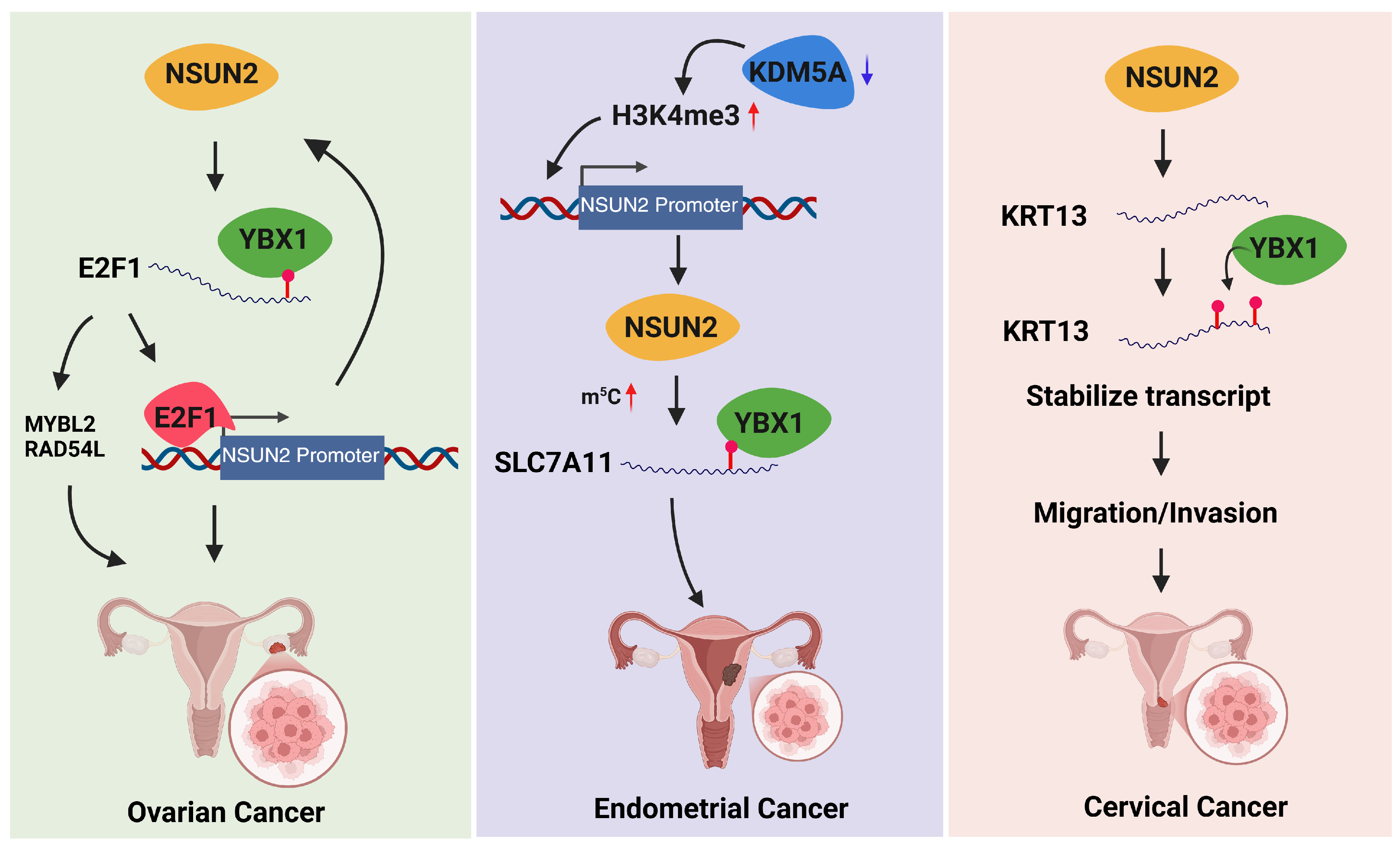
| Ovarian cancer | Human OC patient tumor datasets/tissues and OC cell models | RNA methylation assays, mRNA stability analysis, transcription factor binding studies, phase separation assays, and gene expression/prognostic correlation analysis | NSUN2 mediates E2F1 mRNA(m5C), which is recognized by YBX1 to enhance E2F1 expression, E2F1 then activates NSUN2, forming a positive feedback loop | Upregulation of E2F1, activation of oncogenes MYBL2 and RAD54L, promotion of tumor progression, and association with poorer prognosis in OC | Mar, 2024 | [122] |
| Ovarian cancer | TCGA transcriptomic dataset | Multi-omics bioinformatics clustering and Cox/random forest-based risk modeling | Epigenetic (m6A/m5C/m1A/m7G)-related immune lncRNAs regulate tumor microenvironment and mutation heterogeneity | Defines prognostic subtypes and predicts immune checkpoint and chemotherapy response | Aug, 2023 | [123] |
| Ovarian cancer | HGSOC tissue | m5C profiling of lncRNAs, RNA-seq integration, pathway enrichment, clinical correlation analysis | Dysregulated m5C methylation in lncRNAs alters PI3K-Akt and cancer-related signaling pathways | Increased tumor growth, invasion, metastasis, and poor prognosis | Dec, 2021 | [124] |
| Ovarian cancer | HGSOC and normal fimbria tissues | Expression analysis, functional characterization, downstream target identification, and m5C RNA methylation analysis. | NSUN1 overexpression may increase m5C methylation, enhance RAPGEF4 expression, and subsequently activate pro-tumorigenic signaling pathways. | NSUN1 is involved in cell proliferation, migration, and invasion, and may serve as a potential diagnostic biomarker. | Dec, 2023 | [125] |
| Ovarian cancer | OC cell lines (OVCAR3, SKOV3) and in vivo models | Sevoflurane treatment with DNMT2/APC modulation and functional assays for malignancy and m5C methylation | DNMT2-mediated m5C modification of APC mRNA activates β-catenin signaling under Sevoflurane exposure | Increased proliferation, migration, invasion, cell cycle progression, reduced apoptosis, and enhanced tumor growth | Dec, 2024 | [126] |
| Ovarian cancer | OC tissues, cell lines, and xenografts | m5C binding studies, functional cell assays, and in vivo tumor models | ALYREF stabilizes m5C-modified BIRC5 mRNA to drive oncogenic processes | Promotes OC proliferation, migration, invasion, glycolysis, and tumor growth; knockdown inhibits malignant progression | Aug, 2025 | [127] |
| Ovarian cancer | PDOs, OC cells, and xenograft models | m5C–YBX1 interaction analysis and functional drug-resistance assays | YBX1 binds m5C-modified CHD3 mRNA to enhance homologous recombination and DNA repair | Promotes platinum resistance and tumor survival under chemotherapeutic stress | Aug, 2024 | [128] |
| Ovarian cancer | Cisplatin-resistant OC cell lines and patient samples | Multi-omics profiling and functional ALYREF/NSUN2 modulation assays | NSUN2-mediated m5C on LGR4 mRNA is recognized by ALYREF, stabilizing LGR4 and activating Wnt/β-catenin signaling | Promotes chemoresistance and survival of OC cells | Dec, 2025 | [129] |
| Ovarian cancer | OC tissues, cell lines, and PDX models | m5C profiling, YBX1/MATR3 functional assays, and in vitro/in vivo chemosensitivity testing | YBX1 stabilizes m5C-modified CCNL2 mRNA, cooperating with MATR3 to promote proliferation and chemoresistance | Enhanced tumor growth and cisplatin resistance; YBX1 inhibition restores chemosensitivity | Jul, 2025 | [130] |
| Ovarian cancer | Chemoresistant and chemosensitive tissues, epithelial ovarian cancer (EOC) cell lines, xenograft models | Expression analysis, ubiquitination assays, functional overexpression studies, in vivo validation | SIAH1 ubiquitinates YBX1 at Lys304, promoting its degradation and destabilizing m5C-modified mRNAs | Increased cisplatin sensitivity, reduced tumor growth, and reversal of chemoresistance | Mar, 2022 | [131] |
| Ovarian cancer | OC patient samples from databases and in vitro OC cell lines | Identification of m5C-related lncRNAs, nine-lncRNA risk model via LASSO-Cox regression, survival and nomogram analysis, GSEA, immune infiltration, WGCNA, ceRNA network, and functional assays | m5C-related lncRNAs regulate gene expression and immune infiltration, promoting tumor progression and OC cell proliferation | Risk model stratifies prognosis; AC005562.1 drives OC cell proliferation; pathways and immune profiles differ by risk group; potential biomarkers identified | May, 2023 | [132] |
| Ovarian cancer | OC cell lines and tumor-bearing mice | m5C-modification analysis, lncRNA/miRNA interaction studies, functional and in vivo assays | NSUN2-mediated m5C stabilizes SNHG15, which sponges miR-545-3p to upregulate PD-L1 | Promote proliferation, immune evasion, and tumor growth; knockdown of SNHG15 restores anti-tumor immunity | Aug, 2025 | [133] |
| Uterine corpus endometrial carcinoma (UCEC) | TCGA transcriptomic dataset | Consensus clustering and LASSO-based prognostic risk modeling with immune infiltration analysis | m5C-related lncRNAs influence tumor progression and immune microenvironment | Predicts prognosis and correlates with tumor grade, PD-L1 expression, and immune infiltration | Mar, 2022 | [134] |
| Endometrial cancer | EC patient tissues, EC cell lines, and in vivo tumor xenograft models | Epigenetic and m5C sequencing analyses, ferroptosis assays, knockdown/rescue experiments | NSUN2-mediated m5C stabilizes SLC7A11 via YBX1, suppressing ferroptosis | Promotes proliferation and ferroptosis resistance; targeting NSUN2 enhances ferroptotic tumor suppression | Feb, 2024 | [135] |
| Endometrial cancer | TCGA/GEO datasets and in vitro cell experiments | Clustering, pathway enrichment, and LASSO-based prognostic modeling | m5C regulators (NSUN2, YBX1) modulate cell adhesion pathways | Distinct m5C patterns correlate with prognosis and survival prediction | Oct, 2023 | [136] |
| Cervical cancer | CC cancer cell line | Transcriptome-wide m5C profiling combined with NSUN2 depletion/rescue, RNA-binding assays, and mRNA export analyses. | NSUN2 installs m5C on CG-rich mRNA regions, which are recognized by ALYREF to facilitate nuclear–cytoplasmic mRNA export. | m5C promotes efficient mRNA export and regulates post-transcriptional gene expression in mammalian cells. | Apr, 2017 | [18] |
| Cervical, ovarian, and endometrial cancer | Gynecologic cancer cell lines (cervical, ovarian, endometrial); comparative cellular models across cancer types | NSUN2 knockdown and overexpression, migration and invasion assays, catalytic activity rescue experiments, and molecular interaction analysis. | NSUN2 mediates m5C methylation of KRT13 mRNA, which is recognized and stabilized by the m5C reader YBX1, promoting oncogenic activity. | In CC, NSUN2 depletion inhibits cell migration and invasion, whereas NSUN2 has minimal functional impact in ovarian and EC cells, highlighting a cancer-specific oncogenic pathway | Feb, 2022 | [137] |
| Cervical cancer | CC tissues, cell lines, and in vivo models | Functional assays, m5C modification analysis, RNA-protein interaction studies | NSUN2-mediated m5C stabilizes LRRC8A via YBX1 binding | Promotes proliferation and metastasis by suppressing apoptosis | Jan, 2023 | [138] |
| Cervical cancer | Clinical CC specimens and CC cell lines (Caski, SiHa) | Bioinformatics, proliferation/migration assays, Western blot, knockdown/rescue, m5C RNA analysis | NSUN2-mediated m5C stabilizes LINC01094, which activates SIRT1 via ZNF582, suppressing p53 | Promotes EMT, metastasis, proliferation, and inhibits apoptosis in CC cells | Feb, 2026 | [139] |
| Cervical cancer | Patient samples, cell lines, CDX, and 3D PDO models | LC-MS/MS, RNA-seq, m5C-seq, RIP, functional radiosensitivity assays | NSUN6-mediated m5C modification stabilizes NDRG1 mRNA via ALYREF, enhancing homologous recombination DNA repair | Promotes radioresistance and poor prognosis; NSUN6 inhibition restores radiosensitivity | Jul, 2024 | [140] |
| Cervical cancer | CC cell lines | MeRIP, RIP, Co-IP, RT-qPCR, Western blot, functional ferroptosis and viability assays | ALYREF stabilizes KIF20A mRNA via m5C methylation; KIF20A activates BUB1 to induce ferroptosis resistance | Promotes hypoxia-induced ferroptosis resistance; disruption of the ALYREF/KIF20A/BUB1 axis sensitizes cells to ferroptosis | Jan, 2026 | [141] |
| Cervical cancer | Tumor tissues | m5C mapping, transcriptomics, knockdown/overexpression, proliferation, metastasis, and drug sensitivity assays | m5C stabilizes SERPINB5 mRNA, promoting mitotic regulators (CENPE) to overcome drug-induced mitotic arrest | Drives tumor growth, metastasis, and chemoresistance; SERPINB5 depletion restores sensitivity to paclitaxel and vincristine | Feb, 2026 | [142] |
| Cervical cancer | TCGA/GEO datasets, CC tissues, SiHa cells | NMF clustering of m5C regulators, Cox-based 4-gene signature construction, experimental validation | m5C modification subtypes drive differential expression of oncogenic genes (FNDC3A, VEGFA, OPN3, CPE) | Defines prognostic subtypes; high-risk signature correlates with poor survival and enhanced proliferation and invasion | Sep, 2021 | [143] |
| Cervical cancer | Clinical samples and public datasets (TCGA, GSE39001) | Bioinformatics modeling, COX regression, immune infiltration analysis, IHC/RT-qPCR validation, and drug screening | m6A, m5C, and m1A methylation-related genes regulate tumor progression and immune microenvironment | Predicts survival, correlates with immune infiltration, and identifies potential immunotherapy targets | Dec, 2023 | [144] |
| Cervical cancer | Clinical samples from TCGA database | Bioinformatics-based lncRNA-methylation gene signature construction and validation with survival, immune, and drug response analyses | m1A/m6A/m5C-associated lncRNAs regulate gene expression and immune pathways | Predicts prognosis, immunotherapy response, and potential drug sensitivity | Jan, 2024 | [145] |
| Disease | Sample Type | Approaches | Mechanism | Biological Effects | Publication Date | Ref. |
|---|---|---|---|---|---|---|
| Mitochondrial dysfunction–associated cardiomyopathy | Conditional Nsun4 knockout mouse model; mouse mitochondrial RNA; mitochondrial ribosomal fractions | Conditional knockout; mitochondrial translation assays; bisulfite RNA sequencing; ribosome assembly and NSUN4–MTERF4 interaction analysis; mitoribosome biogenesis assays | NSUN4 independently methylates C911 (m5C911) in mitochondrial 12S rRNA, while the NSUN4–MTERF4 complex promotes assembly of ribosomal subunits into a functional monosome | Loss of Nsun4 abolishes mitochondrial translation and disrupts ribosome maturation, impairing mitochondrial protein synthesis and cellular energy homeostasis | Feb, 2014 | [14] |
| Vascular inflammation/allograft arteriosclerosis | Cultured vascular endothelial cells; wild-type rat vascular tissue; Nsun−/− rat vascular tissue; rat aortic allograft specimens | In vitro and in vivo mRNA methylation assays, leukocyte adhesion assays, TNF-α and homocysteine stimulation, Nsun2 knockout models, and allograft arteriosclerosis assessment | NSUN2-driven m5C methylation amplifies inflammatory endothelial signaling via translational control of ICAM-1 | Nsun2 deficiency reduces ICAM-1 induction and leukocyte adhesion attenuating vascular inflammation | Mar, 2016 | [148] |
| Cardiac hypertrophy | Dnmt2-deficient mouse model; mouse cardiac tissue; isolated cardiomyocytes | Genetic knockout mouse model; echocardiography; cardiac morphometric analysis; cellular hypertrophy assays; molecular analysis of P-TEFb complex activity; noncoding RNA interaction studies | Dnmt2 deficiency promotes dissociation of the inhibitory noncoding RNA 7SK from the P-TEFb complex, resulting in increased P-TEFb activation and transcriptional programs that drive cardiac growth | Loss of Dnmt2 leads to cardiomyocyte enlargement and cardiac hypertrophy while preserving systolic function | Jun, 2016 | [149] |
| Abdominal aortic aneurysm (AAA) | Human AAA tissue (n = 32) vs. normal aortic tissue (n = 12) | Global m5C methylation quantification; gene expression analysis; m5C regulators; protein expression analysis; (RIP-seq; Regulatory network analysis | m5C dysregulation enhances immune-related lncRNA–mRNA networks, promoting inflammatory signaling and macrophage infiltration in AAA tissue | Elevated m5C and its regulators are associated with inflammatory cell infiltration and immune activation, contributing to AAA pathogenesis | Nov, 2021 | [150] |
| Cardiac regeneration | Human iPSCs; hiPSC-derived cardiomyocytes; metabolically matured CMs; biochemically enhanced CMs. | Transcriptome-wide RNA-BS-seq integrated with RNA-seq to profile differential m5C methylation and gene expression during cardiomyocyte differentiation and maturation. | Dynamic remodeling of RNA m5C during pluripotency exit and cardiac lineage commitment modulates lineage-specific transcript regulation. | Biological effect: m5C reprogramming supports cardiomyocyte differentiation and maturation by regulating stem cell pluripotency and cardiac gene expression programs | Dec, 2024 | [157] |
| Atherosclerosis/vascular restenosis | Human aortic vascular smooth muscle cells | nucleolar morphology analysis, oxidative DNA damage assays, protein expression (p53, p21), rDNA transcription assessment, SIRT7 and DNMT2 expression, RNA methylation analysis | Curcumin downregulates SIRT7 and upregulates DNMT2, inhibiting rDNA transcription and promoting RNA methylation, leading to nucleolar stress and cell cycle arrest | Oxidation-dependent cytostatic effect in VSMCs, reducing proliferation and potentially limiting atherosclerosis or post-angioplasty restenosis. | Mar, 2015 | [158] |
| Pressure overload–induced myocardial remodeling/heart failure | TAC-induced hypertrophic mice; angiotensin II–treated H9c2 cells and neonatal rat cardiomyocytes (NRCMs). | Echocardiography, histological staining, Western blot, Dot blot and IHC for m5C, RNA-seq, molecular docking, MST, NSUN2 knockdown, and MeRIP-qPCR validation. | Puerarin upregulates and targets NSUN2, enhancing m5C methylation of NF-κB mRNA, which promotes its degradation and suppresses NF-κB signaling activation. | Increased NSUN2-mediated m5C modification reduces cardiomyocyte hypertrophy and fibrosis, improves cardiac function, and attenuates myocardial remodeling progression | Jul, 2025 | [161] |
| Ischemia/reperfusion–induced cardiomyocyte necroptosis/ischemic heart injury | Hypoxia/reoxygenation (H/R)–treated cardiomyocytes; I/R-injured mouse hearts | piRNA expression analysis, HNEAP knockdown, cardiac function assessment in mice, RNA–protein interaction assays, m5C methylation analysis, and Atf7/Chmp2a expression studies | HNEAP binds DNMT1, suppressing m5C methylation of Atf7 mRNA, elevating ATF7 levels, which represses Chmp2a transcription and promotes cardiomyocyte necroptosis | HNEAP promotes necroptotic cardiomyocyte death and worsens cardiac function, while its inhibition reduces necroptosis and protects the heart | Dec, 2023 | [162] |
| Sepsis-induced myocardial injury (SIMI) | Cecum ligation and puncture (CLP) rat model of SIMI; LPS-treated H9c2 cardiomyocytes in vitro. | RT-qPCR, ELISA, ferroptosis and Western blot assays, MeRIP-qPCR and RIP, luciferase reporter assays, and in vivo/in vitro NSUN2 or NCOA4 overexpression | NSUN2 mediates m5C modification of NCOA4 mRNA, suppressing its expression and thereby inhibiting NCOA4-driven ferroptosis and inflammatory signaling. | NSUN2 overexpression reduces inflammation, ferroptosis, myocardial injury, and cardiac dysfunction in SIMI, while improving cardiomyocyte viability | Jul, 2025 | [163] |
| Doxorubicin-induced cardiotoxicity (DIC) | DOX-treated murine model; cardiomyocyte-specific ALYREF overexpression and knockout mice; cultured cardiomyocytes | Cardiac-specific genetic manipulation (overexpression and CKO), DNA damage and apoptosis assays, protein interaction and binding analysis, phase separation (LLPS) assays, and ubiquitination studies | DOX binds ALYREF, disrupts its liquid–liquid phase separation, promotes ubiquitin-mediated degradation, destabilizes the NORAD-activated ribonucleoprotein complex, and impairs genomic integrity | Loss of ALYREF condensates enhances cardiomyocyte DNA damage, apoptosis, and cardiac dysfunction, whereas preserving ALYREF phase separation mitigates DOX-induced cardiotoxicity | Nov, 2025 | [164] |
| Post-myocardial infarction cardiac fibrosis | Myocardial ischemia mouse model with myofibroblast-specific NSUN2 knockdown; cardiac myofibroblasts. | Conditional myofibroblast-specific Nsun2 knockdown, fibrosis assessment, and RNA stability evaluation of HuR mediated by m5C modification. | NSUN2-mediated m5C modification enhances HuR mRNA stability, promoting pro-fibrotic gene expression in myofibroblasts | Myofibroblast-specific Nsun2 knockdown reduces HuR stability and suppresses extracellular matrix deposition, thereby attenuating cardiac fibrosis after myocardial ischemia | Dec, 2025 | [165] |
| Doxorubicin-induced cardiotoxicity | C57BL/6J mice treated with DOX; H9C2 cardiomyocytes in vitro | NSUN2 knockdown or overexpression, cardiac function assessment, histopathology, apoptosis and ROS assays, NRF2 protein/mRNA analysis, and m5C MeRIP assays | NSUN2 methylates Nrf2 mRNA (m5C), increasing its stability and protein expression, which enhances the antioxidative stress response | NSUN2 protects cardiomyocytes from DOX-induced apoptosis and oxidative damage, preserving cardiac function | Feb, 2023 | [166] |
| Cardiac hypertrophy/heart failure | Human hypertrophic heart tissues; neonatal rat cardiomyocytes; adult mouse cardiomyocytes; cardiac-specific Nsun2 knockout mice under aging, transverse aortic constriction (TAC), and angiotensin II stress. | Bulk and single-cell RNA-seq, m5C-RIP-seq, Cre/LoxP cardiac-specific knockout, rAAV9-mediated gene modulation, RNA pull-down, polysome profiling, reporter assays, and IonOptix contractility measurements. | NSUN2 catalyzes m5C methylation of PRKACA mRNA, enhancing its translation via YBX1-dependent regulation and promoting activation of PKA signaling. | NSUN2-driven PRKACA translation sustains cardiac homeostasis and hypertrophic adaptation, whereas Nsun2 loss impairs PKA signaling, calcium handling, contractility, and accelerates heart failure progression. | Jan, 2025 | [167] |
| Hypoxic pulmonary hypertension | Hypoxia-induced PH models and pulmonary artery smooth muscle cells (PASMCs) | circRNA expression analysis, m5C prediction and validation, NSUN2 knockdown, circRNA overexpression, RNA–protein interaction assays (FXR2 binding), and PASMC proliferation assays under hypoxia. | NSUN2-mediated m5C modification stabilizes circCCNL2, enabling it to bind and sequester FXR2, thereby preventing FXR2 from interacting with CDKL3 and limiting pro-proliferative signaling in PASMCs. | m5C-modified circCCNL2 suppresses PASMC proliferation and attenuates pulmonary vascular remodeling, thereby slowing hypoxic PH progression. | Mar, 2025 | [168] |
| Atherosclerosis (AS) | Public GEO microarray datasets; THP-1–derived macrophages/foam cell model; single-cell RNA-seq immune profiling data. | DE/correlation analysis, PPI, LASSO-ROC modeling, clustering, WGCNA, scRNA-seq, qRT-PCR validation, NSUN3 functional assays, and ceRNA prediction | m5C regulators (NSUN3/NSUN5) correlate with immune infiltration, and NSUN3 enhances macrophage inflammatory cytokine production | Five diagnostic biomarkers were identified, and NSUN3 drives proinflammatory macrophage responses that promote atherosclerosis progression | Jan, 2026 | [169] |
| Myocardial infarction–induced cardiac remodeling | Mouse left anterior descending (LAD) ligation MI model; infarcted mouse myocardial tissue; hypoxia-induced primary neonatal cardiac fibroblasts; single-cell sequencing datasets | In vivo AAV-mediated Alyref knockdown, siRNA silencing in fibroblasts, single-cell RNA-seq analysis, RIP-seq, immunofluorescence, histopathology, Western blot, and ECM gene expression assays. | The m5C reader ALYREF binds target transcripts to enhance extracellular matrix protein synthesis (Col1a2, Col3a1, Eln) via regulation of the Fbln1/Loxl1 pathway in activated cardiac fibroblasts | ALYREF promotes fibroblast proliferation, transdifferentiation, and collagen/elastin deposition, thereby facilitating post-MI cardiac remodeling, while its silencing impairs ECM synthesis and worsens cardiac function | Jun, 2025 | [170] |
| Acute coronary syndrome (ACS) | Serum from ACS patients; ox-LDL–stimulated cardiac microvascular endothelial cells (CMECs). | siRNA TET3 knockdown, RT-qPCR/Western blot, proliferation–apoptosis assays, m5C and RNA stability analysis, RIP, and CCAT2/TRIM14 functional studies | TET3 removes m5C modification from lncRNA CCAT2, reducing its stability and weakening CCAT2–FUS interaction, which releases TRIM14 mRNA from repression and increases TRIM14 expression. | Elevated TET3 promotes CMEC apoptosis and impairs proliferation and angiogenesis, thereby aggravating endothelial damage in ACS | Nov, 2025 | [171] |
Disclaimer/Publisher’s Note: The statements, opinions and data contained in all publications are solely those of the individual author(s) and contributor(s) and not of MDPI and/or the editor(s). MDPI and/or the editor(s) disclaim responsibility for any injury to people or property resulting from any ideas, methods, instructions or products referred to in the content. |
© 2026 by the authors. Licensee MDPI, Basel, Switzerland. This article is an open access article distributed under the terms and conditions of the Creative Commons Attribution (CC BY) license.
Share and Cite
Yang, Q.; Salih, S.M.; Wu, R.; Arora, I.; Mousa, M.; Al-Hendy, A.; Boyer, T.G. 5-Methylcytidine RNA Epitranscriptomics in Women’s Health and Disease: Mechanisms and Clinical Implications. Cells 2026, 15, 847. https://doi.org/10.3390/cells15090847
Yang Q, Salih SM, Wu R, Arora I, Mousa M, Al-Hendy A, Boyer TG. 5-Methylcytidine RNA Epitranscriptomics in Women’s Health and Disease: Mechanisms and Clinical Implications. Cells. 2026; 15(9):847. https://doi.org/10.3390/cells15090847
Chicago/Turabian StyleYang, Qiwei, Sana M. Salih, Rongxue Wu, Itika Arora, Mira Mousa, Ayman Al-Hendy, and Thomas G. Boyer. 2026. "5-Methylcytidine RNA Epitranscriptomics in Women’s Health and Disease: Mechanisms and Clinical Implications" Cells 15, no. 9: 847. https://doi.org/10.3390/cells15090847
APA StyleYang, Q., Salih, S. M., Wu, R., Arora, I., Mousa, M., Al-Hendy, A., & Boyer, T. G. (2026). 5-Methylcytidine RNA Epitranscriptomics in Women’s Health and Disease: Mechanisms and Clinical Implications. Cells, 15(9), 847. https://doi.org/10.3390/cells15090847