


Highlights
What are the main findings?
- Platelet granule-derived mediators and extracellular vesicles cooperatively derive neurovascular dysfunction in neurodegenerative diseases.
- Distinct platelet secretory pathways—including α-granules, dense granules, and platelet-derived EVs—activate inflammatory, oxidative, and protein aggregation mechanisms in Alzheimer’s disease.
What is the implication of the main finding?
- Platelet-derived mediators represent promising biomarkers and therapeutic targets for early detection and intervention in neurodegenerative diseases.
- Integrating platelet secretion and EV signaling into a unified neurovascular framework provides a conceptual basis for future mechanistic and translational studies.
Abstract
Platelets are increasingly recognized as multifunctional regulators that extend beyond hemostasis to actively engage in immunological regulation and neurovascular homeostasis. Platelets employ specialized secretory mechanisms, including granule-dependent release and extracellular vesicle (EV) shedding, to convey diverse bioactive mediators to vascular, immune, and neural cells. Growing evidence indicates that platelet-derived granules and EVs significantly influence the neurovascular unit, regulate inflammatory signaling, and modify neuronal function in both health and disease. In neurodegenerative disorders, particularly Alzheimer’s disease (AD), accumulating evidence suggests that platelet activation may be increased in neurodegenerative conditions, including AD, although the extent and causality of this activation remain under investigation. This review delineates the secretory apparatus of platelets and their mechanistic functions in intercellular communication, underscores platelet contributions to AD and other neurological disorders, and explores novel clinical prospects for biomarker development and therapeutic targeting based on platelet-derived EVs.
1. Introduction
1.1. Platelets in Hemostasis
Platelets are the primary effector cells in the hemostatic response to vascular injury, coordinating a complex series of molecular events that result in clot formation. Following endothelial disruption, platelets are the initial cells to attach to the exposed subendothelial matrix, swiftly converting the injured site into a microenvironment conducive to thrombin production. Thrombin, a serine protease, is generated through the convergence of the extrinsic and intrinsic coagulation pathways, which function synergistically to expedite clot formation [1]. The extrinsic pathway is triggered by the interaction of tissue factor and factor VII, which occurs following endothelial injury; conversely, the intrinsic pathway is activated by factor XII in the presence of phospholipids. Both pathways converge, ultimately leading to the assembly of membrane-associated enzyme complexes on the platelet surface, which significantly amplifies thrombin generation. Following its production, thrombin then retroactively activates platelets, initiating their activation and promoting the conversion of fibrinogen to fibrin, thus constructing the thrombus’s structural foundation. In this process, platelets serve not only as passive substrates but also as active regulators integrating diverse extracellular signals through numerous surface receptors. Key agonists, including thrombin, ADP, and thromboxane A2, activate platelets through G protein-coupled receptors, with thrombin being the most potent by interacting with PAR1 and PAR4, which couple to Gq, Gi, and G12/13 proteins [2]. Simultaneously, collagen exposed at the site of vascular injury interacts with the GPVI/Fc receptor complex, triggering tyrosine kinase-dependent signaling via ITAM-mediated recruitment of SYK [3,4]. The convergence of these signaling pathways ultimately results in the activation of integrin GP IIb/IIIa (αIIbβ3), which facilitates robust platelet–platelet adhesion via fibrin bridging [5,6]. In this meticulously orchestrated process, platelets serve as the pivotal agents of hemostasis—facilitating thrombin production, enhancing activation signals, and forming the stable thrombus that occludes the vascular injury.
1.2. Platelets as Immune Sentinels in Inflammation
In addition to their classical hemostatic roles, platelets serve crucial functions as immune sentinels during critical events such as vascular injury, infection, ischemia, and trauma. In these critical circumstances, they function as swift responders that detect biochemical danger signals and attach to damaged endothelium, commencing both hemostatic and inflammatory processes. Through the facilitation of rapid activation and aggregation, platelets augment thrombin synthesis and fibrin network formation, thus reinstating vascular integrity. Concurrently, they undertake immunomodulatory functions vital for survival in stressful conditions. Platelets exhibit Toll-like receptors (TLR2, TLR4, and TLR9), enabling them to directly identify microbial constituents and circulating pathogens [7,8]. Upon activation, they release antimicrobial peptides and microbicidal molecules, and in certain instances, internalize pathogens via phagocytosis, thereby directly contributing to host defense [9]. These actions establish platelets as initial effector cells that contain and eradicate pathogens prior to the complete recruitment of conventional immune cells [10].
Concurrently, platelets orchestrate a more extensive inflammatory response by enlisting and activating leukocytes. They enhance leukocyte activation and intercellular communication through the secretion of platelet factor 4 (PF4), RANTES, IL-1β, and CD40L. In neutrophils, platelet signals trigger the production of reactive oxygen species (ROS) and the secretion of proteolytic enzymes; in monocytes, they activate prothrombotic and inflammatory gene expression; and in lymphocytes, they facilitate B-cell class-switch recombination and augment cytotoxic T-cell activity [11,12]. This comprehensive platelet–leukocyte interaction establishes a rapid-response network that curtails infection dissemination and stabilizes vascular function during emergencies. Nevertheless, excessive or prolonged activation of this system can be detrimental, resulting in endothelial and tissue damage that may lead to sepsis, ischemic hepatitis, acute lung injury, and other inflammatory disorders [13]. Platelets serve as both protectors and potential enhancers of inflammation, sustaining a fragile balance between protective hemostasis and detrimental thrombosis. This dual function characterizes the nascent concept of immunohemostasis, in which platelets connect coagulation and immune defense as adaptable regulators of vascular homeostasis [14]. In acute emergencies, they concurrently initiate clot formation and regulate immune activation to maintain tissue integrity, while meticulously adjusting these responses to avert collateral damage [15]. The recognition of platelet-specific receptors, including CLEC-2 and TREM-like transcript-1, as regulators of these processes offers promising therapeutic targets for managing platelet activity during severe inflammatory or thrombotic responses [16,17]. Platelets should be regarded not merely as agents of hemostasis but also as protectors of vascular integrity and facilitators of immune coordination, essential for survival during physiological stress [15,18].
1.3. Platelet-Derived Extracellular Vesicles in Inflammation
In acute stress, platelets function as sensors and effectors, converting neuroendocrine and hemodynamic signals into cellular communication signals. Exposure to stress—be it psychosocial or physical—stimulates the sympathetic nervous system and the hypothalamic–pituitary–adrenal axis, resulting in increased levels of catecholamines and glucocorticoids. Hormonal and mechanical stimuli swiftly activate circulating platelets, inducing the release of EVs, specifically small extracellular vesicles (sEVs) and microvesicles originating from the platelet plasma membrane [19,20]. Platelet-derived extracellular vesicles (pEVs) constitute one of the most prevalent vesicle populations in peripheral blood and serve as a vital connection among the vascular, immune, and nervous systems during periods of stress [21]. pEVs encompass a varied array of bioactive molecules, including phosphatidylserine, integrins (notably αIIbβ3), adhesion receptors (CD62P, CD41), enzymes, and small RNAs such as miRNAs, facilitating swift intercellular communication [19,22]. Upon release, they can engage with endothelial cells, leukocytes, and neural or glial cells, affecting vascular tone, permeability, and inflammatory signaling [23,24,25]. During acute physical stress, increased shear stress and catecholamine release facilitate platelet activation and a rise in pEVs that influence coagulation potential and endothelial function [26]. Conversely, psychosocial stress, marked by cortisol-mediated immune modulation, seems to trigger a more nuanced yet coordinated release of pEVs enriched with signaling molecules that regulate immune responsiveness and metabolic equilibrium [21].
pEVs are not simply by-products of platelet activation; they are dynamic messengers that transmit stress-specific information to remote tissues [19]. Surface integrins and tetraspanins enable selective uptake by target cells, while the RNA and protein cargo indicate both platelet activation status and systemic stress conditions [27,28]. Through these mechanisms, pEVs may engage in bidirectional communication between the brain and peripheral organs, augmenting the function of neuroendocrine mediators [29]. During acute psychosocial stress, pEVs may facilitate neurovascular coupling and behavioral regulation by transmitting stress-related miRNAs across the blood–brain barrier, while during physical exertion, they may coordinate metabolic adaptation between muscular and vascular systems [21,22,30,31,32].
Consequently, in the realm of acute stress, platelets expand their role significantly beyond hemostasis. By introducing EVs that reflect and adjust systemic physiological conditions, they contribute to the precise coordination of immuno-metabolic and vascular adaptation [19,32]. Examining the distinctions in platelet-derived vesicle signatures between psychosocial and physical stressors could offer significant insights into individual stress resilience and potentially identify novel biomarkers or therapeutic targets for stress-related disorders [33].
This review analyzes the novel functions of platelet-derived granule contents and EVs as critical mediators of neurovascular dysfunction in neurodegenerative disorders. We examine the impact of platelet secretory products and EV cargo on vascular integrity, neuroinflammation, and neuron-glia interactions, with a specific focus on Alzheimer’s disease (AD). Ultimately, we examine the clinical ramifications of platelet-derived factors as biomarkers and prospective therapeutic targets in neurodegenerative diseases. Although multiple studies have reported enhanced platelet activation markers in patients with neurodegenerative disorders, the degree to which platelet hyperactivation directly contributes to disease progression remains incompletely understood. Notably, functional evidence linking platelet activation to causal neurodegenerative processes is still limited, and many observations remain associative rather than mechanistic. These limitations highlight the need for standardized functional assays and longitudinal studies to better define the role of platelet activation in neurodegeneration.
The conceptual novelty of this review lies in establishing a unified neurovascular framework that integrates platelet granule secretion and EV-mediated signaling into a coordinated pathological axis. Unlike previous reviews that have examined platelet activation of EV biology as separate processes, this review proposes that platelet secretory pathways function as interconnected systems that operate across spatial and temporal scales. Specifically, this framework introduces three key conceptual advances. First, it defines platelet granule release and EV signaling as complementary mechanisms that generate both rapid local responses and sustained long-range communication within the neurovascular unit. Second, it conceptualizes platelet-derived mediators as systemic amplifiers that translate peripheral vascular activation into central neurodegenerative signaling through receptor-mediated and transcriptional pathways. Third, it highlights the convergence of granule-derived factors and EV cargo on shared pathological endpoints, including BBB disruption, neuroinflammation, and protein aggregation, thereby providing a mechanistic bridge linking vascular dysfunction to progressive neurodegeneration. By integrating these processes into a single mechanistic continuum, this review provides a conceptual platform for understanding how platelet-derived signaling contributes to neurovascular dysfunction and offers a framework for identifying novel therapeutic targets and biomarker strategies in neurodegenerative diseases.
2. Platelet Secretory Machinery and Intercellular Communication
Platelets have a specialized secretory apparatus that allows for a swift response to vascular injury and systemic stress [34,35,36]. Although devoid of a nucleus, platelets possess specific intracellular storage compartments and vesicle-producing pathways that enable the secretion of various bioactive mediators [37,38]. Platelet secretory mechanisms can be broadly categorized into two functionally and mechanistically distinct systems: granule-dependent secretion and extracellular vesicle (EV) release (Figure 1). Granule secretion originates from specialized intracellular storage organelles, including α-granules, dense granules, and lysosomes, which rapidly release pre-stored mediators through membrane fusion upon platelet activation. This process enables immediate and localized responses, such as platelet aggregation, vascular modulation, and leukocyte recruitment at sites of vascular injury. In contrast, EV release involves the generation of membrane-derived vesicles, including microvesicles formed by outward budding of the plasma membrane and exosomes originating from multivesicular bodies. Unlike granule secretion, which primarily supports acute local signaling, EVs function as mobile carriers that transport protein, lipids, and nucleic acids to distant target cells, thereby enabling sustained and long-range intercellular communication. These vesicles can circulate systemically and influence endothelial, immune, and neural cells beyond the initial site of platelet activation [39,40]. Recognizing these mechanistic and functional differences between granule-dependent secretion and EV-mediated signaling is essential for understanding how platelet-derived factors contribute differentially to vascular dysfunction, neuroinflammation, and progressive neurodegeneration (Table 1) [41,42].
Figure 1.
Platelet secretory systems and intercellular communication pathways.
2.1. Granules as Expedited Secretory Compartments
2.1.1. α-Granules
α-Granules are the predominant storage organelles in platelets, housing a varied array of proteins that play roles in coagulation, inflammation, angiogenesis, and tissue remodeling. Principal components comprise platelet factor 4 (PF4/CXCL4), P-selectin, vascular endothelial growth factor (VEGF), platelet-derived growth factor, fibrinogen, and amyloid precursor protein (APP). Following platelet activation, α-granule release swiftly modifies the local microenvironment by facilitating leukocyte recruitment, endothelial activation, and extracellular matrix interactions [43,44]. Several components of α-granules are directly pertinent to neurodegenerative disease [45]. PF4 and P-selectin facilitate the formation of platelet-leukocyte aggregates and the activation of microglia, thus exacerbating neuroinflammatory cascades [46]. VEGF, traditionally recognized for its angiogenic properties, can enhance blood–brain barrier (BBB) permeability in pathological states, potentially allowing peripheral inflammatory mediators to infiltrate the central nervous system (CNS) [47]. The presence of APP in α-granules indicates that platelets may directly engage in amyloid-related processes, especially in AD, where aberrant APP processing and Aβ accumulation are key pathological characteristics [45,48].
2.1.2. Dense Granules
Dense granules contain small-molecule mediators, including serotonin, ADP/ATP, calcium ions, and polyphosphates, allowing platelets to swiftly modulate vascular tone, hemostasis, and cellular signaling [49]. Serotonin released from dense granules functions as a powerful vasoactive and neuromodulatory agent, affecting vasoconstriction, synaptic transmission, and neurovascular coupling [50,51,52]. ADP and ATP function as pivotal purinergic signaling molecules, enhancing platelet activation and simultaneously influencing neurons, astrocytes, and microglia via P2 receptor interaction [53,54]. In the realm of neurodegeneration, excessive or dysregulated release of dense granules may lead to neuronal excitotoxicity and persistent neuroinflammation [55,56]. Increased extracellular ATP can activate microglial inflammasome pathways, while disrupted serotonin signaling has been associated with synaptic dysfunction and mood disorders commonly seen in neurodegenerative diseases [57,58].
2.1.3. Lysosomes
Platelet lysosomes house proteases, glycosidases, and various degradative enzymes that facilitate extracellular matrix remodeling and maintain barrier integrity [59,60]. Despite being less thoroughly characterized than α- or dense granules, platelet lysosomal secretion may affect BBB integrity during inflammatory or thrombotic conditions. Activated platelets release lysosomal enzymes that may aid in the remodeling of the extracellular matrix and the degradation of tight junction components, potentially worsening neurovascular dysfunction and promoting leukocyte infiltration into the CNS [61,62,63]. Nonetheless, direct mechanistic evidence connecting platelet lysosomal secretion to BBB disruption in AD is currently insufficient and necessitates additional research.
2.2. Extracellular Vesicles as Long-Range Platelet Messengers
In addition to granule-mediated secretion, platelets release EVs that function as mobile carriers of molecular information. pEVs encompass microparticles, also known as microvesicles and exosomes, which vary in size, biogenesis, and cargo composition [64,65]. Microparticles are produced by outward budding of the plasma membrane during platelet activation, while exosomes derive from intraluminal vesicles within multivesicular bodies. pEVs encompass a multifaceted array of proteins, lipids, and nucleic acids, including integrins (e.g., αIIbβ3), adhesion molecules, SNARE proteins, phosphatidylserine, and regulatory microRNAs (miRNAs). MiRNAs, particularly miR-223 and miR-126, are significantly abundant in pEVs and are associated with the regulation of endothelial integrity, inflammatory signaling, and neuronal survival [66,67,68,69]. pEVs functionally amplify the effects of platelet activation beyond the immediate vascular environment. Through their interaction with endothelial cells, leukocytes, and neural cells, EVs facilitate platelets’ involvement in long-distance communication and systemic adaptation [70,71]. Unlike granule release, which facilitates swift and localized effects, EVs offer a means for prolonged modulation of remote tissues, including the neurovascular unit [31,72].
Table 1.
Platelet Secretory Compartments and Biological Functions in Neurodegenerative Diseases.
3. Integrated Roles of Platelet-Derived Factors in Alzheimer’s Disease
Increasing evidence indicates that platelet hyperactivation plays a pivotal role in influencing the neurovascular and inflammatory environment of AD [90]. Platelets from AD patients exhibit increased sensitivity to amyloid-β (Aβ), oxidative stress, and thrombin, leading to pronounced dysregulation and EV release [91,92,93]. These secretory responses establish platelets as primary enhancers of BBB, microglial activation, and pathological protein aggregation [94,95,96]. Instead of merely functioning as passive participants, platelets actively restructure the neurovascular unit, fostering a conducive environment for chronic neurodegeneration [97].
3.1. α-Granule-Derived Mediators
α-Granule-derived mediators establish a direct mechanistic connection between vascular dysfunction and neuronal pathology in AD [98]. PF4 and P-selectin play central roles in initiating platelet-leukocyte aggregation and promoting adhesion to cerebral endothelial cells through receptor-mediated interactions, including binding of P-selectin to P-selectin glycoprotein ligand-1 (PSGL-1) on leukocytes [46,99,100]. These interactions enhance leukocyte recruitment and sustain microglial activation within the neurovascular unit. Activated microglia subsequently release pro-inflammatory cytokines, ROS, and complement components that directly damage neurons and disrupt synaptic networks [101,102].
Platelet-derived PF4 further modulates microglial phenotype towards a pro-inflammatory state through activation of intracellular signaling pathways associated with tau kinase regulation, including glycogen synthase kinase-3β (GSK-3β) and cyclin-dependent kinase 5 (CDK5), thereby promoting tau hyperphosphorylation and cytoskeletal instability [103,104]. In parallel, inflammatory mediators derived from platelet activation enhance amyloidogenic processing of APP, contributing to increased Aβ accumulation and plaque formation [105].
VEGF, released from activated platelets, exerts potent effects on endothelial barrier function through activation of VEGFR2-dependent signaling pathways [106,107,108]. Sustained VEGF signaling induces cytoskeletal rearrangement and disruption of tight junction proteins, including claudin-5 and occludin, thereby increasing paracellular permeability of the BBB. This vascular permeability facilitates the infiltration of peripheral immune mediators, deposition of fibrinogen, and extravasation of plasma protein, which further activate microglia and astrocytes [109,110]. Fibrinogen accumulation within the brain parenchyma interacts with Aβ aggregates, amplifying inflammatory signaling and neuronal toxicity [111]. Collectively, these interconnected receptor-mediated and intracellular signaling events establish a feed-forward cycle of vascular impairment, neuroinflammation, and protein aggregation that accelerates neurodegenerative progression in AD.
3.2. Dense Granule-Derived Mediators
Neurotransmitters and nucleotides derived from platelet dense granules contribute to neuronal stress and glial activation through coordinated purinergic and serotonergic signaling pathways in AD [97]. Dysregulated serotonin release alters cerebral blood flow regulation and impairs synaptic plasticity through serotonergic receptor-mediated signaling, leading to early cognitive dysfunction [112,113]. Serotonergic imbalance additionally influences microglial reactivity, thereby amplifying inflammatory tone within the neurovascular unit [114]. Extracellular ATP and ADP released from activated platelets function as potent danger-associated molecular signals that activate purinergic receptors, including P2X7 and P2Y receptors, on microglia, astrocytes, and neurons [55,115,116]. At the molecular level, activation of P2XY receptors promotes assembly of the NOD-like receptor pyrin domain-containing 3 (NLRP3) inflammasome complex, leading to caspase-1 activation and subsequent secretion of interleukin-1β (IL-1β), thereby reinforcing neuroinflammatory signaling pathways. Sustained purinergic receptor activation further enhances intracellular calcium influx, triggering mitochondrial dysfunction and excitotoxic neuronal apoptosis [117,118]. The calcium-dependent processes intersect with oxidative stress signaling pathways, including activation of NADPH oxidase, resulting in excessive ROS generation and redox imbalance in AD brains [119]. Elevated ROS levels destabilize synaptic proteins and impair neurotransmission, thereby intensifying synaptic loss and neuronal degeneration, particularly in conjunction with Aβ-mediated toxicity [120,121].
Beyond inflammasome-driven responses, purinergic signaling also induces transcriptional and metabolic alterations that sustain neuronal vulnerability. Activation of purinergic receptors has been associated with downstream regulation of transcription factors such as activator protein-1 (AP-1) and nuclear factor of activated T cells (NFAT), which promotes sustained inflammatory gene expression in microglia and astrocytes [122,123]. These transcriptional responses enhance inducible nitric oxide synthase (iNOS) activity and nitric oxide production, further exacerbating oxidative and nitrosative stress within the neuronal microenvironment [124,125].
In parallel, prolonged nucleotide signaling disrupts neuronal metabolic homeostasis by impairing mitochondrial respiration and reducing ATP production efficiency [126,127]. Energy deficits resulting from mitochondrial dysfunction compromise synaptic vesicle recycling and neurotransmitter release, ultimately weakening synaptic maintenance mechanisms and accelerating dendritic spine loss [128,129]. The metabolic instability enhances neuronal susceptibility to excitotoxic injury and contributes to progressive synaptic remodeling observed during early neurodegenerative stages. Collectively, these interconnected receptor-mediated, transcriptional, and metabolic pathways position dense granule secretion as a critical driver of synaptic dysfunction, neuroinflammatory amplification, and neuronal degeneration in AD.
3.3. Platelet-Derived Extracellular Vesicle Cargo
pEVs have emerged as pivotal mediators of intercellular signaling in AD, amplifying the effects of platelet activation beyond localized vascular environments [93,130]. Circulating pEV levels are markedly increased in patients with AD and correlate with vascular dysfunction, cognitive decline, and inflammatory biomarkers, indicating their active contribution to disease progression rather than representing passive by-products of platelet activation [131,132]. EV uptake by recipient cells is mediated through receptor-dependent endocytosis pathways involving integrins, phosphatidylserine-binding receptors, and scavenger receptors, enabling selective delivery of vesicular cargo into endothelial, immune, and neural cells [133,134,135]. These receptor-mediated interactions trigger intracellular signaling cascades that regulate endothelial permeability, cytoskeletal remodeling, and inflammatory gene expression, thereby establishing pEVs as dynamic regulators of neurovascular integrity (Table 2).
3.3.1. EV Biogenesis and Selective Cargo Loading in AD
In the AD milieu, platelet hyperreactivity induced by Aβ, oxidative stress, and thrombin signaling enhances membrane budding and multivesicular body formation, facilitating increased EV release [64,130]. Importantly, EV cargo loading is not random but is regulated by stress-responsive pathways that selectively incorporate miRNAs, inflammatory proteins, and procoagulant lipids into vesicles [28,136]. Redox-sensitive signaling pathways and cytoskeletal remodeling influence EV composition, resulting in vesicles enriched with inflammatory mediators and endothelial-modulating miRNAs [137]. These selective loading processes generate vesicles capable of transmitting disease-specific molecular signals across the neurovascular unit.
3.3.2. EV-Mediated Enhancement of Neuroinflammation
Upon internalization by microglia and astrocytes, pEV cargo alters inflammatory transcriptional programs that sustain neuroinflammatory signaling [138,139]. MiRNAs such as miR-223 and miR-155 enhance pro-inflammatory gene expression by suppressing negative regulators of cytokine signaling pathways, thereby reinforcing nuclear factor kappa B (NF-κB)-dependent inflammatory responses [140,141]. Concurrently, EV-associated proteins promote activation of NADPH oxidase, leading to increased ROS production and sustained inflammasome activation. These signaling events amplify cytokine release and maintain chronic neuroinflammatory cycles characteristic of AD pathology.
3.3.3. pEVs and the Dynamics of Protein Aggregation
Beyond inflammatory signaling, pEVs actively contribute to the propagation of pathological protein aggregation [142]. The lipid bilayer of EV membranes provides specialized microdomains that facilitate nucleation and aggregation of Aβ oligomer [143]. In addition, oxidative stress induced by EV-associated signaling enhances activation of tau kinase, including GSK-3β, thereby promoting tau hyperphosphorylation and cytoskeletal destabilization. The capacity of pEVs to transport misfolded proteins or aggregation-promoting factors resembles prion-like propagation mechanisms that facilitate spatial spread of neuropathology within the brain [142,144].
3.3.4. Systemic-Central Signaling Axis
A defining feature of pEV-mediated signaling is their ability to bridge peripheral and central pathological processes. Circulating pEVs function as systemic carriers that transmit inflammatory and procoagulant signals from peripheral vascular environments to the central nervous system [145,146]. These vesicles harbor disease-stage-specific molecular signatures that establish a bidirectional communication axis linking vascular dysfunction, immune activation, and neuronal degeneration. Through repeated cycles of EV release, uptake, and intracellular signaling, peripheral platelet activation can be translated into sustained neurovascular injury within the brain.
Collectively, platelet-derived granule secretion and EV-mediated communication converge on a unified neurovascular axis characterized by BBB disruption, immune amplification, oxidative stress, and pathological protein accumulation. These coordinated mechanisms promote fibrinogen deposition, Aβ plaque formation, and sustained inflammatory signaling, ultimately driving progressive neurodegeneration in AD. These integrated signaling pathways are summarized schematically in Figure 2. Despite growing evidence supporting the role of platelet-derived EVs in neurodegenerative processes, several uncertainties remain. Many studies rely on cross-sectional human samples without longitudinal validation, limiting causal inference. Furthermore, variability in EV isolation methods introduces inconsistencies across datasets, complicating direct comparisons across studies [33]. Standardization of EV isolation protocols, quantitative normalization strategies, and validation in mechanistic animal models will be essential for translating EV-based findings into clinical actionable biomarkers and therapeutic targets.
Table 2.
Platelet-derived factors in AD pathology.
Figure 2.
Mechanistic integration of platelet-derived mediators driving neurovascular dysfunction in AD.
4. Platelet-Derived Factors in Other Neurodegenerative Diseases
This review primarily focuses on AD, although platelet-derived mediators also play a role in the pathophysiology of other neurodegenerative diseases (Table 3).
4.1. Ischemic Stroke
Ischemic stroke is essentially a thromboinflammatory condition wherein platelet activation acts as both the initial catalyst for vascular occlusion and a significant enhancer of subsequent neurovascular damage [167,168]. Following cerebral vessel occlusion, platelets rapidly attach to exposed subendothelial matrix proteins, aggregate via integrin αIIbβ3 interaction, and produce thrombin, resulting in clot formation and interruption of cerebral blood flow [169]. In addition to mechanical occlusion, activated platelets significantly influence the post-ischemic microenvironment by releasing granule-derived mediators and EVs that exacerbate inflammation, disrupt the BBB, and induce neuronal injury [170,171].
4.1.1. Granule-Mediated Neurovascular Injury
The secretion of dense granules is pivotal in thrombotic propagation and excitotoxic injury subsequent to ischemia. ADP and ATP released from platelets enhance platelet recruitment via purinergic signaling while concurrently activating P2 receptors on neurons and glial cells [172]. Excess extracellular ATP activates microglial inflammasomes and induces calcium overload in neurons, worsening excitotoxic cell death [173]. Serotonin released from dense granules induces vasoconstriction of cerebral microvessels, thereby exacerbating perfusion deficits in peri-infarct regions and aggravating ischemic injury [174]. Platelet-derived α-granule mediators, including PF4, P-selectin, and VEGF, contribute to post-stroke inflammation and vascular permeability through mechanisms described in Section 3.1 [175,176]. In the ischemic microenvironment, these mediators amplify leukocyte recruitment and endothelial dysfunction, thereby reinforcing thromboinflammatory cascades and BBB disruption.
4.1.2. pEVs as Mediators of Thromboinflammation
pEVs are significantly increased in the bloodstream after ischemic stroke and accumulate in cerebral microvasculature and infarcted tissue. These pEVs present phosphatidylserine and tissue factor, providing procoagulant surfaces that augment thrombin generation and fibrin deposition [177,178]. In addition to coagulation, pEVs convey inflammatory proteins, adhesion molecules, and regulatory miRNAs to endothelial and immune cells, exacerbating vascular dysfunction. The delivery of pro-inflammatory miRNAs and cytokine-associated cargo via pEVs facilitates endothelial activation, leukocyte adhesion, and oxidative stress [179]. Furthermore, pEVs engage directly with microglia and astrocytes, eliciting inflammatory gene expression and the formation of glial scar. Through these mechanisms, pEVs amplify the spatial and temporal effects of platelet activation beyond the initial thrombotic event, leading to secondary neurovascular damage and subsequent neuronal loss.
4.1.3. Integrated Role in Ischemic Pathophysiology
Platelet granule secretion and EV release together initiate a thromboinflammatory cascade that connects vascular occlusion to advancing neurodegeneration. Although antiplatelet therapies significantly diminish the risk of recurrent stroke, their restricted effect on post-ischemic neuroinflammation suggests that researchers need to devise strategies aimed at platelet-derived inflammatory and EV-mediated signaling pathways [180].
4.2. Parkinson’s Disease
Parkinson’s disease (PD) is defined by the progressive degeneration of dopaminergic neurons in the substantia nigra, along with persistent neuroinflammation and vascular changes [181,182]. While traditionally regarded as a neurological disorder, mounting evidence suggests that peripheral immune and vascular elements, including platelets, play a role in disease progression.
4.2.1. Granule-Derived Mediators and Neuroinflammation
Activated platelets in PD patients release α-granule-derived inflammatory mediators that enhance leukocyte recruitment and microglial activation through mechanisms similar to those described in AD (Section 3.1) [97]. Increased serotonin levels affect dopaminergic signaling and vascular tone, possibly leading to the dysregulated cerebral perfusion seen in PD [183,184]. VEGF released from α-granules may initially facilitate vascular repair; however, under chronic inflammatory conditions, it contributes to BBB permeability and leukocyte infiltration [47,182]. The compromised integrity of the barrier permits peripheral immune mediators to infiltrate neural tissue, thereby exacerbating neuroinflammatory cascades.
4.2.2. pEVs and α-Synuclein Propagation
pEVs have emerged as potential carriers of α-synuclein, the pathogenic protein implicated in PD. α-Synuclein associated with pEVs can be transmitted between cells, facilitating misfolding and aggregation in recipient neurons and glial cells. The dissemination mediated by pEV resembles the prion-like propagation mechanisms suggested for the progression of PD [185]. Furthermore, pEV cargo comprises inflammatory proteins and regulatory miRNAs that influence microglial activation and oxidative stress responses. By selective absorption by neural cells, pEVs may exacerbate mitochondrial dysfunction and increase neuronal susceptibility.
4.2.3. Crosstalk Between Vascular and Immune Systems
Platelet-mediated vascular inflammation in PD contributes to microvascular dysfunction and diminished cerebral perfusion, potentially intensifying neuronal stress. The synergistic effects of granule-derived mediators and EV signaling establish platelets as systemic amplifiers of neurodegenerative processes rather than mere passive participants.
4.3. Multiple Sclerosis
Multiple sclerosis is an autoimmune demyelinating disorder marked by recurrent breakdown of the BBB, leukocyte infiltration, and persistent neuroinflammation. Platelets actively participate in the initiation and maintenance of these pathological processes [186,187].
4.3.1. Granule Secretion in the Recruitment of Immune Cells
Upon activation, platelets release α-granule-derived chemokines such as PF4 and RANTES, which attract T cells and monocytes to inflamed cerebral vessels [188,189]. P-selectin expression enhances platelet–leukocyte adhesion, thereby facilitating transendothelial migration into CNS tissue. These interactions are essential for the development of inflammatory lesions typical of multiple sclerosis [190]. ATP derived from dense granules further activates immune cells through purinergic receptors, augmenting cytokine production and inflammatory signaling within the neurovascular compartment [191].
4.3.2. EV-Mediated Immune Modulation
pEVs are abundant in pro-inflammatory proteins and miRNAs that modulate T cell activation and endothelial permeability. These EVs augment the expression of adhesion molecules on endothelial cells, thereby facilitating leukocyte migration across the BBB [192,193]. Platelet activation occurs prior to the onset of clinical disease in experimental autoimmune encephalomyelitis, highlighting a pathogenic role for platelets in neuroinflammation. While direct evidence for pEV involvement is limited, EV-mediated mechanisms may facilitate immune activation and neurovascular dysfunction, ultimately leading to demyelination [187].
4.3.3. Contribution to Chronic Neurovascular Dysfunction
By continuous granule secretion and EV release, platelets maintain a pro-inflammatory vascular milieu that facilitates persistent immune infiltration and neural injury. Targeting platelet-derived pathways may thus serve as a complementary therapeutic strategy in conjunction with immunomodulatory treatments.
Table 3.
Comparative mechanisms of platelet-derived granules and EVs in neurological disorders.
5. Clinical and Translational Implications
Platelet-derived secretory products and EVs constitute a distinctive interface between vascular biology and neurodegenerative pathology, presenting innovative prospects for disease diagnosis, monitoring, and therapeutic intervention [42,198]. In contrast to CNS biomarkers that necessitate invasive cerebrospinal fluid extraction, platelet-derived mediators are prevalent in peripheral blood, facilitating minimally invasive evaluation of neurovascular dysfunction and inflammatory status [199,200]. Moreover, the mechanistic role of platelets in disease progression designates them as both indicators and regulators of neurodegenerative processes.
5.1. Platelet-Derived Factors as Biomarkers
The pursuit of dependable blood-based biomarkers for neurodegenerative diseases has intensified owing to the limitations of neuroimaging and cerebrospinal fluid analyses [199,201]. Platelets and their secretory products provide a biologically relevant and accessible biomarker source indicative of vascular inflammation, immune activation, and neurovascular unit integrity [55,99,198]. Soluble P-selectin and PF4 are well-established markers of platelet activation and are associated with systemic inflammatory burden [54,100]. Increased circulating levels of these mediators have been reported in patients with AD, ischemic stroke, and multiple sclerosis, associating platelet activation with disease severity and progression [202,203,204]. Serotonin, primarily contained in platelet dense granules, demonstrates modified plasma concentrations in neurodegenerative disorders and correlates with cognitive impairment and mood disturbances [58,194]. pEVs augment biomarker specificity through the encapsulation of disease-relevant cargo [40]. EV-associated miRNAs, including miR-223 and miR-126, modulate endothelial function, inflammatory signaling, and neuronal survival pathways, with their circulating levels exhibiting disease-specific alterations [205,206,207]. Proteomic profiling of pEVs has revealed inflammatory mediators, oxidative stress-associated proteins, and aggregation-prone peptides that reflect pathological processes in the CNS [179,208]. Significantly, EV cargo reflects dynamic disease states, enabling longitudinal monitoring of neurodegenerative progression and therapeutic efficacy [209,210,211]. This temporal sensitivity establishes platelet-derived biomarkers as potent instruments for early diagnosis and precision medicine approaches [198,212].
5.2. Therapeutics Targeting of Platelet-Derived Factors
Given their pivotal role in neurovascular dysfunction and inflammation, platelet secretory mechanisms constitute promising therapeutic targets [170,213]. Conventional antiplatelet treatment, such as aspirin and P2Y12 receptor inhibitors, diminishes thrombotic risk in ischemic stroke and exhibits limited protective effects against cognitive impairment [214,215]. Nonetheless, extensive inhibition of platelet aggregation carries bleeding risks and may inadequately mitigate platelet-mediated inflammatory signaling [216,217]. Focusing on granule secretion presents a more refined strategy [218]. Inhibitors of platelet degranulation pathways may attenuate the release of pro-inflammatory mediators while preserving essential hemostatic function [170]. Modulation of α-granule-derived VEGF signaling may stabilize BBB integrity, while regulating dense granule ATP/serotonin release could diminish excitotoxic and inflammatory cascades [219,220]. Oxidative stress pathways, particularly NADPH oxidase-dependent platelet activation, have been identified as upstream regulators of platelet secretion and EV release [141,221]. Pharmacological NADPH oxidase inhibition and antioxidant strategies may mitigate platelet-induced neurovascular damage by reducing ROS-mediated activation [222,223]. EV-targeted therapies represent an emerging frontier. Strategies include inhibiting EV biogenesis, blocking EV uptake by target cells, or engineering EV cargo to deliver neuroprotective molecules [224]. Modified pEVs may be utilized as natural nanocarriers for anti-inflammatory miRNAs or therapeutic proteins, capitalizing on their intrinsic biocompatibility and targeting ability [27,200]. Emerging therapeutic strategies targeting platelet-mediated inflammation are currently under investigation. In addition to conventional antiplatelet agents, novel pharmacological approaches targeting P2Y12 signaling, inflammasome activation, and oxidative stress pathways are being explored in preclinical and early clinical studies [225]. Furthermore, EV-based diagnostics are being actively evaluated as minimally invasive tools for disease monitoring and patient stratification. Several ongoing clinical studies are evaluating the impact of antiplatelet and anti-inflammatory interventions on cognitive decline and vascular dysfunction, highlighting the growing interest in platelet-targeted therapeutic strategies.
5.3. Liquid Biopsy Approaches Using Extracellular Vesicles
Liquid biopsy technologies utilizing circulating EVs are transforming disease diagnostics across oncology and cardiovascular medicine and are increasingly applied to neurodegenerative disorders [226,227]. pEVs are notably appealing because of their abundance, stability, and cargo profiles that respond to diseases [40,228]. Advanced isolation techniques combined with high-throughput omics platforms enable the comprehensive characterization of EV protein, lipid, and RNA content from small plasma volumes [229,230]. These approaches allow identification of disease-specific EV signatures indicative of neurovascular dysfunction, inflammatory activation, and protein aggregation processes [231,232]. Longitudinal liquid biopsy profiling of pEVs provides unprecedented opportunities to track disease progression, stratify patients, and monitor therapeutic efficacy [211,233]. Furthermore, the integration of EV biomarkers with clinical parameters and imaging data may facilitate early identification of neurodegenerative disease prior to overt neurological decline [234]. Challenges remain in standardizing EV isolation, normalization strategies, and cargo quantification [33,235]. Rapid technological advancements are accelerating the clinical translation of EV-based diagnostics, establishing pEVs as central components of future precision neurology frameworks [236].
5.4. Knowledge Gaps and Limitations in Platelet-Mediated Neurodegeneration Research
Despite growing recognition of platelet-derived mediators and EVs as contributors to neurodegenerative processes, several important knowledge gaps remain. Many clinical studies investigating platelet activation in neurodegenerative diseases rely on cross-sectional human cohorts, limiting the ability to establish causal relationships between platelet dysregulation and disease progression. Furthermore, variability in platelet isolation procedures and EV characterization methods introduces methodological inconsistencies across studies, complicating comparison or results. Another significant limitation arises from confounding factors commonly present in aging populations. Neurodegenerative disease cohorts frequently include individuals with cardiovascular disease, diabetes, chronic inflammation, and other systemic conditions known to independently alter platelet reactivity. In addition, widely prescribed medications such as antiplatelet agents, statins, and anti-inflammatory drugs can substantially influence platelet activation profiles and EV release patterns. These variables complicate interpretation of platelet-derived biomarkers and necessitate careful study design and stratification strategies.
At the mechanistic level, the precise molecular pathways linking platelet activation to neuronal dysfunction remain incompletely defined. While emerging evidence supports roles for purinergic signaling, oxidative stress pathways, and receptor-mediated EV uptake, many downstream signaling cascades have yet to be fully characterized in vivo. In particular, the extent to which platelet-derived EVs directly transport pathological cargo across the BBB remains an area of active investigation. Future research should prioritize standardized platelet phenotyping protocols, longitudinal cohort studies, and integration of multi-omics approaches, including proteomics, lipidomics, and single-cell transcriptomics. These efforts will be essential to clarify the causal role of platelet-derived factors in neurodegeneration and to support the development of clinically actionable biomarkers and therapeutic targets.
6. Conclusions
Platelets are increasingly recognized as multifunctional regulators that extend far beyond their classical hemostatic role. This review highlights how platelet-derived secretory systems—including α-granule release, dense granule secretion, and EV signaling—collectively function as integrated mediators of neurovascular dysfunction in neurodegenerative disease, particularly AD. Several key mechanistic insights emerge from this synthesis. First, α-granule-derived mediators promote endothelial activation, leukocyte recruitment, and BBB disruption, establishing a vascular environment that facilitates sustained neuroinflammation. Second, dense granule-derived nucleotides and neurotransmitters activate purinergic signaling pathways that amplify inflammasome activation, oxidative stress, and neuronal vulnerability. Third, pEVs extend these effects beyond local vascular sites by transporting bioactive cargo that modulates endothelial integrity, glial activation, and pathological protein aggregation. Together, these interconnected mechanisms support the concept that platelet secretory pathways function not as isolated processes but as coordinated amplifiers of neurovascular and neuroinflammatory signaling.
Importantly, this review proposes a unified neurovascular framework in which platelet granule secretion and EV-mediated communication converge to bridge peripheral vascular activation with central neurodegenerative progression. By integrating these traditionally separated mechanisms into a single conceptual axis, this framework provides a clearer mechanistic understanding of how systemic inflammatory and vascular signals propagate into neuronal injury and cognitive decline. Despite significant advances, several critical research priorities remain. Future studies should prioritize: (1) mechanistic validation using longitudinal animal models to establish causal relationships between platelet-derived mediators and neurodegenerative progression; (2) standardization of EV isolation, characterization, and quantification methods to improve reproducibility across studies; (3) integration of multi-omics approaches, including proteomics, transcriptomics, and single-cell analyses, to define disease-stage-specific platelet signatures; and (4) translational investigations assessing platelet-derived granules and EV cargo as predictive biomarkers and therapeutic targets in clinical settings.
Collectively, advancing mechanistic resolution and translational validation of platelet-derived signaling pathways will be essential for transforming platelet biology from a descriptive framework into a clinically actionable platform. These efforts may ultimately enable the development of targeted strategies that mitigate neurovascular dysfunction and slow the progression of neurodegenerative diseases.
Funding
This research was supported by the GRRC program of Gyeonggi province (GRRCAjou2023-B01), Republic of Korea.
Data Availability Statement
No new data were created or analyzed in this study.
Conflicts of Interest
The authors declare no conflicts of interest.
Abbreviations
The following abbreviations are used in this manuscript:
| AD | Alzheimer’s disease |
| APP | Amyloid precursor protein |
| Aβ | Amyloid-β |
| ATP | Adenosine triphosphate |
| ADP | Adenosine diphosphate |
| BBB | Blood–brain barrier |
| CNS | Central nervous system |
| CDK5 | Cyclin-dependent kinase 5 |
| EV | Extracellular vesicle |
| GSK-3β | Glycogen synthase kinase-3 beta |
| IL-1β | Interleukin-1 beta |
| MiRNA | MicroRNA |
| NF-κB | Nuclear factor kappa B |
| NLRP3 | NOD-like receptor pyrin domain-containing 3 |
| PD | Parkinson’s disease |
| PF4 | Platelet factor 4 |
| pEV | Platelet-derived extracellular vesicle |
| P2X7 | Purinergic receptor P2X7 |
| P2Y12 | Purinergic receptor P2Y12 |
| RANTES | Regulated upon Activation, Normal T cell Expressed and Secreted |
| ROS | Reactive oxygen species |
| VEGF | Vascular endothelial growth factor |
References
- Hoffman, M.M.; Monroe, D.M. Rethinking the coagulation cascade. Curr. Hematol. Rep. 2005, 4, 391–396. [Google Scholar] [CrossRef] [Scilit]
- Brass, L.F.; Manning, D.R.; Cichowski, K.; Abrams, C.S. Signaling through G proteins in platelets: To the integrins and beyond. Thromb. Haemost. 1997, 78, 581–589. [Google Scholar] [CrossRef] [Scilit]
- Watson, S.P.; Auger, J.M.; McCarty, O.J.; Pearce, A.C. GPVI and integrin αIIbβ3 signaling in platelets. J. Thromb. Haemost. 2005, 3, 1752–1762. [Google Scholar] [CrossRef] [Scilit] [PubMed]
- Nieswandt, B.; Watson, S.P. Platelet-collagen interaction: Is GPVI the central receptor? Blood 2003, 102, 449–461. [Google Scholar] [CrossRef] [Scilit] [PubMed]
- Tadokoro, S.; Shattil, S.J.; Eto, K.; Tai, V.; Liddington, R.C.; de Pereda, J.M.; Ginsberg, M.H.; Calderwood, D.A. Talin binding to integrin β tails: A final common step in integrin activation. Science 2003, 302, 103–106. [Google Scholar] [CrossRef] [Scilit] [PubMed]
- Kim, B.; Lee, B.K.; Kim, J.-A.; Jung, Y.-S. Aβ1-40 Enhances Platelet Sensitivity to Thrombin via NADPH Oxidase–ROS Signaling and Integrin αIIbβ3 Engagement. Biomol. Ther. 2026, 34, 365–378. [Google Scholar] [CrossRef] [Scilit]
- Andonegui, G.; Kerfoot, S.M.; McNagny, K.; Ebbert, K.V.; Patel, K.D.; Kubes, P. Platelets express functional Toll-like receptor-4. Blood 2005, 106, 2417–2423. [Google Scholar] [CrossRef] [Scilit]
- Cognasse, F.; Hamzeh, H.; Chavarin, P.; Acquart, S.; Genin, C.; Garraud, O. Evidence of Toll-like receptor molecules on human platelets. Immunol. Cell Biol. 2005, 83, 196–198. [Google Scholar] [CrossRef] [Scilit]
- Tang, Y.Q.; Yeaman, M.R.; Selsted, M.E. Antimicrobial peptides from human platelets. Infect. Immun. 2002, 70, 6524–6533. [Google Scholar] [CrossRef] [Scilit]
- Wong, C.H.; Jenne, C.N.; Petri, B.; Chrobok, N.L.; Kubes, P. Nucleation of platelets with blood-borne pathogens on Kupffer cells precedes other innate immunity and contributes to bacterial clearance. Nat. Immunol. 2013, 14, 785–792. [Google Scholar] [CrossRef] [Scilit]
- Vieira-de-Abreu, A.; Campbell, R.A.; Weyrich, A.S.; Zimmerman, G.A. Platelets: Versatile effector cells in hemostasis, inflammation, and the immune continuum. Semin. Immunopathol. 2012, 34, 5–30. [Google Scholar] [CrossRef] [Scilit]
- Gros, A.; Ollivier, V.; Ho-Tin-Noe, B. Platelets in inflammation: Regulation of leukocyte activities and vascular repair. Front. Immunol. 2014, 5, 678. [Google Scholar] [CrossRef] [Scilit] [PubMed]
- Morris, S.M.; Chauhan, A. The role of platelet mediated thromboinflammation in acute liver injury. Front. Immunol. 2022, 13, 1037645. [Google Scholar] [CrossRef] [Scilit] [PubMed]
- Guo, L.; Rondina, M.T. The Era of Thromboinflammation: Platelets Are Dynamic Sensors and Effector Cells During Infectious Diseases. Front. Immunol. 2019, 10, 2204. [Google Scholar] [CrossRef] [Scilit] [PubMed]
- Ho-Tin-Noe, B.; Boulaftali, Y.; Camerer, E. Platelets and vascular integrity: How platelets prevent bleeding in inflammation. Blood 2018, 131, 277–288. [Google Scholar] [CrossRef] [Scilit]
- Bourne, J.H.; Smith, C.W.; Jooss, N.J.; Di, Y.; Brown, H.C.; Montague, S.J.; Thomas, M.R.; Poulter, N.S.; Rayes, J.; Watson, S.P. CLEC-2 Supports Platelet Aggregation in Mouse but not Human Blood at Arterial Shear. Thromb. Haemost. 2022, 122, 1988–2000. [Google Scholar] [CrossRef] [Scilit]
- Jessica, M.O.; Fiorella, R.; Ocatavio, S.; Linnette, R.; Nahomy, L.; Kanth, M.B.; Bismarck, M.; Rondina, M.T.; Valance, W.A. Tlt-1-Controls Early Thrombus Formation and Stability by Facilitating Aiibb3 Outside-in Signaling in Mice. Int. J. Adv. Res. 2018, 6, 1143–1149. [Google Scholar] [CrossRef] [Scilit]
- Goerge, T.; Ho-Tin-Noe, B.; Carbo, C.; Benarafa, C.; Remold-O’Donnell, E.; Zhao, B.Q.; Cifuni, S.M.; Wagner, D.D. Inflammation induces hemorrhage in thrombocytopenia. Blood 2008, 111, 4958–4964. [Google Scholar] [CrossRef] [Scilit]
- Puhm, F.; Boilard, E.; Machlus, K.R. Platelet Extracellular Vesicles: Beyond the Blood. Arterioscler. Thromb. Vasc. Biol. 2021, 41, 87–96. [Google Scholar] [CrossRef] [Scilit]
- Heijnen, H.; van der Sluijs, P. Platelet secretory behaviour: As diverse as the granules... or not? J. Thromb. Haemost. 2015, 13, 2141–2151. [Google Scholar] [CrossRef] [Scilit]
- Moser, D.A.; Tertel, T.; Berg, F.; Hummel, E.M.; Platen, P.; Giebel, B.; Kumsta, R. Stress type-specific small extracellular vesicle signatures reflect divergent biological responses to acute psychosocial and physical challenges Do I understand correctly that there were no queries regarding the manuscript? Sci. Rep. 2025, 15, 35231. [Google Scholar] [CrossRef] [Scilit]
- Laffont, B.; Corduan, A.; Ple, H.; Duchez, A.C.; Cloutier, N.; Boilard, E.; Provost, P. Activated platelets can deliver mRNA regulatory Ago2*microRNA complexes to endothelial cells via microparticles. Blood 2013, 122, 253–261. [Google Scholar] [CrossRef] [Scilit] [PubMed]
- Rosinska, J.; Lukasik, M.; Kozubski, W. The Impact of Vascular Disease Treatment on Platelet-Derived Microvesicles. Cardiovasc. Drugs Ther. 2017, 31, 627–644. [Google Scholar] [CrossRef] [Scilit] [PubMed]
- Hayon, Y.; Dashevsky, O.; Shai, E.; Varon, D.; Leker, R.R. Platelet microparticles promote neural stem cell proliferation, survival and differentiation. J. Mol. Neurosci. 2012, 47, 659–665. [Google Scholar] [CrossRef] [Scilit] [PubMed]
- Miyazawa, B.; Trivedi, A.; Togarrati, P.P.; Potter, D.; Baimukanova, G.; Vivona, L.; Lin, M.; Lopez, E.; Callcut, R.; Srivastava, A.K.; et al. Regulation of endothelial cell permeability by platelet-derived extracellular vesicles. J. Trauma. Acute Care Surg. 2019, 86, 931–942. [Google Scholar] [CrossRef] [Scilit]
- Nomura, S.; Tandon, N.N.; Nakamura, T.; Cone, J.; Fukuhara, S.; Kambayashi, J. High-shear-stress-induced activation of platelets and microparticles enhances expression of cell adhesion molecules in THP-1 and endothelial cells. Atherosclerosis 2001, 158, 277–287. [Google Scholar] [CrossRef] [Scilit]
- Liu, Y.J.; Wang, C. A review of the regulatory mechanisms of extracellular vesicles-mediated intercellular communication. Cell Commun. Signal. 2023, 21, 77. [Google Scholar] [CrossRef] [Scilit]
- Eustes, A.S.; Dayal, S. The Role of Platelet-Derived Extracellular Vesicles in Immune-Mediated Thrombosis. Int. J. Mol. Sci. 2022, 23, 7837. [Google Scholar] [CrossRef] [Scilit]
- Zhou, W.; Zhao, L.; Mao, Z.; Wang, Z.; Zhang, Z.; Li, M. Bidirectional Communication Between the Brain and Other Organs: The Role of Extracellular Vesicles. Cell. Mol. Neurobiol. 2023, 43, 2675–2696. [Google Scholar] [CrossRef] [Scilit]
- Faille, D.; El-Assaad, F.; Mitchell, A.J.; Alessi, M.C.; Chimini, G.; Fusai, T.; Grau, G.E.; Combes, V. Endocytosis and intracellular processing of platelet microparticles by brain endothelial cells. J. Cell. Mol. Med. 2012, 16, 1731–1738. [Google Scholar] [CrossRef] [Scilit]
- Ruan, J.; Miao, X.; Schluter, D.; Lin, L.; Wang, X. Extracellular vesicles in neuroinflammation: Pathogenesis, diagnosis, and therapy. Mol. Ther. 2021, 29, 1946–1957. [Google Scholar] [CrossRef] [Scilit] [PubMed]
- Brahmer, A.; Neuberger, E.; Esch-Heisser, L.; Haller, N.; Jorgensen, M.M.; Baek, R.; Mobius, W.; Simon, P.; Kramer-Albers, E.M. Platelets, endothelial cells and leukocytes contribute to the exercise-triggered release of extracellular vesicles into the circulation. J. Extracell. Vesicles 2019, 8, 1615820. [Google Scholar] [CrossRef] [Scilit] [PubMed]
- Kumar, M.A.; Baba, S.K.; Sadida, H.Q.; Marzooqi, S.A.; Jerobin, J.; Altemani, F.H.; Algehainy, N.; Alanazi, M.A.; Abou-Samra, A.B.; Kumar, R.; et al. Extracellular vesicles as tools and targets in therapy for diseases. Signal Transduct. Target. Ther. 2024, 9, 27. [Google Scholar] [CrossRef] [Scilit] [PubMed]
- Golebiewska, E.M.; Poole, A.W. Platelet secretion: From haemostasis to wound healing and beyond. Blood Rev. 2015, 29, 153–162. [Google Scholar] [CrossRef] [Scilit]
- Scridon, A. Platelets and Their Role in Hemostasis and Thrombosis-From Physiology to Pathophysiology and Therapeutic Implications. Int. J. Mol. Sci. 2022, 23, 12772. [Google Scholar] [CrossRef] [Scilit]
- Smith, T.L.; Weyrich, A.S. Platelets as central mediators of systemic inflammatory responses. Thromb. Res. 2011, 127, 391–394. [Google Scholar] [CrossRef] [Scilit]
- Sharda, A.; Flaumenhaft, R. The life cycle of platelet granules. F1000Research 2018, 7, 236. [Google Scholar] [CrossRef] [Scilit]
- Burnouf, T.; Chou, M.L.; Lundy, D.J.; Chuang, E.Y.; Tseng, C.L.; Goubran, H. Expanding applications of allogeneic platelets, platelet lysates, and platelet extracellular vesicles in cell therapy, regenerative medicine, and targeted drug delivery. J. Biomed. Sci. 2023, 30, 79. [Google Scholar] [CrossRef] [Scilit]
- Joshi, S.; Whiteheart, S.W. The nuts and bolts of the platelet release reaction. Platelets 2017, 28, 129–137. [Google Scholar] [CrossRef] [Scilit]
- Lou, C.; Cai, X. The emerging roles of platelet-derived extracellular vesicles in disease. Ann. Med. 2025, 57, 2499029. [Google Scholar] [CrossRef] [Scilit]
- Koupenova, M.; Clancy, L.; Corkrey, H.A.; Freedman, J.E. Circulating Platelets as Mediators of Immunity, Inflammation, and Thrombosis. Circ. Res. 2018, 122, 337–351. [Google Scholar] [CrossRef] [Scilit]
- Leiter, O.; Walker, T.L. Platelets in Neurodegenerative Conditions-Friend or Foe? Front. Immunol. 2020, 11, 747. [Google Scholar] [CrossRef] [Scilit] [PubMed]
- Ambrosio, A.L.; Di Pietro, S.M. The winding road to platelet alpha-granules. Front. Cell Dev. Biol. 2025, 13, 1584059. [Google Scholar] [CrossRef] [Scilit] [PubMed]
- Blair, P.; Flaumenhaft, R. Platelet α-granules: Basic biology and clinical correlates. Blood Rev. 2009, 23, 177–189. [Google Scholar] [CrossRef] [Scilit] [PubMed]
- Evin, G.; Li, Q.X. Platelets and Alzheimer’s disease: Potential of APP as a biomarker. World J. Psychiatry 2012, 2, 102–113. [Google Scholar] [CrossRef] [Scilit]
- D’Mello, C.; Riazi, K.; Le, T.; Stevens, K.M.; Wang, A.; McKay, D.M.; Pittman, Q.J.; Swain, M.G. P-selectin-mediated monocyte-cerebral endothelium adhesive interactions link peripheral organ inflammation to sickness behaviors. J. Neurosci. 2013, 33, 14878–14888. [Google Scholar] [CrossRef] [Scilit]
- Argaw, A.T.; Asp, L.; Zhang, J.; Navrazhina, K.; Pham, T.; Mariani, J.N.; Mahase, S.; Dutta, D.J.; Seto, J.; Kramer, E.G.; et al. Astrocyte-derived VEGF-A drives blood-brain barrier disruption in CNS inflammatory disease. J. Clin. Investig. 2012, 122, 2454–2468. [Google Scholar] [CrossRef] [Scilit]
- Brandl, S.; Reindl, M. Blood-Brain Barrier Breakdown in Neuroinflammation: Current In Vitro Models. Int. J. Mol. Sci. 2023, 24, 12699. [Google Scholar] [CrossRef] [Scilit]
- Dupuis, A.; Bordet, J.C.; Eckly, A.; Gachet, C. Platelet δ-Storage Pool Disease: An Update. J. Clin. Med. 2020, 9, 2508. [Google Scholar] [CrossRef] [Scilit]
- Shi, G.; Morrell, C.N. Platelets as initiators and mediators of inflammation at the vessel wall. Thromb. Res. 2011, 127, 387–390. [Google Scholar] [CrossRef] [Scilit]
- Watts, S.W.; Davis, R.P. 5-hydroxtryptamine receptors in systemic hypertension: An arterial focus. Cardiovasc. Ther. 2011, 29, 54–67. [Google Scholar] [CrossRef] [Scilit] [PubMed]
- Ciranna, L. Serotonin as a modulator of glutamate- and GABA-mediated neurotransmission: Implications in physiological functions and in pathology. Curr. Neuropharmacol. 2006, 4, 101–114. [Google Scholar] [CrossRef] [Scilit] [PubMed]
- Mitrugno, A.; Rigg, R.A.; Laschober, N.B.; Ngo, A.T.P.; Pang, J.; Williams, C.D.; Aslan, J.E.; McCarty, O.J.T. Potentiation of TRAP-6-induced platelet dense granule release by blockade of P2Y(12) signaling with MRS2395. Platelets 2018, 29, 383–394. [Google Scholar] [CrossRef] [Scilit] [PubMed]
- Abbracchio, M.P.; Ceruti, S. Roles of P2 receptors in glial cells: Focus on astrocytes. Purinergic Signal. 2006, 2, 595–604. [Google Scholar] [CrossRef] [Scilit]
- Rodriguez Moore, G.; Melo-Escobar, I.; Stegner, D.; Bracko, O. One immune cell to bind them all: Platelet contribution to neurodegenerative disease. Mol. Neurodegener. 2024, 19, 65. [Google Scholar] [CrossRef] [Scilit]
- Cisneros-Mejorado, A.J.; Perez-Samartin, A.; Domercq, M.; Arellano, R.O.; Gottlieb, M.; Koch-Nolte, F.; Matute, C. P2X7 Receptors as a Therapeutic Target in Cerebrovascular Diseases. Front. Mol. Neurosci. 2020, 13, 92. [Google Scholar] [CrossRef] [Scilit]
- Campagno, K.E.; Mitchell, C.H. The P2X(7) Receptor in Microglial Cells Modulates the Endolysosomal Axis, Autophagy, and Phagocytosis. Front. Cell. Neurosci. 2021, 15, 645244. [Google Scholar] [CrossRef] [Scilit]
- Chakraborty, S.; Lennon, J.C.; Malkaram, S.A.; Zeng, Y.; Fisher, D.W.; Dong, H. Serotonergic system, cognition, and BPSD in Alzheimer’s disease. Neurosci. Lett. 2019, 704, 36–44. [Google Scholar] [CrossRef] [Scilit]
- Houlahan, C.B.; Kong, Y.; Johnston, B.; Cielesh, M.; Chau, T.H.; Fenwick, J.; Coleman, P.R.; Hao, H.; Haltiwanger, R.S.; Thaysen-Andersen, M.; et al. Analysis of the Healthy Platelet Proteome Identifies a New Form of Domain-Specific O-Fucosylation. Mol. Cell. Proteom. 2024, 23, 100717. [Google Scholar] [CrossRef] [Scilit]
- Ludwig, N.; Hilger, A.; Zarbock, A.; Rossaint, J. Platelets at the Crossroads of Pro-Inflammatory and Resolution Pathways during Inflammation. Cells 2022, 11, 1957. [Google Scholar] [CrossRef] [Scilit]
- Ciferri, S.; Emiliani, C.; Guglielmini, G.; Orlacchio, A.; Nenci, G.G.; Gresele, P. Platelets release their lysosomal content in vivo in humans upon activation. Thromb. Haemost. 2000, 83, 157–164. [Google Scholar] [CrossRef] [Scilit]
- Del Maschio, A.; Corvazier, E.; Maillet, F.; Kazatchkine, M.D.; Maclouf, J. Platelet-dependent induction and amplification of polymorphonuclear leucocyte lysosomal enzyme release. Br. J. Haematol. 1989, 72, 329–335. [Google Scholar] [CrossRef] [Scilit]
- Park, S.M.; Jung, H.Y.; Kim, H.O.; Rhim, H.; Paik, S.R.; Chung, K.C.; Park, J.H.; Kim, J. Evidence that α-synuclein functions as a negative regulator of Ca(++)-dependent α-granule release from human platelets. Blood 2002, 100, 2506–2514. [Google Scholar] [CrossRef] [Scilit] [PubMed]
- Heijnen, H.F.G.; Schiel, A.E.; Fijnheer, R.; Geuze, H.J.; Sixma, J.J. Activated Platelets Release Two Types of Membrane Vesicles: Microvesicles by Surface Shedding and Exosomes Derived From Exocytosis of Multivesicular Bodies and α-Granules. Blood 1999, 94, 3791–3799. [Google Scholar] [CrossRef] [Scilit] [PubMed]
- Zmigrodzka, M.; Guzera, M.; Miskiewicz, A.; Jagielski, D.; Winnicka, A. The biology of extracellular vesicles with focus on platelet microparticles and their role in cancer development and progression. Tumour Biol. 2016, 37, 14391–14401. [Google Scholar] [CrossRef] [Scilit] [PubMed]
- Mabrouk, M.; Guessous, F.; Naya, A.; Merhi, Y.; Zaid, Y. The Pathophysiological Role of Platelet-Derived Extracellular Vesicles. Semin. Thromb. Hemost. 2023, 49, 279–283. [Google Scholar] [CrossRef] [Scilit]
- Szilagyi, B.; Fejes, Z.; Rusznyak, A.; Fenyvesi, F.; Pocsi, M.; Halmi, S.; Griger, Z.; Kunapuli, S.P.; Kappelmayer, J.; Nagy, B., Jr. Platelet Microparticles Enriched in miR-223 Reduce ICAM-1-Dependent Vascular Inflammation in Septic Conditions. Front. Physiol. 2021, 12, 658524. [Google Scholar] [CrossRef] [Scilit]
- Sibilano, M.; Tullio, V.; Adorno, G.; Savini, I.; Gasperi, V.; Catani, M.V. Platelet-Derived miR-126-3p Directly Targets AKT2 and Exerts Anti-Tumor Effects in Breast Cancer Cells: Further Insights in Platelet-Cancer Interplay. Int. J. Mol. Sci. 2022, 23, 5484. [Google Scholar] [CrossRef] [Scilit]
- Xi, T.; Jin, F.; Zhu, Y.; Wang, J.; Tang, L.; Wang, Y.; Liebeskind, D.S.; He, Z. MicroRNA-126-3p attenuates blood-brain barrier disruption, cerebral edema and neuronal injury following intracerebral hemorrhage by regulating PIK3R2 and Akt. Biochem. Biophys. Res. Commun. 2017, 494, 144–151. [Google Scholar] [CrossRef] [Scilit]
- Vachon, L.; Jean, G.; Milasan, A.; Babran, S.; Lacroix, E.; Guadarrama Bello, D.; Villeneuve, L.; Rak, J.; Nanci, A.; Mihalache-Avram, T.; et al. Platelet extracellular vesicles preserve lymphatic endothelial cell integrity and enhance lymphatic vessel function. Commun. Biol. 2024, 7, 975. [Google Scholar] [CrossRef] [Scilit]
- Kuravi, S.J.; Harrison, P.; Rainger, G.E.; Nash, G.B. Ability of Platelet-Derived Extracellular Vesicles to Promote Neutrophil-Endothelial Cell Interactions. Inflammation 2019, 42, 290–305. [Google Scholar] [CrossRef] [Scilit]
- Stalker, T.J.; Welsh, J.D.; Brass, L.F. Shaping the platelet response to vascular injury. Curr. Opin. Hematol. 2014, 21, 410–417. [Google Scholar] [CrossRef] [Scilit]
- Cognasse, F.; Laradi, S.; Berthelot, P.; Bourlet, T.; Marotte, H.; Mismetti, P.; Garraud, O.; Hamzeh-Cognasse, H. Platelet Inflammatory Response to Stress. Front. Immunol. 2019, 10, 1478. [Google Scholar] [CrossRef] [Scilit]
- Sadeghi-Ardebili, M.; Hasannia, S.; Dabirmanesh, B.; Khavari-Nejad, R.A. Functional characterization of the dimeric form of PDGF-derived fusion peptide fabricated based on theoretical arguments. Sci. Rep. 2024, 14, 1003. [Google Scholar] [CrossRef] [Scilit] [PubMed]
- Sweeney, M.D.; Sagare, A.P.; Zlokovic, B.V. Blood-brain barrier breakdown in Alzheimer disease and other neurodegenerative disorders. Nat. Rev. Neurol. 2018, 14, 133–150. [Google Scholar] [CrossRef] [Scilit] [PubMed]
- Ma, L.; Perini, R.; McKnight, W.; Dicay, M.; Klein, A.; Hollenberg, M.D.; Wallace, J.L. Proteinase-activated receptors 1 and 4 counter-regulate endostatin and VEGF release from human platelets. Proc. Natl. Acad. Sci. USA 2005, 102, 216–220. [Google Scholar] [CrossRef] [Scilit] [PubMed]
- Falati, S.; Gross, P.; Merrill-Skoloff, G.; Furie, B.C.; Furie, B. Real-time in vivo imaging of platelets, tissue factor and fibrin during arterial thrombus formation in the mouse. Nat. Med. 2002, 8, 1175–1181. [Google Scholar] [CrossRef] [Scilit]
- Canobbio, I.; Abubaker, A.A.; Visconte, C.; Torti, M.; Pula, G. Role of amyloid peptides in vascular dysfunction and platelet dysregulation in Alzheimer’s disease. Front. Cell. Neurosci. 2015, 9, 65. [Google Scholar] [CrossRef] [Scilit]
- Burnstock, G. Physiology and pathophysiology of purinergic neurotransmission. Physiol. Rev. 2007, 87, 659–797. [Google Scholar] [CrossRef] [Scilit]
- Gawaz, M.; Neumann, F.J.; Ott, I.; May, A.; Schömig, A. Platelet activation and coronary stent implantation. Effect of antithrombotic therapy. Circulation 1996, 94, 279–285. [Google Scholar] [CrossRef] [Scilit]
- Miguelez, C.; Morera-Herreras, T.; Torrecilla, M.; Ruiz-Ortega, J.A.; Ugedo, L. Interaction between the 5-HT system and the basal ganglia: Functional implication and therapeutic perspective in Parkinson’s disease. Front. Neural Circuits 2014, 8, 21. [Google Scholar] [CrossRef] [Scilit]
- Paquette, M.A.; Foley, K.; Brudney, E.G.; Meshul, C.K.; Johnson, S.W.; Berger, S.P. The sigma-1 antagonist BMY-14802 inhibits L-DOPA-induced abnormal involuntary movements by a WAY-100635-sensitive mechanism. Psychopharmacology 2009, 204, 743–754. [Google Scholar] [CrossRef] [Scilit] [PubMed]
- Del Zoppo, G.J.; Milner, R.; Mabuchi, T.; Hung, S.; Wang, X.; Berg, G.I.; Koziol, J.A. Microglial activation and matrix protease generation during focal cerebral ischemia. Stroke 2007, 38, 646–651. [Google Scholar] [CrossRef] [Scilit]
- Gidlöf, O.; Sathanoori, R.; Magistri, M.; Faghihi, M.A.; Wahlestedt, C.; Olde, B.; Erlinge, D. Extracellular Uridine Triphosphate and Adenosine Triphosphate Attenuate Endothelial Inflammation through miR-22-Mediated ICAM-1 Inhibition. J. Vasc. Res. 2015, 52, 71–80. [Google Scholar] [CrossRef] [Scilit] [PubMed]
- Jansen, F.; Yang, X.; Hoelscher, M.; Cattelan, A.; Schmitz, T.; Proebsting, S.; Wenzel, D.; Vosen, S.; Franklin, B.S.; Fleischmann, B.K.; et al. Endothelial microparticle-mediated transfer of MicroRNA-126 promotes vascular endothelial cell repair via SPRED1 and is abrogated in glucose-damaged endothelial microparticles. Circulation 2013, 128, 2026–2038. [Google Scholar] [CrossRef] [Scilit] [PubMed]
- Mause, S.F.; Weber, C. Microparticles: Protagonists of a novel communication network for intercellular information exchange. Circ. Res. 2010, 107, 1047–1057. [Google Scholar] [CrossRef] [Scilit]
- Owens, A.P., 3rd; Mackman, N. Microparticles in hemostasis and thrombosis. Circ. Res. 2011, 108, 1284–1297. [Google Scholar] [CrossRef] [Scilit]
- Goetzl, E.J.; Goetzl, L.; Karliner, J.S.; Tang, N.; Pulliam, L. Human plasma platelet-derived exosomes: Effects of aspirin. FASEB J. 2016, 30, 2058–2063. [Google Scholar] [CrossRef] [Scilit]
- Fiandaca, M.S.; Kapogiannis, D.; Mapstone, M.; Boxer, A.; Eitan, E.; Schwartz, J.B.; Abner, E.L.; Petersen, R.C.; Federoff, H.J.; Miller, B.L.; et al. Identification of preclinical Alzheimer’s disease by a profile of pathogenic proteins in neurally derived blood exosomes: A case-control study. Alzheimers Dement. 2015, 11, 600–607.e601. [Google Scholar] [CrossRef] [Scilit]
- De Sousa, D.M.B.; Poupardin, R.; Villeda, S.A.; Schroer, A.B.; Frohlich, T.; Frey, V.; Staffen, W.; Mrowetz, H.; Altendorfer, B.; Unger, M.S.; et al. The platelet transcriptome and proteome in Alzheimer’s disease and aging: An exploratory cross-sectional study. Front. Mol. Biosci. 2023, 10, 1196083. [Google Scholar] [CrossRef] [Scilit]
- Koch-Paszkowski, J.; Sennett, C.; Pula, G. Alzheimer’s Disease-Thrombosis Comorbidity: A Growing Body of Evidence from Patients and Animal Models. Cells 2025, 14, 1069. [Google Scholar] [CrossRef] [Scilit]
- Winfree, R.L.; Erreger, K.; Phillips, J.; Seto, M.; Wang, Y.; Schneider, J.A.; Bennett, D.A.; Schrag, M.S.; Hohman, T.J.; Hamm, H.E. Elevated protease-activated receptor 4 (PAR4) gene expression in Alzheimer’s disease predicts cognitive decline. Neurobiol. Aging 2024, 140, 93–101. [Google Scholar] [CrossRef] [Scilit]
- Odaka, H.; Hiemori, K.; Shimoda, A.; Akiyoshi, K.; Tateno, H. Platelet-derived extracellular vesicles are increased in sera of Alzheimer’s disease patients, as revealed by Tim4-based assays. FEBS Open Bio 2021, 11, 741–752. [Google Scholar] [CrossRef] [Scilit] [PubMed]
- Wu, T.; Chen, L.; Zhou, L.; Xu, J.; Guo, K. Platelets transport β-amyloid from the peripheral blood into the brain by destroying the blood-brain barrier to accelerate the process of Alzheimer’s disease in mouse models. Aging 2021, 13, 7644. [Google Scholar] [CrossRef] [Scilit] [PubMed]
- Kniewallner, K.M.; Foidl, B.M.; Humpel, C. Platelets isolated from an Alzheimer mouse damage healthy cortical vessels and cause inflammation in an organotypic ex vivo brain slice model. Sci. Rep. 2018, 8, 15483. [Google Scholar] [CrossRef] [Scilit] [PubMed]
- Donner, L.; Feige, T.; Freiburg, C.; Toska, L.M.; Reichert, A.S.; Chatterjee, M.; Elvers, M. Impact of Amyloid-β on Platelet Mitochondrial Function and Platelet-Mediated Amyloid Aggregation in Alzheimer’s Disease. Int. J. Mol. Sci. 2021, 22, 9633. [Google Scholar] [CrossRef] [Scilit] [PubMed]
- Lan, Y.; Ding, J.; Yu, T.; Cheng, C. Research progress of platelets in neurodegenerative diseases. Front. Aging Neurosci. 2025, 17, 1544605. [Google Scholar] [CrossRef] [Scilit]
- Morrell, C.N.; Aggrey, A.A.; Chapman, L.M.; Modjeski, K.L. Emerging roles for platelets as immune and inflammatory cells. Blood 2014, 123, 2759–2767. [Google Scholar] [CrossRef] [Scilit]
- Lv, W.; Jiang, X.; Zhang, Y. The role of platelets in the blood-brain barrier during brain pathology. Front. Cell. Neurosci. 2023, 17, 1298314. [Google Scholar] [CrossRef] [Scilit]
- Muller, L.; Gebicka, P.; Handtke, S.; Schonborn, L.; Thiele, T. Advances in our understanding of anti-PF4 related immunothrombosis. Front. Immunol. 2025, 16, 1724207. [Google Scholar] [CrossRef] [Scilit]
- Yang, G.; Xu, X.; Gao, W.; Wang, X.; Zhao, Y.; Xu, Y. Microglia-orchestrated neuroinflammation and synaptic remodeling: Roles of pro-inflammatory cytokines and receptors in neurodegeneration. Front. Cell. Neurosci. 2025, 19, 1700692. [Google Scholar] [CrossRef] [Scilit] [PubMed]
- Gao, C.; Jiang, J.; Tan, Y.; Chen, S. Microglia in neurodegenerative diseases: Mechanism and potential therapeutic targets. Signal Transduct. Target. Ther. 2023, 8, 359. [Google Scholar] [CrossRef] [Scilit] [PubMed]
- De Jong, E.K.; de Haas, A.H.; Brouwer, N.; van Weering, H.R.; Hensens, M.; Bechmann, I.; Pratley, P.; Wesseling, E.; Boddeke, H.W.; Biber, K. Expression of CXCL4 in microglia in vitro and in vivo and its possible signaling through CXCR3. J. Neurochem. 2008, 105, 1726–1736. [Google Scholar] [CrossRef] [Scilit] [PubMed]
- Kitazawa, M.; Cheng, D.; Tsukamoto, M.R.; Koike, M.A.; Wes, P.D.; Vasilevko, V.; Cribbs, D.H.; LaFerla, F.M. Blocking IL-1 signaling rescues cognition, attenuates tau pathology, and restores neuronal β-catenin pathway function in an Alzheimer’s disease model. J. Immunol. 2011, 187, 6539–6549. [Google Scholar] [CrossRef] [Scilit]
- Chen, Z.; Balachandran, Y.L.; Chong, W.P.; Chan, K.W.Y. Roles of Cytokines in Alzheimer’s Disease. Int. J. Mol. Sci. 2024, 25, 5803. [Google Scholar] [CrossRef] [Scilit]
- Huebner, B.R.; Moore, E.E.; Moore, H.B.; Stettler, G.R.; Nunns, G.R.; Lawson, P.; Sauaia, A.; Kelher, M.; Banerjee, A.; Silliman, C.C. Thrombin Provokes Degranulation of Platelet α-Granules Leading to the Release of Active Plasminogen Activator Inhibitor-1 (PAI-1). Shock 2018, 50, 671–676. [Google Scholar] [CrossRef] [Scilit]
- Argaw, A.T.; Gurfein, B.T.; Zhang, Y.; Zameer, A.; John, G.R. VEGF-mediated disruption of endothelial CLN-5 promotes blood-brain barrier breakdown. Proc. Natl. Acad. Sci. USA 2009, 106, 1977–1982. [Google Scholar] [CrossRef] [Scilit]
- Ali, M.; Falkenhain, K.; Njiru, B.N.; Murtaza-Ali, M.; Ruiz-Uribe, N.E.; Haft-Javaherian, M.; Catchers, S.; Nishimura, N.; Schaffer, C.B.; Bracko, O. VEGF signalling causes stalls in brain capillaries and reduces cerebral blood flow in Alzheimer’s mice. Brain 2022, 145, 1449–1463. [Google Scholar] [CrossRef] [Scilit]
- Simoes-Pires, E.N.; Torrente, D.; Singh, P.; Strickland, S.; Norris, E.H. Synergistic effects of the Aβ/fibrinogen complex on synaptotoxicity, neuroinflammation, and blood-brain barrier damage in Alzheimer’s disease models. Alzheimers Dement. 2025, 21, e70119. [Google Scholar] [CrossRef] [Scilit]
- Mendiola, A.S.; Yan, Z.; Dixit, K.; Johnson, J.R.; Bouhaddou, M.; Meyer-Franke, A.; Shin, M.G.; Yong, Y.; Agrawal, A.; MacDonald, E.; et al. Defining blood-induced microglia functions in neurodegeneration through multiomic profiling. Nat. Immunol. 2023, 24, 1173–1187. [Google Scholar] [CrossRef] [Scilit]
- Badimon, A.; Torrente, D.; Norris, E.H. Vascular Dysfunction in Alzheimer’s Disease: Alterations in the Plasma Contact and Fibrinolytic Systems. Int. J. Mol. Sci. 2023, 24, 7046. [Google Scholar] [CrossRef] [Scilit] [PubMed]
- Dolgacheva, L.P.; Zinchenko, V.P.; Nadeev, A.D.; Goncharov, N.V. Serotonergic Regulation in Alzheimer’s Disease. Int. J. Mol. Sci. 2025, 26, 5218. [Google Scholar] [CrossRef] [Scilit] [PubMed]
- Smith, G.S.; Protas, H.; Kuwabara, H.; Savonenko, A.; Nassery, N.; Gould, N.F.; Kraut, M.; Avramopoulos, D.; Holt, D.; Dannals, R.F.; et al. Molecular imaging of the association between serotonin degeneration and beta-amyloid deposition in mild cognitive impairment. Neuroimage Clin. 2023, 37, 103322. [Google Scholar] [CrossRef] [Scilit] [PubMed]
- Zheng, Y.; Xu, L. Bidirectional crosstalk between microglia and serotonin signaling in neuroinflammation and CNS disorders. Front. Immunol. 2025, 16, 1646740. [Google Scholar] [CrossRef] [Scilit]
- Tewari, M.; Michalski, S.; Egan, T.M. Modulation of Microglial Function by ATP-Gated P2X7 Receptors: Studies in Rat, Mice and Human. Cells 2024, 13, 161. [Google Scholar] [CrossRef] [Scilit]
- Suzuki, T.; Kohyama, K.; Moriyama, K.; Ozaki, M.; Hasegawa, S.; Ueno, T.; Saitoe, M.; Morio, T.; Hayashi, M.; Sakuma, H. Extracellular ADP augments microglial inflammasome and NF-κB activation via the P2Y12 receptor. Eur. J. Immunol. 2020, 50, 205–219. [Google Scholar] [CrossRef] [Scilit]
- Nishida, K.; Nakatani, T.; Ohishi, A.; Okuda, H.; Higashi, Y.; Matsuo, T.; Fujimoto, S.; Nagasawa, K. Mitochondrial dysfunction is involved in P2X7 receptor-mediated neuronal cell death. J. Neurochem. 2012, 122, 1118–1128. [Google Scholar] [CrossRef] [Scilit]
- Adinolfi, E.; Callegari, M.G.; Ferrari, D.; Bolognesi, C.; Minelli, M.; Wieckowski, M.R.; Pinton, P.; Rizzuto, R.; Di Virgilio, F. Basal activation of the P2X7 ATP receptor elevates mitochondrial calcium and potential, increases cellular ATP levels, and promotes serum-independent growth. Mol. Biol. Cell 2005, 16, 3260–3272. [Google Scholar] [CrossRef] [Scilit]
- Shimohama, S.; Tanino, H.; Kawakami, N.; Okamura, N.; Kodama, H.; Yamaguchi, T.; Hayakawa, T.; Nunomura, A.; Chiba, S.; Perry, G.; et al. Activation of NADPH oxidase in Alzheimer’s disease brains. Biochem. Biophys. Res. Commun. 2000, 273, 5–9. [Google Scholar] [CrossRef] [Scilit]
- Saez-Orellana, F.; Godoy, P.A.; Bastidas, C.Y.; Silva-Grecchi, T.; Guzman, L.; Aguayo, L.G.; Fuentealba, J. ATP leakage induces P2XR activation and contributes to acute synaptic excitotoxicity induced by soluble oligomers of beta-amyloid peptide in hippocampal neurons. Neuropharmacology 2016, 100, 116–123. [Google Scholar] [CrossRef] [Scilit]
- Martin, E.; Amar, M.; Dalle, C.; Youssef, I.; Boucher, C.; Le Duigou, C.; Bruckner, M.; Prigent, A.; Sazdovitch, V.; Halle, A.; et al. New role of P2X7 receptor in an Alzheimer’s disease mouse model. Mol. Psychiatry 2019, 24, 108–125. [Google Scholar] [CrossRef] [Scilit] [PubMed]
- Guo, Q.; Gobbo, D.; Zhao, N.; Zhang, H.; Awuku, N.O.; Liu, Q.; Fang, L.P.; Gampfer, T.M.; Meyer, M.R.; Zhao, R.; et al. Adenosine triggers early astrocyte reactivity that provokes microglial responses and drives the pathogenesis of sepsis-associated encephalopathy in mice. Nat. Commun. 2024, 15, 6340. [Google Scholar] [CrossRef] [Scilit] [PubMed]
- Bedetta, M.; Pizzo, P.; Lia, A. The Multifaceted Role of P2X7R in Microglia and Astrocytes. Neurochem. Res. 2025, 50, 239. [Google Scholar] [CrossRef] [Scilit] [PubMed]
- Kim, J.E.; Lee, D.S.; Wang, S.H.; Kang, T.C. P2X7 receptor augments kainic acid-induced nitrosative stress by abrogating GS-HSP25-mediated iNOS inhibition and GSH synthesis in the mouse hippocampus. Mol. Cell. Neurosci. 2025, 133, 103995. [Google Scholar] [CrossRef] [Scilit]
- Zhang, W.; Xiao, D.; Mao, Q.; Xia, H. Role of neuroinflammation in neurodegeneration development. Signal Transduct. Target. Ther. 2023, 8, 267. [Google Scholar] [CrossRef] [Scilit]
- Zheng, H.; Liu, Q.; Zhou, S.; Luo, H.; Zhang, W. Role and therapeutic targets of P2X7 receptors in neurodegenerative diseases. Front. Immunol. 2024, 15, 1345625. [Google Scholar] [CrossRef] [Scilit]
- Wei, S.; Song, X.; Mou, Y.; Yang, T.; Wang, Y.; Wang, H.; Ren, C.; Song, X. New insights into pathogenisis and therapies of P2X7R in Parkinson’s disease. NPJ Park. Dis. 2025, 11, 108. [Google Scholar] [CrossRef] [Scilit]
- Myeong, J.; Stunault, M.I.; Klyachko, V.A.; Ashrafi, G. Metabolic regulation of single synaptic vesicle exo- and endocytosis in hippocampal synapses. Cell Rep. 2024, 43, 114218. [Google Scholar] [CrossRef] [Scilit]
- Bapat, O.; Purimetla, T.; Kruessel, S.; Shah, M.; Fan, R.; Thum, C.; Rupprecht, F.; Langer, J.D.; Rangaraju, V. VAP spatially stabilizes dendritic mitochondria to locally support synaptic plasticity. Nat. Commun. 2024, 15, 205. [Google Scholar] [CrossRef] [Scilit]
- Suades, R.; Padro, T.; Vilahur, G.; Badimon, L. Platelet-released extracellular vesicles: The effects of thrombin activation. Cell Mol. Life Sci. 2022, 79, 190. [Google Scholar] [CrossRef] [Scilit]
- Hosseinzadeh, S.; Noroozian, M.; Mortaz, E.; Mousavizadeh, K. Plasma microparticles in Alzheimer’s disease: The role of vascular dysfunction. Metab. Brain Dis. 2018, 33, 293–299. [Google Scholar] [CrossRef] [Scilit] [PubMed]
- Gonçalves, G.S.; Duarte, R.C.F.; Campos, F.M.F.; de Fátima Oliveira Silva, I.; de Sousa, L.P.; Faria, M.C.; Filho, O.A.M.; Bicalho, M.A.C.; de Moraes, E.N.; Teixeira, A.L.; et al. Elevated platelet microparticles levels are associated with lipidic oxidation and inflammatory profiles in Alzheimer’s disease. Eur. Geriatr. Med. 2016, 7, 352–359. [Google Scholar] [CrossRef] [Scilit]
- Hirosawa, K.M.; Sato, Y.; Kasai, R.S.; Yamaguchi, E.; Komura, N.; Ando, H.; Hoshino, A.; Yokota, Y.; Suzuki, K.G.N. Uptake of small extracellular vesicles by recipient cells is facilitated by paracrine adhesion signaling. Nat. Commun. 2025, 16, 2419. [Google Scholar] [CrossRef] [Scilit] [PubMed]
- Brodeur, A.; Migneault, F.; Lanoie, M.; Beillevaire, D.; Turgeon, J.; Karakeussian-Rimbaud, A.; Thibodeau, N.; Boilard, E.; Dieude, M.; Hebert, M.J. Apoptotic exosome-like vesicles transfer specific and functional mRNAs to endothelial cells by phosphatidylserine-dependent macropinocytosis. Cell Death Dis. 2023, 14, 449. [Google Scholar] [CrossRef] [Scilit] [PubMed]
- Forero, A.; Pipicelli, F.; Moser, S.; Baumann, N.; Gratz, C.; Gonzalez Pisfil, M.; Pfaffl, M.W.; Putz, B.; Kielkowski, P.; Cernilogar, F.M.; et al. Extracellular vesicle-mediated trafficking of molecular cues during human brain development. Cell Rep. 2024, 43, 114755. [Google Scholar] [CrossRef] [Scilit]
- Carnino, J.M.; Ni, K.; Jin, Y. Post-translational Modification Regulates Formation and Cargo-Loading of Extracellular Vesicles. Front. Immunol. 2020, 11, 948. [Google Scholar] [CrossRef] [Scilit]
- Chiaradia, E.; Tancini, B.; Emiliani, C.; Delo, F.; Pellegrino, R.M.; Tognoloni, A.; Urbanelli, L.; Buratta, S. Extracellular Vesicles under Oxidative Stress Conditions: Biological Properties and Physiological Roles. Cells 2021, 10, 1763. [Google Scholar] [CrossRef] [Scilit]
- Ogaki, A.; Ikegaya, Y.; Koyama, R. Extracellular Vesicles Taken up by Astrocytes. Int. J. Mol. Sci. 2021, 22, 10553. [Google Scholar] [CrossRef] [Scilit]
- Bhat, S.A.; Goel, R.; Shukla, R.; Hanif, K. Platelet CD40L induces activation of astrocytes and microglia in hypertension. Brain Behav. Immun. 2017, 59, 173–189. [Google Scholar] [CrossRef] [Scilit]
- Zingale, V.D.; Gugliandolo, A.; Mazzon, E. MiR-155: An Important Regulator of Neuroinflammation. Int. J. Mol. Sci. 2021, 23, 90. [Google Scholar] [CrossRef] [Scilit]
- Gaspar, R.S.; Ferreira, P.M.; Mitchell, J.L.; Pula, G.; Gibbins, J.M. Platelet-derived extracellular vesicles express NADPH oxidase-1 (Nox-1), generate superoxide and modulate platelet function. Free Radic. Biol. Med. 2021, 165, 395–400. [Google Scholar] [CrossRef] [Scilit]
- Fowler, S.L.; Behr, T.S.; Turkes, E.; O’Brien, D.P.; Cauhy, P.M.; Rawlinson, I.; Edmonds, M.; Foiani, M.S.; Schaler, A.; Crowley, G.; et al. Tau filaments are tethered within brain extracellular vesicles in Alzheimer’s disease. Nat. Neurosci. 2025, 28, 40–48. [Google Scholar] [CrossRef] [Scilit]
- Dinkins, M.B.; Dasgupta, S.; Wang, G.; Zhu, G.; Bieberich, E. Exosome reduction in vivo is associated with lower amyloid plaque load in the 5XFAD mouse model of Alzheimer’s disease. Neurobiol. Aging 2014, 35, 1792–1800. [Google Scholar] [CrossRef] [Scilit] [PubMed]
- Ruan, Z.; Pathak, D.; Venkatesan Kalavai, S.; Yoshii-Kitahara, A.; Muraoka, S.; Bhatt, N.; Takamatsu-Yukawa, K.; Hu, J.; Wang, Y.; Hersh, S.; et al. Alzheimer’s disease brain-derived extracellular vesicles spread tau pathology in interneurons. Brain 2021, 144, 288–309. [Google Scholar] [CrossRef] [Scilit] [PubMed]
- Horn, M.D.; MacLean, A.G. Extracellular Vesicles as a Means of Viral Immune Evasion, CNS Invasion, and Glia-Induced Neurodegeneration. Front. Cell. Neurosci. 2021, 15, 695899. [Google Scholar] [CrossRef] [Scilit] [PubMed]
- Ramos-Zaldivar, H.M.; Polakovicova, I.; Salas-Huenuleo, E.; Corvalan, A.H.; Kogan, M.J.; Yefi, C.P.; Andia, M.E. Extracellular vesicles through the blood-brain barrier: A review. Fluids Barriers CNS 2022, 19, 60. [Google Scholar] [CrossRef] [Scilit]
- Lin, L.; Huang, L.; Huang, S.; Chen, W.; Huang, H.; Chi, L.; Su, F.; Liu, X.; Yuan, K.; Jiang, Q.; et al. MSC-Derived Extracellular Vesicles Alleviate NLRP3/GSDMD-Mediated Neuroinflammation in Mouse Model of Sporadic Alzheimer’s Disease. Mol. Neurobiol. 2024, 61, 5494–5509. [Google Scholar] [CrossRef] [Scilit]
- Zernecke, A.; Bidzhekov, K.; Noels, H.; Shagdarsuren, E.; Gan, L.; Denecke, B.; Hristov, M.; Köppel, T.; Jahantigh, M.N.; Lutgens, E.; et al. Delivery of microRNA-126 by apoptotic bodies induces CXCL12-dependent vascular protection. Sci. Signal. 2009, 2, ra81. [Google Scholar] [CrossRef] [Scilit]
- Sheedy, F.J.; Palsson-McDermott, E.; Hennessy, E.J.; Martin, C.; O’Leary, J.J.; Ruan, Q.; Johnson, D.S.; Chen, Y.; O’Neill, L.A. Negative regulation of TLR4 via targeting of the proinflammatory tumor suppressor PDCD4 by the microRNA miR-21. Nat. Immunol. 2010, 11, 141–147. [Google Scholar] [CrossRef] [Scilit]
- Wang, Z.; Brandt, S.; Medeiros, A.; Wang, S.; Wu, H.; Dent, A.; Serezani, C.H. MicroRNA 21 is a homeostatic regulator of macrophage polarization and prevents prostaglandin E2-mediated M2 generation. PLoS ONE 2015, 10, e0115855. [Google Scholar] [CrossRef] [Scilit]
- Cardoso, A.L.; Guedes, J.R.; Pereira de Almeida, L.; Pedroso de Lima, M.C. miR-155 modulates microglia-mediated immune response by down-regulating SOCS-1 and promoting cytokine and nitric oxide production. Immunology 2012, 135, 73–88. [Google Scholar] [CrossRef] [Scilit] [PubMed]
- Thome, A.D.; Harms, A.S.; Volpicelli-Daley, L.A.; Standaert, D.G. microRNA-155 Regulates Alpha-Synuclein-Induced Inflammatory Responses in Models of Parkinson Disease. J. Neurosci. 2016, 36, 2383–2390. [Google Scholar] [CrossRef] [Scilit] [PubMed]
- Mause, S.F.; Ritzel, E.; Liehn, E.A.; Hristov, M.; Bidzhekov, K.; Müller-Newen, G.; Soehnlein, O.; Weber, C. Platelet microparticles enhance the vasoregenerative potential of angiogenic early outgrowth cells after vascular injury. Circulation 2010, 122, 495–506. [Google Scholar] [CrossRef] [Scilit] [PubMed]
- Barry, S.T.; Flinn, H.M.; Humphries, M.J.; Critchley, D.R.; Ridley, A.J. Requirement for Rho in integrin signalling. Cell Adhes. Commun. 1997, 4, 387–398. [Google Scholar] [CrossRef] [Scilit]
- Furie, B.; Furie, B.C. Mechanisms of thrombus formation. N. Engl. J. Med. 2008, 359, 938–949. [Google Scholar] [CrossRef] [Scilit]
- Falati, S.; Liu, Q.; Gross, P.; Merrill-Skoloff, G.; Chou, J.; Vandendries, E.; Celi, A.; Croce, K.; Furie, B.C.; Furie, B. Accumulation of tissue factor into developing thrombi in vivo is dependent upon microparticle P-selectin glycoprotein ligand 1 and platelet P-selectin. J. Exp. Med. 2003, 197, 1585–1598. [Google Scholar] [CrossRef] [Scilit]
- Chen, M.; Inestrosa, N.C.; Ross, G.S.; Fernandez, H.L. Platelets are the primary source of amyloid β-peptide in human blood. Biochem. Biophys. Res. Commun. 1995, 213, 96–103. [Google Scholar] [CrossRef] [Scilit]
- Evin, G.; Zhu, A.; Holsinger, R.M.; Masters, C.L.; Li, Q.X. Proteolytic processing of the Alzheimer’s disease amyloid precursor protein in brain and platelets. J. Neurosci. Res. 2003, 74, 386–392. [Google Scholar] [CrossRef] [Scilit]
- Boilard, E.; Lai, Y.; Larabee, K.; Balestrieri, B.; Ghomashchi, F.; Fujioka, D.; Gobezie, R.; Coblyn, J.S.; Weinblatt, M.E.; Massarotti, E.M.; et al. A novel anti-inflammatory role for secretory phospholipase A2 in immune complex-mediated arthritis. EMBO Mol. Med. 2010, 2, 172–187. [Google Scholar] [CrossRef] [Scilit]
- Goto, S.; Deguchi, J.; Nishio, N.; Nomura, N.; Funabashi, H. Hepatotoxicants induce cytokine imbalance in response to innate immune system. J. Toxicol. Sci. 2015, 40, 389–404. [Google Scholar] [CrossRef] [Scilit]
- Kidd, L.; Geddings, J.; Hisada, Y.; Sueda, M.; Concannon, T.; Nichols, T.; Merricks, E.; Mackman, N. Procoagulant microparticles in dogs with immune-mediated hemolytic anemia. J. Vet. Intern. Med. 2015, 29, 908–916. [Google Scholar] [CrossRef] [Scilit]
- Del Conde, I.; Nabi, F.; Tonda, R.; Thiagarajan, P.; López, J.A.; Kleiman, N.S. Effect of P-selectin on phosphatidylserine exposure and surface-dependent thrombin generation on monocytes. Arterioscler. Thromb. Vasc. Biol. 2005, 25, 1065–1070. [Google Scholar] [CrossRef] [Scilit]
- Trajkovic, K.; Hsu, C.; Chiantia, S.; Rajendran, L.; Wenzel, D.; Wieland, F.; Schwille, P.; Brügger, B.; Simons, M. Ceramide triggers budding of exosome vesicles into multivesicular endosomes. Science 2008, 319, 1244–1247. [Google Scholar] [CrossRef] [Scilit] [PubMed]
- Jana, A.; Hogan, E.L.; Pahan, K. Ceramide and neurodegeneration: Susceptibility of neurons and oligodendrocytes to cell damage and death. J. Neurol. Sci. 2009, 278, 5–15. [Google Scholar] [CrossRef] [Scilit] [PubMed]
- Berliner, J.A.; Gharavi, N.M. Endothelial cell regulation by phospholipid oxidation products. Free Radic. Biol. Med. 2008, 45, 119–123. [Google Scholar] [CrossRef] [Scilit] [PubMed]
- Reed, A.; Ware, T.; Li, H.; Fernando Bazan, J.; Cravatt, B.F. TMEM164 is an acyltransferase that forms ferroptotic C20:4 ether phospholipids. Nat. Chem. Biol. 2023, 19, 378–388. [Google Scholar] [CrossRef] [Scilit]
- Burkard, P.; Vogtle, T.; Nieswandt, B. Platelets in Thrombo-Inflammation: Concepts, Mechanisms, and Therapeutic Strategies for Ischemic Stroke. Hamostaseologie 2020, 40, 153–164. [Google Scholar] [CrossRef] [Scilit]
- Denorme, F. Targeting platelets in ischemic stroke: Balancing on a tightrope. J. Cereb. Blood Flow Metab. 2026. [Google Scholar] [CrossRef] [Scilit]
- Rehman, S.; Nadeem, A.; Akram, U.; Sarwar, A.; Quraishi, A.; Siddiqui, H.; Malik, M.A.J.; Nabi, M.; Ul Haq, I.; Cho, A.; et al. Molecular Mechanisms of Ischemic Stroke: A Review Integrating Clinical Imaging and Therapeutic Perspectives. Biomedicines 2024, 12, 812. [Google Scholar] [CrossRef] [Scilit]
- Rawish, E.; Nording, H.; Munte, T.; Langer, H.F. Platelets as Mediators of Neuroinflammation and Thrombosis. Front. Immunol. 2020, 11, 548631. [Google Scholar] [CrossRef] [Scilit]
- Gob, V.; Zimmermann, L.; Hemmen, K.; Haarmann, A.; Heinze, K.G.; Schuhmann, M.K.; Stegner, D. Platelet-Derived PDGF-A Disrupts Blood-Brain Barrier Integrity in Ischemic Stroke-Brief Report. Arterioscler. Thromb. Vasc. Biol. 2025, 45, 1166–1174. [Google Scholar] [CrossRef] [Scilit] [PubMed]
- Arbeloa, J.; Perez-Samartin, A.; Gottlieb, M.; Matute, C. P2X7 receptor blockade prevents ATP excitotoxicity in neurons and reduces brain damage after ischemia. Neurobiol. Dis. 2012, 45, 954–961. [Google Scholar] [CrossRef] [Scilit] [PubMed]
- Cisneros-Mejorado, A.; Perez-Samartin, A.; Gottlieb, M.; Matute, C. ATP signaling in brain: Release, excitotoxicity and potential therapeutic targets. Cell. Mol. Neurobiol. 2015, 35, 1–6. [Google Scholar] [CrossRef] [Scilit] [PubMed]
- Berger, M.; Gray, J.A.; Roth, B.L. The expanded biology of serotonin. Annu. Rev. Med. 2009, 60, 355–366. [Google Scholar] [CrossRef] [Scilit]
- Stegner, D.; Klaus, V.; Nieswandt, B. Platelets as Modulators of Cerebral Ischemia/Reperfusion Injury. Front. Immunol. 2019, 10, 2505. [Google Scholar] [CrossRef] [Scilit]
- Hu, Y.; Zheng, Y.; Wang, T.; Jiao, L.; Luo, Y. VEGF, a Key Factor for Blood Brain Barrier Injury After Cerebral Ischemic Stroke. Aging Dis. 2022, 13, 647–654. [Google Scholar] [CrossRef] [Scilit]
- Badimon, L.; Suades, R.; Fuentes, E.; Palomo, I.; Padro, T. Role of Platelet-Derived Microvesicles As Crosstalk Mediators in Atherothrombosis and Future Pharmacology Targets: A Link between Inflammation, Atherosclerosis, and Thrombosis. Front. Pharmacol. 2016, 7, 293. [Google Scholar] [CrossRef] [Scilit]
- Carandina, A.; Favero, C.; Sacco, R.M.; Hoxha, M.; Torgano, G.; Montano, N.; Bollati, V.; Tobaldini, E. The Role of Extracellular Vesicles in Ischemic Stroke Severity. Biology 2022, 11, 1489. [Google Scholar] [CrossRef] [Scilit]
- Vajen, T.; Benedikter, B.J.; Heinzmann, A.C.A.; Vasina, E.M.; Henskens, Y.; Parsons, M.; Maguire, P.B.; Stassen, F.R.; Heemskerk, J.W.M.; Schurgers, L.J.; et al. Platelet extracellular vesicles induce a pro-inflammatory smooth muscle cell phenotype. J. Extracell. Vesicles 2017, 6, 1322454. [Google Scholar] [CrossRef] [Scilit]
- Shah, J.; Liu, S.; Yu, W. Contemporary antiplatelet therapy for secondary stroke prevention: A narrative review of current literature and guidelines. Stroke Vasc. Neurol. 2022, 7, 406–414. [Google Scholar] [CrossRef] [Scilit]
- Guatteo, E.; Berretta, N.; Monda, V.; Ledonne, A.; Mercuri, N.B. Pathophysiological Features of Nigral Dopaminergic Neurons in Animal Models of Parkinson’s Disease. Int. J. Mol. Sci. 2022, 23, 4508. [Google Scholar] [CrossRef] [Scilit]
- Lau, K.; Kotzur, R.; Richter, F. Blood-brain barrier alterations and their impact on Parkinson’s disease pathogenesis and therapy. Transl. Neurodegener. 2024, 13, 37. [Google Scholar] [CrossRef] [Scilit] [PubMed]
- Politis, M.; Loane, C. Serotonergic dysfunction in Parkinson’s disease and its relevance to disability. Sci. World J. 2011, 11, 1726–1734. [Google Scholar] [CrossRef] [Scilit] [PubMed]
- Pelizzari, L.; Lagana, M.M.; Rossetto, F.; Bergsland, N.; Galli, M.; Baselli, G.; Clerici, M.; Nemni, R.; Baglio, F. Cerebral blood flow and cerebrovascular reactivity correlate with severity of motor symptoms in Parkinson’s disease. Ther. Adv. Neurol. Disord. 2019, 12, 1756286419838354. [Google Scholar] [CrossRef] [Scilit] [PubMed]
- Loov, C.; Scherzer, C.R.; Hyman, B.T.; Breakefield, X.O.; Ingelsson, M. α-Synuclein in Extracellular Vesicles: Functional Implications and Diagnostic Opportunities. Cell. Mol. Neurobiol. 2016, 36, 437–448. [Google Scholar] [CrossRef] [Scilit]
- Schreiner, T.G.; Romanescu, C.; Popescu, B.O. The Blood-Brain Barrier-A Key Player in Multiple Sclerosis Disease Mechanisms. Biomolecules 2022, 12, 538. [Google Scholar] [CrossRef] [Scilit]
- Orian, J.M.; D’Souza, C.S.; Kocovski, P.; Krippner, G.; Hale, M.W.; Wang, X.; Peter, K. Platelets in Multiple Sclerosis: Early and Central Mediators of Inflammation and Neurodegeneration and Attractive Targets for Molecular Imaging and Site-Directed Therapy. Front. Immunol. 2021, 12, 620963. [Google Scholar] [CrossRef] [Scilit]
- Kral, J.B.; Schrottmaier, W.C.; Salzmann, M.; Assinger, A. Platelet Interaction with Innate Immune Cells. Transfus. Med. Hemotherapy 2016, 43, 78–88. [Google Scholar] [CrossRef] [Scilit]
- Von Hundelshausen, P.; Koenen, R.R.; Sack, M.; Mause, S.F.; Adriaens, W.; Proudfoot, A.E.; Hackeng, T.M.; Weber, C. Heterophilic interactions of platelet factor 4 and RANTES promote monocyte arrest on endothelium. Blood 2005, 105, 924–930. [Google Scholar] [CrossRef] [Scilit]
- Langer, H.F.; Choi, E.Y.; Zhou, H.; Schleicher, R.; Chung, K.J.; Tang, Z.; Göbel, K.; Bdeir, K.; Chatzigeorgiou, A.; Wong, C.; et al. Platelets contribute to the pathogenesis of experimental autoimmune encephalomyelitis. Circ. Res. 2012, 110, 1202–1210. [Google Scholar] [CrossRef] [Scilit]
- Burnstock, G.; Boeynaems, J.M. Purinergic signalling and immune cells. Purinergic Signal. 2014, 10, 529–564. [Google Scholar] [CrossRef] [Scilit] [PubMed]
- Sheremata, W.A.; Jy, W.; Horstman, L.L.; Ahn, Y.S.; Alexander, J.S.; Minagar, A. Evidence of platelet activation in multiple sclerosis. J. Neuroinflammation 2008, 5, 27. [Google Scholar] [CrossRef] [Scilit] [PubMed]
- Fang, W.; Geng, X.; Deng, Y.; Li, Y.; Shang, E.; Cen, J.; Lv, P. Platelet activating factor induces blood brain barrier permeability alteration in vitro. J. Neuroimmunol. 2011, 230, 42–47. [Google Scholar] [CrossRef] [Scilit] [PubMed]
- Sevush, S.; Jy, W.; Horstman, L.L.; Mao, W.W.; Kolodny, L.; Ahn, Y.S. Platelet activation in Alzheimer disease. Arch. Neurol. 1998, 55, 530–536. [Google Scholar] [CrossRef] [Scilit]
- Sinauridze, E.I.; Kireev, D.A.; Popenko, N.Y.; Pichugin, A.V.; Panteleev, M.A.; Krymskaya, O.V.; Ataullakhanov, F.I. Platelet microparticle membranes have 50- to 100-fold higher specific procoagulant activity than activated platelets. Thromb. Haemost. 2007, 97, 425–434. [Google Scholar] [CrossRef] [Scilit]
- Li, Q.X.; Campbell, B.C.; McLean, C.A.; Thyagarajan, D.; Gai, W.P.; Kapsa, R.M.; Beyreuther, K.; Masters, C.L.; Culvenor, J.G. Platelet α- and γ-synucleins in Parkinson’s disease and normal control subjects. J. Alzheimers Dis. 2002, 4, 309–315. [Google Scholar] [CrossRef] [Scilit]
- Pan, C.; Zhou, Y.; Dator, R.; Ginghina, C.; Zhao, Y.; Movius, J.; Peskind, E.; Zabetian, C.P.; Quinn, J.; Galasko, D.; et al. Targeted discovery and validation of plasma biomarkers of Parkinson’s disease. J. Proteome Res. 2014, 13, 4535–4545. [Google Scholar] [CrossRef] [Scilit]
- Cadoni, M.P.L.; Coradduzza, D.; Congiargiu, A.; Sedda, S.; Zinellu, A.; Medici, S.; Nivoli, A.M.; Carru, C. Platelet Dynamics in Neurodegenerative Disorders: Investigating the Role of Platelets in Neurological Pathology. J. Clin. Med. 2024, 13, 2102. [Google Scholar] [CrossRef] [Scilit]
- Valletta, M.; Briel, N.; Yuksekel, I.; Barboure, M.; Coward, A.; De Houwer, J.F.H.; Fawad, A.; Gonzalez-Mayoral, A.; Iaccarino, G.; Martinez-Dubarbie, F.; et al. Fluid biomarkers for neurodegenerative diseases: A comprehensive update. Alzheimers Res. Ther. 2025, 18, 12. [Google Scholar] [CrossRef] [Scilit]
- Dai, Z.; Zhao, T.; Song, N.; Pan, K.; Yang, Y.; Zhu, X.; Chen, P.; Zhang, J.; Xia, C. Platelets and platelet extracellular vesicles in drug delivery therapy: A review of the current status and future prospects. Front. Pharmacol. 2022, 13, 1026386. [Google Scholar] [CrossRef] [Scilit]
- Padala, S.P.; Newhouse, P.A. Blood-based biomarkers in Alzheimer’s disease: A mini-review. Metab. Brain Dis. 2023, 38, 185–193. [Google Scholar] [CrossRef] [Scilit] [PubMed]
- Jaremo, P.; Milovanovic, M.; Buller, C.; Nilsson, S.; Winblad, B. P-selectin paradox and dementia of the Alzheimer type: Circulating P-selectin is increased but platelet-bound P-selectin after agonist provocation is compromised. Scand. J. Clin. Lab. Investig. 2013, 73, 170–174. [Google Scholar] [CrossRef] [Scilit] [PubMed]
- Kozuka, K.; Kohriyama, T.; Nomura, E.; Ikeda, J.; Kajikawa, H.; Nakamura, S. Endothelial markers and adhesion molecules in acute ischemic stroke--sequential change and differences in stroke subtype. Atherosclerosis 2002, 161, 161–168. [Google Scholar] [CrossRef] [Scilit] [PubMed]
- Dziedzic, A.; Miller, E.; Bijak, M.; Przyslo, L.; Saluk-Bijak, J. Increased Pro-Thrombotic Platelet Activity Associated with Thrombin/PAR1-Dependent Pathway Disorder in Patients with Secondary Progressive Multiple Sclerosis. Int. J. Mol. Sci. 2020, 21, 7722. [Google Scholar] [CrossRef] [Scilit]
- Cao, Y.; Zhang, P.Y. miRNAs in vascular integrity. Exp. Ther. Med. 2017, 14, 473–477. [Google Scholar] [CrossRef] [Scilit]
- Wei, H.; Xu, Y.; Xu, W.; Zhou, Q.; Chen, Q.; Yang, M.; Feng, F.; Liu, Y.; Zhu, X.; Yu, M.; et al. Serum Exosomal miR-223 Serves as a Potential Diagnostic and Prognostic Biomarker for Dementia. Neuroscience 2018, 379, 167–176. [Google Scholar] [CrossRef] [Scilit]
- Chen, Y.; Song, Y.; Huang, J.; Qu, M.; Zhang, Y.; Geng, J.; Zhang, Z.; Liu, J.; Yang, G.Y. Increased Circulating Exosomal miRNA-223 Is Associated with Acute Ischemic Stroke. Front. Neurol. 2017, 8, 57. [Google Scholar] [CrossRef] [Scilit]
- Kasprzyk-Pochopien, J.; Kaminska, A.; Mielczarek, P.; Porada, R.; Stepien, E.; Piekoszewski, W. The Proteomic Analysis of Platelet Extracellular Vesicles in Diabetic Patients by nanoLC-MALDI-MS/MS and nanoLC-TIMS-MS/MS. Molecules 2025, 30, 1384. [Google Scholar] [CrossRef] [Scilit]
- Leggio, L.; Paterno, G.; Vivarelli, S.; Falzone, G.G.; Giachino, C.; Marchetti, B.; Iraci, N. Extracellular Vesicles as Novel Diagnostic and Prognostic Biomarkers for Parkinson’s Disease. Aging Dis. 2021, 12, 1494–1515. [Google Scholar] [CrossRef] [Scilit]
- Candelario, K.M.; Steindler, D.A. The role of extracellular vesicles in the progression of neurodegenerative disease and cancer. Trends Mol. Med. 2014, 20, 368–374. [Google Scholar] [CrossRef] [Scilit]
- Chatterjee, M.; Ozdemir, S.; Fritz, C.; Mobius, W.; Kleineidam, L.; Mandelkow, E.; Biernat, J.; Dogdu, C.; Peters, O.; Cosma, N.C.; et al. Plasma extracellular vesicle tau and TDP-43 as diagnostic biomarkers in FTD and ALS. Nat. Med. 2024, 30, 1771–1783. [Google Scholar] [CrossRef] [Scilit]
- Yu, H.; Liu, Y.; He, B.; He, T.; Chen, C.; He, J.; Yang, X.; Wang, J.Z. Platelet biomarkers for a descending cognitive function: A proteomic approach. Aging Cell 2021, 20, e13358. [Google Scholar] [CrossRef] [Scilit] [PubMed]
- Sun, Y.; Langer, H.F. Platelets, Thromboinflammation and Neurovascular Disease. Front. Immunol. 2022, 13, 843404. [Google Scholar] [CrossRef] [Scilit] [PubMed]
- Huang, W.Y.; Ovbiagele, B.; Lee, M. P2Y12 receptor inhibitor plus aspirin versus aspirin treated within 24 hours of acute noncardioembolic ischemic stroke or TIA: Meta-analysis. J. Formos. Med. Assoc. 2022, 121, 1053–1061. [Google Scholar] [CrossRef] [Scilit] [PubMed]
- Kitt, K.; Murphy, R.; Clarke, A.; Reddin, C.; Ferguson, J.; Bosch, J.; Whiteley, W.; Canavan, M.; Judge, C.; O’Donnell, M. Antiplatelet therapy and incident cognitive impairment or dementia-a systematic review and meta-analysis of randomised clinical trials. Age Ageing 2023, 52, afad197. [Google Scholar] [CrossRef] [Scilit]
- Eikelboom, J.W.; Hirsh, J.; Spencer, F.A.; Baglin, T.P.; Weitz, J.I. Antiplatelet drugs: Antithrombotic Therapy and Prevention of Thrombosis, 9th ed: American College of Chest Physicians Evidence-Based Clinical Practice Guidelines. Chest 2012, 141, e89S–e119S. [Google Scholar] [CrossRef] [Scilit]
- Morena, L.; Cieri, I.F.; Mendes, D.M.; Suarez Ferreira, S.P.; Patel, S.; Ghandour, S.; Andrade, M.F.; Manchella, M.; Rodriguez, A.A.; Davies, H.; et al. The impact of platelets and antiplatelets medications on immune mediation. JVS Vasc. Sci. 2025, 6, 100278. [Google Scholar] [CrossRef] [Scilit]
- Ren, Q.; Ye, S.; Whiteheart, S.W. The platelet release reaction: Just when you thought platelet secretion was simple. Curr. Opin. Hematol. 2008, 15, 537–541. [Google Scholar] [CrossRef] [Scilit]
- Reusswig, F.; Yilmaz, M.; Brechtenkamp, M.; Krueger, I.; Metz, L.M.; Klocker, N.; Lammert, E.; Elvers, M. The NMDA receptor regulates integrin activation, ATP release and arterial thrombosis through store-operated Ca(2+) entry in platelets. Front. Cardiovasc. Med. 2023, 10, 1171831. [Google Scholar] [CrossRef] [Scilit]
- Kopeikina, E.; Ponomarev, E.D. The Role of Platelets in the Stimulation of Neuronal Synaptic Plasticity, Electric Activity, and Oxidative Phosphorylation: Possibilities for New Therapy of Neurodegenerative Diseases. Front. Cell. Neurosci. 2021, 15, 680126. [Google Scholar] [CrossRef] [Scilit]
- Oh, E.B.; Kong, Y.J.; Kim, T.; Jang, J.; Yu, H.; Park, J.W.; Kim, S.; Kim, T.; Sohn, J.; Chang, T.S. NADPH oxidase 2 inhibitor GSK2795039 exerts antiplatelet and antithrombotic activity. Sci. Rep. 2025, 15, 36270. [Google Scholar] [CrossRef] [Scilit]
- Dionisio, L.M.; Zheng, Y.; Cancelas, J.A. Redox Control in Platelet Activity and Therapy. Antioxidants 2025, 14, 1286. [Google Scholar] [CrossRef] [Scilit] [PubMed]
- Kahles, T.; Brandes, R.P. Which NADPH oxidase isoform is relevant for ischemic stroke? The case for nox 2. Antioxid. Redox Signal. 2013, 18, 1400–1417. [Google Scholar] [CrossRef] [Scilit] [PubMed]
- Hu, Z.; Xie, S.; Chen, C.; Luo, Z.; Deng, X.; Xiao, R. Targeting the P2X7 receptor in cerebrovascular diseases: From molecular mechanisms to preclinical therapeutic potential. Front. Pharmacol. 2025, 16, 1713748. [Google Scholar] [CrossRef] [Scilit] [PubMed]
- Park, J.W.; Oh, E.B.; Chang, T.-S. Hemostasis-Sparing Antiplatelet Therapy: Current Concepts and Emerging Targets in Arterial Thrombosis. Biomol. Ther. 2026, 34, 225–237. [Google Scholar] [CrossRef] [Scilit]
- Tamura, T.; Yoshioka, Y.; Sakamoto, S.; Ichikawa, T.; Ochiya, T. Extracellular vesicles as a promising biomarker resource in liquid biopsy for cancer. Extracell. Vesicles Circ. Nucleic Acids 2021, 2, 148–174. [Google Scholar] [CrossRef] [Scilit]
- Raghav, A.; Singh, M.; Jeong, G.B.; Giri, R.; Agarwal, S.; Kala, S.; Gautam, K.A. Extracellular vesicles in neurodegenerative diseases: A systematic review. Front. Mol. Neurosci. 2022, 15, 1061076. [Google Scholar] [CrossRef] [Scilit]
- Spakova, T.; Janockova, J.; Rosocha, J. Characterization and Therapeutic Use of Extracellular Vesicles Derived from Platelets. Int. J. Mol. Sci. 2021, 22, 9701. [Google Scholar] [CrossRef] [Scilit]
- Tan, Y.; Kao, W.C.; Boppart, M.; Sweedler, J.V. Plasma-Derived Extracellular Vesicle Proteomics. J. Proteome Res. 2025, 24, 4309–4321. [Google Scholar] [CrossRef] [Scilit]
- Chen, J.; Tian, C.; Xiong, X.; Yang, Y.; Zhang, J. Extracellular vesicles: New horizons in neurodegeneration. EBioMedicine 2025, 113, 105605. [Google Scholar] [CrossRef] [Scilit]
- Zanirati, G.; Dos Santos, P.G.; Alcara, A.M.; Bruzzo, F.; Ghilardi, I.M.; Wietholter, V.; Xavier, F.A.C.; Goncalves, J.I.B.; Marinowic, D.; Shetty, A.K.; et al. Extracellular Vesicles: The Next Generation of Biomarkers and Treatment for Central Nervous System Diseases. Int. J. Mol. Sci. 2024, 25, 7371. [Google Scholar] [CrossRef] [Scilit]
- Ollen-Bittle, N.; Roseborough, A.D.; Wang, W.; Wu, J.D.; Whitehead, S.N. Mechanisms and Biomarker Potential of Extracellular Vesicles in Stroke. Biology 2022, 11, 1231. [Google Scholar] [CrossRef] [Scilit]
- Wang, Z.; Zheng, Y.; Cai, H.; Yang, C.; Li, S.; Lv, H.; Feng, T.; Yu, Z. Aβ1-42-containing platelet-derived extracellular vesicle is associated with cognitive decline in Parkinson’s disease. Front. Aging Neurosci. 2023, 15, 1170663. [Google Scholar] [CrossRef] [Scilit]
- Cano, A.; Esteban-de-Antonio, E.; Bernuz, M.; Puerta, R.; Garcia-Gonzalez, P.; de Rojas, I.; Olive, C.; Perez-Cordon, A.; Montrreal, L.; Nunez-Llaves, R.; et al. Plasma extracellular vesicles reveal early molecular differences in amyloid positive patients with early-onset mild cognitive impairment. J. Nanobiotechnology 2023, 21, 54. [Google Scholar] [CrossRef] [Scilit]
- Li, K.; Chen, M.M.; Xu, S.X.; Liu, Z. Normalization strategies for protein quantification in plasma astrocyte-derived extracellular vesicles: A clinical applicability study. BMC Psychiatry 2026, 26, 137. [Google Scholar] [CrossRef] [Scilit]
- Yu, Y.; Wang, Z.; Chai, Z.; Ma, S.; Li, A.; Li, Y. Central Nervous System-Derived Extracellular Vesicles as Biomarkers in Alzheimer’s Disease. Int. J. Mol. Sci. 2025, 26, 8272. [Google Scholar] [CrossRef] [Scilit]
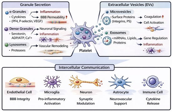
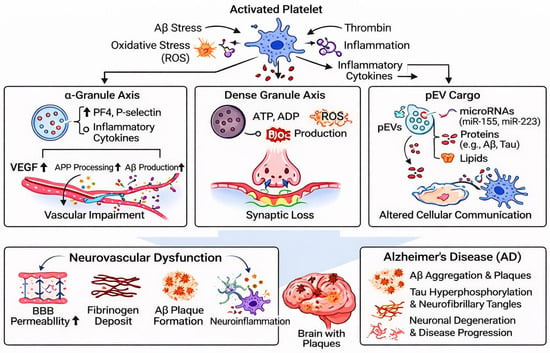
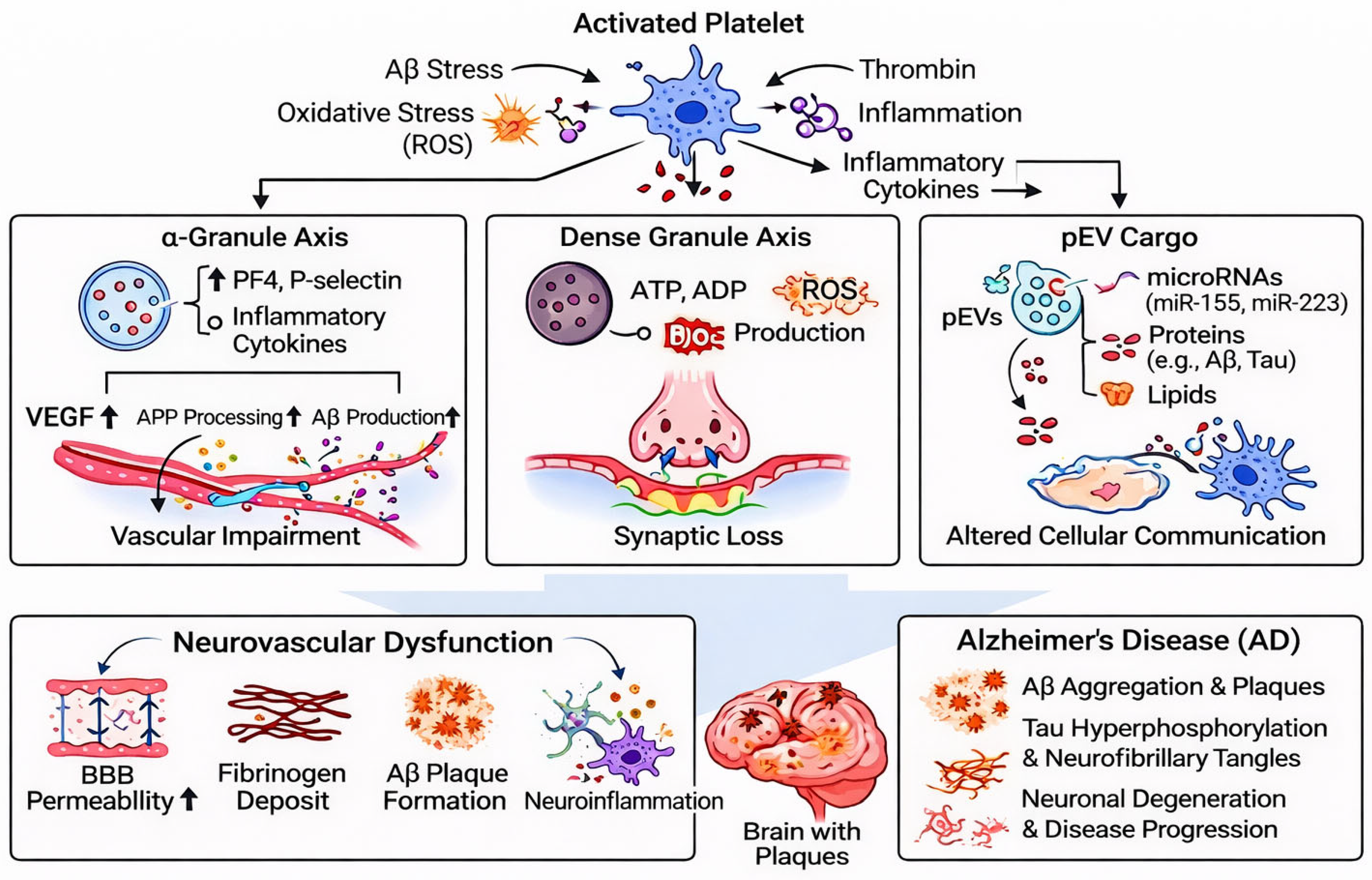
Disclaimer/Publisher’s Note: The statements, opinions and data contained in all publications are solely those of the individual author(s) and contributor(s) and not of MDPI and/or the editor(s). MDPI and/or the editor(s) disclaim responsibility for any injury to people or property resulting from any ideas, methods, instructions or products referred to in the content. |
© 2026 by the authors. Licensee MDPI, Basel, Switzerland. This article is an open access article distributed under the terms and conditions of the Creative Commons Attribution (CC BY) license.