Lipoprotein(a) and Cardiovascular Disease: From Genetic Risk Factor to Therapeutic Target
 , ,
, ,  and
and
Highlights
- Lipoprotein(a) [Lp(a)] acts as a distinct, genetically determined driver of cardiovascular disease by integrating pro-atherogenic, pro-inflammatory, and pro-thrombotic mechanisms.
- Emerging RNA-targeted platforms, such as antisense oligonucleotides and siRNAs, can achieve potent and durable Lp(a) reductions of over 90%, overcoming the limitations of conventional therapies.
- The development of potent Lp(a)-lowering therapeutics facilitates a transition from identifying Lp(a) as a risk marker to utilizing it as a specific therapeutic target.
- Ongoing Phase 3 clinical trials will define the clinical necessity of routine Lp(a) screening and the potential for mitigating residual risk in patients with ASCVD and aortic stenosis.
Abstract
1. Introduction
2. Structure, Genetics and Metabolism of Lp(a)
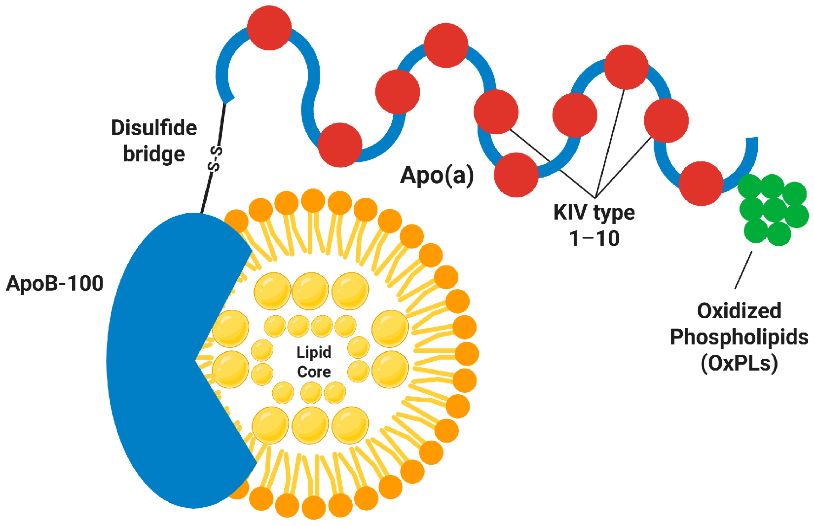
2.1. Unique Molecular Architecture
2.2. Genetics and Isoform Size Heterogeneity
2.3. Metabolism: Synthesis and Clearance
3. Pathologic Mechanisms Between Lp(a) and Cardiovascular Disease
4. Epidemiology and Clinical Associations
4.1. Coronary Artery Disease and Myocardial Infarction
4.2. Ischemic Stroke and Peripheral Artery Disease
4.3. Calcific Aortic Valve Stenosis
4.4. Heart Failure and Long-Term Prognosis
5. Measurement and Clinical Interpretation
5.1. Analytical Considerations and Standardization
5.2. Guideline Thresholds for Risk Assessment
5.3. Clinical Indications and Timing
6. Current Management Strategies
6.1. Lifestyle and Conventional Lipid-Lowering Therapy
6.2. Lipoprotein Apheresis
6.3. Practical Approach in the Absence of Lp(a)-Targeted Drugs
7. Emerging Lp(a)-Targeted Therapies
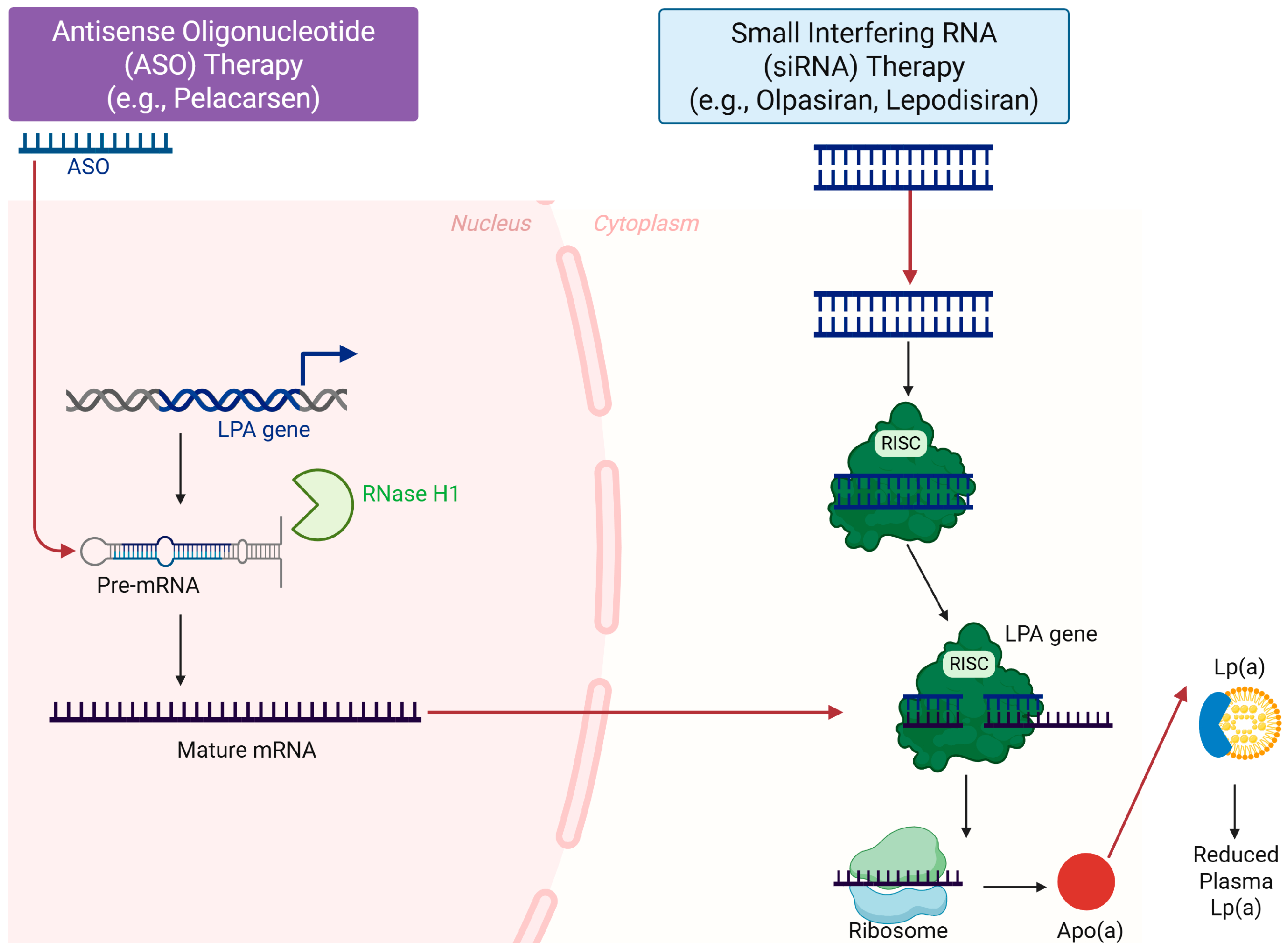
7.1. Antisense Oligonucleotides: Pelacarsen
7.2. Small Interfering RNA: Olpasiran
7.3. Lepodisiran and Other Emerging Agents
8. Knowledge Gaps and Future Directions
9. Conclusions
Author Contributions
Funding
Institutional Review Board Statement
Informed Consent Statement
Data Availability Statement
Conflicts of Interest
Abbreviations
| ACC | American College of Cardiology |
| AHA | American Heart Association |
| apo(a) | Apolipoprotein(a) |
| apoB-100 | Apolipoprotein B-100 |
| ASCVD | Atherosclerotic cardiovascular disease |
| ASO(s) | Antisense oligonucleotide(s) |
| AVR | Aortic valve replacement |
| BMP2 | Bone morphogenetic protein 2 |
| CAD | Coronary artery disease |
| CAVS | Calcific aortic valve stenosis |
| CNV | Copy number variation |
| EAS | European Atherosclerosis Society |
| EF | Ejection fraction |
| ER | Endoplasmic reticulum |
| ESC | European Society of Cardiology |
| GalNAc | N-acetylgalactosamine |
| GSK3β | Glycogen synthase kinase 3 beta |
| GWAS | Genome-wide association study |
| HF | Heart failure |
| ICAM-1 | Intercellular adhesion molecule-1 |
| IFCC | International Federation of Clinical Chemistry and Laboratory Medicine |
| IL-1β | Interleukin-1 beta |
| IL-6 | Interleukin-6 |
| JAHA | Journal of the American Heart Association |
| KI–KV | Kringle I–Kringle V |
| KIV | Kringle IV |
| KIV-10 | Kringle IV type 10 |
| KIV-2 | Kringle IV type 2 |
| KIV-9 | Kringle IV type 9 |
| LDL | Low-density lipoprotein |
| LDL-C | Low-density lipoprotein cholesterol |
| LDLR | Low-density lipoprotein receptor |
| Lp(a) | Lipoprotein(a) |
| LPA | LPA gene (apolipoprotein(a) gene locus) |
| MACE | Major adverse cardiovascular events |
| MALE(s) | Major adverse limb event(s) |
| MAPK | Mitogen-activated protein kinase |
| MCP-1 | Monocyte chemoattractant protein-1 |
| MI | Myocardial infarction |
| NF-κB | Nuclear factor kappa B |
| NLA | National Lipid Association |
| NLRP3 | NLR family pyrin domain containing 3 (inflammasome) |
| OxPL(s) | Oxidized phospholipid(s) |
| PAD | Peripheral artery disease |
| PCSK9 | Proprotein convertase subtilisin/kexin type 9 |
| PlgRKT | Plasminogen receptor with a C-terminal lysine (PlgRKT) |
| RISC | RNA-induced silencing complex |
| Runx2 | Runt-related transcription factor 2 |
| siRNA | Small interfering RNA |
| SNP(s) | Single-nucleotide polymorphism(s) |
| SR-BI | Scavenger receptor class B type I |
| TNF-α | Tumor necrosis factor-alpha |
| tPA | Tissue plasminogen activator |
| VCAM-1 | Vascular cell adhesion molecule-1 |
| VEC(s) | Valvular endothelial cell(s) |
| VIC(s) | Valvular interstitial cell(s) |
| WHO | World Health Organization |
| Wnt | Wingless/Int-1 signaling pathway |
References
- Clarke, R.; Peden, J.F.; Hopewell, J.C.; Kyriakou, T.; Goel, A.; Heath, S.C.; Parish, S.; Barlera, S.; Franzosi, M.G.; Rust, S.; et al. Genetic variants associated with Lp(a) lipoprotein level and coronary disease. N. Engl. J. Med. 2009, 361, 2518–2528. [Google Scholar] [CrossRef] [Scilit]
- Thanassoulis, G.; Campbell, C.Y.; Owens, D.S.; Smith, J.G.; Smith, A.V.; Peloso, G.M.; Kerr, K.F.; Pechlivanis, S.; Budoff, M.J.; Harris, T.B.; et al. Genetic associations with valvular calcification and aortic stenosis. N. Engl. J. Med. 2013, 368, 503–512. [Google Scholar] [CrossRef] [Scilit] [PubMed]
- Bjornson, E.; Adiels, M.; Taskinen, M.R.; Burgess, S.; Chapman, M.J.; Packard, C.J.; Boren, J. Lipoprotein(a) Is Markedly More Atherogenic Than LDL: An Apolipoprotein B-Based Genetic Analysis. J. Am. Coll. Cardiol. 2024, 83, 385–395. [Google Scholar] [CrossRef] [Scilit] [PubMed]
- Schwartz, G.G.; Szarek, M.; Bittner, V.A.; Diaz, R.; Goodman, S.G.; Jukema, J.W.; Landmesser, U.; Lopez-Jaramillo, P.; Manvelian, G.; Pordy, R.; et al. Lipoprotein(a) and Benefit of PCSK9 Inhibition in Patients With Nominally Controlled LDL Cholesterol. J. Am. Coll. Cardiol. 2021, 78, 421–433. [Google Scholar] [CrossRef] [Scilit] [PubMed]
- Kaur, G.; Abdelrahman, K.; Berman, A.N.; Biery, D.W.; Shiyovich, A.; Huck, D.; Garshick, M.; Blankstein, R.; Weber, B. Lipoprotein(a): Emerging insights and therapeutics. Am. J. Prev. Cardiol. 2024, 18, 100641. [Google Scholar] [CrossRef] [Scilit]
- Gudbjartsson, D.F.; Thorgeirsson, G.; Sulem, P.; Helgadottir, A.; Gylfason, A.; Saemundsdottir, J.; Bjornsson, E.; Norddahl, G.L.; Jonasdottir, A.; Jonasdottir, A.; et al. Lipoprotein(a) Concentration and Risks of Cardiovascular Disease and Diabetes. J. Am. Coll. Cardiol. 2019, 74, 2982–2994. [Google Scholar] [CrossRef] [Scilit] [PubMed]
- Clarke, R.; Wright, N.; Lin, K.; Yu, C.; Walters, R.G.; Lv, J.; Hill, M.; Kartsonaki, C.; Millwood, I.Y.; Bennett, D.A.; et al. Causal Relevance of Lp(a) for Coronary Heart Disease and Stroke Types in East Asian and European Ancestry Populations: A Mendelian Randomization Study. Circulation 2025, 151, 1699–1711. [Google Scholar] [CrossRef] [Scilit]
- Leibundgut, G.; Scipione, C.; Yin, H.; Schneider, M.; Boffa, M.B.; Green, S.; Yang, X.; Dennis, E.; Witztum, J.L.; Koschinsky, M.L.; et al. Determinants of binding of oxidized phospholipids on apolipoprotein (a) and lipoprotein (a). J. Lipid Res. 2013, 54, 2815–2830. [Google Scholar] [CrossRef] [Scilit] [PubMed]
- Greco, A.; Finocchiaro, S.; Spagnolo, M.; Faro, D.C.; Mauro, M.S.; Raffo, C.; Sangiorgio, G.; Imbesi, A.; Laudani, C.; Mazzone, P.M.; et al. Lipoprotein(a) as a Pharmacological Target: Premises, Promises, and Prospects. Circulation 2025, 151, 400–415. [Google Scholar] [CrossRef] [Scilit] [PubMed]
- Nordestgaard, B.G.; Langsted, A. Lipoprotein(a) and cardiovascular disease. Lancet 2024, 404, 1255–1264. [Google Scholar] [CrossRef] [Scilit]
- Mach, F.; Baigent, C.; Catapano, A.L.; Koskinas, K.C.; Casula, M.; Badimon, L.; Chapman, M.J.; De Backer, G.G.; Delgado, V.; Ference, B.A.; et al. 2019 ESC/EAS Guidelines for the management of dyslipidaemias: Lipid modification to reduce cardiovascular risk. Eur. Heart J. 2020, 41, 111–188, Corrigendum in Eur. Heart J. 2020, 41, 4255. https://doi.org/10.1093/eurheartj/ehz826. [Google Scholar] [CrossRef] [Scilit]
- Kronenberg, F.; Mora, S.; Stroes, E.S.G.; Ference, B.A.; Arsenault, B.J.; Berglund, L.; Dweck, M.R.; Koschinsky, M.; Lambert, G.; Mach, F.; et al. Lipoprotein(a) in atherosclerotic cardiovascular disease and aortic stenosis: A European Atherosclerosis Society consensus statement. Eur. Heart J. 2022, 43, 3925–3946. [Google Scholar] [CrossRef] [Scilit]
- Xie, S.; Galimberti, F.; Olmastroni, E.; Carugo, S.; Catapano, A.L.; Casula, M.; Group, M.-L. Effect of lipid-lowering therapies on lipoprotein(a) levels: A comprehensive meta-analysis of randomized controlled trials. Atherosclerosis 2025, 408, 120420. [Google Scholar] [CrossRef] [Scilit]
- Enkhmaa, B.; Berglund, L. Non-genetic influences on lipoprotein(a) concentrations. Atherosclerosis 2022, 349, 53–62. [Google Scholar] [CrossRef] [Scilit] [PubMed]
- Chung, F.Z.; Wu, L.H.; Lee, H.T.; Mueller, W.T.; Spahr, M.A.; Eaton, S.R.; Tian, Y.; Settimi, P.D.; Oxender, D.L.; Ramharack, R. Bacterial expression and characterization of human recombinant apolipoprotein(a) kringle IV type 9. Protein Expr. Purif. 1998, 13, 222–228. [Google Scholar] [CrossRef] [Scilit]
- Buciu, I.C.; Tieranu, E.N.; Pircalabu, A.S.; Zlatian, O.M.; Donoiu, I.; Cioboata, R.; Militaru, C.; Militaru, S.; Botezat, M.M.; Militaru, C. Lipoprotein (a) in the context of atherosclerosis: Pathological implications and therapeutic perspectives in myocardial infarction. A narrative review. Rom. J. Morphol. Embryol. 2024, 65, 609–616. [Google Scholar] [CrossRef] [Scilit] [PubMed]
- Eaton, D.L.; Fless, G.M.; Kohr, W.J.; McLean, J.W.; Xu, Q.T.; Miller, C.G.; Lawn, R.M.; Scanu, A.M. Partial amino acid sequence of apolipoprotein(a) shows that it is homologous to plasminogen. Proc. Natl. Acad. Sci. USA 1987, 84, 3224–3228. [Google Scholar] [CrossRef] [Scilit]
- Schmidt, K.; Noureen, A.; Kronenberg, F.; Utermann, G. Structure, function, and genetics of lipoprotein (a). J. Lipid Res. 2016, 57, 1339–1359. [Google Scholar] [CrossRef] [Scilit] [PubMed]
- Tsioulos, G.; Kounatidis, D.; Vallianou, N.G.; Poulaki, A.; Kotsi, E.; Christodoulatos, G.S.; Tsilingiris, D.; Karampela, I.; Skourtis, A.; Dalamaga, M. Lipoprotein(a) and Atherosclerotic Cardiovascular Disease: Where Do We Stand? Int. J. Mol. Sci. 2024, 25, 3537. [Google Scholar] [CrossRef] [Scilit] [PubMed]
- Mihalich, A.; Magnaghi, P.; Sessa, L.; Trubia, M.; Acquati, F.; Taramelli, R. Genomic structure and organization of kringles type 3 to 10 of the apolipoprotein(a) gene in 6q26–27. Gene 1997, 196, 1–8. [Google Scholar] [CrossRef] [Scilit] [PubMed]
- Koschinsky, M.L.; Beisiegel, U.; Henne-Bruns, D.; Eaton, D.L.; Lawn, R.M. Apolipoprotein(a) size heterogeneity is related to variable number of repeat sequences in its mRNA. Biochemistry 1990, 29, 640–644. [Google Scholar] [CrossRef] [Scilit] [PubMed]
- Berglund, L.; Ramakrishnan, R. Lipoprotein(a): An elusive cardiovascular risk factor. Arterioscler. Thromb. Vasc. Biol. 2004, 24, 2219–2226. [Google Scholar] [CrossRef] [Scilit] [PubMed]
- Koschinsky, M.L.; Boffa, M.B. Oxidized phospholipid modification of lipoprotein(a): Epidemiology, biochemistry and pathophysiology. Atherosclerosis 2022, 349, 92–100. [Google Scholar] [CrossRef] [Scilit] [PubMed]
- Chajek-Shaul, T.; Friedman, G.; Bengtsson-Olivecrona, G.; Vlodavsky, I.; Bar-Shavit, R. Interaction of lipoprotein lipase with subendothelial extracellular matrix. Biochim. Biophys. Acta 1990, 1042, 168–175. [Google Scholar] [CrossRef] [Scilit] [PubMed]
- Mochalkin, I.; Cheng, B.; Klezovitch, O.; Scanu, A.M.; Tulinsky, A. Recombinant kringle IV-10 modules of human apolipoprotein(a): Structure, ligand binding modes, and biological relevance. Biochemistry 1999, 38, 1990–1998. [Google Scholar] [CrossRef] [Scilit] [PubMed]
- van der Valk, F.M.; Bekkering, S.; Kroon, J.; Yeang, C.; Van den Bossche, J.; van Buul, J.D.; Ravandi, A.; Nederveen, A.J.; Verberne, H.J.; Scipione, C.; et al. Oxidized Phospholipids on Lipoprotein(a) Elicit Arterial Wall Inflammation and an Inflammatory Monocyte Response in Humans. Circulation 2016, 134, 611–624. [Google Scholar] [CrossRef] [Scilit]
- Boerwinkle, E.; Leffert, C.C.; Lin, J.; Lackner, C.; Chiesa, G.; Hobbs, H.H. Apolipoprotein(a) gene accounts for greater than 90% of the variation in plasma lipoprotein(a) concentrations. J. Clin. Investig. 1992, 90, 52–60. [Google Scholar] [CrossRef] [Scilit] [PubMed]
- Frank, S.L.; Klisak, I.; Sparkes, R.S.; Mohandas, T.; Tomlinson, J.E.; McLean, J.W.; Lawn, R.M.; Lusis, A.J. The apolipoprotein(a) gene resides on human chromosome 6q26–27, in close proximity to the homologous gene for plasminogen. Hum. Genet. 1988, 79, 352–356. [Google Scholar] [CrossRef] [Scilit]
- Coassin, S.; Kronenberg, F. Lipoprotein(a) beyond the kringle IV repeat polymorphism: The complexity of genetic variation in the LPA gene. Atherosclerosis 2022, 349, 17–35. [Google Scholar] [CrossRef] [Scilit] [PubMed]
- Noureen, A.; Fresser, F.; Utermann, G.; Schmidt, K. Sequence variation within the KIV-2 copy number polymorphism of the human LPA gene in African, Asian, and European populations. PLoS ONE 2015, 10, e0121582. [Google Scholar] [CrossRef] [Scilit] [PubMed]
- Fogacci, F.; Di Micoli, V.; Avagimyan, A.; Giovannini, M.; Imbalzano, E.; Cicero, A.F.G. Assessment of Apolipoprotein(a) Isoform Size Using Phenotypic and Genotypic Methods. Int. J. Mol. Sci. 2023, 24, 13886. [Google Scholar] [CrossRef] [Scilit]
- Brunner, C.; Lobentanz, E.M.; Petho-Schramm, A.; Ernst, A.; Kang, C.; Dieplinger, H.; Muller, H.J.; Utermann, G. The number of identical kringle IV repeats in apolipoprotein(a) affects its processing and secretion by HepG2 cells. J. Biol. Chem. 1996, 271, 32403–32410. [Google Scholar] [CrossRef] [Scilit] [PubMed]
- White, A.L.; Guerra, B.; Lanford, R.E. Influence of allelic variation on apolipoprotein(a) folding in the endoplasmic reticulum. J. Biol. Chem. 1997, 272, 5048–5055. [Google Scholar] [CrossRef] [Scilit]
- White, A.L.; Guerra, B.; Wang, J.; Lanford, R.E. Presecretory degradation of apolipoprotein[a] is mediated by the proteasome pathway. J. Lipid Res. 1999, 40, 275–286. [Google Scholar] [CrossRef] [Scilit] [PubMed]
- Wang, J.; White, A.L. Role of calnexin, calreticulin, and endoplasmic reticulum mannosidase I in apolipoprotein(a) intracellular targeting. Biochemistry 2000, 39, 8993–9000. [Google Scholar] [CrossRef] [Scilit]
- Reyes-Soffer, G.; Ginsberg, H.N.; Berglund, L.; Duell, P.B.; Heffron, S.P.; Kamstrup, P.R.; Lloyd-Jones, D.M.; Marcovina, S.M.; Yeang, C.; Koschinsky, M.L.; et al. Lipoprotein(a): A Genetically Determined, Causal, and Prevalent Risk Factor for Atherosclerotic Cardiovascular Disease: A Scientific Statement From the American Heart Association. Arterioscler. Thromb. Vasc. Biol. 2022, 42, e48–e60. [Google Scholar] [CrossRef] [Scilit] [PubMed]
- Schachtl-Riess, J.F.; Kheirkhah, A.; Gruneis, R.; Di Maio, S.; Schoenherr, S.; Streiter, G.; Losso, J.L.; Paulweber, B.; Eckardt, K.U.; Kottgen, A.; et al. Frequent LPA KIV-2 Variants Lower Lipoprotein(a) Concentrations and Protect Against Coronary Artery Disease. J. Am. Coll. Cardiol. 2021, 78, 437–449. [Google Scholar] [CrossRef] [Scilit]
- Lee, S.R.; Prasad, A.; Choi, Y.S.; Xing, C.; Clopton, P.; Witztum, J.L.; Tsimikas, S. LPA Gene, Ethnicity, and Cardiovascular Events. Circulation 2017, 135, 251–263. [Google Scholar] [CrossRef] [Scilit] [PubMed]
- Reyes-Soffer, G. The impact of race and ethnicity on lipoprotein(a) levels and cardiovascular risk. Curr. Opin. Lipidol. 2021, 32, 163–166. [Google Scholar] [CrossRef] [Scilit]
- Kim, J.A.; Kim, N.H. Lipoprotein(a) and Cardiovascular Risk in Asian Populations: A Comprehensive Review. J. Lipid Atheroscler. 2025, 14, 174–187. [Google Scholar] [CrossRef] [Scilit]
- Rubin, J.; Kim, H.J.; Pearson, T.A.; Holleran, S.; Ramakrishnan, R.; Berglund, L. Apo[a] size and PNR explain African American-Caucasian differences in allele-specific apo[a] levels for small but not large apo[a]. J. Lipid Res. 2006, 47, 982–989. [Google Scholar] [CrossRef] [Scilit]
- Rader, D.J.; Mann, W.A.; Cain, W.; Kraft, H.G.; Usher, D.; Zech, L.A.; Hoeg, J.M.; Davignon, J.; Lupien, P.; Grossman, M.; et al. The low density lipoprotein receptor is not required for normal catabolism of Lp(a) in humans. J. Clin. Investig. 1995, 95, 1403–1408. [Google Scholar] [CrossRef] [Scilit] [PubMed]
- Huby, T.; Afzal, V.; Doucet, C.; Lawn, R.M.; Gong, E.L.; Chapman, M.J.; Thillet, J.; Rubin, E.M. Regulation of the expression of the apolipoprotein(a) gene: Evidence for a regulatory role of the 5′ distal apolipoprotein(a) transcription control region enhancer in yeast artificial chromosome transgenic mice. Arterioscler. Thromb. Vasc. Biol. 2003, 23, 1633–1639. [Google Scholar] [CrossRef] [Scilit] [PubMed]
- Chemello, K.; Chan, D.C.; Lambert, G.; Watts, G.F. Recent advances in demystifying the metabolism of lipoprotein(a). Atherosclerosis 2022, 349, 82–91. [Google Scholar] [CrossRef] [Scilit]
- Sharma, M.; Redpath, G.M.; Williams, M.J.; McCormick, S.P. Recycling of Apolipoprotein(a) After PlgRKT-Mediated Endocytosis of Lipoprotein(a). Circ. Res. 2017, 120, 1091–1102. [Google Scholar] [CrossRef] [Scilit]
- Siddiqui, H.; Deo, N.; Rutledge, M.T.; Williams, M.J.A.; Redpath, G.M.I.; McCormick, S.P.A. Plasminogen Receptors Promote Lipoprotein(a) Uptake by Enhancing Surface Binding and Facilitating Macropinocytosis. Arterioscler. Thromb. Vasc. Biol. 2023, 43, 1851–1866. [Google Scholar] [CrossRef] [Scilit]
- Mooser, V.; Seabra, M.C.; Abedin, M.; Landschulz, K.T.; Marcovina, S.; Hobbs, H.H. Apolipoprotein(a) kringle 4-containing fragments in human urine. Relationship to plasma levels of lipoprotein(a). J. Clin. Investig. 1996, 97, 858–864. [Google Scholar] [CrossRef] [Scilit]
- Nurmohamed, N.S.; Gaillard, E.L.; Malkasian, S.; de Groot, R.J.; Ibrahim, S.; Bom, M.J.; Kaiser, Y.; Earls, J.P.; Min, J.K.; Kroon, J.; et al. Lipoprotein(a) and Long-Term Plaque Progression, Low-Density Plaque, and Pericoronary Inflammation. JAMA Cardiol. 2024, 9, 826–834. [Google Scholar] [CrossRef] [Scilit]
- Rosenson, R.S.; Tate, A.M.; Grushko, O.G.; Damodaran, D.; Chen, Q.; Boffa, M.; Koschinsky, M.; Narula, J.; Goonewardena, S.N. Lipoprotein (a) integrates monocyte-mediated thrombosis and inflammation in atherosclerotic cardiovascular disease. J. Lipid Res. 2025, 66, 100820. [Google Scholar] [CrossRef] [Scilit]
- Bhatia, H.S.; Dweck, M.R.; Craig, N.; Capoulade, R.; Pibarot, P.; Trainor, P.J.; Whelton, S.P.; Rikhi, R.; Lidani, K.C.F.; Post, W.S.; et al. Oxidized Phospholipids and Calcific Aortic Valvular Disease. J. Am. Coll. Cardiol. 2024, 84, 2430–2441. [Google Scholar] [CrossRef] [Scilit] [PubMed]
- Boffa, M.B.; Koschinsky, M.L. Oxidized phospholipids as a unifying theory for lipoprotein(a) and cardiovascular disease. Nat. Rev. Cardiol. 2019, 16, 305–318. [Google Scholar] [CrossRef] [Scilit]
- Gilliland, T.C.; Liu, Y.; Mohebi, R.; Miksenas, H.; Haidermota, S.; Wong, M.; Hu, X.; Cristino, J.R.; Browne, A.; Plutzky, J.; et al. Lipoprotein(a), Oxidized Phospholipids, and Coronary Artery Disease Severity and Outcomes. J. Am. Coll. Cardiol. 2023, 81, 1780–1792. [Google Scholar] [CrossRef] [Scilit]
- Boffa, M.B.; Koschinsky, M.L. Lipoprotein (a): Truly a direct prothrombotic factor in cardiovascular disease? J. Lipid Res. 2016, 57, 745–757. [Google Scholar] [CrossRef] [Scilit] [PubMed]
- Yeang, C.; Wilkinson, M.J.; Tsimikas, S. Lipoprotein(a) and oxidized phospholipids in calcific aortic valve stenosis. Curr. Opin. Cardiol. 2016, 31, 440–450. [Google Scholar] [CrossRef] [Scilit]
- Nielsen, L.B.; Nordestgaard, B.G.; Stender, S.; Niendorf, A.; Kjeldsen, K. Transfer of lipoprotein(a) and LDL into aortic intima in normal and in cholesterol-fed rabbits. Arterioscler. Thromb. Vasc. Biol. 1995, 15, 1492–1502. [Google Scholar] [CrossRef] [Scilit]
- Nielsen, L.B.; Stender, S.; Jauhiainen, M.; Nordestgaard, B.G. Preferential influx and decreased fractional loss of lipoprotein(a) in atherosclerotic compared with nonlesioned rabbit aorta. J. Clin. Investig. 1996, 98, 563–571. [Google Scholar] [CrossRef] [Scilit]
- Klezovitch, O.; Edelstein, C.; Zhu, L.; Scanu, A.M. Apolipoprotein(a) binds via its C-terminal domain to the protein core of the proteoglycan decorin. Implications for the retention of lipoprotein(a) in atherosclerotic lesions. J. Biol. Chem. 1998, 273, 23856–23865. [Google Scholar] [CrossRef] [Scilit]
- Aikas, L.; Kovanen, P.T.; Lorey, M.B.; Laaksonen, R.; Holopainen, M.; Ruhanen, H.; Kakela, R.; Jauhiainen, M.; Hermansson, M.; Oorni, K. Icosapent ethyl-induced lipoprotein remodeling and its impact on cardiovascular disease risk markers in normolipidemic individuals. JCI Insight 2025, 10, e193637. [Google Scholar] [CrossRef] [Scilit]
- Goldberg, I.J.; Cabodevilla, A.G.; Younis, W. In the Beginning, Lipoproteins Cross the Endothelial Barrier. J. Atheroscler. Thromb. 2024, 31, 854–860. [Google Scholar] [CrossRef] [Scilit] [PubMed]
- Skalen, K.; Gustafsson, M.; Rydberg, E.K.; Hulten, L.M.; Wiklund, O.; Innerarity, T.L.; Boren, J. Subendothelial retention of atherogenic lipoproteins in early atherosclerosis. Nature 2002, 417, 750–754. [Google Scholar] [CrossRef] [Scilit] [PubMed]
- Boffa, M.B.; Koschinsky, M.L. Lipoprotein(a) and cardiovascular disease. Biochem. J. 2024, 481, 1277–1296. [Google Scholar] [CrossRef] [Scilit] [PubMed]
- Tan, J.M.E.; Cheng, L.; Calhoun, R.P.; Weller, A.H.; Drareni, K.; Fong, S.; Barbara, E.; Lim, H.W.; Xue, C.; Winter, H.; et al. PRDM16 regulates smooth muscle cell identity and atherosclerotic plaque composition. Nat. Cardiovasc. Res. 2025, 4, 1573–1588. [Google Scholar] [CrossRef] [Scilit] [PubMed]
- Clarke, S.L.; Huang, R.D.L.; Hilliard, A.T.; Levin, M.G.; Sharma, D.; Thomson, B.; Lynch, J.; Tsao, P.S.; Gaziano, J.M.; Assimes, T.L.; et al. Genetically predicted lipoprotein(a) associates with coronary artery plaque severity independent of low-density lipoprotein cholesterol. Eur. J. Prev. Cardiol. 2025, 32, 116–127. [Google Scholar] [CrossRef] [Scilit] [PubMed]
- Schnitzler, J.G.; Hoogeveen, R.M.; Ali, L.; Prange, K.H.M.; Waissi, F.; van Weeghel, M.; Bachmann, J.C.; Versloot, M.; Borrelli, M.J.; Yeang, C.; et al. Atherogenic Lipoprotein(a) Increases Vascular Glycolysis, Thereby Facilitating Inflammation and Leukocyte Extravasation. Circ. Res. 2020, 126, 1346–1359. [Google Scholar] [CrossRef] [Scilit] [PubMed]
- Polkinghorne, M.D.; Badi, I.; Baragetti, A.; Chauhan, J.; Xie, C.; Wahome, E.; Akoumianakis, I.; Foran, D.; Patel, P.; de Araujo, E.; et al. Lipoprotein(a) and Vascular Redox State in Patients With Advanced Coronary Atherosclerosis. Arterioscler. Thromb. Vasc. Biol. 2025, 46, 232–247. [Google Scholar] [CrossRef] [Scilit]
- Bergmark, C.; Dewan, A.; Orsoni, A.; Merki, E.; Miller, E.R.; Shin, M.J.; Binder, C.J.; Horkko, S.; Krauss, R.M.; Chapman, M.J.; et al. A novel function of lipoprotein [a] as a preferential carrier of oxidized phospholipids in human plasma. J. Lipid Res. 2008, 49, 2230–2239. [Google Scholar] [CrossRef] [Scilit]
- Kumarapperuma, H.; Chia, Z.J.; Malapitan, S.M.; Wight, T.N.; Little, P.J.; Kamato, D. Response to retention hypothesis as a source of targets for arterial wall-directed therapies to prevent atherosclerosis: A critical review. Atherosclerosis 2024, 397, 118552. [Google Scholar] [CrossRef] [Scilit]
- Wang, X.; Ma, C.; Hou, X.; Zhang, G.; Huang, Y. Circular RNA circ_0002984 Promotes Cell Proliferation and Migration by Regulating miR-181b-5p/Vascular Endothelial Growth Factor Axis and PI3K-AKT Signaling Pathway in Oxidized Low-Density Lipoprotein-Treated Vascular Smooth Muscle Cells. J. Cardiovasc. Pharmacol. 2022, 79, 501–511. [Google Scholar] [CrossRef] [Scilit]
- Tasdighi, E.; Adhikari, R.; Almaadawy, O.; Leucker, T.M.; Blaha, M.J. LP(a): Structure, Genetics, Associated Cardiovascular Risk, and Emerging Therapeutics. Annu. Rev. Pharmacol. Toxicol. 2024, 64, 135–157. [Google Scholar] [CrossRef] [Scilit]
- Podrez, E.A.; Poliakov, E.; Shen, Z.; Zhang, R.; Deng, Y.; Sun, M.; Finton, P.J.; Shan, L.; Febbraio, M.; Hajjar, D.P.; et al. A novel family of atherogenic oxidized phospholipids promotes macrophage foam cell formation via the scavenger receptor CD36 and is enriched in atherosclerotic lesions. J. Biol. Chem. 2002, 277, 38517–38523. [Google Scholar] [CrossRef] [Scilit]
- Ichikawa, T.; Unoki, H.; Sun, H.; Shimoyamada, H.; Marcovina, S.; Shikama, H.; Watanabe, T.; Fan, J. Lipoprotein(a) promotes smooth muscle cell proliferation and dedifferentiation in atherosclerotic lesions of human apo(a) transgenic rabbits. Am. J. Pathol. 2002, 160, 227–236. [Google Scholar] [CrossRef] [Scilit] [PubMed]
- Rader, D.J.; Bajaj, A. Lipoprotein(a) and Oxidized Phospholipids: Partners in Crime or Individual Perpetrators in Cardiovascular Disease? J. Am. Coll. Cardiol. 2023, 81, 1793–1796. [Google Scholar] [CrossRef] [Scilit]
- Yao, M.; Dickeson, S.K.; Dhanabalan, K.; Solomevich, S.; Dennewitz, C.; Gailani, D.; Song, W.L. Investigation of the Influence of Lipoprotein(a) and Oxidized Lipoprotein(a) on Plasminogen Activation and Fibrinolysis. J. Lipid Atheroscler. 2025, 14, 229–235. [Google Scholar] [CrossRef] [Scilit]
- Clark, J.R.; Sutherland, F.S.; Assini, J.M.; Daichedt, L.; Szabla, R.; Junop, M.W.; Koschinsky, M.L.; Boffa, M.B. Lipoprotein(a) prolongs ex vivo plasma clot lysis times through effects on clot formation rate and fibrin structure. J. Thromb. Haemost. 2025, 24, 701–715. [Google Scholar] [CrossRef] [Scilit] [PubMed]
- Rogers, M.A.; Bartoli-Leonard, F.; Zheng, K.H.; Small, A.M.; Chen, H.Y.; Clift, C.L.; Asano, T.; Kuraoka, S.; Blaser, M.C.; Perez, K.A.; et al. Major Facilitator Superfamily Domain Containing 5 Inhibition Reduces Lipoprotein(a) Uptake and Calcification in Valvular Heart Disease. Circulation 2024, 149, 391–401. [Google Scholar] [CrossRef] [Scilit]
- Torzewski, M.; Ravandi, A.; Yeang, C.; Edel, A.; Bhindi, R.; Kath, S.; Twardowski, L.; Schmid, J.; Yang, X.; Franke, U.F.W.; et al. Lipoprotein(a) Associated Molecules are Prominent Components in Plasma and Valve Leaflets in Calcific Aortic Valve Stenosis. JACC Basic Transl. Sci. 2017, 2, 229–240. [Google Scholar] [CrossRef] [Scilit]
- Zheng, K.H.; Tsimikas, S.; Pawade, T.; Kroon, J.; Jenkins, W.S.A.; Doris, M.K.; White, A.C.; Timmers, N.; Hjortnaes, J.; Rogers, M.A.; et al. Lipoprotein(a) and Oxidized Phospholipids Promote Valve Calcification in Patients With Aortic Stenosis. J. Am. Coll. Cardiol. 2019, 73, 2150–2162. [Google Scholar] [CrossRef] [Scilit] [PubMed]
- Yu, B.; Hafiane, A.; Thanassoulis, G.; Ott, L.; Filwood, N.; Cerruti, M.; Gourgas, O.; Shum-Tim, D.; Al Kindi, H.; de Varennes, B.; et al. Lipoprotein(a) Induces Human Aortic Valve Interstitial Cell Calcification. JACC Basic Transl. Sci. 2017, 2, 358–371. [Google Scholar] [CrossRef] [Scilit]
- Lan, N.S.R.; Khan, Z.; Watts, G.F. Lipoprotein(a) and calcific aortic valve disease: Current evidence and future directions. Curr. Opin. Clin. Nutr. Metab. Care 2024, 27, 77–86. [Google Scholar] [CrossRef] [Scilit]
- Dzobo, K.E.; Cupido, A.J.; Mol, B.M.; Stiekema, L.C.A.; Versloot, M.; Winkelmeijer, M.; Peter, J.; Pennekamp, A.M.; Havik, S.R.; Vaz, F.M.; et al. Diacylglycerols and Lysophosphatidic Acid, Enriched on Lipoprotein(a), Contribute to Monocyte Inflammation. Arterioscler. Thromb. Vasc. Biol. 2024, 44, 720–740. [Google Scholar] [CrossRef] [Scilit]
- Moncla, L.M.; Briend, M.; Bosse, Y.; Mathieu, P. Calcific aortic valve disease: Mechanisms, prevention and treatment. Nat. Rev. Cardiol. 2023, 20, 546–559. [Google Scholar] [CrossRef] [Scilit]
- Otto, C.M.; Newby, D.E.; Hillis, G.S. Calcific Aortic Stenosis: A Review. JAMA 2024, 332, 2014–2026. [Google Scholar] [CrossRef] [Scilit]
- Peletier, M.; Ali, L.; Dzobo, K.; El Bouazzati, T.; Gaillard, E.L.; Romero Prats, M.L.; Versloot, M.; Peter, J.; Tsimikas, S.; Dweck, M.; et al. Fuelling aortic stenosis: The integral role of 6-phosphofructo-2-kinase/fructose-2,6-biphosphatase 3-mediated glycolysis in Lp(a)-induced valve inflammation. Eur. Heart J. Open 2025, 5, oeaf068. [Google Scholar] [CrossRef] [Scilit]
- Sanchez-Bayuela, T.; Peral-Rodrigo, M.; Parra-Izquierdo, I.; Lopez, J.; Gomez, C.; Montero, O.; Perez-Riesgo, E.; San Roman, J.A.; Butcher, J.T.; Sanchez Crespo, M.; et al. Inflammation via JAK-STAT/HIF-1alpha Drives Metabolic Changes in Pentose Phosphate Pathway and Glycolysis That Support Aortic Valve Cell Calcification. Arterioscler. Thromb. Vasc. Biol. 2025, 45, e232–e249. [Google Scholar] [CrossRef] [Scilit] [PubMed]
- Wong, N.D.; Fan, W.; Hu, X.; Ballantyne, C.; Hoodgeveen, R.C.; Tsai, M.Y.; Browne, A.; Budoff, M.J. Lipoprotein(a) and Long-Term Cardiovascular Risk in a Multi-Ethnic Pooled Prospective Cohort. J. Am. Coll. Cardiol. 2024, 83, 1511–1525. [Google Scholar] [CrossRef] [Scilit]
- Small, A.M.; Pournamdari, A.; Melloni, G.E.M.; Scirica, B.M.; Bhatt, D.L.; Raz, I.; Braunwald, E.; Giugliano, R.P.; Sabatine, M.S.; Peloso, G.M.; et al. Lipoprotein(a), C-Reactive Protein, and Cardiovascular Risk in Primary and Secondary Prevention Populations. JAMA Cardiol. 2024, 9, 385–391. [Google Scholar] [CrossRef] [Scilit] [PubMed]
- Yu, M.M.; Wang, M.L.; Wang, J.J.; Lin, B.L.; Zhao, X.; Tao, X.W.; Chen, Y.Y.; Li, P.Y.; Zhang, J.K.; Ge, J.B.; et al. Association of Lipoprotein(a) Levels With Myocardial Infarction in Patients With Low-Attenuation Plaque. J. Am. Coll. Cardiol. 2024, 83, 1743–1755. [Google Scholar] [CrossRef] [Scilit]
- Tsimikas, S. Lipoprotein(a) in the Year 2024: A Look Back and a Look Ahead. Arterioscler Thromb. Vasc. Biol. 2024, 44, 1485–1490. [Google Scholar] [CrossRef] [Scilit]
- Berman, A.N.; Biery, D.W.; Besser, S.A.; Singh, A.; Shiyovich, A.; Weber, B.N.; Huck, D.M.; Divakaran, S.; Hainer, J.; Kaur, G.; et al. Lipoprotein(a) and Major Adverse Cardiovascular Events in Patients With or Without Baseline Atherosclerotic Cardiovascular Disease. J. Am. Coll. Cardiol. 2024, 83, 873–886. [Google Scholar] [CrossRef] [Scilit]
- Bhatia, H.S.; Wandel, S.; Willeit, P.; Lesogor, A.; Bailey, K.; Ridker, P.M.; Nestel, P.; Simes, J.; Tonkin, A.; Schwartz, G.G.; et al. Independence of Lipoprotein(a) and Low-Density Lipoprotein Cholesterol-Mediated Cardiovascular Risk: A Participant-Level Meta-Analysis. Circulation 2025, 151, 312–321. [Google Scholar] [CrossRef] [Scilit] [PubMed]
- Shiyovich, A.; Berman, A.N.; Besser, S.A.; Biery, D.W.; Cardoso, R.; Divakaran, S.; Singh, A.; Huck, D.M.; Weber, B.; Plutzky, J.; et al. Lipoprotein(a) as a cardiovascular risk factor among patients with and without diabetes Mellitus: The Mass General Brigham Lp(a) Registry. Cardiovasc. Diabetol. 2024, 23, 257. [Google Scholar] [CrossRef] [Scilit] [PubMed]
- Zeng, G.; Zhu, P.; Yuan, D.; Wang, P.; Li, T.; Li, Q.; Xu, J.; Tang, X.; Song, Y.; Chen, Y.; et al. Renal function alters the association of lipoprotein(a) with cardiovascular outcomes in patients undergoing percutaneous coronary intervention: A prospective cohort study. Clin. Kidney J. 2024, 17, sfae032. [Google Scholar] [CrossRef] [Scilit] [PubMed]
- Enkhmaa, B.; Roshanravan, B.; Berglund, L. Lipoprotein(a) and Chronic Kidney Disease: Insights into New Therapeutic Opportunities. Clin. J. Am. Soc. Nephrol. 2025. Epub ahead of printing. [Google Scholar] [CrossRef] [Scilit]
- Mohammadnia, N.; van Broekhoven, A.; Bax, W.A.; Eikelboom, J.W.; Mosterd, A.; Fiolet, A.T.L.; Tijssen, J.G.P.; Thompson, P.L.; de Kleijn, D.P.V.; Tsimikas, S.; et al. Interleukin-6 modifies Lipoprotein(a) and oxidized phospholipids associated cardiovascular disease risk in a secondary prevention cohort. Atherosclerosis 2025, 405, 119211. [Google Scholar] [CrossRef] [Scilit] [PubMed]
- Chevalier, C.; Rodrigues Oliveira, A.; Blanchard, V.; Le May, C.; Cariou, B.; Hadjadj, S.; Croyal, M. Post-translational modifications of apolipoproteins as promising biomarkers for diabetes-related cardiovascular diseases: A comprehensive review. Diabetes Metab. 2025, 51, 101683. [Google Scholar] [CrossRef] [Scilit]
- Tsimikas, S.; Witztum, J.L. Oxidized phospholipids in cardiovascular disease. Nat. Rev. Cardiol. 2024, 21, 170–191. [Google Scholar] [CrossRef] [Scilit]
- Verbeek, R.; Boekholdt, S.M.; Stoekenbroek, R.M.; Hovingh, G.K.; Witztum, J.L.; Wareham, N.J.; Sandhu, M.S.; Khaw, K.T.; Tsimikas, S. Population and assay thresholds for the predictive value of lipoprotein (a) for coronary artery disease: The EPIC-Norfolk Prospective Population Study. J. Lipid Res. 2016, 57, 697–705. [Google Scholar] [CrossRef] [Scilit]
- Kamstrup, P.R.; Tybjaerg-Hansen, A.; Steffensen, R.; Nordestgaard, B.G. Genetically elevated lipoprotein(a) and increased risk of myocardial infarction. JAMA 2009, 301, 2331–2339. [Google Scholar] [CrossRef] [Scilit]
- Grundy, S.M.; Stone, N.J.; Bailey, A.L.; Beam, C.; Birtcher, K.K.; Blumenthal, R.S.; Braun, L.T.; de Ferranti, S.; Faiella-Tommasino, J.; Forman, D.E.; et al. 2018 AHA/ACC/AACVPR/AAPA/ABC/ACPM/ADA/AGS/APhA/ASPC/NLA/PCNA Guideline on the Management of Blood Cholesterol: A Report of the American College of Cardiology/American Heart Association Task Force on Clinical Practice Guidelines. Circulation 2019, 139, e1082–e1143. [Google Scholar] [CrossRef] [Scilit]
- MacDougall, D.E.; Tybjaerg-Hansen, A.; Knowles, J.W.; Stern, T.P.; Hartsuff, B.K.; McGowan, M.P.; Baum, S.J.; Wilemon, K.A.; Nordestgaard, B.G. Lipoprotein(a) and recurrent atherosclerotic cardiovascular events: The US Family Heart Database. Eur. Heart J. 2025, 46, 4762–4775. [Google Scholar] [CrossRef] [Scilit]
- Rudin, S.; Kriemler, L.; Dittrich, T.D.; Zietz, A.; Schweizer, J.; Arnold, M.; Peters, N.; Barinka, F.; Jung, S.; Arnold, M.; et al. Lipoprotein(a) as a blood marker for large artery atherosclerosis stroke etiology: Validation in a prospective cohort from a swiss stroke center. Swiss Med. Wkly. 2024, 154, 3633. [Google Scholar] [CrossRef] [Scilit] [PubMed]
- Gurdasani, D.; Sjouke, B.; Tsimikas, S.; Hovingh, G.K.; Luben, R.N.; Wainwright, N.W.; Pomilla, C.; Wareham, N.J.; Khaw, K.T.; Boekholdt, S.M.; et al. Lipoprotein(a) and risk of coronary, cerebrovascular, and peripheral artery disease: The EPIC-Norfolk prospective population study. Arterioscler. Thromb. Vasc. Biol. 2012, 32, 3058–3065. [Google Scholar] [CrossRef] [Scilit]
- Kao, A.S.; Kermanshahchi, J.; Khosrowjerdi, S.; Razavi, A.C.; Levene, J.; Reyes, M.; Garg, P.; Longstreth, W.T., Jr.; Rannikmae, K.; Sudlow, C.; et al. Demographics of lipoprotein(a) and stroke: The UK biobank. Am. J. Prev. Cardiol. 2025, 23, 101055. [Google Scholar] [CrossRef] [Scilit] [PubMed]
- Thomas, P.E.; Vedel-Krogh, S.; Nielsen, S.F.; Nordestgaard, B.G.; Kamstrup, P.R. Lipoprotein(a) and Risks of Peripheral Artery Disease, Abdominal Aortic Aneurysm, and Major Adverse Limb Events. J. Am. Coll. Cardiol. 2023, 82, 2265–2276. [Google Scholar] [CrossRef] [Scilit]
- Masson, W.; Lobo, M.; Barbagelata, L.; Molinero, G.; Bluro, I.; Nogueira, J.P. Elevated lipoprotein (a) levels and risk of peripheral artery disease outcomes: A systematic review. Vasc. Med. 2022, 27, 385–391. [Google Scholar] [CrossRef] [Scilit]
- Garagoli, F.; Masson, W.; Barbagelata, L. Association between elevated lipoprotein(a) levels and vulnerability of carotid atherosclerotic plaque: A systematic review. J. Stroke Cerebrovasc. Dis. 2024, 33, 108020. [Google Scholar] [CrossRef] [Scilit]
- Small, A.M.; Peloso, G.M.; Linefsky, J.; Aragam, J.; Galloway, A.; Tanukonda, V.; Wang, L.C.; Yu, Z.; Sunitha Selvaraj, M.; Farber-Eger, E.H.; et al. Multiancestry Genome-Wide Association Study of Aortic Stenosis Identifies Multiple Novel Loci in the Million Veteran Program. Circulation 2023, 147, 942–955. [Google Scholar] [CrossRef] [Scilit]
- Pantelidis, P.; Oikonomou, E.; Lampsas, S.; Zakynthinos, G.E.; Lysandrou, A.; Kalogeras, K.; Katsianos, E.; Theofilis, P.; Siasos, G.; Vavuranakis, M.A.; et al. Lipoprotein(a) and calcific aortic valve disease initiation and progression: A systematic review and meta-analysis. Cardiovasc. Res. 2023, 119, 1641–1655. [Google Scholar] [CrossRef] [Scilit]
- Arsenault, B.J.; Loganath, K.; Girard, A.; Botezatu, S.; Zheng, K.H.; Tzolos, E.; Abdoun, K.; Tastet, L.; Capoulade, R.; Cote, N.; et al. Lipoprotein(a) and Calcific Aortic Valve Stenosis Progression: A Systematic Review and Meta-Analysis. JAMA Cardiol. 2024, 9, 835–842. [Google Scholar] [CrossRef] [Scilit]
- Nomura, S.; Guan, W.; Zhang, Y.; Tison, G.H.; Ikezaki, H.; Diffenderfer, M.R.; Liu, C.T.; Hoogeveen, R.C.; Ballantyne, C.M.; Schaefer, E.J.; et al. Lipoprotein(a) and Heart Failure Among Black and White Participants in Atherosclerosis Risk in Communities Study, Framingham Offspring Study, and Multi-Ethnic Study of Atherosclerosis: The Pooling Project. J. Am. Heart Assoc. 2025, 14, e038608. [Google Scholar] [CrossRef] [Scilit] [PubMed]
- Wang, H.P.; Zhang, N.; Liu, Y.J.; Xia, T.L.; Chen, G.C.; Yang, J.; Li, F.R. Lipoprotein(a), family history of cardiovascular disease, and incidence of heart failure. J. Lipid Res. 2023, 64, 100398. [Google Scholar] [CrossRef] [Scilit]
- Kamstrup, P.R.; Nordestgaard, B.G. Elevated Lipoprotein(a) Levels, LPA Risk Genotypes, and Increased Risk of Heart Failure in the General Population. JACC Heart Fail. 2016, 4, 78–87. [Google Scholar] [CrossRef] [Scilit] [PubMed]
- Yadalam, A.K.; Gangavelli, A.; Razavi, A.C.; Ko, Y.A.; Alkhoder, A.; Haroun, N.; Lodhi, R.; Eldaidamouni, A.; Kasem, M.A.; Quyyumi, A.A. Lipoprotein(a) Levels and Adverse Outcomes in Heart Failure. J. Card. Fail. 2025. [Google Scholar] [CrossRef] [Scilit]
- Bhatia, H.S.; Rikhi, R.; Allen, T.S.; Yeang, C.; Guan, W.; Garg, P.K.; Tsai, M.Y.; Criqui, M.H.; Shapiro, M.D.; Tsimikas, S. Lipoprotein(a) and the pooled cohort equations for ASCVD risk prediction: The Multi-Ethnic Study of Atherosclerosis. Atherosclerosis 2023, 381, 117217. [Google Scholar] [CrossRef] [Scilit] [PubMed]
- Fan, W.; Wu, C.; Wong, N.D. Lipoprotein(a) Atherosclerotic Cardiovascular Disease Risk Score Development and Prediction in Primary Prevention from Real-World Data. Circ. Genom. Precis. Med. 2025, 18, e004631. [Google Scholar] [CrossRef] [Scilit] [PubMed]
- Marcovina, S.M.; Albers, J.J. Lipoprotein (a) measurements for clinical application. J. Lipid Res. 2016, 57, 526–537. [Google Scholar] [CrossRef] [Scilit]
- Lamon-Fava, S.; Marcovina, S.M.; Albers, J.J.; Kennedy, H.; DeLuca, C.; White, C.C.; Cupples, L.A.; McNamara, J.R.; Seman, L.J.; Bongard, V.; et al. Lipoprotein(a) levels, apo(a) isoform size, and coronary heart disease risk in the Framingham Offspring Study. J. Lipid Res. 2011, 52, 1181–1187. [Google Scholar] [CrossRef] [Scilit]
- Cegla, J.; France, M.; Marcovina, S.M.; Neely, R.D.G. Lp(a): When and how to measure it. Ann. Clin. Biochem. 2021, 58, 16–21. [Google Scholar] [CrossRef] [Scilit]
- Dati, F.; Tate, J.R.; Marcovina, S.M.; Steinmetz, A. First WHO/IFCC International Reference Reagent for Lipoprotein(a) for Immunoassay--Lp(a) SRM 2B. Clin. Chem. Lab. Med. 2004, 42, 670–676. [Google Scholar] [CrossRef] [Scilit]
- Koschinsky, M.L.; Bajaj, A.; Boffa, M.B.; Dixon, D.L.; Ferdinand, K.C.; Gidding, S.S.; Gill, E.A.; Jacobson, T.A.; Michos, E.D.; Safarova, M.S.; et al. A focused update to the 2019 NLA scientific statement on use of lipoprotein(a) in clinical practice. J. Clin. Lipidol. 2024, 18, e308–e319. [Google Scholar] [CrossRef] [Scilit]
- Arnett, D.K.; Blumenthal, R.S.; Albert, M.A.; Buroker, A.B.; Goldberger, Z.D.; Hahn, E.J.; Himmelfarb, C.D.; Khera, A.; Lloyd-Jones, D.; McEvoy, J.W.; et al. 2019 ACC/AHA Guideline on the Primary Prevention of Cardiovascular Disease: A Report of the American College of Cardiology/American Heart Association Task Force on Clinical Practice Guidelines. Circulation 2019, 140, e596–e646, Correction in Circulation 2020, 141, e774. https://doi.org/10.1161/CIR.0000000000000771. [Google Scholar] [CrossRef] [Scilit]
- Mach, F.; Koskinas, K.C.; van Lennep, J.E.R.; Tokgözoğlu, L.; Badimon, L.; Baigent, C.; Benn, M.; Binder, C.J.; Catapano, A.L.; De Backer, G.G.; et al. 2025 Focused Update of the 2019 ESC/EAS Guidelines for the management of dyslipidaemias: Developed by the task force for the management of dyslipidaemias of the European Society of Cardiology (ESC) and the European Atherosclerosis Society (EAS). Eur. Heart J. 2025, 46, 4359–4378, Correction in Eur. Heart J. 2025, ehaf1036. https://doi.org/10.1093/eurheartj/ehaf1036. [Google Scholar] [CrossRef] [Scilit]
- Khovidhunkit, W. Lipoprotein(a). In Endotext; Feingold, K.R., Adler, R.A., Ahmed, S.F., Anawalt, B., Blackman, M.R., Chrousos, G., Corpas, E., de Herder, W.W., Dhatariya, K., Dungan, K., et al., Eds.; MDText.com, Inc.: Dartmouth, MA, USA, 2000. [Google Scholar]
- Nazli, S.A.; Rosman, A.; Mohd Kasim, N.A.; Al-Khateeb, A.; Ul-Saufie, A.Z.; Md Radzi, A.B.; Ibrahim, K.S.; Kasim, S.S.; Nawawi, H. Coronary risk factor profiles according to different age categories in premature coronary artery disease patients who have undergone percutaneous coronary intervention. Sci. Rep. 2024, 14, 15326. [Google Scholar] [CrossRef] [Scilit]
- Loh, W.J.; Pang, J.; Chakraborty, A.; Ward, N.C.; Chan, D.C.; Hooper, A.J.; Bell, D.A.; Burnett, J.R.; Martin, A.C.; Watts, G.F. Cascade testing of children and adolescents for elevated Lp(a) in pedigrees with familial hypercholesterolaemia. J. Clin. Lipidol. 2024, 18, e33–e37. [Google Scholar] [CrossRef] [Scilit]
- Annink, M.E.; Janssen, E.S.; Reeskamp, L.F. Effectiveness of cascade screening for elevated lipoprotein(a), an underdiagnosed family disorder. Curr. Opin. Lipidol. 2024, 35, 290–296. [Google Scholar] [CrossRef] [Scilit] [PubMed]
- Santos, R.D.; Gidding, S.S.; Bourbon, M.; Iatan, I.; Harada-Shiba, M.; Raal, F.J.; Vallejo-Vaz, A.J.; Wiegman, A.; Watts, G.F. Recent advances in research and care of familial hypercholesterolaemia. Lancet Diabetes Endocrinol. 2025, 13, 1054–1071. [Google Scholar] [CrossRef] [Scilit] [PubMed]
- Girard, A.S.; Paulin, A.; Manikpurage, H.D.; Lajeunesse, E.; Clavel, M.A.; Pibarot, P.; Krege, J.H.; Mathieu, P.; Theriault, S.; Arsenault, B.J. Impact of Lipoprotein(a) on Valvular and Cardiovascular Outcomes in Patients With Calcific Aortic Valve Stenosis. J. Am. Heart Assoc. 2025, 14, e038955. [Google Scholar] [CrossRef] [Scilit] [PubMed]
- Riley, T.M.; Sapp, P.A.; Kris-Etherton, P.M.; Petersen, K.S. Effects of saturated fatty acid consumption on lipoprotein (a): A systematic review and meta-analysis of randomized controlled trials. Am. J. Clin. Nutr. 2024, 120, 619–629. [Google Scholar] [CrossRef] [Scilit]
- Fogacci, F.; Di Micoli, V.; Sabouret, P.; Giovannini, M.; Cicero, A.F.G. Lifestyle and Lipoprotein(a) Levels: Does a Specific Counseling Make Sense? J. Clin. Med. 2024, 13, 751. [Google Scholar] [CrossRef] [Scilit]
- de Boer, L.M.; Oorthuys, A.O.J.; Wiegman, A.; Langendam, M.W.; Kroon, J.; Spijker, R.; Zwinderman, A.H.; Hutten, B.A. Statin therapy and lipoprotein(a) levels: A systematic review and meta-analysis. Eur. J. Prev. Cardiol. 2022, 29, 779–792. [Google Scholar] [CrossRef] [Scilit]
- Chou, R.; Cantor, A.; Dana, T.; Wagner, J.; Ahmed, A.Y.; Fu, R.; Ferencik, M. Statin Use for the Primary Prevention of Cardiovascular Disease in Adults: Updated Evidence Report and Systematic Review for the US Preventive Services Task Force. JAMA 2022, 328, 754–771. [Google Scholar] [CrossRef] [Scilit]
- Rivera, F.B.; Cha, S.W.; Linnaeus Louisse, C.; Carado, G.P.; Magalong, J.V.; Tang, V.A.; Enriquez, M.G.; Gulati, M.; Enkhmaa, B.; Pagidipati, N.; et al. Impact of Proprotein Convertase Subtilisin/Kexin Type 9 Inhibitors on Lipoprotein(a): A Meta-Analysis and Meta-Regression of Randomized Controlled Trials. JACC Adv. 2025, 4, 101549. [Google Scholar] [CrossRef] [Scilit] [PubMed]
- O’Donoghue, M.L.; Fazio, S.; Giugliano, R.P.; Stroes, E.S.G.; Kanevsky, E.; Gouni-Berthold, I.; Im, K.; Lira Pineda, A.; Wasserman, S.M.; Ceska, R.; et al. Lipoprotein(a), PCSK9 Inhibition, and Cardiovascular Risk. Circulation 2019, 139, 1483–1492. [Google Scholar] [CrossRef] [Scilit]
- Gianos, E.; Duell, P.B.; Toth, P.P.; Moriarty, P.M.; Thompson, G.R.; Brinton, E.A.; Hudgins, L.C.; Nametka, M.; Byrne, K.H.; Raghuveer, G.; et al. Lipoprotein Apheresis: Utility, Outcomes, and Implementation in Clinical Practice: A Scientific Statement From the American Heart Association. Arterioscler. Thromb. Vasc. Biol. 2024, 44, e304–e321. [Google Scholar] [CrossRef] [Scilit] [PubMed]
- Marlega-Linert, J.; Gasecka, A.; van der Pol, E.; Kuchta, A.; Filipiak, K.J.; Fijalkowski, M.; Gruchala, M.; Nieuwland, R.; Mickiewicz, A. Lipoprotein apheresis affects the concentration of extracellular vesicles in patients with elevated lipoprotein (a). Sci. Rep. 2024, 14, 2762. [Google Scholar] [CrossRef] [Scilit]
- Klingel, R.; Julius, U.; Bernhardt, W.M.; Heigl, F.; Spitthoever, R.; Leebmann, J.; Schettler, V.J.J.; Lehmacher, W.; Nordestgaard, B.G.; Kronenberg, F.; et al. Lipoprotein apheresis for lipoprotein(a)-associated progressive atherosclerotic cardiovascular disease: 12-years follow-up. Atherosclerosis 2025, 410, 120508. [Google Scholar] [CrossRef] [Scilit] [PubMed]
- Schumann, F.; Kassner, U.; Spira, D.; Zimmermann, F.F.; Bobbert, T.; Steinhagen-Thiessen, E.; Hollstein, T. Long-term lipoprotein apheresis reduces cardiovascular events in high-risk patients with isolated lipoprotein(a) elevation. J. Clin. Lipidol. 2024, 18, e738–e745. [Google Scholar] [CrossRef] [Scilit]
- Nissen, S.E.; Navar, A.M.; Krege, J.H. Lepodisiran—A Long-Duration Small Interfering RNA Targeting Lipoprotein(a). Reply. N. Engl. J. Med. 2025, 393, 411–412. [Google Scholar] [CrossRef] [Scilit]
- O’Donoghue, M.L.; Rosenson, R.S.; Lopez, J.A.G.; Lepor, N.E.; Baum, S.J.; Stout, E.; Gaudet, D.; Knusel, B.; Kuder, J.F.; Murphy, S.A.; et al. The Off-Treatment Effects of Olpasiran on Lipoprotein(a) Lowering: OCEAN(a)-DOSE Extension Period Results. J. Am. Coll. Cardiol. 2024, 84, 790–797. [Google Scholar] [CrossRef] [Scilit]
- Nissen, S.E.; Wang, Q.; Nicholls, S.J.; Navar, A.M.; Ray, K.K.; Schwartz, G.G.; Szarek, M.; Stroes, E.S.G.; Troquay, R.; Dorresteijn, J.A.N.; et al. Zerlasiran-A Small-Interfering RNA Targeting Lipoprotein(a): A Phase 2 Randomized Clinical Trial. JAMA 2024, 332, 1992–2002. [Google Scholar] [CrossRef] [Scilit]
- Nicholls, S.J.; Ni, W.; Rhodes, G.M.; Nissen, S.E.; Navar, A.M.; Michael, L.F.; Haupt, A.; Krege, J.H. Oral Muvalaplin for Lowering of Lipoprotein(a): A Randomized Clinical Trial. JAMA 2025, 333, 222–231. [Google Scholar] [CrossRef] [Scilit]
- Sabatine, M.S.; Giugliano, R.P.; Keech, A.C.; Honarpour, N.; Wiviott, S.D.; Murphy, S.A.; Kuder, J.F.; Wang, H.; Liu, T.; Wasserman, S.M.; et al. Evolocumab and Clinical Outcomes in Patients with Cardiovascular Disease. N. Engl. J. Med. 2017, 376, 1713–1722. [Google Scholar] [CrossRef] [Scilit]
- Schwartz, G.G.; Steg, P.G.; Szarek, M.; Bhatt, D.L.; Bittner, V.A.; Diaz, R.; Edelberg, J.M.; Goodman, S.G.; Hanotin, C.; Harrington, R.A.; et al. Alirocumab and Cardiovascular Outcomes after Acute Coronary Syndrome. N. Engl. J. Med. 2018, 379, 2097–2107. [Google Scholar] [CrossRef] [Scilit] [PubMed]
- Force, U.S.P.S.T.; Davidson, K.W.; Barry, M.J.; Mangione, C.M.; Cabana, M.; Chelmow, D.; Coker, T.R.; Davis, E.M.; Donahue, K.E.; Jaen, C.R.; et al. Aspirin Use to Prevent Cardiovascular Disease: US Preventive Services Task Force Recommendation Statement. JAMA 2022, 327, 1577–1584. [Google Scholar] [CrossRef] [Scilit]
- Virani, S.S.; Newby, L.K.; Arnold, S.V.; Bittner, V.; Brewer, L.C.; Demeter, S.H.; Dixon, D.L.; Fearon, W.F.; Hess, B.; Johnson, H.M.; et al. 2023 AHA/ACC/ACCP/ASPC/NLA/PCNA Guideline for the Management of Patients With Chronic Coronary Disease: A Report of the American Heart Association/American College of Cardiology Joint Committee on Clinical Practice Guidelines. Circulation 2023, 148, e9–e119, Correction in Circulation 2023, 148, e148. https://doi.org/10.1161/CIR.0000000000001183. [Google Scholar] [CrossRef] [Scilit]
- Gaine, S.P.; Blumenthal, R.S.; Sharma, G. Coronary Artery Calcium Score as a Graded Decision Tool. JACC Adv. 2023, 2, 100664. [Google Scholar] [CrossRef] [Scilit]
- Pavlovic, J.; Bos, D.; Ikram, M.K.; Ikram, M.A.; Kavousi, M.; Leening, M.J.G. Guideline-Directed Application of Coronary Artery Calcium Scores for Primary Prevention of Atherosclerotic Cardiovascular Disease. JACC Cardiovasc. Imaging 2025, 18, 465–475. [Google Scholar] [CrossRef] [Scilit]
- Multimodality Writing Group for Chronic Coronary Disease; Winchester, D.E.; Maron, D.J.; Blankstein, R.; Chang, I.C.; Kirtane, A.J.; Kwong, R.Y.; Pellikka, P.A.; Prutkin, J.M.; Russell, R.; et al. ACC/AHA/ASE/ASNC/ASPC/HFSA/HRS/SCAI/SCCT/SCMR/STS 2023 Multimodality Appropriate Use Criteria for the Detection and Risk Assessment of Chronic Coronary Disease. J. Am. Coll. Cardiol. 2023, 81, 2445–2467. [Google Scholar] [CrossRef] [Scilit] [PubMed]
- Gulati, M.; Levy, P.D.; Mukherjee, D.; Amsterdam, E.; Bhatt, D.L.; Birtcher, K.K.; Blankstein, R.; Boyd, J.; Bullock-Palmer, R.P.; Conejo, T.; et al. 2021 AHA/ACC/ASE/CHEST/SAEM/SCCT/SCMR Guideline for the Evaluation and Diagnosis of Chest Pain: A Report of the American College of Cardiology/American Heart Association Joint Committee on Clinical Practice Guidelines. Circulation 2021, 144, e368–e454, Correction in Circulation 2023, 148, e281. https://doi.org/10.1161/CIR.0000000000001198. [Google Scholar]
- Viney, N.J.; van Capelleveen, J.C.; Geary, R.S.; Xia, S.; Tami, J.A.; Yu, R.Z.; Marcovina, S.M.; Hughes, S.G.; Graham, M.J.; Crooke, R.M.; et al. Antisense oligonucleotides targeting apolipoprotein(a) in people with raised lipoprotein(a): Two randomised, double-blind, placebo-controlled, dose-ranging trials. Lancet 2016, 388, 2239–2253. [Google Scholar] [CrossRef] [Scilit] [PubMed]
- Karwatowska-Prokopczuk, E.; Lesogor, A.; Yan, J.H.; Hurh, E.; Hoenlinger, A.; Margolskee, A.; Xia, S.; Tsimikas, S. Efficacy and safety of pelacarsen in lowering Lp(a) in healthy Japanese subjects. J. Clin. Lipidol. 2023, 17, 181–188. [Google Scholar] [CrossRef] [Scilit]
- Bhatia, H.S.; Bajaj, A.; Goonewardena, S.N.; Moriarty, P.M. Pelacarsen: Mechanism of action and Lp(a)-lowering effect. J. Clin. Lipidol. 2025, 19, 1208–1222. [Google Scholar] [CrossRef] [Scilit] [PubMed]
- Tsimikas, S.; Karwatowska-Prokopczuk, E.; Xia, S. Lipoprotein(a) Reduction in Persons with Cardiovascular Disease. Reply. N. Engl. J. Med. 2020, 382, e65. [Google Scholar] [CrossRef] [Scilit] [PubMed]
- Yeang, C.; Karwatowska-Prokopczuk, E.; Su, F.; Dinh, B.; Xia, S.; Witztum, J.L.; Tsimikas, S. Effect of Pelacarsen on Lipoprotein(a) Cholesterol and Corrected Low-Density Lipoprotein Cholesterol. J. Am. Coll. Cardiol. 2022, 79, 1035–1046. [Google Scholar] [CrossRef] [Scilit]
- Cho, L.; Nicholls, S.J.; Nordestgaard, B.G.; Landmesser, U.; Tsimikas, S.; Blaha, M.J.; Leitersdorf, E.; Lincoff, A.M.; Lesogor, A.; Manning, B.; et al. Design and Rationale of Lp(a)HORIZON Trial: Assessing the Effect of Lipoprotein(a) Lowering With Pelacarsen on Major Cardiovascular Events in Patients With CVD and Elevated Lp(a). Am. Heart J. 2025, 287, 1–9. [Google Scholar] [CrossRef] [Scilit]
- O’Donoghue, M.L.; Rosenson, R.S.; Gencer, B.; Lopez, J.A.G.; Lepor, N.E.; Baum, S.J.; Stout, E.; Gaudet, D.; Knusel, B.; Kuder, J.F.; et al. Small Interfering RNA to Reduce Lipoprotein(a) in Cardiovascular Disease. N. Engl. J. Med. 2022, 387, 1855–1864. [Google Scholar] [CrossRef] [Scilit]
- Yan, J.H.; Taylor, A.J.; Clough, T.; Burmeister-Getz, E.; Pazdirkova, M.; Russo, C. Pharmacokinetics and Safety of Pelacarsen, a GalNAc(3)-Conjugated Antisense Oligonucleotide Targeting Apo(a), in Participants With Mild Hepatic Impairment. Clin. Transl. Sci. 2025, 18, e70344. [Google Scholar] [CrossRef] [Scilit]
- Malick, W.A.; Goonewardena, S.N.; Koenig, W.; Rosenson, R.S. Clinical Trial Design for Lipoprotein(a)-Lowering Therapies: JACC Focus Seminar 2/3. J. Am. Coll. Cardiol. 2023, 81, 1633–1645. [Google Scholar] [CrossRef] [Scilit]
- Nissen, S.E.; Linnebjerg, H.; Shen, X.; Wolski, K.; Ma, X.; Lim, S.; Michael, L.F.; Ruotolo, G.; Gribble, G.; Navar, A.M.; et al. Lepodisiran, an Extended-Duration Short Interfering RNA Targeting Lipoprotein(a): A Randomized Dose-Ascending Clinical Trial. JAMA 2023, 330, 2075–2083. [Google Scholar] [CrossRef] [Scilit] [PubMed]
- Nissen, S.E.; Wolski, K.; Watts, G.F.; Koren, M.J.; Fok, H.; Nicholls, S.J.; Rider, D.A.; Cho, L.; Romano, S.; Melgaard, C.; et al. Single Ascending and Multiple-Dose Trial of Zerlasiran, a Short Interfering RNA Targeting Lipoprotein(a): A Randomized Clinical Trial. JAMA 2024, 331, 1534–1543. [Google Scholar] [CrossRef] [Scilit] [PubMed]
- Baker, B.F.; Xia, S.; Partridge, W.; Engelhardt, J.A.; Tsimikas, S.; Crooke, S.T.; Bhanot, S.; Geary, R.S. Safety and Tolerability of GalNAc(3)-Conjugated Antisense Drugs Compared to the Same-Sequence 2′-O-Methoxyethyl-Modified Antisense Drugs: Results from an Integrated Assessment of Phase 1 Clinical Trial Data. Nucleic Acid. Ther. 2024, 34, 18–25. [Google Scholar] [CrossRef] [Scilit] [PubMed]
- Nicholls, S.J.; Nissen, S.E.; Fleming, C.; Urva, S.; Suico, J.; Berg, P.H.; Linnebjerg, H.; Ruotolo, G.; Turner, P.K.; Michael, L.F. Muvalaplin, an Oral Small Molecule Inhibitor of Lipoprotein(a) Formation: A Randomized Clinical Trial. JAMA 2023, 330, 1042–1053. [Google Scholar] [CrossRef] [Scilit] [PubMed]
- Diaz, N.; Perez, C.; Escribano, A.M.; Sanz, G.; Priego, J.; Lafuente, C.; Barberis, M.; Calle, L.; Espinosa, J.F.; Priest, B.T.; et al. Discovery of potent small-molecule inhibitors of lipoprotein(a) formation. Nature 2024, 629, 945–950. [Google Scholar] [CrossRef] [Scilit] [PubMed]


| Strategy | Specific Intervention | Effect on Lp(a) | Clinical Status |
|---|---|---|---|
| Conventional | Statins/ Ezetimibe | Neutral/Slight increase | Standard of care for LDL-C |
| Established | PCSK9 Inhibitors | 20~30% reduction | Approval for ASCVD/FH |
| Emerging (RNA) | Lipoprotein Apheresis | 60~80% (acute) | Reserved for refractory cases |
| Pelacarsen (ASO) | 70~80% reduction | Phase 3 [Lp(a)HORIZON] | |
| Olpasiran (siRNA) | >90% reduction | Phase 3 [OCEAN(a) outcome] | |
| Muvalaplin (Small molecule) | 65~90% reduction | Phase 2 (Oral) |
Disclaimer/Publisher’s Note: The statements, opinions and data contained in all publications are solely those of the individual author(s) and contributor(s) and not of MDPI and/or the editor(s). MDPI and/or the editor(s) disclaim responsibility for any injury to people or property resulting from any ideas, methods, instructions or products referred to in the content. |
© 2026 by the authors. Licensee MDPI, Basel, Switzerland. This article is an open access article distributed under the terms and conditions of the Creative Commons Attribution (CC BY) license.
Share and Cite
Yun, H.R.; Singh, M.K.; Han, S.; Ranbhise, J.S.; Ha, J.; Kim, S.S.; Kang, I. Lipoprotein(a) and Cardiovascular Disease: From Genetic Risk Factor to Therapeutic Target. Cells 2026, 15, 315. https://doi.org/10.3390/cells15040315
Yun HR, Singh MK, Han S, Ranbhise JS, Ha J, Kim SS, Kang I. Lipoprotein(a) and Cardiovascular Disease: From Genetic Risk Factor to Therapeutic Target. Cells. 2026; 15(4):315. https://doi.org/10.3390/cells15040315
Chicago/Turabian StyleYun, Hyeong Rok, Manish Kumar Singh, Sunhee Han, Jyotsna S. Ranbhise, Joohun Ha, Sung Soo Kim, and Insug Kang. 2026. "Lipoprotein(a) and Cardiovascular Disease: From Genetic Risk Factor to Therapeutic Target" Cells 15, no. 4: 315. https://doi.org/10.3390/cells15040315
APA StyleYun, H. R., Singh, M. K., Han, S., Ranbhise, J. S., Ha, J., Kim, S. S., & Kang, I. (2026). Lipoprotein(a) and Cardiovascular Disease: From Genetic Risk Factor to Therapeutic Target. Cells, 15(4), 315. https://doi.org/10.3390/cells15040315