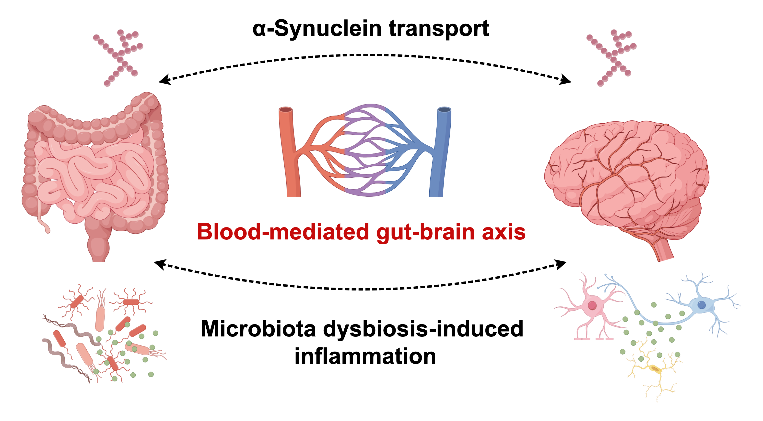
Blood-Mediated Gut–Brain Axis in Parkinson’s Disease: Focus on α-Synuclein Transport and Microbiota Dysbiosis-Induced Inflammation
 , and
, and
Highlights
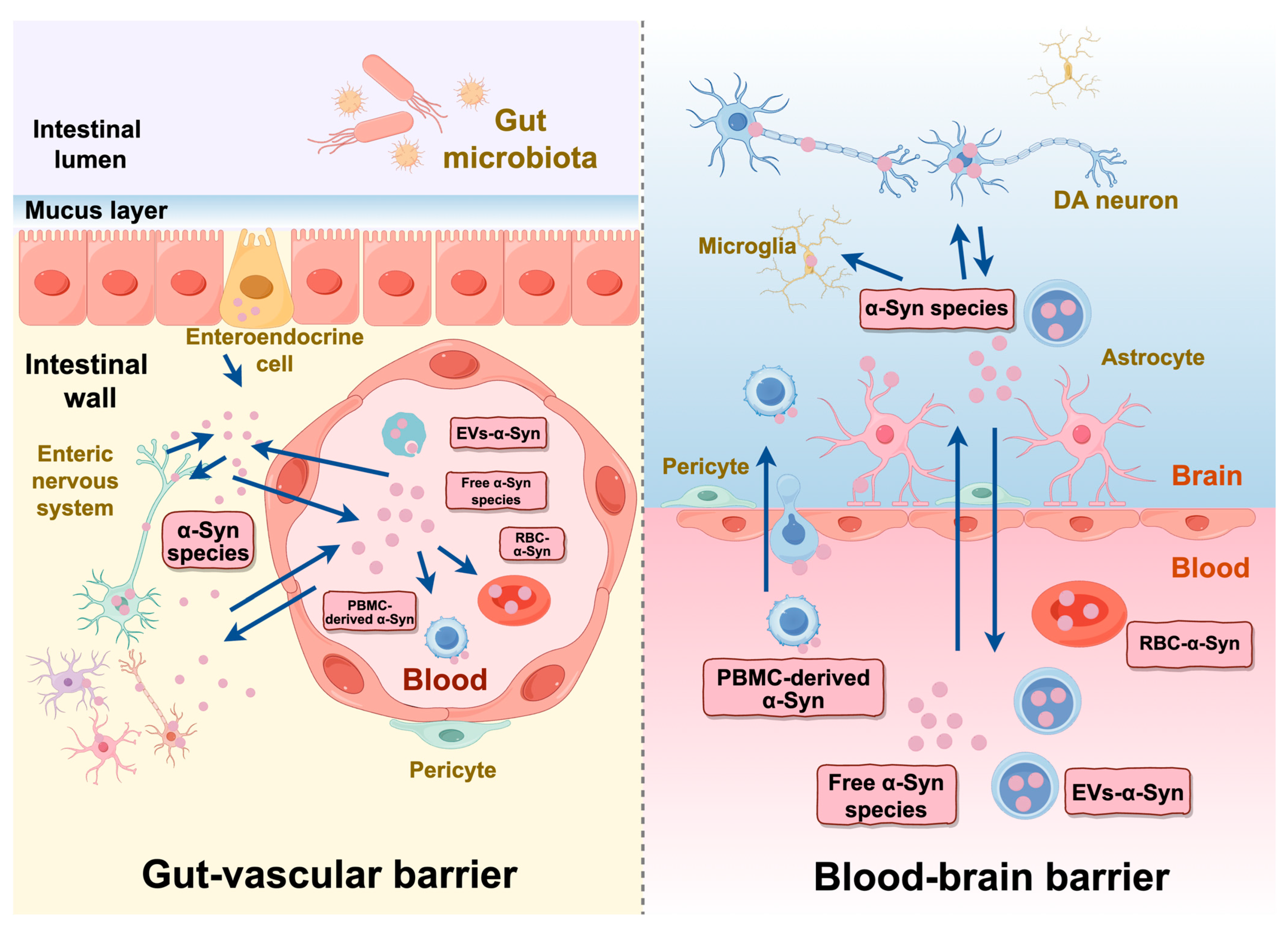
- Reviews the pathological transport of α-synuclein across the gut barrier and blood–brain barrier.
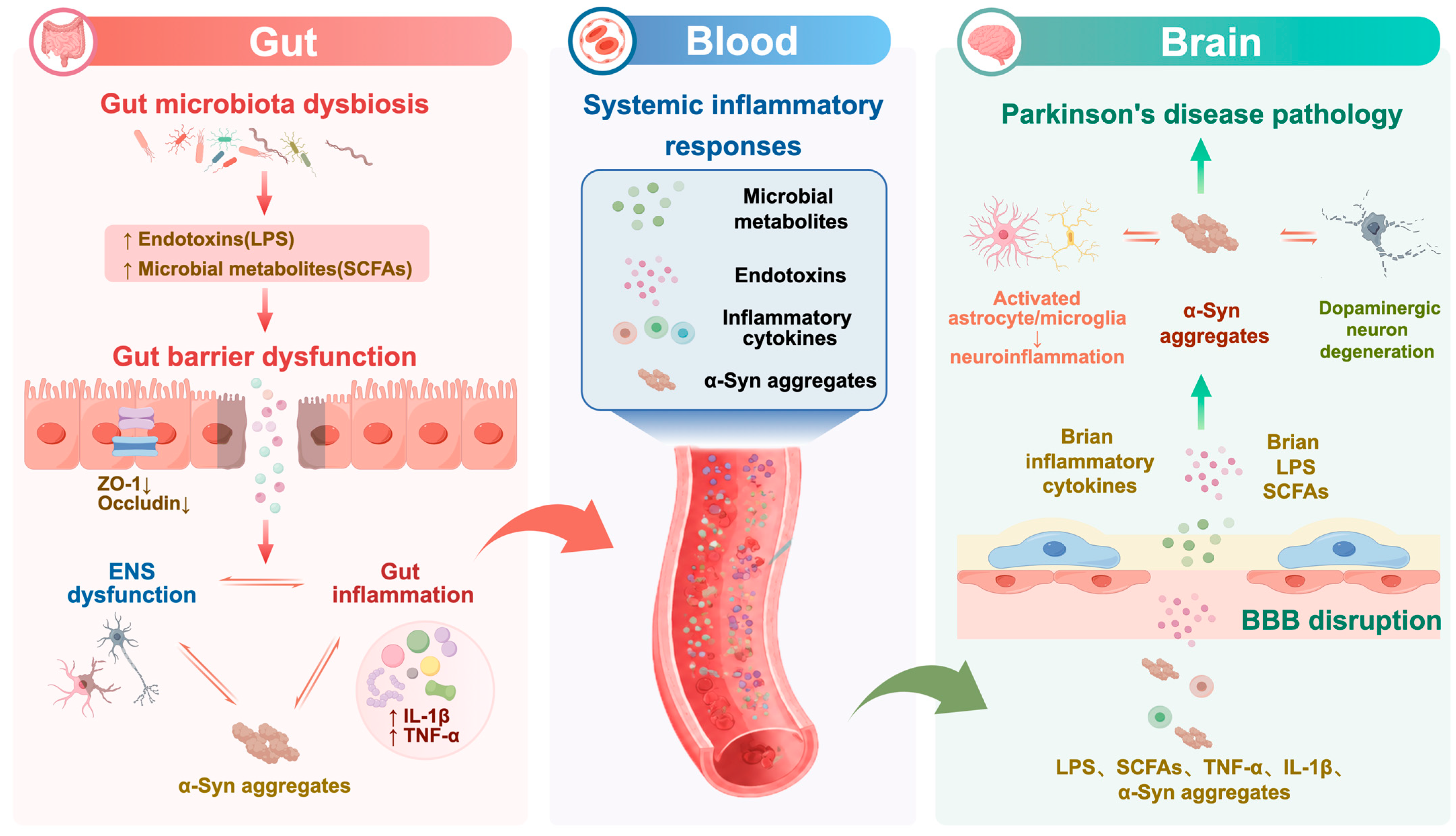
- Summarizes how gut microbiota dysbiosis may affect brain pathology via blood-mediated inflammation.
- Highlights the potential role of blood in mediating gut–brain crosstalk in Parkinson’s disease.
- Identifies potential biomarkers and therapeutic strategies targeting the gut–blood–brain axis.
Abstract
1. Introduction
2. Search Strategy
3. Pathological α-Syn Transport Between the Gut and Brain via Blood
3.1. Distribution of α-Syn in the Gut, Brain and Blood
3.2. Blood–Brain Barrier and Gut Barrier Dysfunction in PD
3.3. α-Syn Transport Across the BBB and Gut Barrier
3.4. Role of Peripheral Blood Mononuclear Cells in α-Syn Transport
4. Influence of Gut Microbiota Dysbiosis on the Brain Through Blood-Mediated Systemic Inflammation
4.1. Characteristics of Gut Microbiota Dysbiosis in PD
4.2. Gut Infection Induces Increased Microbial Endotoxin and Brain Inflammation Through Blood
4.3. Gut Microbiome-Driven Changes in Circulating Metabolites and Their Effects on the Brain
4.3.1. Short-Chain Fatty Acids (SCFAs)
4.3.2. Bile Acids (BAs)
4.3.3. Amino Acids
4.3.4. Trimethylamine N-Oxide (TMAO)
5. Application of the Gut–Blood–Brain Axis
5.1. Blood Biomarkers Related to Gastrointestinal Dysfunction and Brain Pathology in PD
5.2. Therapeutic Strategies Targeting the Gut–Brain Axis
6. Conclusions
Author Contributions
Funding
Institutional Review Board Statement
Informed Consent Statement
Data Availability Statement
Conflicts of Interest
Abbreviations
| AAAS | Aromatic Amino Acids |
| α-Syn | α-Synuclein |
| BAs | Bile Acids |
| BCAAs | Branched-Chain Amino Acids |
| BBB | Blood–Brain Barrier |
| CNS | Central Nervous System |
| CSF | Cerebrospinal Fluid |
| DA | Dopamine |
| ECs | Endothelial Cells |
| EECs | Enteroendocrine Cells |
| ENS | Enteric Nervous System |
| EVs | Extracellular Vesicles |
| FABP | Fatty Acid-Binding Protein |
| FABP2 | Intestinal-Type Fatty Acid-Binding Protein |
| FFAR3 | Free Fatty Acid Receptor 3 |
| GI | Gastrointestinal |
| GIT | Gastrointestinal Tract |
| GVB | Gut–Vascular Barrier |
| IBD | Inflammatory Bowel Disease |
| IFN-β | Interferon Beta |
| IFN-γ | Interferon Gamma |
| IL-1β | Interleukin-1 Beta |
| IL-6 | Interleukin-6 |
| IL-8 | Interleukin-8 |
| IL-13Rα1 | Interleukin-13 Receptor Alpha 1 |
| IPA | Indole-3-Propionic Acid |
| LAG3 | Lymphocyte-Activation Gene 3 |
| LBs | Lewy Bodies |
| LPS | Lipopolysaccharide |
| LRP-1 | Low-Density Lipoprotein Receptor-Related Protein 1 |
| MPTP | 1-methyl-4-phenyl-1,2,3,6-tetrahydropyridine |
| MSC | Mesenchymal Stem Cell |
| NF-κB | Nuclear Factor κB |
| NGAL | Neutrophil Gelatinase-Associated Lipocalin |
| NO | Nitric Oxide |
| PBMCs | Peripheral Blood Mononuclear Cells |
| PD | Parkinson’s Disease |
| RBCs | Red Blood Cells |
| ROS | Reactive Oxygen Species |
| RT-QuIC | Real-Time Quaking-Induced Conversion |
| SCFAs | Short-Chain Fatty Acids |
| SN | Substantia Nigra |
| TEER | Transepithelial Electrical Resistance |
| TLR4 | Toll-Like Receptor 4 |
| TMAO | Trimethylamine N-Oxide |
| TUDCA | Tauroursodeoxycholic Acid |
| UDCA | Ursodeoxycholic Acid |
| VEGFA | Vascular Endothelial Growth Factor A |
| ZO-1 | Zonula Occludens-1 |
References
- Morris, H.R.; Spillantini, M.G.; Sue, C.M.; Williams-Gray, C.H. The Pathogenesis of Parkinson’s Disease. Lancet 2024, 403, 293–304. [Google Scholar] [CrossRef] [Scilit] [PubMed]
- Knudsen, K.; Krogh, K.; Østergaard, K.; Borghammer, P. Constipation in Parkinson’s Disease: Subjective Symptoms, Objective Markers, and New Perspectives. Mov. Disord. 2017, 32, 94–105. [Google Scholar] [CrossRef] [Scilit] [PubMed]
- Beach, T.G.; Adler, C.H.; Sue, L.I.; Vedders, L.; Lue, L.; White Iii, C.L.; Akiyama, H.; Caviness, J.N.; Shill, H.A.; Sabbagh, M.N.; et al. Multi-Organ Distribution of Phosphorylated Alpha-Synuclein Histopathology in Subjects with Lewy Body Disorders. Acta Neuropathol. 2010, 119, 689–702. [Google Scholar] [CrossRef] [Scilit] [PubMed]
- O’Donovan, S.M.; Crowley, E.K.; Brown, J.R.-M.; O’Sullivan, O.; O’Leary, O.F.; Timmons, S.; Nolan, Y.M.; Clarke, D.J.; Hyland, N.P.; Joyce, S.A.; et al. Nigral Overexpression of α-Synuclein in a Rat Parkinson’s Disease Model Indicates Alterations in the Enteric Nervous System and the Gut Microbiome. Neurogastroenterol. Motil. 2020, 32, e13726. [Google Scholar] [CrossRef] [Scilit] [PubMed]
- Singaram, C.; Ashraf, W.; Gaumnitz, E.A.; Torbey, C.; Sengupta, A.; Pfeiffer, R.; Quigley, E.M. Dopaminergic Defect of Enteric Nervous System in Parkinson’s Disease Patients with Chronic Constipation. Lancet 1995, 346, 861–864. [Google Scholar] [CrossRef] [Scilit] [PubMed]
- Tan, A.H.; Lim, S.Y.; Lang, A.E. The Microbiome-Gut-Brain Axis in Parkinson Disease—From Basic Research to the Clinic. Nat. Rev. Neurol. 2022, 18, 476–495. [Google Scholar] [CrossRef] [Scilit] [PubMed]
- Braak, H.; Del Tredici, K.; Rüb, U.; de Vos, R.A.I.; Jansen Steur, E.N.H.; Braak, E. Staging of Brain Pathology Related to Sporadic Parkinson’s Disease. Neurobiol. Aging 2003, 24, 197–211. [Google Scholar] [CrossRef] [Scilit] [PubMed]
- Klingelhoefer, L.; Reichmann, H. Pathogenesis of Parkinson Disease—The Gut-Brain Axis and Environmental Factors. Nat. Rev. Neurol. 2015, 11, 625–636. [Google Scholar] [CrossRef] [Scilit] [PubMed]
- Sian-Hülsmann, J. Wilful Pathogens Provoke a Gut Feeling in Parkinson’s Disease. J. Neural. Transm. 2022, 129, 557–562. [Google Scholar] [CrossRef] [Scilit] [PubMed]
- Jellinger, K.A. A Critical Reappraisal of Current Staging of Lewy-Related Pathology in Human Brain. Acta Neuropathol. 2008, 116, 1–16. [Google Scholar] [CrossRef] [Scilit] [PubMed]
- Kalaitzakis, M.E.; Graeber, M.B.; Gentleman, S.M.; Pearce, R.K.B. The Dorsal Motor Nucleus of the Vagus Is Not an Obligatory Trigger Site of Parkinson’s Disease: A Critical Analysis of Alpha-Synuclein Staging. Neuropathol. Appl. Neurobiol. 2008, 34, 284–295. [Google Scholar] [CrossRef] [Scilit] [PubMed]
- Kim, S.; Kwon, S.-H.; Kam, T.-I.; Panicker, N.; Karuppagounder, S.S.; Lee, S.; Lee, J.H.; Kim, W.R.; Kook, M.; Foss, C.A.; et al. Transneuronal Propagation of Pathologic α-Synuclein from the Gut to the Brain Models Parkinson’s Disease. Neuron 2019, 103, 627–641.e7. [Google Scholar] [CrossRef] [Scilit] [PubMed]
- Manfredsson, F.P.; Luk, K.C.; Benskey, M.J.; Gezer, A.; Garcia, J.; Kuhn, N.C.; Sandoval, I.M.; Patterson, J.R.; O’Mara, A.; Yonkers, R.; et al. Induction of Alpha-Synuclein Pathology in the Enteric Nervous System of the Rat and Non-Human Primate Results in Gastrointestinal Dysmotility and Transient CNS Pathology. Neurobiol. Dis. 2018, 112, 106–118. [Google Scholar] [CrossRef] [Scilit] [PubMed]
- Foffani, G.; Obeso, J.A. A Cortical Pathogenic Theory of Parkinson’s Disease. Neuron 2018, 99, 1116–1128. [Google Scholar] [CrossRef] [Scilit] [PubMed]
- Wang, P.; Lan, G.; Xu, B.; Yu, Z.; Tian, C.; Lei, X.; Meissner, W.G.; Feng, T.; Yang, Y.; Zhang, J. α-Synuclein-Carrying Astrocytic Extracellular Vesicles in Parkinson Pathogenesis and Diagnosis. Transl. Neurodegener. 2023, 12, 40. [Google Scholar] [CrossRef] [Scilit] [PubMed]
- Brown, G.C.; Camacho, M.; Williams-Gray, C.H. The Endotoxin Hypothesis of Parkinson’s Disease. Mov. Disord. 2023, 38, 1143–1155. [Google Scholar] [CrossRef] [Scilit] [PubMed]
- Gibbons, C.H.; Levine, T.; Adler, C.; Bellaire, B.; Wang, N.; Stohl, J.; Agarwal, P.; Aldridge, G.M.; Barboi, A.; Evidente, V.G.H.; et al. Skin Biopsy Detection of Phosphorylated α-Synuclein in Patients with Synucleinopathies. JAMA 2024, 331, 1298–1306. [Google Scholar] [CrossRef] [Scilit] [PubMed]
- Shu, H.; Zhang, P.; Gu, L. Alpha-Synuclein in Peripheral Body Fluid as a Biomarker for Parkinson’s Disease. Acta Neurol. Belg. 2024, 124, 831–842. [Google Scholar] [CrossRef] [Scilit] [PubMed]
- Erskine, D.; Patterson, L.; Alexandris, A.; Hanson, P.S.; McKeith, I.G.; Attems, J.; Morris, C.M. Regional Levels of Physiological α-Synuclein Are Directly Associated with Lewy Body Pathology. Acta Neuropathol. 2018, 135, 153–154. [Google Scholar] [CrossRef] [Scilit] [PubMed]
- Qualman, S.J.; Haupt, H.M.; Yang, P.; Hamilton, S.R. Esophageal Lewy Bodies Associated with Ganglion Cell Loss in Achalasia. Similarity to Parkinson’s Disease. Gastroenterology 1984, 87, 848–856. [Google Scholar] [PubMed]
- Wakabayashi, K.; Takahashi, H.; Takeda, S.; Ohama, E.; Ikuta, F. Parkinson’s Disease: The Presence of Lewy Bodies in Auerbach’s and Meissner’s Plexuses. Acta Neuropathol. 1988, 76, 217–221. [Google Scholar] [CrossRef] [Scilit] [PubMed]
- Vascellari, S.; Orrù, C.D.; Groveman, B.R.; Parveen, S.; Fenu, G.; Pisano, G.; Piga, G.; Serra, G.; Oppo, V.; Murgia, D.; et al. α-Synuclein Seeding Activity in Duodenum Biopsies from Parkinson’s Disease Patients. PLoS Pathog. 2023, 19, e1011456. [Google Scholar] [CrossRef] [Scilit] [PubMed]
- Chandra, R.; Sokratian, A.; Chavez, K.R.; King, S.; Swain, S.M.; Snyder, J.C.; West, A.B.; Liddle, R.A. Gut Mucosal Cells Transfer α-Synuclein to the Vagus Nerve. JCI Insight 2023, 8, e172192. [Google Scholar] [CrossRef] [Scilit] [PubMed]
- Rodrigues, P.V.; de Godoy, J.V.P.; Bosque, B.P.; Amorim Neto, D.P.; Tostes, K.; Palameta, S.; Garcia-Rosa, S.; Tonoli, C.C.C.; de Carvalho, H.F.; de Castro Fonseca, M. Transcellular Propagation of Fibrillar α-Synuclein from Enteroendocrine to Neuronal Cells Requires Cell-to-Cell Contact and Is Rab35-Dependent. Sci. Rep. 2022, 12, 4168. [Google Scholar] [CrossRef] [Scilit] [PubMed]
- Choi, M.L.; Chappard, A.; Singh, B.P.; Maclachlan, C.; Rodrigues, M.; Fedotova, E.I.; Berezhnov, A.V.; De, S.; Peddie, C.J.; Athauda, D.; et al. Pathological Structural Conversion of α-Synuclein at the Mitochondria Induces Neuronal Toxicity. Nat. Neurosci. 2022, 25, 1134–1148. [Google Scholar] [CrossRef] [Scilit] [PubMed]
- Sarkar, S.; Dammer, E.B.; Malovic, E.; Olsen, A.L.; Raza, S.A.; Gao, T.; Xiao, H.; Oliver, D.L.; Duong, D.; Joers, V.; et al. Molecular Signatures of Neuroinflammation Induced by αSynuclein Aggregates in Microglial Cells. Front. Immunol. 2020, 11, 33. [Google Scholar] [CrossRef] [Scilit] [PubMed]
- Lamontagne-Proulx, J.; Coulombe, K.; Dahhani, F.; Côté, M.; Guyaz, C.; Tremblay, C.; Di Marzo, V.; Flamand, N.; Calon, F.; Soulet, D. Effect of Docosahexaenoic Acid (DHA) at the Enteric Level in a Synucleinopathy Mouse Model. Nutrients 2021, 13, 4218. [Google Scholar] [CrossRef] [Scilit]
- Emmi, A.; Sandre, M.; Russo, F.P.; Tombesi, G.; Garrì, F.; Campagnolo, M.; Carecchio, M.; Biundo, R.; Spolverato, G.; Macchi, V.; et al. Duodenal Alpha-Synuclein Pathology and Enteric Gliosis in Advanced Parkinson’s Disease. Mov. Disord. 2023, 38, 885–894. [Google Scholar] [CrossRef] [Scilit] [PubMed]
- Chopra, A.; Outeiro, T.F. Aggregation and beyond: Alpha-Synuclein-Based Biomarkers in Synucleinopathies. Brain 2024, 147, 81–90. [Google Scholar] [CrossRef] [Scilit] [PubMed]
- Schaeffer, E.; Kluge, A.; Schulte, C.; Deuschle, C.; Bunk, J.; Welzel, J.; Maetzler, W.; Berg, D. Association of Misfolded α-Synuclein Derived from Neuronal Exosomes in Blood with Parkinson’s Disease Diagnosis and Duration. J. Park. Dis. 2024, 14, 667–679. [Google Scholar] [CrossRef] [Scilit] [PubMed]
- Kluge, A.; Schaeffer, E.; Bunk, J.; Sommerauer, M.; Röttgen, S.; Schulte, C.; Roeben, B.; von Thaler, A.-K.; Welzel, J.; Lucius, R.; et al. Detecting Misfolded α-Synuclein in Blood Years before the Diagnosis of Parkinson’s Disease. Mov. Disord. 2024, 39, 1289–1299. [Google Scholar] [CrossRef] [Scilit] [PubMed]
- Barbour, R.; Kling, K.; Anderson, J.P.; Banducci, K.; Cole, T.; Diep, L.; Fox, M.; Goldstein, J.M.; Soriano, F.; Seubert, P.; et al. Red Blood Cells Are the Major Source of Alpha-Synuclein in Blood. Neurodegener. Dis. 2008, 5, 55–59. [Google Scholar] [CrossRef] [Scilit] [PubMed]
- Li, Q.-X.; Mok, S.S.; Laughton, K.M.; McLean, C.A.; Cappai, R.; Masters, C.L.; Culvenor, J.G.; Horne, M.K. Plasma Alpha-Synuclein Is Decreased in Subjects with Parkinson’s Disease. Exp. Neurol. 2007, 204, 583–588. [Google Scholar] [CrossRef] [Scilit] [PubMed]
- Wang, L.; Wang, G.; Duan, Y.; Wang, F.; Lin, S.; Zhang, F.; Li, H.; Li, A.; Li, H. A Comparative Study of the Diagnostic Potential of Plasma and Erythrocytic α-Synuclein in Parkinson’s Disease. Neurodegener. Dis. 2019, 19, 204–210. [Google Scholar] [CrossRef] [Scilit] [PubMed]
- Li, X.-Y.; Li, W.; Li, X.; Li, X.-R.; Sun, L.; Yang, W.; Cai, Y.; Chen, Z.; Wu, J.; Wang, C.; et al. Alterations of Erythrocytic Phosphorylated Alpha-Synuclein in Different Subtypes and Stages of Parkinson’s Disease. Front. Aging Neurosci. 2021, 13, 623977. [Google Scholar] [CrossRef] [Scilit] [PubMed]
- Liu, G.; Yu, Z.; Gao, L.; Zheng, Y.; Feng, T. Erythrocytic Alpha-Synuclein in Early Parkinson’s Disease: A 3-Year Longitudinal Study. Park. Relat. Disord. 2022, 104, 44–48. [Google Scholar] [CrossRef] [Scilit] [PubMed]
- Wang, X.; Yu, S.; Li, F.; Feng, T. Detection of α-Synuclein Oligomers in Red Blood Cells as a Potential Biomarker of Parkinson’s Disease. Neurosci. Lett. 2015, 599, 115–119. [Google Scholar] [CrossRef] [Scilit] [PubMed]
- Braak, H.; de Vos, R.A.I.; Bohl, J.; Del Tredici, K. Gastric Alpha-Synuclein Immunoreactive Inclusions in Meissner’s and Auerbach’s Plexuses in Cases Staged for Parkinson’s Disease-Related Brain Pathology. Neurosci. Lett. 2006, 396, 67–72. [Google Scholar] [CrossRef] [Scilit] [PubMed]
- Wang, H.L.; Lu, C.S.; Yeh, T.H.; Shen, Y.M.; Weng, Y.H.; Huang, Y.Z.; Chen, R.S.; Liu, Y.C.; Cheng, Y.C.; Chang, H.C.; et al. Combined Assessment of Serum Alpha-Synuclein and Rab35 Is a Better Biomarker for Parkinson’s Disease. J. Clin. Neurol. 2019, 15, 488–495. [Google Scholar] [CrossRef] [Scilit] [PubMed]
- Beck, G.; Hori, Y.; Hayashi, Y.; Morii, E.; Takehara, T.; Mochizuki, H. Detection of Phosphorylated Alpha-Synuclein in the Muscularis Propria of the Gastrointestinal Tract Is a Sensitive Predictor for Parkinson’s Disease. Park. Dis. 2020, 2020, 4687530. [Google Scholar] [CrossRef] [Scilit] [PubMed]
- Visanji, N.P.; Marras, C.; Kern, D.S.; Al Dakheel, A.; Gao, A.; Liu, L.W.C.; Lang, A.E.; Hazrati, L.-N. Colonic Mucosal A-Synuclein Lacks Specificity as a Biomarker for Parkinson Disease. Neurology 2015, 84, 609–616. [Google Scholar] [CrossRef] [Scilit] [PubMed]
- Parnetti, L.; Chiasserini, D.; Persichetti, E.; Eusebi, P.; Varghese, S.; Qureshi, M.M.; Dardis, A.; Deganuto, M.; De Carlo, C.; Castrioto, A.; et al. Cerebrospinal Fluid Lysosomal Enzymes and Alpha-Synuclein in Parkinson’s Disease. Mov. Disord. 2014, 29, 1019–1027. [Google Scholar] [CrossRef] [Scilit] [PubMed]
- Antunes, L.; Frasquilho, S.; Ostaszewski, M.; Weber, J.; Longhino, L.; Antony, P.; Baumuratov, A.; Buttini, M.; Shannon, K.M.; Balling, R.; et al. Similar α-Synuclein Staining in the Colon Mucosa in Patients with Parkinson’s Disease and Controls. Mov. Disord. Off. J. Mov. Disord. Soc. 2016, 31, 1567–1570. [Google Scholar] [CrossRef] [Scilit] [PubMed]
- Shin, C.; Park, S.-H.; Yun, J.Y.; Shin, J.H.; Yang, H.-K.; Lee, H.-J.; Kong, S.-H.; Suh, Y.-S.; Shen, G.; Kim, Y.; et al. Fundamental Limit of Alpha-Synuclein Pathology in Gastrointestinal Biopsy as a Pathologic Biomarker of Parkinson’s Disease: Comparison with Surgical Specimens. Park. Relat. Disord. 2017, 44, 73–78. [Google Scholar] [CrossRef] [Scilit] [PubMed]
- Yu, Z.; Liu, G.; Li, Y.; Arkin, E.; Zheng, Y.; Feng, T. Erythrocytic α-Synuclein Species for Parkinson’s Disease Diagnosis and the Correlations with Clinical Characteristics. Front. Aging Neurosci. 2022, 14, 827493. [Google Scholar] [CrossRef] [Scilit] [PubMed]
- Compta, Y.; Valente, T.; Saura, J.; Segura, B.; Iranzo, Á.; Serradell, M.; Junqué, C.; Tolosa, E.; Valldeoriola, F.; Muñoz, E.; et al. Correlates of Cerebrospinal Fluid Levels of Oligomeric- and Total-α-Synuclein in Premotor, Motor and Dementia Stages of Parkinson’s Disease. J. Neurol. 2015, 262, 294–306. [Google Scholar] [CrossRef] [Scilit] [PubMed]
- Tian, C.; Liu, G.; Gao, L.; Soltys, D.; Pan, C.; Stewart, T.; Shi, M.; Xie, Z.; Liu, N.; Feng, T.; et al. Erythrocytic α-Synuclein as a Potential Biomarker for Parkinson’s Disease. Transl. Neurodegener. 2019, 8, 15. [Google Scholar] [CrossRef] [Scilit] [PubMed]
- Sánchez-Ferro, Á.; Rábano, A.; Catalán, M.J.; Rodríguez-Valcárcel, F.C.; Fernández Díez, S.; Herreros-Rodríguez, J.; García-Cobos, E.; Álvarez-Santullano, M.M.; López-Manzanares, L.; Mosqueira, A.J.; et al. In Vivo Gastric Detection of α-Synuclein Inclusions in Parkinson’s Disease. Mov. Disord. 2015, 30, 517–524. [Google Scholar] [CrossRef] [Scilit] [PubMed]
- Hu, Y.; Yu, S.-Y.; Zuo, L.-J.; Cao, C.-J.; Wang, F.; Chen, Z.-J.; Du, Y.; Lian, T.-H.; Wang, Y.-J.; Chan, P.; et al. Parkinson Disease with REM Sleep Behavior Disorder: Features, α-Synuclein, and Inflammation. Neurology 2015, 84, 888–894. [Google Scholar] [CrossRef] [Scilit] [PubMed]
- Gündner, A.L.; Duran-Pacheco, G.; Zimmermann, S.; Ruf, I.; Moors, T.; Baumann, K.; Jagasia, R.; van de Berg, W.D.J.; Kremer, T. Path Mediation Analysis Reveals GBA Impacts Lewy Body Disease Status by Increasing α-Synuclein Levels. Neurobiol. Dis. 2019, 121, 205–213. [Google Scholar] [CrossRef] [Scilit] [PubMed]
- Ruffmann, C.; Bengoa-Vergniory, N.; Poggiolini, I.; Ritchie, D.; Hu, M.T.; Alegre-Abarrategui, J.; Parkkinen, L. Detection of Alpha-Synuclein Conformational Variants from Gastro-Intestinal Biopsy Tissue as a Potential Biomarker for Parkinson’s Disease. Neuropathol. Appl. Neurobiol. 2018, 44, 722–736. [Google Scholar] [CrossRef] [Scilit] [PubMed]
- El-Agnaf, O.M.A.; Salem, S.A.; Paleologou, K.E.; Curran, M.D.; Gibson, M.J.; Court, J.A.; Schlossmacher, M.G.; Allsop, D. Detection of Oligomeric Forms of Alpha-Synuclein Protein in Human Plasma as a Potential Biomarker for Parkinson’s Disease. FASEB J. 2006, 20, 419–425. [Google Scholar] [CrossRef] [Scilit] [PubMed]
- Kramer, M.L.; Schulz-Schaeffer, W.J. Presynaptic Alpha-Synuclein Aggregates, Not Lewy Bodies, Cause Neurodegeneration in Dementia with Lewy Bodies. J. Neurosci. 2007, 27, 1405–1410. [Google Scholar] [CrossRef] [Scilit] [PubMed]
- Emelyanov, A.; Andoskin, P.; Pchelina, S. Dataset of Total, Oligomeric Alpha-Synuclein and Hemoglobin Levels in Plasma in Parkinson׳s Disease. Data Brief. 2017, 10, 182–185. [Google Scholar] [CrossRef] [Scilit] [PubMed]
- Tokuda, T.; Qureshi, M.M.; Ardah, M.T.; Varghese, S.; Shehab, S.a.S.; Kasai, T.; Ishigami, N.; Tamaoka, A.; Nakagawa, M.; El-Agnaf, O.M.A. Detection of Elevated Levels of α-Synuclein Oligomers in CSF from Patients with Parkinson Disease. Neurology 2010, 75, 1766–1772. [Google Scholar] [CrossRef] [Scilit] [PubMed]
- Lebouvier, T.; Neunlist, M.; Bruley des Varannes, S.; Coron, E.; Drouard, A.; N’Guyen, J.-M.; Chaumette, T.; Tasselli, M.; Paillusson, S.; Flamand, M.; et al. Colonic Biopsies to Assess the Neuropathology of Parkinson’s Disease and Its Relationship with Symptoms. PLoS ONE 2010, 5, e12728. [Google Scholar] [CrossRef] [Scilit] [PubMed]
- Chatterjee, K.; Roy, A.; Banerjee, R.; Choudhury, S.; Mondal, B.; Halder, S.; Basu, P.; Shubham, S.; Dey, S.; Kumar, H. Inflammasome and α-Synuclein in Parkinson’s Disease: A Cross-Sectional Study. J. Neuroimmunol. 2020, 338, 577089. [Google Scholar] [CrossRef] [Scilit] [PubMed]
- Wang, X.; Chi, J.; Huang, D.; Ding, L.; Zhao, X.; Jiang, L.; Yu, Y.; Gao, F. α-Synuclein Promotes Progression of Parkinson’s Disease by Upregulating Autophagy Signaling Pathway to Activate NLRP3 Inflammasome. Exp. Ther. Med. 2020, 19, 931–938. [Google Scholar] [CrossRef] [Scilit] [PubMed]
- Foulds, P.G.; Mitchell, J.D.; Parker, A.; Turner, R.; Green, G.; Diggle, P.; Hasegawa, M.; Taylor, M.; Mann, D.; Allsop, D. Phosphorylated α-Synuclein Can Be Detected in Blood Plasma and Is Potentially a Useful Biomarker for Parkinson’s Disease. FASEB J. 2011, 25, 4127–4137. [Google Scholar] [CrossRef] [Scilit] [PubMed]
- Majbour, N.K.; Vaikath, N.N.; van Dijk, K.D.; Ardah, M.T.; Varghese, S.; Vesterager, L.B.; Montezinho, L.P.; Poole, S.; Safieh-Garabedian, B.; Tokuda, T.; et al. Oligomeric and Phosphorylated Alpha-Synuclein as Potential CSF Biomarkers for Parkinson’s Disease. Mol. Neurodegener. 2016, 11, 7. [Google Scholar] [CrossRef] [Scilit] [PubMed]
- Harapan, B.N.; Frydrychowicz, C.; Classen, J.; Wittekind, C.; Gradistanac, T.; Rumpf, J.-J.; Mueller, W. No Enhanced (p-) α-Synuclein Deposition in Gastrointestinal Tissue of Parkinson’s Disease Patients. Park. Relat. Disord. 2020, 80, 82–88. [Google Scholar] [CrossRef] [Scilit] [PubMed]
- Norman, M.; Gilboa, T.; Walt, D.R. High-Sensitivity Single Molecule Array Assays for Pathological Isoforms in Parkinson’s Disease. Clin. Chem. 2022, 68, 431–440. [Google Scholar] [CrossRef] [Scilit] [PubMed]
- Chung, S.J.; Kim, J.; Lee, H.J.; Ryu, H.-S.; Kim, K.; Lee, J.H.; Jung, K.W.; Kim, M.J.; Kim, M.-J.; Kim, Y.J.; et al. Alpha-Synuclein in Gastric and Colonic Mucosa in Parkinson’s Disease: Limited Role as a Biomarker. Mov. Disord. 2016, 31, 241–249. [Google Scholar] [CrossRef] [Scilit] [PubMed]
- Silva, A.M.; Hickford, E.S.; Cutler, P. An Immunoassay for the Quantification of Phosphorylated α-Synuclein at Serine 129 in Human Cerebrospinal Fluid. Bioanalysis 2024, 16, 1125–1139. [Google Scholar] [CrossRef] [Scilit] [PubMed]
- Si, X.; Tian, J.; Chen, Y.; Yan, Y.; Pu, J.; Zhang, B. Central Nervous System-Derived Exosomal Alpha-Synuclein in Serum May Be a Biomarker in Parkinson’s Disease. Neuroscience 2019, 413, 308–316. [Google Scholar] [CrossRef] [Scilit] [PubMed]
- Stuendl, A.; Kunadt, M.; Kruse, N.; Bartels, C.; Moebius, W.; Danzer, K.M.; Mollenhauer, B.; Schneider, A. Induction of α-Synuclein Aggregate Formation by CSF Exosomes from Patients with Parkinson’s Disease and Dementia with Lewy Bodies. Brain 2016, 139, 481–494. [Google Scholar] [CrossRef] [Scilit] [PubMed]
- Stuendl, A.; Kraus, T.; Chatterjee, M.; Zapke, B.; Sadowski, B.; Moebius, W.; Hobert, M.A.; Deuschle, C.; Brockmann, K.; Maetzler, W.; et al. α-Synuclein in Plasma-Derived Extracellular Vesicles Is a Potential Biomarker of Parkinson’s Disease. Mov. Disord. 2021, 36, 2508–2518. [Google Scholar] [CrossRef] [Scilit] [PubMed]
- Hong, Z.; Tian, C.; Stewart, T.; Aro, P.; Soltys, D.; Bercow, M.; Sheng, L.; Borden, K.; Khrisat, T.; Pan, C.; et al. Development of a Sensitive Diagnostic Assay for Parkinson Disease Quantifying α-Synuclein-Containing Extracellular Vesicles. Neurology 2021, 96, e2332–e2345. [Google Scholar] [CrossRef] [Scilit] [PubMed]
- Emmi, A.; Mammana, A.; Sandre, M.; Baiardi, S.; Weis, L.; Rossi, M.; Magliocchetti, F.; Savarino, E.; Russo, F.P.; Porzionato, A.; et al. Alpha-Synuclein RT-QuIC Assay in Gastroduodenal and Skin Biopsies of Parkinson Disease Patients. Ann. Clin. Transl. Neurol. 2025, 12, 637–642. [Google Scholar] [CrossRef] [Scilit] [PubMed]
- Christenson, P.R.; Jeong, H.; Li, M.; Ahn, H.; Schmeichel, A.M.; Misra, P.; Li, D.; Savica, R.; Low, P.A.; Singer, W.; et al. Blood-Based Nanoparticle-Enhanced Quaking-Induced Conversion (Nano-QuIC): Inhibitor-Resistant Detection of Seeding Activity in Patients Diagnosed with Parkinson’s Disease. Nano Lett. 2024, 24, 15016–15024. [Google Scholar] [CrossRef] [Scilit] [PubMed]
- Wiseman, J.A.; Turner, C.P.; Faull, R.L.M.; Halliday, G.M.; Dieriks, B.V. Refining α-Synuclein Seed Amplification Assays to Distinguish Parkinson’s Disease from Multiple System Atrophy. Transl. Neurodegener. 2025, 14, 7. [Google Scholar] [CrossRef] [Scilit] [PubMed]
- Poggiolini, I.; Gupta, V.; Lawton, M.; Lee, S.; El-Turabi, A.; Querejeta-Coma, A.; Trenkwalder, C.; Sixel-Döring, F.; Foubert-Samier, A.; Pavy-Le Traon, A.; et al. Diagnostic Value of Cerebrospinal Fluid Alpha-Synuclein Seed Quantification in Synucleinopathies. Brain 2022, 145, 584–595. [Google Scholar] [CrossRef] [Scilit] [PubMed]
- Bargar, C.; Wang, W.; Gunzler, S.A.; LeFevre, A.; Wang, Z.; Lerner, A.J.; Singh, N.; Tatsuoka, C.; Appleby, B.; Zhu, X.; et al. Streamlined Alpha-Synuclein RT-QuIC Assay for Various Biospecimens in Parkinson’s Disease and Dementia with Lewy Bodies. Acta Neuropathol. Commun. 2021, 9, 62. [Google Scholar] [CrossRef] [Scilit] [PubMed]
- Wood, H. VEGFA Mediates Blood-Brain Barrier Disruption in Parkinson Disease. Nat. Rev. Neurol. 2022, 18, 1. [Google Scholar] [CrossRef] [Scilit] [PubMed]
- Lau, K.; Kotzur, R.; Richter, F. Blood-Brain Barrier Alterations and Their Impact on Parkinson’s Disease Pathogenesis and Therapy. Transl. Neurodegener. 2024, 13, 37. [Google Scholar] [CrossRef] [Scilit] [PubMed]
- Gray, M.T.; Woulfe, J.M. Striatal Blood-Brain Barrier Permeability in Parkinson’s Disease. J. Cereb. Blood Flow Metab. 2015, 35, 747–750. [Google Scholar] [CrossRef] [Scilit] [PubMed]
- Gorlé, N.; Blaecher, C.; Bauwens, E.; Vandendriessche, C.; Balusu, S.; Vandewalle, J.; Van Cauwenberghe, C.; Van Wonterghem, E.; Van Imschoot, G.; Liu, C.; et al. The Choroid Plexus Epithelium as a Novel Player in the Stomach-Brain Axis during Helicobacter Infection. Brain Behav. Immun. 2018, 69, 35–47. [Google Scholar] [CrossRef] [Scilit] [PubMed]
- Gu, R.; Pan, J.; Awan, M.U.N.; Sun, X.; Yan, F.; Bai, L.; Bai, J. The Major Histocompatibility Complex Participates in Parkinson’s Disease. Pharmacol. Res. 2024, 203, 107168. [Google Scholar] [CrossRef] [Scilit] [PubMed]
- Pellegrini, C.; Fornai, M.; D’Antongiovanni, V.; Antonioli, L.; Bernardini, N.; Derkinderen, P. The Intestinal Barrier in Disorders of the Central Nervous System. Lancet Gastroenterol. Hepatol. 2023, 8, 66–80. [Google Scholar] [CrossRef] [Scilit] [PubMed]
- Forsyth, C.B.; Shannon, K.M.; Kordower, J.H.; Voigt, R.M.; Shaikh, M.; Jaglin, J.A.; Estes, J.D.; Dodiya, H.B.; Keshavarzian, A. Increased Intestinal Permeability Correlates with Sigmoid Mucosa Alpha-Synuclein Staining and Endotoxin Exposure Markers in Early Parkinson’s Disease. PLoS ONE 2011, 6, e28032. [Google Scholar] [CrossRef] [Scilit] [PubMed]
- Clairembault, T.; Leclair-Visonneau, L.; Coron, E.; Bourreille, A.; Le Dily, S.; Vavasseur, F.; Heymann, M.-F.; Neunlist, M.; Derkinderen, P. Structural Alterations of the Intestinal Epithelial Barrier in Parkinson’s Disease. Acta Neuropathol. Commun. 2015, 3, 12. [Google Scholar] [CrossRef] [Scilit] [PubMed]
- Grathwohl, S.; Quansah, E.; Maroof, N.; Steiner, J.A.; Spycher, L.; Benmansour, F.; Duran-Pacheco, G.; Siebourg-Polster, J.; Oroszlan-Szovik, K.; Remy, H.; et al. Specific Immune Modulation of Experimental Colitis Drives Enteric Alpha-Synuclein Accumulation and Triggers Age-Related Parkinson-like Brain Pathology. Free Neuropathol. 2021, 2, 13. [Google Scholar] [CrossRef] [Scilit] [PubMed]
- Dodiya, H.B.; Forsyth, C.B.; Voigt, R.M.; Engen, P.A.; Patel, J.; Shaikh, M.; Green, S.J.; Naqib, A.; Roy, A.; Kordower, J.H.; et al. Chronic Stress-Induced Gut Dysfunction Exacerbates Parkinson’s Disease Phenotype and Pathology in a Rotenone-Induced Mouse Model of Parkinson’s Disease. Neurobiol. Dis. 2020, 135, 104352. [Google Scholar] [CrossRef] [Scilit] [PubMed]
- Spadoni, I.; Zagato, E.; Bertocchi, A.; Paolinelli, R.; Hot, E.; Di Sabatino, A.; Caprioli, F.; Bottiglieri, L.; Oldani, A.; Viale, G.; et al. A Gut-Vascular Barrier Controls the Systemic Dissemination of Bacteria. Science 2015, 350, 830–834. [Google Scholar] [CrossRef] [Scilit] [PubMed]
- Benvenuti, L.; D’Antongiovanni, V.; Pellegrini, C.; Antonioli, L.; Bernardini, N.; Blandizzi, C.; Fornai, M. Enteric Glia at the Crossroads between Intestinal Immune System and Epithelial Barrier: Implications for Parkinson Disease. Int. J. Mol. Sci. 2020, 21, 9199. [Google Scholar] [CrossRef] [Scilit]
- Carloni, S.; Bertocchi, A.; Mancinelli, S.; Bellini, M.; Erreni, M.; Borreca, A.; Braga, D.; Giugliano, S.; Mozzarelli, A.M.; Manganaro, D.; et al. Identification of a Choroid Plexus Vascular Barrier Closing during Intestinal Inflammation. Science 2021, 374, 439–448. [Google Scholar] [CrossRef] [Scilit] [PubMed]
- Brescia, P.; Rescigno, M. The Gut Vascular Barrier: A New Player in the Gut-Liver-Brain Axis. Trends Mol. Med. 2021, 27, 844–855. [Google Scholar] [CrossRef] [Scilit] [PubMed]
- Carloni, S.; Rescigno, M. The Gut-Brain Vascular Axis in Neuroinflammation. Semin. Immunol. 2023, 69, 101802. [Google Scholar] [CrossRef] [Scilit] [PubMed]
- Serafino, A.; Cozzolino, M. The Wnt/β-Catenin Signaling: A Multifunctional Target for Neuroprotective and Regenerative Strategies in Parkinson’s Disease. Neural. Regen. Res. 2023, 18, 306–308. [Google Scholar] [CrossRef] [Scilit] [PubMed]
- Vallée, A.; Vallée, J.-N.; Lecarpentier, Y. Parkinson’s Disease: Potential Actions of Lithium by Targeting the WNT/β-Catenin Pathway, Oxidative Stress, Inflammation and Glutamatergic Pathway. Cells 2021, 10, 230. [Google Scholar] [CrossRef] [Scilit]
- Sui, Y.-T.; Bullock, K.M.; Erickson, M.A.; Zhang, J.; Banks, W.A. Alpha Synuclein Is Transported into and out of the Brain by the Blood-Brain Barrier. Peptides 2014, 62, 197–202. [Google Scholar] [CrossRef] [Scilit] [PubMed]
- Lohmann, S.; Bernis, M.E.; Tachu, B.J.; Ziemski, A.; Grigoletto, J.; Tamgüney, G. Oral and Intravenous Transmission of α-Synuclein Fibrils to Mice. Acta Neuropathol. 2019, 138, 515–533. [Google Scholar] [CrossRef] [Scilit] [PubMed]
- Olaoye, O.J.; Farrow, S.L.; Nyaga, D.M.; Cooper, A.A.; O’Sullivan, J.M. From Blood Vessels to Brain Cells: Connecting the Circulatory System and Parkinson’s Disease. J. Park. Dis. 2025, 15, 255–268. [Google Scholar] [CrossRef] [Scilit] [PubMed]
- Sharma, T.; Kumar, R.; Mukherjee, S. Neuronal Vulnerability to Degeneration in Parkinson’s Disease and Therapeutic Approaches. CNS Neurol. Disord. Drug Targets 2024, 23, 715–730. [Google Scholar] [CrossRef] [Scilit] [PubMed]
- Dryanovski, D.I.; Guzman, J.N.; Xie, Z.; Galteri, D.J.; Volpicelli-Daley, L.A.; Lee, V.M.-Y.; Miller, R.J.; Schumacker, P.T.; Surmeier, D.J. Calcium Entry and α-Synuclein Inclusions Elevate Dendritic Mitochondrial Oxidant Stress in Dopaminergic Neurons. J. Neurosci. 2013, 33, 10154–10164. [Google Scholar] [CrossRef] [Scilit] [PubMed]
- Guzman, J.N.; Sanchez-Padilla, J.; Wokosin, D.; Kondapalli, J.; Ilijic, E.; Schumacker, P.T.; Surmeier, D.J. Oxidant Stress Evoked by Pacemaking in Dopaminergic Neurons Is Attenuated by DJ-1. Nature 2010, 468, 696–700. [Google Scholar] [CrossRef] [Scilit] [PubMed]
- Kuan, W.-L.; Bennett, N.; He, X.; Skepper, J.N.; Martynyuk, N.; Wijeyekoon, R.; Moghe, P.V.; Williams-Gray, C.H.; Barker, R.A. α-Synuclein Pre-Formed Fibrils Impair Tight Junction Protein Expression without Affecting Cerebral Endothelial Cell Function. Exp. Neurol. 2016, 285, 72–81. [Google Scholar] [CrossRef] [Scilit] [PubMed]
- Sheng, L.; Stewart, T.; Yang, D.; Thorland, E.; Soltys, D.; Aro, P.; Khrisat, T.; Xie, Z.; Li, N.; Liu, Z.; et al. Erythrocytic α-Synuclein Contained in Microvesicles Regulates Astrocytic Glutamate Homeostasis: A New Perspective on Parkinson’s Disease Pathogenesis. Acta Neuropathol. Commun. 2020, 8, 102. [Google Scholar] [CrossRef] [Scilit] [PubMed]
- Matsumoto, J.; Stewart, T.; Sheng, L.; Li, N.; Bullock, K.; Song, N.; Shi, M.; Banks, W.A.; Zhang, J. Transmission of α-Synuclein-Containing Erythrocyte-Derived Extracellular Vesicles across the Blood-Brain Barrier via Adsorptive Mediated Transcytosis: Another Mechanism for Initiation and Progression of Parkinson’s Disease? Acta Neuropathol. Commun. 2017, 5, 71. [Google Scholar] [CrossRef] [Scilit] [PubMed]
- Yang, Y.; Liu, Q.; Zhou, T.; Chen, J.; Li, W.; Chen, W.; Zeng, S.; Liu, H.; Wang, P.; Yang, P.; et al. Parkinson’s Disease beyond the Brain: Erythrocyte α-Synuclein Transfer across the Blood-Brain Barrier. Brain 2026, awag179. [Google Scholar] [CrossRef] [Scilit] [PubMed]
- Hurley, M.J.; Menozzi, E.; Koletsi, S.; Bates, R.; Gegg, M.E.; Chau, K.-Y.; Blottière, H.M.; Macnaughtan, J.; Schapira, A.H.V. α-Synuclein Expression in Response to Bacterial Ligands and Metabolites in Gut Enteroendocrine Cells: An in Vitro Proof of Concept Study. Brain Commun. 2023, 5, fcad285. [Google Scholar] [CrossRef] [Scilit] [PubMed]
- Shi, M.; Liu, C.; Cook, T.J.; Bullock, K.M.; Zhao, Y.; Ginghina, C.; Li, Y.; Aro, P.; Dator, R.; He, C.; et al. Plasma Exosomal α-Synuclein Is Likely CNS-Derived and Increased in Parkinson’s Disease. Acta Neuropathol. 2014, 128, 639–650. [Google Scholar] [CrossRef] [Scilit] [PubMed]
- Yang, Y.; Stewart, T.; Zhang, C.; Wang, P.; Xu, Z.; Jin, J.; Huang, Y.; Liu, Z.; Lan, G.; Liang, X.; et al. Erythrocytic α-Synuclein and the Gut Microbiome: Kindling of the Gut-Brain Axis in Parkinson’s Disease. Mov. Disord. 2024, 39, 40–52. [Google Scholar] [CrossRef] [Scilit] [PubMed]
- Liu, Z.; Chan, R.B.; Cai, Z.; Liu, X.; Wu, Y.; Yu, Z.; Feng, T.; Yang, Y.; Zhang, J. α-Synuclein-Containing Erythrocytic Extracellular Vesicles: Essential Contributors to Hyperactivation of Monocytes in Parkinson’s Disease. J. Neuroinflam. 2022, 19, 53. [Google Scholar] [CrossRef] [Scilit] [PubMed]
- Magistrelli, L.; Amoruso, A.; Mogna, L.; Graziano, T.; Cantello, R.; Pane, M.; Comi, C. Probiotics May Have Beneficial Effects in Parkinson’s Disease: In Vitro Evidence. Front. Immunol. 2019, 10, 969. [Google Scholar] [CrossRef] [Scilit] [PubMed]
- Kong, W.-X.; Zhang, Z.-L.; Li, J.; Zhu, S.; Li, C.; Dong, X.-L.; Gan, T.-T.; Hu, D.; Liang, F.-C.; Xu, P.-Y.; et al. LAG3 Limits Regulatory T Cell Proliferation in α-Synuclein Gut-to-Brain Transmission Model. J. Neuroinflam. 2025, 22, 174. [Google Scholar] [CrossRef] [Scilit] [PubMed]
- Campos-Acuña, J.; Elgueta, D.; Pacheco, R. T-Cell-Driven Inflammation as a Mediator of the Gut-Brain Axis Involved in Parkinson’s Disease. Front. Immunol. 2019, 10, 239. [Google Scholar] [CrossRef] [Scilit] [PubMed]
- De Schepper, S.; Konstantellos, V.; Conway, J.A.; Sokolova, D.; Zaccagnini, L.; Cowley, M.V.; Sierksma, A.; Yudina, M.; Edmonds, M.; Gavriouchkina, D.; et al. Intestinal Macrophages Modulate Synucleinopathy along the Gut–Brain Axis. Nature 2026, 651, 174–184. [Google Scholar] [CrossRef] [Scilit] [PubMed]
- Sampson, T.R.; Challis, C.; Jain, N.; Moiseyenko, A.; Ladinsky, M.S.; Shastri, G.G.; Thron, T.; Needham, B.D.; Horvath, I.; Debelius, J.W.; et al. A Gut Bacterial Amyloid Promotes α-Synuclein Aggregation and Motor Impairment in Mice. eLife 2020, 9, e53111. [Google Scholar] [CrossRef] [Scilit] [PubMed]
- Challis, C.; Hori, A.; Sampson, T.R.; Yoo, B.B.; Challis, R.C.; Hamilton, A.M.; Mazmanian, S.K.; Volpicelli-Daley, L.A.; Gradinaru, V. Gut-Seeded α-Synuclein Fibrils Promote Gut Dysfunction and Brain Pathology Specifically in Aged Mice. Nat. Neurosci. 2020, 23, 327–336. [Google Scholar] [CrossRef] [Scilit] [PubMed]
- Quigley, E.M.M. Microbiota-Brain-Gut Axis and Neurodegenerative Diseases. Curr. Neurol. Neurosci. Rep. 2017, 17, 94. [Google Scholar] [CrossRef] [Scilit] [PubMed]
- Claudino Dos Santos, J.C.; Lima, M.P.P.; Brito, G.A.d.C.; Viana, G.S.d.B. Role of Enteric Glia and Microbiota-Gut-Brain Axis in Parkinson Disease Pathogenesis. Ageing Res. Rev. 2023, 84, 101812. [Google Scholar] [CrossRef] [Scilit] [PubMed]
- Keshavarzian, A.; Green, S.J.; Engen, P.A.; Voigt, R.M.; Naqib, A.; Forsyth, C.B.; Mutlu, E.; Shannon, K.M. Colonic Bacterial Composition in Parkinson’s Disease. Mov. Disord. 2015, 30, 1351–1360. [Google Scholar] [CrossRef] [Scilit] [PubMed]
- Scheperjans, F.; Aho, V.; Pereira, P.A.B.; Koskinen, K.; Paulin, L.; Pekkonen, E.; Haapaniemi, E.; Kaakkola, S.; Eerola-Rautio, J.; Pohja, M.; et al. Gut Microbiota Are Related to Parkinson’s Disease and Clinical Phenotype. Mov. Disord. 2015, 30, 350–358. [Google Scholar] [CrossRef] [Scilit] [PubMed]
- Lin, A.; Zheng, W.; He, Y.; Tang, W.; Wei, X.; He, R.; Huang, W.; Su, Y.; Huang, Y.; Zhou, H.; et al. Gut Microbiota in Patients with Parkinson’s Disease in Southern China. Park. Relat. Disord. 2018, 53, 82–88. [Google Scholar] [CrossRef] [Scilit] [PubMed]
- Lin, C.-H.; Chen, C.-C.; Chiang, H.-L.; Liou, J.-M.; Chang, C.-M.; Lu, T.-P.; Chuang, E.Y.; Tai, Y.-C.; Cheng, C.; Lin, H.-Y.; et al. Altered Gut Microbiota and Inflammatory Cytokine Responses in Patients with Parkinson’s Disease. J. Neuroinflam. 2019, 16, 129. [Google Scholar] [CrossRef] [Scilit] [PubMed]
- Munoz-Pinto, M.F.; Candeias, E.; Melo-Marques, I.; Esteves, A.R.; Maranha, A.; Magalhães, J.D.; Carneiro, D.R.; Sant’Anna, M.; Pereira-Santos, A.R.; Abreu, A.E.; et al. Gut-First Parkinson’s Disease Is Encoded by Gut Dysbiome. Mol. Neurodegener. 2024, 19, 78. [Google Scholar] [CrossRef] [Scilit] [PubMed]
- Vandeputte, D.; Falony, G.; Vieira-Silva, S.; Tito, R.Y.; Joossens, M.; Raes, J. Stool Consistency Is Strongly Associated with Gut Microbiota Richness and Composition, Enterotypes and Bacterial Growth Rates. Gut 2016, 65, 57–62. [Google Scholar] [CrossRef] [Scilit] [PubMed]
- Hill-Burns, E.M.; Debelius, J.W.; Morton, J.T.; Wissemann, W.T.; Lewis, M.R.; Wallen, Z.D.; Peddada, S.D.; Factor, S.A.; Molho, E.; Zabetian, C.P.; et al. Parkinson’s Disease and Parkinson’s Disease Medications Have Distinct Signatures of the Gut Microbiome. Mov. Disord. 2017, 32, 739–749. [Google Scholar] [CrossRef] [Scilit] [PubMed]
- Wijeyekoon, R.S.; Kronenberg-Versteeg, D.; Scott, K.M.; Hayat, S.; Kuan, W.-L.; Evans, J.R.; Breen, D.P.; Cummins, G.; Jones, J.L.; Clatworthy, M.R.; et al. Peripheral Innate Immune and Bacterial Signals Relate to Clinical Heterogeneity in Parkinson’s Disease. Brain Behav. Immun. 2020, 87, 473–488. [Google Scholar] [CrossRef] [Scilit] [PubMed]
- Harms, A.S.; Ferreira, S.A.; Romero-Ramos, M. Periphery and Brain, Innate and Adaptive Immunity in Parkinson’s Disease. Acta Neuropathol. 2021, 141, 527–545. [Google Scholar] [CrossRef] [Scilit] [PubMed]
- Chang, J.J.; Kulkarni, S.; Pasricha, T.S. Upper Gastrointestinal Mucosal Damage and Subsequent Risk of Parkinson Disease. JAMA Netw. Open 2024, 7, e2431949. [Google Scholar] [CrossRef] [Scilit] [PubMed]
- White, A.J.; Wijeyekoon, R.S.; Scott, K.M.; Gunawardana, N.P.; Hayat, S.; Solim, I.H.; McMahon, H.T.; Barker, R.A.; Williams-Gray, C.H. The Peripheral Inflammatory Response to Alpha-Synuclein and Endotoxin in Parkinson’s Disease. Front. Neurol. 2018, 9, 946. [Google Scholar] [CrossRef] [Scilit] [PubMed]
- Brown, G.C. The Endotoxin Hypothesis of Neurodegeneration. J. Neuroinflam. 2019, 16, 180. [Google Scholar] [CrossRef] [Scilit] [PubMed]
- Wang, H.; Liu, X.; Tan, C.; Zhou, W.; Jiang, J.; Peng, W.; Zhou, X.; Mo, L.; Chen, L. Bacterial, Viral, and Fungal Infection-Related Risk of Parkinson’s Disease: Meta-Analysis of Cohort and Case-Control Studies. Brain Behav. 2020, 10, e01549. [Google Scholar] [CrossRef] [Scilit] [PubMed]
- Rungratanawanich, W.; Lin, Y.; Wang, X.; Kawamoto, T.; Chidambaram, S.B.; Song, B.-J. ALDH2 Deficiency Increases Susceptibility to Binge Alcohol-Induced Gut Leakiness, Endotoxemia, and Acute Liver Injury in Mice through the Gut-Liver Axis. Redox Biol. 2023, 59, 102577. [Google Scholar] [CrossRef] [Scilit] [PubMed]
- Luo, X.; Yang, X.; Tan, S.; Zhang, Y.; Liu, Y.; Tian, X.; Huang, Y.; Zhou, Y.; He, C.; Yin, K.; et al. Gut Microbiota Mediates Anxiety-like Behaviors Induced by Chronic Infection of Toxoplasma Gondii in Mice. Gut Microbes 2024, 16, 2391535. [Google Scholar] [CrossRef] [Scilit] [PubMed]
- Vargas-Caraveo, A.; Sayd, A.; Maus, S.R.; Caso, J.R.; Madrigal, J.L.M.; García-Bueno, B.; Leza, J.C. Lipopolysaccharide Enters the Rat Brain by a Lipoprotein-Mediated Transport Mechanism in Physiological Conditions. Sci. Rep. 2017, 7, 13113. [Google Scholar] [CrossRef] [Scilit] [PubMed]
- Kim, C.; Lv, G.; Lee, J.S.; Jung, B.C.; Masuda-Suzukake, M.; Hong, C.-S.; Valera, E.; Lee, H.-J.; Paik, S.R.; Hasegawa, M.; et al. Exposure to Bacterial Endotoxin Generates a Distinct Strain of α-Synuclein Fibril. Sci. Rep. 2016, 6, 30891. [Google Scholar] [CrossRef] [Scilit] [PubMed]
- Sender, R.; Fuchs, S.; Milo, R. Revised Estimates for the Number of Human and Bacteria Cells in the Body. PLoS Biol. 2016, 14, e1002533. [Google Scholar] [CrossRef] [Scilit] [PubMed]
- Roy Sarkar, S.; Banerjee, S. Gut Microbiota in Neurodegenerative Disorders. J. Neuroimmunol. 2019, 328, 98–104. [Google Scholar] [CrossRef] [Scilit] [PubMed]
- Chapelet, G.; Leclair-Visonneau, L.; Clairembault, T.; Neunlist, M.; Derkinderen, P. Can the Gut Be the Missing Piece in Uncovering PD Pathogenesis? Park. Relat. Disord. 2019, 59, 26–31. [Google Scholar] [CrossRef] [Scilit] [PubMed]
- Guo, S.; Nighot, M.; Al-Sadi, R.; Alhmoud, T.; Nighot, P.; Ma, T.Y. Lipopolysaccharide Regulation of Intestinal Tight Junction Permeability Is Mediated by TLR4 Signal Transduction Pathway Activation of FAK and MyD88. J. Immunol. 2015, 195, 4999–5010. [Google Scholar] [CrossRef] [Scilit] [PubMed]
- Block, M.L.; Zecca, L.; Hong, J.-S. Microglia-Mediated Neurotoxicity: Uncovering the Molecular Mechanisms. Nat. Rev. Neurosci. 2007, 8, 57–69. [Google Scholar] [CrossRef] [Scilit] [PubMed]
- Qin, L.; Wu, X.; Block, M.L.; Liu, Y.; Breese, G.R.; Hong, J.-S.; Knapp, D.J.; Crews, F.T. Systemic LPS Causes Chronic Neuroinflammation and Progressive Neurodegeneration. Glia 2007, 55, 453–462. [Google Scholar] [CrossRef] [Scilit] [PubMed]
- Bolen, M.L.; Gomes, B.N.; Gill, B.; Menees, K.B.; Staley, H.; Jernigan, J.; McFarland, N.R.; Zimmermann, E.M.; Forsmark, C.E.; Tansey, M.G. Peripheral Blood Immune Cells from Individuals with Parkinson’s Disease or Inflammatory Bowel Disease Share Deficits in Iron Storage and Transport That Are Modulated by Non-Steroidal Anti-Inflammatory Drugs. Neurobiol. Dis. 2025, 207, 106794. [Google Scholar] [CrossRef] [Scilit] [PubMed]
- Scheers, N.; Rossander-Hulthen, L.; Torsdottir, I.; Sandberg, A.-S. Increased Iron Bioavailability from Lactic-Fermented Vegetables Is Likely an Effect of Promoting the Formation of Ferric Iron (Fe3+). Eur. J. Nutr. 2016, 55, 373–382. [Google Scholar] [CrossRef] [Scilit] [PubMed]
- Jiao, J.; Guo, H.; He, Y.; Wang, J.; Yuan, J.; Hu, W. Meta-Analysis of the Association between Serum Iron Levels and Parkinson’s Disease: Evidence from 11 Publications. Brain Res. 2016, 1646, 490–493. [Google Scholar] [CrossRef] [Scilit] [PubMed]
- Dexter, D.T.; Wells, F.R.; Agid, F.; Agid, Y.; Lees, A.J.; Jenner, P.; Marsden, C.D. Increased Nigral Iron Content in Postmortem Parkinsonian Brain. Lancet 1987, 2, 1219–1220. [Google Scholar] [CrossRef] [Scilit] [PubMed]
- Kortekaas, R.; Leenders, K.L.; van Oostrom, J.C.H.; Vaalburg, W.; Bart, J.; Willemsen, A.T.M.; Hendrikse, N.H. Blood-Brain Barrier Dysfunction in Parkinsonian Midbrain in Vivo. Ann. Neurol. 2005, 57, 176–179. [Google Scholar] [CrossRef] [Scilit] [PubMed]
- Yu, H.; Chang, Q.; Sun, T.; He, X.; Wen, L.; An, J.; Feng, J.; Zhao, Y. Metabolic Reprogramming and Polarization of Microglia in Parkinson’s Disease: Role of Inflammasome and Iron. Ageing Res. Rev. 2023, 90, 102032. [Google Scholar] [CrossRef] [Scilit] [PubMed]
- Yang, X.; Ai, P.; He, X.; Mo, C.; Zhang, Y.; Xu, S.; Lai, Y.; Qian, Y.; Xiao, Q. Parkinson’s Disease Is Associated with Impaired Gut-Blood Barrier for Short-Chain Fatty Acids. Mov. Disord. 2022, 37, 1634–1643. [Google Scholar] [CrossRef] [Scilit] [PubMed]
- Chen, S.-J.; Chen, C.-C.; Liao, H.-Y.; Wu, Y.-W.; Liou, J.-M.; Wu, M.-S.; Kuo, C.-H.; Lin, C.-H. Alteration of Gut Microbial Metabolites in the Systemic Circulation of Patients with Parkinson’s Disease. J. Park. Dis. 2022, 12, 1219–1230. [Google Scholar] [CrossRef] [Scilit] [PubMed]
- Mayerhofer, R.; Fröhlich, E.E.; Reichmann, F.; Farzi, A.; Kogelnik, N.; Fröhlich, E.; Sattler, W.; Holzer, P. Diverse Action of Lipoteichoic Acid and Lipopolysaccharide on Neuroinflammation, Blood-Brain Barrier Disruption, and Anxiety in Mice. Brain Behav. Immun. 2017, 60, 174–187. [Google Scholar] [CrossRef] [Scilit] [PubMed]
- Oldendorf, W.H. Carrier-Mediated Blood-Brain Barrier Transport of Short-Chain Monocarboxylic Organic Acids. Am. J. Physiol. 1973, 224, 1450–1453. [Google Scholar] [CrossRef] [Scilit] [PubMed]
- Silva, Y.P.; Bernardi, A.; Frozza, R.L. The Role of Short-Chain Fatty Acids from Gut Microbiota in Gut-Brain Communication. Front. Endocrinol. 2020, 11, 25. [Google Scholar] [CrossRef] [Scilit] [PubMed]
- Blacher, E.; Levy, M.; Tatirovsky, E.; Elinav, E. Microbiome-Modulated Metabolites at the Interface of Host Immunity. J. Immunol. 2017, 198, 572–580. [Google Scholar] [CrossRef] [Scilit] [PubMed]
- Frost, G.; Sleeth, M.L.; Sahuri-Arisoylu, M.; Lizarbe, B.; Cerdan, S.; Brody, L.; Anastasovska, J.; Ghourab, S.; Hankir, M.; Zhang, S.; et al. The Short-Chain Fatty Acid Acetate Reduces Appetite via a Central Homeostatic Mechanism. Nat. Commun. 2014, 5, 3611. [Google Scholar] [CrossRef] [Scilit] [PubMed]
- Wang, Y.; Chen, R.; Shi, G.; Huang, X.; Li, K.; Wang, R.; Cao, X.; Yang, Z.; Zhao, N.; Yan, J. Chitosan Alleviates Symptoms of Parkinson’s Disease by Reducing Acetate Levels, Which Decreases Inflammation and Promotes Repair of the Intestinal Barrier and Blood-Brain Barrier. Neural Regen. Res. 2026, 21, 377–391. [Google Scholar] [CrossRef] [Scilit] [PubMed]
- Nielsen, D.S.G.; Jensen, B.B.; Theil, P.K.; Nielsen, T.S.; Knudsen, K.E.B.; Purup, S. Effect of Butyrate and Fermentation Products on Epithelial Integrity in a Mucus-Secreting Human Colon Cell Line. J. Funct. Foods 2018, 40, 9–17. [Google Scholar] [CrossRef] [Scilit]
- Zapała, B.; Stefura, T.; Wójcik-Pędziwiatr, M.; Kabut, R.; Bałajewicz-Nowak, M.; Milewicz, T.; Dudek, A.; Stój, A.; Rudzińska-Bar, M. Differences in the Composition of Gut Microbiota between Patients with Parkinson’s Disease and Healthy Controls: A Cohort Study. J. Clin. Med. 2021, 10, 5698. [Google Scholar] [CrossRef] [Scilit]
- Zhao, H.; Wang, C.; Zhao, N.; Li, W.; Yang, Z.; Liu, X.; Le, W.; Zhang, X. Potential Biomarkers of Parkinson’s Disease Revealed by Plasma Metabolic Profiling. J. Chromatogr. B Anal. Technol. Biomed. Life Sci. 2018, 1081–1082, 101–108. [Google Scholar] [CrossRef] [Scilit] [PubMed]
- Wei, Q.; Wang, H.; Liu, Y.; Wang, F.; Wu, X.; Xu, C.; Cui, G.; Xiang, J. Aerobic Exercise Improves Clinical Symptoms in People with Parkinson’s Disease and Its Potential Mechanism. Front. Neurol. 2025, 16, 1658162. [Google Scholar] [CrossRef] [Scilit] [PubMed]
- Shao, Y.; Li, T.; Liu, Z.; Wang, X.; Xu, X.; Li, S.; Xu, G.; Le, W. Comprehensive Metabolic Profiling of Parkinson’s Disease by Liquid Chromatography-Mass Spectrometry. Mol. Neurodegener. 2021, 16, 4. [Google Scholar] [CrossRef] [Scilit] [PubMed]
- Razavi, S.M.; Esmaealzadeh, N.; Ataei, M.; Afshari, N.; Saleh, M.; Amini, Y.; Hasrati, S.; Ghazizadeh Hashemi, F.; Mortazavi, A.; Mohaghegh Shalmani, L.; et al. The Effects of Ursodeoxycholic Acid on Parkinson’s Disease, a Mechanistic Review of the Recent Evidence. Metab. Brain Dis. 2025, 40, 115. [Google Scholar] [CrossRef] [Scilit] [PubMed]
- Abdelkader, N.F.; Safar, M.M.; Salem, H.A. Ursodeoxycholic Acid Ameliorates Apoptotic Cascade in the Rotenone Model of Parkinson’s Disease: Modulation of Mitochondrial Perturbations. Mol. Neurobiol. 2016, 53, 810–817. [Google Scholar] [CrossRef] [Scilit] [PubMed]
- Rosa, A.I.; Duarte-Silva, S.; Silva-Fernandes, A.; Nunes, M.J.; Carvalho, A.N.; Rodrigues, E.; Gama, M.J.; Rodrigues, C.M.P.; Maciel, P.; Castro-Caldas, M. Tauroursodeoxycholic Acid Improves Motor Symptoms in a Mouse Model of Parkinson’s Disease. Mol. Neurobiol. 2018, 55, 9139–9155. [Google Scholar] [CrossRef] [Scilit] [PubMed]
- Fan, P.; Liu, P.; Song, P.; Chen, X.; Ma, X. Moderate Dietary Protein Restriction Alters the Composition of Gut Microbiota and Improves Ileal Barrier Function in Adult Pig Model. Sci. Rep. 2017, 7, 43412. [Google Scholar] [CrossRef] [Scilit] [PubMed]
- Zhang, Y.; He, X.; Qian, Y.; Xu, S.; Mo, C.; Yan, Z.; Yang, X.; Xiao, Q. Plasma Branched-Chain and Aromatic Amino Acids Correlate with the Gut Microbiota and Severity of Parkinson’s Disease. npj Park. Dis. 2022, 8, 48. [Google Scholar] [CrossRef] [Scilit] [PubMed]
- Li, J.; Zhang, L.; Wu, T.; Li, Y.; Zhou, X.; Ruan, Z. Indole-3-Propionic Acid Improved the Intestinal Barrier by Enhancing Epithelial Barrier and Mucus Barrier. J. Agric. Food Chem. 2021, 69, 1487–1495. [Google Scholar] [CrossRef] [Scilit] [PubMed]
- Xie, L.; Wu, Q.; Li, K.; Khan, M.A.S.; Zhang, A.; Sinha, B.; Li, S.; Chang, S.L.; Brody, D.L.; Grinstaff, M.W.; et al. Tryptophan Metabolism in Alzheimer’s Disease with the Involvement of Microglia and Astrocyte Crosstalk and Gut-Brain Axis. Aging Dis. 2024, 15, 2168–2190. [Google Scholar] [CrossRef] [Scilit] [PubMed]
- Pan, C.; Wang, J.; Mao, Z.; Jiang, X.; Xu, Y.; Zhang, Y.; Chen, L.; Zhang, Z.-Y.; Wang, X. Tryptophan-Rich Diet Improves High-Fat Diet-Induced Cognitive Dysfunction and Blood-Brain Barrier Disruption in C57BL/6 Mice through FFAR3 Activation. J. Agric. Food Chem. 2025, 73, 17696–17712. [Google Scholar] [CrossRef] [Scilit] [PubMed]
- Owe-Larsson, M.; Drobek, D.; Iwaniak, P.; Kloc, R.; Urbanska, E.M.; Chwil, M. Microbiota-Derived Tryptophan Metabolite Indole-3-Propionic Acid-Emerging Role in Neuroprotection. Molecules 2025, 30, 3628. [Google Scholar] [CrossRef] [Scilit]
- Mimori, S.; Kawada, K.; Saito, R.; Takahashi, M.; Mizoi, K.; Okuma, Y.; Hosokawa, M.; Kanzaki, T. Indole-3-Propionic Acid Has Chemical Chaperone Activity and Suppresses Endoplasmic Reticulum Stress-Induced Neuronal Cell Death. Biochem. Biophys. Res. Commun. 2019, 517, 623–628. [Google Scholar] [CrossRef] [Scilit] [PubMed]
- Kim, C.-S.; Jung, S.; Hwang, G.-S.; Shin, D.-M. Gut Microbiota Indole-3-Propionic Acid Mediates Neuroprotective Effect of Probiotic Consumption in Healthy Elderly: A Randomized, Double-Blind, Placebo-Controlled, Multicenter Trial and in Vitro Study. Clin. Nutr. 2023, 42, 1025–1033. [Google Scholar] [CrossRef] [Scilit] [PubMed]
- Quan, W.; Qiao, C.-M.; Niu, G.-Y.; Wu, J.; Zhao, L.-P.; Cui, C.; Zhao, W.-J.; Shen, Y.-Q. Trimethylamine N-Oxide Exacerbates Neuroinflammation and Motor Dysfunction in an Acute MPTP Mice Model of Parkinson’s Disease. Brain Sci. 2023, 13, 790. [Google Scholar] [CrossRef] [Scilit]
- Su, M.-L.; Duan, H.-S.; Wang, Q.-L.; Zhang, Y.; Shi, X.-M.; Zeng, J.; Tan, W.-N.; Chang, Y.; Wang, S. Trimethylamine-N-Oxide Damages Astrocytes and Lymphatic Endothelial Cells in the Cerebral Lymphatic System. IBRO Neurosci. Rep. 2025, 19, 614–623. [Google Scholar] [CrossRef] [Scilit] [PubMed]
- Chen, S.-J.; Kuo, C.-H.; Kuo, H.-C.; Chen, C.-C.; Wu, W.-K.; Liou, J.-M.; Wu, M.-S.; Lin, C.-H. The Gut Metabolite Trimethylamine N-Oxide Is Associated with Parkinson’s Disease Severity and Progression. Mov. Disord. 2020, 35, 2115–2116. [Google Scholar] [CrossRef] [Scilit] [PubMed]
- Xu, Z.; Chen, H.; Zheng, G.; Zhang, W.; Wang, J.; Zhang, J.; Gao, Q.; Wu, J.; Xue, X. Plasma Trimethylamine N-Oxide, Carnitine, Choline, and Betaine Distinguish Cognition Impairment in Parkinson’s Disease. Park. Relat. Disord. 2025, 135, 107844. [Google Scholar] [CrossRef] [Scilit] [PubMed]
- Morais, L.H.; Boktor, J.C.; MahmoudianDehkordi, S.; Kaddurah-Daouk, R.; Mazmanian, S.K. α-Synuclein Overexpression and the Microbiome Shape the Gut and Brain Metabolome in Mice. npj Park. Dis. 2024, 10, 208. [Google Scholar] [CrossRef] [Scilit] [PubMed]
- Aho, V.T.E.; Houser, M.C.; Pereira, P.A.B.; Chang, J.; Rudi, K.; Paulin, L.; Hertzberg, V.; Auvinen, P.; Tansey, M.G.; Scheperjans, F. Relationships of Gut Microbiota, Short-Chain Fatty Acids, Inflammation, and the Gut Barrier in Parkinson’s Disease. Mol. Neurodegener. 2021, 16, 6. [Google Scholar] [CrossRef] [Scilit] [PubMed]
- Houser, M.C.; Chang, J.; Factor, S.A.; Molho, E.S.; Zabetian, C.P.; Hill-Burns, E.M.; Payami, H.; Hertzberg, V.S.; Tansey, M.G. Stool Immune Profiles Evince Gastrointestinal Inflammation in Parkinson’s Disease. Mov. Disord. 2018, 33, 793–804. [Google Scholar] [CrossRef] [Scilit] [PubMed]
- Eidson, L.N.; Kannarkat, G.T.; Barnum, C.J.; Chang, J.; Chung, J.; Caspell-Garcia, C.; Taylor, P.; Mollenhauer, B.; Schlossmacher, M.G.; Ereshefsky, L.; et al. Candidate Inflammatory Biomarkers Display Unique Relationships with Alpha-Synuclein and Correlate with Measures of Disease Severity in Subjects with Parkinson’s Disease. J. Neuroinflam. 2017, 14, 164. [Google Scholar] [CrossRef] [Scilit] [PubMed]
- Shateri, S.; Khatami, S.H.; Haghbin Toutounchi, A.; Rajaei, S.; Mahdavi, M.; Mahmoodi Baram, S.; Shahidi, G.-A.; Habibi, A.H.; Aghamollaii, V.; Ghlichnia, B.; et al. Plasma Cytokines Profile in Patients with Alzheimer’s and Parkinson’s Disease: A Comparative Study in Terms of Inflammation. Int. J. Neurosci. 2025, 135, 158–167. [Google Scholar] [CrossRef] [Scilit] [PubMed]
- Bellini, G.; Benvenuti, L.; Ippolito, C.; Frosini, D.; Segnani, C.; Rettura, F.; Pancetti, A.; Bertani, L.; D’Antongiovanni, V.; Palermo, G.; et al. Intestinal Histomorphological and Molecular Alterations in Patients with Parkinson’s Disease. Eur. J. Neurol. 2023, 30, 3440–3450. [Google Scholar] [CrossRef] [Scilit] [PubMed]
- Devos, D.; Lebouvier, T.; Lardeux, B.; Biraud, M.; Rouaud, T.; Pouclet, H.; Coron, E.; Bruley des Varannes, S.; Naveilhan, P.; Nguyen, J.-M.; et al. Colonic Inflammation in Parkinson’s Disease. Neurobiol. Dis. 2013, 50, 42–48. [Google Scholar] [CrossRef] [Scilit] [PubMed]
- Qu, Y.; Li, J.; Qin, Q.; Wang, D.; Zhao, J.; An, K.; Mao, Z.; Min, Z.; Xiong, Y.; Li, J.; et al. A Systematic Review and Meta-Analysis of Inflammatory Biomarkers in Parkinson’s Disease. npj Park. Dis. 2023, 9, 18. [Google Scholar] [CrossRef] [Scilit] [PubMed]
- Dumitrescu, L.; Marta, D.; Dănău, A.; Lefter, A.; Tulbă, D.; Cozma, L.; Manole, E.; Gherghiceanu, M.; Ceafalan, L.C.; Popescu, B.O. Serum and Fecal Markers of Intestinal Inflammation and Intestinal Barrier Permeability Are Elevated in Parkinson’s Disease. Front. Neurosci. 2021, 15, 689723. [Google Scholar] [CrossRef] [Scilit] [PubMed]
- Boncuk Ulaş, S.; Güzey Aras, Y.; Irmak Gözükara, S.; Acar, T.; Acar, B.A. Correlates of Zonulin and Claudin-5, Markers of Intestinal and Brain Endothelial Permeability, in Parkinson’s Disease: A Pilot Study. Park. Relat. Disord. 2023, 110, 105361. [Google Scholar] [CrossRef] [Scilit] [PubMed]
- Tezcan Yavuz, B.; Kabartan Cokeli, E.; Sirin Tomruk, C.; Hacioglu, G.; Cirrik, S.; Tomruk, C. Alpha Pinene Affects Intestinal Permeability and Protects the Gastrointestinal System Against Rotenone Toxicity via the Keap1/Nrf2 Pathway in Rats. Neurotox. Res. 2025, 43, 31. [Google Scholar] [CrossRef] [Scilit] [PubMed]
- Ahmad Fadzuli, N.I.; Lim, S.M.; Neoh, C.F.; Majeed, A.B.A.; Tan, M.P.; Khor, H.M.; Tan, A.H.; Ramasamy, K. Faecal Intestinal Permeability and Intestinal Inflammatory Markers in Older Adults with Age-Related Disorders: A Systematic Review and Meta-Analysis. Ageing Res. Rev. 2024, 101, 102506. [Google Scholar] [CrossRef] [Scilit] [PubMed]
- Rajkovaca Latic, I.; Popovic, Z.; Mijatovic, K.; Sahinovic, I.; Pekic, V.; Vucic, D.; Cosic, V.; Miskic, B.; Tomic, S. Association of Intestinal Inflammation and Permeability Markers with Clinical Manifestations of Parkinson’s Disease. Park. Relat. Disord. 2024, 123, 106948. [Google Scholar] [CrossRef] [Scilit] [PubMed]
- Li, P.; Killinger, B.A.; Ensink, E.; Beddows, I.; Yilmaz, A.; Lubben, N.; Lamp, J.; Schilthuis, M.; Vega, I.E.; Woltjer, R.; et al. Gut Microbiota Dysbiosis Is Associated with Elevated Bile Acids in Parkinson’s Disease. Metabolites 2021, 11, 29. [Google Scholar] [CrossRef] [Scilit]
- Fan, X.; Xiao, Z.; Chen, Y.; Yang, H.; Diao, M.; Hu, W.; Wang, S. Interactions between Gut Microbiota and Parkinson’s Disease: The Role of Tryptophan Metabolism. Cell Commun. Signal. 2025, 23, 424. [Google Scholar] [CrossRef] [Scilit] [PubMed]
- Tan, A.H.; Chong, C.W.; Lim, S.-Y.; Yap, I.K.S.; Teh, C.S.J.; Loke, M.F.; Song, S.-L.; Tan, J.Y.; Ang, B.H.; Tan, Y.Q.; et al. Gut Microbial Ecosystem in Parkinson Disease: New Clinicobiological Insights from Multi-Omics. Ann. Neurol. 2021, 89, 546–559. [Google Scholar] [CrossRef] [Scilit] [PubMed]
- Grau, A.J.; Willig, V.; Fogel, W.; Werle, E. Assessment of Plasma Lactoferrin in Parkinson’s Disease. Mov. Disord. 2001, 16, 131–134. [Google Scholar] [CrossRef] [Scilit]
- Smathers, R.L.; Petersen, D.R. The Human Fatty Acid-Binding Protein Family: Evolutionary Divergences and Functions. Hum. Genom. 2011, 5, 170–191. [Google Scholar] [CrossRef] [Scilit] [PubMed]
- Sekimori, T.; Fukunaga, K.; Oizumi, H.; Baba, T.; Totsune, T.; Takeda, A.; Sasaki, T.; Kawahata, I. FABP2 Is Involved in Intestinal α-Synuclein Pathologies. J. Integr. Neurosci. 2024, 23, 44. [Google Scholar] [CrossRef] [Scilit] [PubMed]
- Liu, S.; Zhao, Q.; Tang, J.; Li, X.; Wang, J.; Zhao, Y.; Yang, Z.; Pan, X.; Xiang, R.; Tian, J.; et al. Unraveling the Relation of Parkinson’s Disease and Metabolites: A Combined Analysis of Stool and Plasma Metabolites Based on Untargeted Metabolomics Technology. CNS Neurosci. Ther. 2025, 31, e70424. [Google Scholar] [CrossRef] [Scilit] [PubMed]
- Wu, J.; Li, C.-S.; Huang, W.-Y.; Zhou, S.-Y.; Zhao, L.-P.; Li, T.; Li, M.-A.; Zhang, M.-X.; Qiao, C.-M.; Zhao, W.-J.; et al. Gut Microbiota Promote the Propagation of Pathologic α-Syn from Gut to Brain in a Gut-Originated Mouse Model of Parkinson’s Disease. Brain Behav. Immun. 2025, 128, 152–169. [Google Scholar] [CrossRef] [Scilit] [PubMed]
- Zhang, D.; Meng, Y.; Li, D.; Zhu, S. LRG and NGAL Are Potential Diagnosis and Disease Activity Biomarkers for Inflammatory Bowel Disease. Clin. Biochem. 2025, 139, 110981. [Google Scholar] [CrossRef] [Scilit] [PubMed]
- van Kessel, S.P.; Frye, A.K.; El-Gendy, A.O.; Castejon, M.; Keshavarzian, A.; van Dijk, G.; El Aidy, S. Gut Bacterial Tyrosine Decarboxylases Restrict Levels of Levodopa in the Treatment of Parkinson’s Disease. Nat. Commun. 2019, 10, 310. [Google Scholar] [CrossRef] [Scilit] [PubMed]
- Ai, P.; Xu, S.; Yuan, Y.; Xu, Z.; He, X.; Mo, C.; Zhang, Y.; Yang, X.; Xiao, Q. Targeted Gut Microbiota Modulation Enhances Levodopa Bioavailability and Motor Recovery in MPTP Parkinson’s Disease Models. Int. J. Mol. Sci. 2025, 26, 5282. [Google Scholar] [CrossRef] [Scilit]
- Maini Rekdal, V.; Bess, E.N.; Bisanz, J.E.; Turnbaugh, P.J.; Balskus, E.P. Discovery and Inhibition of an Interspecies Gut Bacterial Pathway for Levodopa Metabolism. Science 2019, 364, eaau6323. [Google Scholar] [CrossRef] [Scilit] [PubMed]
- Reunanen, S.; Ghemtio, L.; Patel, J.Z.; Patel, D.R.; Airavaara, K.; Yli-Kauhaluoma, J.; Jeltsch, M.; Xhaard, H.; Piepponen, P.T.; Tammela, P. Targeting Bacterial and Human Levodopa Decarboxylases for Improved Drug Treatment of Parkinson’s Disease: Discovery and Characterization of New Inhibitors. Eur. J. Pharm. Sci. 2025, 211, 107133. [Google Scholar] [CrossRef] [Scilit] [PubMed]
- Nápoles-Medina, A.Y.; Aguilar-Uscanga, B.R.; Solís-Pacheco, J.R.; Tejeda-Martínez, A.R.; Ramírez-Jirano, L.J.; Urmeneta-Ortiz, M.F.; Chaparro-Huerta, V.; Flores-Soto, M.E. Oral Administration of Lactobacillus Inhibits the Permeability of Blood-Brain and Gut Barriers in a Parkinsonism Model. Behav. Neurol. 2023, 2023, 6686037. [Google Scholar] [CrossRef] [Scilit] [PubMed]
- Perez-Pardo, P.; de Jong, E.M.; Broersen, L.M.; van Wijk, N.; Attali, A.; Garssen, J.; Kraneveld, A.D. Promising Effects of Neurorestorative Diets on Motor, Cognitive, and Gastrointestinal Dysfunction after Symptom Development in a Mouse Model of Parkinson’s Disease. Front. Aging Neurosci. 2017, 9, 57. [Google Scholar] [CrossRef] [Scilit] [PubMed]
- Cirstea, M.S.; Yu, A.C.; Golz, E.; Sundvick, K.; Kliger, D.; Radisavljevic, N.; Foulger, L.H.; Mackenzie, M.; Huan, T.; Finlay, B.B.; et al. Microbiota Composition and Metabolism Are Associated with Gut Function in Parkinson’s Disease. Mov. Disord. 2020, 35, 1208–1217. [Google Scholar] [CrossRef] [Scilit] [PubMed]
- Zhao, Z.; Ning, J.; Bao, X.-Q.; Shang, M.; Ma, J.; Li, G.; Zhang, D. Fecal Microbiota Transplantation Protects Rotenone-Induced Parkinson’s Disease Mice via Suppressing Inflammation Mediated by the Lipopolysaccharide-TLR4 Signaling Pathway through the Microbiota-Gut-Brain Axis. Microbiome 2021, 9, 226. [Google Scholar] [CrossRef] [Scilit] [PubMed]
- Hong, C.-T.; Chan, L.; Chen, K.-Y.; Lee, H.-H.; Huang, L.-K.; Yang, Y.-C.S.H.; Liu, Y.-R.; Hu, C.-J. Rifaximin Modifies Gut Microbiota and Attenuates Inflammation in Parkinson’s Disease: Preclinical and Clinical Studies. Cells 2022, 11, 3468. [Google Scholar] [CrossRef] [Scilit]
- Guo, L.; Hu, H.; Jiang, N.; Yang, H.; Sun, X.; Xia, H.; Ma, J.; Liu, H. Electroacupuncture Blocked Motor Dysfunction and Gut Barrier Damage by Modulating Intestinal NLRP3 Inflammasome in MPTP-Induced Parkinson’s Disease Mice. Heliyon 2024, 10, e30819. [Google Scholar] [CrossRef] [Scilit] [PubMed]
- Ma, X.; Qiao, H.-F.; Wang, Q.; Wang, Y.; Yuan, W.; Liu, Z.-B. Effect of electroacupuncture on expression of α-syn in colon and substantia nigra of Parkinson’s disease mice. Zhen Ci Yan Jiu 2021, 46, 362–367. [Google Scholar] [CrossRef] [Scilit] [PubMed]
- Jang, J.-H.; Yeom, M.-J.; Ahn, S.; Oh, J.-Y.; Ji, S.; Kim, T.-H.; Park, H.-J. Acupuncture Inhibits Neuroinflammation and Gut Microbial Dysbiosis in a Mouse Model of Parkinson’s Disease. Brain Behav. Immun. 2020, 89, 641–655. [Google Scholar] [CrossRef] [Scilit] [PubMed]


| Different Forms α-Syn | Gut | Blood | Brain | ||||||
|---|---|---|---|---|---|---|---|---|---|
| Sample | Changes | Methods | Sample | Changes | Methods | Sample | Changes | Methods | |
| Total α-Syn | Stomach | Up↑ [38] | Immunohistochemical analysis | Serum | Up↑ [39] | Immunomagnetic reduction (IMR) | Substantia nigra | Up↑ [7] | Immunohistochemical analysis |
| Gastrointestinal tissues | Unchanged [40] | Immunohistochemical analysis | Plasma | Up↑ [34] | Enzyme linked immunosorbent assay (ELISA) | Cortex | Up↑ [7] | Immunohistochemical analysis | |
| Colon | Up↑ [41] | Immunohistochemical analysis | Plasma | Down↓ [33] | Western blot analysis | CSF | Down↓ [42] | ELISA | |
| Colon | Unchanged [43,44] | Immunohistochemical analysis | RBCs | Up↑ [45] | Electrochemilumin-escence (ECL) immunoassay | CSF | Unchanged [46] | ELISA | |
| RBCs cytosolic α-Syn | Unchanged [47] | ECL immunoassay | |||||||
| RBCs membrane α-Syn | Up↑ [47] | ECL immunoassay | |||||||
| α-Syn aggregates | Stomach | Up↑ [48] | Immunohistochemical analysis | Serum | Up↑ [49] | ELISA | Substantia nigra | Up↑ [50] | Immuno-based assays |
| Gastro- Intestinal Biopsy tissue | Unchanged [51] | Proximity ligation assay (PLA) | Plasma | Up↑ [52] | ELISA | Cortex | Up↑ [53] | Paraffin-embedded tissue blot | |
| Plasma | Down↓ [54] | ELISA | CSF | Up↑ [55] | ELISA | ||||
| RBCs | Up↑ [37] | ELISA | |||||||
| Phosphorylated α-Syn | Colon | Up↑ [56] | Immunohistochemical analysis | Serum | Unchanged [57] | ELISA | Substantia nigra and cortex | Up↑ [50] | ELISA |
| Colon | Up↑ [41] | Immunohistochemical analysis | Plasma | Up↑ [58] | ELISA | Frontal cortex | Down↓ [50] | Mass spectrometry | |
| Plasma | Unchanged [59] | ELISA | CSF | Up↑ [60] | ELISA | ||||
| Small intestine and colon | Down↓ [61] | Immunohistochemical techniques | RBCs | Up↑ [47] | ECL immunoassay | CSF | Down↓ [62] | SIMOA | |
| Stomach and colon | Unchanged [63] | Immunohistochemical analysis | Up↑ [35] | ELISA | CSF | Unchanged [64] | ECL immunoassay | ||
| EV- associated α-Syn | / | / | / | Serum | Down↓ [65] | ELISA | CSF | Down↓ [66] | ECL immunoassay |
| Plasma | Up↑ [67] | ECL immunoassay | CSF | Down↓ [68] | Apogee nanoscale flow cytometry technology | ||||
| α-Syn fibril (seed activity) | Gastric biopsies | Up↑ [69] | Real-time quaking-induced conversion (RT-QuIC) | Serum | Up↑ [70] | Nanoparticle-enhanced Quaking- induced Conversion (Nano-QuIC) | Substantia nigra | Up↑ [71] | RT-QuIC |
| Duodenum | Up↑ [22] | RT-QuIC | Plasma | Up↑ [70] | Nano-QuIC | CSF | Up↑ [72] | RT-QuIC | |
| Colon | Up↑ [73] | RT-QuIC | CSF | Up↑ [72] | Protein misfolding cyclic amplification (PMCA) | ||||
| Type | Biomarker | Changes in Blood | Changes in Gut | ||
|---|---|---|---|---|---|
| Blood Fraction | Changes | Tissue or Sample | Changes | ||
| Inflammatory cytokines | IL-1β | Plasma | Unchanged [116,175] | Colon | Up↑ [176] |
| Blood | Up↑ [177] | ||||
| TNF-α | Plasma | Up↑ [116,175] | Feces | Up↑ [175] | |
| IFN-γ | Plasma | Up↑ [116] | Colon | Up↑ [176] | |
| Blood | Down↓ [177] | ||||
| IL-6 | Serum Plasma | Up↑ [58,123] | Feces | Up↑ [171] | |
| IL-8 | Serum Plasma | Up↑ [173,174] | Feces | Up↑ [171,172] | |
| Intestinal permeability marker | Zonulin | Serum | Up↑ [178,179] | Stomach and jejunum | Up↑ [180] |
| Feces | Unchanged [181] | ||||
| Feces | Up↑ [182] | ||||
| Microbial metabolites | Short chain Fatty acids (SCFAs) | Plasma | Up↑ [142] | Feces | Down↓ [142] |
| Bile acids (BAs) | Plasma | Up↑ [143] | Ileum | Up↑ [183] | |
| Down↓ [154,155] | |||||
| Indole -3- propionic acid (IPA) | Plasma | Up↑ [143,165] | Colon | Up↑ [184] | |
| Trimethylamine N-oxide (TMAO) | Plasma | Up↑ [168,169] | Feces | Up↑ [185] | |
| Others | Calprotectin | Serum | Up↑ [178] | Feces | Up↑ [181] |
| Lactoferrin | Plasma | Unchanged [186] | Feces | Up↑ [182] | |
| Neutrophil gelatinase-associated lipocalin (NGAL) | Plasma | Up↑ [173] | Feces | Up↑ [171] | |
Disclaimer/Publisher’s Note: The statements, opinions and data contained in all publications are solely those of the individual author(s) and contributor(s) and not of MDPI and/or the editor(s). MDPI and/or the editor(s) disclaim responsibility for any injury to people or property resulting from any ideas, methods, instructions or products referred to in the content. |
© 2026 by the authors. Licensee MDPI, Basel, Switzerland. This article is an open access article distributed under the terms and conditions of the Creative Commons Attribution (CC BY) license.
Share and Cite
Xu, C.; Li, W.; Ma, X.; Han, L.; Cui, G.; Zhao, J.; Wang, X.; Guan, Y. Blood-Mediated Gut–Brain Axis in Parkinson’s Disease: Focus on α-Synuclein Transport and Microbiota Dysbiosis-Induced Inflammation. Cells 2026, 15, 1718. https://doi.org/10.3390/cells15181718
Xu C, Li W, Ma X, Han L, Cui G, Zhao J, Wang X, Guan Y. Blood-Mediated Gut–Brain Axis in Parkinson’s Disease: Focus on α-Synuclein Transport and Microbiota Dysbiosis-Induced Inflammation. Cells. 2026; 15(18):1718. https://doi.org/10.3390/cells15181718
Chicago/Turabian StyleXu, Chunjie, Wei Li, Xiaonan Ma, Luyu Han, Guangxu Cui, Jingtong Zhao, Xin Wang, and Yingjun Guan. 2026. "Blood-Mediated Gut–Brain Axis in Parkinson’s Disease: Focus on α-Synuclein Transport and Microbiota Dysbiosis-Induced Inflammation" Cells 15, no. 18: 1718. https://doi.org/10.3390/cells15181718
APA StyleXu, C., Li, W., Ma, X., Han, L., Cui, G., Zhao, J., Wang, X., & Guan, Y. (2026). Blood-Mediated Gut–Brain Axis in Parkinson’s Disease: Focus on α-Synuclein Transport and Microbiota Dysbiosis-Induced Inflammation. Cells, 15(18), 1718. https://doi.org/10.3390/cells15181718