E3 Ubiquitin Ligases in Neurodevelopmental Disorders

 ,
,
Highlights
- •
- E3-related neurodevelopmental disorders are not limited to single E3 ubiquitin ligases but also involve E3 complex components, substrate recognition factors, and other regulatory proteins associated with ubiquitination; genetic abnormalities in different genes entail distinct genetic and functional consequences.
- •
- GO enrichment analysis and mechanistic evidence indicate that various E3-related abnormalities exhibit functional convergence in specific neurodevelopmental and cellular processes; however, the stages of therapeutic translation for these mechanisms differ significantly.
- •
- UBE3A-related Angelman syndrome currently offers the clearest mechanism-based therapeutic pathway, whereas research into most non-UBE3A mechanisms remains at the stage of preclinical pathway analysis or candidate substrate validation.
- •
- Future studies need to further elucidate the specific functional consequences of various pathogenic variants, validate disease-relevant direct substrates and reproducible functional biomarkers, and consider factors such as developmental stage, dosage, and cell type in therapeutic development.
Abstract
1. Introduction
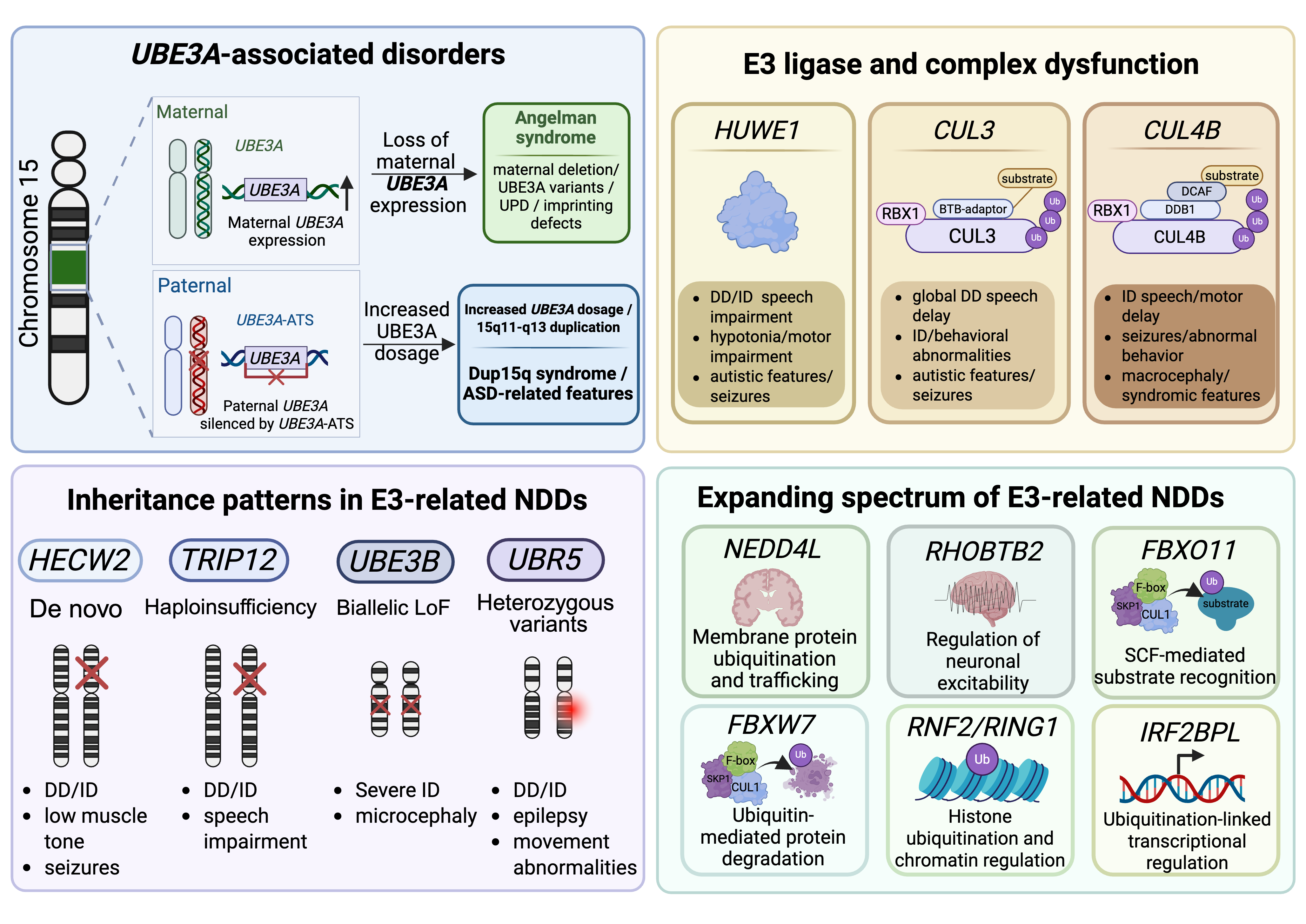
2. NDD-Associated E3 Ligases and E3-Complex Components

2.1. UBE3A
2.2. HUWE1, CUL3, and CUL4B
2.3. HECW2, TRIP12, UBE3B, and UBR5
2.4. NEDD4L, RHOBTB2, FBXO11, FBXW7, RNF2/RING1, and IRF2BPL
3. Neurodevelopmental Processes and Specific Mechanisms Affected by E3 Ubiquitin Ligase Abnormalities
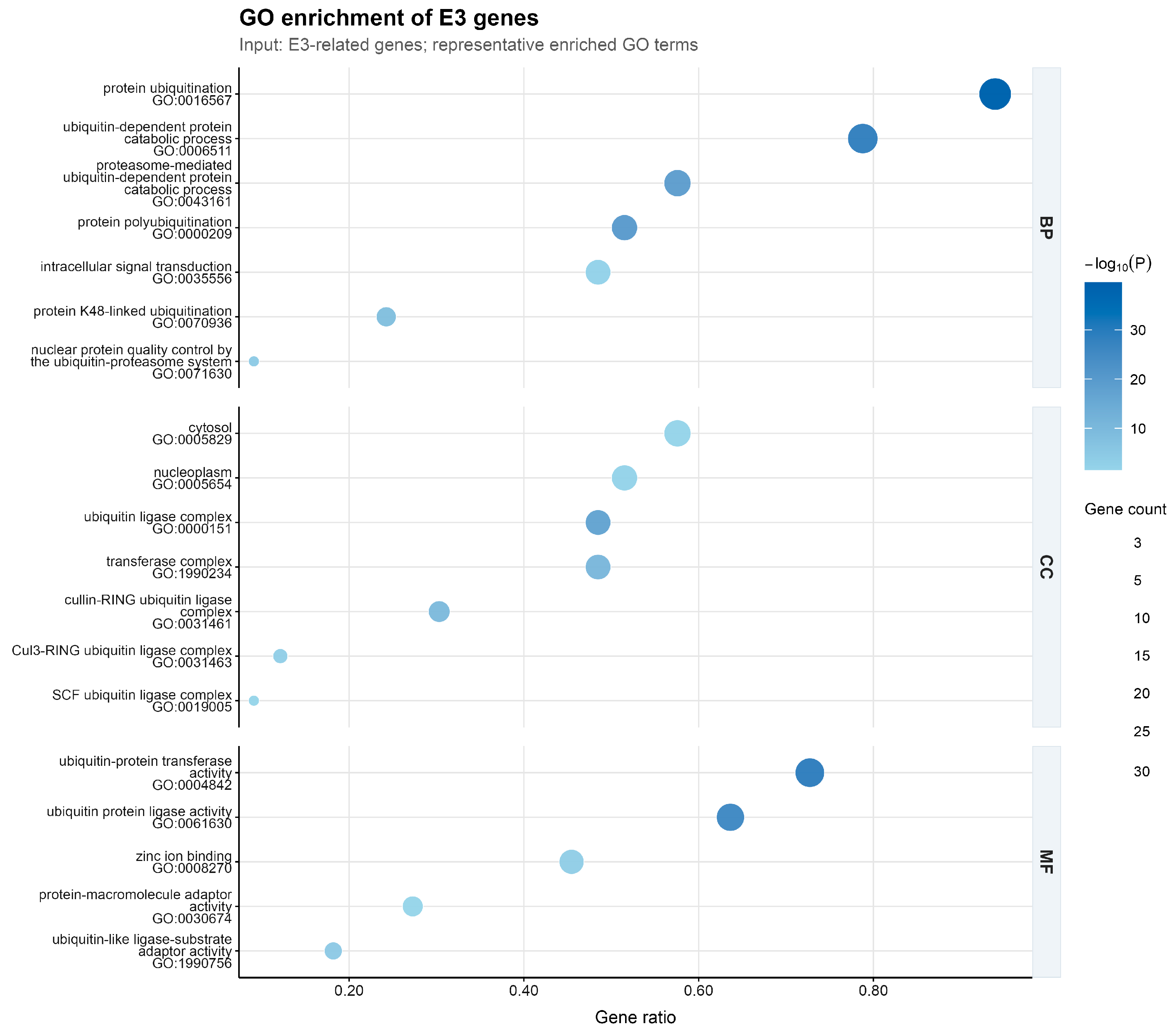
3.1. Gene Ontology Enrichment Analysis of E3-Related Genes
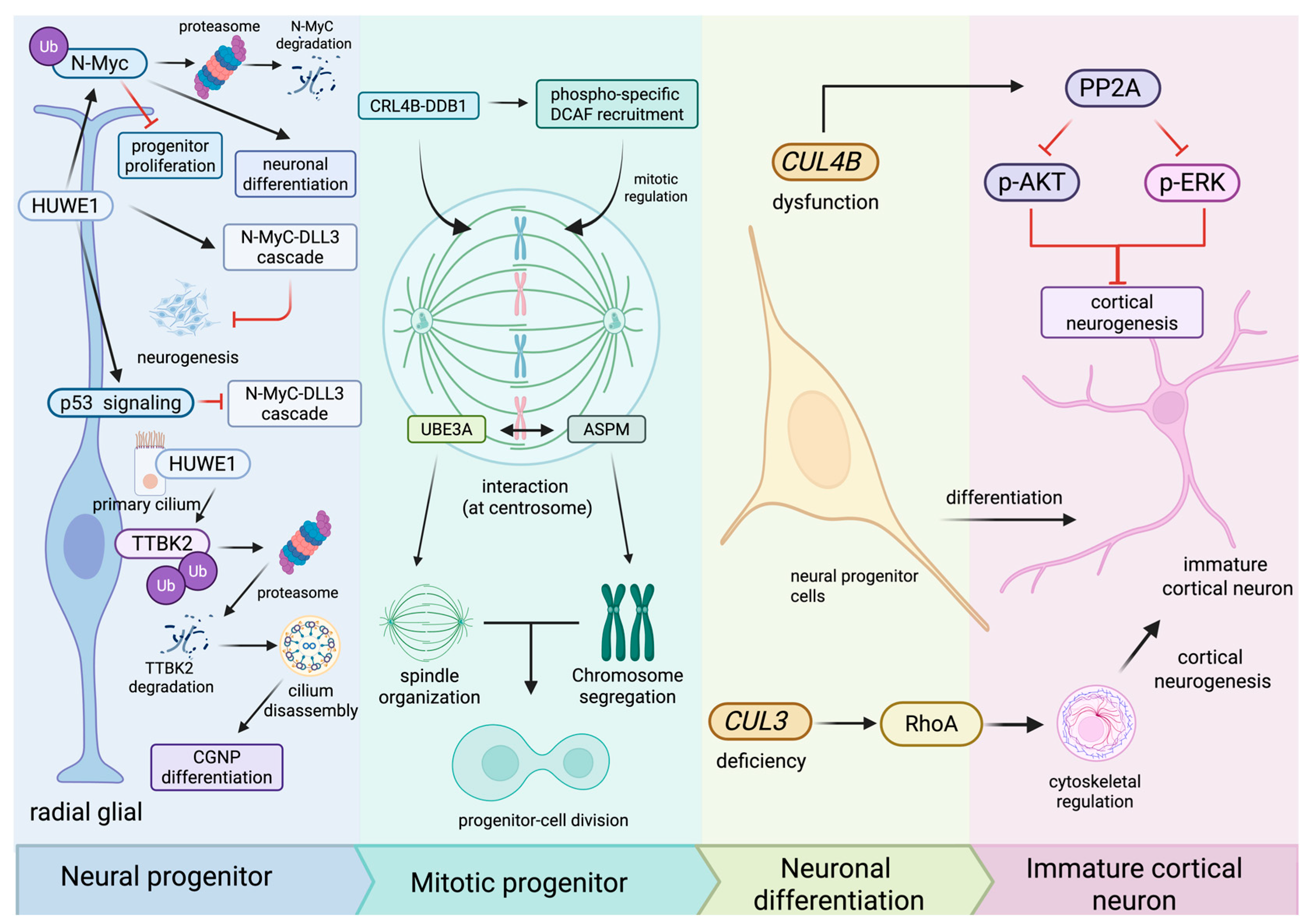
3.2. Neural Progenitor Proliferation, Differentiation, and Early Brain Development
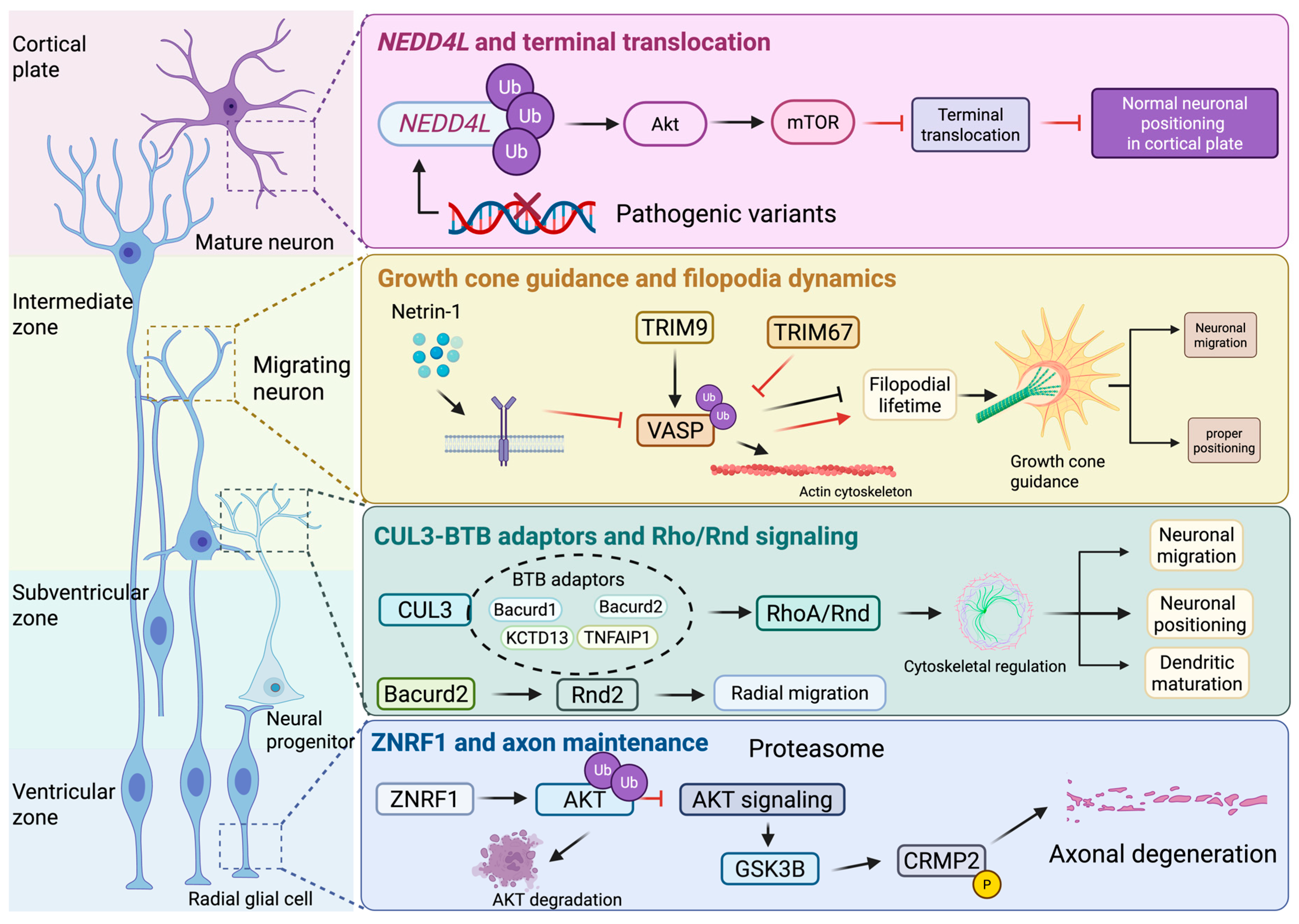
3.3. Neuronal Migration, Cytoskeletal Regulation, and Cortical Organization
3.4. Synaptic Development, E/I Balance, and Neural Circuit Maturation
3.5. Proteostasis, Metabolic Regulation, and Cellular Homeostasis

| Gene | E3-Related Category | Associated Disorder/Syndrome | Variant or Genetic Mechanism | Inheritance Pattern | Main NDD-Related Features | Genetic Evidence Summarized | References |
|---|---|---|---|---|---|---|---|
| UBE3A | HECT-type E3 ligase | Angelman syndrome | Maternal 15q11-q13 deletion, UBE3A variant, paternal UPD, imprinting defect | Maternal allele/imprinting | DD, ID, limited speech, ataxia, seizures, sleep and behavioral features | Clinical genetics and review synthesis | [17] |
| UBE3A | HECT-type E3 ligase | Angelman syndrome | Clinical diagnosis of UBE3A-related/imprinting disorder | Usually maternal allele or imprinting defect | Atypical early presentation of AS | Human case report | [18] |
| UBE3A | HECT-type E3 ligase | Angelman syndrome | AS-related genetic etiologies | Maternal allele/imprinting | Developmental milestones and daily living impairment across age | Human cohort/clinical assessment | [19] |
| UBE3A | HECT-type E3 ligase | Angelman syndrome | AS-related genetic etiologies | Maternal allele/imprinting | Autistic traits in the context of AS | Human behavioral cohort | [20] |
| UBE3A | HECT-type E3 ligase | Angelman syndrome | AS-related genetic etiologies | Maternal allele/imprinting | Sleep disturbance and challenging behavior | Human sleep/behavior cohort | [21] |
| UBE3A | HECT-type E3 ligase | UBE3A-related ID/DD | Familial pathogenic UBE3A variant | Familial inheritance; allele effect requires interpretation | Multigenerational ID and DD | Human family study | [22] |
| UBE3A | HECT-type E3 ligase | Angelman syndrome mechanistic basis | Paternal UBE3A silencing in neurons | Allele-specific expression | Basis for neuronal UBE3A deficiency | Patient-derived neurons | [23] |
| UBE3A | HECT-type E3 ligase | Angelman syndrome imprinting context | Allele-specific Ube3a expression during development | Maternal/paternal allele regulation | Developmental dependence on maternal expression | Mouse brain expression mapping | [24] |
| UBE3A | HECT-type E3 ligase | UBE3A imprinting/dosage context | Brain-region and cell-type distribution of UBE3A/E6AP and antisense transcript | Allele-specific regulatory background | Developmental brain expression context | Rhesus monkey brain mapping | [25] |
| UBE3A | HECT-type E3 ligase | 15q11-q13 duplication syndrome/Dup15q syndrome | Chromosome 15q11.2-q13.1 duplication; increased UBE3A dosage context | Usually maternal duplication in classic Dup15q | ASD-related features, DD/ID, abnormal sleep EEG | Human sleep EEG cohort | [26] |
| UBE3A | HECT-type E3 ligase | UBE3A gain-of-function-related ASD/ID | UBE3A gain-of-function variant | Dominant/dosage-related | ASD and ID | Human genetic report with functional interpretation | [28] |
| UBE3A | HECT-type E3 ligase | UBE3A gain-of-function ASD model | Autism-linked gain-of-function mutation | Maternal or paternal inheritance in model context | Behavioral phenotypes and interneuron abnormalities | Mouse model | [29] |
| UBE3A | HECT-type E3 ligase | 15q11-q13 duplication syndrome/Dup15q syndrome model | 15q11.2-q13.1 duplication model | Dosage-related model | Sleep EEG signatures | Mouse model sleep EEG | [27] |
| HUWE1 | HECT-type E3 ligase | HUWE1-related X-linked intellectual disability/NDD | Whole-gene duplication or copy-number gain involving HUWE1 | X-linked dosage alteration | ID and syndromic developmental phenotype | Human CNV/family genetics | [31] |
| HUWE1 | HECT-type E3 ligase | HUWE1-related NDD/X-linked intellectual disability context | Pathogenic HUWE1 sequence variants | X-linked | DD, ID, speech impairment, seizures, autistic features, hypotonia | Human cohort genetics | [32] |
| HUWE1 | HECT-type E3 ligase | HUWE1-related NDD/X-linked intellectual disability context | Pathogenic HUWE1 variants | X-linked | DD, ID, speech/language impairment and variable syndromic features | Human cohort and suggested evaluations | [33] |
| CUL3 | Cullin-RING ligase scaffold | Neurodevelopmental disorder with or without autism or seizures | Heterozygous/de novo CUL3 variants | Autosomal dominant/de novo | DD, ID, ASD-related features, seizures | Human cohort genetics | [37] |
| CUL3 | Cullin-RING ligase scaffold | CUL3-related disorder / NEDAUS spectrum | Missense variants affecting BTB-adaptor binding | Autosomal dominant/de novo | Diverse NDD phenotypes | Human genetics plus binding assay | [40] |
| CUL3 | Cullin-RING ligase scaffold | CUL3-related syndromic NDD/NEDAUS | Loss-of-function variants | Autosomal dominant/de novo | DD/ID and syndromic features | Human cohort genetics | [38] |
| CUL3 | Cullin-RING ligase scaffold | CUL3-related NDD/NEDAUS | CUL3 pathogenic variants | Autosomal dominant/de novo | DD, ID, ASD-related features, seizures and variable congenital features | Human cohort and episignature analysis | [39] |
| CUL4B | Cullin-RING ligase scaffold/core component | X-linked intellectual developmental disorder, Cabezas type | Pathogenic CUL4B variants | X-linked | ID, speech/motor delay, seizures and syndromic features | Human genetics | [44] |
| CUL4B | Cullin-RING ligase scaffold/core component | CUL4B-associated XLID/Cabezas spectrum | Pathogenic CUL4B variants | X-linked | ID, brain malformations and syndromic features | Human cohort genetics | [45] |
| HECW2 | HECT-type E3 ligase | HECW2-related NDD | De novo missense variants | Autosomal dominant/de novo | Neurodevelopmental delay, ID, hypotonia | Human genetics with functional context | [50] |
| HECW2 | HECT-type E3 ligase | Developmental and epileptic encephalopathy in HECW2-related reports | HECW2 variants; zebrafish hecw2a knockdown | Autosomal dominant/de novo; model knockdown | Epilepsy, DD and abnormal brain development | Human genetics plus zebrafish model | [51] |
| HECW2 | HECT-type E3 ligase | HECW2-related developmental delay/NDD | Novel HECW2 variant | Likely de novo/dominant in reported case context | DD, neurodevelopmental delay, hypotonia | Human case report | [52] |
| TRIP12 | HECT-type E3 ligase | Clark-Baraitser syndrome/TRIP12-related NDD | TRIP12 pathogenic variants/haploinsufficiency | Autosomal dominant/de novo | DD, ID, speech delay, behavioral abnormalities | Human cohort genetics | [54] |
| TRIP12 | HECT-type E3 ligase | Clark-Baraitser syndrome/TRIP12-related NDD | TRIP12 haploinsufficiency or pathogenic variants | Autosomal dominant/de novo | ID, DD, speech impairment and ASD-related features | Human genetics | [55] |
| TRIP12 | HECT-type E3 ligase | TRIP12-related NDD/Clark-Baraitser syndrome | TRIP12 variants | Autosomal dominant/de novo | DD, ID, speech impairment, behavioral problems and facial phenotype | Human cohort phenotype study | [56] |
| UBE3B | HECT-type E3 ligase | Kaufman oculocerebrofacial syndrome | UBE3B deficiency/biallelic loss-of-function | Autosomal recessive | Severe ID, DD, ocular/facial anomalies, congenital features | Human genetics plus model organism data | [57] |
| UBE3B | HECT-type E3 ligase | Blepharophimosis-ptosis-intellectual-disability syndrome/KOS | Biallelic UBE3B variants | Autosomal recessive | ID, growth delay and multisystem congenital features | Human genetics | [58] |
| UBE3B | HECT-type E3 ligase | Kaufman oculocerebrofacial syndrome/UBE3B-related syndrome | Biallelic UBE3B pathogenic variants | Autosomal recessive | DD/ID, microcephaly, hypotonia and syndromic features | Human case series | [59] |
| UBE3B | HECT-type E3 ligase | Kaufman oculocerebrofacial syndrome/UBE3B-related syndrome | Novel biallelic UBE3B mutations | Autosomal recessive | DD/ID and additional dysmorphic/congenital findings | Human case series | [60] |
| UBR5 | N-recognin/HECT-type E3 ligase | UBR5-related neurodevelopmental syndrome | Heterozygous pathogenic UBR5 variants | Autosomal dominant/often de novo | DD, ASD, ID, speech/language impairment and epilepsy | Human cohort genetics | [62] |
| UBR5 | N-recognin/HECT-type E3 ligase | ASD/ID associated with UBR5 loss-of-function | UBR5 loss-of-function variants | Autosomal dominant/de novo or inherited context | ASD and ID with variable NDD features | Human case series and literature review | [63] |
| UBR5/UBR7 | N-recognin-related E3 factors | UBR7/UBR5-associated Notch-related neurodevelopmental syndrome | UBR7 disease gene with UBR5 participation in Notch pathway | Mostly de novo/dominant for UBR7 syndrome context | Epilepsy, ptosis, hypothyroidism and DD/ID-related features | Human genetics plus functional assays | [64] |
| NEDD4L | HECT-type E3 ligase | Periventricular nodular heterotopia 7 | Pathogenic NEDD4L variants | Autosomal dominant/de novo or inherited in reported families | DD/ID, epilepsy, cortical malformation, speech/motor abnormalities | Human genetics plus functional/model data | [65] |
| NEDD4L | HECT-type E3 ligase | NEDD4L-related disorder/PVNH7 | Pathogenic NEDD4L variant | Autosomal dominant/reported case context | PVNH, DD, syndromic features and ophthalmic finding | Human case report and literature summary | [66] |
| RHOBTB2 | BTB-domain atypical Rho GTPase; CUL3-related ubiquitination context | Developmental and epileptic encephalopathy 64 | Pathogenic RHOBTB2 variants | Autosomal dominant/de novo | Early-onset epilepsy, DD/ID, movement disorder, hypotonia/dystonia | Human genetics | [67] |
| RHOBTB2 | BTB-domain atypical Rho GTPase; CUL3-related ubiquitination context | RHOBTB2-associated NDD/DEE64 spectrum | Pathogenic RHOBTB2 variants at different protein positions | Autosomal dominant/mostly de novo | Epilepsy, DD/ID, motor and tone abnormalities | Human genotype–phenotype cohort | [68] |
| FBXO11 | F-box substrate receptor in SCF-type CRL complex | FBXO11-related IDDFBA/NDD | De novo missense variants altering protein behavior | Autosomal dominant/de novo | ID/DD and variable syndromic features | Human genetics plus in vitro functional assays | [70] |
| FBXW7 | F-box substrate receptor in SCF-type CRL complex | FBXW7 neurodevelopmental syndrome | Heterozygous pathogenic FBXW7 variants | Autosomal dominant/de novo | Global DD, ID, hypotonia, speech delay, epilepsy and brain anomalies | Human genetics plus functional assays | [71] |
| FBXW7 | F-box substrate receptor in SCF-type CRL complex | FBXW7-related NDD/FBXW7 neurodevelopmental syndrome | Pathogenic FBXW7 variants | Autosomal dominant/de novo or inherited context | DD/ID, hypotonia, language delay and variable congenital features | Human case report/series | [72] |
| RNF2/RING1 | Polycomb-associated RING-type E3 ligase/PRC1 component | RNF2-related syndromic neurodevelopmental disorder | De novo RNF2 variants | Autosomal dominant/de novo | DD/ID, behavioral abnormalities, seizures and congenital/syndromic features | Human genetics and chromatin-regulation context | [74] |
| RING1 | Polycomb-associated RING-type E3 ligase/PRC1 component | RING1-related neurodevelopmental/chromatin disorder | Missense variants affecting RING1 function | Autosomal dominant/de novo | Neurodevelopmental abnormalities with syndromic features | Human variants plus functional neurogenesis assays | [75] |
| RNF2/RING1 | Polycomb-associated RING-type E3 ligase/PRC1 component | Polycomb-related NDD involving RNF2/RING1 | Variants altering chromatin binding of Polycomb complexes | Autosomal dominant/de novo context | Syndromic neurodevelopmental disorders | Human genetics plus chromatin/model studies | [76] |
| IRF2BPL | Transcriptional/ubiquitination-associated regulator | Neurodevelopmental disorder with regression, abnormal movements, loss of speech and seizures | Pathogenic/truncating IRF2BPL variants | Autosomal dominant/de novo | DD/ID, regression, movement disorder, speech loss, seizures | Human genotype–phenotype study | [77] |
| IRF2BPL | Transcriptional/ubiquitination-associated regulator | IRF2BPL syndrome/NEDAMSS | Pathogenic IRF2BPL variants | Autosomal dominant/de novo or inherited context | DD/ID, regression, epilepsy, ataxia and abnormal movements | Human cohort genetics | [78] |
| KCTD13 | BTB/KCTD adaptor linked to CUL3-type ubiquitination | 16p11.2 CNV-associated neurodevelopmental phenotype | 16p11.2 copy-number alteration involving KCTD13; KCTD13 dosage | CNV dosage-related | ASD-related features and developmental/metabolic phenotype | Human CNV/proteomic or cellular model context | [111] |
| KCTD13 | BTB/KCTD adaptor linked to CUL3-type ubiquitination | 16p11.2 CNV-associated neurodevelopmental phenotype | 16p11.2 CNV network involving KCTD13 | CNV dosage-related | Psychiatric/NDD risk with cortical developmental context | Spatiotemporal protein network analysis | [88] |
| KLHL15 | Kelch-like substrate adaptor / E3 ligase-related protein | KLHL15-related X-linked intellectual disability | KLHL15 pathogenic variant | X-linked | Intellectual disability and neurodevelopmental impairment | Human disease context plus biochemical/cell assays | [113] |
| Process Category | E3-Related Gene | Molecular Module/Key Substrate or Pathway | Main Functional Findings | Neurodevelopmental Relevance | Evidence Model or System | References |
|---|---|---|---|---|---|---|
| Neural progenitor proliferation, differentiation, and early brain development | CUL4B | NPC growth and cell-cycle regulation | CUL4B regulates neural progenitor growth and cell-cycle control. | Supports early progenitor expansion and cortical neurogenesis. | Neural progenitor cell models | [81] |
| Neural progenitor proliferation, differentiation, and early brain development | CUL4B | PP2A-dependent AKT/ERK signaling | CUL4B mutations impair cortical neurogenesis through PP2A-dependent inhibition of AKT and ERK signaling. | Links CUL4B dysfunction to impaired human cortical neurogenesis. | Human cortical neurogenesis models | [47] |
| Neural progenitor proliferation, differentiation, and early brain development | CRL4B-DDB1 complex | Phosphorylation-specific DCAF recruitment | CUL4B-based E3 complexes recruit phosphorylation-specific DCAFs during mitotic regulation. | Connects CRL4B complex function with mitosis and brain development. | Mitotic and developmental model systems | [82] |
| Neural progenitor proliferation, differentiation, and early brain development | HUWE1 | N-Myc degradation | HUWE1 controls N-Myc stability and thereby regulates neural progenitor proliferation and neuronal differentiation. | Places HECT-type E3 activity in early cell-number and differentiation control. | Developing brain and neural differentiation models | [34] |
| Neural progenitor proliferation, differentiation, and early brain development | HUWE1 | N-Myc-DLL3 cascade | HUWE1 suppresses the N-Myc-DLL3 cascade to inhibit proliferation and promote neurogenesis. | Links HUWE1-dependent N-Myc regulation to neurogenic progression. | Brain tumor and neurogenesis models | [83] |
| Neural progenitor proliferation, differentiation, and early brain development | HUWE1 | p53 signaling | Enhanced p53 signaling impairs neuronal differentiation in HUWE1-associated intellectual disability models. | Connects HUWE1 dysfunction with differentiation defects in a disease-relevant context. | Models of HUWE1-associated intellectual disability | [35] |
| Neural progenitor proliferation, differentiation, and early brain development | HUWE1 | TTBK2 and primary cilium disassembly | HUWE1-mediated TTBK2 degradation regulates primary cilium disassembly and cerebellar granule neuron progenitor differentiation. | Links protein turnover to ciliary dynamics and cerebellar development. | Cerebellar development and progenitor models | [36] |
| Neural progenitor proliferation, differentiation, and early brain development | UBE3A | ASPM interaction and centrosome localization | UBE3A interacts with ASPM and localizes to centrosomes, with links to spindle organization and chromosome segregation. | Suggests a role for UBE3A in progenitor-cell division-related processes. | Cell-based localization and interaction assays | [84] |
| Neural progenitor proliferation, differentiation, and early brain development | CUL3 | RhoA signaling and cytoskeletal dynamics | Cul3 haploinsufficiency alters RhoA-related cytoskeletal regulation and cortical neurogenesis. | Connects CRL3 dysfunction with early cortical development and cytoskeletal control. | Mouse and neuronal model systems | [42] |
| Neuronal migration, cytoskeletal regulation, and cortical organization | NEDD4L | AKT-mTOR signaling and terminal translocation | NEDD4L activity is linked to AKT-mTOR signaling, neuronal positioning, and terminal translocation in the developing cortex. | Provides a mechanism-oriented link to cortical malformation and abnormal neuronal positioning. | Developing cortex and patient-genetic studies | [65,66] |
| Neuronal migration, cytoskeletal regulation, and cortical organization | CUL3 / CRL3 adaptor system | BTB-domain adaptor interaction | CUL3 loss-of-function variants and variants affecting CUL3-BTB adaptor interaction provide a complex-level basis for downstream cytoskeletal effects. | Connects scaffold or adaptor-binding defects with cytoskeletal pathway disruption. | Patient-genetic and biochemical studies | [38,40] |
| Neuronal migration, cytoskeletal regulation, and cortical organization | CUL3 | Cytoskeletal protein homeostasis and cell migration | Cul3 regulates cytoskeletal protein homeostasis and cell migration during a critical developmental window. | Links CRL3 function to neuronal migration and cortical organization. | Developing brain and cell migration models | [43] |
| Neuronal migration, cytoskeletal regulation, and cortical organization | Bacurd/Kctd13-related adaptors | Rnd/Rho GTPase-related regulation | Bacurd1/Kctd13 and Bacurd2/Tnfaip1 interact with Rnd proteins and influence neuronal positioning and dendritic maturation. | Places CRL3 adaptors in Rho-family cytoskeletal regulation during cortical development. | Cortical neuron developmental models | [86] |
| Neuronal migration, cytoskeletal regulation, and cortical organization | Bacurd2 | Rnd2-dependent radial migration | Bacurd2 interacts with Rnd2 and controls radial migration in the developing mammalian cortex. | Links adaptor-mediated Rnd signaling to neuronal migration. | Developing mammalian cerebral cortex | [87] |
| Neuronal migration, cytoskeletal regulation, and cortical organization | KCTD13-CUL3-RhoA pathway | 16p11.2 protein network | KCTD13, CUL3, and RhoA are connected in a spatiotemporal protein network involving mid-fetal cortical development. | Provides network-level support for RhoA-related cortical developmental biology. | Spatiotemporal protein-network analysis | [88] |
| Neuronal migration, cytoskeletal regulation, and cortical organization | TRIM9/TRIM67 | DCC, VASP ubiquitination, and filopodial stability | TRIM9 ubiquitinates VASP in a non-degradative manner, whereas TRIM67 counteracts VASP ubiquitination to regulate filopodial lifetime. | Links ubiquitination to growth-cone dynamics and netrin-dependent axon guidance. | Neuronal growth-cone and axon-guidance assays | [89,90] |
| Neuronal migration, cytoskeletal regulation, and cortical organization | ZNRF1 | AKT-GSK3B-CRMP2 axis | ZNRF1 promotes AKT degradation, thereby enhancing GSK3B-dependent CRMP2 phosphorylation during axon degeneration. | Connects ubiquitin-mediated protein turnover with axonal integrity. | Axon injury and degeneration models | [91] |
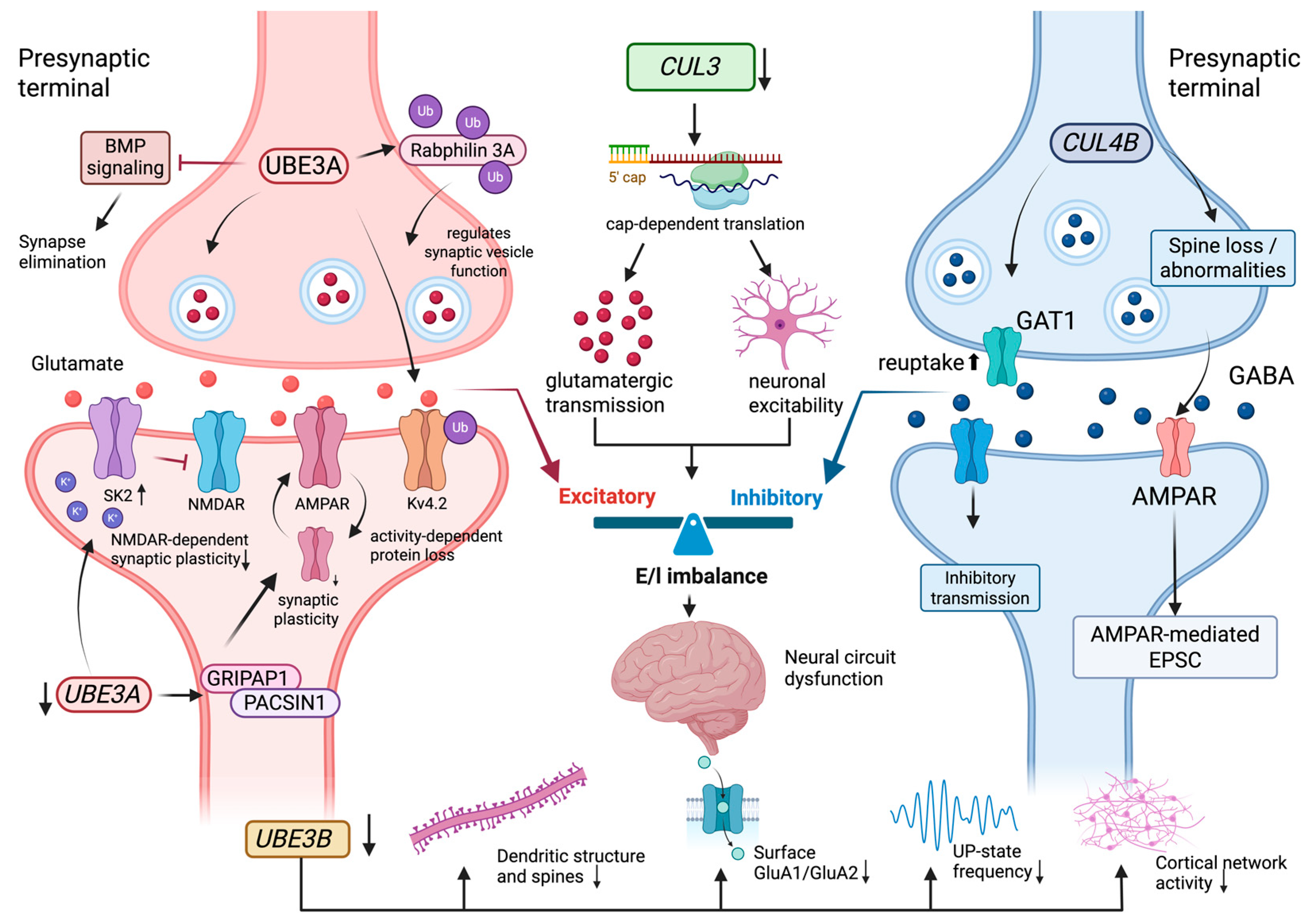
| Synaptic development, E/I balance, and neural circuit maturation | UBE3A | SK2 channel regulation | UBE3A loss increases synaptic SK2 channel levels and impairs NMDAR-dependent synaptic plasticity. | Links UBE3A deficiency with altered synaptic plasticity and learning-related circuitry. | Angelman syndrome mouse and hippocampal models | [93,94] |
| Synaptic development, E/I balance, and neural circuit maturation | UBE3A | Kv4.2 activity-dependent degradation | UBE3A binds and ubiquitinates Kv4.2, promoting activity-dependent Kv4.2 protein loss. | Connects UBE3A-dependent protein turnover with neuronal excitability and synaptic plasticity. | Angelman syndrome mouse and neuronal activity models | [95] |
| Synaptic development, E/I balance, and neural circuit maturation | UBE3A | Rabphilin 3A monoubiquitination | UBE3A mediates non-degradative monoubiquitination of Rabphilin 3A. | Provides a non-degradative ubiquitination mechanism relevant to presynaptic function. | Biochemical and synaptic-function assays | [96] |
| Synaptic development, E/I balance, and neural circuit maturation | UBE3A | GRIPAP1, PACSIN1, and AMPA receptor recycling | UBE3A regulates GRIPAP1 and PACSIN1 proteins linked to AMPA receptor endocytic recycling. | Connects UBE3A loss with receptor trafficking and synaptic plasticity. | Human cortical neuron and cell-based assays | [97] |
| Synaptic development, E/I balance, and neural circuit maturation | UBE3A | BMP signaling and synapse elimination | Presynaptic Ube3a promotes synapse elimination through downregulation of BMP signaling. | Links E3 activity with developmental synapse remodeling. | Synapse-development model systems | [98] |
| Synaptic development, E/I balance, and neural circuit maturation | CUL3 | Cap-dependent translation and eIF4E-eIF4G1 complex | CUL3 deficiency enhances cap-dependent translation; modulation of eIF4E-eIF4G1 or neuronal activity improves selected synaptic and behavioral readouts. | Connects CRL3 dysfunction with glutamatergic transmission, neuronal excitability, and E/I balance. | Cul3-deficient mouse and neuronal models | [100] |
| Synaptic development, E/I balance, and neural circuit maturation | CUL3 | Region-specific circuit effects | Prefrontal and striatal Cul3 deficiency produce partly distinct effects on social and stereotypic behavior-related circuitry. | Links E3-complex dysfunction with brain-region-specific circuit maturation. | Region-specific Cul3 mouse models | [101] |
| Synaptic development, E/I balance, and neural circuit maturation | CUL4B | Synapse loss, spine abnormalities, and AMPAR-mediated EPSCs | Neural Cul4b deficiency causes synapse loss, dendritic spine abnormalities, and reduced AMPAR-mediated EPSCs. | Links CUL4B dysfunction with excitatory synaptic function. | Neural-specific Cul4b mouse models | [48] |
| Synaptic development, E/I balance, and neural circuit maturation | CUL4B | GAT1 accumulation and GABA reuptake | Cul4b deletion causes GAT1 accumulation, increased GABA reuptake, and impaired inhibitory synaptic transmission. | Connects CRL4B dysfunction with inhibitory transmission and epilepsy-related network vulnerability. | Cul4b-deficient mice and pharmacological assays | [49] |
| Synaptic development, E/I balance, and neural circuit maturation | UBE3B | Dendritic complexity, spine density, surface GluA1/GluA2, and cortical UP states | CNS-specific Ube3b loss reduces dendritic complexity, excitatory synapse density, surface GluA1/GluA2, and cortical UP-state frequency. | Places UBE3B in excitatory synaptic development and cortical network maturation. | CNS-specific Ube3b mouse models | [102] |
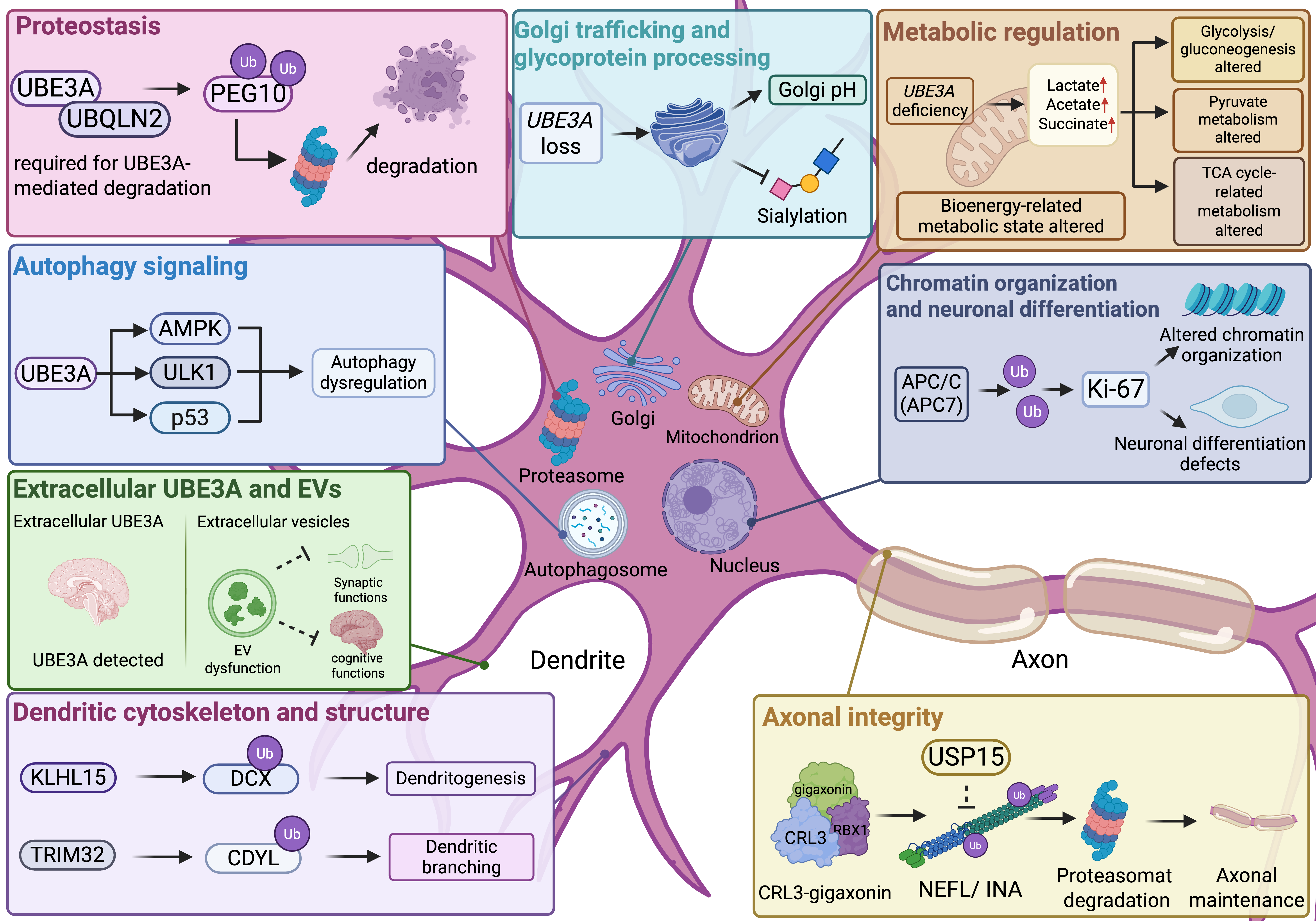
| Proteostasis, metabolic regulation, and cellular homeostasis | UBE3A | Spatiotemporal proteomic changes | UBE3A-deficient models show developmental and region-dependent proteomic changes involving proteasome, translation-related, aminoacyl-tRNA synthetase, and synapse-related pathways. | Links UBE3A loss with proteostasis and synapse-related molecular maturation. | Cross-species, brain-region, and developmental-stage proteomics | [103] |
| Proteostasis, metabolic regulation, and cellular homeostasis | UBE3A | Glycolysis/gluconeogenesis, pyruvate metabolism, TCA cycle-related metabolism, and mitochondrial function-related annotations | Early AS model metabolomics showed altered bioenergy-related metabolic pathways, including increased lactate, acetate, and succinate. | Provides annotation-level evidence for altered metabolic state during early development. | 1H-NMR metabolomics in AS mouse embryos | [104] |
| Proteostasis, metabolic regulation, and cellular homeostasis | UBE3A | Golgi acidification and surface protein sialylation | UBE3A loss affects Golgi acidification and cell-surface protein sialylation. | Links UBE3A dysfunction with trafficking and glycoprotein processing. | Cellular and neuronal assays | [105] |
| Proteostasis, metabolic regulation, and cellular homeostasis | UBE3A/UBQLN2 | PEG10 proteasomal degradation | UBQLN2 contributes to UBE3A-mediated proteasomal degradation of PEG10. | Connects UBE3A-dependent degradation with retroelement-derived protein control. | Proteostasis and cell-based assays | [106,107] |
| Proteostasis, metabolic regulation, and cellular homeostasis | UBE3A | Extracellular vesicles and extracellular UBE3A | UBE3A-related extracellular vesicle dysfunction and extracellular UBE3A detection are linked to synaptic or cognitive functional contexts. | Extends UBE3A-related mechanisms to extracellular signaling and trafficking contexts. | AS model extracellular vesicle, CSF, and extracellular-space assays | [108,109] |
| Proteostasis, metabolic regulation, and cellular homeostasis | UBE3A | AMPK-ULK1 and p53-related autophagy signaling | UBE3A deficiency is associated with autophagy dysregulation involving AMPK-ULK1 and p53 pathways. | Links UBE3A loss with cellular homeostasis pathways. | AS model and cellular assays | [110] |
| Proteostasis, metabolic regulation, and cellular homeostasis | KCTD13/CRL3 adaptor | ADSS ubiquitination and purine metabolism | KCTD13 regulates ADSS ubiquitination and purine metabolism. | Connects a CRL3 adaptor with metabolic homeostasis in 16p11.2-associated ASD biology. | Cellular and metabolic assays | [111] |
| Proteostasis, metabolic regulation, and cellular homeostasis | APC/C-APC7 | Ki-67 and chromatin-associated protein clearance | APC7 supports ubiquitin-dependent clearance of chromatin-associated proteins such as Ki-67. | Links protein clearance with cell-cycle exit, chromatin organization, and neuronal differentiation. | Developing mammalian brain and neural progenitor models | [112] |
| Proteostasis, metabolic regulation, and cellular homeostasis | CUL3-KLHL15 | Doublecortin protein degradation | KLHL15 promotes degradation of doublecortin proteins and constrains dendritogenesis. | Links CRL3 substrate-adaptor function to dendritic structure. | Neuronal morphology and biochemical assays | [113] |
| Proteostasis, metabolic regulation, and cellular homeostasis | TRIM32 | CDYL degradation | TRIM32 promotes CDYL degradation and dendritic branching. | Connects ubiquitin-mediated degradation with dendritic arborization. | Neuronal morphology assays | [114] |
| Proteostasis, metabolic regulation, and cellular homeostasis | CRL3-gigaxonin/USP15 | Neurofilament protein clearance | CRL3-gigaxonin promotes neurofilament clearance, whereas USP15 antagonizes this pathway. | Links E3 complex activity and deubiquitination with axonal cytoskeletal maintenance. | Biochemical and cellular models | [115] |
4. Functional Evidence and Therapeutic Translation
4.1. UBE3A Restoration and Paternal Reactivation in Angelman Syndrome
4.2. Preclinical Translational Mechanisms Beyond UBE3A
4.3. Candidate Substrates and Mechanisms Requiring Further Validation
5. Conclusions and Future Perspectives
Author Contributions
Funding
Institutional Review Board Statement
Informed Consent Statement
Data Availability Statement
Acknowledgments
Conflicts of Interest
Abbreviations
| NDD | Neurodevelopmental disorder |
| NDDs | Neurodevelopmental disorders |
| DD | Developmental delay |
| ID | Intellectual disability |
| ASD | Autism spectrum disorder |
| AS | Angelman syndrome |
| Dup15q | 15q11-q13 duplication syndrome |
| UPS | Ubiquitin-proteasome system |
| Ub | Ubiquitin |
| E1 | Ubiquitin-activating enzyme |
| E2 | Ubiquitin-conjugating enzyme |
| E3 | Ubiquitin ligase |
| HECT | Homologous to the E6AP carboxyl terminus |
| RING | Really interesting new gene |
| CRL | Cullin-RING ligase |
| CRL3 | CUL3-based Cullin-RING ligase complex |
| CRL4B | CUL4B-based Cullin-RING ligase complex |
| BTB | Broad-complex, Tramtrack, and Bric-a-brac |
| DCAF | DDB1- and CUL4-associated factor |
| NPC | Neural progenitor cell |
| CNV | Copy number variant |
| UPD | Uniparental disomy |
| UBE3A-ATS | UBE3A antisense transcript |
| E/I | Excitatory/inhibitory |
| iPSC | Induced pluripotent stem cell |
| hiPSC | Human induced pluripotent stem cell |
| RNA-seq | RNA sequencing |
| WES | Whole-exome sequencing |
| WGS | Whole-genome sequencing |
References
- Parenti, I.; Rabaneda, L.G.; Schoen, H.; Novarino, G. Neurodevelopmental Disorders: From Genetics to Functional Pathways. Trends Neurosci. 2020, 43, 608–621. [Google Scholar] [CrossRef] [Scilit] [PubMed]
- Silbereis, J.C.; Pochareddy, S.; Zhu, Y.; Li, M.; Sestan, N. The Cellular and Molecular Landscapes of the Developing Human Central Nervous System. Neuron 2016, 89, 248–268. [Google Scholar] [CrossRef] [Scilit] [PubMed]
- Deciphering Developmental Disorders Study. Prevalence and architecture of de novo mutations in developmental disorders. Nature 2017, 542, 433–438. [CrossRef] [Scilit] [PubMed]
- Zhou, X.; Feliciano, P.; Shu, C.; Wang, T.; Astrovskaya, I.; Hall, J.B.; Obiajulu, J.U.; Wright, J.R.; Murali, S.C.; Xu, S.X.; et al. Integrating de novo and inherited variants in 42,607 autism cases identifies mutations in new moderate-risk genes. Nat. Genet. 2022, 54, 1305–1319. [Google Scholar] [CrossRef] [Scilit] [PubMed]
- Das, M.; Girirajan, S. Genetic subtypes, allelic effects, and convergent neurodevelopmental mechanisms. Genome Med. 2021, 13, 99. [Google Scholar] [CrossRef] [Scilit] [PubMed]
- Komander, D.; Rape, M. The ubiquitin code. Annu. Rev. Biochem. 2012, 81, 203–229. [Google Scholar] [CrossRef] [Scilit] [PubMed]
- Hegde, A.N.; Upadhya, S.C. The ubiquitin-proteasome pathway in health and disease of the nervous system. Trends Neurosci. 2007, 30, 587–595. [Google Scholar] [CrossRef] [Scilit] [PubMed]
- Jarome, T.J.; Helmstetter, F.J. The ubiquitin-proteasome system as a critical regulator of synaptic plasticity and long-term memory formation. Neurobiol. Learn. Mem. 2013, 105, 107–116. [Google Scholar] [CrossRef] [Scilit] [PubMed]
- Hale, M.; Bashaw, G.J. Emerging roles for E3 ubiquitin ligases in neural development and disease. Front. Cell Dev. Biol. 2025, 13, 1557653. [Google Scholar] [CrossRef] [Scilit] [PubMed]
- Gao, S.; Shan, C.; Zhang, R.; Wang, T. Genetic advances in neurodevelopmental disorders. Med. Rev. 2025, 5, 139–151. [Google Scholar] [CrossRef] [Scilit] [PubMed]
- Ebstein, F.; Kury, S.; Papendorf, J.J.; Kruger, E. Neurodevelopmental Disorders (NDD) Caused by Genomic Alterations of the Ubiquitin-Proteasome System (UPS): The Possible Contribution of Immune Dysregulation to Disease Pathogenesis. Front. Mol. Neurosci. 2021, 14, 733012. [Google Scholar] [CrossRef] [Scilit] [PubMed]
- Lambert, N.; Moise, M.; Nguyen, L. E3 ubiquitin ligases and cerebral cortex development in health and disease. Dev. Neurobiol. 2022, 82, 392–407. [Google Scholar] [CrossRef] [Scilit] [PubMed]
- Zenge, C.; Ordureau, A. Ubiquitin system mutations in neurological diseases. Trends Biochem. Sci. 2024, 49, 875–887. [Google Scholar] [CrossRef] [Scilit] [PubMed]
- Ashitomi, H.; Nakagawa, T.; Nakagawa, M.; Hosoi, T. Cullin-RING Ubiquitin Ligases in Neurodevelopment and Neurodevelopmental Disorders. Biomedicines 2025, 13, 810. [Google Scholar] [CrossRef] [Scilit] [PubMed]
- Kurmashev, R. UBE3A Dosage Imbalance as a Molecular Framework Linking Angelman Syndrome and Dup15q-Associated Autism Phenotypes. Am. J. Med. Genet. B Neuropsychiatr. Genet. 2026. ahead of print. [Google Scholar] [CrossRef] [Scilit] [PubMed]
- Qu, S.; Sun, P.; Dai, L.; Song, C.; Wang, Y. Clinical and Genetic Profiles of 11 Chinese Patients with Angelman Syndrome. Genet. Res. 2025, 2025, 5593007. [Google Scholar] [CrossRef] [Scilit] [PubMed]
- Buiting, K.; Williams, C.; Horsthemke, B. Angelman syndrome—Insights into a rare neurogenetic disorder. Nat. Rev. Neurol. 2016, 12, 584–593. [Google Scholar] [CrossRef] [Scilit] [PubMed]
- Dang, H.; Srinivasa, S.; Lee, S.Y.; Alprin, C. A Case Study of Early Diagnosed Angelman Syndrome: Recognizing Atypical Clinical Presentations. Cureus 2023, 15, e39271. [Google Scholar] [CrossRef] [Scilit] [PubMed]
- Sadhwani, A.; Powers, S.; Wheeler, A.; Miller, H.; Potter, S.N.; Peters, S.U.; Bacino, C.A.; Skinner, S.A.; Wink, L.K.; Erickson, C.A.; et al. Developmental milestones and daily living skills in individuals with Angelman syndrome. J. Neurodev. Disord. 2024, 16, 32. [Google Scholar] [CrossRef] [Scilit] [PubMed]
- Hagenaar, D.A.; Mous, S.E.; Ten Hoopen, L.W.; Rietman, A.B.; Hiralal, K.R.; Bindels-de Heus, K.G.C.B.; de Nijs, P.F.A.; Mohr, T.C.; Lens, E.J.; Hillegers, M.H.J.; et al. Age-Related Trajectories of Autistic Traits in Children with Angelman Syndrome. Autism Res. Off. J. Int. Soc. Autism Res. 2025, 18, 870–880. [Google Scholar] [CrossRef] [Scilit] [PubMed]
- O’Donohoe, D.S.; Whelan, S.; Mannion, A.; Tones, M.; Heussler, H.; Bellgard, M.; Leader, G. Association between sleep disturbances and challenging behavior in children and adolescents with Angelman syndrome. Sleep Med. 2024, 123, 1–6. [Google Scholar] [CrossRef] [Scilit] [PubMed]
- Zhao, X.; Zheng, Y.; Wang, L.; Wang, Y.; Mei, S.; Kong, X. A novel variant in UBE3A in a family with multigenerational intellectual disability and developmental delay. Mol. Genet. Genom. Med. 2022, 10, e1883. [Google Scholar] [CrossRef] [Scilit] [PubMed]
- Stanurova, J.; Neureiter, A.; Hiber, M.; de Oliveira Kessler, H.; Stolp, K.; Goetzke, R.; Klein, D.; Bankfalvi, A.; Klump, H.; Steenpass, L. Angelman syndrome-derived neurons display late onset of paternal UBE3A silencing. Sci. Rep. 2016, 6, 30792, Erratum in Sci. Rep. 2018, 8, 46952. https://doi.org/10.1038/srep46952.. [Google Scholar] [CrossRef] [Scilit] [PubMed]
- Judson, M.C.; Sosa-Pagan, J.O.; Del Cid, W.A.; Han, J.E.; Philpot, B.D. Allelic specificity of Ube3a expression in the mouse brain during postnatal development. J. Comp. Neurol. 2014, 522, 1874–1896. [Google Scholar] [CrossRef] [Scilit] [PubMed]
- Gonzalez Ramirez, C.; Salvador, S.G.; Patel, R.K.R.; Clark, S.; Miller, N.W.; James, L.M.; Ringelberg, N.W.; Simon, J.M.; Bennett, J.; Amaral, D.G.; et al. Regional and cellular organization of the autism-associated protein UBE3A/E6AP and its antisense transcript in the brain of the developing rhesus monkey. Front. Neuroanat. 2024, 18, 1410791. [Google Scholar] [CrossRef] [Scilit] [PubMed]
- Saravanapandian, V.; Nadkarni, D.; Hsu, S.H.; Hussain, S.A.; Maski, K.; Golshani, P.; Colwell, C.S.; Balasubramanian, S.; Dixon, A.; Geschwind, D.H.; et al. Abnormal sleep physiology in children with 15q11.2-13.1 duplication (Dup15q) syndrome. Mol. Autism 2021, 12, 54. [Google Scholar] [CrossRef] [Scilit] [PubMed]
- Saravanapandian, V.; Madani, M.; Nichols, I.; Vincent, S.; Dover, M.; Dikeman, D.; Philpot, B.D.; Takumi, T.; Colwell, C.S.; Jeste, S.; et al. Sleep EEG signatures in mouse models of 15q11.2-13.1 duplication (Dup15q) syndrome. J. Neurodev. Disord. 2024, 16, 39. [Google Scholar] [CrossRef] [Scilit] [PubMed]
- Gunelson, A.M.; Kim, K.S.; Steigerwald, C.G.; Segal, D.; Abreu, N.J.; Yi, J.J. Autism and intellectual disability due to a novel gain-of-function mutation in UBE3A. J. Hum. Genet. 2025, 70, 439–442. [Google Scholar] [CrossRef] [Scilit] [PubMed]
- Xing, L.; Simon, J.M.; Ptacek, T.S.; Yi, J.J.; Loo, L.; Mao, H.; Wolter, J.M.; McCoy, E.S.; Paranjape, S.R.; Taylor-Blake, B.; et al. Autism-linked UBE3A gain-of-function mutation causes interneuron and behavioral phenotypes when inherited maternally or paternally in mice. Cell Rep. 2023, 42, 112706. [Google Scholar] [CrossRef] [Scilit] [PubMed]
- Weston, K.P.; Gunelson, A.M.; Maloney, S.E.; Ge, X.; Stelzer, J.A.; Kim, K.-S.; Collier, S.; Mindt, M.M.; Agajanian, M.J.; Major, M.B.; et al. The gain-of-function UBE3AQ588E variant causes Angelman-like neurodevelopmental phenotypes in mice. Sci. Rep. 2025, 15, 9152. [Google Scholar] [CrossRef] [Scilit] [PubMed]
- Froyen, G.; Corbett, M.; Vandewalle, J.; Jarvela, I.; Lawrence, O.; Meldrum, C.; Bauters, M.; Govaerts, K.; Vandeleur, L.; Van Esch, H.; et al. Submicroscopic duplications of the hydroxysteroid dehydrogenase HSD17B10 and the E3 ubiquitin ligase HUWE1 are associated with mental retardation. Am. J. Hum. Genet. 2008, 82, 432–443. [Google Scholar] [CrossRef] [Scilit] [PubMed]
- Moortgat, S.; Berland, S.; Aukrust, I.; Maystadt, I.; Baker, L.; Benoit, V.; Caro-Llopis, A.; Cooper, N.S.; Debray, F.G.; Faivre, L.; et al. HUWE1 variants cause dominant X-linked intellectual disability: A clinical study of 21 patients. Eur. J. Hum. Genet. 2018, 26, 64–74. [Google Scholar] [CrossRef] [Scilit] [PubMed]
- Li, M.H.; Coleman, D.L.; Hogan, K.; Luz, D.; Bhandari, L.; Belnap, N.; Busa, T.; Coutton, C.; Dieterich, K.; Gorokhova, S.; et al. 35 Individuals with HUWE1-Related Neurodevelopmental Disorder and Suggested Clinical Evaluations. Am. J. Med. Genet. A 2026, 200, 2010–2018. [Google Scholar] [CrossRef] [Scilit] [PubMed]
- Zhao, X.; Heng, J.I.; Guardavaccaro, D.; Jiang, R.; Pagano, M.; Guillemot, F.; Iavarone, A.; Lasorella, A. The HECT-domain ubiquitin ligase Huwe1 controls neural differentiation and proliferation by destabilizing the N-Myc oncoprotein. Nat. Cell Biol. 2008, 10, 643–653. [Google Scholar] [CrossRef] [Scilit] [PubMed]
- Aprigliano, R.; Aksu, M.E.; Bradamante, S.; Mihaljevic, B.; Wang, W.; Rian, K.; Montaldo, N.P.; Grooms, K.M.; Fordyce Martin, S.L.; Bordin, D.L.; et al. Increased p53 signaling impairs neural differentiation in HUWE1-promoted intellectual disabilities. Cell Rep. Med. 2021, 2, 100240. [Google Scholar] [CrossRef] [Scilit] [PubMed]
- Lin, I.H.; Li, Y.R.; Chang, C.H.; Cheng, Y.W.; Wang, Y.T.; Tsai, Y.S.; Lin, P.Y.; Kao, C.H.; Su, T.Y.; Hsu, C.S.; et al. Regulation of primary cilia disassembly through HUWE1-mediated TTBK2 degradation plays a crucial role in cerebellar development and medulloblastoma growth. Cell Death Differ. 2024, 31, 1349–1361. [Google Scholar] [CrossRef] [Scilit] [PubMed]
- Nakashima, M.; Kato, M.; Matsukura, M.; Kira, R.; Ngu, L.-H.; Lichtenbelt, K.D.; van Gassen, K.L.I.; Mitsuhashi, S.; Saitsu, H.; Matsumoto, N. De novo variants in CUL3 are associated with global developmental delays with or without infantile spasms. J. Hum. Genet. 2020, 65, 727–734. [Google Scholar] [CrossRef] [Scilit] [PubMed]
- Blackburn, P.R.; Ebstein, F.; Hsieh, T.C.; Motta, M.; Radio, F.C.; Herkert, J.C.; Rinne, T.; Thiffault, I.; Rapp, M.; Alders, M.; et al. Loss-of-Function Variants in CUL3 Cause a Syndromic Neurodevelopmental Disorder. Ann. Neurol. 2025, 97, 76–89. [Google Scholar] [CrossRef] [Scilit] [PubMed]
- van der Laan, L.; Silva, A.; Kleinendorst, L.; Rooney, K.; Haghshenas, S.; Lauffer, P.; Alanay, Y.; Bhai, P.; Brusco, A.; de Munnik, S.; et al. CUL3-related neurodevelopmental disorder: Clinical phenotype of 20 new individuals and identification of a potential phenotype-associated episignature. HGG Adv. 2024, 6, 100380. [Google Scholar] [CrossRef] [Scilit] [PubMed]
- Kato, K.; Miya, F.; Oka, Y.; Mizuno, S.; Saitoh, S. A novel missense variant in CUL3 shows altered binding ability to BTB-adaptor proteins leading to diverse phenotypes of CUL3-related disorders. J. Hum. Genet. 2021, 66, 491–498. [Google Scholar] [CrossRef] [Scilit] [PubMed]
- Petroski, M.D.; Deshaies, R.J. Function and regulation of cullin-RING ubiquitin ligases. Nat. Rev. Mol. Cell Biol. 2005, 6, 9–20. [Google Scholar] [CrossRef] [Scilit] [PubMed]
- Amar, M.; Pramod, A.B.; Yu, N.K.; Herrera, V.M.; Qiu, L.R.; Moran-Losada, P.; Zhang, P.; Trujillo, C.A.; Ellegood, J.; Urresti, J.; et al. Autism-linked Cullin3 germline haploinsufficiency impacts cytoskeletal dynamics and cortical neurogenesis through RhoA signaling. Mol. Psychiatry 2021, 26, 3586–3613. [Google Scholar] [CrossRef] [Scilit] [PubMed]
- Morandell, J.; Schwarz, L.A.; Basilico, B.; Tasciyan, S.; Dimchev, G.; Nicolas, A.; Sommer, C.; Kreuzinger, C.; Dotter, C.P.; Knaus, L.S.; et al. Cul3 regulates cytoskeleton protein homeostasis and cell migration during a critical window of brain development. Nat. Commun. 2021, 12, 3058. [Google Scholar] [CrossRef] [Scilit] [PubMed]
- Tarpey, P.S.; Raymond, F.L.; O’Meara, S.; Edkins, S.; Teague, J.; Butler, A.; Dicks, E.; Stevens, C.; Tofts, C.; Avis, T.; et al. Mutations in CUL4B, which encodes a ubiquitin E3 ligase subunit, cause an X-linked mental retardation syndrome associated with aggressive outbursts, seizures, relative macrocephaly, central obesity, hypogonadism, pes cavus, and tremor. Am. J. Hum. Genet. 2007, 80, 345–352. [Google Scholar] [CrossRef] [Scilit] [PubMed]
- Vulto-van Silfhout, A.T.; Nakagawa, T.; Bahi-Buisson, N.; Haas, S.A.; Hu, H.; Bienek, M.; Vissers, L.E.; Gilissen, C.; Tzschach, A.; Busche, A.; et al. Variants in CUL4B are associated with cerebral malformations. Hum. Mutat. 2015, 36, 106–117. [Google Scholar] [CrossRef] [Scilit] [PubMed]
- Nakagawa, T.; Xiong, Y. X-linked mental retardation gene CUL4B targets ubiquitylation of H3K4 methyltransferase component WDR5 and regulates neuronal gene expression. Mol. Cell 2011, 43, 381–391. [Google Scholar] [CrossRef] [Scilit] [PubMed]
- Ma, Y.; Liu, X.; Zhou, M.; Sun, W.; Jiang, B.; Liu, Q.; Wang, M.; Zou, Y.; Liu, Q.; Gong, Y.; et al. CUL4B mutations impair human cortical neurogenesis through PP2A-dependent inhibition of AKT and ERK. Cell Death Dis. 2024, 15, 121. [Google Scholar] [CrossRef] [Scilit] [PubMed]
- Jiang, W.; Zhang, J.; Wang, M.; Zou, Y.; Liu, Q.; Song, Y.; Sun, G.; Gong, Y.; Zhang, F.; Jiang, B. The X-linked intellectual disability gene CUL4B is critical for memory and synaptic function. Acta Neuropathol. Commun. 2024, 12, 188. [Google Scholar] [CrossRef] [Scilit] [PubMed]
- Jiang, W.; Ma, Y.-Y.; Wang, Y.-F.; Jin, S.-Q.; Yu, R.-Q.; Chu, S.-X.; Gao, Y.-F.; Wang, M.-L.; Zou, Y.-X.; Liu, Q.; et al. GABA transporter 1 is a promising drug target for CUL4B mutation-associated epilepsy. Acta Pharmacol. Sin. 2025, 46, 1580–1591. [Google Scholar] [CrossRef] [Scilit] [PubMed]
- Berko, E.R.; Cho, M.T.; Eng, C.; Shao, Y.; Sweetser, D.A.; Waxler, J.; Robin, N.H.; Brewer, F.; Donkervoort, S.; Mohassel, P.; et al. De novo missense variants in HECW2 are associated with neurodevelopmental delay and hypotonia. J. Med. Genet. 2017, 54, 84–86. [Google Scholar] [CrossRef] [Scilit] [PubMed]
- Lu, Q.; Zhang, M.N.; Shi, X.Y.; Zhang, L.Q.; Wang, Y.Y.; Liu, L.Y.; He, W.; Chen, H.M.; He, B.; Zou, L.P. Association of HECW2 variants with developmental and epileptic encephalopathy and knockdown of zebrafish hecw2a. Am. J. Med. Genet. A 2021, 185, 377–383. [Google Scholar] [CrossRef] [Scilit] [PubMed]
- Acharya, A.; Kavus, H.; Dunn, P.; Nasir, A.; Folk, L.; Withrow, K.; Wentzensen, I.M.; Ruzhnikov, M.R.Z.; Fallot, C.; Smol, T.; et al. Delineating the genotypic and phenotypic spectrum of HECW2-related neurodevelopmental disorders. J. Med. Genet. 2022, 59, 669–677. [Google Scholar] [CrossRef] [Scilit] [PubMed]
- Miyazaki, K.; Ozaki, T.; Kato, C.; Hanamoto, T.; Fujita, T.; Irino, S.; Watanabe, K.; Nakagawa, T.; Nakagawara, A. A novel HECT-type E3 ubiquitin ligase, NEDL2, stabilizes p73 and enhances its transcriptional activity. Biochem. Biophys. Res. Commun. 2003, 308, 106–113. [Google Scholar] [CrossRef] [Scilit] [PubMed]
- Bramswig, N.C.; Lüdecke, H.J.; Pettersson, M.; Albrecht, B.; Bernier, R.A.; Cremer, K.; Eichler, E.E.; Falkenstein, D.; Gerdts, J.; Jansen, S.; et al. Identification of new TRIP12 variants and detailed clinical evaluation of individuals with non-syndromic intellectual disability with or without autism. Hum. Genet. 2017, 136, 179–192. [Google Scholar] [CrossRef] [PubMed]
- Zhang, J.; Gambin, T.; Yuan, B.; Szafranski, P.; Rosenfeld, J.A.; Balwi, M.A.; Alswaid, A.; Al-Gazali, L.; Shamsi, A.M.A.; Komara, M.; et al. Haploinsufficiency of the E3 ubiquitin-protein ligase gene TRIP12 causes intellectual disability with or without autism spectrum disorders, speech delay, and dysmorphic features. Hum. Genet. 2017, 136, 377–386, Erratum in Hum. Genet. 2017, 136, 1009–1011. https://doi.org/10.1007/s00439-017-1828-1.. [Google Scholar] [CrossRef] [Scilit] [PubMed]
- Aerden, M.; Denomme-Pichon, A.S.; Bonneau, D.; Bruel, A.L.; Delanne, J.; Gerard, B.; Mazel, B.; Philippe, C.; Pinson, L.; Prouteau, C.; et al. The neurodevelopmental and facial phenotype in individuals with a TRIP12 variant. Eur. J. Hum. Genet. 2023, 31, 461–468. [Google Scholar] [CrossRef] [Scilit] [PubMed]
- Basel-Vanagaite, L.; Yilmaz, R.; Tang, S.; Reuter, M.S.; Rahner, N.; Grange, D.K.; Mortenson, M.; Koty, P.; Feenstra, H.; Farwell Gonzalez, K.D.; et al. Expanding the clinical and mutational spectrum of Kaufman oculocerebrofacial syndrome with biallelic UBE3B mutations. Hum. Genet. 2014, 133, 939–949. [Google Scholar] [CrossRef] [Scilit] [PubMed]
- Flex, E.; Ciolfi, A.; Caputo, V.; Fodale, V.; Leoni, C.; Melis, D.; Bedeschi, M.F.; Mazzanti, L.; Pizzuti, A.; Tartaglia, M.; et al. Loss of function of the E3 ubiquitin-protein ligase UBE3B causes Kaufman oculocerebrofacial syndrome. J. Med. Genet. 2013, 50, 493–499. [Google Scholar] [CrossRef] [Scilit] [PubMed]
- Yilmaz, R.; Szakszon, K.; Altmann, A.; Altunoglu, U.; Senturk, L.; McGuire, M.; Calabrese, O.; Madan-Khetarpal, S.; Basel-Vanagaite, L.; Borck, G. Kaufman oculocerebrofacial syndrome: Novel UBE3B mutations and clinical features in four unrelated patients. Am. J. Med. Genet. A 2018, 176, 187–193. [Google Scholar] [CrossRef] [Scilit] [PubMed]
- Albakheet, A.; Almuallami, D.; Almass, R.; Qari, A.; Kenana, R.; AlQudairy, H.; Huma, R.; Binomar, H.; Wakil, S.M.; Alowain, M.; et al. Novel UBE3B mutations: Report of eight patients with Kaufman oculocerebrofacial syndrome with additional clinical findings from a highly consanguineous population. Clin. Dysmorphol. 2024, 33, 55–62. [Google Scholar] [CrossRef] [Scilit] [PubMed]
- Ambrozkiewicz, M.C.; Borisova, E.; Schwark, M.; Ripamonti, S.; Schaub, T.; Smorodchenko, A.; Weber, A.I.; Rhee, H.J.; Altas, B.; Yilmaz, R.; et al. The murine ortholog of Kaufman oculocerebrofacial syndrome protein Ube3b regulates synapse number by ubiquitinating Ppp3cc. Mol. Psychiatry 2021, 26, 1980–1995. [Google Scholar] [CrossRef] [Scilit] [PubMed]
- Sabeh, P.; Dumas, S.A.; Maios, C.; Daghar, H.; Korzeniowski, M.; Rousseau, J.; Lines, M.; Guerin, A.; Millichap, J.J.; Landsverk, M.; et al. Heterozygous UBR5 variants result in a neurodevelopmental syndrome with developmental delay, autism, and intellectual disability. Am. J. Hum. Genet. 2025, 112, 75–86. [Google Scholar] [CrossRef] [Scilit] [PubMed]
- Reuter, M.S.; Salazar, N.B.; Howe, J.L.; Hoang, N.; Sarikaya, E.; Selvanayagam, T.; Mendes de Aquino, M.; Vicente, A.M.; Oliveira, G.; Freitag, C.M.; et al. UBR5 loss-of-function variants in autism spectrum disorder and intellectual disability: Case series and review of the literature. npj Genom. Med. 2025, 11, 1. [Google Scholar] [CrossRef] [Scilit] [PubMed]
- Li, C.; Beauregard-Lacroix, E.; Kondratev, C.; Rousseau, J.; Heo, A.J.; Neas, K.; Graham, B.H.; Rosenfeld, J.A.; Bacino, C.A.; Wagner, M.; et al. UBR7 functions with UBR5 in the Notch signaling pathway and is involved in a neurodevelopmental syndrome with epilepsy, ptosis, and hypothyroidism. Am. J. Hum. Genet. 2021, 108, 134–147. [Google Scholar] [CrossRef] [Scilit] [PubMed]
- Broix, L.; Jagline, H.; Ivanova, E.; Schmucker, S.; Drouot, N.; Clayton-Smith, J.; Pagnamenta, A.T.; Metcalfe, K.A.; Isidor, B.; Louvier, U.W.; et al. Mutations in the HECT domain of NEDD4L lead to AKT-mTOR pathway deregulation and cause periventricular nodular heterotopia. Nat. Genet. 2016, 48, 1349–1358. [Google Scholar] [CrossRef] [Scilit] [PubMed]
- Santilli, C.; Aggarwal, A.; Dailey, C.; McClelland, C. Ophthalmic Findings Associated with NEDD4L-related Disorder. J. AAPOS 2022, 26, 164–167. [Google Scholar] [CrossRef] [Scilit] [PubMed]
- Straub, J.; Konrad, E.D.H.; Gruner, J.; Toutain, A.; Bok, L.A.; Cho, M.T.; Crawford, H.P.; Dubbs, H.; Douglas, G.; Jobling, R.; et al. Missense Variants in RHOBTB2 Cause a Developmental and Epileptic Encephalopathy in Humans, and Altered Levels Cause Neurological Defects in Drosophila. Am. J. Hum. Genet. 2018, 102, 44–57. [Google Scholar] [CrossRef] [Scilit] [PubMed]
- Langhammer, F.; Maroofian, R.; Badar, R.; Gregor, A.; Rochman, M.; Ratliff, J.B.; Koopmans, M.; Herget, T.; Hempel, M.; Kortum, F.; et al. Genotype-phenotype correlations in RHOBTB2-associated neurodevelopmental disorders. Genet. Med. 2023, 25, 100885. [Google Scholar] [CrossRef] [Scilit] [PubMed]
- Langhammer, F.; Gregor, A.; Ntamati, N.R.; Ekici, A.B.; Winner, B.; Nevian, T.; Zweier, C. Deregulated ion channels contribute to RHOBTB2-associated developmental and epileptic encephalopathy. Hum. Mol. Genet. 2025, 34, 639–650. [Google Scholar] [CrossRef] [Scilit] [PubMed]
- Gregor, A.; Meerbrei, T.; Gerstner, T.; Toutain, A.; Lynch, S.A.; Stals, K.; Maxton, C.; Lemke, J.R.; Bernat, J.A.; Bombei, H.M.; et al. De novo missense variants in FBXO11 alter its protein expression and subcellular localization. Hum. Mol. Genet. 2022, 31, 440–454. [Google Scholar] [CrossRef] [Scilit] [PubMed]
- Stephenson, S.E.M.; Costain, G.; Blok, L.E.R.; Silk, M.A.; Nguyen, T.B.; Dong, X.; Alhuzaimi, D.E.; Dowling, J.J.; Walker, S.; Amburgey, K.; et al. Germline variants in tumor suppressor FBXW7 lead to impaired ubiquitination and a neurodevelopmental syndrome. Am. J. Hum. Genet. 2022, 109, 601–617. [Google Scholar] [CrossRef] [Scilit] [PubMed]
- Savasta, S.; Comisi, F.F.; Dell’Isola, G.B.; Di Pasquale, G.; Johnson, I.; Herman, I.; Comisi, A.M.; Operto, F.F.; Bargiacchi, G.; Barge-Schaapveld, D.; et al. Expanding clinical variability in FBXW7-related neurodevelopmental disorder: A multicenter case series. J. Neurodev. Disord. 2026, 18, 42. [Google Scholar] [CrossRef] [Scilit] [PubMed]
- Blackledge, N.P.; Klose, R.J. The molecular principles of gene regulation by Polycomb repressive complexes. Nat. Rev. Mol. Cell Biol. 2021, 22, 815–833. [Google Scholar] [CrossRef] [Scilit] [PubMed]
- Luo, X.; Schoch, K.; Jangam, S.V.; Bhavana, V.H.; Graves, H.K.; Kansagra, S.; Jasien, J.M.; Stong, N.; Keren, B.; Mignot, C.; et al. Rare deleterious de novo missense variants in Rnf2/Ring2 are associated with a neurodevelopmental disorder with unique clinical features. Hum. Mol. Genet. 2021, 30, 1283–1292. [Google Scholar] [CrossRef] [Scilit] [PubMed]
- Ryan, C.W.; Regan, S.L.; Mills, E.F.; McGrath, B.T.; Gong, E.; Lai, Y.T.; Sheingold, J.B.; Patel, K.; Horowitz, T.; Moccia, A.; et al. RING1 missense variants reveal sensitivity of DNA damage repair to H2A monoubiquitination dosage during neurogenesis. Nat. Commun. 2024, 15, 7931. [Google Scholar] [CrossRef] [Scilit] [PubMed]
- Borges, R.L.; Gonzalez-Blanco, G.; Arigela, H.; Huang, Y.; Caeiro, L.D.; Fattakhov, N.; Lepore, S.; Garcia-Martinez, L.; Maurice, M.; Mehta, P.D.; et al. Unbalanced chromatin binding of Polycomb complexes drives neurodevelopmental disorders. Mol. Cell 2026, 86, 604–624.e616. [Google Scholar] [CrossRef] [Scilit] [PubMed]
- Venkateswaran, S.; Michaud, J.; Ito, Y.; Geraghty, M.; Lewis, E.C.; Ellezam, B.; Boycott, K.M.; Dyment, D.A.; Kernohan, K.D.; Care4Rare Canada, C. IRF2BPL-Related Disorder, Causing Neurodevelopmental Disorder with Regression, Abnormal Movements, Loss of Speech and Seizures (NEDAMSS) Is Characterized by Pathology Consistent with DRPLA. Mov. Disord. 2024, 39, 2102–2109. [Google Scholar] [CrossRef] [Scilit] [PubMed]
- Iwama, K.; Kato, M.; Uchiyama, Y.; Sakamoto, M.; Miyamoto, R.; Izumi, Y.; Ohashi, K.; Hattori, A.; Yoshida, N.; Azuma, Y.; et al. Clinical and genetic spectrum of patients with IRF2BPL syndrome. J. Hum. Genet. 2025, 70, 181–188. [Google Scholar] [CrossRef] [Scilit] [PubMed]
- Gene Ontology, C.; Aleksander, S.A.; Balhoff, J.; Carbon, S.; Cherry, J.M.; Drabkin, H.J.; Ebert, D.; Feuermann, M.; Gaudet, P.; Harris, N.L.; et al. The Gene Ontology knowledgebase in 2023. Genetics 2023, 224, iyad031. [Google Scholar] [CrossRef] [Scilit] [PubMed]
- Kolberg, L.; Raudvere, U.; Kuzmin, I.; Adler, P.; Vilo, J.; Peterson, H. g:Profiler-interoperable web service for functional enrichment analysis and gene identifier mapping (2023 update). Nucleic Acids Res. 2023, 51, W207–W212. [Google Scholar] [CrossRef] [Scilit] [PubMed]
- Liu, H.C.; Enikolopov, G.; Chen, Y. Cul4B regulates neural progenitor cell growth. BMC Neurosci. 2012, 13, 112. [Google Scholar] [CrossRef] [Scilit] [PubMed]
- Stier, A.; Gilberto, S.; Mohamed, W.I.; Royall, L.N.; Helenius, J.; Mikicic, I.; Sajic, T.; Beli, P.; Muller, D.J.; Jessberger, S.; et al. The CUL4B-based E3 ubiquitin ligase regulates mitosis and brain development by recruiting phospho-specific DCAFs. EMBO J. 2023, 42, e112847. [Google Scholar] [CrossRef] [Scilit] [PubMed]
- Zhao, X.; D’Arca, D.; Lim, W.K.; Brahmachary, M.; Carro, M.S.; Ludwig, T.; Cardo, C.C.; Guillemot, F.; Aldape, K.; Califano, A.; et al. The N-Myc-DLL3 cascade is suppressed by the ubiquitin ligase Huwe1 to inhibit proliferation and promote neurogenesis in the developing brain. Dev. Cell 2009, 17, 210–221. [Google Scholar] [CrossRef] [Scilit] [PubMed]
- Singhmar, P.; Kumar, A. Angelman syndrome protein UBE3A interacts with primary microcephaly protein ASPM, localizes to centrosomes and regulates chromosome segregation. PLoS ONE 2011, 6, e20397. [Google Scholar] [CrossRef] [Scilit] [PubMed]
- Silva, C.G.; Peyre, E.; Nguyen, L. Cell migration promotes dynamic cellular interactions to control cerebral cortex morphogenesis. Nat. Rev. Neurosci. 2019, 20, 318–329. [Google Scholar] [CrossRef] [Scilit] [PubMed]
- Gladwyn-Ng, I.; Huang, L.; Ngo, L.; Li, S.S.; Qu, Z.; Vanyai, H.K.; Cullen, H.D.; Davis, J.M.; Heng, J.I. Bacurd1/Kctd13 and Bacurd2/Tnfaip1 are interacting partners to Rnd proteins which influence the long-term positioning and dendritic maturation of cerebral cortical neurons. Neural Dev. 2016, 11, 7. [Google Scholar] [CrossRef] [Scilit] [PubMed]
- Gladwyn-Ng, I.E.; Li, S.S.; Qu, Z.; Davis, J.M.; Ngo, L.; Haas, M.; Singer, J.; Heng, J.I. Bacurd2 is a novel interacting partner to Rnd2 which controls radial migration within the developing mammalian cerebral cortex. Neural Dev. 2015, 10, 9. [Google Scholar] [CrossRef] [Scilit] [PubMed]
- Lin, G.N.; Corominas, R.; Lemmens, I.; Yang, X.; Tavernier, J.; Hill, D.E.; Vidal, M.; Sebat, J.; Iakoucheva, L.M. Spatiotemporal 16p11.2 protein network implicates cortical late mid-fetal brain development and KCTD13-Cul3-RhoA pathway in psychiatric diseases. Neuron 2015, 85, 742–754. [Google Scholar] [CrossRef] [Scilit] [PubMed]
- Menon, S.; Boyer, N.P.; Winkle, C.C.; McClain, L.M.; Hanlin, C.C.; Pandey, D.; Rothenfusser, S.; Taylor, A.M.; Gupton, S.L. The E3 Ubiquitin Ligase TRIM9 Is a Filopodia Off Switch Required for Netrin-Dependent Axon Guidance. Dev. Cell 2015, 35, 698–712. [Google Scholar] [CrossRef] [Scilit] [PubMed]
- Boyer, N.P.; McCormick, L.E.; Menon, S.; Urbina, F.L.; Gupton, S.L. A pair of E3 ubiquitin ligases compete to regulate filopodial dynamics and axon guidance. J. Cell Biol. 2020, 219, e201902088. [Google Scholar] [CrossRef] [Scilit] [PubMed]
- Wakatsuki, S.; Saitoh, F.; Araki, T. ZNRF1 promotes Wallerian degeneration by degrading AKT to induce GSK3B-dependent CRMP2 phosphorylation. Nat. Cell Biol. 2011, 13, 1415–1423. [Google Scholar] [CrossRef] [Scilit] [PubMed]
- Exposito-Alonso, D.; Rico, B. Mechanisms Underlying Circuit Dysfunction in Neurodevelopmental Disorders. Annu. Rev. Genet. 2022, 56, 391–422. [Google Scholar] [CrossRef] [Scilit] [PubMed]
- Sun, J.; Zhu, G.; Liu, Y.; Standley, S.; Ji, A.; Tunuguntla, R.; Wang, Y.; Claus, C.; Luo, Y.; Baudry, M.; et al. UBE3A Regulates Synaptic Plasticity and Learning and Memory by Controlling SK2 Channel Endocytosis. Cell Rep. 2015, 12, 449–461. [Google Scholar] [CrossRef] [Scilit] [PubMed]
- Sun, J.; Liu, Y.; Hao, X.; Baudry, M.; Bi, X. Lack of UBE3A-Mediated Regulation of Synaptic SK2 Channels Contributes to Learning and Memory Impairment in the Female Mouse Model of Angelman Syndrome. Neural Plast. 2022, 2022, 3923384. [Google Scholar] [CrossRef] [Scilit] [PubMed]
- Hu, J.H.; Malloy, C.; Liu, Y.; Park, J.M.; Pratt, A.; Welch, M.; Murphy, J.G.; Abebe, D.; Karlsson, R.M.; Cameron, H.A.; et al. Activity-dependent degradation of Kv4.2 contributes to synaptic plasticity and behavior in Angelman syndrome model mice. Cell Rep. 2025, 44, 115583. [Google Scholar] [CrossRef] [Scilit] [PubMed]
- Avagliano Trezza, R.; Punt, A.M.; Mientjes, E.; van den Berg, M.; Zampeta, F.I.; de Graaf, I.J.; van der Weegen, Y.; Demmers, J.A.A.; Elgersma, Y.; Distel, B. Mono-ubiquitination of Rabphilin 3A by UBE3A serves a non-degradative function. Sci. Rep. 2021, 11, 3007. [Google Scholar] [CrossRef] [Scilit] [PubMed]
- Drebushenko, L.; Belous, N.; Lischka, F.W.; Zhou, Q.; Malci, A.; Sidorov, M.S.; Burnett, B.; Doughty, M.L. The Ubiquitin E3 Ligase UBE3A Regulates GRIPAP1 and PACSIN1 Proteins Linked to the Endocytic Recycling of AMPA Receptors. Mol. Cell. Biol. 2025, 45, 353–368. [Google Scholar] [CrossRef] [Scilit] [PubMed]
- Furusawa, K.; Ishii, K.; Tsuji, M.; Tokumitsu, N.; Hasegawa, E.; Emoto, K. Presynaptic Ube3a E3 ligase promotes synapse elimination through down-regulation of BMP signaling. Science 2023, 381, 1197–1205. [Google Scholar] [CrossRef] [Scilit] [PubMed]
- Tang, X.; Jaenisch, R.; Sur, M. The role of GABAergic signalling in neurodevelopmental disorders. Nat. Rev. Neurosci. 2021, 22, 290–307. [Google Scholar] [CrossRef] [Scilit] [PubMed]
- Dong, Z.; Chen, W.; Chen, C.; Wang, H.; Cui, W.; Tan, Z.; Robinson, H.; Gao, N.; Luo, B.; Zhang, L.; et al. CUL3 Deficiency Causes Social Deficits and Anxiety-like Behaviors by Impairing Excitation-Inhibition Balance through the Promotion of Cap-Dependent Translation. Neuron 2019, 105, 475–490.e6. [Google Scholar] [CrossRef] [Scilit] [PubMed]
- Rapanelli, M.; Tan, T.; Wang, W.; Wang, X.; Wang, Z.J.; Zhong, P.; Frick, L.; Qin, L.; Ma, K.; Qu, J.; et al. Behavioral, circuitry, and molecular aberrations by region-specific deficiency of the high-risk autism gene Cul3. Mol. Psychiatry 2021, 26, 1491–1504. [Google Scholar] [CrossRef] [Scilit] [PubMed]
- Vashisth, S.; Shedd, A.; Aiken, A.; Cheon, S.; Bandopadhay, J.; Kaur, K.; Huber, K.M.; Chahrour, M.H. The E3 Ubiquitin Ligase UBE3B Regulates Synaptic Development and Cortical Network Activity. Autism Res. 2026, 19, e70229. [Google Scholar] [CrossRef] [Scilit] [PubMed]
- Pandya, N.J.; Meier, S.; Tyanova, S.; Terrigno, M.; Wang, C.; Punt, A.M.; Mientjes, E.J.; Vautheny, A.; Distel, B.; Kremer, T.; et al. A cross-species spatiotemporal proteomic analysis identifies UBE3A-dependent signaling pathways and targets. Mol. Psychiatry 2022, 27, 2590–2601. [Google Scholar] [CrossRef] [Scilit] [PubMed]
- Gupta, P.K.; Barak, S.; Feuermann, Y.; Goobes, G.; Kaphzan, H. 1H-NMR-based metabolomics reveals metabolic alterations in early development of a mouse model of Angelman syndrome. Mol. Autism 2024, 15, 31. [Google Scholar] [CrossRef] [Scilit] [PubMed]
- Condon, K.H.; Ho, J.; Robinson, C.G.; Hanus, C.; Ehlers, M.D. The Angelman syndrome protein Ube3a/E6AP is required for Golgi acidification and surface protein sialylation. J. Neurosci. 2013, 33, 3799–3814. [Google Scholar] [CrossRef] [Scilit] [PubMed]
- Pandya, N.J.; Wang, C.; Costa, V.; Lopatta, P.; Meier, S.; Zampeta, F.I.; Punt, A.M.; Mientjes, E.; Grossen, P.; Distler, T.; et al. Secreted retrovirus-like GAG-domain-containing protein PEG10 is regulated by UBE3A and is involved in Angelman syndrome pathophysiology. Cell Rep. Med. 2021, 2, 100360. [Google Scholar] [CrossRef] [Scilit] [PubMed]
- Roberts, J.E.; Huynh, P.T.; Carale, L.O.; Whiteley, A.M. UBQLN2 is necessary for UBE3A-mediated proteasomal degradation of the domesticated retroelement PEG10. J. Cell Sci. 2025, 138, jcs264105. [Google Scholar] [CrossRef] [Scilit] [PubMed]
- Penna, E.; Su, W.; Reece, T.; Braga, M.; Shigemitsu, H.; Ta, K.; Baudry, M.; Bi, X. Extracellular vesicle dysfunction contributes to synaptic and cognitive deficits in a mouse model of Angelman syndrome. Prog. Neurobiol. 2026, 257, 102870. [Google Scholar] [CrossRef] [Scilit] [PubMed]
- Dodge, A.; Willman, J.; Willman, M.; Nenninger, A.W.; Morrill, N.K.; Lamens, K.; Greene, H.; Weeber, E.J.; Nash, K.R. Identification of UBE3A Protein in CSF and Extracellular Space of the Hippocampus Suggest a Potential Novel Function in Synaptic Plasticity. Autism Res. 2021, 14, 645–655. [Google Scholar] [CrossRef] [Scilit] [PubMed]
- Hao, X.; Sun, J.; Zhong, L.; Baudry, M.; Bi, X. UBE3A deficiency-induced autophagy is associated with activation of AMPK-ULK1 and p53 pathways. Exp. Neurol. 2023, 363, 114358. [Google Scholar] [CrossRef] [Scilit] [PubMed]
- Madison, J.M.; Duong, K.; Vieux, E.F.; Udeshi, N.D.; Iqbal, S.; Requadt, E.; Fereshetian, S.; Lewis, M.C.; Gomes, A.S.; Pierce, K.A.; et al. Regulation of purine metabolism connects KCTD13 to a metabolic disorder with autistic features. iScience 2021, 24, 101935. [Google Scholar] [CrossRef] [Scilit] [PubMed]
- Ferguson, C.J.; Urso, O.; Bodrug, T.; Gassaway, B.M.; Watson, E.R.; Prabu, J.R.; Lara-Gonzalez, P.; Martinez-Chacin, R.C.; Wu, D.Y.; Brigatti, K.W.; et al. APC7 mediates ubiquitin signaling in constitutive heterochromatin in the developing mammalian brain. Mol. Cell 2022, 82, 90–105.e113. [Google Scholar] [CrossRef] [Scilit] [PubMed]
- Song, J.; Merrill, R.A.; Usachev, A.Y.; Strack, S. The X-linked intellectual disability gene product and E3 ubiquitin ligase KLHL15 degrades doublecortin proteins to constrain neuronal dendritogenesis. J. Biol. Chem. 2021, 296, 100082. [Google Scholar] [CrossRef] [Scilit] [PubMed]
- Liu, L.; Liu, T.T.; Xie, G.G.; Zhu, X.Q.; Wang, Y. Ubiquitin ligase TRIM32 promotes dendrite arborization by mediating degradation of the epigenetic factor CDYL. FASEB J. 2022, 36, e22087. [Google Scholar] [CrossRef] [Scilit] [PubMed]
- Park, H.M.; Le, L.; Nguyen, T.T.; Nam, K.H.; Ordureau, A.; Lee, J.E.; Nguyen, T.V. The CRL3(gigaxonin) ubiquitin ligase-USP15 pathway governs the destruction of neurofilament proteins. Proc. Natl. Acad. Sci. USA 2023, 120, e2306395120. [Google Scholar] [CrossRef] [Scilit] [PubMed]
- Meng, L.; Ward, A.J.; Chun, S.; Bennett, C.F.; Beaudet, A.L.; Rigo, F. Towards a therapy for Angelman syndrome by targeting a long non-coding RNA. Nature 2015, 518, 409–412. [Google Scholar] [CrossRef] [Scilit] [PubMed]
- Milazzo, C.; Mientjes, E.J.; Wallaard, I.; Rasmussen, S.V.; Erichsen, K.D.; Kakunuri, T.; van der Sman, A.S.E.; Kremer, T.; Miller, M.T.; Hoener, M.C.; et al. Antisense oligonucleotide treatment rescues UBE3A expression and multiple phenotypes of an Angelman syndrome mouse model. JCI Insight 2021, 6, e145991. [Google Scholar] [CrossRef] [Scilit] [PubMed]
- Jagasia, R.; Bon, C.; Rasmussen, S.V.; Badillo, S.; Tehler, D.; Buchy, D.; Berrera, M.; Prasad, M.; Chamberlain, S.; Terrigno, M.; et al. Angelman syndrome patient-derived neuron screen leads to clinical ASO rugonersen targeting UBE3A-ATS with long-lasting effect in monkeys. Nucleic Acids Res. 2025, 53, gkaf851. [Google Scholar] [CrossRef] [Scilit] [PubMed]
- Hipp, J.F.; Bacino, C.A.; Bird, L.M.; Bruenig-Traebert, I.; Chan, D.; de Wit, M.C.; Fontoura, P.; Hooper, G.; Jagasia, R.; Krishnan, M.L.; et al. The UBE3A-ATS antisense oligonucleotide rugonersen in children with Angelman syndrome: A phase 1 trial. Nat. Med. 2025, 31, 2936–2945. [Google Scholar] [CrossRef] [Scilit] [PubMed]
- Smeenk, H.; Lendemeijer, B.; Buurma, M.G.; Forgione, M.A.; Slump, D.E.; Monshouwer, R.A.; Wallaard, I.; Mientjes, E.J.; Hoogendijk, W.J.G.; Elgersma, Y.; et al. A xenotransplantation model for reactivation of paternal UBE3A using human-specific antisense oligonucleotides. Sci. Rep. 2026, 16, 11443. [Google Scholar] [CrossRef] [Scilit] [PubMed]
- Clarke, M.T.; Remesal, L.; Lentz, L.; Tan, D.J.; Young, D.; Thapa, S.; Namuduri, S.R.; Borges, B.; Kirn, G.; Valencia, J.; et al. Prenatal delivery of a therapeutic antisense oligonucleotide achieves broad biodistribution in the brain and ameliorates Angelman syndrome phenotype in mice. Mol. Ther. 2024, 32, 935–951. [Google Scholar] [CrossRef] [Scilit] [PubMed]
- Spencer, E.R.; Shi, W.; Komorowski, R.W.; Gilbert, J.P.; Ostrowski, L.M.; Bird, L.M.; Thibert, R.; Bao, C.; Molloy, F.; Calhoun, M.; et al. Longitudinal EEG model detects antisense oligonucleotide treatment effect and increased UBE3A in Angelman syndrome. Brain Commun. 2022, 4, fcac106. [Google Scholar] [CrossRef] [Scilit] [PubMed]
- Milazzo, C.; Narayanan, R.; Badillo, S.; Wang, S.; Almand, R.; Monshouwer, R.; Tzouros, M.; Golling, S.; Mientjes, E.; Chamberlain, S.; et al. UBE3A reinstatement restores behavior and proteome in an Angelman syndrome mouse model of imprinting defects. Mol. Autism 2025, 16, 45. [Google Scholar] [CrossRef] [Scilit] [PubMed]
- Taylor, A.J.; Foley, N.M.; Childers, I.R.; Murphy, W.J.; Dindot, S.V. A de novo genome assembly of an Angelman syndrome pig (Sus scrofa domesticus) model to resolve SNHG14. J. Hered. 2026, 117, 918–926. [Google Scholar] [CrossRef] [Scilit] [PubMed]
- Wolter, J.M.; James, L.M.; Boeshore, S.L.; Mao, H.; McCoy, E.S.; Ryan, D.F.; Fragola, G.; Taylor-Blake, B.; Stein, J.L.; Zylka, M.J. AAV-dCas9 vector unsilences paternal Ube3a in neurons by impeding Ube3a-ATS transcription. Commun. Biol. 2025, 8, 1332. [Google Scholar] [CrossRef] [Scilit] [PubMed]
- Bazick, H.O.; James, L.M.; Taylor-Blake, B.; Wolter, J.M.; Zylka, M.J. Multi-targeting zinc finger nuclease vector unsilences paternal UBE3A in a mouse model of Angelman syndrome. Gene Ther. 2026, 33, 57–67. [Google Scholar] [CrossRef] [Scilit] [PubMed]
- Huang, H.S.; Allen, J.A.; Mabb, A.M.; King, I.F.; Miriyala, J.; Taylor-Blake, B.; Sciaky, N.; Dutton, J.W., Jr.; Lee, H.M.; Chen, X.; et al. Topoisomerase inhibitors unsilence the dormant allele of Ube3a in neurons. Nature 2011, 481, 185–189. [Google Scholar] [CrossRef] [Scilit] [PubMed]
- Powell, W.T.; Coulson, R.L.; Gonzales, M.L.; Crary, F.K.; Wong, S.S.; Adams, S.; Ach, R.A.; Tsang, P.; Yamada, N.A.; Yasui, D.H.; et al. R-loop formation at Snord116 mediates topotecan inhibition of Ube3a-antisense and allele-specific chromatin decondensation. Proc. Natl. Acad. Sci. USA 2013, 110, 13938–13943. [Google Scholar] [CrossRef] [Scilit] [PubMed]
- Yang, X.; Huang, Y.A.; Marshall, J. UBE3A stabilization of beta-catenin preserves synaptic proteins essential for motor and cognitive functions in Angelman Syndrome. Mol. Autism 2025, 16, 60. [Google Scholar] [CrossRef] [Scilit] [PubMed]
- Han, L.; Begum Yagci, Z.; Keung, A.J. A high sensitivity assay of UBE3A ubiquitin ligase activity. Methods 2025, 235, 92–99. [Google Scholar] [CrossRef] [Scilit] [PubMed]
- Xing, L.; Lamberti, S.U.; Nourie, H.C.; Rust, T.E.; Hu, W.; Zylka, M.J. A luminescence-based biosensor to measure endogenous UBE3A activity. iScience 2025, 28, 113684. [Google Scholar] [CrossRef] [Scilit] [PubMed]
- Stelzer, J.A.; Yi, J.J. A Scalable, Cell-based Method for the Functional Assessment of Ube3a Variants. J. Vis. Exp. 2022, e64454. [Google Scholar] [CrossRef] [Scilit] [PubMed]
- Kelkar, G.R.; Stuppy, S.R.; Sen, D.; Yagci, Z.B.; Han, L.; Land, L.; Hartman, J.K.; Keung, A.J. A human Angelman Syndrome class II pluripotent stem cell line with fluorescent paternal UBE3A reporter. Front. Cell Dev. Biol. 2025, 13, 1665693. [Google Scholar] [CrossRef] [Scilit] [PubMed]
- Vihma, H.; James, L.M.; Nourie, H.C.; Smith, A.L.; Liang, S.; Friar, C.A.; Vulli, T.; Xing, L.; Cowley, D.O.; Burette, A.C.; et al. A dual-reporter mouse for therapeutic discovery in Angelman syndrome. JCI Insight 2026, 11, e197028. [Google Scholar] [CrossRef] [Scilit] [PubMed]

Disclaimer/Publisher’s Note: The statements, opinions and data contained in all publications are solely those of the individual author(s) and contributor(s) and not of MDPI and/or the editor(s). MDPI and/or the editor(s) disclaim responsibility for any injury to people or property resulting from any ideas, methods, instructions or products referred to in the content. |
© 2026 by the authors. Licensee MDPI, Basel, Switzerland. This article is an open access article distributed under the terms and conditions of the Creative Commons Attribution (CC BY) license.
Share and Cite
Wang, S.; Pan, X.; Zhang, K.; Zhao, D.; Chen, X.; Wang, Y.; Hu, H.; Zhang, Y. E3 Ubiquitin Ligases in Neurodevelopmental Disorders. Cells 2026, 15, 1703. https://doi.org/10.3390/cells15181703
Wang S, Pan X, Zhang K, Zhao D, Chen X, Wang Y, Hu H, Zhang Y. E3 Ubiquitin Ligases in Neurodevelopmental Disorders. Cells. 2026; 15(18):1703. https://doi.org/10.3390/cells15181703
Chicago/Turabian StyleWang, Shuwan, Xiang Pan, Kang Zhang, Deqiang Zhao, Xiaoxiao Chen, Yibei Wang, Haixia Hu, and Yanfeng Zhang. 2026. "E3 Ubiquitin Ligases in Neurodevelopmental Disorders" Cells 15, no. 18: 1703. https://doi.org/10.3390/cells15181703
APA StyleWang, S., Pan, X., Zhang, K., Zhao, D., Chen, X., Wang, Y., Hu, H., & Zhang, Y. (2026). E3 Ubiquitin Ligases in Neurodevelopmental Disorders. Cells, 15(18), 1703. https://doi.org/10.3390/cells15181703