A Proposed Microneedle–Small Extracellular Vesicle System for Localized Adjunctive Treatment of Established Oral Squamous Cell Carcinoma: A Narrative Review and Preclinical Development Perspective

 ,
,
Abstract
1. Introduction
2. Clinical Rationale for the Proposed Microneedle–sEV System
2.1. Comparison with Alternative Local Delivery Platforms
2.2. Limitations of Current OSCC Treatment
2.3. Advantages and Limitations of Local Administration

| Level of Care | Method | Key Limitations | Representative Outcomes | Main Cause | Refs. |
|---|---|---|---|---|---|
| Systemic treatment | Curative surgery |
|
|
| [54] |
| Concurrent chemoradiotherapy (cisplatin) |
|
|
| [60,61] | |
| Targeted therapy (cetuximab) |
|
|
| [12,13] | |
| Immunotherapy (PD-1/PD-L1 inhibitors) |
|
|
| [62] | |
| Topical treatment | Intratumoral injection |
|
|
| [67,68] |
| Patches/hydrogels |
|
|
| [38,69,70] | |
| Intratumoral formulation |
|
|
| [80] |
3. Proposed Microneedle–sEV System for Established OSCC
3.1. OSCC-Specific Mechanistic Foundation
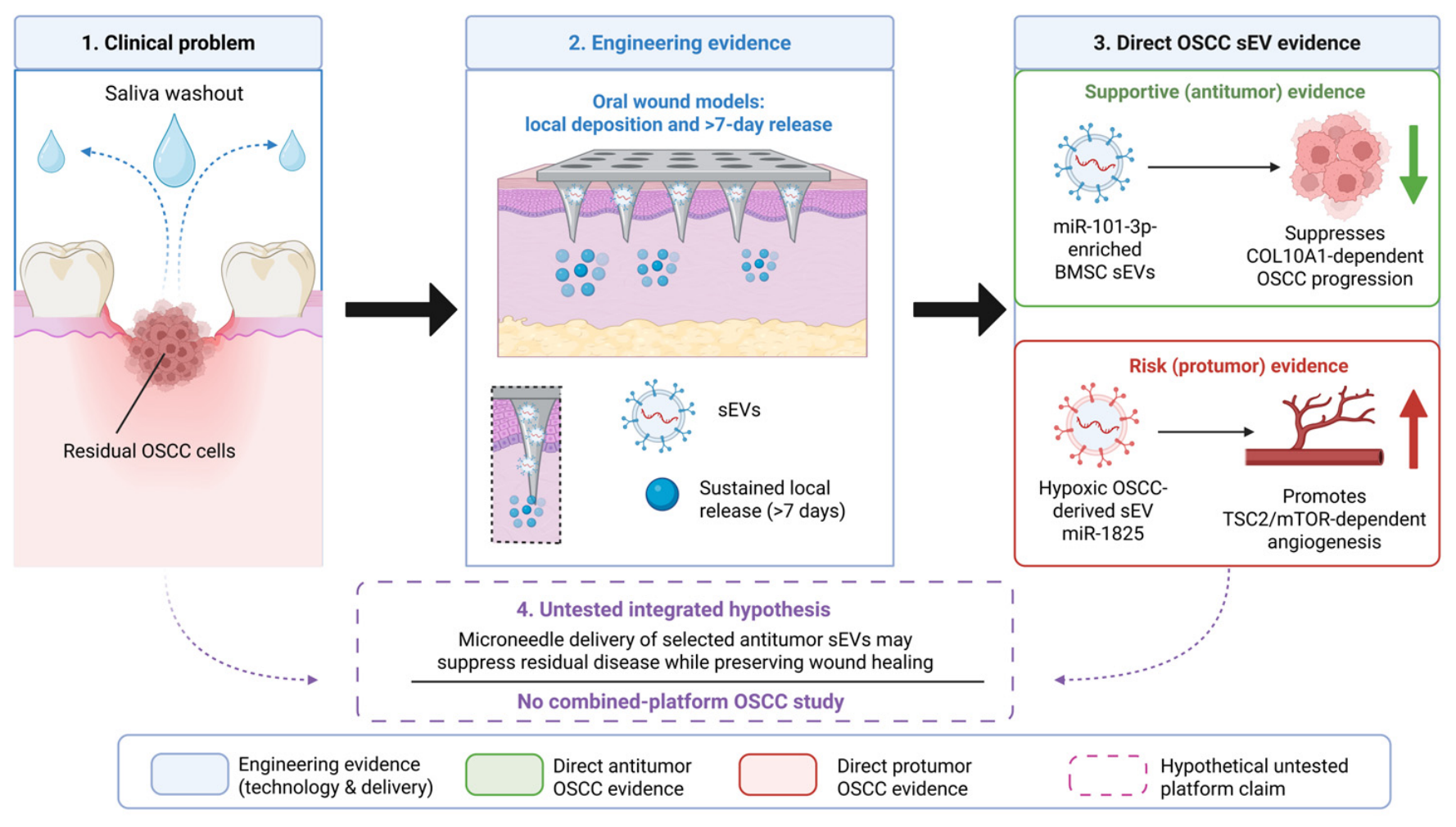
3.2. Proposed Application: Postoperative Residual-Disease Control

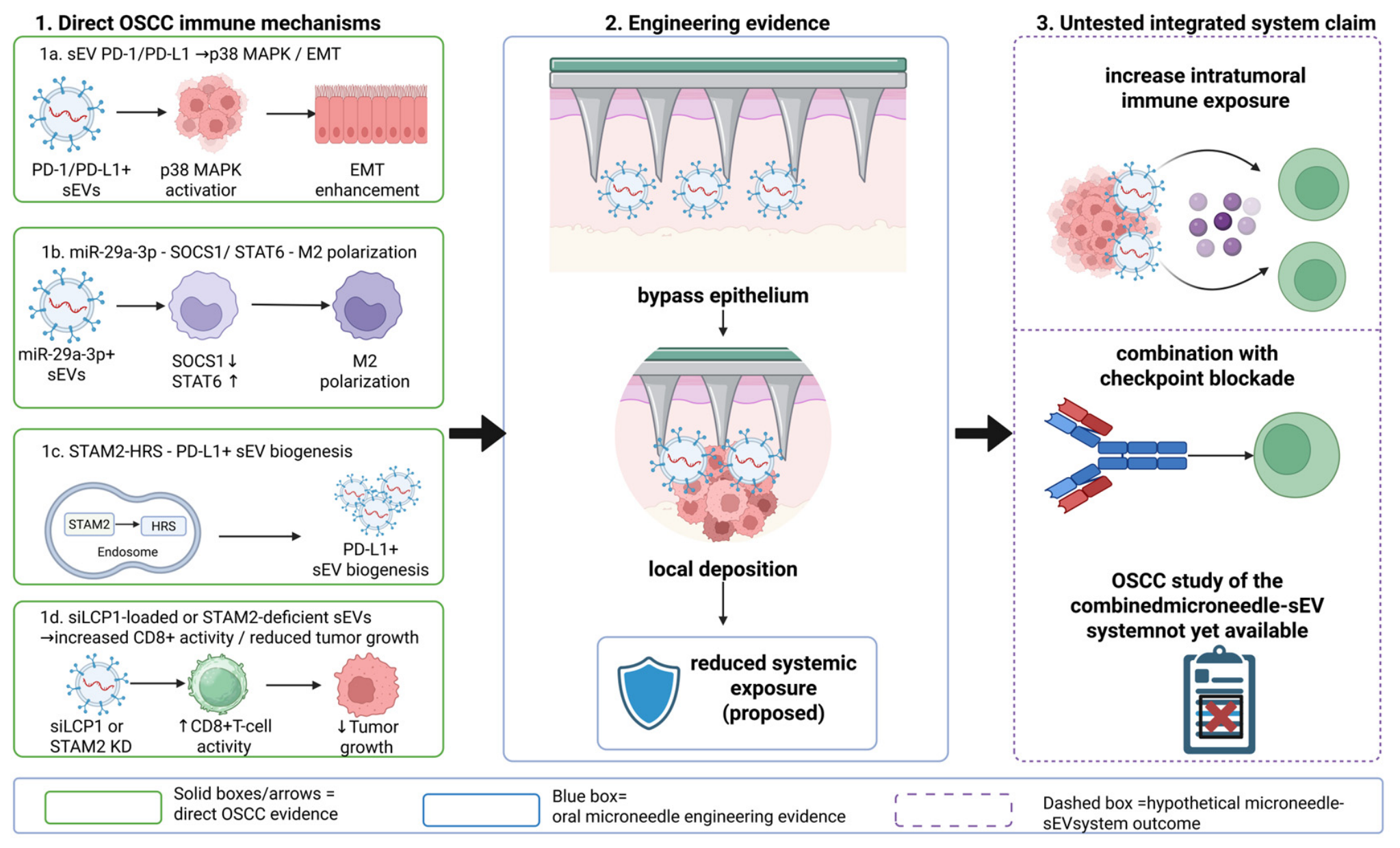
3.3. Proposed Application: Local Immunomodulation and Checkpoint Combination

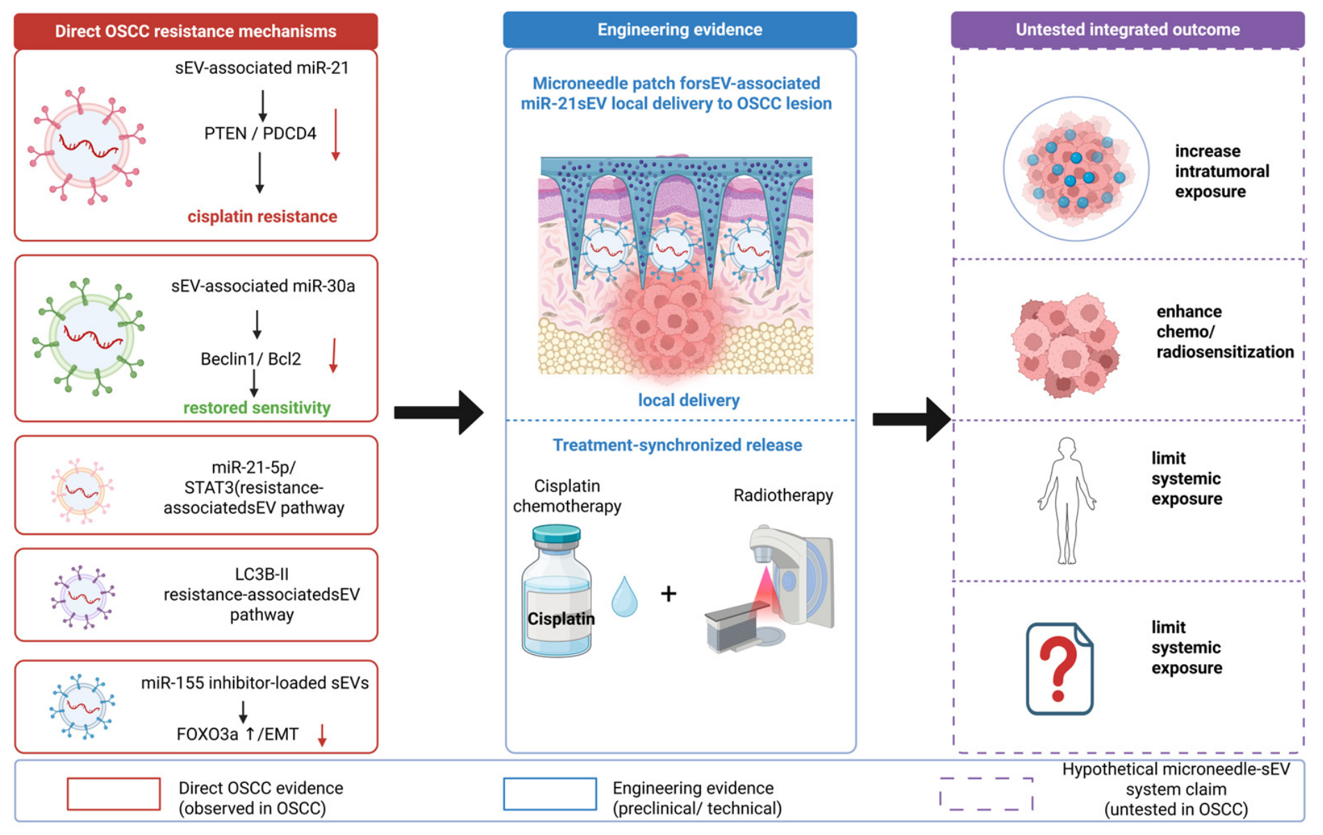
3.4. Proposed Application: Chemoradiotherapy Sensitization

3.5. Cross-Cutting Evidence Boundary and Decision Criteria
| Part A: OSCC-Specific Mechanistic and sEV Evidence (Level 3; No Combined Microneedle Delivery Tested) | |||||
| Context | Reported sEV Finding | MN Role/Boundary | Refs. | Level | Next Test |
| OSCC mechanism: angiogenesis/metastasis | miR-1825 → TSC2/mTOR angiogenesis; sEV PD-1 → PD-L1/p38-MAPK EMT and nodal-metastasis association | OSCC mechanism only; MN delivery not tested | [82,83] | Level 3 | Exclude proangiogenic/prometastatic activity |
| OSCC mechanism: immune escape | miR-29a-3p → SOCS1/STAT6 M2 polarization; mtDNA/PD-L1 → Treg activity; STAM2–HRS → PD-L1-positive sEV biogenesis | OSCC mechanism only; non-OSCC MN data inform engineering | [84,85,89] | Level 3 | Confirm target engagement and immune-cell effects in OSCC |
| OSCC mechanism: cisplatin response | miR-21 → PTEN/PDCD4 suppression and resistance; miR-30a → Beclin1/Bcl2 suppression and resensitization | OSCC mechanism only; MN contribution untested | [86,87] | Level 3 | Confirm cargo delivery, pathway modulation, and causal rescue |
| Postoperative residual disease | Menstrual MSC-derived sEVs: anti-angiogenic and tumor-suppressive effects | Oral studies support wet adhesion and prolonged local delivery | [97,103,104] | Level 3 | Test residual-tumor control + wound safety |
| Postoperative residual disease | miR-101-3p-enriched BMSC sEVs → COL10A1-dependent suppression of proliferation/invasion | Controlled surgical-bed release proposed | [98,103,104] | Level 3 | Verify post-fabrication cargo potency |
| Local immunomodulation | EBI3-displaying, siLCP1-loaded sEVs → lower LCP1 and OSCC progression | Localized immune delivery proposed | [99] | Level 3 | Compare free sEVs, the microneedle–sEV system, and checkpoint blockade |
| Local immunomodulation | STAM2-deficient OSCC sEVs → more proliferating/GzmB-positive CD8+ T cells | Distinct product; local MN delivery untested | [89] | Level 3 | Define source, potency, and immune-cell uptake |
| Chemoradiotherapy sensitization | Ovatodiolide-associated sEV modulation → lower miR-21-5p/STAT3 signaling | Treatment-synchronized delivery proposed | [90] | Level 3 | Test added sensitization vs. standard therapy |
| Chemoradiotherapy sensitization | miR-155 inhibitor-loaded sEVs → FOXO3a↑, EMT↓, cisplatin sensitivity↑ | OSCC spheroid/xenograft evidence; MN delivery untested | [92] | Level 3 | Verify potency after loading and synchronized release |
| Part B: Oral-Delivery and Cross-Disease Engineering Evidence (Levels 2, 4, and 5; Not Used as OSCC Mechanistic Evidence) | |||||
| Context | Reported sEV finding | MN role/boundary | Refs. | Level | Next test |
| Postoperative delivery | Oral-ulcer microneedle–sEV systems → improved healing and reduced inflammation | Oral adhesion/local delivery; not residual OSCC | [103,104] | Level 2 | Guide delivery and wound-safety design |
| Local immunomodulation | Engineered sEVs in superficial non-oral tumors → enhanced local antitumor immunity | Microneedle–sEV system evidence outside OSCC | [28,120] | Level 5 | Use as engineering evidence only |
| Chemoradiotherapy sensitization | Engineered sEVs in non-oral tumors → improved local delivery/antitumor response | Microneedle–sEV system delivery outside OSCC | [28,124] | Level 5 | Validate in orthotopic OSCC |
| Cross-cutting oral delivery | MN without sEV cargo → oral-mucosal penetration | Device-only oral evidence | [26,128] | Level 4 | Define site-specific insertion and safety |
4. Candidate Technical Design of the Proposed Microneedle–sEV System
4.1. Matrix Materials and Microneedle Structure
4.2. sEV Loading and Bioactivity Preservation
4.3. Release Kinetics and Targeting
4.4. Preclinical In Vitro and In Vivo Evaluation
- Preserve sEV identity and mechanism-linked potency after fabrication.
- Deliver a reproducible local dose with adequate retention and limited systemic escape.
- Show added value over free sEVs, blank microneedles, and an established local comparator.
- Meet context-specific efficacy and oral-safety requirements.
5. Consolidated Preclinical Development Barriers

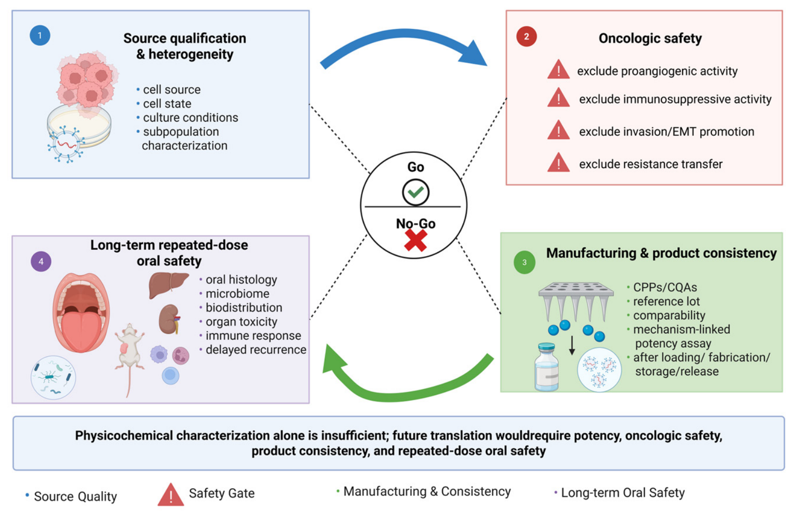
5.1. sEV Heterogeneity and Oncologic Safety
5.2. In Vivo Fate and Repeated-Dose Oral Safety
5.3. Manufacturing, Standardization, and Scale-Up
- Product safety: sterility, endotoxin, and residual process reagents.
- Device performance: loading/dose uniformity, release, strength, insertion, and oral stability.
- Biological potency: a mechanism-linked assay on the released sEV product.
5.4. Integration, Usability, and Future Translation
| Bottleneck | Main Issue | Evidence Status | Priority Action | Refs. |
|---|---|---|---|---|
| Product heterogeneity |
|
|
| [19,158,159] |
| Oncologic safety |
|
|
| [82,84,85,86,88,89] |
| Mechanistic evidence |
|
|
| [181,182,183] |
| Quality control/consistency |
|
|
| [19,158,159,160,161,184] |
| Long-term/repeated-dose safety |
|
|
| [82,84,85,86,88,89,136,139,162,163,164] |
| Future translation |
|
|
| [66,199] |
6. Conclusions and Outlook
- Source: Qualify candidate sEV sources and exclude OSCC-relevant pro-tumor activity.
- Product: Define critical process/quality attributes and mechanism-linked release assays.
- Safety: Quantify biodistribution and repeated-dose oral safety in relevant models.
- Performance: Compare the proposed microneedle–sEV system with free sEVs, blank microneedles, established local delivery, and standard therapy.
Author Contributions
Funding
Institutional Review Board Statement
Informed Consent Statement
Data Availability Statement
Conflicts of Interest
References
- Akram, K.; Aslam, M.; Waheed, T.; Ayesha, N.; Alamri, F.S.; Mirdad, A.R.; Rehman, A. Deep visual detection system for oral squamous cell carcinoma. Sci. Rep. 2026, 16, 4219. [Google Scholar] [CrossRef] [Scilit]
- Bray, F.; Laversanne, M.; Sung, H.; Ferlay, J.; Siegel, R.L.; Soerjomataram, I.; Jemal, A. Global cancer statistics 2022: GLOBOCAN estimates of incidence and mortality worldwide for 36 cancers in 185 countries. CA Cancer J. Clin. 2024, 74, 229–263. [Google Scholar] [CrossRef] [Scilit] [PubMed]
- He, Y.; Gong, X.; Tao, H.; He, Y.; Cao, Y.; Zhang, Y.; Tang, X.; Liu, J. The effect of postoperative adjuvant chemotherapy on survival outcomes in patients with early stage oral squamous cell carcinoma. Sci. Rep. 2025, 15, 27157. [Google Scholar] [CrossRef] [Scilit]
- Struckmeier, A.K.; Buchbender, M.; Lutz, R.; Kesting, M. Improved recurrence rates and progression-free survival in primarily surgically treated oral squamous cell carcinoma—Results from a German tertiary medical center. Clin. Oral Investig. 2024, 28, 262. [Google Scholar] [CrossRef] [Scilit]
- Ren, Z.H.; Liu, K.; Chen, Y.; Yang, Z.M.; Wu, K.; Wu, H.J. Prospective observational study of surgery alone for locally advanced oral squamous cell carcinoma: A real-world study. BMC Oral Health 2024, 24, 156. [Google Scholar] [CrossRef] [Scilit]
- Wu, J.; Chen, H.; Liu, Y.; Yang, R.; An, N. The global, regional, and national burden of oral cancer, 1990-2021: A systematic analysis for the Global Burden of Disease Study 2021. J. Cancer Res. Clin. Oncol. 2025, 151, 53. [Google Scholar] [CrossRef] [Scilit]
- Mohamad, I.; Glaun, M.D.E.; Prabhash, K.; Busheri, A.; Lai, S.Y.; Noronha, V.; Hosni, A. Current Treatment Strategies and Risk Stratification for Oral Carcinoma. Am. Soc. Clin. Oncol. Educ. Book 2023, 43, e389810. [Google Scholar] [CrossRef] [Scilit]
- Nauta, I.H.; Peferoen, L.A.N.; Brakenhoff, R.H.; Leemans, C.R.; Bloemena, E. Architectural dysplasia in surgical margins and the risk of local relapse in oral cancer. J. Oral Pathol. Med. 2024, 53, 544–550. [Google Scholar] [CrossRef] [Scilit]
- Szewczyk, M.; Pazdrowski, J.; Pieńkowski, P.; Wojtera, B.; Więckowska, B.; Golusiński, P.; Golusiński, W. A Matter of Margins in Oral Cancer-How Close Is Enough? Cancers 2024, 16, 1488. [Google Scholar] [CrossRef] [Scilit]
- Cheng, Y.; Li, S.; Gao, L.; Zhi, K.; Ren, W. The Molecular Basis and Therapeutic Aspects of Cisplatin Resistance in Oral Squamous Cell Carcinoma. Front. Oncol. 2021, 11, 761379. [Google Scholar] [CrossRef] [Scilit]
- Zhang, C.; Xu, C.; Gao, X.; Yao, Q. Platinum-based drugs for cancer therapy and anti-tumor strategies. Theranostics 2022, 12, 2115–2132. [Google Scholar] [CrossRef] [Scilit]
- Hartmann, S.; Kriegebaum, U.; Küchler, N.; Lessner, G.; Brands, R.C.; Linz, C.; Schneider, T.; Kübler, A.C.; Müller-Richter, U.D. Efficacy of cetuximab and panitumumab in oral squamous cell carcinoma cell lines: Prognostic value of MAGE-A subgroups for treatment success. J. Cranio-Maxillo-Facial Surg. 2013, 41, 623–629. [Google Scholar] [CrossRef] [Scilit]
- Yang, H.; Mo, C.; Xun, Y.; Liu, L.G.; Li, W.; Guan, J.; Liu, J.; Wu, J.; Yang, A.; Zheng, S.; et al. Combination of cetuximab with met inhibitor in control of cetuximab-resistant oral squamous cell carcinoma. Am. J. Transl. Res. 2019, 11, 2370–2381. [Google Scholar]
- Lee, J.; Roh, J.L. Ferroptosis in Oral Cancer: Mechanistic Insights and Clinical Prospects. Cells 2025, 14, 1685. [Google Scholar] [CrossRef] [Scilit]
- Wang, Y.; Chen, K.; Tian, G.; Hou, C.; Huang, Y.; Song, F.; Zhang, M.; Hou, J. PLS3 Is a Prognostic Biomarker and Correlates with Immune Infiltrates in Head and Neck Squamous Cell Carcinoma. Cancers 2025, 17, 3882. [Google Scholar] [CrossRef] [Scilit]
- Liu, H.; Huang, Y.; Huang, M.; Huang, Z.; Wang, Q.; Qing, L.; Li, L.; Xu, S.; Jia, B. Current Status, Opportunities, and Challenges of Exosomes in Oral Cancer Diagnosis and Treatment. Int. J. Nanomed. 2022, 17, 2679–2705. [Google Scholar] [CrossRef] [Scilit]
- Lu, Y.; Zheng, Z.; Yuan, Y.; Pathak, J.L.; Yang, X.; Wang, L.; Ye, Z.; Cho, W.C.; Zeng, M.; Wu, L. The Emerging Role of Exosomes in Oral Squamous Cell Carcinoma. Front. Cell Dev. Biol. 2021, 9, 628103. [Google Scholar] [CrossRef] [Scilit]
- Li, M.; Yin, S.; Xu, A.; Kang, L.; Ma, Z.; Liu, F.; Yang, T.; Sun, P.; Tang, Y. Synergistic Phototherapy-Molecular Targeted Therapy Combined with Tumor Exosome Nanoparticles for Oral Squamous Cell Carcinoma Treatment. Pharmaceutics 2023, 16, 33. [Google Scholar] [CrossRef] [Scilit]
- Welsh, J.A.; Goberdhan, D.C.; O’Driscoll, L.; Buzas, E.I.; Blenkiron, C.; Bussolati, B.; Cai, H.; Di Vizio, D.; Driedonks, T.A.; Erdbrügger, U.; et al. Minimal information for studies of extracellular vesicles (MISEV2023): From basic to advanced approaches. J. Extracell. Vesicles 2024, 13, e12404. [Google Scholar] [CrossRef] [Scilit] [PubMed]
- Dubashynskaya, N.V.; Petrova, V.A.; Skorik, Y.A. Biopolymer Drug Delivery Systems for Oromucosal Application: Recent Trends in Pharmaceutical R&D. Int. J. Mol. Sci. 2024, 25, 5359. [Google Scholar] [CrossRef] [Scilit]
- Zhai, T.; Tang, W.; Liu, P.; Liang, Y.; Ma, Z.; Fan, L. Therapeutic value of mesenchymal stem cell-derived extracellular vesicles in hypertrophic and keloid scars: A systematic review and meta-analysis. Front. Cell Dev. Biol. 2026, 14, 1739106. [Google Scholar] [CrossRef] [Scilit]
- Liu, M.; Wang, K.; Zeng, C.; Jia, Y.; Wang, J.; Taledaohan, A.; Wang, Y.; Guan, X. Gelatin/Lignin Hydrogel Loaded with Mesenchymal Stem Cell-Derived Exosomes Enriched in Microrna-185 Inhibits Progression of Oral Cancer. Pharmaceutics 2026, 18, 363. [Google Scholar] [CrossRef] [Scilit]
- Xu, C.; Chen, L.; Liu, G.; Xu, J.; Lv, W.; Gao, X.; Xu, P.; Tang, M.; Wang, Y.; Zhao, X.; et al. Tailoring an intravenously injectable oncolytic virus for augmenting radiotherapy. Cell Rep. Med. 2025, 6, 102078. [Google Scholar] [CrossRef] [Scilit]
- Meng, Y.; Li, X.J.; Li, Y.; Zhang, T.Y.; Liu, D.; Wu, Y.Q.; Hou, F.F.; Ye, L.; Wu, C.J.; Feng, X.D.; et al. Novel Double-Layer Dissolving Microneedles for Transmucosal Sequential Delivery of Multiple Drugs in the Treatment of Oral Mucosa Diseases. ACS Appl. Mater. Interfaces 2023, 15, 13892–13906. [Google Scholar] [CrossRef] [Scilit]
- Shi, S.; Yu, Z.L.; Jia, J. The Roles of Exosomes in the Diagnose, Development and Therapeutic Resistance of Oral Squamous Cell Carcinoma. Int. J. Mol. Sci. 2023, 24, 1968. [Google Scholar] [CrossRef] [Scilit]
- Caffarel-Salvador, E.; Kim, S.; Soares, V.; Tian, R.Y.; Stern, S.R.; Minahan, D.; Yona, R.; Lu, X.; Zakaria, F.R.; Collins, J.; et al. A microneedle platform for buccal macromolecule delivery. Sci. Adv. 2021, 7, eabe2620. [Google Scholar] [CrossRef] [Scilit]
- Zheng, L.; Sun, J.; Wang, L.; Ding, Z.; Tang, Y.; Dai, M.; Tang, H. Construction and applications of exosome-microneedle integrated systems. Int. J. Pharm. X 2025, 10, 100360. [Google Scholar] [CrossRef] [Scilit]
- Chen, Z.; Hu, F.; Xiang, J.; Zhou, X.; Wu, B.; Fan, B.; Tang, H.; Liu, B.; Chen, L. Mesoporous Microneedles Enabled Localized Controllable Delivery of Stimulator of Interferon Gene Agonist Nanoexosomes for FLASH Radioimmunotherapy against Breast Cancer. ACS Appl. Mater. Interfaces 2024, 16, 58180–58190. [Google Scholar] [CrossRef] [Scilit]
- Qi, M.; Hong, Y.; Liu, Y.; Wang, H.; Xiong, Y.; Tang, Y.; Ma, M.; Gao, Z.; Zhang, D. Carboxymethyl chitosan-based injectable hydrogel immobilizing single-atom nanozymes for localized ROS amplification and ferroptosis-enhanced postoperative oral cancer therapy. Carbohydr. Polym. 2026, 373, 124668. [Google Scholar] [CrossRef] [Scilit]
- Li, W.; Zhang, P.; Fu, H.; Yan, S.; Zhu, D. Targeted therapeutic strategy for oral squamous carcinoma using celecoxib-loaded GABA/wheat gluten-alginate nanocarrier hydrogel with glutathione down-regulation and enhanced CCND2-mediated apoptosis. Int. J. Biol. Macromol. 2025, 303, 140679. [Google Scholar] [CrossRef] [Scilit]
- Shi, C.; Liu, B.; Feng, L.; Wei, B.; Yu, S.; Chen, P. Design and evaluation of multi-responsive PNIPAM-g-Dextran-sodium alginate blended smart hydrogel for enhanced chemo-photodynamic therapy against oral squamous cell carcinoma. Colloids Surf. B Biointerfaces 2026, 259, 115293. [Google Scholar] [CrossRef] [Scilit]
- Wu, Y.; Chen, F.; Huang, N.; Li, J.; Wu, C.; Tan, B.; Liu, Y.; Li, L.; Yang, C.; Shao, D.; et al. Near-infrared light-responsive hybrid hydrogels for the synergistic chemo-photothermal therapy of oral cancer. Nanoscale 2021, 13, 17168–17182. [Google Scholar] [CrossRef] [Scilit]
- Ghahramani, Y.; Razavi, N.; Tavakkol, O.; Gholami, M.; Khandani, F.; Khaksar, E.; Khaksar, E.; Derakhshan, M.; Yari Kalashgrani, M.; Karganroudi, S.S.; et al. Dual smart monitoring and predictive non-destructive evaluation: A review of advanced hydrogel and stem cell-based strategies for oral cancer theragnostic applications. Int. J. Pharm. 2026, 687, 126398. [Google Scholar] [CrossRef] [Scilit]
- Li, X.; Xie, X.; Wu, Y.; Zhang, Z.; Liao, J. Microneedles: Structure, classification, and application in oral cancer theranostics. Drug Deliv. Transl. Res. 2023, 13, 2195–2212. [Google Scholar] [CrossRef] [Scilit]
- Ferreira, L.E.N.; Franz-Montan, M.; Benso, B.; Gill, H.S. Microneedles for oral mucosal delivery—Current trends and perspective on future directions. Expert Opin. Drug Deliv. 2023, 20, 1251–1265. [Google Scholar] [CrossRef] [Scilit]
- Liu, J.; Zheng, W.; Sun, H.; González, F.E.; Zhou, D.; Xu, X. Environment-adaptive microneedle design strategies for precision oral disease treatment: From material innovation to clinical translation. J. Control. Release 2025, 388, 114402. [Google Scholar] [CrossRef] [Scilit]
- Habib, L.; Alyan, M.; Ghantous, Y.; Shklover, J.; Shainsky, J.; Abu El-Naaj, I.; Bianco-Peled, H.; Schroeder, A. A mucoadhesive patch loaded with freeze-dried liposomes for the local treatment of oral tumors. Drug Deliv. Transl. Res. 2023, 13, 1228–1245. [Google Scholar] [CrossRef] [Scilit]
- Goldberg, M.; Manzi, A.; Birdi, A.; Laporte, B.; Conway, P.; Cantin, S.; Mishra, V.; Singh, A.; Pearson, A.T.; Goldberg, E.R.; et al. A nanoengineered topical transmucosal cisplatin delivery system induces anti-tumor response in animal models and patients with oral cancer. Nat. Commun. 2022, 13, 4829. [Google Scholar] [CrossRef] [Scilit]
- Chen, Y.W.; He, A.C.; Huang, T.Y.; Lai, D.H.; Wang, Y.P.; Liu, W.W.; Kuo, W.T.; Hou, H.H.; Cheng, S.J.; Lee, C.Y.; et al. Iontophoresis-Enhanced Buccal Delivery of Cisplatin-Encapsulated Chitosan Nanoparticles for Treating Oral Cancer in a Mouse Model. Int. J. Nanomed. 2024, 19, 10435–10453. [Google Scholar] [CrossRef] [Scilit]
- Zhong, J.; He, G.; Ma, X.; Ye, J.; Tao, Z.Y.; Li, Z.; Zhang, F.; Feng, P.; Wang, Y.; Lan, X.; et al. Triterpene-Based Prodrug for Self-Boosted Drug Release and Targeted Oral Squamous Cell Carcinoma Chemotherapy. ACS Appl. Mater. Interfaces 2024, 16, 41960–41972. [Google Scholar] [CrossRef] [Scilit]
- Wang, H.; Yu, Y.; Zhou, W.; Miao, B.; Zhang, Y.; Zhu, J.; Lin, Z.; Han, J.; Xu, X.; Zhang, F.; et al. Dual-loaded homotypic exosomes with endogenous IR808 and pH-responsive DOX drive sequenced chemo-PDT and convert “cold” OSCC tumors “hot”. J. Nanobiotechnol. 2026, 24, 456. [Google Scholar] [CrossRef] [Scilit]
- Tsai, Y.F.; Chan, L.P.; Chen, Y.K.; Su, C.W.; Hsu, C.W.; Wang, Y.Y.; Yuan, S.F. RAD51 is a poor prognostic marker and a potential therapeutic target for oral squamous cell carcinoma. Cancer Cell Int. 2023, 23, 231. [Google Scholar] [CrossRef] [Scilit]
- Wang, C.; Li, Y.; Shen, X.; Xiao, S.; Du, H.; Zhao, Q.; Wang, Y.; Zhang, Q.; Wen, S. Localized chemo-immunotherapy for HNSCC via CpG-synergized doxorubicin release from injectable TPGS/Na2CO3 hydrogel remodeling tumor immunity. Biomater. Adv. 2026, 180, 214588. [Google Scholar] [CrossRef] [Scilit]
- Mousavi, S.M.; Abbasifard, M.; Abbasi, R.; Tivay, S.; Hosseini, H.; Taghipoor, M.; Kalashgrani, M.Y.; Aljabri, M.D.; Rahman, M.M.; Chiang, W.H.; et al. Smart nano exosomes as precision drug carriers: A new frontier in targeted treatment of oral cancer. Int. J. Pharm. 2025, 682, 125923. [Google Scholar] [CrossRef] [Scilit]
- Quadri, Z.; Elsherbini, A.; Bieberich, E. Extracellular vesicles in pharmacology: Novel approaches in diagnostics and therapy. Pharmacol. Res. 2022, 175, 105980. [Google Scholar] [CrossRef] [Scilit]
- Senevirathna, K.; Jayawickrama, S.M.; Jayasinghe, Y.A.; Prabani, K.I.P.; Akshala, K.; Pradeep, R.G.G.R.; Damayanthi, H.D.W.T.; Hettiarachchi, K.; Dorji, T.; Lucero-Prisno, D.E.; et al. Nanoplatforms: The future of oral cancer treatment. Health Sci. Rep. 2023, 6, e1471. [Google Scholar] [CrossRef] [Scilit]
- Tutty, M.A.; Vella, G.; Vennemann, A.; Wiemann, M.; Prina-Mello, A. Evaluating nanobiomaterial-induced DNA strand breaks using the alkaline comet assay. Drug Deliv. Transl. Res. 2022, 12, 2243–2258. [Google Scholar] [CrossRef] [Scilit]
- Janivarad, T.; Kumbar, V.M.; Marakatti, R.; Kotabagi, S.; Kambi, M.; Lolsure, K.; Paranjape, R. Targeting oral cancer with MicroRNA-based therapeutics: The role of tumor-suppressor miRNAs and exosome delivery. Arch. Oral Biol. 2026, 186, 106562. [Google Scholar] [CrossRef] [Scilit]
- Baena, J.C.; Cabrera-Salcedo, S.C.; Carrera Suárez, Y.; Biancha-Vasco, J.M.; Rios-Serna, L.J.; García-Mantilla, M.D.; Estrada-Schweineberg, M.; Victoria Hincapie, J.S.; Toro-Pedroza, A.; Garcia-Robledo, J.E.; et al. The avatar principle: Exosomal dynamics guiding tumor adaptation and next-generation therapeutic strategies. J. Nanobiotechnol. 2026, 24, 159. [Google Scholar] [CrossRef] [Scilit]
- Rayamajhi, S.; Nguyen, T.D.T.; Marasini, R.; Aryal, S. Macrophage-derived exosome-mimetic hybrid vesicles for tumor targeted drug delivery. Acta Biomater. 2019, 94, 482–494. [Google Scholar] [CrossRef] [Scilit]
- Elkhoury, K.; Koçak, P.; Kang, A.; Arab-Tehrany, E.; Ellis Ward, J.; Shin, S.R. Engineering Smart Targeting Nanovesicles and Their Combination with Hydrogels for Controlled Drug Delivery. Pharmaceutics 2020, 12, 849. [Google Scholar] [CrossRef] [Scilit]
- Zhang, L.; Yang, Y.; Mo, L.; Che, F.; Li, B.; Lei, H.; Sun, Y.; Zhou, Y.; Zhang, C.; Guo, Y. Aptamer-targeted hybrid nanoparticles based on human exosomes and LXR agonist-loaded liposomes for enhanced anti-AML therapy. Nucleos. Nucleot. Nucl. Acids 2026. Advance online publication. [Google Scholar] [CrossRef] [Scilit]
- van Oorschot, H.D.; Hardillo, J.A.; van Es, R.J.; van den Broek, G.B.; Takes, R.P.; Halmos, G.B.; de Jel, D.V.C.; Dirven, R.; Lacko, M.; Vaassen, L.A.A.; et al. Hospital-level variation in resection margins after oral cavity cancer surgery and associated survival outcomes. Br. J. Oral Maxillofac. Surg. 2025, 63, 664–671. [Google Scholar] [CrossRef] [Scilit]
- Rubert, A.; Bagan, L.; Proaño, A.; Sarrion, G.; Bagan, J. Impact of surgical margins on recurrence and survival rate in patients with oral squamous cell carcinoma: A systematic review and meta-analysis. Med. Oral Patol. Oral Cir. Bucal 2026, 31, e307–e316. [Google Scholar] [CrossRef] [Scilit]
- Evans, M.; Bonomo, P.; Chan, P.C.; Chua, M.L.K.; Eriksen, J.G.; Hunter, K.; Jones, T.M.; Laskar, S.G.; Maroldi, R.; O’Sullivan, B.; et al. Post-operative radiotherapy for oral cavity squamous cell carcinoma: Review of the data guiding the selection and the delineation of post-operative target volumes. Radiother. Oncol. 2025, 207, 110880. [Google Scholar] [CrossRef] [Scilit]
- Cooper, J.S.; Pajak, T.F.; Forastiere, A.A.; Jacobs, J.; Campbell, B.H.; Saxman, S.B.; Kish, J.A.; Kim, H.E.; Cmelak, A.J.; Rotman, M.; et al. Postoperative concurrent radiotherapy and chemotherapy for high-risk squamous-cell carcinoma of the head and neck. N. Engl. J. Med. 2004, 350, 1937–1944. [Google Scholar] [CrossRef] [Scilit]
- Ye, A.Y.; Hay, J.H.; Laskin, J.J.; Wu, J.S.; Ho, C.C. Toxicity and outcomes in combined modality treatment of head and neck squamous cell carcinoma: Cisplatin versus cetuximab. J. Cancer Res. Ther. 2013, 9, 607–612. [Google Scholar] [CrossRef] [Scilit]
- Ang, K.K.; Zhang, Q.; Rosenthal, D.I.; Nguyen-Tan, P.F.; Sherman, E.J.; Weber, R.S.; Galvin, J.M.; Bonner, J.A.; Harris, J.; El-Naggar, A.K.; et al. Randomized phase III trial of concurrent accelerated radiation plus cisplatin with or without cetuximab for stage III to IV head and neck carcinoma: RTOG 0522. J. Clin. Oncol. 2014, 32, 2940–2950. [Google Scholar] [CrossRef] [Scilit]
- Baronzio, G.; Parmar, G.; Baronzio, M. Overview of Methods for Overcoming Hindrance to Drug Delivery to Tumors, with Special Attention to Tumor Interstitial Fluid. Front. Oncol. 2015, 5, 165. [Google Scholar] [CrossRef] [Scilit]
- Pulito, C.; Brandi, R.; Sestito, R.; Orlandi, G.; Sanguineti, G.; Blandino, G.; Strano, S. Circulating miR-18b-3p is a novel biomarker predicting chemo-radiotherapy induced oral mucositis in head and neck cancer. Signal Transduct. Target. Ther. 2025, 10, 380. [Google Scholar] [CrossRef] [Scilit]
- Bhide, S.A.; Ahmed, M.; Barbachano, Y.; Newbold, K.; Harrington, K.J.; Nutting, C.M. Sequential induction chemotherapy followed by radical chemo-radiation in the treatment of locoregionally advanced head-and-neck cancer. Br. J. Cancer 2008, 99, 57–62. [Google Scholar] [CrossRef] [Scilit]
- Burtness, B.; Harrington, K.J.; Greil, R.; Soulières, D.; Tahara, M.; de Castro, G.; Psyrri, A., Jr.; Basté, N.; Neupane, P.; Bratland, Å.; et al. Pembrolizumab alone or with chemotherapy versus cetuximab with chemotherapy for recurrent or metastatic squamous cell carcinoma of the head and neck (KEYNOTE-048): A randomised, open-label, phase 3 study. Lancet 2019, 394, 1915–1928. [Google Scholar] [CrossRef] [Scilit] [PubMed]
- Li, F.; Han, Y.; Ou, F.; Deng, L.; Li, H.; Yu, X.; Yi, Y.; Ma, R.; Wu, Z.; You, Z.; et al. Spatial heterogeneity of MDSCs mediated by ANXA1-FPRs signaling drives immune suppression in OSCC progression. Nat. Commun. 2026, 17, 2535. [Google Scholar] [CrossRef] [Scilit]
- Xue, N.; Wang, Y.; Wang, Z.; Zeng, X.; Wang, J.; Zhang, X. Trends in immunotherapy for oral squamous cell carcinoma. Cell. Oncol. 2025, 48, 1159–1179. [Google Scholar] [CrossRef] [Scilit]
- Xiang, Z.; Wei, X.; Zhang, Z.; Tang, Y.; Chen, L.; Tan, C.; Zeng, Y.; Wang, J.; Zhao, G.; Dai, Z.; et al. Efficacy, safety and single-cell analysis of neoadjuvant immunochemotherapy in locally advanced oral squamous cell carcinoma: A phase II trial. Nat. Commun. 2025, 16, 3968. [Google Scholar] [CrossRef] [Scilit]
- Herrada Céspedes, A.; Reyes, M.; Morales, J.O. Advanced drug delivery systems for oral squamous cell carcinoma: A comprehensive review of nanotechnology-based and other innovative approaches. Front. Drug Deliv. 2025, 5, 1596964. [Google Scholar] [CrossRef] [Scilit]
- Elsaady, S.A.; Aboushelib, M.N.; Al-Wakeel, E.; Badawi, M.F. A novel intra-tumoral drug delivery carrier for treatment of oral squamous cell carcinoma. Sci. Rep. 2023, 13, 11984. [Google Scholar] [CrossRef] [Scilit]
- Hussein, N.I.; Molina, A.H.; Sunga, G.M.; Amit, M.; Lei, Y.L.; Zhao, X.; Hartgerink, J.D.; Sikora, A.G.; Young, S. Localized intratumoral delivery of immunomodulators for oral cancer and oral potentially malignant disorders. Oral Oncol. 2024, 158, 106986. [Google Scholar] [CrossRef] [Scilit]
- Ma, T.; Wang, X.; Wang, Y.; Hao, Y.; Yang, X.; Yan, X.; Huang, Q.; Li, Z.; Cong, B.; Li, D. Curcumin analogue AC17-loaded dissolvable microneedles activate FOXO3 and enhance localized drug delivery for oral squamous cell carcinoma treatment. Int. J. Pharm. 2024, 661, 124385. [Google Scholar] [CrossRef] [Scilit]
- Malhotra, S.; Lijnse, T.; Cearbhaill, E.O.; Brayden, D.J. Devices to overcome the buccal mucosal barrier to administer therapeutic peptides. Adv. Drug Deliv. Rev. 2025, 220, 115572. [Google Scholar] [CrossRef] [Scilit]
- Campos, J.C.; Cunha, J.D.; Ferreira, D.C.; Reis, S.; Costa, P.J. Challenges in the local delivery of peptides and proteins for oral mucositis management. Eur. J. Pharm. Biopharm. 2018, 128, 131–146. [Google Scholar] [CrossRef] [Scilit]
- Ibrahim, Y.H.Y.; Regdon, G.; Hamedelniel, E.I., Jr.; Sovány, T. Review of recently used techniques and materials to improve the efficiency of orally administered proteins/peptides. Daru J. Fac. Pharm. Tehran Univ. Med. Sci. 2020, 28, 403–416. [Google Scholar] [CrossRef] [Scilit]
- Onishi, H.; Yumoto, K.; Sakata, O. Oral Biodisposition Study of Ritodrine after Its Buccal Administration in Rats. Curr. Drug Deliv. 2017, 14, 634–640. [Google Scholar] [CrossRef] [Scilit]
- Pérez-González, N.; Morales-Molina, J.A.; Calpena-Campmany, A.C.; Halbaut, L.; Rodríguez-Lagunas, M.J.; Bozal-de Febrer, N.; Souto, E.B.; Mallandrich, M.; Clares-Naveros, B. Caspofungin formulations for buccal and sublingual mucosae anti-fungal infections: Physicochemical characterization, rheological analysis, release and ex vivopermeability profiles. Pharm. Dev. Technol. 2024, 29, 1042–1063. [Google Scholar] [CrossRef] [Scilit]
- Dwivedi, P.; Kalra, P.; Zhou, H.; Alam, K.; Tsakalozou, E.; Al-Ghabeish, M.; Kelchen, M.; Pauletti, G.M. In Vitro Oral Cavity Permeability Assessment to Enable Simulation of Drug Absorption. Pharmaceutics 2025, 17, 924. [Google Scholar] [CrossRef] [Scilit]
- Wanasathop, A.; Patel, P.B.; Choi, H.A.; Li, S.K. Permeability of Buccal Mucosa. Pharmaceutics 2021, 13, 1814. [Google Scholar] [CrossRef] [Scilit]
- Majid, H.; Puzik, A.; Maier, T.; Merk, R.; Bartel, A.; Mueller, H.C.; Burckhardt, B.B. Formulation Development of Sublingual Cyclobenzaprine Tablets Empowered by Standardized and Physiologically Relevant Ex Vivo Permeation Studies. Pharmaceutics 2021, 13, 1409. [Google Scholar] [CrossRef] [Scilit]
- Liu, C.; Kou, Y.; Zhang, X.; Cheng, H.; Chen, X.; Mao, S. Strategies and industrial perspectives to improve oral absorption of biological macromolecules. Expert Opin. Drug Deliv. 2018, 15, 223–233. [Google Scholar] [CrossRef] [Scilit]
- Solanki, V.R.; Parmar, V.K. Recent Advances in Development of Buccal Formulations: From Small to Macromolecules. AAPS PharmSciTech 2025, 26, 156. [Google Scholar] [CrossRef] [Scilit]
- Bakhrushina, E.O.; Mikhel, I.B.; Buraya, L.M.; Moiseev, E.D.; Zubareva, I.M.; Belyatskaya, A.V.; Evzikov, G.Y.; Bondarenko, A.P.; Krasnyuk, I.I., Jr.; Krasnyuk, I.I. Implantation of In Situ Gelling Systems for the Delivery of Chemotherapeutic Agents. Gels 2024, 10, 44. [Google Scholar] [CrossRef] [Scilit]
- Špiljak, B.; Somogyi Škoc, M.; Rezić Meštrović, I.; Bašić, K.; Bando, I.; Šutej, I. Targeting the Oral Mucosa: Emerging Drug Delivery Platforms and the Therapeutic Potential of Glycosaminoglycans. Pharmaceutics 2025, 17, 1212. [Google Scholar] [CrossRef] [Scilit]
- Capik, O.; Gumus, R.; Karatas, O.F. Hypoxia-induced tumor exosomes promote angiogenesis through miR-1825/TSC2/mTOR axis in oral squamous cell carcinoma. Head Neck 2023, 45, 2259–2273. [Google Scholar] [CrossRef] [Scilit]
- Zhang, L.Z.; Yang, J.G.; Xia, H.F.; Huang, J.; Liu, H.M.; Wu, M.; Liu, B.; Wang, W.M.; Chen, G. PD-1 Carried on Small Extracellular Vesicles Leads to OSCC Metastasis. J. Dent. Res. 2023, 102, 795–805. [Google Scholar] [CrossRef] [Scilit]
- Cai, J.; Qiao, B.; Gao, N.; Lin, N.; He, W. Oral squamous cell carcinoma-derived exosomes promote M2 subtype macrophage polarization mediated by exosome-enclosed miR-29a-3p. Am. J. Physiol. Cell Physiol. 2019, 316, C731–C740. [Google Scholar] [CrossRef] [Scilit]
- Ko, H.H.; Peng, H.H.; Cheng, A.N.; Chou, H.Y.E.; Hou, H.H.; Kuo, W.T.; Liu, W.W.; Kuo, M.Y.P.; Lee, A.Y.L.; Cheng, S.J. Metastasis and immunosuppression promoted by mtDNA and PD-L1 in extracellular vesicles are reversed by WGP β-glucan in oral squamous cell carcinoma. Cancer Sci. 2023, 114, 3857–3872. [Google Scholar] [CrossRef] [Scilit]
- Liu, T.; Chen, G.; Sun, D.; Lei, M.; Li, Y.; Zhou, C.; Li, X.; Xue, W.; Wang, H.; Liu, C.; et al. Exosomes containing miR-21 transfer the characteristic of cisplatin resistance by targeting PTEN and PDCD4 in oral squamous cell carcinoma. Acta Biochim. Biophys. Sin. 2017, 49, 808–816. [Google Scholar] [CrossRef] [Scilit]
- Kulkarni, B.; Gondaliya, P.; Kirave, P.; Rawal, R.; Jain, A.; Garg, R.; Kalia, K. Exosome-mediated delivery of miR-30a sensitize cisplatin-resistant variant of oral squamous carcinoma cells via modulating Beclin1 and Bcl2. Oncotarget 2020, 11, 1832–1845. [Google Scholar] [CrossRef] [Scilit]
- Li, J.; Bao, Y.; Peng, S.; Jiang, C.; Zhu, L.; Zou, S.; Xu, J.; Li, Y. M2 Macrophages-Derived Exosomal miRNA-23a-3p Promotes the Progression of Oral Squamous Cell Carcinoma by Targeting PTEN. Curr. Issues Mol. Biol. 2023, 45, 4936–4947. [Google Scholar] [CrossRef] [Scilit]
- Wang, X.L.; Xia, H.F.; Chen, Z.K.; Wang, K.M.; Zhang, W.; Chen, G. STAM2-mediated fine-tuning of PD-L1 secretion via small extracellular vesicles in oral squamous cell carcinoma. Int. J. Biol. Macromol. 2025, 319, 145350. [Google Scholar] [CrossRef] [Scilit]
- Chen, J.H.; Wu, A.T.H.; Bamodu, O.A.; Yadav, V.K.; Chao, T.Y.; Tzeng, Y.M.; Mukhopadhyay, D.; Hsiao, M.; Lee, J.C. Ovatodiolide Suppresses Oral Cancer Malignancy by Down-Regulating Exosomal Mir-21/STAT3/β-Catenin Cargo and Preventing Oncogenic Transformation of Normal Gingival Fibroblasts. Cancers 2019, 12, 56. [Google Scholar] [CrossRef] [Scilit]
- Su, K.Y.; Khoo, X.H.; Paterson, I.C.; Goh, B.H.; Lee, W.L. Autophagy and extracellular vesicles mediate cisplatin resistance in oral squamous cell carcinoma with LC3B-II as a potential non-invasive biomarker. Sci. Rep. 2025, 15, 15945. [Google Scholar] [CrossRef] [Scilit]
- Sayyed, A.A.; Gondaliya, P.; Mali, M.; Pawar, A.; Bhat, P.; Khairnar, A.; Arya, N.; Kalia, K. MiR-155 Inhibitor-Laden Exosomes Reverse Resistance to Cisplatin in a 3D Tumor Spheroid and Xenograft Model of Oral Cancer. Mol. Pharm. 2021, 18, 3010–3025. [Google Scholar] [CrossRef] [Scilit]
- Słonimska, P.; Sachadyn, P.; Zieliński, J.; Skrzypski, M.; Pikuła, M. Chemotherapy-Mediated Complications of Wound Healing: An Understudied Side Effect. Adv. Wound Care 2024, 13, 187–199. [Google Scholar] [CrossRef] [Scilit]
- Ghanaati, S.; Udeabor, S.E.; Winter, A.; Sader, R.; Heselich, A. Cancer Recurrence in Operated Primary Oral Squamous Cell Carcinoma Patients Seems to Be Independent of the Currently Available Postoperative Therapeutic Approach: A Retrospective Clinical Study. Curr. Oncol. 2025, 32, 208. [Google Scholar] [CrossRef] [Scilit]
- Blatt, S.; Krüger, M.; Sagheb, K.; Barth, M.; Kämmerer, P.W.; Al-Nawas, B.; Sagheb, K. Tumor Recurrence and Follow-Up Intervals in Oral Squamous Cell Carcinoma. J. Clin. Med. 2022, 11, 7061. [Google Scholar] [CrossRef] [Scilit]
- Wang, Q.; Sun, J.; Jiang, H.; Yu, M. Emerging roles of extracellular vesicles in oral and maxillofacial areas. Int. J. Oral Sci. 2025, 17, 11. [Google Scholar] [CrossRef] [Scilit]
- Rosenberger, L.; Ezquer, M.; Lillo-Vera, F.; Pedraza, P.L.; Ortúzar, M.I.; González, P.L.; Figueroa-Valdés, A.I.; Cuenca, J.; Ezquer, F.; Khoury, M.; et al. Stem cell exosomes inhibit angiogenesis and tumor growth of oral squamous cell carcinoma. Sci. Rep. 2019, 9, 663. [Google Scholar] [CrossRef] [Scilit]
- Xie, C.; Du, L.Y.; Guo, F.; Li, X.; Cheng, B. Exosomes derived from microRNA-101-3p-overexpressing human bone marrow mesenchymal stem cells suppress oral cancer cell proliferation, invasion, and migration. Mol. Cell. Biochem. 2019, 458, 11–26. [Google Scholar] [CrossRef] [Scilit]
- Kase, Y.; Uzawa, K.; Wagai, S.; Yoshimura, S.; Yamamoto, J.I.; Toeda, Y.; Okubo, M.; Eizuka, K.; Ando, T.; Nobuchi, T.; et al. Engineered exosomes delivering specific tumor-suppressive RNAi attenuate oral cancer progression. Sci. Rep. 2021, 11, 5897. [Google Scholar] [CrossRef] [Scilit]
- Adel, N.; Stankovic, N.; Gindi, A.; Kolenda, J.; Mikhaylova, N.; Miladze, T.; Cervantes, G.; Ragaei, A.; Shawky, L.M.; Sabry, M.A. Elysee Exosome Versus Cellbooster Lift in Oral Wound Healing. Plast. Reconstr. Surg. Glob. Open 2025, 13, e7181. [Google Scholar] [CrossRef] [Scilit]
- Hao, M.; Duan, M.; Yang, Z.; Zhou, H.; Li, S.; Xiang, J.; Wu, H.; Liu, H.; Chang, L.; Wang, D.; et al. Engineered stem cell exosomes for oral and maxillofacial wound healing. Front. Bioeng. Biotechnol. 2022, 10, 1038261. [Google Scholar] [CrossRef] [Scilit]
- Raj, A.T.; Kheur, S.; Bhonde, R.; Mani, V.R.; Baeshen, H.A.; Patil, S. Assessing the effect of human dental pulp mesenchymal stem cell secretome on human oral, breast, and melanoma cancer cell lines. Saudi J. Biol. Sci. 2021, 28, 6556–6567. [Google Scholar] [CrossRef] [Scilit]
- Ge, W.; Gao, Y.; Zeng, Y.; Yu, Y.; Xie, X.; Liu, L. Silk Fibroin Microneedles Loaded with Lipopolysaccharide-Pretreated Bone Marrow Mesenchymal Stem Cell-Derived Exosomes for Oral Ulcer Treatment. ACS Appl. Mater. Interfaces 2024, 16, 37486–37496. [Google Scholar] [CrossRef] [Scilit]
- Zeng, Y.; Xie, X.; Gao, Y.; Ge, W.; Fang, B.; Han, C. Antibacterial composite protein microneedle loaded with hypoxia-treated exosomes for oral ulcer healing. J. Drug Deliv. Sci. Technol. 2024, 99, 105952. [Google Scholar] [CrossRef] [Scilit]
- Daroonpan, P.; Ouchi, R.; Zhang, C.; Nagai, S.; Nishii, N.; Kashima, Y.; Tsushima, F.; Harada, H.; Hamagaki, M.; Ikeda, T.; et al. Personal immune profiles: Diversity and prognostic value for oral tongue squamous cell carcinoma evaluated by comprehensive immune parameter analyses with multiplex immunofluorescence. Oral Oncol. 2023, 143, 106458. [Google Scholar] [CrossRef] [Scilit]
- Schroeder, J.C.; Puntigam, L.; Hofmann, L.; Jeske, S.S.; Beccard, I.J.; Doescher, J.; Laban, S.; Hoffmann, T.K.; Brunner, C.; Theodoraki, M.N.; et al. Circulating Exosomes Inhibit B Cell Proliferation and Activity. Cancers 2020, 12, 2110. [Google Scholar] [CrossRef] [Scilit]
- Wang, X.; Chen, X.; Mei, Z. Changes of immune function mediated by Gasdermin-E in the immune microenvironment of primary oral squamous cell carcinoma. Oral Oncol. 2024, 159, 107088. [Google Scholar] [CrossRef] [Scilit]
- Federspiel, J.; Dudas, J.; Hofauer, B.G.; Wollenberg, B.; Steinbichler, T.B. Cancer-Associated Fibroblasts-Derived Exosomes as Mediators of Immunotherapy Resistance in Head and Neck Squamous Cell Carcinoma. Cells 2025, 14, 1978. [Google Scholar] [CrossRef] [Scilit]
- Theodoraki, M.N.; Yerneni, S.S.; Hoffmann, T.K.; Gooding, W.E.; Whiteside, T.L. Clinical Significance of PD-L1+ Exosomes in Plasma of Head and Neck Cancer Patients. Clin. Cancer Res. 2018, 24, 896–905. [Google Scholar] [CrossRef] [Scilit]
- You, Y.; Tian, Z.; Du, Z.; Wu, K.; Xu, G.; Dai, M.; Wang, Y.; Xiao, M. M1-like tumor-associated macrophages cascade a mesenchymal/stem-like phenotype of oral squamous cell carcinoma via the IL6/Stat3/THBS1 feedback loop. J. Exp. Clin. Cancer Res. 2022, 41, 10. [Google Scholar] [CrossRef] [Scilit]
- You, T.; Mao, F.; Zhang, K.; Zhang, X.; Hu, X. Hypoxia-Mediated Downregulation of Exosomal miR-128-3p Promotes Lymphangiogenesis via VEGF-C in Oral Squamous Cell Carcinoma. Int. J. Dent. 2026, 2026, 3514074. [Google Scholar] [CrossRef] [Scilit]
- Sinha, D.; Roy, S.; Saha, P.; Chatterjee, N.; Bishayee, A. Trends in Research on Exosomes in Cancer Progression and Anticancer Therapy. Cancers 2021, 13, 326. [Google Scholar] [CrossRef] [Scilit]
- Papademetrio, D.L.; Garcia, M.N.; Grasso, D.; Alvarez, É. Autophagy-Mediated Exosomes as Immunomodulators of Natural Killer Cells in Pancreatic Cancer Microenvironment. Front. Oncol. 2021, 10, 622956. [Google Scholar] [CrossRef] [Scilit]
- Allegra, A.; Petrarca, C.; Di Gioacchino, M.; Casciaro, M.; Musolino, C.; Gangemi, S. Exosome-Mediated Therapeutic Strategies for Management of Solid and Hematological Malignancies. Cells 2022, 11, 1128. [Google Scholar] [CrossRef] [Scilit]
- Xie, Q.H.; Zheng, J.Q.; Ding, J.Y.; Wu, Y.F.; Liu, L.; Yu, Z.L.; Chen, G. Exosome-Mediated Immunosuppression in Tumor Microenvironments. Cells 2022, 11, 1946. [Google Scholar] [CrossRef] [Scilit]
- Li, Q. Role of exosomes in cellular communication between tumor cells and the tumor microenvironment. Oncol. Lett. 2022, 24, 240. [Google Scholar] [CrossRef] [Scilit]
- Jayathilaka, B.; Mian, F.; Franchini, F.; Au-Yeung, G.; IJzerman, M. Cancer and treatment specific incidence rates of immune-related adverse events induced by immune checkpoint inhibitors: A systematic review. Br. J. Cancer 2025, 132, 51–57. [Google Scholar] [CrossRef] [Scilit]
- Liu, Z.; Wang, D.; Li, G.; Yi, M.; Zhang, Z.; Zhong, G.; Xu, L.; Jiang, R.; Zheng, Y.; Huang, L.; et al. Neoadjuvant with low-dose radiotherapy, tislelizumab, albumin-bound paclitaxel, and cisplatin for resectable locally advanced head and neck squamous cell carcinoma: Phase II single-arm trial. Nat. Commun. 2025, 16, 4608. [Google Scholar] [CrossRef] [Scilit]
- Chen, F.; Zhao, Z.; Liu, X.; Chen, H.; An, L.; Wang, Y.; Xu, W.; Guo, S.; Jiang, S.; Chen, G.-Q.; et al. A multifunctional microneedle patch loading exosomes and magnetic nanoparticles synergistically for treating oral mucosal lesions. Appl. Mater. Today 2024, 40, 102382. [Google Scholar] [CrossRef] [Scilit]
- Mu, S.; Qian, T.; Li, Z.; Zhao, P.; Tao, Y.; Chang, H.; Qu, F. Long-term storage and intradermal vaccination of tumor-derived exosomes via sugar microneedles for improving tumor immunotherapies. Chem. Eng. J. 2024, 497, 155595. [Google Scholar] [CrossRef] [Scilit]
- Kelly, S.A.; Zhao, L.; Nzakizwanayo, J.; Rodgers, A.M.; Thompson, T.P.; Lee, A.J.; McCarthy, H.O.; McGrath, J.W.; Ingram, R.J.; Jones, B.V.; et al. Preserving the gut microbiome: Hydrogel-forming microneedle delivery of tetracycline reduces gut microbiome disruption compared to oral administration. Drug Deliv. Transl. Res. 2026. Advance online publication. [Google Scholar] [CrossRef] [Scilit]
- de Morais, E.F.; de Oliveira, L.Q.R.; Marques, C.E.; Téo, F.H.; Rocha, G.V.; Rodini, C.O.; Gurgel, C.A.; Salo, T.; Graner, E.; Coletta, R.D. Generation and Characterization of Cisplatin-Resistant Oral Squamous Cell Carcinoma Cells Displaying an Epithelial-Mesenchymal Transition Signature. Cells 2025, 14, 1311. [Google Scholar] [CrossRef] [Scilit]
- Harnekar, S.H.; Prakash, N.; Nagarkar, R.; Pradeep, G.L.; Mahajan, A.; Patil, R.K.A. Comparative evaluation of oral mucositis in oral cancer patients undergoing 3-dimensional conformal radiation therapy and intensity modulated radiation therapy with or without chemotherapy. J. Oral Maxillofac. Pathol. 2023, 27, 720–726. [Google Scholar] [CrossRef] [Scilit]
- Cao, Y.; Hu, X.; Wu, D.; Jiang, Y.; Yu, Y.; Wang, S.; Chen, W.; Long, Y.; Xu, L.; Qu, J.; et al. Targeting capacity, safety and efficacy of engineered extracellular vesicles delivered by transdermal microneedles to treat plasmacytoma in mice. Clin. Transl. Med. 2025, 15, e70327. [Google Scholar] [CrossRef] [Scilit]
- Yavuz, B.; Chambre, L.; Harrington, K.; Kluge, J.; Valenti, L.; Kaplan, D.L. Silk Fibroin Microneedle Patches for the Sustained Release of Levonorgestrel. ACS Appl. Bio Mater. 2020, 3, 5375–5382. [Google Scholar] [CrossRef] [Scilit]
- Du, G.; Sun, X. Current advances in sustained release microneedles. Pharm. Front. 2020, 2, e11–e22. [Google Scholar] [CrossRef] [Scilit]
- Ayala-Mar, S.; Jeon, O.; Chacón-Ponce, P.; González-Valdez, J.; Alsberg, E. Photocrosslinkable and biodegradable hydrogels for the controlled delivery of exosomes. bioRxiv 2025. [Google Scholar] [CrossRef] [Scilit]
- Cheng, X.; Yang, Y.; Liao, Z.; Yi, Q.; Zhou, Y.; Dai, X.; Liu, Y.; Liu, O. Drug-loaded mucoadhesive microneedle patch for the treatment of oral submucous fibrosis. Front. Bioeng. Biotechnol. 2023, 11, 1251583. [Google Scholar] [CrossRef] [Scilit]
- Nison, M.; Kotian, M.; Pai, V.R.; Bandi, S.P.; Mohite, P.; Datta, D. Harnessing Polymeric Dissolvable Microneedles: Precision Delivery of Therapeutics for Oral Ulcers. ACS Omega 2025, 10, 56932–56963. [Google Scholar] [CrossRef] [Scilit]
- Li, X.J.; Li, Y.; Meng, Y.; Pu, X.Q.; Qin, J.W.; Xie, R.; Wang, W.; Liu, Z.; Jiang, L.; Ju, X.J.; et al. Composite dissolvable microneedle patch for therapy of oral mucosal diseases. Biomater. Adv. 2022, 139, 213001. [Google Scholar] [CrossRef] [Scilit]
- Bui, V.D.; Son, S.; Xavier, W.; Nguyen, V.Q.; Jung, J.M.; Lee, J.; Shin, S.; Um, W.; An, J.Y.; Kim, C.H.; et al. Dissolving microneedles for long-term storage and transdermal delivery of extracellular vesicles. Biomaterials 2022, 287, 121644. [Google Scholar] [CrossRef] [Scilit]
- Li, Y.; Bi, D.; Hu, Z.; Yang, Y.; Liu, Y.; Leung, W.K. Hydrogel-Forming Microneedles with Applications in Oral Diseases Management. Materials 2023, 16, 4805. [Google Scholar] [CrossRef] [Scilit]
- Bao, S.; Yang, X.; Reis, R.L.; Xiao, B.; Kundu, S.C.; Duan, L. Synthesis and application of silk fibroin nanoparticles for drug delivery. Commun. Mater. 2026, 7, 100370. [Google Scholar] [CrossRef] [Scilit]
- Fantini, A.; Delledonne, A.; Casula, L.; Nicoli, S.; Pescina, S.; Cardia, M.C.; Lai, F.; Sissa, C.; Santi, P.; Padula, C. Application of Microneedles for High-Molecular-Weight Dextran Penetration Across the Buccal Mucosa. Pharmaceuticals 2025, 18, 158. [Google Scholar] [CrossRef] [Scilit]
- Lau, S.; Fei, J.; Liu, H.; Chen, W.; Liu, R. Multilayered pyramidal dissolving microneedle patches with flexible pedestals for improving effective drug delivery. J. Control. Release 2017, 265, 113–119. [Google Scholar] [CrossRef] [Scilit]
- Kiriishi, K.; Ayuse, T.; Tanoue, N.; Komeyama, K.; Magata, N.; Takahashi, H.; Tominaga, T.; Hayashida, Y.; Imai, H.; Sanuki, T.; et al. Microneedle patch-enhanced palatal anesthesia with benzocaine: Efficacy and safety in a randomized double-blind crossover trial. Drug Discov. Ther. 2026, 20, 63–70. [Google Scholar] [CrossRef] [Scilit]
- Ma, W.; Tang, X.; Han, Y.; Xie, S.; Shi, H. Advances in microneedle therapeutics for recurrent aphthous ulcers. Biomed. Mater. 2026, 21, 032002. [Google Scholar] [CrossRef] [Scilit]
- Serpe, L.; Jain, A.; de Macedo, C.G.; Volpato, M.C.; Groppo, F.C.; Gill, H.S.; Franz-Montan, M. Influence of salivary washout on drug delivery to the oral cavity using coated microneedles: An in vitro evaluation. Eur. J. Pharm. Sci. 2016, 93, 215–223. [Google Scholar] [CrossRef] [Scilit]
- Di Carla Santos, S.; Fávaro-Moreira, N.C.; Abdalla, H.B.; Augusto, G.G.X.; Costa, Y.M.; Volpato, M.C.; Groppo, F.C.; Gill, H.S.; Franz-Montan, M. A crossover clinical study to evaluate pain intensity from microneedle insertion in different parts of the oral cavity. Int. J. Pharm. 2021, 592, 120050. [Google Scholar] [CrossRef] [Scilit]
- Creighton, R.L.; Faber, K.A.; Tobos, C.I.; Doan, M.A.; Guo, T.; Woodrow, K.A. Oral mucosal vaccination using integrated fiber microneedles. J. Control. Release 2024, 367, 649–660. [Google Scholar] [CrossRef] [Scilit]
- Liu, D.; Gao, J.; Wu, X.; Hao, X.; Hu, W.; Han, L. Conductive Microneedles Loaded with Polyphenol-Engineered Exosomes Reshape Diabetic Neurovascular Niches for Chronic Wound Healing. Adv. Sci. 2025, 12, e07974. [Google Scholar] [CrossRef] [Scilit]
- Wu, J.; Jin, Z.; Fu, T.; Qian, Y.; Bian, X.; Zhang, X.; Zhang, J. Extracellular Vesicle-Based Drug Delivery Systems in Cancer Therapy. Int. J. Mol. Sci. 2025, 26, 4835. [Google Scholar] [CrossRef] [Scilit]
- Wang, X.; Cheng, W.; Su, J. Research Progress of Extracellular Vesicles-Loaded Microneedle Technology. Pharmaceutics 2024, 16, 326. [Google Scholar] [CrossRef] [Scilit]
- Wen, W.; Yang, J.; Liang, X.; Li, Y.; Zhang, W.; Sun, X.; Wang, R. Construction of cryomicroneedles loaded with milk-derived exosomes encapsulated TNF-α siRNA and efficacy of percutaneous acupoint administration in rheumatoid arthritis. Int. J. Pharm. 2024, 657, 124159. [Google Scholar] [CrossRef] [Scilit]
- Wang, H.; Chen, W.; Liu, Y.; Zhu, Y.; Huang, Y.; Lu, Z. Janus adhesive microneedle patch loaded with exosomes for intrauterine adhesion treatment. J. Mater. Chem. B 2024, 12, 3543–3555. [Google Scholar] [CrossRef] [Scilit]
- Kang, W.Y.; Shin, E.K.; Kim, E.H.; Kang, M.H.; Bang, C.Y.; Bang, O.Y.; Cha, J.M. Lyoprotectant Constituents Suited for Lyophilization and Reconstitution of Stem-Cell-Derived Extracellular Vesicles. Biomater. Res. 2024, 28, 0005. [Google Scholar] [CrossRef] [Scilit]
- Mu, S.; Chang, H.; Qu, F. Fabrication and Characterization of Microneedle Patches for Loading and Delivery of Exosomes. J. Vis. Exp. 2024, 209, e67109. [Google Scholar] [CrossRef] [Scilit]
- Xu, J.; Lin, S.; Chen, H.; Yang, G.; Zhou, M.; Liu, Y.; Li, A.; Yin, S.; Jiang, X. Highly Active Frozen Nanovesicles Microneedles for Senile Wound Healing via Antibacteria, Immunotherapy, and Skin Regeneration. Adv. Healthc. Mater. 2024, 13, e2304315. [Google Scholar] [CrossRef] [Scilit]
- Panda, A.; Shettar, A.; Sharma, P.K.; Repka, M.A.; Murthy, S.N. Development of lysozyme loaded microneedles for dermal applications. Int. J. Pharm. 2021, 593, 120104. [Google Scholar] [CrossRef] [Scilit]
- Zhao, B.; Yan, C.; Qi, H.; Ma, H.; Sun, C. Sequential release of MTH1 inhibitor and chemotherapeutic drug using a pH-sensitive polymeric delivery system induces apoptosis in human tongue squamous cell carcinoma CAL-27 cells in vitro. World J. Surg. Oncol. 2025, 23, 390. [Google Scholar] [CrossRef] [Scilit]
- Liu, J.; Gao, Z.; Dai, M.; Chen, H.; Xie, X.; Yang, L. Core-shell microneedles with MMP/pH-triggered release for periodontitis bone repair. J. Nanobiotechnol. 2026, 24, 163. [Google Scholar] [CrossRef] [Scilit]
- Solomon, M.C.; Chandrashekar, C.; Kulkarni, S.; Shetty, N.; Pandey, A. Exosomes: Mediators of cellular communication in potentially malignant oral lesions and head and neck cancers. F1000Research 2023, 12, 58. [Google Scholar] [CrossRef] [Scilit]
- Zhao, S.; Li, Y.; Cheng, B. A tumor microenvironment-responsive microneedle patch for chemodynamic therapy of oral squamous cell carcinoma. Nanoscale Adv. 2023, 5, 6162–6169. [Google Scholar] [CrossRef] [Scilit]
- Zhang, M.; Hu, S.; Liu, L.; Dang, P.; Liu, Y.; Sun, Z.; Qiao, B.; Wang, C. Engineered exosomes from different sources for cancer-targeted therapy. Signal Transduct. Target. Ther. 2023, 8, 124. [Google Scholar] [CrossRef] [Scilit]
- Liang, Y.; Duan, L.; Lu, J.; Xia, J. Engineering exosomes for targeted drug delivery. Theranostics 2021, 11, 3183–3195. [Google Scholar] [CrossRef] [Scilit]
- Zhang, X.Y.; Sui, Y.; Shan, X.F.; Wang, L.M.; Zhang, L.; Xie, S.; Cai, Z.G. Construction of oral squamous cell carcinoma organoids in vitro 3D-culture for drug screening. Oral Dis. 2025, 31, 99–109. [Google Scholar] [CrossRef] [Scilit]
- Behren, A.; Kamenisch, Y.; Muehlen, S.; Flechtenmacher, C.; Haberkorn, U.; Hilber, H.; Myers, J.N.; Bergmann, Z.; Plinkert, P.K.; Simon, C. Development of an oral cancer recurrence mouse model after surgical resection. Int. J. Oncol. 2010, 36, 849–855. [Google Scholar] [CrossRef] [Scilit]
- Takakura, Y.; Hanayama, R.; Akiyoshi, K.; Futaki, S.; Hida, K.; Ichiki, T.; Ishii-Watabe, A.; Kuroda, M.; Maki, K.; Miura, Y.; et al. Quality and Safety Considerations for Therapeutic Products Based on Extracellular Vesicles. Pharm. Res. 2024, 41, 1573–1594. [Google Scholar] [CrossRef] [Scilit] [PubMed]
- Gimona, M.; Brizzi, M.F.; Choo, A.B.H.; Dominici, M.; Davidson, S.M.; Grillari, J.; Hermann, D.M.; Hill, A.F.; de Kleijn, D.; Lai, R.C.; et al. Critical considerations for the development of potency tests for therapeutic applications of mesenchymal stromal cell-derived small extracellular vesicles. Cytotherapy 2021, 23, 373–380. [Google Scholar] [CrossRef] [Scilit]
- Lee, K.W.A.; Chan, L.K.W.; Hung, L.C.; Phoebe, L.K.W.; Park, Y.; Yi, K.H. Clinical Applications of Exosomes: A Critical Review. Int. J. Mol. Sci. 2024, 25, 7794. [Google Scholar] [CrossRef] [Scilit]
- Adamo, G.; Picciotto, S.; Gargano, P.; Paterna, A.; Raccosta, S.; Rao, E.; Romancino, D.P.; Ghersi, G.; Manno, M.; Salamone, M.; et al. DetectEV: A functional enzymatic assay to assess integrity and bioactivity of extracellular vesicles. J. Extracell. Vesicles 2025, 14, e70030. [Google Scholar] [CrossRef] [Scilit]
- Flores Rodríguez, J.C.; Toledo Avelar, L.E.; Yi, K.; Merino Arellano, R.; López Rodríguez, J.A.; Garza Vargas, M.S. Efficacy of Exosome-Based Therapies for Skin Rejuvenation: A Systematic Review of Human Studies. Cureus 2026, 18, e104182. [Google Scholar] [CrossRef] [Scilit]
- Van Delen, M.; Derdelinckx, J.; Wouters, K.; Nelissen, I.; Cools, N. A systematic review and meta-analysis of clinical trials assessing safety and efficacy of human extracellular vesicle-based therapy. J. Extracell. Vesicles 2024, 13, e12458. [Google Scholar] [CrossRef] [Scilit] [PubMed]
- Rodrigues, S.C.; Cardoso, R.M.S.; Gomes, C.F.; Duarte, F.V.; Freire, P.C.; Neves, R.; Simoes-Correia, J. Toxicological Profile of Umbilical Cord Blood-Derived Small Extracellular Vesicles. Membranes 2021, 11, 647. [Google Scholar] [CrossRef] [Scilit]
- Wang, M.; Zhang, B. The Immunomodulation Potential of Exosomes in Tumor Microenvironment. J. Immunol. Res. 2021, 2021, 3710372. [Google Scholar] [CrossRef] [Scilit] [PubMed]
- Wang, Y.; Wang, S.; Chen, A.; Wang, R.; Li, L.; Fang, X. Efficient exosome subpopulation isolation and proteomic profiling using a Sub-ExoProfile chip towards cancer diagnosis and treatment. Analyst 2022, 147, 4237–4248. [Google Scholar] [CrossRef] [Scilit]
- Guo, Z.; Zhuang, H.; Liu, Q. Origin dictates function: The dual roles of exosomes derived from diverse origins in the onset and progression of colorectal cancer (Review). Oncol. Rep. 2026, 55, 64. [Google Scholar] [CrossRef] [Scilit]
- Guerra-Andrés, M.; Fernández, Á.F.; Fontanil, T. Exosomes, autophagy, and cancer: A complex triad. Int. J. Cancer 2025, 157, 405–415. [Google Scholar] [CrossRef] [Scilit]
- Zhou, M.; Li, H.; Zhao, J.; Zhang, Q.; Han, Z.; Han, Z.C.; Zhu, L.; Wang, H.; Li, Z. Extracellular vesicles derived from mesenchymal stem cells suppress breast cancer progression by inhibiting angiogenesis. Mol. Med. Rep. 2024, 30, 192. [Google Scholar] [CrossRef] [Scilit]
- Morini, M.; Vitale, C.; Ardito, M.; Dondero, A.; Cortese, K.; Bottino, C.; Castriconi, R. Exosomes and immune modulation: Implications for neuroblastoma immunotherapy. Front. Immunol. 2025, 16, 1600062. [Google Scholar] [CrossRef] [Scilit]
- Chen, J.; Verdiell, A.; Formoso, C.; Luciano, M.; Chen, C.; Thakur, A. Tumor-derived extracellular vesicles: Bridging communication and next-generation theranostics. Biomed. Pharmacother. 2025, 193, 118815. [Google Scholar] [CrossRef] [Scilit]
- Olejarz, W.; Kubiak-Tomaszewska, G.; Chrzanowska, A.; Lorenc, T. Exosomes in Angiogenesis and Anti-angiogenic Therapy in Cancers. Int. J. Mol. Sci. 2020, 21, 5840. [Google Scholar] [CrossRef] [Scilit]
- You, X.; Sun, W.; Wang, Y.; Liu, X.; Wang, A.; Liu, L.; Han, S.; Sun, Y.; Zhang, J.; Guo, L.; et al. Cervical cancer-derived exosomal miR-663b promotes angiogenesis by inhibiting vinculin expression in vascular endothelial cells. Cancer Cell Int. 2021, 21, 684. [Google Scholar] [CrossRef] [Scilit]
- Hyung, S.; Ko, J.; Heo, Y.J.; Blum, S.M.; Kim, S.T.; Park, S.H.; Park, J.O.; Kang, W.K.; Lim, H.Y.; Klempner, S.J.; et al. Patient-derived exosomes facilitate therapeutic targeting of oncogenic MET in advanced gastric cancer. Sci. Adv. 2023, 9, eadk1098. [Google Scholar] [CrossRef] [Scilit]
- Mashouri, L.; Yousefi, H.; Aref, A.R.; Ahadi, A.M.; Molaei, F.; Alahari, S.K. Exosomes: Composition, biogenesis, and mechanisms in cancer metastasis and drug resistance. Mol. Cancer 2019, 18, 75. [Google Scholar] [CrossRef] [Scilit]
- Qi, R.; Zhao, Y.; Guo, Q.; Mi, X.; Cheng, M.; Hou, W.; Zheng, H.; Hua, B. Exosomes in the lung cancer microenvironment: Biological functions and potential use as clinical biomarkers. Cancer Cell Int. 2021, 21, 333. [Google Scholar] [CrossRef] [Scilit]
- Jena, B.C.; Mandal, M. The emerging roles of exosomes in anti-cancer drug resistance and tumor progression: An insight towards tumor-microenvironment interaction. Biochim. Biophys. Acta Rev. Cancer 2021, 1875, 188488. [Google Scholar] [CrossRef] [Scilit]
- Dehkordi, N.R.; Dehkordi, N.R.; Farjoo, M.H. Therapeutic properties of stem cell-derived exosomes in ischemic heart disease. Eur. J. Pharmacol. 2022, 920, 174839. [Google Scholar] [CrossRef] [Scilit]
- Xie, X.; Guan, H.; Li, M.; Gao, W.; Wu, K. Advances in exosomal non-coding RNAs in cervical cancer (Review). Mol. Clin. Oncol. 2025, 23, 104. [Google Scholar] [CrossRef] [Scilit]
- Long, S.; Wang, B.; Tian, S.; Tang, H.; Wu, H.; Yang, X.; Zhang, C. Biomaterial Engineering for Spatiotemporal Regulation of Exosome Functions: From Design Principles to Key Applications in Regenerative Medicine. Pharmaceuticals 2026, 19, 672. [Google Scholar] [CrossRef] [Scilit]
- da Costa, V.R.; Araldi, R.P.; Vigerelli, H.; D’Ámelio, F.; Mendes, T.B.; Gonzaga, V.; Policíquio, B.; Colozza-Gama, G.A.; Valverde, C.W.; Kerkis, I. Exosomes in the Tumor Microenvironment: From Biology to Clinical Applications. Cells 2021, 10, 2617. [Google Scholar] [CrossRef] [Scilit]
- Lázaro-Ibáñez, E.; Faruqu, F.N.; Saleh, A.F.; Silva, A.M.; Wang, J.T.W.; Rak, J.; Al-Jamal, K.T.; Dekker, N. Selection of fluorescent, bioluminescent, and radioactive tracers to accurately reflect extracellular vesicle biodistribution in vivo. ACS Nano 2021, 15, 3212–3227. [Google Scholar] [CrossRef] [Scilit]
- Kang, M.; Jordan, V.; Blenkiron, C.; Chamley, L.W. Biodistribution of extracellular vesicles following administration into animals: A systematic review. J. Extracell. Vesicles 2021, 10, e12085. [Google Scholar] [CrossRef] [Scilit] [PubMed]
- US Food and Drug Administration. Current Good Manufacturing Practice Requirements for Combination Products. Guidance for Industry and FDA Staff. 2017. Available online: https://www.fda.gov/regulatory-information/search-fda-guidance-documents/current-good-manufacturing-practice-requirements-combination-products (accessed on 15 August 2026).
- Ansar, S.M.; Mudalige, T. Characterization of doxorubicin liposomal formulations for size-based distribution of drug and excipients using asymmetric-flow field-flow fractionation (AF4) and liquid chromatography-mass spectrometry (LC-MS). Int. J. Pharm. 2020, 574, 118906. [Google Scholar] [CrossRef] [Scilit]
- Coronel Arrechea, C.; Marioni, J.; Ardanaz, E.; Bianco, I.; Mizutamari, R.K. Limitations of Limulus amebocyte lysate test for endotoxin control in raw materials for liposomal nanoformulations. Nanomedicine 2024, 19, 2289–2300. [Google Scholar] [CrossRef] [Scilit]
- Neun, B.W.; Dobrovolskaia, M.A. Detection of Endotoxin in Nano-formulations Using Limulus Amoebocyte Lysate (LAL) Assays. J. Vis. Exp. 2019, 143, e58830. [Google Scholar] [CrossRef] [Scilit]
- Rashid, F.; Carter, P.; Childs, S. Novel Injectable Hydrogel Formulations and Gas Chromatography Analysis of the Residual Crosslinker in Formulations Intended for Pharmaceutical and Cosmetic Applications. Gels 2024, 10, 280. [Google Scholar] [CrossRef] [Scilit]
- Lyu, S.; Liu, Q.; Yuen, H.Y.; Xie, H.; Yang, Y.; Yeung, K.W.; Tang, C.Y.; Wang, S.; Liu, Y.; Li, B.; et al. A differential-targeting core-shell microneedle patch with coordinated and prolonged release of mangiferin and MSC-derived exosomes for scarless skin regeneration. Mater. Horiz. 2024, 11, 2667–2684. [Google Scholar] [CrossRef] [Scilit]
- Gopan, G.; Jose, J.; Khot, K.B. RP-HPLC-Validated Method for Hesperidin Estimation in Rat Skin Following Transdermal Delivery via Dissolving Microneedle Patch. Biomed. Chromatogr. 2025, 39, e70257. [Google Scholar] [CrossRef] [Scilit]
- Tsuboko, Y.; Sakoda, H.; Okamoto, Y.; Nomura, Y.; Yamamoto, E. Mechanical Characterization of Individual Needles in Microneedle Arrays: Factors Affecting Compression Test Results. Pharmaceutics 2024, 16, 1480. [Google Scholar] [CrossRef] [Scilit]
- Anbazhagan, G.; Suseela, S.B.; Sankararajan, R. Design, analysis and fabrication of solid polymer microneedle patch using CO2 laser and polymer molding. Drug Deliv. Transl. Res. 2023, 13, 1813–1827. [Google Scholar] [CrossRef] [Scilit]
- Loizidou, E.Z.; Inoue, N.T.; Ashton-Barnett, J.; Barrow, D.A.; Allender, C.J. Evaluation of geometrical effects of microneedles on skin penetration by CT scan and finite element analysis. Eur. J. Pharm. Biopharm. 2016, 107, 1–6. [Google Scholar] [CrossRef] [Scilit]
- Kim, J.; Park, S.; Nam, G.; Choi, Y.; Woo, S.; Yoon, S.H. Bioinspired microneedle insertion for deep and precise skin penetration with low force: Why the application of mechanophysical stimuli should be considered. J. Mech. Behav. Biomed. Mater. 2018, 78, 480–490. [Google Scholar] [CrossRef] [Scilit]
- Menon, I.; Bagwe, P.; Gomes, K.B.; Bajaj, L.; Gala, R.; Uddin, M.N.; D’Souza, M.J.; Zughaier, S.M. Microneedles: A New Generation Vaccine Delivery System. Micromachines 2021, 12, 435. [Google Scholar] [CrossRef] [Scilit]
- Jiang, Y.; Huang, W.J.; Zhou, R. Advancing human adipose-derived mesenchymal stem cell-derived exosomes for androgenetic alopecia: Appraisal and methodological recommendations. World J. Stem Cells 2025, 17, 113456. [Google Scholar] [CrossRef] [Scilit]
- Elakkawi, M.M.J.; Zhu, Y.; Xiong, Y.; Li, C.; Li, H.; Chen, J.; Xu, T.; Qi, X. Study on the mechanism of hUCMSCs-derived 3D exosome combined with HAMA microneedles in the treatment of deep second-degree burns. Bioact. Mater. 2025, 56, 468–482. [Google Scholar] [CrossRef] [Scilit]
- Jain, A.; Verma, S.; Jadhav, A.; John, S.; Gupta, S. Role of nanotechnology in modulating the tumor microenvironment to enhance immunotherapy efficacy. World J. Clin. Oncol. 2026, 17, 111294. [Google Scholar] [CrossRef] [Scilit]
- Machiels, J.-P.; Leemans, C.R.; Golusinski, W.; Grau, C.; Licitra, L.; Grégoire, V. Squamous cell carcinoma of the oral cavity, larynx, oropharynx and hypopharynx: EHNS–ESMO–ESTRO Clinical Practice Guidelines for diagnosis, treatment and follow-up. Ann. Oncol. 2020, 31, 1462–1475. [Google Scholar] [CrossRef] [Scilit]
- Aldeen Salaymeh, E.; Steinberg, D.; Abu Ammar, A. Chlorhexidine-loaded microneedles for treatment of oral diseases. Int. J. Pharm. 2025, 670, 125143. [Google Scholar] [CrossRef] [Scilit]
- Zeng, Y.; Gao, Y.; He, L.; Ge, W.; Liu, J.; Yu, Y.; Xie, X. Multifunctional polysaccharide composited microneedle for oral ulcers healing. Mater. Today Bio 2023, 22, 100782. [Google Scholar] [CrossRef] [Scilit]
- Sang, Z.; Zhu, T.; Qu, X.; Zhang, Z.; Wang, W.; Hao, Y. A hyaluronic acid-based dissolving microneedle patch loaded with 5-aminolevulinic acid for improved oral leukoplakia treatment. Colloids Surf. B Biointerfaces 2025, 245, 114216. [Google Scholar] [CrossRef] [Scilit]
- Ge, W.; Gao, Y.; He, L.; Zeng, Y.; Liu, J.; Yu, Y.; Xie, X.; Xu, R.A. Combination therapy using multifunctional dissolvable hyaluronic acid microneedles for oral ulcers. Int. J. Biol. Macromol. 2023, 251, 126333. [Google Scholar] [CrossRef] [Scilit]
- Kang, H.; Zuo, Z.; Lin, R.; Yao, M.; Han, Y.; Han, J. The most promising microneedle device: Present and future of hyaluronic acid microneedle patch. Drug Deliv. 2022, 29, 3087–3110. [Google Scholar] [CrossRef] [Scilit]
- Milián-Guimerá, C.; Tollemeto, M.; Mortensen, J.S.; Badillo-Ramírez, I.; Jernskæg, D.R.; Sam, M.; Essabar, A.; Chang, T.J.; Zhang, Z.; Nielsen, H.M.; et al. Fabrication and characterization of enteric microneedle patches for oral delivery of small and macromolecule compounds. Int. J. Pharm. 2026, 687, 126376. [Google Scholar] [CrossRef] [Scilit]
- US Food and Drug Administration. Application of Human Factors Engineering Principles for Combination Products: Questions and Answers. Guidance for Industry and FDA Staff. 2023. Available online: https://www.fda.gov/regulatory-information/search-fda-guidance-documents/application-human-factors-engineering-principles-combination-products-questions-and-answers (accessed on 15 August 2026).
- Tong, Y.; Sun, J.; Jiang, X.; Jia, X.; Jia, Y.; Wang, H.; Wu, J.; Li, Z.; Sun, H.; Yang, G. Stable production of immortalized hPC-MSC-derived extracellular vesicles: Integrated cargo analysis and functional validation. Stem Cell Res. Ther. 2025, 16, 619. [Google Scholar] [CrossRef] [Scilit]
- Gong, S.; Li, N.; Peng, Q.; Wang, F.; Du, R.; Zhang, B.; Wang, J.; Han, L.; Zhang, Y.; Ning, Z.; et al. A scalable platform for EPSC-Induced MSC extracellular vesicles with therapeutic potential. Stem Cell Res. Ther. 2025, 16, 426. [Google Scholar] [CrossRef] [Scilit]
- Levy, D.; Abadchi, S.N.; Shababi, N.; Ravari, M.R.; Pirolli, N.H.; Bergeron, C.; Obiorah, A.; Mokhtari-Esbuie, F.; Gheshlaghi, S.; Abraham, J.M.; et al. Induced Pluripotent Stem Cell-Derived Extracellular Vesicles Promote Wound Repair in a Diabetic Mouse Model via an Anti-Inflammatory Immunomodulatory Mechanism. Adv. Healthc. Mater. 2023, 12, e2300879. [Google Scholar] [CrossRef] [Scilit]
- Silva, R.M.; Azevedo, A.M.; Bonifácio, V.D.B.; Fernandez-Becerra, C.; Pinto, S.N. Unlocking the potential of extracellular vesicles: One stimulus away from clinical implementation. Biomater. Sci. 2025, 13, 6483–6509. [Google Scholar] [CrossRef] [Scilit]
- Kronstadt, S.M.; Patel, D.B.; Born, L.J.; Levy, D.; Lerman, M.J.; Mahadik, B.; McLoughlin, S.T.; Fasuyi, A.; Fowlkes, L.; Van Heyningen, L.H.; et al. Mesenchymal Stem Cell Culture within Perfusion Bioreactors Incorporating 3D-Printed Scaffolds Enables Improved Extracellular Vesicle Yield with Preserved Bioactivity. Adv. Healthc. Mater. 2023, 12, e2300584. [Google Scholar] [CrossRef] [Scilit]
- Chen, Y.W.; Lin, Y.H.; Ho, C.C.; Chen, C.Y.; Yu, M.H.; Lee, A.K.; Chiu, S.C.; Cho, D.Y.; Shie, M.Y. High-yield extracellular vesicle production from HEK293T cells encapsulated in 3D auxetic scaffolds with cyclic mechanical stimulation for effective drug carrier systems. Biofabrication 2024, 16, 045035. [Google Scholar] [CrossRef] [Scilit]
- Yuan, F.; Li, Y.M.; Wang, Z. Preserving extracellular vesicles for biomedical applications: Consideration of storage stability before and after isolation. Drug Deliv. 2021, 28, 1501–1509. [Google Scholar] [CrossRef] [Scilit]
- Gelibter, S.; Marostica, G.; Mandelli, A.; Siciliani, S.; Podini, P.; Finardi, A.; Furlan, R. The impact of storage on extracellular vesicles: A systematic study. J. Extracell. Vesicles 2022, 11, e12162. [Google Scholar] [CrossRef] [Scilit]
- Dogan, A.B.; Marsh, S.R.; Tschetter, R.J.; EBeard, C.; Amin, M.R.; Jane Jourdan, L.; Gourdie, R.G. Stabilizing milk-derived extracellular vesicles (mEVs) through lyophilization: A novel trehalose and tryptophan formulation for maintaining structure and Bioactivity during long-term storage. J. Biol. Eng. 2025, 19, 4. [Google Scholar] [CrossRef] [Scilit]
- Wang, L.; Liu, S.; Li, K.; Ma, A.; Hu, C.; Wang, C.; Cao, N.; Zhao, Y.; Fu, R.; Jia, W.; et al. General requirements for the production of extracellular vesicles derived from human stem cells. Cell Prolif. 2024, 57, e13554. [Google Scholar] [CrossRef] [Scilit]
- Marcianti, A.; Spampinato, E.; Nava, S.; Stella, G.M.; Perego, P.; Pogliani, S.; Frigerio, S.; Mirra, L.; Gagni, P.; Moda, F.; et al. Extracellular vesicles isolated from adipose tissue-derived mesenchymal stromal cells as carriers for Paclitaxel delivery. Stem Cell Res. Ther. 2025, 16, 307. [Google Scholar] [CrossRef] [Scilit]
- Humbert, C.; Cordier, C.; Drut, I.; Hamrick, M.; Wong, J.; Bellamy, V.; Flaire, J.; Bakshy, K.; Dingli, F.; Loew, D.; et al. GMP-Compliant Process for the Manufacturing of an Extracellular Vesicles-Enriched Secretome Product Derived from Cardiovascular Progenitor Cells Suitable for a Phase I Clinical Trial. J. Extracell. Vesicles 2025, 14, e70145. [Google Scholar] [CrossRef] [Scilit]
- Das, S.; Basak, S.; Sarkar, S. Decoding Salivary ncRNAomes as Novel Biomarkers for Oral Cancer Detection and Prognosis. Non-Coding RNA 2025, 11, 28. [Google Scholar] [CrossRef] [Scilit]
- Wong, D.T.W. Salivary Extracellular Noncoding RNA: Emerging Biomarkers for Molecular Diagnostics. Clin. Ther. 2015, 37, 540–551. [Google Scholar] [CrossRef] [Scilit]
- Liu, C.J.; Lin, S.C.; Yang, C.C.; Cheng, H.W.; Chang, K.W. Exploiting salivary miR-31 as a clinical biomarker of oral squamous cell carcinoma. Head Neck 2012, 34, 219–224. [Google Scholar] [CrossRef] [Scilit]
- Zhao, S.Y.; Wang, J.; Ouyang, S.B.; Huang, Z.K.; Liao, L. Salivary Circular RNAs Hsa_Circ_0001874 and Hsa_Circ_0001971 as Novel Biomarkers for the Diagnosis of Oral Squamous Cell Carcinoma. Cell. Physiol. Biochem. 2018, 47, 2511–2521. [Google Scholar] [CrossRef] [Scilit]
- Gai, C.; Camussi, F.; Broccoletti, R.; Gambino, A.; Cabras, M.; Molinaro, L.; Carossa, S.; Camussi, G.; Arduino, P.G. Salivary extracellular vesicle-associated miRNAs as potential biomarkers in oral squamous cell carcinoma. BMC Cancer 2018, 18, 439. [Google Scholar] [CrossRef] [Scilit]
- Patel, A.; Patel, S.; Patel, P.; Mandlik, D.; Patel, K.; Tanavde, V. Salivary Exosomal miRNA-1307-5p Predicts Disease Aggressiveness and Poor Prognosis in Oral Squamous Cell Carcinoma Patients. Int. J. Mol. Sci. 2022, 23, 10639. [Google Scholar] [CrossRef] [Scilit]
- Park, W.; Maeng, S.W.; Mok, J.W.; Choi, M.; Cha, H.J.; Joo, C.K.; Hahn, S.K. Hydrogel Microneedles Extracting Exosomes for Early Detection of Colorectal Cancer. Biomacromolecules 2023, 24, 1445–1452. [Google Scholar] [CrossRef] [Scilit]
| Domain | Option | Advantages | Main Limitation | Candidate Design | Proposed Use | Refs. |
|---|---|---|---|---|---|---|
| Matrix materials | Hyaluronic acid (HA) |
|
|
|
| [130,131] |
| Chitosan (CS) |
|
|
|
| [119,132] | |
| Silk fibroin (SF) |
|
|
|
| [128,133] | |
| sEV loading | Matrix embedding |
|
|
|
| [144] |
| Physical adsorption |
|
|
|
| [145] | |
| Chemical immobilization |
|
|
|
| [144] | |
| Release kinetics | Sustained release |
|
|
|
| [125,126] |
| Pulsed/responsive release |
|
|
|
| [127,152] | |
| Targeting | Physical targeting |
|
|
|
| [152,153] |
| Molecular targeting |
|
|
|
| [154,155] |
Disclaimer/Publisher’s Note: The statements, opinions and data contained in all publications are solely those of the individual author(s) and contributor(s) and not of MDPI and/or the editor(s). MDPI and/or the editor(s) disclaim responsibility for any injury to people or property resulting from any ideas, methods, instructions or products referred to in the content. |
© 2026 by the authors. Licensee MDPI, Basel, Switzerland. This article is an open access article distributed under the terms and conditions of the Creative Commons Attribution (CC BY) license.
Share and Cite
He, H.; Chen, Z.; Liu, H.; Zhang, X.; Li, Z.; Yang, H.; Chen, X.; Yang, Y.; Wang, Z. A Proposed Microneedle–Small Extracellular Vesicle System for Localized Adjunctive Treatment of Established Oral Squamous Cell Carcinoma: A Narrative Review and Preclinical Development Perspective. Cells 2026, 15, 1527. https://doi.org/10.3390/cells15171527
He H, Chen Z, Liu H, Zhang X, Li Z, Yang H, Chen X, Yang Y, Wang Z. A Proposed Microneedle–Small Extracellular Vesicle System for Localized Adjunctive Treatment of Established Oral Squamous Cell Carcinoma: A Narrative Review and Preclinical Development Perspective. Cells. 2026; 15(17):1527. https://doi.org/10.3390/cells15171527
Chicago/Turabian StyleHe, Hai, Zishuai Chen, Hengxiang Liu, Xiaohua Zhang, Zhiqiang Li, Heqiang Yang, Xiaoyong Chen, Yufei Yang, and Zifan Wang. 2026. "A Proposed Microneedle–Small Extracellular Vesicle System for Localized Adjunctive Treatment of Established Oral Squamous Cell Carcinoma: A Narrative Review and Preclinical Development Perspective" Cells 15, no. 17: 1527. https://doi.org/10.3390/cells15171527
APA StyleHe, H., Chen, Z., Liu, H., Zhang, X., Li, Z., Yang, H., Chen, X., Yang, Y., & Wang, Z. (2026). A Proposed Microneedle–Small Extracellular Vesicle System for Localized Adjunctive Treatment of Established Oral Squamous Cell Carcinoma: A Narrative Review and Preclinical Development Perspective. Cells, 15(17), 1527. https://doi.org/10.3390/cells15171527