A Single Ribonucleotide and the Various Possibilities for Charge Transfer Modulation Through ds-DNA: A Density Functional Theory Study


Abstract
1. Introduction
2. Materials and Methods
3. Results and Discussion
3.1. Influence of Riboadenosine on the ds-DNA Double-Helix Structure
3.2. Electronic Properties of ds-DNA Containing Riboadenosine
3.3. Charge Transfer Through the Double Helix and the Differences Between R-DNA, RE-R-DNA, IM-R-DNA and SSB-R-DNA
3.4. The Influence of Riboadenine as Part of ds-DNA on Charge Migration
4. Conclusions
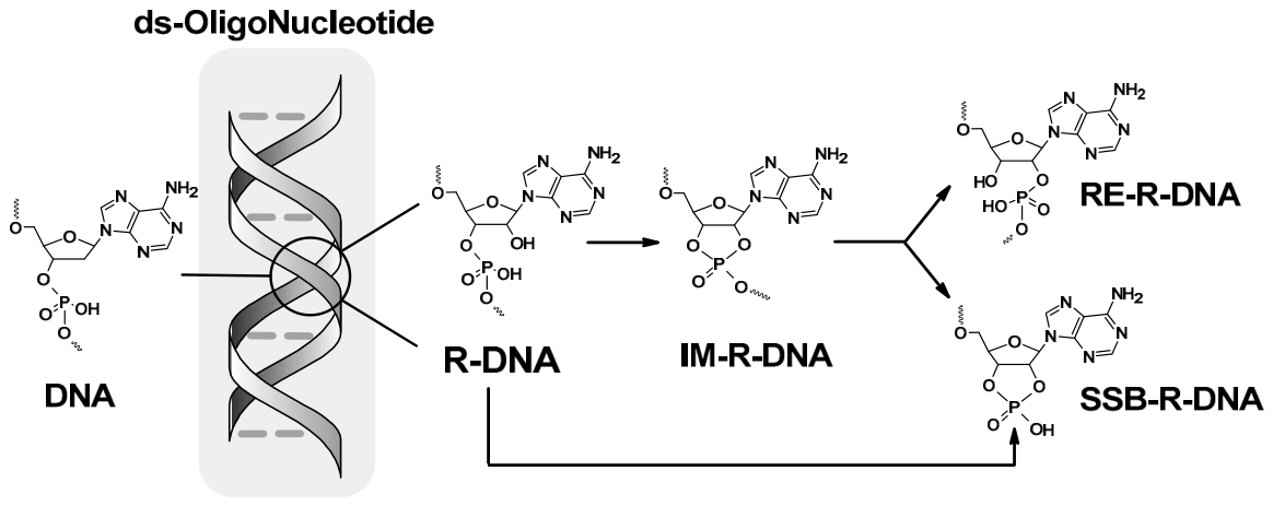
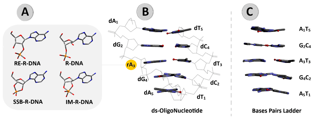
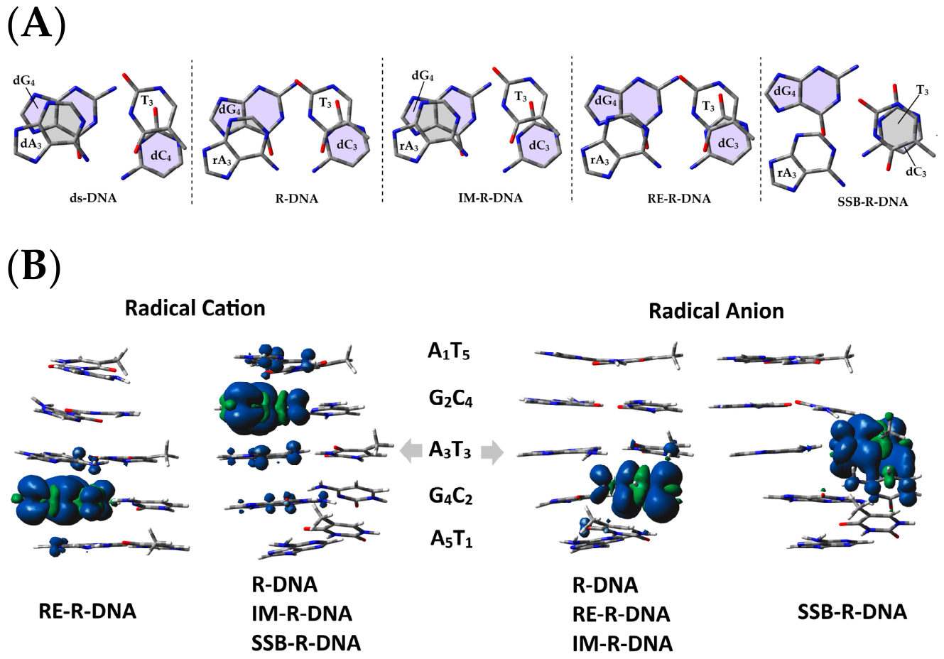
- The influence on ds-DNA structure was observed mainly in the nucleoside pair dimer formed by ribonucleosides and 3′-end 2′-deoxynucleosides [A3G4]*[C2T3], as shown in Table 1. This was particularly evident for heterocycle ring overlapping of the neutral form of the discussed ds-oligonucleotides in comparison with ds-DNA, and becomes more obvious after negative or positive charge adoption. This was well supported by the stacking interaction energies calculated for the base-pair and nucleoside-pair dimer. The differences in EST for each ds-oligo indicate the different effect of the modified internucleotide bond on the double-helix geometry.
- An investigation of global electronic properties of R-DNA, RE-R-DNA, IM-R-DNA, and SSB-R-DNA in their complete double-helix form, as well as the nucleoside skeleton or base-pair ladder, revealed the following: (a) the highest ionisation potential and lowest electron affinity for R-DNA with the native internucleotide bond and (b) the lowest ionisation potential and highest electron affinity for SSB-R-DNA, where the internucleotide bond was cleaved, leaving the 3′,2-cyclic phosphate on the rA3 moiety and a free 5′-OH on dG4. The above results suggest that the ribonucleoside subunit present in ds-DNA is unavailable to enzymes that utilise charge transfer for communication. This is evidenced by the electron–hole and electron rate constant (kHT).
- Careful analysis of base-pair electronic properties elucidated that the G2C4 base pair adopted the lowest ionisation potential among all ds-oligonucleotides containing riboadenosine (A3), with the exception of RE-R-DNA, for which G4C2 was assigned. Moreover, the highest electron affinity was found for the G4C2 base pair of R-DNA, IM-R-DNA, RE-R-DNA and A3T3 of SSB-R-DNA. This indicates that rearrangement of an internucleotide bond can force double-helix geometry fluctuation and bring about changes in the electronic properties of the single base pair.
- A comparative analysis of the electron–hole kHT reveals its relationship to spatial geometry fluctuation as a result of a different form of internucleotide phosphodiester between rA3 and dG4 (Table 8). The lowest rate constants in (s−1) were found for A3T3|G4C2 and G4C2|A5T1 of R-DNA, IM-R-DNA, and SSB-R-DNA, whereas, for RE-RDNA, it was noted as almost unaffected (Table 8). The above observation corresponds well with the highest electron activation energy (Ea) of the previously discussed cases.
- In contrast, it was observed that electron migration through stacked base pairs was almost unaffected for all the discussed ds-oligonucleotides. The kHT values were found to be in the range of 1010–1015 (s−1) for each base-pair dimer.
Supplementary Materials
Funding
Institutional Review Board Statement
Informed Consent Statement
Data Availability Statement
Acknowledgments
Conflicts of Interest
References
- Sudhir Ambekar, S. DNA: Damage and Repair Mechanisms in Humans. Glob. J. Pharm. Pharm. Sci. 2017, 3, 555613. [Google Scholar] [CrossRef] [Scilit]
- Oke, O.E.; Akosile, O.A.; Oni, A.I.; Opowoye, I.O.; Ishola, C.A.; Adebiyi, J.O.; Odeyemi, A.J.; Adjei-Mensah, B.; Uyanga, V.A.; Abioja, M.O. Oxidative Stress in Poultry Production. Poult. Sci. 2024, 103, 104003. [Google Scholar] [CrossRef] [Scilit] [PubMed]
- Romero Romero, M.L.; Landerer, C.; Poehls, J.; Toth-Petroczy, A. Phenotypic Mutations Contribute to Protein Diversity and Shape Protein Evolution. Protein Sci. 2022, 31, e4397. [Google Scholar] [CrossRef] [Scilit] [PubMed]
- Gerasimavicius, L.; Livesey, B.J.; Marsh, J.A. Loss-of-Function, Gain-of-Function and Dominant-Negative Mutations Have Profoundly Different Effects on Protein Structure. Nat. Commun. 2022, 13, 3895. [Google Scholar] [CrossRef] [Scilit] [PubMed]
- Gorinia, F.; Scalab, G.; Cooke, M.S.; Majello, B.; Amente, S. Towards a Comprehensive View of 8-Oxo-7,8-Dihydro-2’- Deoxyguanosine: Highlighting the Intertwined Roles of DNA Damage and Epigenetics in Genomic Instability. DNA Repair 2021, 97, 103027. [Google Scholar] [CrossRef] [Scilit] [PubMed]
- Besaratinia, A.; Caliri, A.W.; Tommasi, S. The Interplay of DNA Damage and Repair, Gene Expression, and Mutagenesis in Mammalian Cells during Oxidative Stress. Carcinogenesis 2024, 45, 868–879. [Google Scholar] [CrossRef] [Scilit] [PubMed]
- Iyama, T.; Wilson, D.M. DNA Repair Mechanisms in Dividing and Non-Dividing Cells. DNA Repair 2013, 12, 620–636. [Google Scholar] [CrossRef] [Scilit] [PubMed]
- Fuss, J.O.; Tsai, C.L.; Ishida, J.P.; Tainer, J.A. Emerging Critical Roles of Fe-S Clusters in DNA Replication and Repair. Biochim. Biophys. Acta—Mol. Cell Res. 2015, 1853, 1253–1271. [Google Scholar] [CrossRef] [Scilit] [PubMed]
- Genereux, J.C.; Barton, J.K. Mechanisms for DNA Charge Transport. Mechanisms 2011, 110, 1642–1662. [Google Scholar] [CrossRef] [Scilit] [PubMed]
- O’Brien, E.; Holt, M.E.; Thompson, M.K.; Salay, L.E.; Aaron, C.; Chazin, W.J.; Barton, J.K. The [4Fe4S] cluster of human DNA primase functions as a redox switch using DNA charge transport. Science 2017, 355, eaag1789. [Google Scholar] [CrossRef] [Scilit] [PubMed]
- Tse, E.C.M.; Zwang, T.J.; Bedoya, S.; Barton, J.K. Effective Distance for DNA-Mediated Charge Transport between Repair Proteins. ACS Cent. Sci. 2019, 5, 65–72. [Google Scholar] [CrossRef] [Scilit] [PubMed]
- Novoa-aponte, L.; Leon-torres, A.; Philpott, C.C. Guardians of the Genome: Iron—Sulfur Proteins in the Nucleus. Inorganics 2024, 12, 316. [Google Scholar] [CrossRef] [Scilit]
- Shi, R.; Hou, W.; Wang, Z.Q.; Xu, X. Biogenesis of Iron–Sulfur Clusters and Their Role in DNA Metabolism. Front. Cell Dev. Biol. 2021, 9, 735678. [Google Scholar] [CrossRef] [Scilit] [PubMed]
- Hassan, A.; Lima, F.C.D.A.; Crespilho, F.N. Redox-Guided DNA Scanning by the Dynamic Repair Enzyme Endonuclease III. Biochemistry 2025, 64, 782–790. [Google Scholar] [CrossRef] [Scilit] [PubMed]
- Lujan, S.A.; Williams, J.S.; Kunkel, T.A. DNA Polymerases Divide the Labor of Genome Replication. Trends Cell Biol. 2016, 129, 640–654. [Google Scholar] [CrossRef] [Scilit] [PubMed]
- Stillman, B. Reconsidering DNA Polymerases at the Replication Fork in Eukaryotes. Mol. Cell 2015, 59, 139–141. [Google Scholar] [CrossRef] [Scilit] [PubMed]
- Bainbridge, L.J.; Zabrady, K.; Doherty, A.J. Primase-Polymerases: How to Make a Primer from Scratch. Biosci. Rep. 2023, 43, BSR20221986. [Google Scholar] [CrossRef] [Scilit]
- Sui, Y.; Epstein, A.; Dominska, M.; Zheng, D.Q.; Petes, T.D.; Klein, H.L. Ribodysgenesis: Sudden Genome Instability in the Yeast Saccharomyces Cerevisiae Arising from RNase H2 Cleavage at Genomic-Embedded Ribonucleotides. Nucleic Acids Res. 2022, 50, 6890–6902. [Google Scholar] [CrossRef] [Scilit] [PubMed]
- Hiller, B.; Achleitner, M.; Glage, S.; Naumann, R.; Behrendt, R.; Roers, A. Mammalian RNase H2 Removes Ribonucleotides from DNA to Maintain Genome Integrity. J. Exp. Med. 2012, 209, 1419–1426. [Google Scholar] [CrossRef] [Scilit] [PubMed]
- Cerritelli, S.M.; Crouch, R.J. The Balancing Act of Ribonucleotides in DNA. Trends Biochem. Sci. 2016, 41, 434–445. [Google Scholar] [CrossRef] [Scilit] [PubMed]
- Nick McElhinny, S.A.; Watts, B.E.; Kumar, D.; Watt, D.L.; Lundström, E.B.; Burgers, P.M.J.; Johansson, E.; Chabes, A.; Kunkel, T.A. Abundant Ribonucleotide Incorporation into DNA by Yeast Replicative Polymerases. Proc. Natl. Acad. Sci. USA 2010, 107, 4949–4954. [Google Scholar] [CrossRef] [Scilit]
- Beard, W.A.; Wilson, S.H. Structural Insights into the Origins of DNA Polymerase Fidelity. Structure 2003, 11, 489–496. [Google Scholar] [CrossRef] [Scilit] [PubMed]
- Hiller, B.; Hoppe, A.; Haase, C.; Hiller, C.; Schubert, N.; Müller, W.; Reijns, M.A.M.; Jackson, A.P.; Kunkel, T.A.; Wenzel, J.; et al. Ribonucleotide Excision Repair Is Essential to Prevent Squamous Cell Carcinoma of the Skin. Cancer Res. 2018, 78, 5917–5926. [Google Scholar] [CrossRef] [Scilit] [PubMed]
- Kellner, V.; Luke, B. Molecular and Physiological Consequences of Faulty Eukaryotic Ribonucleotide Excision Repair. EMBO J. 2020, 39, e102309. [Google Scholar] [CrossRef] [Scilit]
- Sparks, J.L.; Chon, H.; Cerritelli, S.M.; Kunkel, T.A.; Johansson, E.; Crouch, R.J.; Burgers, P.M. RNase H2-Initiated Ribonucleotide Excision Repair. Mol. Cell 2012, 47, 980–986. [Google Scholar] [CrossRef] [Scilit] [PubMed]
- Mikkola, S.; Lönnberg, T.; Lönnberg, H. Phosphodiester Models for Cleavage of Nucleic Acids. Beilstein J. Org. Chem. 2018, 14, 803–837. [Google Scholar] [CrossRef] [Scilit] [PubMed]
- BIOVIA. Discovery Studio Visualizer, v16.1.0.15350; BIOVIA: San Diego, CA, USA, 2015.
- Karwowski, B.T. The Consequence of the Presence of Ribonucleotide for Ds-DNA’s Electronic Properties: Preliminary Theoretical Studies. Cells 2025, 14, 881. [Google Scholar] [CrossRef] [Scilit]
- Rayne, S.; Forest, K. Performance of the M062X Density Functional against the ISOL Set of Benchmark Isomerization Energies for Large Organic Molecules. Nat. Preced. 2010. [Google Scholar] [CrossRef] [Scilit]
- Zhao, Y.; Truhlar, D.G. The M06 Suite of Density Functionals for Main Group Thermochemistry, Thermochemical Kinetics, Noncovalent Interactions, Excited States, and Transition Element: Two New Functionals and Systematic Testing of Four M06-Class Functionals and 12 Other Functionals. Theor. Chem. Acc. 2008, 120, 215–241. [Google Scholar] [CrossRef] [Scilit]
- Cammi, R.; Corni, S.; Mennucci, B.; Tomasi, J. Electronic Excitation Energies of Molecules in Solution: State Specific and Linear Response Methods for Nonequilibrium Continuum Solvation Models. J. Chem. Phys. 2005, 122, 104513. [Google Scholar] [CrossRef] [Scilit] [PubMed]
- Li, T.C.; Tong, P.Q. Time-Dependent Density-Functional Theory for Multicomponent Systems. Phys. Rev. A 1986, 34, 529–532. [Google Scholar] [CrossRef] [Scilit] [PubMed]
- Cave, R.J.; Newton, M.D. Generalization of the Mulliken-Hush Treatment for the Calculation of Electron Transfer Matrix Elements. Chem. Phys. Lett. 1996, 249, 15–19. [Google Scholar] [CrossRef] [Scilit]
- Miertuš, S.; Tomasi, J. Approximate Evaluations of the Electrostatic Free Energy and Internal Energy Changes in Solution Processes. Chem. Phys. 1982, 65, 239–245. [Google Scholar] [CrossRef] [Scilit]
- Marcus, R.A. Electron Transfer Reactions in Chemistry: Theory and Experiment (Nobel Lecture). Angew. Chem. Int. Ed. Engl. 1993, 32, 1111–1121. [Google Scholar] [CrossRef] [Scilit]
- Rösch, N.; Voityuk, A.A. Quantum Chemical Calculation of Donor–Acceptor Coupling for Charge Transfer in DNA. In Long-Range Charge Transfer in DNA II; Springer: Berlin/Heidelberg, Germany, 2012; pp. 37–72. [Google Scholar] [CrossRef] [Scilit]
- Senet, P. Kohn-Sham Orbital Formulation of the Chemical Electronic Responses, Including the Hardness. J. Chem. Phys. 1997, 107, 2516–2524. [Google Scholar] [CrossRef] [Scilit]
- Dreuw, A.; Head-Gordon, M. Single-Reference Ab Initio Methods for the Calculation of Excited States of Large Molecules. Chem. Rev. 2005, 105, 4009–4037. [Google Scholar] [CrossRef] [Scilit] [PubMed]
- Frisch, M.J.; Trucks, G.W.; Schlegel, H.B.; Scuseria, G.E.; Robb, M.A.; Cheeseman, J.R.; Scalmani, G.; Barone, V.; Petersson, G.A.; Nakatsuji, H.; et al. Gaussian 16, Revision C.01; Gaussian, Inc.: Wallingford, CT, USA, 2016.
- Li, S.; Olson, W.K.; Lu, X.J. Web 3DNA 2.0 for the Analysis, Visualization, and Modeling of 3D Nucleic Acid Structures. Nucleic Acids Res. 2019, 47, W26–W34. [Google Scholar] [CrossRef] [Scilit] [PubMed]
- Editor, G.; Hobza, P.; Cozzi, F.; Annunziata, R.; Benaglia, M.; Kim, K.; Aguirre, G.; Estrada, J.; Siegel, J.S.; Chem, P.C.; et al. Nature and Magnitude of Aromatic Stacking of Nucleic Acid Bases. Phys. Chem. Chem. Phys. 2008, 10, 2595–2610. [Google Scholar] [CrossRef] [Scilit] [PubMed]
- Karwowski, B.T. The 2Ih and OXOG Proximity Consequences on Charge Transfer through ds-DNA: Theoretical Studies of Clustered DNA Damage. Molecules 2023, 28, 2180. [Google Scholar] [CrossRef] [Scilit] [PubMed]
- Gu, J.; Wang, J.; Leszczynski, J. Electron Attachment-Induced DNA Single-Strand Breaks at the Pyrimidine Sites. Nucleic Acids Res. 2010, 38, 5280–5290. [Google Scholar] [CrossRef] [Scilit] [PubMed]
- Kabasch, W. A Solution for the Best Rotation to Relate Two Sets of Vectors. Acta Cryst. 1976, 32, 922–923. [Google Scholar] [CrossRef] [Scilit]
- Kabsch, W. A Discussion of the Solution for the Best Rotation to Relate Two Sets of Vectors. Acta Cryst. 1978, A34, 827–828. [Google Scholar] [CrossRef] [Scilit]
- Nieuwland, C.; Hamlin, T.A.; Guerra, F.; Barone, G. B-DNA Structure and Stability: The Role of Nucleotide Composition and Order. ChemistryOpen 2022, 11, e202100231. [Google Scholar] [CrossRef] [Scilit] [PubMed]
- License, A.; Scotia, N.; Scotia, N.; Scotia, N. Modeling Biophysical and Biological Properties from the Characteristics of the Molecular Electron Density, Electron Localization and Delocalization Matrices, and the Electrostatic Potential. J. Comput. Chem. 2014, 35, 1165–1198. [Google Scholar] [CrossRef] [Scilit] [PubMed]
- Driver, M.D.; Williamson, M.J.; Cook, J.L.; Hunter, C.A. Functional Group Interaction pro Fi Les: A General Treatment of Solvent effects on Non-Covalent Interactions†. Chem. Sci. 2020, 11, 4456–4466. [Google Scholar] [CrossRef] [Scilit] [PubMed]
- Kumar, A.; Adhikary, A.; Sevilla, M.D.; Close, D.M. One-Electron Oxidation of Ds(5′-GGG-3′) and Ds(5′-G(8OG)G-3′) and the Nature of Hole Distribution: A Density Functional Theory (DFT) Study. Phys. Chem. Chem. Phys. 2020, 22, 5078–5089. [Google Scholar] [CrossRef] [Scilit] [PubMed]
- Olson, W.K. The flexible DNA double helix. I. Average dimensions and distribution functions. Biopolymers 1979, 18, 1213–1233. [Google Scholar] [CrossRef] [Scilit] [PubMed]
- O’Flaherty, D.K.; Guengerich, F.P.; Egli, M.; Wilds, C.J. Backbone Flexibility Influences Nucleotide Incorporation by Human Translesion DNA Polymerase η Opposite Intrastrand Cross-Linked DNA. Biochemistry 2016, 54, 7449–7456. [Google Scholar] [CrossRef] [Scilit] [PubMed]
- Mukaka, M.M. Statistics Corner: A Guide to Appropriate Use of Correlation Coefficient in Medical Research. Malawi Med. J. 2012, 24, 69–71. [Google Scholar] [PubMed]
- Mester, D. Vertical Ionization Potentials and Electron Affinities at the Double- Hybrid Density Functional Level. J. Chem. Theory Comput. 2023, 19, 3982–3995. [Google Scholar] [CrossRef] [Scilit] [PubMed]
- Clore, A.M.G. Investigation of the solution structures of short nucleic acid fragments by means of nuclear overhauser enhancement measurements. Prog. NMR Spectrosc. 1985, 17, 1–32. [Google Scholar] [CrossRef] [Scilit]
- Karwowski, B.T. The AT Interstrand Cross-Link: Structure, Electronic Properties, and Influence on Charge Transfer in DsDNA. Mol. Ther. Nucleic Acids 2018, 13, 665–685. [Google Scholar] [CrossRef] [Scilit] [PubMed]
- Karwowski, B. How Clustered DNA Damage Can Change the Electronic Properties of Ds-DNA, Differences between GAG, GAOXOG, and OXOGAOXOG. Biomolecules 2023, 13, 517. [Google Scholar] [CrossRef] [Scilit] [PubMed]
- Zhou, H.; Lin, H.; Wang, Q.; Hao, T.; Hu, Y.; Wang, S.; Guo, Z. Tunneling or Hopping? A Direct Electrochemical Observation of Electron Transfer in DNA. Anal. Chem. 2022, 94, 15324–15331. [Google Scholar] [CrossRef] [Scilit] [PubMed]
- Capobianco, A.; Caruso, T.; Celentano, M.; D’Ursi, A.M.; Scrima, M.; Peluso, A. Stacking Interactions between Adenines in Oxidized Oligonucleotides. J. Phys. Chem. B 2013, 117, 8947–8953. [Google Scholar] [CrossRef] [Scilit] [PubMed]
- Hüsken, D.; Goodall, G.; Blommers, M.J.J.; Jahnke, W.; Hall, J.; Häner, R.; Moser, H.E. Creating RNA Bulges: Cleavage of RNA in RNA/DNA Duplexes by Metal Ion Catalysis. Biochemistry 1996, 35, 16591–16600. [Google Scholar] [CrossRef] [Scilit] [PubMed]
- Kaukinen, U.; Lyytikäinen, S.; Mikkola, S.; Lönnberg, H. The Reactivity of Phosphodiester Bonds within Linear Single-Stranded Oligoribonucleotides in Strongly Dependent on the Base Sequence. Nucleic Acids Res. 2002, 30, 468–474. [Google Scholar] [CrossRef] [Scilit] [PubMed]
- O’Neill, M.A.; Barton, J.K. DNA Charge Transport: Conformationally Gated Hopping through Stacked Domains. J. Am. Chem. Soc. 2004, 126, 11471–11483. [Google Scholar] [CrossRef] [Scilit] [PubMed]
- Shuai, Z.; Li, W.; Ren, J.; Jiang, Y.; Geng, H. Applying Marcus Theory to Describe the Carrier Transports in Organic Semiconductors: Limitations and Beyond. J. Chem. Phys. 2020, 153, 080902. [Google Scholar] [CrossRef] [Scilit] [PubMed]
- Sontz, P.A.; Muren, N.B.; Barton, J.K. DNA Charge Transport for Sensing and Signaling. Acc. Chem. Res. 2012, 45, 1792–1800. [Google Scholar] [CrossRef] [Scilit] [PubMed]
- Xiang, L.; Palma, J.L.; Bruot, C.; Mujica, V.; Ratner, M.A.; Tao, N. Corrigendum: Intermediate Tunnelling—Hopping Regime in DNA Charge Transport. Nat. Publ. Gr. 2017, 9, 295. [Google Scholar] [CrossRef] [Scilit] [PubMed]
- Giese, B. Long-Distance Charge Transport in DNA: The Hopping Mechanism. Acc. Chem. Res. 2000, 33, 631–636. [Google Scholar] [CrossRef] [Scilit] [PubMed]
- Khan, A. Reorganization Energy, Activation Energy, and Mechanism of Hole Transfer Process in DNA: A Theoretical Study. J. Chem. Phys. 2008, 128, 075101. [Google Scholar] [CrossRef] [Scilit] [PubMed]
- Li, P.; Zhao, C.; Smith, M.D.; Shimizu, K.D. Comprehensive Experimental Study of N-Heterocyclic π-Stacking Interactions of Neutral and Cationic Pyridines. J. Org. Chem. 2013, 78, 5303–5313. [Google Scholar] [CrossRef] [Scilit] [PubMed]
- Fleming, A.M.; Muller, J.G.; Dlouhy, A.C.; Burrows, C.J. Structural Context Effects in the Oxidation of 8-Oxo-7,8-Dihydro-2′- Deoxyguanosine to Hydantoin Products: Electrostatics, Base Stacking, and Base Pairing. J. Am. Chem. Soc. 2012, 134, 15091–15102. [Google Scholar] [CrossRef] [Scilit] [PubMed]



| Base Pair Dimer | Base-Pair Heterocycles Overlap [Å2] | Rise (h) [Å] | ||||||||
|---|---|---|---|---|---|---|---|---|---|---|
| R-DNA | IM-R-DNA | RE-R-DNA | SSB-R-DNA | DNA | R-DNA | IM-R-DNA | RE-R-DNA | SSB-R-DNA | DNA | |
| Neutral Form | ||||||||||
| A1T5|G2C4 | 3.67 | 3.60 | 3.66 | 3.49 | 3.81 | 3.09 | 3.02 | 3.04 | 2.95 | 2.96 |
| G2C4|A3T3 | 4.82 | 4.49 | 4.07 | 3.17 | 3.44 | 3.19 | 3.28 | 3.16 | 3.17 | 3.22 |
| A3T3|G4C2 | 1.33 | 3.31 | 1.17 | 2.93 | 3.73 | 2.98 | 2.86 | 2.94 | 2.85 | 2.93 |
| G4C2|A5T1 | 4.01 | 3.38 | 4.33 | 4.70 | 3.96 | 2.97 | 2.90 | 2.88 | 3.03 | 2.95 |
| Radical Cation Form | ||||||||||
| A1T5|G2C4 | 4.79 | 4.60 | 3.27 | 4.56 | 4.43 | 3.02 | 3.08 | 3.02 | 2.95 | 2.94 |
| G2C4|A3T3 | 4.46 | 1.86 | 4.10 | 3.04 | 2.37 | 3.12 | 3.17 | 3.12 | 3.15 | 3.00 |
| A3T3|G4C2 | 0.63 | 3.18 | 0.22 | 2.87 | 3.83 | 2.84 | 2.82 | 2.84 | 2.84 | 2.97 |
| G4C2|A5T1 | 4.52 | 3.78 | 4.52 | 4.47 | 4.01 | 2.85 | 2.89 | 2.85 | 2.83 | 2.92 |
| Radical Anion Form | ||||||||||
| A1T5|G2C4 | 3.33 | 3.31 | 3.44 | 4.35 | 3.80 | 3.07 | 3.00 | 3.07 | 3.03 | 3.00 |
| G2C4|A3T3 | 5.00 | 4.16 | 3.55 | 3.53 | 3.82 | 3.30 | 3.66 | 3.30 | 3.25 | 3.52 |
| A3T3|G4C2 | 2.24 | 3.21 | 2.68 | 0.86 | 3.89 | 2.99 | 2.82 | 2.99 | 3.43 | 2.87 |
| G4C2|A5T1 | 4.00 | 3.90 | 4.35 | 4.94 | 4.03 | 2.79 | 2.84 | 2.79 | 2.69 | 2.90 |
| Root Mean Square Deviation (RMSD) of atomic positions, in [Å2], of the ds-oligonucleotide structures after charge adoption | ||||||||||
| Anion vs. Neutral | (a)0.37 (b)0.44b | (a)0.34 (b)0.34 | (a)0.48 (b)0.47 | (a)1.50 (b)1.37 | (a)0.17 (b)0.16 |  | ||||
| Cation vs. Neutral | (a)0.72 (b)0.69 | (a)0.88 (b)0.91 | (a)0.47 (b)0.45 | (a)0.50 (b)0.48 | (a)0.36 (b)0.31 | |||||
| Base Pairs Dimer | R-DNA | RE-R-DNA | IM-R-DNA | SSB-R-DNA | ||||||||
|---|---|---|---|---|---|---|---|---|---|---|---|---|
| Neutr | Cation | Anion | Neutr | Cation | Anion | Neutr | Cation | Anion | Neutr | Cation | Anion | |
| A1T5|G2C4 | 14.80 | 13.02 | 14.97 | 15.09 | 15.17 | 15.35 | 15.49 | 14.42 | 15.29 | 14.60 | 13.74 | 14.92 |
| G2C4|A3T3 | 13.36 | 14.00 | 12.85 | 14.34 | 14.69 | 13.46 | 13.58 | 11.52 | 13.33 | 14.81 | 18.66 | 14.12 |
| A3T3|G4C2 | 13.06 | 12.92 | 13.72 | 13.16 | 10.99 | 10.88 | 14.05 | 14.69 | 12.39 | 12.31 | 11.88 | 12.48 |
| G4C2|A5T1 | 15.92 | 14.94 | 16.07 | 15.47 | 13.91 | 15.17 | 15.38 | 15.83 | 15.87 | 15.26 | 16.19 | 14.83 |
| A1T5|A3A5 | 0.46 | 0.51 | 0.47 | 0.46 | 0.51 | 0.48 | 0.46 | 0.49 | 0.48 | 0.47 | 0.56 | 0.44 |
| G2C4|G4C2 | 0.46 | 0.48 | 0.38 | 0.39 | 0.41 | 0.35 | 0.44 | 0.53 | 0.35 | 0.56 | 0.56 | 0.41 |
| A3T3|A5T1 | 0.51 | 0.53 | 0.53 | 0.52 | 0.51 | 0.42 | 0.49 | 0.54 | 0.42 | 0.61 | 0.63 | 0.52 |
| Nucleoside-Pair Dimer in Neutral Form | ||||||||
|---|---|---|---|---|---|---|---|---|
| ds-DNA | ds-DNA | R-DNA | RE-R-DNA | IM-R-DNA | SSB-R-DNA | |||
| Neutr * | Cation * | Anion * | ||||||
| A1T5|G2C4 | 14.84 | 14.09 | 15.87 | 26.71 | 27.91 | 28.12 | 28.39 | 27.22 |
| G2C4|A3T3 | 14.60 | 13.63 | 14.20 | 25.29 | 23.82 | 25.01 | 23.46 | 27.80 |
| A3T3|G4C2 | 14.24 | 14.75 | 12.94 | 24.59 | 25.12 | 17.82 | 18.65 | 18.32 |
| G4C2|A5T1 | 15.07 | 15.15 | 15.44 | 26.88 | 27.79 | 27.53 | 26.85 | 22.80 |
| ds-Oligonucleotide [28] | Nucleoside-Pair Skeleton | |||||||||||
|---|---|---|---|---|---|---|---|---|---|---|---|---|
| NEVIP | EQVIP | AIP | NEVEA | EQVEA | AEA | NEVIP | EQVIP | AIP | NEVEA | EQVEA | AEA | |
| DNA | 6.72 | 6.08 | 5.65 | 0.84 | 1.58 | 2.09 | 6.68 | 6.00 | 5.57 | 0.71 | 1.29 | 1.97 |
| R-DNA | 7.03 | 6.21 | 7.41 | 0.73 | 1.38 | 1.76 | 6.23 | 6.12 | 6.88 | 0.56 | 1.31 | 1.94 |
| IM-R-DNA | 6.91 | 6.25 | 4.49 | 0.68 | 1.42 | 3.47 | 6.87 | 6.16 | 5.40 | 0.57 | 1.33 | 2.38 |
| RE-R-DNA | 6.84 | 6.12 | 3.84 | 0.62 | 1.33 | 4.08 | 6.70 | 6.01 | 5.23 | 0.49 | 1.26 | 3.00 |
| SSB-R-DNA | 6.75 | 6.09 | 2.76 | 0.82 | 1.62 | 5.55 | 6.51 | 6.04 | 3.00 | 0.86 | 1.43 | 5.17 |
| Base-Pair Ladders | ||||||
|---|---|---|---|---|---|---|
| NEVIP | EQVIP | AIP | NEVEA | EQVEA | AEA | |
| DNA | 6.47 | 5.98 | 5.58 | 0.60 | 1.34 | 1.90 |
| R-DNA | 6.87 | 5.89 | 6.61 | 0.55 | 1.51 | 1.80 |
| IM-R-DNA | 6.82 | 6.05 | 5.56 | 0.56 | 1.34 | 1.85 |
| RE-R-DNA | 6.81 | 6.01 | 5.71 | 0.53 | 1.29 | 1.99 |
| SSB-R-DNA | 6.81 | 5.96 | 4.59 | 0.72 | 1.42 | 2.94 |
| DNA | R-DNA | RE-R-DNA | IM-R-DNA | SSB-R-DNA | ||||||
|---|---|---|---|---|---|---|---|---|---|---|
| AIP | VIP | AIP | VIP | AIP | VIP | AIP | VIP | AIP | VIP | |
| (a)dA1T5 | 6.53 | 6.57 | 6.69 | 6.59 | 6.59 | 6.56 | 6.46 | 6.56 | 6.55 | 6.69 |
| (b)A1T5 | 6.60 | 6.65 | 6.82 | 6.68 | 6.66 | 6.65 | 6.58 | 6.64 | 6.39 | 6.67 |
| (a)dG2C4 | 5.90 | 6.16 | 6.05 | 6.23 | 6.26 | 6.20 | 5.86 | 6.22 | 5.62 | 6.19 |
| (b)G2C4 | 5.83 | 6.13 | 6.01 | 6.19 | 6.23 | 6.17 | 5.82 | 6.18 | 5.65 | 6.11 |
| (a)rA3T3 | 6.64 | 6.67 | 7.04 | 6.69 | 6.55 | 6.71 | 6.44 | 6.70 | 6.44 | 6.79 |
| *(a)rA3T3 | 5.73 | 6.83 | ||||||||
| (b)A3T3 | 6.60 | 6.65 | 6.87 | 6.61 | 6.75 | 6.66 | 6.67 | 6.63 | 6.56 | 6.71 |
| (a)dG4dC2 | 6.14 | 6.16 | 6.42 | 6.20 | 5.45 | 6.20 | 6.14 | 6.20 | 5.58 | 6.04 |
| (b)G4C2 | 6.11 | 6.13 | 6.29 | 6.17 | 5.82 | 6.17 | 6.17 | 6.17 | 6.02 | 6.19 |
| (a)dA5T1 | 6.72 | 6.76 | 6.89 | 6.76 | 6.70 | 6.74 | 6.75 | 6.79 | 6.31 | 6.57 |
| (b)A5T1 | 6.72 | 6.74 | 6.85 | 6.74 | 6.68 | 6.72 | 6.73 | 6.75 | 6.55 | 6.67 |
| AEA | VEA | AEA | VEA | AEA | VEA | AEA | VEA | AEA | VEA | |
| (a)dA1T5 | 1.42 | 1.41 | 1.51 | 1.50 | 1.49 | 1.49 | 1.47 | 1.49 | 1.63 | 1.61 |
| (b)A1T5 | 1.49 | 1.48 | 1.41 | 1.41 | 1.41 | 1.41 | 1.41 | 1.41 | 1.60 | 1.54 |
| (a)dG2dC4 | 1.47 | 1.49 | 1.70 | 1.54 | 1.54 | 1.55 | 1.54 | 1.55 | 1.72 | 1.67 |
| (b)G2C4 | 1.53 | 1.56 | 1.46 | 1.47 | 1.48 | 1.49 | 1.48 | 1.49 | 1.68 | 1.62 |
| (a)rA3T3 | 1.40 | 1.40 | 1.44 | 1.48 | 1.61 | 1.45 | 1.69 | 1.48 | 2.22 | 1.58 |
| *(a)rA3T3 | 2.74 | 1.58 | ||||||||
| (b)A3T3 | 1.48 | 1.46 | 1.37 | 1.42 | 1.42 | 1.38 | 1.42 | 1.41 | 2.04 | 1.51 |
| (a)dG4dC2 | 1.95 | 1.52 | 1.80 | 1.57 | 2.51 | 1.56 | 2.18 | 1.57 | 2.19 | 1.72 |
| (b)G4C2 | 1.99 | 1.59 | 1.86 | 1.50 | 2.15 | 1.48 | 1.95 | 1.50 | 1.76 | 1.62 |
| (a)dA5T1 | 1.39 | 1.42 | 1.34 | 1.38 | 1.61 | 1.38 | 1.33 | 1.36 | 1.65 | 1.54 |
| (b)A5T1 | 1.35 | 1.38 | 1.40 | 1.43 | 1.39 | 1.43 | 1.39 | 1.41 | 1.64 | 1.58 |
| Hole Migration | [A1G2A3]*[T5C4T3] | [G2A3G4]*[C4T3C2] | [A3G4A5]*[T3C2T1] | [A1G2A3G4A5]*[T5C4T3C2T1] | |||||
|---|---|---|---|---|---|---|---|---|---|
| A1 → G2 | G2 ← A3 | G2 ← A3 | A3 → G4 | A3 → G4 | G4 ← A5 | A1 ← A3 | G2 ← G4 | A3 ← A5 | |
| DNA [44] | −0.52(a) | −0.84 | −0.85 | −0.84 | −0.51 | −0.60 | −0.33 | −0.01 | −0.09 |
| −0.76(b) | −1.09 | −1.10 | −0.81 | −0.53 | −0.60 | −0.33 | −0.29 | −0.07 | |
| R–DNA | −0.29 | −0.72 | −0.91 | −0.72 | −0.23 | −0.56 | −0.42 | −0.19 | −0.33 |
| −0.62 | −1.17 | −1.36 | −0.87 | −0.11 | −0.56 | −0.55 | −0.48 | −0.44 | |
| RE–R–DNA | −0.47 | −0.51 | −0.52 | −0.50 | −0.48 | −1.24 | −0.04 | −0.02 | −0.76 |
| −0.43 | −0.53 | −0.54 | −0.93 | −0.83 | −1.55 | −0.11 | 0.39 | −0.72 | |
| IM–R–DNA | −0.46 | −0.73 | −0.73 | −0.73 | −0.44 | −0.55 | −0.27 | −0.01 | −0.11 |
| −0.76 | −1.14 | −1.14 | −0.78 | −0.44 | −0.54 | −0.38 | −0.36 | −0.09 | |
| SSB–R–DNA | −0.75 | −0.90 | −0.71 | −0.83 | −0.71 | −0.44 | −0.15 | 0.11 | 0.27 |
| −0.93 | −1.21 | −1.03 | −0.85 | −0.88 | −0.49 | −0.28 | −0.18 | 0.39 | |
| Electron Migration | |||||||||
| A1 → G2 | G2 ← A3 | G2 ← A3 | A3 → G4 | A3 → G4 | G4 ← A5 | A1 ← A3 | G2 → G4 | A3 ← A5 | |
| DNA [44] | −0.08 | −0.13 | −0.12 | −0.15 | −0.12 | −0.62 | −0.05 | −0.03 | −0.50 |
| −0.05 | −0.10 | −0.10 | −0.57 | −0.55 | −1.07 | −0.05 | −0.47 | −0.53 | |
| R–DNA | −0.06 | −0.05 | −0.05 | −0.03 | −0.03 | −0.60 | 0.01 | 0.02 | −0.57 |
| −0.05 | −0.10 | −0.10 | −0.45 | −0.38 | −0.99 | −0.05 | −0.34 | −0.61 | |
| RE–R–DNA | −0.06 | −0.11 | −0.12 | −0.11 | −0.10 | −1.00 | −0.05 | 0.01 | −0.89 |
| −0.06 | −0.05 | −0.06 | −0.73 | −0.77 | −1.69 | 0.00 | −0.67 | −0.93 | |
| IM–R–DNA | −0.07 | −0.08 | −0.09 | −0.09 | −0.08 | −0.60 | −0.01 | 0.00 | −0.52 |
| −0.06 | −0.06 | −0.07 | −0.53 | −0.54 | −1.07 | 0.01 | −0.46 | −0.54 | |
| SSB–R–DNA | A1 → G2 | G2 → A3 | G2 → A3 | A3 ← G4 | A3 ← G4 | G4 → A5 | A1 → A3 | G2 ← G4 | A3 → A5 |
| −0.27 | 0.18 | −0.01 | −0.30 | −0.18 | −0.39 | −0.09 | −0.30 | −0.21 | |
| −0.27 | −0.29 | −0.48 | −0.68 | −0.03 | −0.30 | −0.56 | −0.21 | −0.27 | |
 | |||||||||
| ds–Oligo | Base–Pair Dimer | Hole Transfer | Electron Transfer | ||||||||
|---|---|---|---|---|---|---|---|---|---|---|---|
| λ | ΔG | Ea | HDA | kHT | λ | ΔG | Ea | HDA | kHT | ||
| R–DNA | A1T5|G2C4 | 0.37 | −0.81 | 0.13 | 0.26 | 1.38 × 10¹³ | −0.002 | −0.05 | −0.37 | 0.02 | n.d |
| G2C4|rA3T3 | 0.39 | −0.86 | 0.14 | 0.20 | 4.07 × 10¹² | 0.05 | −0.09 | 0.01 | 0.03 | 4.86 × 10¹³ | |
| A3T3|G4C2 | 0.09 | −0.59 | 0.66 | 0.21 | 1.59 × 1004 | 0.41 | −0.49 | 0.00 | 0.04 | 3.61 × 10¹³ | |
| G4C2|A5T1 | 0.08 | −0.57 | 0.69 | 0.27 | 1.06 × 1004 | 0.41 | −0.49 | 0.00 | 0.04 | 3.21 × 10¹³ | |
| A1T5|A3T3 | −0.35 | −0.06 | −0.12 | 0.03 | n.d. | −0.06 | −0.04 | −0.04 | 0.09 | n.d. | |
| G2C4|G4C2 | 0.38 | −0.28 | 0.01 | 0.01 | 2.05 × 10¹² | 0.37 | −0.40 | 0.00 | 0.05 | 6.73 × 10¹³ | |
| A3T3|A5T1 | −0.32 | −0.02 | −0.09 | 0.06 | n.d. | 0.02 | −0.03 | 0.0001 | 0.08 | 6.97 × 10¹⁴ | |
| RE–R–DNA | A1T5|G2C4 | −0.05 | −0.43 | −1.10 | 0.27 | n.d. | 0.01 | −0.07 | 0.17 | 0.01 | 2.97 × 10¹⁰ |
| G2C4|A3T3 | −0.04 | −0.52 | −1.86 | 0.24 | n.d. | 0.06 | −0.06 | 0.00 | 0.04 | 1.14 × 10¹⁴ | |
| A3T3|G4C2 | 0.37 | −0.93 | 0.21 | 0.24 | 4.70 × 10¹¹ | 0.67 | −0.72 | 0.00 | 0.07 | 9.62 × 10¹³ | |
| G4C2|A5T1 | 0.74 | −0.87 | 0.01 | 0.27 | 1.15 × 10¹⁵ | 0.91 | −0.76 | 0.01 | 0.03 | 1.23 × 10¹³ | |
| A1T5|A3T3 | 0.00 | −0.09 | 0.48 | 0.01 | 2.26 × 1005 | 0.04 | −0.01 | 0.00 | 0.09 | 6.24 × 10¹⁴ | |
| G2C4|G4C2 | 0.38 | −0.41 | 0.0007 | 0.002 | 1.06 × 10¹¹ | 0.67 | −0.67 | 0.00 | 0.05 | 5.11 × 10¹³ | |
| A3T3|A5T1 | 0.05 | −0.06 | 0.0004 | 0.03 | 6.45 × 10¹³ | 0.08 | −0.03 | 0.01 | 0.08 | 2.77 × 10¹⁴ | |
| IM–R–DNA | A1T5|G2C4 | 0.30 | −0.77 | 0.18 | 0.26 | 1.63 × 10¹² | −0.0003 | −0.07 | −0.41 | 0.01 | n.d. |
| G2C4|A3T3 | 0.24 | −0.86 | 0.40 | 0.23 | 3.54 × 10⁸ | −0.01 | −0.06 | −0.15 | 0.03 | n.d. | |
| A3T3|G4C2 | 0.02 | −0.51 | 3.01 | 0.20 | 7.61 × 10−36 | 0.46 | −0.53 | 0.00 | 0.06 | 7.96 × 10¹³ | |
| G4C2|A5T1 | 0.01 | −0.56 | 7.91 | 0.29 | 2.69 × 10−118 | 0.49 | −0.56 | 0.00 | 0.04 | 3.48 × 10¹³ | |
| A1T5|A3T3 | 0.02 | −0.51 | 3.01 | 0.20 | 5.80 × 10¹² | −0.0003 | −0.01 | −0.11 | 0.09 | n.d. | |
| G2C4|G4C2 | 0.01 | −0.56 | 7.91 | 0.29 | 2.83 × 10⁸ | 0.46 | −0.47 | 0.00 | 0.05 | 6.20 × 10¹³ | |
| A3T3|A5T1 | 0.08 | −0.10 | 0.00 | 0.01 | 3.97 × 10¹² | 0.04 | −0.03 | 0.00 | 0.05 | 1.99 × 10¹⁴ | |
| SSB–R–DNA | A1T5|G2C4 | 0.28 | −0.74 | 0.20 | 0.25 | 9.85 × 10¹¹ | −0.13 | −0.09 | −0.09 | 0.06 | n.d. |
| G2C4|A3T3 | 0.27 | −0.91 | 0.38 | 0.26 | 8.27 × 1008 | 0.41 | −0.36 | 0.00 | 0.05 | 6.15 × 10¹³ | |
| A3T3|G4C2 | 0.02 | −0.54 | 4.46 | 0.30 | 4.36 × 10−60 | 0.40 | −0.28 | 0.01 | 0.11 | 2.24 × 10¹⁴ | |
| G4C2|A5T1 | 0.01 | −0.53 | 4.46 | 0.23 | 3.62 × 10−60 | −0.03 | −0.12 | −0.20 | 0.02 | n.d. | |
| A1T5|A3T3 | 0.09 | −0.17 | 0.02 | 0.02 | 1.13 × 10¹³ | 0.72 | −0.44 | 0.03 | 0.04 | 1.15 × 10¹³ | |
| G2C4|G4C2 | 0.24 | −0.37 | 0.02 | 0.04 | 2.62 × 10¹³ | 0.27 | −0.08 | 0.03 | 0.02 | 3.56 × 10¹² | |
| A3T3|A5T1 | 0.31 | −0.01 | 0.07 | 0.02 | 7.54 × 10¹¹ | 0.36 | −0.40 | 0.00 | 0.04 | 4.31 × 10¹³ | |
Disclaimer/Publisher’s Note: The statements, opinions and data contained in all publications are solely those of the individual author(s) and contributor(s) and not of MDPI and/or the editor(s). MDPI and/or the editor(s) disclaim responsibility for any injury to people or property resulting from any ideas, methods, instructions or products referred to in the content. |
© 2026 by the author. Licensee MDPI, Basel, Switzerland. This article is an open access article distributed under the terms and conditions of the Creative Commons Attribution (CC BY) license.
Share and Cite
Karwowski, B.T. A Single Ribonucleotide and the Various Possibilities for Charge Transfer Modulation Through ds-DNA: A Density Functional Theory Study. Cells 2026, 15, 1194. https://doi.org/10.3390/cells15131194
Karwowski BT. A Single Ribonucleotide and the Various Possibilities for Charge Transfer Modulation Through ds-DNA: A Density Functional Theory Study. Cells. 2026; 15(13):1194. https://doi.org/10.3390/cells15131194
Chicago/Turabian StyleKarwowski, Boleslaw T. 2026. "A Single Ribonucleotide and the Various Possibilities for Charge Transfer Modulation Through ds-DNA: A Density Functional Theory Study" Cells 15, no. 13: 1194. https://doi.org/10.3390/cells15131194
APA StyleKarwowski, B. T. (2026). A Single Ribonucleotide and the Various Possibilities for Charge Transfer Modulation Through ds-DNA: A Density Functional Theory Study. Cells, 15(13), 1194. https://doi.org/10.3390/cells15131194