Review

Curriculum Vitae of WEE2 Kinase in Homeostasis and Diseases: A Systematic Review

1
State Key Laboratory for Quality and Safety of Agro-Products, School of Marine Sciences, Ningbo University, Ningbo 315211, China
2
Laboratory of Biochemistry and Molecular Biology, School of Marine Sciences, Meishan Campus, Ningbo University, Ningbo 315832, China
3
Key Laboratory of Aquacultural Biotechnology of Ministry of Education, Ningbo University, Ningbo 315211, China
*
Authors to whom correspondence should be addressed.
Cells 2026, 15(13), 1147; https://doi.org/10.3390/cells15131147
Submission received: 29 May 2026 / Revised: 18 June 2026 / Accepted: 22 June 2026 / Published: 24 June 2026
(This article belongs to the Special Issue Clockwork Cells: Decoding the Language of Cell Cycle Regulators)

Highlights

What are the main findings?
  • WEE2 is an oocyte-specific kinase essential for meiotic arrest and progression.
  • WEE2 mutations are a major cause of total fertilization failure and female infertility.
What are the implications of the main findings?
  • WEE2 structure, function, and mutational mechanisms lay a core molecular foundation for understanding associated infertility.
  • Latest progress on WEE2 inhibitors offers new avenues for precise diagnosis and targeted treatment of reproductive disorders.

Abstract

WEE2, an oocyte-specific kinase of the WEE family, is a core regulator of oocyte meiosis. It maintains germinal vesicle (GV) arrest and prevents premature meiotic resumption by phosphorylating cyclin-dependent kinase 1 (CDK1), thereby inhibiting maturation-promoting factor (MPF) activity. WEE2 also regulates exit from metaphase II (MII), ensuring orderly meiotic progression. Consequently, the functional integrity of WEE2 is essential for female reproduction. Homozygous or compound heterozygous mutations in the WEE2 gene represent a major genetic cause of total fertilization failure and primary infertility, as these mutations lead to reduced or abolished kinase activity, impair meiotic control, and disrupt oocyte maturation and embryonic development. This review systematically summarizes the protein structure, core functions, and mutation types of WEE2, along with its association with total fertilization failure and female primary infertility. It also highlights research advances in WEE2-targeted inhibitors and discusses the potential applications and future directions of WEE2 in the diagnosis and management of reproductive disorders.

1. Introduction

WEE2 is an oocyte-specific tyrosine kinase belonging to the WEE kinase family, which also includes WEE1 (Wee1-like protein kinase) and PKMYT1 (membrane-associated tyrosine- and threonine-specific cyclin-dependent kinase 1 (CDK1)-inhibitory kinase, also known as MYT1) [1]. As dual-specificity protein kinases in the serine/threonine kinase superfamily, WEE family members function as core negative regulators of the G2/M cell cycle checkpoint by phosphorylating CDK1 and inhibiting maturation-promoting factor (MPF) activity, thereby temporally controlling eukaryotic cell division [2]. WEE1 and PKMYT1 regulate the somatic cell cycle; their inhibition causes premature CDK1 activation, leading to cell cycle dysregulation and genomic instability [2].
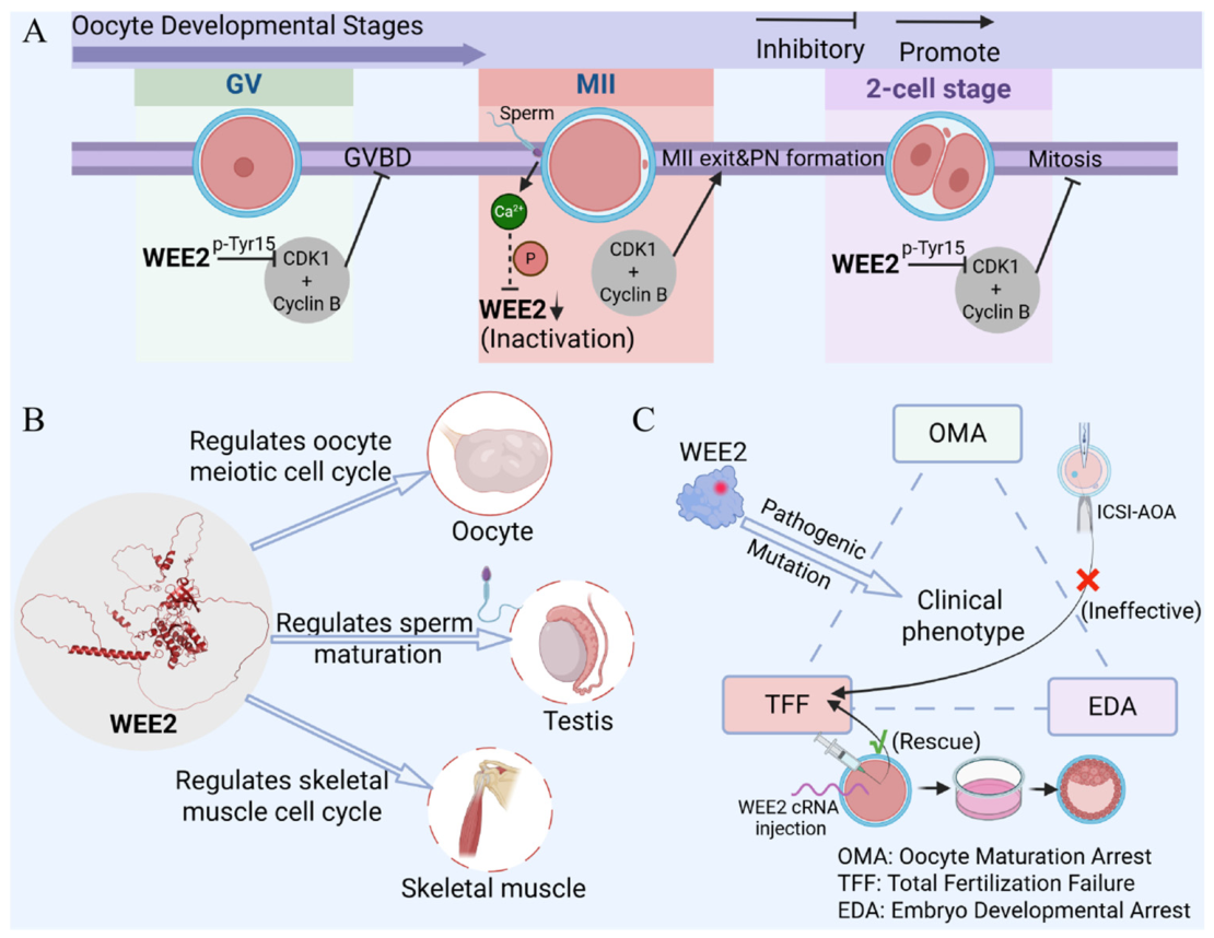
Unlike its homologs, WEE2 primarily governs oocyte meiosis: it maintains germinal vesicle (GV) arrest and regulates exit from metaphase II (MII) [3,4]. During the GV stage, WEE2 phosphorylates CDK1 at Tyr15 within the CDK1/cyclin B (MPF) complex, reducing MPF activity and sustaining meiotic arrest. Following an LH surge, cAMP degradation suppresses WEE2 activity, activates CDK1, and triggers GV breakdown (GVBD) and meiotic resumption [5,6]. In MII, WEE2 again inhibits MPF activity via the same mechanism, promoting oocyte exit and pronuclear formation. Pathogenic variants of WEE2 are predominantly autosomal recessive meaning that the mutations must come from both parents to be phenotypically observable. Existing research indicates that pathogenic WEE2 mutations occur at a frequency below 0.01% among the general population [7,8]. Homozygous or compound heterozygous mutations cause loss or marked reduction in WEE2 function, resulting in persistent MPF activation, premature GVBD, and failure of MII exit and pronuclear formation. This constitutes a major pathogenic mechanism underlying oocyte maturation defects (OMD), total fertilization failure (TFF), and primary infertility. Recent studies have identified WEE2 as a causative gene for early embryonic arrest (EDA); mutations in its kinase domain block embryo development at the 2–4-cell stage, expanding the phenotypic spectrum of WEE2-related disorders and confirming WEE2 as a maternal effect gene with marked phenotypic heterogeneity [9].
Functional differences between WEE2 and WEE1/PKMYT1 arise from distinct expression patterns, substrate specificities, and functional localizations [10]. WEE2 is highly expressed during oocyte maturation but barely detectable in somatic cells, contrasting with the ubiquitous expression of WEE1 and PKMYT1 [11]. Non-human primate tissue profiling confirms predominant oocyte expression of WEE2, with low testicular and no detectable somatic expression [12]. However, a recent high-sensitivity proteomic study (LC–MS/MS) identified low-abundance WEE2 in mouse skeletal muscle, suggesting a conserved role in aerobic capacity and potential non-reproductive functions [13]. Regarding substrate specificity, WEE1 and WEE2 primarily target CDK1 Tyr15, whereas PKMYT1 phosphorylates both Thr14 and Tyr15 [12,14]. Functionally, WEE2 plays unique roles in germ cells under specific regulation by PKA, while WEE1 and PKMYT1 are more broadly involved in somatic cell cycle checkpoints and responses to DNA damage or developmental signals.
Mutations in WEE2 disrupt oocyte meiotic regulation and pronuclear formation after fertilization, leading to irreversible fertilization failure and female infertility [15]. In vitro fertilization (IVF) is a current treatment approach. Studies show that microinjection of WEE2 cRNA into human oocytes can effectively rescue fertilization function, offering a promising therapeutic strategy [16]. Recently, WEE2 has become a popular target in reproductive medicine and non-hormonal contraception research [14,15]. Selective inhibition of its kinase activity, based on WEE2 structural specificity, may serve as an effective non-hormonal contraceptive method. WEE1 and PKMYT1 inhibitors have been extensively developed as candidate chemotherapeutic agents for oncology [11,17]. As a representative WEE1 inhibitor, AZD1775 also non-specifically inhibits WEE2 due to conserved kinase domains, which results in severe off-target effects [18]. Therefore, novel WEE2 inhibitors with improved target specificity are urgently needed in clinical settings to treat a wide range of diseases caused by WEE2 dysfunction. Systematic reviews on WEE2 structure and function remain limited. This article systematically summarizes progress on the protein structure, biological functions, pathogenic mutations, associated reproductive diseases, and targeted inhibitors of WEE2. It further dissects the regulatory mechanisms of WEE2, aiming to provide genetic markers for diagnosing reproductive disorders and to lay a foundation for targeted therapy and novel contraceptive strategies.

2. Materials and Methods

2.1. Study Design and Reporting Guidelines

The design and drafting of this systematic review strictly adhere to the 2020 Preferred Reporting Items for Systematic Reviews and Meta-Analyses (PRISMA) statement. The primary objective of this review is to comprehensively collate evidence concerning WEE2, including its structural features, biological functions, mutation profiles and disease correlations, as well as preclinical and clinical research data of WEE2-targeted small-molecule inhibitors. This work further elaborates on the regulatory mechanisms of WEE2, aiming to identify genetic biomarkers for the diagnosis of reproductive system diseases and lay a theoretical foundation for targeted therapy and innovative contraceptive strategies.

2.2. Literature Search Strategy

This study performed comprehensive and independent electronic literature searches in PubMed to screen relevant investigations on the biological structure and function of the WEE2 protein, its gene mutations, and corresponding targeted inhibitors. The search scope included original research articles published between January 2000 and June 2026, and the complete retrieval procedure was finalized on 18 June 2026. In an initial search, a total of 82 search papers were identified in PubMed using the keyword search terms WEE2.

2.3. Eligibility Criteria

This study included all relevant publications released globally from 2000 to the present. The research scope covered comprehensive studies on the structural and functional features of WEE2 protein, pathogenic gene mutations, pathogenic mechanisms of female infertility, functional validation using gene-edited animal models, WEE2 cRNA rescue assays, and the development of targeted small-molecule inhibitors. No restrictions were set for article types, formats or publication platforms, and all studies focusing on functional WEE2 protein were eligible for inclusion in this systematic analysis. By contrast, studies that only explored homologous kinases WEE1 and PKMYT1, non-coding RNAs such as WEE2-AS1, or other molecules without direct regulatory connections to WEE2 protein were excluded.

2.4. Study Selection

All retrieved records were deduplicated by automatic software matching together with manual verification. Two researchers independently conducted two rounds of screening: they first performed preliminary screening by titles and abstracts and then retrieved full texts of potentially relevant articles to assess eligibility. Any disagreements between the two reviewers were discussed with a third senior researcher until a consensus was reached. A flow diagram complying with the PRISMA 2020 guidelines (Figure 1) was plotted to count the number of retained and excluded records at each screening stage and specify all reasons for exclusion.

2.5. Data Extraction

Extracted data fell into three categories: clinical data of WEE2 variants in infertile cohorts, cellular and animal data with WEE2 knockout or overexpression, and experimental results of WEE2 kinase inhibitors. Key extracted information included study profiles, variant characteristics, model construction protocols, reproductive phenotypes and functional parameters of inhibitors. Discrepancies were resolved by rechecking full articles; no subjective speculation was made for incomplete data reported in included studies.

2.6. Data Synthesis

Included studies exhibited substantial heterogeneity in participant cohorts, WEE2 gene manipulation protocols, oocyte experimental procedures and chemical structures of inhibitors; therefore, quantitative meta-analysis was not performed. We synthesized evidence via qualitative narrative synthesis covering three themes: clinical data of WEE2 variants linked to infertility, relevant disease phenotypes, and preclinical studies on selective WEE2 inhibitors. All experimental and clinical findings were summarized qualitatively without a unified table of included studies.

2.7. Risk of Bias/Methodological Quality Assessment

Included studies encompassed human clinical sequencing cohorts, gene-edited animal experiments, and in vitro and in vivo functional assays of WEE2 kinase inhibitors. There was substantial heterogeneity across diverse study designs, and no universal standardized quality assessment tool was available. Accordingly, standardized risk-of-bias assessment was not performed in this review.

3. Structure of WEE2

3.1. Gene Location and Evolutionary Conservation of WEE2

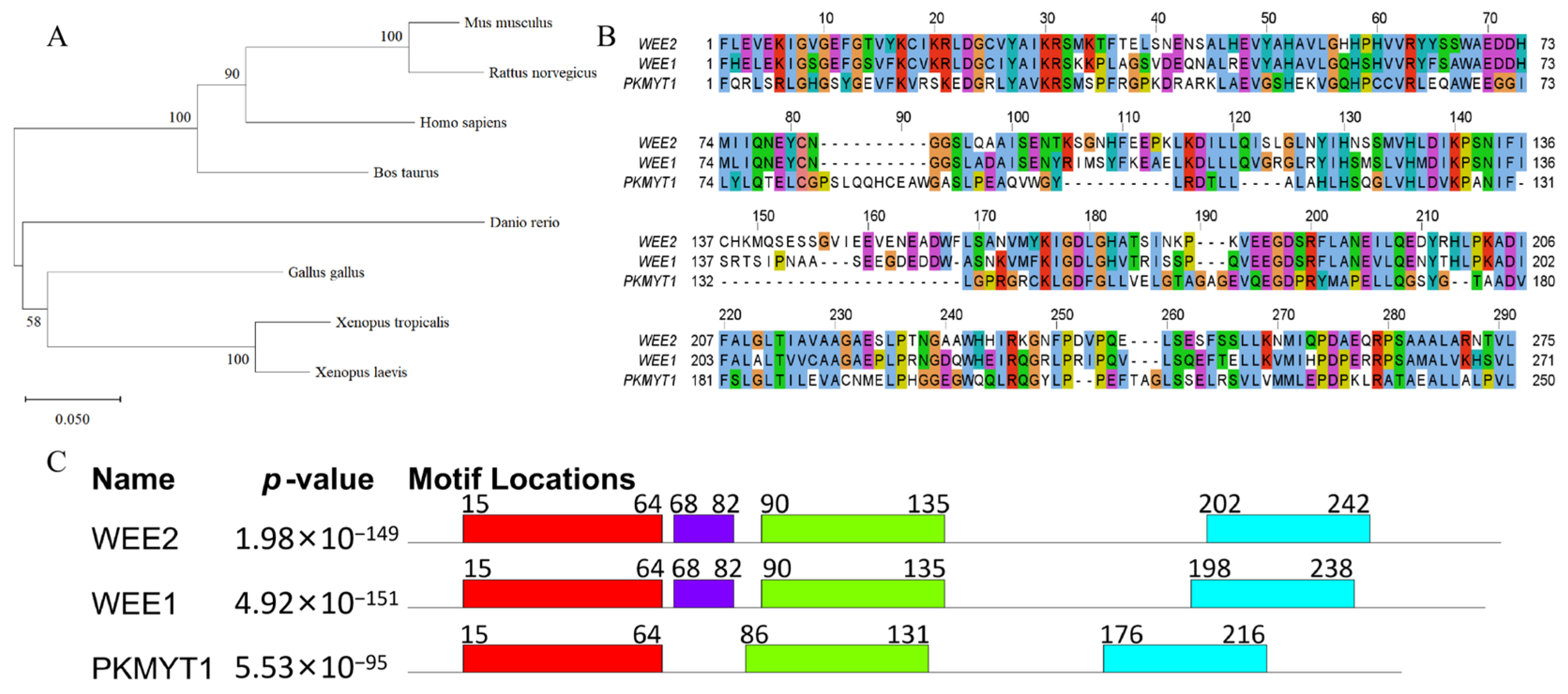
Each member of the WEE family is distributed on distinct chromosomes. The WEE2 gene (also known as WEE1B) is located on human chromosome 7. According to the NCBI database, its full-length genomic sequence spans 22,919 nucleotides (nt) and comprises 12 exons. The canonical mRNA transcript is 3061 nt in length, encoding a protein of 567 amino acids (aa). As an oocyte-specific cell cycle regulatory kinase, WEE2 exhibits a highly evolutionarily conserved kinase domain across species from lower vertebrates (Xenopus laevis) to higher mammals (Homo sapiens), with >93% sequence identity among closely related species, indicating essential and irreplaceable biological functions (Figure 2A). Multiple sequence alignment using Jalview (v2.11.5.1) revealed that the kinase domain of human WEE2 shares 63.27% identity with WEE1, markedly higher than the 33.7% identity with PKMYT1, suggesting a closer phylogenetic relationship between WEE2 and WEE1. Nevertheless, the core functional motifs within the kinase domains remain highly conserved across all three proteins, reflecting the functional constraint characteristic of the protein kinase family (Figure 2B). Homologous motif prediction using the MEME Suite (v5.5.9) identified four highly significant conserved motifs (p < 1 × 10−90) in human WEE2 and its family kinases. WEE2 shares all four motifs with WEE1, whereas PKMYT1 shares only motifs 1, 2, and 3. These results support the homologous evolutionary relationship among the three kinases and underscore the functional conservation of CDK1 phosphorylation and cell cycle arrest mediated by their conserved domains (Figure 2C).

3.2. Domain Architecture and Protein Sequence Features of WEE2

WEE2 is a 567-amino-acid (aa) protein, whereas WEE1 and PKMYT1 comprise 646 aa and 499 aa, respectively. Jalview alignment [19] shows that the overall sequence identity among the three proteins is considerably lower than within the kinase domain: 44.98% between WEE2 and WEE1, 26.39% between WEE2 and PKMYT1, and 26.65% between WEE1 and PKMYT1. Thus, functional divergence among the three kinases arises not only from subtle differences within the kinase domain but also largely from distinct sequences and phosphorylation sites in the N- and C-terminal regions.
Experimental 3D structures of catalytic domains from human WEE kinases are all derived from conventional X-ray crystallography, and no resolved structures exist for their N- and C-terminal regulatory regions [18]. Domain annotations from UniProt (Figure 3A) reveal marked divergence in N-/C-terminal sequence features distribution among WEE family members, forming the structural basis for their functional specificity. WEE2 contains intrinsically disordered regions (IDRs) at both the N-terminus (residues 1–117) and C-terminus (514–567), linked to the central kinase domain by flexible linkers. In contrast, WEE1 possesses a longer N-terminal IDR but no C-terminal IDR. PKMYT1 harbors two short N-terminal IDRs, one C-terminal IDR, and additional interaction regions for PIN1(Peptidylprolyl Cis/Trans Isomerase, NIMA-Interacting 1) [20] and the CDK1–cyclin B complex. PIN1 interacts with the C-terminal domain of Myt1 in a phosphorylation-dependent way. These differences collectively constitute critical molecular evidence for the functional divergence of WEE kinases.
A search of the PhosphoSitePlus database [21] (https://www.phosphosite.org/, accessed on 23 May 2026) revealed a total of 6 phosphorylation sites on the full-length human WEE2 protein that have been verified by both high-throughput mass spectrometry and low-throughput experiments. Among these, T225 and T423 are located within the kinase domain (Figure 3B). Alignment with the UniProt database confirmed that T225 is homologous to S312 of WEE1 and S120 of PKMYT1, both of which reside within the ATP-binding pocket. Phosphorylation at this site may regulate kinase catalytic activity by modulating ATP binding efficiency. Prediction results from the NetPhos 3.1 server [22] (https://services.healthtech.dtu.dk/services/NetPhos-3.1/, accessed on 23 May 2026) indicated that high-confidence phosphorylation sites (>0.7 threshold) in WEE2 are highly enriched in the N- and C-terminal IDRs, and are specifically recognized only by PKA and PKC, demonstrating high kinase specificity. In contrast, WEE1 is recognized by a broader spectrum of kinases, including PKA, PKC, CDK5, CKII, and PKB. PKMYT1 is primarily targeted by PKA, PKC, and PKB, exhibiting intermediate kinase specificity between WEE2 and WEE1.

3.3. WEE2 Secondary and Tertiary Structures

AlphaFold2 prediction combined with ESPript 3.2 visualization shows that the core α-helices and β-sheets in the kinase domains of WEE2, WEE1, and PKMYT1 are highly conserved in arrangement. The ATP-binding region in all three proteins adopts a β-sheet scaffold, reflecting conserved catalytic function within the family. Full-length WEE2 contains 12 α-helices, 4 310-helices (η), 9 β-sheets, and several random coils. The core kinase domain forms a stable catalytic scaffold, whereas the N- and C-terminal random coil regions serve as independent regulatory modules, providing sufficient binding space for PKA/PKC-mediated reproductive-specific phosphorylation. This is highly consistent with the terminal regulatory features predicted by UniProt and NetPhos 3.1, together constituting the structural basis for WEE2-specific regulation of oocyte meiosis.
The full-length tertiary structure of WEE2 features a compact core and flexible termini. Its kinase domain adopts a classic bilobal folding pattern: a small N-terminal subdomain and a large C-terminal subdomain linked by a flexible hinge, forming a catalytic pocket for ATP binding and substrate phosphorylation. Random coils appear as flexible disordered arms attached to the kinase domain, capable of regulating its activity through spatial conformational changes. The small N-terminal subdomain contains the ATP-binding motif and key salt bridges that stabilize the ATP-bound conformation. The large C-terminal subdomain houses the substrate-binding region, active site, magnesium ion-binding site, and activation loop; conserved residues in the catalytic loop promote phosphate transfer. Compared with WEE1, WEE2 possesses a shorter and conformationally more stable N-terminal random coil region, which prevents interference from non-specific signals. Unlike the membrane-localized PKMYT1, full-length WEE2 contains no transmembrane domain; its N- and C-terminal flexible disordered arms can swing freely, giving WEE2 a typical soluble protein conformation (Figure 3B–D). All above structural comparisons and phosphorylation predictions are derived from public bioinformatic webservers and the precomputed AlphaFold2 database, rather than original experimental structural data generated in this study.
In summary, structural differences between WEE2 and WEE1/PKMYT1 are mainly concentrated in reproduction-specific regulatory terminal amino acid sequences, secondary structure diversity, tissue specificity of substrate recognition, and inhibitor-binding structures. These differences likely arise from key residue mutations or local conformational changes, and they underpin the exclusive role of WEE2 in oocyte meiosis regulation. This makes WEE2 a specific target for the diagnosis and treatment of reproductive diseases, highlighting its unique biological value [23].

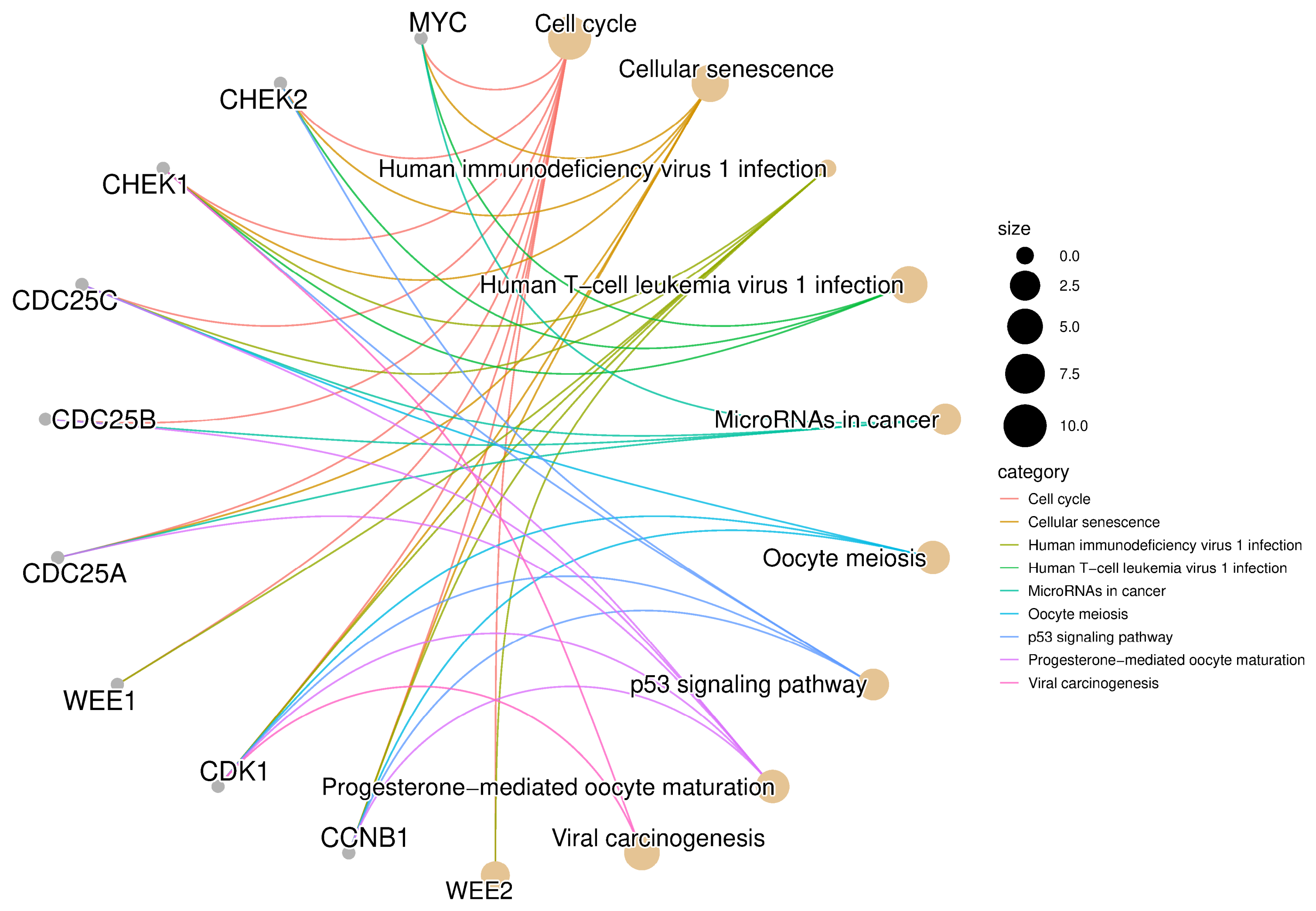
4. WEE2 Regulatory Network and Signaling Pathways

WEE2 does not function independently but is embedded in multiple highly conserved signaling pathways to form a precise regulatory network that collectively governs the arrest, maintenance and resumption of meiosis in oocytes (Figure 4). In mouse oocytes, WEE2 and PKMYT1 jointly maintain GV stage arrest: WEE2 inhibits nuclear CDK1, whereas PKMYT1 inhibits cytoplasmic CDK1, establishing a dual inhibitory mechanism. WEE2 also antagonizes CDC25 phosphatases: nuclear localized CDC25A and cytoplasm nucleus shuttling CDC25B can relieve the inhibition by WEE2 through dephosphorylating CDK1, thus driving the resumption of meiosis. In addition, the APC–CDH1 complex prevents premature CDK1 activation by degrading cyclin B1 and blocking its binding to CDK1. CDC14B phosphatase maintains APC–CDH1 activity (Anaphase-Promoting Complex–Cdc20 Homolog 1, a core E3 ubiquitin ligase, maintains GV arrest in oocytes), while EMI1 (Early Mitotic Inhibitor 1, an oocyte-specific endogenous inhibitor of APC–CDH1) inhibits its activity to balance cell cycle progression [5]. In the regulation of prophase of meiosis, the cAMP/PKA signaling pathway acts as the core upstream pathway [12], a mechanism clearly demonstrated in rhesus macaques: high intracellular levels of cAMP activate PKA, which in turn stabilizes and activates WEE2 via phosphorylation [1,24]. Activated WEE2 continuously phosphorylates the Tyr15 site of CDK1, suppresses the activity of MPF, maintains oocyte arrest at the GV stage, and prevents precocious meiotic entry. Meanwhile, PKA can phosphorylate CDC25B, causing it to bind 14-3-3 proteins and be retained in the cytoplasm [25].
Studies have shown that the surge of luteinizing hormone (LH) in vivo serves as a key signal for the resumption of GV stage arrest in mouse oocytes [6]. LH initiates a paracrine signaling pathway in oocytes by inducing granulosa cells to secrete EGF like factors, which closes gap junctions between oocytes and cumulus cells and lowers intracellular cGMP levels in oocytes. This in turn activates PDE3A to degrade cAMP and reduces PKA activity, thereby indirectly releasing meiotic arrest. In vitro experiments have confirmed that when oocytes are removed from follicles, intracellular cAMP spontaneously declines, directly leading to attenuated PKA activity. This results in decreased phosphorylation of WEE2 and nuclear translocation of CDC25B; once CDK1 is activated, meiosis resumes [5].
The CaMKII pathway mediates the exit of oocytes from MII arrest [16,26]. Sperm–oocyte fusion triggers intracellular Ca2+ oscillations, activating CaMKII. Activated CaMKII directly phosphorylates Ser15 of WEE2 and enhances its kinase activity [27], activated WEE2 phosphorylates and inhibits CDK1, lowering MPF activity. The downstream APC/C-CDC20 pathway is then activated, degrading cyclin B and inactivating MPF. MPF inactivation completes meiosis, extrudes the second polar body, and forms pronuclei, accomplishing fertilization [26]. When WEE2 function is lost or reduced, CDK1 remains uninhibited, causing persistent MPF activation. Even if sperm entry triggers Ca2+ oscillations, defective WEE2-mediated signal conversion prevents MPF inactivation, ultimately leading to MII exit failure and fertilization arrest.
These pathways and interacting molecules collectively form a WEE2-centered meiotic regulatory module that ensures the proper timing, fidelity, and developmental competence of oocyte maturation.

5. Biological Functions of WEE2

As an oocyte-specific protein kinase, WEE2 functions as a molecular switch for meiotic progression. It exhibits strict tissue specificity: high expression in the ovary (oocytes), low expression in the testis, and negligible expression in somatic tissues. KEGG pathway enrichment analysis indicates that WEE2 and its interacting proteins cooperatively regulate core reproductive pathways, such as oocyte meiosis and progesterone-mediated oocyte maturation, thereby ensuring precise control of the cell cycle and reproductive development (Figure 5). WEE2 localizes to the nucleus at the GV stage and disperses into the cytoplasm following GVBD [12]. It is continuously expressed throughout the GV, MI, and MII stages (Figure 6), declines after fertilization, and becomes undetectable in early embryonic development. Thus, WEE2 functions across the entire process from oocyte maturation to fertilization.

5.1. Dual Regulation of Oocyte Meiosis

During the GV stage, WEE2 phosphorylates CDK1 at Tyr15, thereby inhibiting CDK1 (a core subunit of MPF) and suppressing MPF activity. This arrests the cell cycle at the GV stage and prevents premature GVBD [3,4,5]. This process is coordinately regulated by the cAMP–PKA axis and the APC–CDH1 complex. Following a gonadotropin surge or a decrease in intracellular cAMP, the inhibitory effect of WEE2 is relieved, promoting meiotic resumption. At the MII stage, WEE2 serves as the core negative regulator maintaining meiotic arrest. Sperm-triggered Ca2+ oscillations inactivate and degrade WEE2, relieving its inhibition of CDK1 and leading to MPF inactivation. This ultimately promotes oocyte exit from MII, second polar body extrusion, and pronuclear formation, completing fertilization [26]. Functional abnormalities in WEE2 prevent oocyte exit from MII, resulting in fertilization arrest.

5.2. Factors Affecting Early Embryonic Development

WEE2 is a key regulatory molecule linking oocyte maturation and early embryonic development, playing an irreplaceable role in fertilization and the initiation of embryogenesis. In rhesus monkeys, WEE2 is significantly degraded after fertilization and becomes undetectable in the embryo [12]. Extensive WEE2 degradation relieves CDK1 inhibition, initiating the first zygotic mitosis and marking the official start of embryonic development. Insufficient WEE2 degradation leads to persistent CDK1 inhibition, preventing normal mitotic initiation and causing embryonic arrest. Multiple studies have shown that homozygous or compound heterozygous mutations in WEE2 result in marked absence or reduction in WEE2 expression. While most cases present with complete fertilization failure (no 2PN formation), a small subset of patients exhibit a very low rate of 2PN formation; however, all embryos show fragmented developmental arrest and fail to implant normally [28,29]. Neither conventional intracytoplasmic sperm injection (ICSI) nor artificial oocyte activation (AOA) improves fertilization outcomes [7].

5.3. Functions of WEE2 in Testis

According to the Genotype–Tissue Expression (GTEx) project, WEE2 is expressed in the testis and may downregulate spermatocyte meiotic progression during spermatogenesis. Studies have shown that a bitter taste receptor gene polymorphism in Caucasian men is associated with male infertility. The minor allele of TAS2R3-rs11763979 is associated with a decrease in the number of sperm with a normal morphology and with an increase in WEE2-AS1 expression. It is plausible to hypothesize that the increased WEE2-AS1 expression may downregulate WEE2, which in turn can alter the natural timing of sperm maturation, increasing the number of abnormal sperm cells [30].

5.4. Potential Functions of WEE2 in Skeletal Muscle

Beyond its reproductive roles, it is speculated that WEE2 may also possess non-reproductive functions. WEE2 was significantly enriched in a study investigating the association between aerobic capacity (AC) and the skeletal muscle proteome, where it was negatively correlated with AC and identified as a cross-model conserved associated protein. Pathway enrichment analysis revealed that in wild-type mice, WEE2 is enriched in the “cell cycle regulation” pathway, whereas in McArdle disease mice, it is enriched in “organic nitrogen compound metabolism” and “protein metabolism” pathways and classified under “phosphoprotein.” It is speculated that WEE2 may regulate phosphorylation of proteins involved in skeletal muscle cell cycle or metabolism [13]. This function awaits validation by in vivo and in vitro experiments but provides a new direction for expanding the tissue expression profile and functional scope of WEE2.

6. WEE2 and Diseases

6.1. Oocyte Maturation Defect (OMD)

OMD is a major cause of primary infertility in women, characterized by failure of oocytes to complete meiotic maturation either in vivo or during in vitro culture, thereby preventing normal fertilization. Monogenic factors are important pathogenic contributors, and WEE2 is one of the core causative genes [31]. Pathogenic mutations in WEE2 produce non-functional or functionally impaired truncated proteins, leading to defective kinase activity [32]. This results in premature GVBD or persistent MII arrest, ultimately causing asynchrony between nuclear and cytoplasmic maturation.

6.2. Total Fertilization Failure (TFF)

TFF is a serious complication in IVF and ICSI cycles, referring to failure of all mature oocytes to fertilize normally, with an incidence of 1–3% [33]. Its core etiology is oocyte activation defect (OAD), which can be sperm- or oocyte-related; WEE2 is the core oocyte-related pathogenic gene [34]. Clinical studies report a WEE2 mutation frequency of 6–20.8% in TFF patients [16]. Compound heterozygous mutations (e.g., c.1535+3A>G/c.946C>T) lead to loss of WEE2 function and continuous MPF activation. Even ICSI combined with artificial oocyte activation (ICSI-AOA) using calcium ionophore cannot effectively activate the ooplasm or form pronuclei, resulting in fertilization failure [28,35]. A prospective controlled study showed that the rs1476640 polymorphism in WEE2 is significantly associated with fertilization failure in women undergoing ICSI. The C allele increases risk (OR = 9.06, 95% CI: 3.27–25.14), while the T allele is protective. This polymorphism can be used to screen high-risk patients and guide personalized treatment strategies [36].

6.3. Early Embryonic Development Arrest (EDA)

EDA is a key cause of embryo transfer failure and reduced clinical pregnancy rates in IVF/ICSI cycles. It refers to developmental stagnation at a specific stage (typically 2- to 8-cell), preventing progression to blastocyst and implantation. Abnormalities in maternal effect genes, including WEE2, are core pathogenic factors. The detection rate of WEE2-related pathogenic mutations is approximately 5.95%, with embryos arresting at the 2–4-cell stage [9]. WEE2 abnormalities can lead to OMD, indirectly inducing fertilization disorders and EDA. Moreover, low-level residual expression of WEE2 in early post-fertilization embryos directly participates in regulating the MPF signaling pathway. Abnormal WEE2 expression or function disrupts MPF signaling, leading to developmental arrest.
All these diseases stem from pathological alterations in the WEE2 gene (Figure 6) and are primarily associated with human reproductive health, especially female fertility. The far higher detection rate of WEE2 pathogenic variants in infertile patients relative to healthy individuals highlights its essential function in governing the full reproductive cascade—oocyte maturation, fertilization activation, and early embryonic development—providing a basis for genetic diagnosis and treatment of infertility.

7. WEE2 Mutations

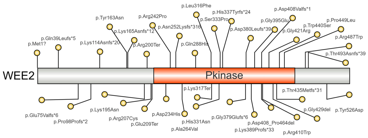
The protein encoded by WEE2 is a key kinase regulating oocyte meiosis. At present, whole-exome sequencing (WES) is widely used to identify pathogenic mutations in infertile female patients with a variety of specific phenotypes, such as oocyte maturation defects [37] and empty follicle syndrome [38,39], while not a universal routine assay applicable to all infertile patients [40], WES serves as a critical step in etiological diagnosis. Accumulating evidence confirms that WEE2 mutations play a critical role in oocyte maturation, fertilization, and early embryonic development, making it an important genetic cause of female infertility. Affected patients typically present with recurrent or complete fertilization failure, whereas heterozygous carriers generally show no clinical phenotype. Most WEE2 mutations are pathogenic, usually occurring as homozygous or compound heterozygous variants following an autosomal recessive inheritance pattern. The main mutation types include frameshift, nonsense, missense, start codon, and splice site mutations (Table 1). All relative positions of variants are shown in the lollipop diagram (Figure 7). According to ACMG criteria, most are classified as “pathogenic” or “likely pathogenic”.
The kinase activity and physiological function of WEE2 depend critically on its intact spatial conformation, particularly the correct folding and arrangement of the kinase domain, ATP-binding pocket, nuclear export sequence (NES), catalytic loop, and substrate-binding region. Most pathogenic mutations disrupt protein conformation integrity, directly or indirectly, leading to loss of kinase function. The reversibility of such conformational alterations is markedly site-dependent and mutation-type-specific. Different mutation types cause distinct aberrant conformations, all ultimately abolishing or significantly reducing kinase activity, primarily through disruption of the ATP-binding site, substrate-binding region, or autophosphorylation capacity.
Missense mutations induce local conformational disturbances and spatial rearrangements. For example, c.619C>T (p.R207C) destabilizes local conformation and impairs kinase domain function [8]; c.1346C>T (p.P449L) reduces cavity volume within the kinase domain [28], interfering with ATP binding or CDK recognition; c.946C>T occurs in the protein kinase domain and NES [16], potentially disrupting both kinase conformation and nuclear-cytoplasmic shuttling. Frameshift and nonsense mutations cause protein truncation and complete loss of native conformation. The nonsense mutation c.949A>T [28] and the frameshift mutation c.1006_1007dup14 introduce premature stop codons, leading to early translational termination and deletion of core structures such as the kinase domain. Splicing mutations in WEE2 cause domain deletions and abnormal conformations. The c.1222-1G>A variant leads to exon 9 loss in mRNA, producing an in-frame deletion of 57 amino acids in the kinase active domain, directly disrupting structural integrity and CDK1 phosphorylation function [32]. Additionally, several mutations induce aberrant subcellular localization. Missense mutations c.585G>C [7] and c.1319G>C [41] do not directly damage core kinase domain conformation but alter overall spatial folding, shifting WEE2 localization from nuclear to nucleocytoplasmic distribution, thereby abolishing its specific regulation of nuclear CDK1 and indirectly causing functional loss.
Table 1. Summary of the Types and Functional Consequences of WEE2 Gene Mutations.
LocationSequence
Variation
Amino Acid ChangeMutation TypeResultReference
Exon 1c.220_223delAAAGp.Glu75Valfs*6Frameshift deletionWEE2 degradation[26,35,42]
Exon 4c.700G>Cp.Asp234HisMissenseWEE2 degradation[35]
Exon 6c.1006_1007insTAp.His337Tyrfs*24Frameshift insertionWEE2 degradation[35]
Exon 5c.864G>Cp.Gln288HisMissenseAmino acid substitution[14]
Exon 1c.1A>Gp.Met1?Initiation
codon variant
Disruption of the start codon[14]
Exon 4c.619C>Tp.Arg207CysMissenseDisrupts the hydrogen bond and impairs the α-helix conformation[8]
Exon 9c.1228C>Tp.Arg410TrpMissenseAlters hydrogen bonds between residues in the secondary structure[15,26]
Exon 11c.1576T>Gp.Tyr526AspMissenseMay interfere with the C-terminal structure of WEE2 and reduce its function.[14]
Exon 9c.1261G>Ap.Gly421ArgMissenseMay impair the kinase domain and abolish its function.[14]
Exon 4c.725G>Cp.Arg242ProMissenseImpair the kinase domain and reduce its activity[26]
Exon 6c.997T>Cp.Ser333ProMissenseImpair the kinase domain and reduce its activity[26]
Exon 6c.991C>Ap.His331AsnMissenseImpair the kinase domain and reduce its activity[14]
Exon 8c.1184G>Ap.Gly395GluMissenseImpair the kinase domain and reduce its activity[26]
Exon 9c.1319G>Cp.Trp440SerMissenseAbnormal subcellular localization and reduced WEE2[41]
Exon 9c.1286_1288delGAGp.Gly429delIn-frame deletionDecreased WEE2[26]
Exon 10c.1473dupAp.Thr493Asnfs*39Frameshift insertionDecreased WEE2[35]
Exon 6c.949A>Tp.Lys317TerNonsenseDecreased WEE2[28]
Exon 9c.1346C>Tp.Pro449LeuMissenseDecreased WEE2[28]
Exon 1c.115_116insTp.Gln39Leufs*5Frameshift insertionDecreased WEE2[7]
Exon 4c.756_758delTGAp.Asn252Lysfs*316Frameshift deletionDecreased WEE2[7]
Exon 10c.1459C>Tp.Arg487TrpMissenseDecreased WEE2[7]
IVS 10c.1535+3A>Gp.?SplicingImpairs RNA splicing[16]
Exon 8c.1221G>Ap.Asp408Valfs*1Splicing variantDisrupt RNA splicing;
Produce truncated WEE2
[26,28]
Exon 9c.1222-1G>Ap.Asp408_Pro464delSplicingResults in exon 9 deletion of WEE2 mRNA[32]
IVS 4c.759-2A>Gp.?SplicingDisrupts the normal splicing site[43]
Exon 6c.946C>Tp.Leu316PheMissenseMay affect WEE2 phosphorylation and its nuclear localization[16]
Exon 3c.585G>Cp.Lys195AsnMissenseInduces enhanced nuclear export of WEE2[15]
Exon 2c.487T>Ap.Tyr163AsnMissenseDecreased WEE2 kinase activity[9]
Exon 9c.1304_1307delCCAAp.Thr435Metfs*31Frameshift deletionCDK1 phosphorylation fails [14]
Exon 5c.791C>Tp.Ala264ValMissenseDecreased pY15 of CDK1[44]
Exon 8c.1165_1168delAAACp.Lys389Profs*33Frameshift deletionMay produce truncated WEE2 and disrupt protein structure[9]
Exon 1c.293_294insCTGAGACACCAGCCCAACCp.Pro98Profs*2Frameshift insertionProduce truncated WEE2[14]
Exon 4c.598C>Tp.Arg200TerNonsenseProduce truncated WEE2[26,41]
Exon 1c.341_342delAAp.Lys114Asnfs*20Frameshift deletionProduce truncated WEE2 [14]
IVS 7c.1136-2A>Gp.Gly379Glufs*6/p.Asp380Leufs*39SplicingProduce truncated WEE2 [15]
Exon 6c.1006_1007dupp.His337Tyrfs*24Frameshift duplicationProduce truncated WEE2 [15]
Exon 4c.625G>Tp.Glu209TerNonsenseProduce truncated WEE2 [43]
Exon 3c.495del ap.Lys165Asnfs*12Frameshift deletionProduce truncated protein[45]
All mutations were identified in females with primary infertility, including novel private variants from individual patients and recurrent mutations detected across independent infertility cohorts. Variants were preliminarily screened via WES and further verified by Sanger sequencing. a Relevant nucleotide information was not reported in the original text. WEE2: WEE2 protein. The * represents a stop codon.

8. WEE2 Inhibitors for Non-Hormonal Female Contraception

Current oral emergency contraceptives are predominantly hormone-based (e.g., levonorgestrel and ulipristal acetate), which must be administered before the LH surge to block ovulation, have no effect on fertilization or implantation, and disrupt menstrual cycle timing [46]. Therefore, developing a non-hormonal, on-demand contraceptive that inhibits fertilization without disturbing the menstrual cycle represents a breakthrough for women’s autonomous fertility control.
Kinases are highly druggable; their activity can be modulated by small-molecule drugs. Humans encode 518 kinases across seven groups [47]. Most kinase-targeted drugs are Type I inhibitors that bind the ATP-binding pocket, with selectivity enhanced by unique amino acid sequences; allosteric modulators have also emerged. WEE2, an oocyte-specific kinase, uniquely suppresses meiosis during oocyte maturation initiation and regulates meiosis at fertilization. Existing research indicates that WEE2 rescues the lethal phenotype of WEE1 mutants in fission yeast, and WEE2 overexpression induces cell cycle arrest at the G2/M transition. Accordingly, we speculate that WEE2 overexpression may affect somatic cell mitosis, which offers theoretical support for developing WEE2 inhibitors (instead of structural analogs) to inhibit fertilization [48,49]. Selective WEE2 inhibitors exert contraceptive activity without affecting embryonic development or somatic cell function, a non-hormonal mechanism validated in mouse and non-human primate models [12,24].
Beyond conventional high-throughput screening (HTS), virtual HTS (vHTS) evaluates small-molecule binding via computational simulation, enabling rapid identification of active compounds. Combining vHTS with stepwise in vitro functional assays allows selection of selective WEE2 inhibitors [46,49]. Based on mechanism, WEE2 inhibitors fall into two categories: allosteric inhibitors (bind outside the ATP pocket, alter conformation, higher selectivity, less ATP competition) and ATP-competitive inhibitors (occupy the ATP pocket, block catalysis, most common type) (Table 2).
Table 2. Information Related to WEE2 Inhibitor.
NumberNameStructureWEE2
ΔTm (°C)
WEE1
ΔTm (°C)
PKMYT1
ΔTm
(°C)
WEE2
IC50
(μM)
WEE1
IC50
(μM)
WEE2 Inhibition Rate (%)WEE1 Inhibition Rate (%)Embryonic Cleavage Rate (%)HEK293 Cells Proliferation InhibitionReference
1GPHR-00336382Not disclosed3.5--5.8 ± 1.2>100≥80~0--[4]
2GPHR-00355672Not disclosed5.5--5.5 ± 0.9>100≥80~0--[4]
3Compound 2Cells 15 01147 i00113.3----946511Significant[48]
4Compound 12Cells 15 01147 i00211.6----73~6536None[48]
5Compound 16Cells 15 01147 i0037.0----70~5747None[48]
6Bosutinib isomerCells 15 01147 i00412.3103-----Significant *[18]
Kinase inhibition rates were determined using compounds 3, 4, and 5 at 1 μM, and cleavage rates were measured at 10 μM. * The cellular proliferation IC50 values of Bosutinib isomer were 1.6 ± 0.19 μM in HEK293T cells. -: Data not provided in the cited literature.

8.1. Allosteric Inhibitors: GPHR-00336382 and GPHR-00355672

Using a full-length WEE2 homology model and the kinase domain crystal structure (PDB: 5VDK), researchers applied vHTS and HTS to comprehensive and allosteric-focused libraries [4]. After multiple rounds of validation to eliminate interfering and non-specific compounds, two highly selective allosteric WEE2 inhibitors were successfully identified: GPHR-00336382 and GPHR-00355672, with IC50 values against WEE2 of 5.8 ± 1.2 μM and 5.5 ± 0.9 μM, respectively. GPHR-00336382 binds to full-length WEE2 but not to the kinase domain, whereas GPHR-00355672 binds to the kinase domain. ELISA (Enzyme-Linked Immunosorbent Assay) results showed that both compounds reduced WEE2 activity by at least 80% but exerted almost no effect on WEE1 activity. Up to now, functional verification of the two inhibitors is limited to in vitro enzyme assays. Lack of cellular, embryonic and in vivo animal data leaves their cellular permeability, in vivo potency and toxicity uncharacterized.

8.2. ATP-Competitive Inhibitors

8.2.1. Non-Selective Inhibitors

Hanna et al. identified potent and selective WEE2-targeted compounds using vHTS, molecular docking, and structural similarity analysis [48]. Compound 2 is a broad-spectrum protein tyrosine kinase inhibitor. ELISA assays confirmed that Compound 2 exhibited remarkable WEE2 inhibitory activity. Furthermore, biological activity evaluation demonstrated that Compound 2 significantly reduced embryonic cleavage rates, with only 11% of embryos initiating development. Notably, cell proliferation assays revealed that Compound 2 also displayed pronounced cross-inhibition against WEE1, thereby interfering with mitosis. Therefore, the inhibitor has low target specificity with severe off-target effects, cannot selectively regulate WEE2 function alone, and carries major safety hazards in vivo.

8.2.2. Selective Inhibitors Targeting WEE2

Compounds 12 and 16 are both commercially available analogs of Compound 2 identified via similarity searching. ELISA assays confirmed that both compounds also reduce WEE2 activity. Biological activity tests showed that they decreased fertilization rates to 36% and 47%, respectively, while their effects on mitosis were negligible. These results indicate that both compounds represent promising candidates for further development or structural modification [48]. Like the aforementioned Compound 2, these two inhibitors have only been tested for activity in embryonic models without accurate IC50 quantification. The shortage of cellular kinetic and in vivo efficacy data warrants further scaffold optimization.

8.2.3. Broad-Spectrum Kinase Inhibitors with Preferential Activity Against WEE2

A study confirmed that bosutinib isomer is a potent and selective broad-spectrum inhibitor with a preference for WEE2 [18]. Zhu et al. characterized its binding to WEE1, WEE2 and PKMYT1 using isothermal titration calorimetry (ITC) and found that it exhibits potent nanomolar affinity for WEE2 (Kd = 4.7 ± 2.3 nM), the strongest among the tested compounds. Thermodynamic and affinity analyses revealed a markedly enhanced specific binding preference for WEE2: its affinity for WEE2 is approximately 9.3-fold higher than for WEE1 (WEE1: Kd = 43.7 ± 10 nM), whereas binding to PKMYT1 is significantly reduced (Kd = 444 ± 41 nM), nearly 100-fold weaker than to WEE2, allowing efficient discrimination among the three kinases. Notably, it shows relatively weak antiproliferative activity at the cellular level, with an IC50 of 1.6–3.9 μM, and its in vivo utility may be limited by cellular permeability or its kinase selectivity profile. Therefore, these limitations may substantially restrict their subsequent in vivo animal studies and translational applications.
Three distinct strategies have emerged for WEE2 inhibitor development: (i) combined screening of allosteric-focused and comprehensive libraries; (ii) classical vHTS integrated with core scaffold derivatization and optimization; and (iii) repurposing known kinase inhibitors. Additionally, Shahid et al. recently identified candidate drugs including midostaurin and nilotinib through drug repurposing studies via virtual docking and molecular dynamics simulations [50]. Allosteric inhibitors target a unique WEE2 site with no cross-inhibition, while ATP-competitive inhibitors achieve selectivity via structural differences between WEE2 and WEE1 and side-chain modifications. Both categories show definite WEE2 inhibitory activity, laying a foundation for druggability optimization and clinical translation, and promise to provide novel non-hormonal options for female-controlled contraception.

9. Discussion

9.1. Functional Differentiation and Evolution of the WEE Family

WEE2, together with WEE1 and PKMYT1, belongs to the WEE kinase family. As an oocyte-specific WEE kinase, WEE2 exhibits clear functional divergence from the ubiquitously expressed WEE1 and PKMYT1. Both WEE2 and WEE1 are primarily localized in the nucleus, whereas PKMYT1 is mainly distributed in the endoplasmic reticulum and Golgi apparatus. In addition to blocking the G2/M transition [51,52], WEE1 can also participate in replication fork stability and S-phase checkpoint regulation by phosphorylating CDK2 [53,54]. PKMYT1 exhibits dual substrate specificity and functional redundancy, being capable of phosphorylating both Tyr15 and Thr14 residues simultaneously [55,56], and can specifically compensate for the loss of WEE1 function when WEE1 is impaired [57]. In contrast, WEE2 is not involved in somatic DNA damage repair or cell cycle regulation. Its core functions are confined to maintaining the timing of oocyte meiosis, promoting oocyte maturation, and initiating fertilization, with no functional redundancy with PKMYT1 [58].
In normal cells, WEE1 functions as a tumor suppressor. In tumor cells, however, WEE1 can exhibit “pseudo-oncogene” properties, and its high expression conversely becomes critical for cancer cell survival [59]. For example, WEE1 is highly expressed and exerts pro tumorigenic effects in multiple cancers including gastric cancer [60], leukemia, and melanoma [61]. Similarly, PKMYT1 is highly expressed and exerts pro-tumorigenic effects in most cancers, including chronic myeloid leukemia (CML) [62], breast cancer [63,64], and colorectal cancer (CRC) [65], but is downregulated and functions as a tumor suppressor in lung adenocarcinoma (LUAD) [66]. In addition, PKMYT1 also exhibits non-canonical functions in pancreatic cancer [67] and prostate cancer [68]. In contrast, WEE2 is not involved in tumorigenesis and has not been implicated in cancer development, invasion, or metastasis. Its absence results exclusively in female reproductive disorders, rather than somatic abnormalities or cancer susceptibility.

9.2. Effects of WEE2 Mutations on Protein Conformation and Functional Reversibility

Conformational changes induced by WEE2 mutations generally lack reversibility, and the potential for functional restoration is highly dependent on mutation type, site, and extent of damage. Most pathogenic mutations that directly disrupt backbone folding or core functional structures—including frameshift, nonsense, and splicing mutations, as well as missense mutations affecting core catalytic residues or conserved hydrogen bonds—cause irreversible alterations, leading to complete loss of function and representing major genetic causes of human fertilization failure. Conventional AOA cannot reverse these deficiencies. In contrast, a small number of weakly pathogenic missense mutations that cause only mild local conformational disturbances (e.g., disruption of non-core hydrogen or ionic bonds without affecting overall kinase domain folding) can be rescued by exogenous interventions. For example, microinjection of wild-type WEE2 cRNA into oocytes carrying such mutations enables expression of properly folded wild-type WEE2 protein, which functionally replaces the mutant variant and restores fertilization capacity. In vitro studies confirm that this approach supports morphologically normal blastocyst formation with no significant chromosomal abnormalities [16].

9.3. Challenges and Clinical Applications of WEE2-Targeted Inhibitor Development

While WEE2 loss in humans causes irreversible fertilization failure and full female sterility, two CRISPR-edited WEE2 knockout mouse strains with either a 17 bp deletion or a 21,878 bp large genomic deletion display markedly different reproductive traits. Both knockout mouse lines displayed normal growth, ovarian development, ovulation and estrous cycles. Only full-length knockout mice had slightly reduced litter sizes, without a complete infertility phenotype. Thus, mouse models fail to mimic human WEE2-mediated infertility and are unsuitable for assessing the in vivo contraceptive activity of WEE2 inhibitors [3]. Currently, the development of WEE2 inhibitors still faces critical technical bottlenecks. The limited screening scope of compound libraries makes it difficult to obtain candidates with high activity and affinity. The full-length structure of WEE2 remains unsolved, and its kinase domain lacks catalytic activity [18], rendering homology modeling and virtual screening unable to accurately reflect its true allosteric sites and conformational dynamics. Meanwhile, conventional cell systems hardly mimic the physiological microenvironment of oocytes; related experiments are costly and technically challenging. Therefore, it is urgent to establish a somatic cell-based high-throughput screening model to improve the efficiency and accuracy of validation. In clinical translation, there are also multiple challenges including off-target effects, toxic side effects, and the lack of a comprehensive evaluation system.
Early studies showed that there are species-specific conservation differences in the phosphorylation sites of WEE2, and its nuclear localization is critical for its function, making target positioning highly significant for drug design [69,70]. Studies have confirmed that the activity of porcine WEE2 entirely depends on the cAMP/PKA pathway [71], which is highly conserved among humans, pigs, and mice [6], suggesting that WEE2 can serve as a key pharmacological target. Currently, most WEE2 inhibitors target the ATP-binding pocket of the kinase catalytic domain. However, this pocket is highly conserved across the WEE kinase family, resulting in poor inhibitor selectivity and frequent off-target effects [23]. Functional homology in other domains further increases the risk of non-specific binding [18]. In addition, the selectivity data of existing inhibitors are only based on three kinases: WEE1, WEE2, and PKMYT3, lacking full verification of the entire kinase family. Therefore, non-specific binding to other kinases cannot be ruled out, indicating potential safety risks.
At present, inhibitors targeting WEE1 and PKMYT1 have entered clinical trials. WEE1 inhibitors (e.g., AZD1775) and PKMYT1 inhibitors (e.g., RP-6306) both induce prematurely mitotic entry and subsequent death of DNA-damaged cells [72,73]. Nevertheless, since both WEE1 and PKMYT1 are also expressed in normal cells, therapeutic administration is accompanied by adverse effects. Specifically, AZD1775 can severely impair bone marrow function and even induce aplastic anemia [74], whereas PKMYT1 inhibitors exhibit relatively mild toxicity, predominantly manifesting as rash and gastrointestinal reactions [75]. WEE1, PKMYT1 and WEE2 belong to the same gene family and share high structural and functional homology; accordingly, inhibitors with poor selectivity are prone to off-target cross-inhibition. For this reason, pregnant or fertile-age cancer patients receiving WEE1/PKMYT1 inhibitors must strictly follow medical advice to avoid reproductive toxicity. Similarly, insufficiently selective WEE2 inhibitors may also cause adverse reactions via off-target cross-inhibition. Therefore, great attention must be paid to the selectivity of WEE2 inhibitors in clinical applications, and standardized medication regimens should be strictly implemented to minimize risks.

10. Conclusions and Perspectives

As an oocyte-specific WEE kinase, WEE2 regulates meiosis and fertilization by phosphorylating CDK1 at Tyr15, and its structure determines its germline-specific function. Pathogenic mutations in WEE2 are a major genetic cause of female infertility. Although some progress has been made in targeted inhibitors, issues such as limited molecules, insufficient activity, and low selectivity remain. Future efforts are needed to characterize its structure, improve the mutation spectrum, optimize inhibitors, and explore non-reproductive functions and gene therapies to promote precision diagnosis and treatment in reproductive medicine and improve female reproductive health.

Author Contributions

R.W.: Project administration, methodology, visualization, resources writing—original draft preparation; J.Y., Y.-J.L., G.-S.Z., X.L., Y.-F.J. and C.-H.L.: project administration, validation, visualization; G.-J.Y. and J.C.: conceptualization, funding acquisition, investigation, supervision, writing—review and editing. All authors have read and agreed to the published version of the manuscript.

Funding

This research was funded by the Natural Science Foundation of Zhejiang Province (LY24C190001) and Natural Science Foundation of Ningbo Municipality (2024J168).

Institutional Review Board Statement

Not applicable.

Informed Consent Statement

Not applicable.

Data Availability Statement

No new data were created or analyzed in this study.

Acknowledgments

The authors express gratitude for English editing software, such as ChatGPT-5.5 Instant and DeepSeek-V4, for refining language, checking grammatical errors, and enhancing logical coherence in the manuscript.

Conflicts of Interest

The authors declare no conflicts of interest.

Abbreviations

The following abbreviations are used in this manuscript:
GVGerminal vesicle
CDK1Cyclin-dependent kinase 1
MPFMaturation-promoting factor
MIIMetaphase II
WEE2Wee1-like protein kinase 2
WEE1Wee1-like protein kinase
PKMYT1Membrane-associated tyrosine- and threonine-specific cdc2-inhibitory kinase
GVBDGerminal vesicle breakdown
OMDOocyte maturation defects
TFFTotal fertilization failure
EDAEarly embryonic arrest
IVFIn vitro fertilization
IDRsIntrinsically disordered regions
PKAProtein Kinase A
cAMPCyclic Adenosine Monophosphate
cGMPCyclic Guanosine Monophosphate
CaMCalmodulin
PDE3APhosphodiesterase 3A
CaMKIICalcium/Calmodulin-Dependent Protein Kinase II
IP3Inositol 1,4,5-trisphosphate
PIP2Phosphatidylinositol 4,5-bisphosphate
EGFEpidermal Growth Factor
LHLuteinizing Hormone
CDC25ACell Division Cycle 25 Homolog A
CDC25BCell Division Cycle 25 Homolog B
CDC14BCell Division Cycle 14 Homolog B
APC-CDH1Anaphase-Promoting Complex-Cdc20 Homolog 1
APC/C-CDC20Anaphase-Promoting Complex/Cyclosome-CDC20
EMI1Early Mitotic Inhibitor 1
14-3-3Tyrosine 3-Monooxygenase/Tryptophan 5-Monooxygenase Activation Protein
EREndoplasmic Reticulum
GolgiGolgi Apparatus
ICSIIntracytoplasmic sperm injection
AOAArtificial oocyte activation
GTExGenotype-Tissue Expression
ACAerobic capacity
MYCMYC proto-oncogene
CHEK1Checkpoint kinase 1
CHEK2Checkpoint kinase 2
CDC25CCell division cycle 25 homolog C
CCNB1Cyclin B1
OADOocyte activation defect
PNPronuclear stage
NESNuclear export sequence
HTSHigh-throughput screening
vHTSVirtual HTS
ITCIsothermal titration calorimetry
CMLChronic myeloid leukemia
CRCColorectal cancer
LUADLung adenocarcinoma
PIN1Peptidylprolyl Cis/Trans Isomerase, NIMA-Interacting 1

References

  1. Liu, C.; Liu, Y.; Liu, Y.; Wu, D.; Luan, Z.; Wang, E.; Yu, B. Ser 15 of WEE1B is a potential PKA phosphorylation target in G2/M transition in one-cell stage mouse embryos. Mol. Med. Rep. 2013, 7, 1929–1937. [Google Scholar] [CrossRef] [Scilit] [PubMed]
  2. Aarts, M.; Sharpe, R.; Garcia-Murillas, I.; Gevensleben, H.; Hurd, M.S.; Shumway, S.D.; Toniatti, C.; Ashworth, A.; Turner, N.C. Forced mitotic entry of S-phase cells as a therapeutic strategy induced by inhibition of WEE1. Cancer Discov. 2012, 2, 524–539. [Google Scholar] [CrossRef] [Scilit] [PubMed]
  3. Nozawa, K.; Liao, Z.; Satouh, Y.; Geng, T.; Ikawa, M.; Monsivais, D.; Matzuk, M.M. Oocyte-specific Wee1-like protein kinase 2 is dispensable for fertility in mice. PLoS ONE 2023, 18, e0289083. [Google Scholar] [CrossRef] [Scilit] [PubMed]
  4. Hanna, C.B.; Mudaliar, D.; John, K.; Allen, C.L.; Sun, L.; Hawkinson, J.E.; Schönbrunn, E.; Georg, G.I.; Jensen, J.T. Development of WEE2 kinase inhibitors as novel non-hormonal female contraceptives that target meiosis. Biol. Reprod. 2020, 103, 368–377. [Google Scholar] [CrossRef] [Scilit] [PubMed]
  5. Solc, P.; Schultz, R.M.; Motlik, J. Prophase I arrest and progression to metaphase I in mouse oocytes: Comparison of resumption of meiosis and recovery from G2-arrest in somatic cells. Mol. Hum. Reprod. 2010, 16, 654–664. [Google Scholar] [CrossRef] [Scilit] [PubMed]
  6. Han, S.J.; Conti, M. New pathways from PKA to the Cdc2/cyclin B complex in oocytes: Wee1B as a potential PKA substrate. Cell Cycle 2006, 5, 227–231. [Google Scholar] [CrossRef] [Scilit] [PubMed]
  7. Jin, J.; Tong, X.; Zhang, Y.L.; Yang, W.; Ma, Y.; Ren, P.; Zhou, F.; Zhang, S. Novel WEE2 compound heterozygous mutations identified in patients with fertilization failure or poor fertilization. J. Assist. Reprod. Genet. 2021, 38, 2861–2869. [Google Scholar] [CrossRef] [Scilit] [PubMed]
  8. Yang, X.; Shu, L.; Cai, L.; Sun, X.; Cui, Y.; Liu, J. Homozygous missense mutation Arg207Cys in the WEE2 gene causes female infertility and fertilization failure. J. Assist. Reprod. Genet. 2019, 36, 965–971. [Google Scholar] [CrossRef] [Scilit] [PubMed]
  9. Zhao, H.; Ouyang, N.; Ou, S.; Lin, H.; Liao, Z.; Liu, W.; Chen, H.; Yuan, P. Identification of novel variants and expansion of the phenotypic spectrum in PATL2, WEE2, and TUBB8 associated with human early embryonic arrest. J. Assist. Reprod. Genet. 2025, 42, 1961–1973. [Google Scholar] [CrossRef] [Scilit] [PubMed]
  10. Mueller, P.R.; Leise, W.F., 3rd. Measurement of Wee kinase activity. Methods Mol. Biol. 2005, 296, 299–328. [Google Scholar] [CrossRef] [Scilit] [PubMed]
  11. Zhang, Z.; Harish, R.; Elahi, N.; Saini, S.; Telia, A.; Kundlas, M.; Koroleva, A.; Umoh, I.N.; Lota, M.; Bilkhu, M.; et al. Targeting WEE1 Kinase for Breast Cancer Therapeutics: An Update. Int. J. Mol. Sci. 2025, 26, 5701. [Google Scholar] [CrossRef] [Scilit] [PubMed]
  12. Hanna, C.B.; Yao, S.; Patta, M.C.; Jensen, J.T.; Wu, X. WEE2 is an oocyte-specific meiosis inhibitor in rhesus macaque monkeys. Biol. Reprod. 2010, 82, 1190–1197. [Google Scholar] [CrossRef] [Scilit] [PubMed]
  13. Plaza-Florido, A.; Santos-Lozano, A.; López-Ortiz, S.; Gálvez, B.G.; Arenas, J.; Martín, M.A.; Valenzuela, P.L.; Pinós, T.; Lucia, A.; Fiuza-Luces, C. Aerobic capacity and muscle proteome: Insights from a mouse model. Exp. Physiol. 2025, 110, 293–306. [Google Scholar] [CrossRef] [Scilit] [PubMed]
  14. Zhao, S.; Chen, T.; Yu, M.; Bian, Y.; Cao, Y.; Ning, Y.; Su, S.; Zhang, J.; Zhao, S. Novel WEE2 gene variants identified in patients with fertilization failure and female infertility. Fertil. Steril. 2019, 111, 519–526, Erratum in Fertil. Steril. 2019, 111, 1032. https://doi.org/10.1016/j.fertnstert.2019.03.029. [Google Scholar] [CrossRef] [Scilit] [PubMed]
  15. Dai, J.; Zheng, W.; Dai, C.; Guo, J.; Lu, C.; Gong, F.; Li, Y.; Zhou, Q.; Lu, G.; Lin, G. New biallelic mutations in WEE2: Expanding the spectrum of mutations that cause fertilization failure or poor fertilization. Fertil. Steril. 2019, 111, 510–518. [Google Scholar] [CrossRef] [Scilit] [PubMed]
  16. Tian, Y.; Wang, G.; Wang, J.; Mu, X.; Chen, H.; Song, X.; Bai, X. Novel compound heterozygous mutation in WEE2 is associated with fertilization failure: Case report of an infertile woman and literature review. BMC Womens Health 2020, 20, 246. [Google Scholar] [CrossRef] [Scilit] [PubMed]
  17. Wang, Y.; Wang, C.; Ren, F.; Zhavoronkov, A.; Ding, X. Recent advances in the discovery of PKMYT1-targeting drugs: A patent review (2021–2025). Expert Opin. Ther. Pat. 2026, 36, 555–579. [Google Scholar] [CrossRef] [Scilit] [PubMed]
  18. Zhu, J.Y.; Cuellar, R.A.; Berndt, N.; Lee, H.E.; Olesen, S.H.; Martin, M.P.; Jensen, J.T.; Georg, G.I.; Schönbrunn, E. Structural Basis of Wee Kinases Functionality and Inactivation by Diverse Small Molecule Inhibitors. J. Med. Chem. 2017, 60, 7863–7875. [Google Scholar] [CrossRef] [Scilit] [PubMed]
  19. Guo, Y.; Zhu, X.; Song, L.; Wang, Y.; Gao, J.; Yuan, E.; Yu, H.; Fang, Y.; Shi, Q.; Zhao, D.; et al. Genetic analysis of isolated methylmalonic acidemia in Henan, China: c.1663G>A variant of MMUT prevalent in the Henan population. Clin. Chim. Acta 2024, 553, 117729. [Google Scholar] [CrossRef] [Scilit] [PubMed]
  20. Wells, N.J.; Watanabe, N.; Tokusumi, T.; Jiang, W.; Verdecia, M.A.; Hunter, T. The C-terminal domain of the Cdc2 inhibitory kinase Myt1 interacts with Cdc2 complexes and is required for inhibition of G(2)/M progression. J. Cell Sci. 1999, 112, 3361–3371. [Google Scholar] [CrossRef] [Scilit] [PubMed]
  21. Rinehart, J. Cryptic phosphorylation sites provide new structural and functional insights into the human phosphoproteome. Trends Biochem. Sci. 2026, 51, 98–99. [Google Scholar] [CrossRef] [Scilit] [PubMed]
  22. Ma, Y.F.; Wu, Z.H.; Gao, M.; Loor, J.J. Nuclear factor erythroid 2-related factor 2-antioxidant activation through the action of ataxia telangiectasia-mutated serine/threonine kinase is essential to counteract oxidative stress in bovine mammary epithelial cells. J. Dairy Sci. 2018, 101, 5317–5328. [Google Scholar] [CrossRef] [Scilit] [PubMed]
  23. Qi, X.; Li, G.; Liu, J.; Mou, L.; Zhang, Y.; Guo, S.; Chen, X.; Li, W. Structural and energetic insights into the selective inhibition of PKMYT1 against WEE1. J. Biomol. Struct. Dyn. 2024, 42, 3010–3018. [Google Scholar] [CrossRef] [Scilit] [PubMed]
  24. Han, S.J.; Chen, R.; Paronetto, M.P.; Conti, M. Wee1B is an oocyte-specific kinase involved in the control of meiotic arrest in the mouse. Curr. Biol. 2005, 15, 1670–1676. [Google Scholar] [CrossRef] [Scilit] [PubMed]
  25. Kaláb, P.; Solc, P.; Motlík, J. The role of RanGTP gradient in vertebrate oocyte maturation. Results Probl. Cell Differ. 2011, 53, 235–267. [Google Scholar] [CrossRef] [Scilit] [PubMed]
  26. Zhang, Z.; Mu, J.; Zhao, J.; Zhou, Z.; Chen, B.; Wu, L.; Yan, Z.; Wang, W.; Zhao, L.; Dong, J.; et al. Novel mutations in WEE2: Expanding the spectrum of mutations responsible for human fertilization failure. Clin. Genet. 2019, 95, 520–524. [Google Scholar] [CrossRef] [Scilit] [PubMed]
  27. Oh, J.S.; Susor, A.; Conti, M. Protein tyrosine kinase Wee1B is essential for metaphase II exit in mouse oocytes. Science 2011, 332, 462–465. [Google Scholar] [CrossRef] [Scilit] [PubMed]
  28. Guo, H.; Wu, H.; Yan, Z.; Yin, M.; Wu, L.; Li, B. Novel WEE2 homozygous mutations c.1346C>T and c.949A>T identified in primary infertile women due to unexplained fertilization failure. Clin. Genet. 2023, 104, 700–704. [Google Scholar] [CrossRef] [Scilit] [PubMed]
  29. Sang, Q.; Zhou, Z.; Mu, J.; Wang, L. Genetic factors as potential molecular markers of human oocyte and embryo quality. J. Assist. Reprod. Genet. 2021, 38, 993–1002. [Google Scholar] [CrossRef] [Scilit] [PubMed]
  30. Gentiluomo, M.; Crifasi, L.; Luddi, A.; Locci, D.; Barale, R.; Piomboni, P.; Campa, D. Taste receptor polymorphisms and male infertility. Hum. Reprod. 2017, 32, 2324–2331. [Google Scholar] [CrossRef] [Scilit] [PubMed]
  31. Loeuillet, C.; Dhellemmes, M.; Cazin, C.; Kherraf, Z.E.; Fourati Ben Mustapha, S.; Zouari, R.; Thierry-Mieg, N.; Arnoult, C.; Ray, P.F. A recurrent ZP1 variant is responsible for oocyte maturation defect with degenerated oocytes in infertile females. Clin. Genet. 2022, 102, 22–29. [Google Scholar] [CrossRef] [Scilit] [PubMed]
  32. Liu, Z.; Zhu, L.; He, H.; Hou, M.; Jia, W.; Jin, L.; Xi, Q.; Zhang, X. Novel splicing mutations in PATL2 and WEE2 cause oocyte degradation and fertilization failure. J. Assist. Reprod. Genet. 2024, 41, 3337–3345. [Google Scholar] [CrossRef] [Scilit] [PubMed]
  33. Sfontouris, I.A.; Nastri, C.O.; Lima, M.L.; Tahmasbpourmarzouni, E.; Raine-Fenning, N.; Martins, W.P. Artificial oocyte activation to improve reproductive outcomes in women with previous fertilization failure: A systematic review and meta-analysis of RCTs. Hum. Reprod. 2015, 30, 1831–1841. [Google Scholar] [CrossRef] [Scilit] [PubMed]
  34. Campos, G.; Sciorio, R.; Esteves, S.C. Total fertilization failure after ICSI: Insights into pathophysiology, diagnosis, and management through artificial oocyte activation. Hum. Reprod. Update 2023, 29, 369–394. [Google Scholar] [CrossRef] [Scilit] [PubMed]
  35. Sang, Q.; Li, B.; Kuang, Y.; Wang, X.; Zhang, Z.; Chen, B.; Wu, L.; Lyu, Q.; Fu, Y.; Yan, Z.; et al. Homozygous Mutations in WEE2 Cause Fertilization Failure and Female Infertility. Am. J. Hum. Genet. 2018, 102, 649–657. [Google Scholar] [CrossRef] [Scilit] [PubMed]
  36. Al-Gawwam, Z.H.M.; Abdulhussein, R.H.; Abed, H.S.; Wei, W.; Cai, M. Association of WEE2 Gene Polymorphism with Fertilization Failure in Women Undergoing Intracytoplasmic Sperm Injection. Int. J. Womens Health 2025, 17, 3045–3053. [Google Scholar] [CrossRef] [Scilit] [PubMed]
  37. Pan, Z.; Ye, Z.; Ye, H.; Chen, B.; Mu, J.; Zhang, Z.; Li, Q.; Kuang, Y.; Wang, L.; Sang, Q.; et al. Novel variants in PABPC1L cause female infertility due to oocyte maturation defects. J. Assist. Reprod. Genet. 2026, 43, 443–455. [Google Scholar] [CrossRef] [Scilit] [PubMed]
  38. Wang, J.; Yang, X.; Sun, X.; Ma, L.; Yin, Y.; He, G.; Zhang, Y.; Zhou, J.; Cai, L.; Liu, J.; et al. A novel homozygous nonsense mutation in zona pellucida 1 (ZP1) causes human female empty follicle syndrome. J. Assist. Reprod. Genet. 2021, 38, 1459–1468. [Google Scholar] [CrossRef] [Scilit] [PubMed]
  39. Jia, W.; Xi, Q.; Zhu, L.; Luo, Y.; Li, Z.; Hou, M.; Zhang, D.; Yang, X.; Hu, J.; Jin, L.; et al. Novel mutations in ZP2 and ZP3 cause female infertility in three patients. J. Assist. Reprod. Genet. 2022, 39, 1205–1215. [Google Scholar] [CrossRef] [Scilit] [PubMed]
  40. Van Der Kelen, A.; Okutman, Ö.; Javey, E.; Serdarogullari, M.; Janssens, C.; Ghosh, M.S.; Dequeker, B.J.H.; Perold, F.; Kastner, C.; Kieffer, E.; et al. A systematic review and evidence assessment of monogenic gene-disease relationships in human female infertility and differences in sex development. Hum. Reprod. Update 2023, 29, 218–232. [Google Scholar] [CrossRef] [Scilit] [PubMed]
  41. Zhou, X.; Zhu, L.; Hou, M.; Wu, Y.; Li, Z.; Wang, J.; Liu, Z.; Zhang, D.; Jin, L.; Zhang, X. Novel compound heterozygous mutations in WEE2 causes female infertility and fertilization failure. J. Assist. Reprod. Genet. 2019, 36, 1957–1962. [Google Scholar] [CrossRef] [Scilit] [PubMed]
  42. Weiner, H.S.; Ulrich, N.D.; Hipp, L.; Hammoud, A.; Xu, M.; Schon, S.B. Total fertilization failure with in vitro fertilization-intracytoplasmic sperm injection related to WEE2 mutation highlights emerging importance of genetic causes of in vitro fertilization failure. FS Rep. 2022, 3, 355–360. [Google Scholar] [CrossRef] [Scilit] [PubMed]
  43. Wang, A.; Huang, S.; Liu, M.; Wang, B.; Wu, F.; Zhu, D.; Zhao, X. Clinical exome sequencing identifies novel compound heterozygous mutations of the WEE2 gene in primary infertile women with fertilization failure. Gynecol. Endocrinol. 2021, 37, 1096–1101. [Google Scholar] [CrossRef] [Scilit] [PubMed]
  44. Xie, S.; Zhang, M.; Wu, F.; Yu, R.; Fu, J.; Shi, R.; Li, Q.; Chen, B.; Shi, J.; Wu, T.; et al. Novel homozygous variants in ASTL and WEE2 responsible for female infertility characterized by abnormal fertilization. J. Assist. Reprod. Genet. 2025, 42, 3817–3827. [Google Scholar] [CrossRef] [Scilit] [PubMed]
  45. Yang, J.; Wang, Z.; Guo, Y.; Yan, B.; Ding, Z.; Ni, Y. Analysis of a Chinese pedigree with female infertility due to WEE2 gene c.495del homozygous frameshifting variant induced fertilization disorder. Chin. J. Med. Genet. 2024, 41, 1478–1482. [Google Scholar] [CrossRef] [PubMed]
  46. Gemzell-Danielsson, K.; Berger, C.; Lalitkumar, P.G. Mechanisms of action of oral emergency contraception. Gynecol. Endocrinol. 2014, 30, 685–687. [Google Scholar] [CrossRef] [Scilit] [PubMed]
  47. Manning, G.; Whyte, D.B.; Martinez, R.; Hunter, T.; Sudarsanam, S. The protein kinase complement of the human genome. Science 2002, 298, 1912–1934. [Google Scholar] [CrossRef] [Scilit] [PubMed]
  48. Hanna, C.B.; Yao, S.; Martin, M.; Schönbrunn, E.; Georg, G.I.; Jensen, J.T.; Cuellar, R.A.D. Identification and Screening of Selective WEE2 Inhibitors to Develop Non-Hormonal Contraceptives that Specifically Target Meiosis. ChemistrySelect 2019, 4, 13363–13369. [Google Scholar] [CrossRef] [Scilit] [PubMed]
  49. Nakanishi, M.; Ando, H.; Watanabe, N.; Kitamura, K.; Ito, K.; Okayama, H.; Miyamoto, T.; Agui, T.; Sasaki, M. Identification and characterization of human Wee1B, a new member of the Wee1 family of Cdk-inhibitory kinases. Genes Cells 2000, 5, 839–847. [Google Scholar] [CrossRef] [Scilit] [PubMed]
  50. Ali, S.; Elasbali, A.M.; Alzahrani, W.; Mohammad, T.; Hassan, M.I.; Zhou, T. Identification of FDA-Approved Drugs as Potential Inhibitors of WEE2: Structure-Based Virtual Screening and Molecular Dynamics with Perspectives for Machine Learning-Assisted Prioritization. Life 2026, 16, 185. [Google Scholar] [CrossRef] [Scilit] [PubMed]
  51. Schmidt, M.; Rohe, A.; Platzer, C.; Najjar, A.; Erdmann, F.; Sippl, W. Regulation of G2/M Transition by Inhibition of WEE1 and PKMYT1 Kinases. Molecules 2017, 22, 2045. [Google Scholar] [CrossRef] [Scilit] [PubMed]
  52. Serpico, A.F.; D’Alterio, G.; Vetrei, C.; Della Monica, R.; Nardella, L.; Visconti, R.; Grieco, D. Wee1 Rather Than Plk1 Is Inhibited by AZD1775 at Therapeutically Relevant Concentrations. Cancers 2019, 11, 819. [Google Scholar] [CrossRef] [Scilit] [PubMed]
  53. Li, C.; Shen, Q.; Zhang, P.; Wang, T.; Liu, W.; Li, R.; Ma, X.; Zeng, X.; Yin, Y.; Tao, K. Targeting MUS81 promotes the anticancer effect of WEE1 inhibitor and immune checkpoint blocking combination therapy via activating cGAS/STING signaling in gastric cancer cells. J. Exp. Clin. Cancer Res. 2021, 40, 315. [Google Scholar] [CrossRef] [Scilit] [PubMed]
  54. Payliss, B.J.; Tse, Y.W.E.; Reichheld, S.E.; Lemak, A.; Yun, H.Y.; Houliston, S.; Patel, A.; Arrowsmith, C.H.; Sharpe, S.; Wyatt, H.D.M. Phosphorylation of the DNA repair scaffold SLX4 drives folding of the SAP domain and activation of the MUS81-EME1 endonuclease. Cell Rep. 2022, 41, 111537. [Google Scholar] [CrossRef] [Scilit] [PubMed]
  55. Zhang, H.; Yu, J.; Yang, Z.; Guo, Z.; Liu, R.; Qin, Q.; Sun, Y.; Liu, N.; Gao, Z.; Zhao, D.; et al. Structure-based virtual screening discovers novel PKMYT1 inhibitors. RSC Med. Chem. 2024, 15, 3114–3124. [Google Scholar] [CrossRef] [Scilit]
  56. Wang, C.; Wang, Y.; Meng, F.; Liu, T.; Wang, X.; Cai, X.; Zhang, M.; Aliper, A.; Ren, F.; Zhavoronkov, A.; et al. Discovery of pyrrolopyrimidinone derivatives as potent PKMYT1 inhibitors for the treatment of cancer. Eur. J. Med. Chem. 2025, 281, 117025. [Google Scholar] [CrossRef] [Scilit] [PubMed]
  57. Li, J.; Lu, J.; Xu, M.; Yang, S.; Yu, T.; Zheng, C.; Huang, X.; Pan, Y.; Chen, Y.; Long, J.; et al. ODF2L acts as a synthetic lethal partner with WEE1 inhibition in epithelial ovarian cancer models. J. Clin. Investig. 2023, 133, e161544. [Google Scholar] [CrossRef] [Scilit] [PubMed]
  58. Nishimura, T.; Shimaoka, T.; Kano, K.; Naito, K. Insufficient amount of Cdc2 and continuous activation of Wee1 B are the cause of meiotic failure in porcine growing oocytes. J. Reprod. Dev. 2009, 55, 553–557. [Google Scholar] [CrossRef] [Scilit]
  59. Ghelli Luserna di Rorà, A.; Cerchione, C.; Martinelli, G.; Simonetti, G. A WEE1 family business: Regulation of mitosis, cancer progression, and therapeutic target. J. Hematol. Oncol. 2020, 13, 126. [Google Scholar] [CrossRef] [Scilit]
  60. Feng, Y.; Wang, C.; Shi, T.; Liu, W.; Liu, H.; Zhu, B.; Gao, F. Enhancer of zeste homolog 2 exerts functions in gastric cancer development via modulating microRNA-222-3p methylation and WEE1 expression. Chem. Biol. Drug Des. 2022, 100, 320–333. [Google Scholar] [CrossRef] [Scilit] [PubMed]
  61. Matheson, C.J.; Backos, D.S.; Reigan, P. Targeting WEE1 Kinase in Cancer. Trends Pharmacol. Sci. 2016, 37, 872–881. [Google Scholar] [CrossRef] [Scilit] [PubMed]
  62. Okabe, S.; Arai, Y.; Gotoh, A.; Akahane, D. Combination of WEE1 Inhibitor and Vitamin K2 Enhances Therapeutic Efficacy in Chronic Myeloid Leukemia. Cancer Innov. 2025, 4, e70024. [Google Scholar] [CrossRef] [Scilit] [PubMed]
  63. Chen, A.; Kim, B.J.; Mitra, A.; Vollert, C.T.; Lei, J.T.; Fandino, D.; Anurag, M.; Holt, M.V.; Gou, X.; Pilcher, J.B.; et al. PKMYT1 Is a Marker of Treatment Response and a Therapeutic Target for CDK4/6 Inhibitor-Resistance in ER+ Breast Cancer. Mol. Cancer Ther. 2024, 23, 1494–1510. [Google Scholar] [CrossRef] [Scilit]
  64. Liu, Y.; Qi, J.; Dou, Z.; Hu, J.; Lu, L.; Dai, H.; Wang, H.; Yang, W. Systematic expression analysis of WEE family kinases reveals the importance of PKMYT1 in breast carcinogenesis. Cell Prolif. 2020, 53, e12741. [Google Scholar] [CrossRef] [Scilit] [PubMed]
  65. Zhang, Y.; Chang, Y.; Hu, Y.; Ding, Z.; Zhang, Z.; Wei, Y.; Liu, B.; Yang, M.; Chen, W. Exploring PKMYT1 as a potential marker for colorectal cancer progression through bioinformatics analyses and experimental validation. Sci. Rep. 2025, 15, 33016. [Google Scholar] [CrossRef] [Scilit] [PubMed]
  66. Wang, S.; Liu, X.; Zhou, T.; Li, J.; Lin, Y.; Zhou, A.; Huang, J.; Zhao, J.; Cai, J.; Cai, X.; et al. PKMYT1 inhibits lung adenocarcinoma progression by abrogating AKT1 activity. Cell. Oncol. 2023, 46, 195–209. [Google Scholar] [CrossRef] [Scilit] [PubMed]
  67. Wang, S.; Xiong, Y.; Luo, Y.; Shen, Y.; Zhang, F.; Lan, H.; Pang, Y.; Wang, X.; Li, X.; Zheng, X.; et al. Genome-wide CRISPR screens identify PKMYT1 as a therapeutic target in pancreatic ductal adenocarcinoma. EMBO Mol. Med. 2024, 16, 1115–1142. [Google Scholar] [CrossRef] [Scilit]
  68. Wang, J.; Wang, L.; Chen, S.; Peng, H.; Xiao, L.; Du, E.; Liu, Y.; Lin, D.; Wang, Y.; Xu, Y.; et al. PKMYT1 is associated with prostate cancer malignancy and may serve as a therapeutic target. Gene 2020, 744, 144608. [Google Scholar] [CrossRef] [Scilit] [PubMed]
  69. Shimaoka, T.; Nishimura, T.; Kano, K.; Naito, K. Analyses of the regulatory mechanism of porcine WEE1B: The phosphorylation sites of porcine WEE1B and mouse WEE1B are different. J. Reprod. Dev. 2011, 57, 223–228. [Google Scholar] [CrossRef] [Scilit] [PubMed]
  70. Oh, J.S.; Han, S.J.; Conti, M. Wee1B, Myt1, and Cdc25 function in distinct compartments of the mouse oocyte to control meiotic resumption. J. Cell Biol. 2010, 188, 199–207. [Google Scholar] [CrossRef] [Scilit] [PubMed]
  71. Shimaoka, T.; Nishimura, T.; Kano, K.; Naito, K. Critical effect of pigWee1B on the regulation of meiotic resumption in porcine immature oocytes. Cell Cycle 2009, 8, 2375–2384. [Google Scholar] [CrossRef] [Scilit]
  72. Wang, C.; Lu, X. WEE-family kinases in cancer: Synthetic lethal interactions and drug discovery. Trends Pharmacol. Sci. 2026, 47, 184–197. [Google Scholar] [CrossRef] [Scilit]
  73. Gallo, D.; Young, J.T.F.; Fourtounis, J.; Martino, G.; Álvarez-Quilón, A.; Bernier, C.; Duffy, N.M.; Papp, R.; Roulston, A.; Stocco, R.; et al. CCNE1 amplification is synthetic lethal with PKMYT1 kinase inhibition. Nature 2022, 604, 749–756. [Google Scholar] [CrossRef] [Scilit] [PubMed]
  74. Guler, S.; DiPoto, M.C.; Crespo, A.; Caldwell, R.; Doerfel, B.; Grossmann, N.; Ho, K.; Huck, B.; Jones, C.C.; Lan, R.; et al. Selective Wee1 Inhibitors Led to Antitumor Activity In Vitro and Correlated with Myelosuppression. ACS Med. Chem. Lett. 2023, 14, 566–576. [Google Scholar] [CrossRef] [Scilit] [PubMed]
  75. Repare Therapeutics Reports Proof of Concept for Lunresertib (RP-6306) in Clinic, Initial Monotherapy Data from Phase 1 MYTHIC Clinical Trial and Early Insights from Ongoing Combination Trials. Available online: https://ir.reparerx.com/news-releases/news-release-details/repare-therapeutics-reports-proof-concept-lunresertib-rp-6306/ (accessed on 18 June 2026).
Figure 1. PRISMA flowchart. Flow diagram summarizing the performed systematic review.
Cells 15 01147 g001
Figure 2. Evolutionary conservation and sequence analysis of WEE2 kinase domain. (A) Phylogenetic tree of WEE2 kinase domain sequences from different species. Analysis and tree construction were performed using MEGA12 software. Node values represent bootstrap support (%), indicating the reliability of evolutionary branches. (B) Multiple sequence alignment results of the kinase domains of WEE2, WEE1 and PKMYT1. This figure was constructed using the MAFFT tool in Jalview software with the classic Clustal color scheme. Different colors correspond to amino acids with distinct physicochemical properties, and darker shading indicates higher sequence conservation at this site. (C) Conserved motif analysis of the kinase domains of WEE2, WEE1 and PKMYT1. Analysis in this figure was performed using the MEME online suite. Different colors represent distinct conserved motifs.
Cells 15 01147 g002
Figure 3. Domain architecture and 3D structural mapping of WEE2, WEE1 and PKMYT1 kinases. (A) Comparative analysis of protein structural characteristics among the three members of the WEE kinase family. Data in this figure were retrieved from the UniProt database and visualized using the IBS 2.0 online server. (B) Schematic diagram of the tertiary structure of WEE2. (C) Schematic diagram of the tertiary structure of WEE1. (D) Schematic diagram of the tertiary structure of PKMYT1. (BD) were predicted by AlphaFold2 and visualized using PyMOL (3.1.6.1). Functional regions including N-terminal/C-terminal disordered regions, kinase domain, activation loop, ATP-binding sites and selected phosphorylation sites are highlighted in different colors with corresponding annotations.
Cells 15 01147 g003
Figure 4. Regulatory network and signaling pathways of WEE2. This figure depicts the core regulatory network of WEE2, including its upstream regulators, downstream target molecules, and the major signaling pathways involved in WEE2-mediated biological processes. Abbreviations: Protein Kinase A (PKA), Cyclic Adenosine Monophosphate (cAMP), Cyclic Guanosine Monophosphate (cGMP), Phosphodiesterase 3A (PDE3A), Calmodulin (CaM), Calcium/Calmodulin-Dependent Protein Kinase II (CaMKII), Inositol 1,4,5-trisphosphate (IP3), Phosphatidylinositol 4,5-bisphosphate (PIP2), Epidermal Growth Factor (EGF), Luteinizing Hormone (LH), Cell Division Cycle 25 Homolog A (CDC25A), Cell Division Cycle 25 Homolog B (CDC25B), Cell Division Cycle 14 Homolog B (CDC14B), Anaphase-Promoting Complex–Cdc20 Homolog 1 (APC–CDH1), Anaphase-Promoting Complex/Cyclosome–CDC20 (APC/C–CDC20), Early Mitotic Inhibitor 1 (EMI1), Tyrosine 3-Monooxygenase/Tryptophan 5-Monooxygenase Activation Protein (14-3-3), Endoplasmic Reticulum (ER), Golgi Apparatus (Golgi). P represents phosphorylation, and -P represents dephosphorylation.
Cells 15 01147 g004
Figure 5. Network analysis of WEE2-interacting proteins and their associated KEGG pathways. This figure demonstrates that these genes are predominantly enriched in the oocyte meiosis, progesterone-mediated oocyte maturation, cell cycle, and p53 signaling pathways, constituting a core molecular network regulating oocyte meiotic arrest and resumption. Interacting protein data were derived from STRING (https://string-db.org, accessed on 23 May 2026) and BioGRID (https://thebiogrid.org, accessed on 23 May 2026). Heat maps were plotted using the Bioinformatics Tools Platform (https://www.bioinformatics.com.cn, accessed on 23 May 2026). Abbreviations: MYC proto-oncogene (MYC), Checkpoint kinase 1 (CHEK1), Checkpoint kinase 2 (CHEK2), Cell division cycle 25 homolog C (CDC25C), Cyclin B1 (CCNB1).
Cells 15 01147 g005
Figure 6. WEE2 regulatory network, physiological functions, and clinical consequences of pathogenic mutations. (A) Regulatory function of WEE2 during oocyte development. (B) Systemic physiological functions of WEE2 in multiple tissues. (C) Clinical phenotypes and targeted interventions of pathogenic WEE2 mutations. Abbreviations: Germinal vesicle (GV), Metaphase II (MII), Cyclin-dependent kinase 1 (CDK1), Pronuclear stage (PN).
Cells 15 01147 g006
Figure 7. Lollipop diagram of pathogenic variant residue distribution in human WEE2 protein. The red shaded region represents the core protein kinase catalytic domain. Yellow circles mark the positions of amino acid alterations. The * represents a stop codon.
Cells 15 01147 g007
Disclaimer/Publisher’s Note: The statements, opinions and data contained in all publications are solely those of the individual author(s) and contributor(s) and not of MDPI and/or the editor(s). MDPI and/or the editor(s) disclaim responsibility for any injury to people or property resulting from any ideas, methods, instructions or products referred to in the content.