Cone Opsins and Inherited Retinal Disease


Abstract
1. Introduction
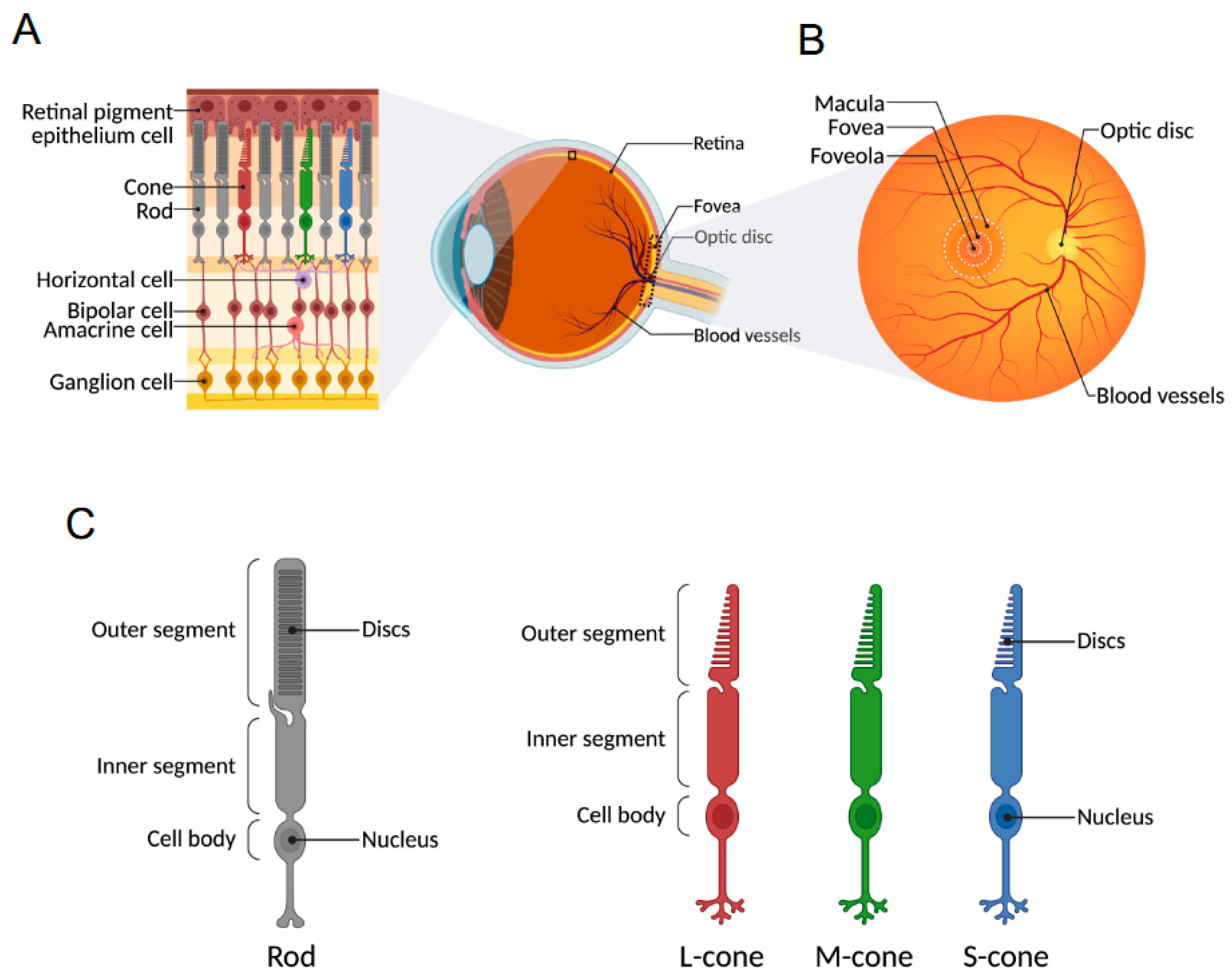
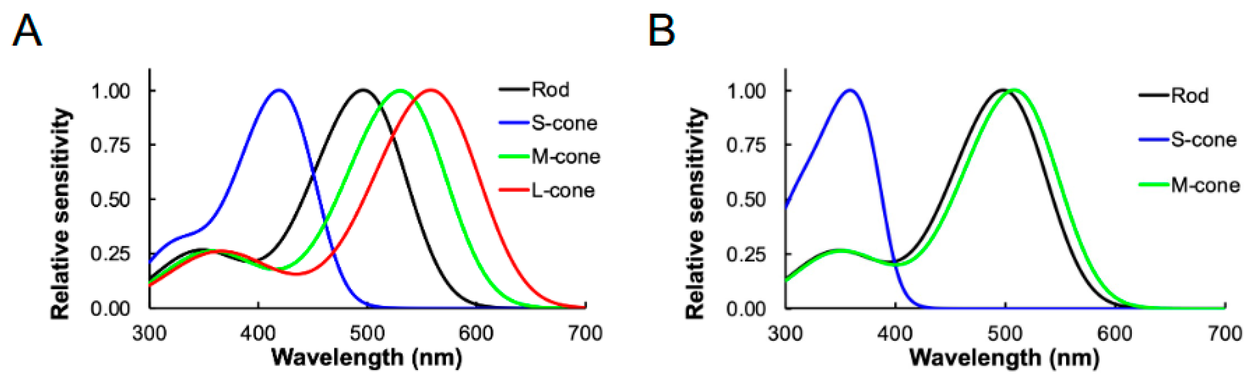
2. Cone Photoreceptor Cells and Cone Opsins
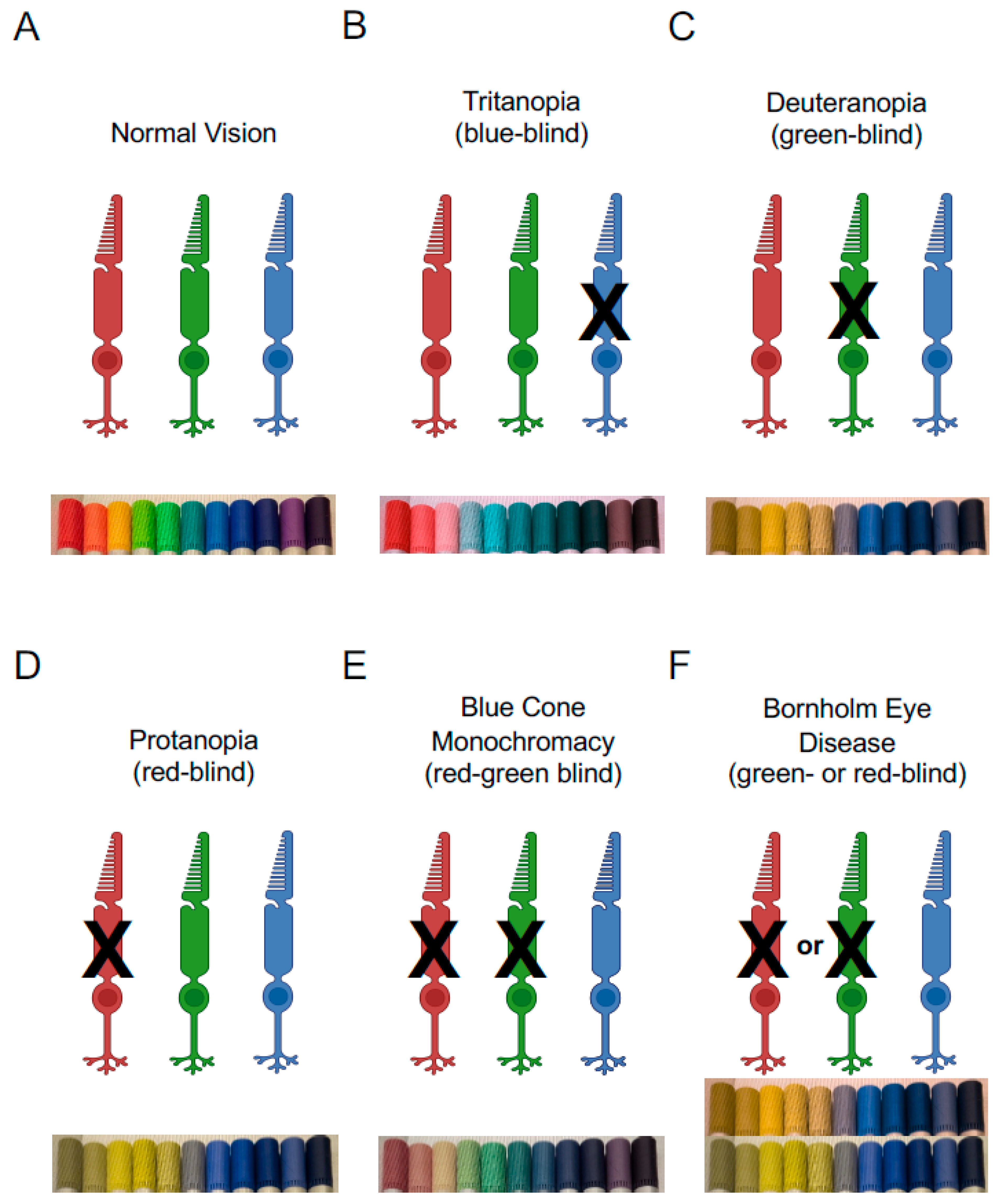
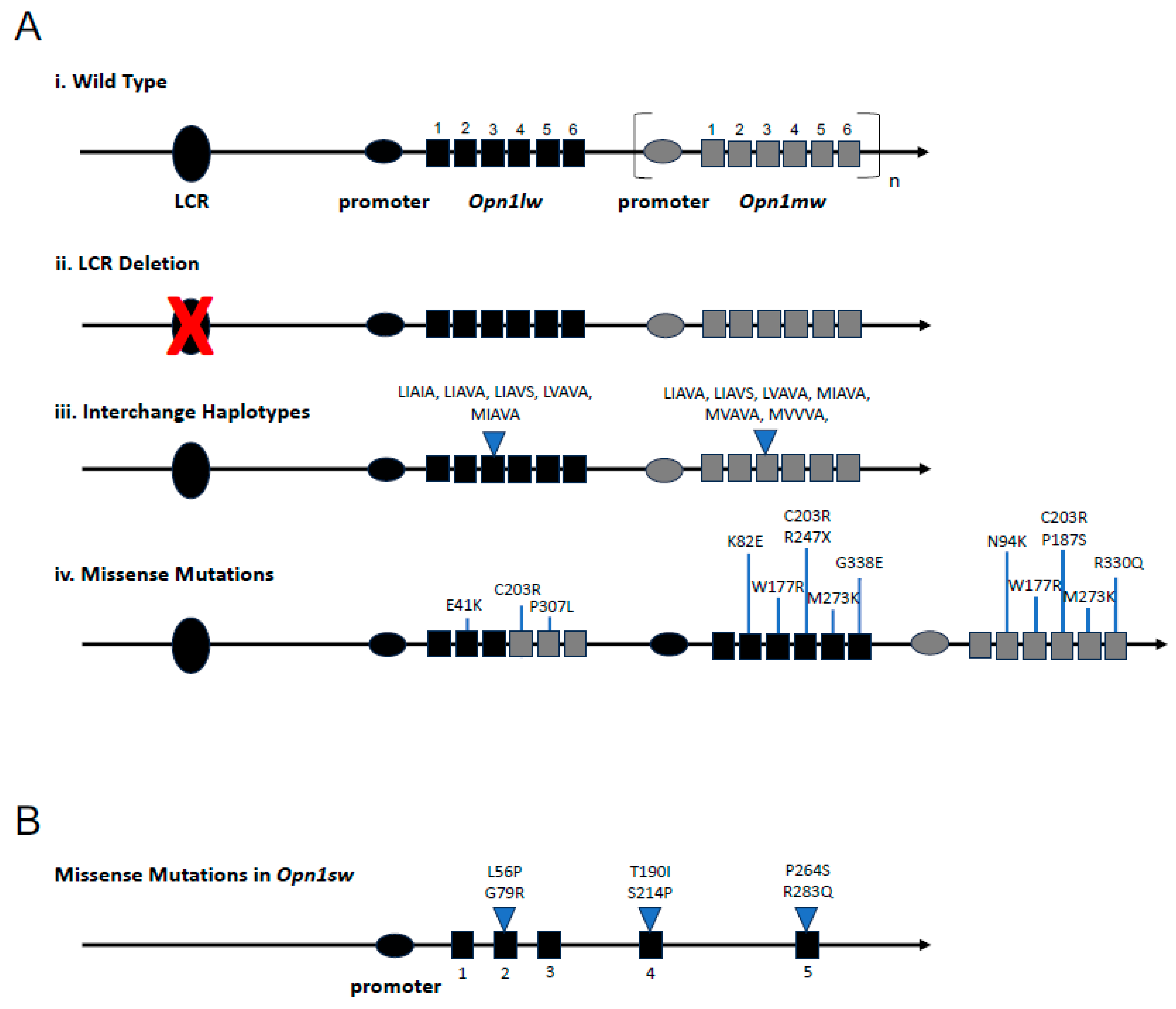
3. Cone Opsin Mutations and Disease
3.1. LCR Deletion
3.2. Interchange Haplotypes
3.3. Missense Mutations
4. Mouse Models with Cone Opsin Mutations
4.1. Mouse Models of Blue Cone Monochromacy
4.2. Mouse Models of Bornholm Eye Disease
5. Concluding Remarks
Author Contributions
Funding
Data Availability Statement
Conflicts of Interest
References
- Lamb, T.D. Why rods and cones? Eye 2016, 30, 179–185. [Google Scholar] [CrossRef] [PubMed]
- Baylor, D.A.; Lamb, T.D.; Yau, K.W. Responses of retinal rods to single photons. J. Physiol. 1979, 288, 613–634. [Google Scholar] [CrossRef]
- Chen, J.; Rattner, A.; Nathans, J. The rod photoreceptor-specific nuclear receptor Nr2e3 represses transcription of multiple cone-specific genes. J. Neurosci. 2005, 25, 118–129. [Google Scholar] [CrossRef] [PubMed]
- Cheng, H.; Khanna, H.; Oh, E.C.; Hicks, D.; Mitton, K.P.; Swaroop, A. Photoreceptor-specific nuclear receptor NR2E3 functions as a transcriptional activator in rod photoreceptors. Hum. Mol. Genet. 2004, 13, 1563–1575. [Google Scholar] [CrossRef] [PubMed]
- Curcio, C.A.; Sloan, K.R.; Kalina, R.E.; Hendrickson, A.E. Human photoreceptor topography. J. Comp. Neurol. 1990, 292, 497–523. [Google Scholar] [CrossRef] [PubMed]
- Ding, J.D.; Salinas, R.Y.; Arshavsky, V.Y. Discs of mammalian rod photoreceptors form through the membrane evagination mechanism. J. Cell Biol. 2015, 211, 495–502. [Google Scholar] [CrossRef] [PubMed]
- Arikawa, K.; Molday, L.L.; Molday, R.S.; Williams, D.S. Localization of peripherin/rds in the disk membranes of cone and rod photoreceptors: Relationship to disk membrane morphogenesis and retinal degeneration. J. Cell Biol. 1992, 116, 659–667. [Google Scholar] [CrossRef] [PubMed]
- Park, P.S. Constitutively active rhodopsin and retinal disease. Adv. Pharmacol. 2014, 70, 1–36. [Google Scholar] [CrossRef] [PubMed]
- Lamb, T.D. Photoreceptor physiology and evolution: Cellular and molecular basis of rod and cone phototransduction. J. Physiol. 2022, 600, 4585–4601. [Google Scholar] [CrossRef] [PubMed]
- Sjoberg, M.; Vennstrom, B.; Forrest, D. Thyroid hormone receptors in chick retinal development: Differential expression of mRNAs for alpha and N-terminal variant beta receptors. Development 1992, 114, 39–47. [Google Scholar] [CrossRef] [PubMed]
- Ng, L.; Ma, M.; Curran, T.; Forrest, D. Developmental expression of thyroid hormone receptor beta2 protein in cone photoreceptors in the mouse. Neuroreport 2009, 20, 627–631. [Google Scholar] [CrossRef] [PubMed]
- Mori, M.; Ghyselinck, N.B.; Chambon, P.; Mark, M. Systematic immunolocalization of retinoid receptors in developing and adult mouse eyes. Investig. Ophthalmol. Vis. Sci. 2001, 42, 1312–1318. [Google Scholar]
- Xu, J.; Zhao, C.; Kang, Y. The Formation and Renewal of Photoreceptor Outer Segments. Cells 2024, 13, 1357. [Google Scholar] [CrossRef] [PubMed]
- Nathans, J.; Thomas, D.; Hogness, D.S. Molecular genetics of human color vision: The genes encoding blue, green, and red pigments. Science 1986, 232, 193–202. [Google Scholar] [CrossRef] [PubMed]
- Curcio, C.A.; Allen, K.A.; Sloan, K.R.; Lerea, C.L.; Hurley, J.B.; Klock, I.B.; Milam, A.H. Distribution and morphology of human cone photoreceptors stained with anti-blue opsin. J. Comp. Neurol. 1991, 312, 610–624. [Google Scholar] [CrossRef] [PubMed]
- Ahnelt, P.K.; Kolb, H.; Pflug, R. Identification of a subtype of cone photoreceptor, likely to be blue sensitive, in the human retina. J. Comp. Neurol. 1987, 255, 18–34. [Google Scholar] [CrossRef] [PubMed]
- Szel, A.; Diamantstein, T.; Rohlich, P. Identification of the blue-sensitive cones in the mammalian retina by anti-visual pigment antibody. J. Comp. Neurol. 1988, 273, 593–602. [Google Scholar] [CrossRef] [PubMed]
- Hagen, L.A.; Arnegard, S.; Kuchenbecker, J.A.; Gilson, S.J.; Neitz, M.; Neitz, J.; Baraas, R.C. The association between L:M cone ratio, cone opsin genes and myopia susceptibility. Vis. Res. 2019, 162, 20–28. [Google Scholar] [CrossRef] [PubMed]
- Lu, Y.; Shiau, F.; Yi, W.; Lu, S.; Wu, Q.; Pearson, J.D.; Kallman, A.; Zhong, S.; Hoang, T.; Zuo, Z.; et al. Single-Cell Analysis of Human Retina Identifies Evolutionarily Conserved and Species-Specific Mechanisms Controlling Development. Dev. Cell 2020, 53, 473–491.e479. [Google Scholar] [CrossRef] [PubMed]
- Peirson, S.N.; Brown, L.A.; Pothecary, C.A.; Benson, L.A.; Fisk, A.S. Light and the laboratory mouse. J. Neurosci. Methods 2018, 300, 26–36. [Google Scholar] [CrossRef] [PubMed]
- Lucas, R.J.; Peirson, S.N.; Berson, D.M.; Brown, T.M.; Cooper, H.M.; Czeisler, C.A.; Figueiro, M.G.; Gamlin, P.D.; Lockley, S.W.; O’Hagan, J.B.; et al. Measuring and using light in the melanopsin age. Trends Neurosci. 2014, 37, 1–9. [Google Scholar] [CrossRef] [PubMed]
- Govardovskii, V.I.; Fyhrquist, N.; Reuter, T.; Kuzmin, D.G.; Donner, K. In search of the visual pigment template. Vis. Neurosci. 2000, 17, 509–528. [Google Scholar] [CrossRef] [PubMed]
- Thoreson, W.B.; Dacey, D.M. Diverse Cell Types, Circuits, and Mechanisms for Color Vision in the Vertebrate Retina. Physiol. Rev. 2019, 99, 1527–1573. [Google Scholar] [CrossRef] [PubMed]
- Jordan, G.; Deeb, S.S.; Bosten, J.M.; Mollon, J.D. The dimensionality of color vision in carriers of anomalous trichromacy. J. Vis. 2010, 10, 12. [Google Scholar] [CrossRef] [PubMed]
- Jordan, G.; Mollon, J.D. A study of women heterozygous for colour deficiencies. Vis. Res. 1993, 33, 1495–1508. [Google Scholar] [CrossRef] [PubMed]
- Nagy, A.L.; MacLeod, D.I.; Heyneman, N.E.; Eisner, A. Four cone pigments in women heterozygous for color deficiency. J. Opt. Soc. Am. 1981, 71, 719–722. [Google Scholar] [CrossRef] [PubMed]
- Bosten, J.M.; Robinson, J.D.; Jordan, G.; Mollon, J.D. Multidimensional scaling reveals a color dimension unique to ‘color-deficient’ observers. Curr. Biol. 2005, 15, R950–R952. [Google Scholar] [CrossRef] [PubMed][Green Version]
- Neitz, M.; Kraft, T.W.; Neitz, J. Expression of L cone pigment gene subtypes in females. Vis. Res. 1998, 38, 3221–3225. [Google Scholar] [CrossRef] [PubMed][Green Version]
- Mustafi, D.; Engel, A.H.; Palczewski, K. Structure of cone photoreceptors. Prog. Retin. Eye Res. 2009, 28, 289–302. [Google Scholar] [CrossRef] [PubMed]
- Neitz, J.; Neitz, M. The genetics of normal and defective color vision. Vis. Res. 2011, 51, 633–651. [Google Scholar] [CrossRef] [PubMed]
- Winderickx, J.; Battisti, L.; Hibiya, Y.; Motulsky, A.G.; Deeb, S.S. Haplotype diversity in the human red and green opsin genes: Evidence for frequent sequence exchange in exon 3. Hum. Mol. Genet. 1993, 2, 1413–1421. [Google Scholar] [CrossRef] [PubMed]
- Aboshiha, J.; Dubis, A.M.; Carroll, J.; Hardcastle, A.J.; Michaelides, M. The cone dysfunction syndromes. Br. J. Ophthalmol. 2016, 100, 115–121. [Google Scholar] [CrossRef] [PubMed]
- Reitner, A.; Sharpe, L.T.; Zrenner, E. Is colour vision possible with only rods and blue-sensitive cones? Nature 1991, 352, 798–800. [Google Scholar] [CrossRef] [PubMed]
- Gardner, J.C.; Michaelides, M.; Holder, G.E.; Kanuga, N.; Webb, T.R.; Mollon, J.D.; Moore, A.T.; Hardcastle, A.J. Blue cone monochromacy: Causative mutations and associated phenotypes. Mol. Vis. 2009, 15, 876–884. [Google Scholar] [PubMed]
- Haim, M.; Fledelius, H.C.; Skarsholm, D. X-linked myopia in Danish family. Acta Ophthalmol. 1988, 66, 450–456. [Google Scholar] [CrossRef]
- Schwartz, M.; Haim, M.; Skarsholm, D. X-linked myopia: Bornholm eye disease. Linkage to DNA markers on the distal part of Xq. Clin. Genet. 1990, 38, 281–286. [Google Scholar] [PubMed]
- Michaelides, M.; Johnson, S.; Bradshaw, K.; Holder, G.E.; Simunovic, M.P.; Mollon, J.D.; Moore, A.T.; Hunt, D.M. X-linked cone dysfunction syndrome with myopia and protanopia. Ophthalmology 2005, 112, 1448–1454. [Google Scholar] [CrossRef] [PubMed]
- Gardner, J.C.; Liew, G.; Quan, Y.H.; Ermetal, B.; Ueyama, H.; Davidson, A.E.; Schwarz, N.; Kanuga, N.; Chana, R.; Maher, E.R.; et al. Three different cone opsin gene array mutational mechanisms with genotype-phenotype correlation and functional investigation of cone opsin variants. Hum. Mutat. 2014, 35, 1354–1362. [Google Scholar] [CrossRef] [PubMed]
- McClements, M.; Davies, W.I.; Michaelides, M.; Young, T.; Neitz, M.; MacLaren, R.E.; Moore, A.T.; Hunt, D.M. Variations in opsin coding sequences cause x-linked cone dysfunction syndrome with myopia and dichromacy. Investig. Ophthalmol. Vis. Sci. 2013, 54, 1361–1369. [Google Scholar] [CrossRef] [PubMed]
- Weitz, C.J.; Miyake, Y.; Shinzato, K.; Montag, E.; Zrenner, E.; Went, L.N.; Nathans, J. Human tritanopia associated with two amino acid substitutions in the blue-sensitive opsin. Am. J. Hum. Genet. 1992, 50, 498–507. [Google Scholar] [PubMed]
- Nathans, J.; Davenport, C.M.; Maumenee, I.H.; Lewis, R.A.; Hejtmancik, J.F.; Litt, M.; Lovrien, E.; Weleber, R.; Bachynski, B.; Zwas, F.; et al. Molecular genetics of human blue cone monochromacy. Science 1989, 245, 831–838. [Google Scholar] [CrossRef] [PubMed]
- Smallwood, P.M.; Wang, Y.; Nathans, J. Role of a locus control region in the mutually exclusive expression of human red and green cone pigment genes. Proc. Natl. Acad. Sci. USA 2002, 99, 1008–1011. [Google Scholar] [CrossRef] [PubMed]
- Wang, Y.; Smallwood, P.M.; Cowan, M.; Blesh, D.; Lawler, A.; Nathans, J. Mutually exclusive expression of human red and green visual pigment-reporter transgenes occurs at high frequency in murine cone photoreceptors. Proc. Natl. Acad. Sci. USA 1999, 96, 5251–5256. [Google Scholar] [CrossRef] [PubMed]
- Carroll, J.; Rossi, E.A.; Porter, J.; Neitz, J.; Roorda, A.; Williams, D.R.; Neitz, M. Deletion of the X-linked opsin gene array locus control region (LCR) results in disruption of the cone mosaic. Vis. Res. 2010, 50, 1989–1999. [Google Scholar] [CrossRef] [PubMed]
- Ayyagari, R.; Kakuk, L.E.; Coats, C.L.; Bingham, E.L.; Toda, Y.; Felius, J.; Sieving, P.A. Bilateral macular atrophy in blue cone monochromacy (BCM) with loss of the locus control region (LCR) and part of the red pigment gene. Mol. Vis. 1999, 5, 13. [Google Scholar] [PubMed]
- Neitz, M.; Neitz, J. Intermixing the OPN1LW and OPN1MW Genes Disrupts the Exonic Splicing Code Causing an Array of Vision Disorders. Genes 2021, 12, 1180. [Google Scholar] [CrossRef] [PubMed]
- Buena-Atienza, E.; Ruther, K.; Baumann, B.; Bergholz, R.; Birch, D.; De Baere, E.; Dollfus, H.; Greally, M.T.; Gustavsson, P.; Hamel, C.P.; et al. De novo intrachromosomal gene conversion from OPN1MW to OPN1LW in the male germline results in Blue Cone Monochromacy. Sci. Rep. 2016, 6, 28253. [Google Scholar] [CrossRef] [PubMed]
- Neitz, M.; Carroll, J.; Renner, A.; Knau, H.; Werner, J.S.; Neitz, J. Variety of genotypes in males diagnosed as dichromatic on a conventional clinical anomaloscope. Vis. Neurosci. 2004, 21, 205–216. [Google Scholar] [CrossRef] [PubMed]
- Carroll, J.; Neitz, M.; Hofer, H.; Neitz, J.; Williams, D.R. Functional photoreceptor loss revealed with adaptive optics: An alternate cause of color blindness. Proc. Natl. Acad. Sci. USA 2004, 101, 8461–8466. [Google Scholar] [CrossRef] [PubMed]
- Ueyama, H.; Muraki-Oda, S.; Yamade, S.; Tanabe, S.; Yamashita, T.; Shichida, Y.; Ogita, H. Unique haplotype in exon 3 of cone opsin mRNA affects splicing of its precursor, leading to congenital color vision defect. Biochem. Biophys. Res. Commun. 2012, 424, 152–157. [Google Scholar] [CrossRef] [PubMed]
- Mizrahi-Meissonnier, L.; Merin, S.; Banin, E.; Sharon, D. Variable retinal phenotypes caused by mutations in the X-linked photopigment gene array. Invest. Ophthalmol. Vis. Sci. 2010, 51, 3884–3892. [Google Scholar] [CrossRef] [PubMed]
- Patterson, E.J.; Wilk, M.; Langlo, C.S.; Kasilian, M.; Ring, M.; Hufnagel, R.B.; Dubis, A.M.; Tee, J.J.; Kalitzeos, A.; Gardner, J.C.; et al. Cone Photoreceptor Structure in Patients With X-Linked Cone Dysfunction and Red-Green Color Vision Deficiency. Investig. Ophthalmol. Vis. Sci. 2016, 57, 3853–3863. [Google Scholar] [CrossRef] [PubMed]
- Orosz, O.; Rajta, I.; Vajas, A.; Takacs, L.; Csutak, A.; Fodor, M.; Kolozsvari, B.; Resch, M.; Senyi, K.; Lesch, B.; et al. Myopia and Late-Onset Progressive Cone Dystrophy Associate to LVAVA/MVAVA Exon 3 Interchange Haplotypes of Opsin Genes on Chromosome X. Investig. Ophthalmol. Vis. Sci. 2017, 58, 1834–1842. [Google Scholar] [CrossRef] [PubMed]
- McClements, M.; Davies, W.I.; Michaelides, M.; Carroll, J.; Rha, J.; Mollon, J.D.; Neitz, M.; MacLaren, R.E.; Moore, A.T.; Hunt, D.M. X-linked cone dystrophy and colour vision deficiency arising from a missense mutation in a hybrid L/M cone opsin gene. Vis. Res. 2013, 80, 41–50. [Google Scholar] [CrossRef] [PubMed]
- Cai, B.; Li, Z.; Sun, S.; Wang, L.; Chen, L.; Yang, J.; Li, X. Novel mutations in the OPN1LW and NR2R3 genes in a patient with blue cone monochromacy. Ophthalmic Genet. 2019, 40, 43–48. [Google Scholar] [CrossRef] [PubMed]
- Ueyama, H.; Kuwayama, S.; Imai, H.; Tanabe, S.; Oda, S.; Nishida, Y.; Wada, A.; Shichida, Y.; Yamade, S. Novel missense mutations in red/green opsin genes in congenital color-vision deficiencies. Biochem. Biophys. Res. Commun. 2002, 294, 205–209. [Google Scholar] [CrossRef] [PubMed]
- Gardner, J.C.; Webb, T.R.; Kanuga, N.; Robson, A.G.; Holder, G.E.; Stockman, A.; Ripamonti, C.; Ebenezer, N.D.; Ogun, O.; Devery, S.; et al. X-linked cone dystrophy caused by mutation of the red and green cone opsins. Am. J. Hum. Genet. 2010, 87, 26–39. [Google Scholar] [CrossRef] [PubMed]
- Winderickx, J.; Sanocki, E.; Lindsey, D.T.; Teller, D.Y.; Motulsky, A.G.; Deeb, S.S. Defective colour vision associated with a missense mutation in the human green visual pigment gene. Nat. Genet. 1992, 1, 251–256. [Google Scholar] [CrossRef] [PubMed]
- Carroll, J.; Baraas, R.C.; Wagner-Schuman, M.; Rha, J.; Siebe, C.A.; Sloan, C.; Tait, D.M.; Thompson, S.; Morgan, J.I.; Neitz, J.; et al. Cone photoreceptor mosaic disruption associated with Cys203Arg mutation in the M-cone opsin. Proc. Natl. Acad. Sci. USA 2009, 106, 20948–20953. [Google Scholar] [CrossRef] [PubMed]
- Nathans, J.; Maumenee, I.H.; Zrenner, E.; Sadowski, B.; Sharpe, L.T.; Lewis, R.A.; Hansen, E.; Rosenberg, T.; Schwartz, M.; Heckenlively, J.R.; et al. Genetic heterogeneity among blue-cone monochromats. Am. J. Hum. Genet. 1993, 53, 987–1000. [Google Scholar] [PubMed]
- Katagiri, S.; Iwasa, M.; Hayashi, T.; Hosono, K.; Yamashita, T.; Kuniyoshi, K.; Ueno, S.; Kondo, M.; Ueyama, H.; Ogita, H.; et al. Genotype determination of the OPN1LW/OPN1MW genes: Novel disease-causing mechanisms in Japanese patients with blue cone monochromacy. Sci. Rep. 2018, 8, 11507. [Google Scholar] [CrossRef] [PubMed]
- Gunther, K.L.; Neitz, J.; Neitz, M. A novel mutation in the short-wavelength-sensitive cone pigment gene associated with a tritan color vision defect. Vis. Neurosci. 2006, 23, 403–409. [Google Scholar] [CrossRef] [PubMed]
- Weitz, C.J.; Went, L.N.; Nathans, J. Human tritanopia associated with a third amino acid substitution in the blue-sensitive visual pigment. Am. J. Hum. Genet. 1992, 51, 444–446. [Google Scholar] [PubMed]
- Baraas, R.C.; Hagen, L.A.; Dees, E.W.; Neitz, M. Substitution of isoleucine for threonine at position 190 of S-opsin causes S-cone-function abnormalities. Vis. Res. 2012, 73, 1–9. [Google Scholar] [CrossRef] [PubMed]
- Baraas, R.C.; Carroll, J.; Gunther, K.L.; Chung, M.; Williams, D.R.; Foster, D.H.; Neitz, M. Adaptive optics retinal imaging reveals S-cone dystrophy in tritan color-vision deficiency. J. Opt. Soc. Am. A Opt. Image Sci. Vis. 2007, 24, 1438–1447. [Google Scholar] [CrossRef] [PubMed]
- Zhen, F.; Zou, T.; Wang, T.; Zhou, Y.; Dong, S.; Zhang, H. Rhodopsin-associated retinal dystrophy: Disease mechanisms and therapeutic strategies. Front. Neurosci. 2023, 17, 1132179. [Google Scholar] [CrossRef] [PubMed]
- Athanasiou, D.; Aguila, M.; Bellingham, J.; Li, W.; McCulley, C.; Reeves, P.J.; Cheetham, M.E. The molecular and cellular basis of rhodopsin retinitis pigmentosa reveals potential strategies for therapy. Prog. Retin. Eye Res. 2018, 62, 1–23. [Google Scholar] [CrossRef] [PubMed]
- Vasudevan, S.; Tang, M.; Park, P.S. Molecular and cellular impact of a C203R/C198R M-opsin mutation. Biochim. Biophys. Acta Mol. Basis Dis. 2026, 1872, 168254. [Google Scholar] [CrossRef] [PubMed]
- Karnik, S.S.; Sakmar, T.P.; Chen, H.B.; Khorana, H.G. Cysteine residues 110 and 187 are essential for the formation of correct structure in bovine rhodopsin. Proc. Natl. Acad. Sci. USA 1988, 85, 8459–8463. [Google Scholar] [CrossRef] [PubMed]
- Karnik, S.S.; Khorana, H.G. Assembly of functional rhodopsin requires a disulfide bond between cysteine residues 110 and 187. J. Biol. Chem. 1990, 265, 17520–17524. [Google Scholar] [CrossRef]
- Richards, J.E.; Scott, K.M.; Sieving, P.A. Disruption of conserved rhodopsin disulfide bond by Cys187Tyr mutation causes early and severe autosomal dominant retinitis pigmentosa. Ophthalmology 1995, 102, 669–677. [Google Scholar] [CrossRef] [PubMed]
- Hwa, J.; Reeves, P.J.; Klein-Seetharaman, J.; Davidson, F.; Khorana, H.G. Structure and function in rhodopsin: Further elucidation of the role of the intradiscal cysteines, Cys-110, -185, and -187, in rhodopsin folding and function. Proc. Natl. Acad. Sci. USA 1999, 96, 1932–1935. [Google Scholar] [CrossRef] [PubMed]
- Vasudevan, S.; Senapati, S.; Pendergast, M.; Park, P.S. Aggregation of rhodopsin mutants in mouse models of autosomal dominant retinitis pigmentosa. Nat. Commun. 2024, 15, 1451. [Google Scholar] [CrossRef] [PubMed]
- Vasudevan, S.; Park, P.S. A Y178C rhodopsin mutation causes aggregation and comparatively severe retinal degeneration. Cell Death Discov. 2025, 11, 32. [Google Scholar] [CrossRef] [PubMed]
- Vasudevan, S.; Prakash, V.; Park, P.S. Aggregation of the Constitutively Active K296E Rhodopsin Mutant Contributes to Retinal Degeneration. FASEB J. 2025, 39, e70848. [Google Scholar] [CrossRef] [PubMed]
- Sechrest, E.R.; Ma, X.; Cahill, M.E.; Barbera, R.J.; Wang, Y.; Deng, W.T. Structural and functional rescue of cones carrying the most common cone opsin C203R missense mutation. JCI Insight 2024, 9, e172834. [Google Scholar] [CrossRef] [PubMed]
- Sechrest, E.R.; Barbera, R.J.; Ma, X.; Dyka, F.; Ahn, J.; Brothers, B.A.; Cahill, M.E.; Hall, I.; Baehr, W.; Deng, W.T. Expression of red/green-cone opsin mutants K82E, P187S, M273K result in unique pathobiological perturbations to cone structure and function. Front. Neurosci. 2024, 18, 1368089. [Google Scholar] [CrossRef] [PubMed]
- Neitz, M.; Krekling, E.D.; Hagen, L.A.; Pedersen, H.R.; Rowlan, J.; Barborek, R.; Neitz, J.; Crain, A.; Baraas, R.C. Tritan color vision deficiency may be associated with an OPN1SW splicing defect and haploinsufficiency. J. Opt. Soc. Am. A Opt. Image Sci. Vis. 2020, 37, A26–A34. [Google Scholar] [CrossRef] [PubMed]
- Chang, B. Mouse models for studies of retinal degeneration and diseases. Methods Mol. Biol. 2013, 935, 27–39. [Google Scholar] [CrossRef] [PubMed]
- Krebs, M.P.; Collin, G.B.; Hicks, W.L.; Yu, M.; Charette, J.R.; Shi, L.Y.; Wang, J.; Naggert, J.K.; Peachey, N.S.; Nishina, P.M. Mouse models of human ocular disease for translational research. PLoS ONE 2017, 12, e0183837. [Google Scholar] [CrossRef] [PubMed]
- Carter-Dawson, L.D.; LaVail, M.M. Rods and cones in the mouse retina. II. Autoradiographic analysis of cell generation using tritiated thymidine. J. Comp. Neurol. 1979, 188, 263–272. [Google Scholar] [CrossRef] [PubMed]
- Carter-Dawson, L.D.; LaVail, M.M. Rods and cones in the mouse retina. I. Structural analysis using light and electron microscopy. J. Comp. Neurol. 1979, 188, 245–262. [Google Scholar] [CrossRef] [PubMed]
- Fu, Y.; Yau, K.W. Phototransduction in mouse rods and cones. Pflug. Arch. 2007, 454, 805–819. [Google Scholar] [CrossRef] [PubMed]
- Applebury, M.L.; Antoch, M.P.; Baxter, L.C.; Chun, L.L.; Falk, J.D.; Farhangfar, F.; Kage, K.; Krzystolik, M.G.; Lyass, L.A.; Robbins, J.T. The murine cone photoreceptor: A single cone type expresses both S and M opsins with retinal spatial patterning. Neuron 2000, 27, 513–523. [Google Scholar] [CrossRef] [PubMed]
- Nadal-Nicolas, F.M.; Kunze, V.P.; Ball, J.M.; Peng, B.T.; Krishnan, A.; Zhou, G.; Dong, L.; Li, W. True S-cones are concentrated in the ventral mouse retina and wired for color detection in the upper visual field. Elife 2020, 9, e56840. [Google Scholar] [CrossRef] [PubMed]
- Zhang, Y.; Deng, W.T.; Du, W.; Zhu, P.; Li, J.; Xu, F.; Sun, J.; Gerstner, C.D.; Baehr, W.; Boye, S.L.; et al. Gene-based Therapy in a Mouse Model of Blue Cone Monochromacy. Sci. Rep. 2017, 7, 6690. [Google Scholar] [CrossRef] [PubMed]
- Cahill, M.E.; Chmelik, K.; Ashcraft, M.E.; Brothers, B.A.; Guan, T.; Nguyen, J.; Puja, A.; Eminhizer, M.; Shaw, L.M.; Barbera, R.J.; et al. Gene therapy rescues cone function in opn1mw−/−/opn1sw−/−/Nrl−/− mice, an all-cone model of blue cone monochromacy. Mol. Ther. Adv. 2026, 34, 201657. [Google Scholar] [CrossRef] [PubMed]
- Greenwald, S.H.; Kuchenbecker, J.A.; Roberson, D.K.; Neitz, M.; Neitz, J. S-opsin knockout mice with the endogenous M-opsin gene replaced by an L-opsin variant. Vis. Neurosci. 2014, 31, 25–37. [Google Scholar] [CrossRef] [PubMed]
- Greenwald, S.H.; Kuchenbecker, J.A.; Rowlan, J.S.; Neitz, J.; Neitz, M. Role of a Dual Splicing and Amino Acid Code in Myopia, Cone Dysfunction and Cone Dystrophy Associated with L/M Opsin Interchange Mutations. Transl. Vis. Sci. Technol. 2017, 6, 2. [Google Scholar] [CrossRef] [PubMed]



| Haplotype | Opsin Affected | Phenotype | References |
|---|---|---|---|
| LIAIA | L | Myopia; color confusion; reduced photopic ERG | [47] |
| LIAVA | L, M | Moderate to high myopia; protanopia, astigmatism; reduced cone function; reduced visual acuity | [38,46,48,49,50,51] |
| LIAVS | L, M | Myopia; deuteranopia if LIAVS and no other opsin gene or second opsin with LIAVA; reduced photopic ERG; poor visual acuity | [46,51] |
| LVAVA | L, M | Myopia; in isolation, no red–green color vision defect; in combination with defects in other genes in the array, can range from normal color vision to protanopia to deuteranopia; reduced cone function; reduced visual acuity | [38,46,52] |
| MIAVA | L, M | Myopia, deuteranopia or protanopia depending on combination defects in gene array; reduced cone function; reduced photopic ERG | [38,46,47] |
| MVAVA | M | Severe myopia; normal red–green color vision; relatively preserved visual acuity | [46,47,53] |
| MVVVA | M | In isolation, normal visual acuity, normal color vision, normal cone function; combined with other mutations (e.g., LIAVA, LVAVA) can increase disease severity | [38,47] |
| Mutation | Opsin Affected | Phenotype | References |
|---|---|---|---|
| E41K | Hybrid L/M | Myopia; cone dysfunction; protanopia; photophobia; astigmatism | [54] |
| K82E | L | Decreased visual acuity; poor night vision; poor color discrimination; cone dysfunction | [55] |
| N94K | M | Deuteranomaly | [56] |
| W177R | L, M | Myopia; reduced visual acuity; nystagmus; macular atrophy; reduced photopic ERG; blue cone monochromacy-like color vision (functioning S-cones; non-functional L-, M-cones) | [57] |
| P187S | M | Deuteranopia | [48] |
| C203R | L, M, hybrid L/M | Myopia; nystagmus; Severe protan and deutan defects with preserved tritan discrimination; severe cone dysfunction; loss of central foveal photoreceptor structure; variable macular atrophy | [38,41,58,59] |
| R247X | L | Myopia; reduced visual acuity; nystagmus; photophobia; severe protan and deutan defects, with preserved tritan discrimination; progressive thinning of outer nuclear layer of foveola | [60] |
| M273K | L, M | Myopia; reduced visual acuity; nystagmus; photophobia; severe protan and deutan defects, with preserved tritan discrimination | [48,61] |
| P307L | Hybrid L/M | Myopia; reduced visual acuity; nystagmus; photophobia; severe protan and deutan defects, with preserved tritan discrimination | [60] |
| R330Q | M | Deuteranopia | [56] |
| G338E | L | Protanopia | [56] |
| L56P | S | Tritanopia | [62] |
| G79R | S | Tritanopia | [63] |
| T190I | S | Mild tritanopia | [64] |
| S214P | S | Tritanopia | [40] |
| P264S | S | Tritanopia | [63] |
| R283Q | S | Tritanopia; progressive disruption in cone mosaic over time | [65] |
Disclaimer/Publisher’s Note: The statements, opinions and data contained in all publications are solely those of the individual author(s) and contributor(s) and not of MDPI and/or the editor(s). MDPI and/or the editor(s) disclaim responsibility for any injury to people or property resulting from any ideas, methods, instructions or products referred to in the content. |
© 2026 by the authors. Licensee MDPI, Basel, Switzerland. This article is an open access article distributed under the terms and conditions of the Creative Commons Attribution (CC BY) license.
Share and Cite
Tang, M.; Park, P.S.-H. Cone Opsins and Inherited Retinal Disease. Cells 2026, 15, 1098. https://doi.org/10.3390/cells15121098
Tang M, Park PS-H. Cone Opsins and Inherited Retinal Disease. Cells. 2026; 15(12):1098. https://doi.org/10.3390/cells15121098
Chicago/Turabian StyleTang, Maya, and Paul S.-H. Park. 2026. "Cone Opsins and Inherited Retinal Disease" Cells 15, no. 12: 1098. https://doi.org/10.3390/cells15121098
APA StyleTang, M., & Park, P. S.-H. (2026). Cone Opsins and Inherited Retinal Disease. Cells, 15(12), 1098. https://doi.org/10.3390/cells15121098