Review

Regionalization of the Developing Hypothalamus: The Prosomeric and Tripartite Models

Institut of Biology of the Ecole Normale Superieure (IBENS), INSERM U1024, 75005 Paris, France
Cells 2026, 15(12), 1085; https://doi.org/10.3390/cells15121085
Submission received: 20 April 2026 / Revised: 9 June 2026 / Accepted: 10 June 2026 / Published: 15 June 2026
(This article belongs to the Section Tissues and Organs)

Abstract

The hypothalamus is a conserved structure in the brain of vertebrates that plays key roles in the organism’s homeostasis through different functions, including regulation of temperature, sleep, energy balance and reproduction. The hypothalamus is composed of juxtaposed nuclei, showing dense organization. During development, anterior, tuberal and posterior subdivisions are observed. However, because of the complexity of its organization, the development of hypothalamic architecture is the subject of different developmental models. First, the prosomeric model argues that the hypothalamus is an anterior structure, divided into two distinct hypothalamic prosomeres, and proposes subdivisions within them based on gene expression patterns. Second, the recently established tripartite model based on gene expression patterns, scRNAseq, fate mapping, and functional assays proposes that the hypothalamus is largely a diencephalic structure, organized in areas/nuclei, and the retromammillary area is a posterior hypothalamic boundary within the diencephalon, ventral to the ZLI. In addition to the prosomeric and tripartite models, key molecular processes that underlie the formation of hypothalamic areas are discussed. Finally, this work argues for the tripartite model as the key model of hypothalamic formation in vertebrate species.

1. Introduction

The hypothalamus is one of the most ancient structures in the brain of vertebrates. It maintains energy balance, regulates temperature and sleep, controls growth, enables reproduction and parenting behavior, and establishes circadian rhythms. Anatomically, the hypothalamus is divided along the antero-posterior (A-P) axis into the anterior hypothalamus, tuberal hypothalamus and mammillary hypothalamus. Each one of these hypothalamic areas is further organized into specific nuclei which contain an extraordinary diversity of neuronal and glial types [1,2]. In particular, in addition to glutamate, GABA or dopamine, hypothalamic neurons synthetize one or more of the following peptides and receptors: Agrp, Avp, Avpr1a, Cck, Crh, Dynorphin, Enk, Esr1, Gal, Ghrh, Glp1r, Gnrh, Grp, Grpr, Hcrt, Kiss1, Kiss1r, Lepr, MCH, neurokinin B, Nms, NPY, Nts, Oxt, POMC, Pmch, Pnoc, PR, Rprm, Prok2, Sst, Tac1, Trh, Ucn3, Vip, Vipr2. Research during the last 30 years has revealed many functions of these peptides into intra-hypothalamic or hypothalamic-extra-hypothalamic neuronal circuits [1,3,4].
Despite the importance of the hypothalamus in the life of vertebrate species, the regionalization of the developing hypothalamus has been unclear until recently. The aim of this review is to discuss recent progress in our comprehension of the regionalization of the developing hypothalamus, in particular by taking into account the prosomeric [5] and tripartite hypothalamic subdivisions [6].

2. Developing Hypothalamic Progenitor Domains Define Adult Nuclei

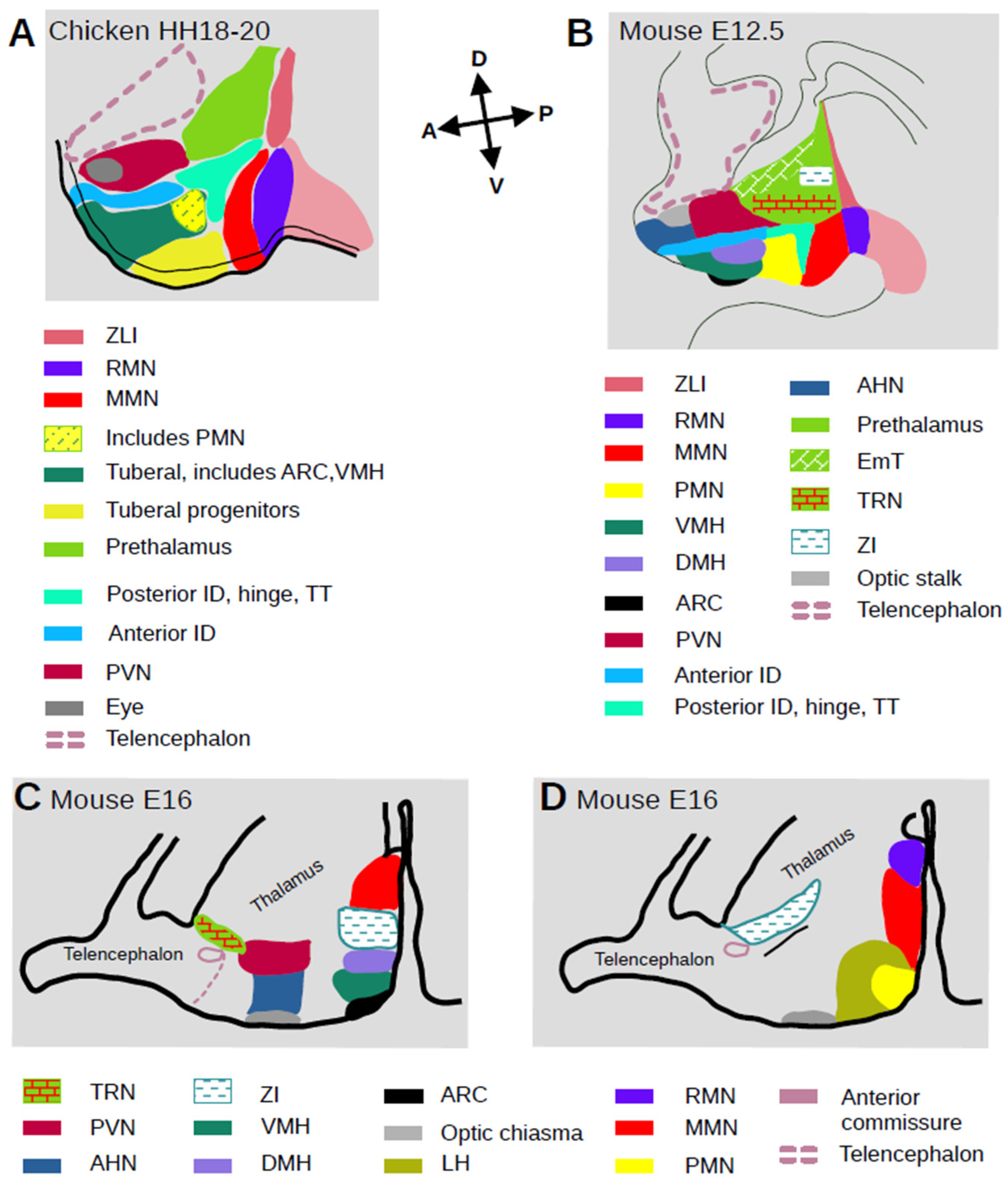
In mammals, the preoptic area has been debated as a telencephalic rather than a diencephalic region [5], and it is analyzed apart from the hypothalamus. Therefore, it is considered that the anterior hypothalamus is composed of the anterior hypothalamic (AHN), supraoptic (SON), suprachiasmatic (SCN), and paraventricular (PVN) nuclei. The tuberal hypothalamus contains the lateral hypothalamic area (LH), arcuate (ARC), dorsomedial (DMH), and the ventromedial (VMH), and its migrated part, tuberal (TuN) hypothalamic nuclei. The more posterior or mammillary hypothalamus includes the premammillary (PMN), mammillary (MMN), supramammillary or retromammillary (RMN) and posterior hypothalamic (PHN) nuclei (Figure 1A,B). The median eminence and pituitary stalk lie ventral to the tuberal hypothalamus. It is also important for this review to remind readers that the prethalamus (PTh) includes the thalamic eminentia (EmT), zona incerta (ZI) and thalamic reticular nucleus (TRN, [7] Figure 1A–D and Table 1 for abbreviations).
Single-cell RNA sequencing (scRNAseq) and gene regulatory networks (GRN) studies show that these adult anatomical subdivisions are already predicted by distinct progenitor subsets at embryonic stages [8,9]. These studies suggest how progenitors progress to differentiated neurons and provide a more accurate way of defining neurons than simply on the basis of their neuropeptide/neurotransmitter expression [9,10,11,12]. Each of the major subdivisions of the developing hypothalamus is identified at the peak of neurogenesis (E11–E13) along the A-P axis: postmitotic neuronal precursor cells of the paraventricular nucleus/supraoptic nucleus (PVN/SON), intrahypothalamic diagonal (ID) and tuberomammillary terminal (TT), ventromedial hypothalamus (VMH), arcuate nucleus (ARC), premammillary nucleus (PMN), mammillary nucleus (MMN), and retromammillary nucleus (RMN) (Figure 1A,B). The intrahypothalamic diagonal (ID) is parallel to the hypothalamus A-P axis and extends anterior until the optic recess. The tuberomamillary terminal (TT) is the border between mammillary and premammillary neuroepithelium (Figure 1A,B).

3. Induction and Regionalization of the Developing Hypothalamus
The hypothalamus (expressing Foxd1) is surrounded antero-dorsal by the telenephalon (expressing Foxg1), postero-dorsal by the prethalamus (expressing Pax6) and postero-ventral by the floorplate and diencephalic tegmentum (expressing Arx, Fox1/2, Shh) [8,9,11,13,14]. The hypothalamus originates from the hypothalamic floorplate (HypFP) cells, also known as rostral diencephalic ventral midline cells. In mouse, chicken and zebrafish neural plate stages, HypFP cells express Shh, Bmp7 and Nkx2.1 and lie anterior to the diencephalic floorplate cells which express Foxa2 [15,16,17,18,19,20]. HypFP cells are induced through the combined action of Nodal, SHH and BMP signals from prechordal mesendoderm [15,16,17,18,21,22].
Figure 1. Color-coded cartoons showing the regional subdivisions in the developing hypothalamus. (A): chicken (HH18-20), (B): mouse (E12.5). Arrows indicate A-P and D-V axes. Adapted from [6,14]. (C,D): cartoons of sagittal sections of a mouse E16 brain showing related positions of hypothalamic and prethalamic nuclei. Section (D) is lateral to (C). Adapted from [7].
Cells 15 01085 g001
Nodal from the prechordal mesoderm triggers hypothalamic induction and patterning [21,23,24]. Abrogation of Nodal signaling in the zebrafish mutant Nodal-related 2 ligand (cyc-/-) or teratocarcinoma-derived growth factor 1 Nodal co-receptor (oep-/-) results in complete loss of hypothalamic tissue, consistent with cyclopia in these mutants [20]. Nodal signaling is required for the expression of both Nkx2.1a and Nk2.1b paralogs in the zebrafish hypothalamus.
In cultured chicken Hamburger–Hamilton stage 4 (HH4) explants, most cells express Pax6, but no Nkx2.1, while in HH6 explants, Nkx2.1 expressing cells are present, some co-expressing Pax6 [13]. At HH8, prethalamic and hypothalamic scRNAseq and RNA velocity show a trajectory from prethalamic-like to HypFP clusters [13,25]. From HH8 through HH20/21, there are progressively fewer prethalamic-like progenitors (expressing Pax6) and more hypothalamic cells (expressing Shh/Nkx2.1). Altogether, these results show that prethalamic cells are induced to hypothalamic fate. Therefore, they support the point that prethalamic and hypothalamic fate are tightly linked and argue for the formation of an initially common domain.
scRNA-seq analysis shows that Pax6 expressing prethalamic-like cells express FST, a potent Nodal antagonist at HH8–HH10. In chicken embryo and dissected prospective hypothalamus cultured explants at HH4, treatment with anti-FST increases the Nkx2.1/Shh expressing hypothalamic domain and reduces the prethalamic Pax6 domain, relative to controls. Conversely, exposure to FST almost eliminates Nkx2.1 expression, and the number of Pax6-positive cells is increased in the embryo or hypothalamic explant. Therefore, prethalamic-derived FST inhibits hypothalamic specification and constrains the size of the developing hypothalamus [13]. In situ hybridization and RNAseq analysis of Nkx2-1CreER/CreER mutants and heterozygous littermate controls shows that Nkx2.1 expression is required for the formation of the mediobasal hypothalamus including ARC, VMH, PMN, and MMN, but not RMN. At mouse E12.5, Nkx2.1CreER/CreER mutants show that both the relative expression levels and the number of cells expressing hypothalamic genes is reduced, while the number of cells in the prethalamic cluster is increased with no substantial overall change in the portion of neural progenitors. RNA velocity analysis shows altered trajectories between mediobasal hypothalamus, mammillary hypothalamus and prethalamus. Therefore, Nkx2.1 not only promotes and maintains the identity of ventral hypothalamic progenitors but also actively represses prethalamic gene expression [8]. These results point out the cellular continuity between the prethalamus and hypothalamus, suggesting their formation from a common territory (prosomere 3, p3, Figure 2A–C) and that the existence of a boundary between the developing prethalamus (p3) and hypothalamus is highly unlikely (Figure 1A–D). They also provide a hint for a distinct developmental pathway of RMN compared to the Nkx2.1-dependent mediobasal hypothalamus [6,8].
BMP signaling is active in the developing tuberal hypothalamus. Remarkably, phosphorylated Smad1/5/8, key effectors of BMP signaling, are detected immediately after HH8 in anterior HypFP cells and propagate to more posterior HypFP cells. As tuberal cells are generated, the zone of active BMP signaling and underlying tissues becomes relatively posterior while the anterior tuberal domain expands. The late-specified HypFP cells show more sustained activation of phosphorylated Smad1/5/8 and become posterior tuberal gliogenic progenitors [26]. In conclusion, hypothalamic neuroepithelium-intrinsic factors are sufficient to maintain tuberal hypothalamic regionalization and neurogenesis.
BMP upregulates Tbx2 in tuberal progenitors and, as a consequence, Shh expression is downregulated in a cell-autonomous manner [17]. This likely occurs by Shh-induced abrogation of Smo expression in ventral tubero-mammillary precursors. Downregulation of SHH signaling first promotes further proliferation of ventral tubero-mammillary progenitors and, second, induces Emx2 expression. When BMP signaling is abrogated by Chordin, hypothalamic Tbx2 expression is lost but restored by Wif (Wnt-inhibitory factor) activation [17]. Complementary to this, simultaneous activation of Nodal signaling and inhibition of Wnt-beta catenin signaling is required within the ventral midline floorplate-like cells to form a regionalized hypothalamus along the A-P axis. In zebrafish, abrogation of Axin1, an intracellular Wnt/beta-catenin inhibitor, results in a smaller hypothalamus of posterior identity expressing Emx2 [27,28]. Interestingly, Wnt8b expression flanks the RMN hypothalamus, suggesting that it may receive more sustained posteriorizing signaling than the tuberal and mammillary hypothalamus and thus has a different fate [6].
SHH derived from the ventral forebrain neuroepithelium is essential for the differentiation of the anterior and tuberal hypothalamus [14]. Shh is initially broadly expressed in ventral hypothalamic progenitors and then rapidly downregulated in the ventral midline at the level of the tuberal hypothalamus [17,29]. When Shh is selectively removed from the hypothalamic basal plate and basal telencephalon in Nkx2.1-CRE × ShhloxP/loxP mice, Foxb1, Rx, Sim1 and Irx5 expression is maintained, showing that hypothalamic mammillary and retromammillary areas are present, respectively. However, hypothalamic cells are devoid of Pomc and Nkx6.2 expression, showing that antero-tuberal hypothalamic nuclei are absent; Nr5a1, showing that differentiation of VMH nucleus is compromised; and Lhx1, Lhx9, Lhx6 showing that the ID and tuberomamillary terminal are undifferentiated [30,31]. Lef1 expression is decreased, suggesting that the premammillary neuroepithelium is reduced. As telencephalic Foxg1 expression is not expanded, altogether these results suggest that ventral diencephalic cells still retain their hypothalamic identity but fail to undergo differentiation in Nkx2.1-CRE × ShhloxP/loxP mice [9,14]. In addition, these data highlight that there is no cell fate conversion between hypothalamic and telencephalic tissue, suggesting a possible boundary mechanism between the two regions, an argument against the prosomeric model.
In the E9.5 mouse hypothalamus, Shh is expressed in bilateral stripes adjacent to the ventral midline [32]. Neural progenitors immediately dorsal to the bilateral stripes of Shh are responsive to SHH signaling by Gli1 expression. Shh expressing progenitors contribute to a subset of ARC neurons and ventrolateral, ventral and central neurons of the VMH. Descendants of Gli1 expressing progenitors contribute to the DMH and dorsolateral VMH. Conditional loss of SHH signaling in the tuberal hypothalamus, by Smo deletion in Gli1 expressing cells, results in a cell-autonomous reduction of proliferating progenitors of distinct DMH and VMH neurons. Furthermore, ShhΔhyp hypothalamus shows that SHH signaling is active after the dorso-ventral (D-V) identity is established. In particular, the non-dividing ventral midline cell population is laterally expanded, Ascl1 expression and consequent neurogenesis are downregulated and Pax6 expression, normally restricted to dorsal diencephalic areas (prethalamus), is ventrally expanded. As a result, tuberal hypothalamic nuclei are absent in ShhΔhyp embryos [33]. This is also a hint regarding the tight relation (fate conversion) between dorsal diencephalic (prethalamic) and hypothalamic cells as both derivatives of a common domain.
The anterior hypothalamic progenitor cells express Rax/Rx/Rx3 [14]. When Rx is inactivated prior to E8.5 in Rxf/f;CreER-expressing [34,35] mouse embryos, the anterior and tuberal hypothalamus are devoid of Shh and the dorsomedial hypothalamus of Otp, Pomc, Th1 and Sst expression. In a complementary manner, the ventromedial expression of Fgf10, Otx2 and Tbx3 expands dorso-medially [36]. Rx-CreERT2:MADMGT/TG tamoxifen-induced mice at E9.5 show at E11.5/E12.5 that the majority of Rx progenitors are multipotent and generate multiple neuronal subpopulations including glutamatergic VMH neurons and GABAergic TuN neurons [37,38]. Timed controlled inhibition of SHH signaling in zebrafish, at the hypothalamic cell differentiation stage, shows that Shh first induces and then downregulates Rx3 expression. SHH-induced Rx3 expression in proliferating progenitors is required for elongation of anterior/tuberal hypothalamus. Subsequent Rx downregulation is required for hypothalamic anisotropic growth, specification of Shh expressing (Rx3 non expressing) cell fate in the anterior ventricular recess, and differentiation into Pomc (ARC), Nr5a1a (VMN) and Th1, Otpb tuberal/anterior cell fates [39].
RNA velocity on scRNAseq data infers that a hypothalamic differentiation trajectory is composed of Six6/Fgf10-expressing progenitor cells, Six6/Ascl1/Isl1 expressing neurogenic precursors, and Six6/Nr5aA1/Pomc expressing neurons [13] In HH8-9 chicken, Six6 and Tbx2 are simultaneously expressed in anterior HypFP cells and resolve shortly after in anterior tuberal Six6/Isl1 expressing cells, Six6/Shh/Ascl1 expressing neurogenic progenitors, posterior tuberal Tbx2 expressing progenitors, and Foxa2/Shh expressing HypFP cells [26]. In situ hybridization in E5 chicken shows that anterior, tuberal and mammillary domains are defined as Six3, Six3 and Fgf10, and Emx2 expressing progenitors, respectively. However, at 9–10 somite chicken, only tuberal Six3 and Fgf10 expressing progenitors are present. Anterior progenitors appear at 11 somites and mammillary progenitors even later at HH16. Despite the ~8-fold expansion of the entire hypothalamic region in the developing chick, the Fgf10 expressing progenitor domain retains a constant size. Fgf10 expressing progenitors are retained centrally and contribute to the tuberal domain in a cell autonomous manner. Furthermore, EdU and DiI/DiO injections show that anterior and mammillary progenitors develop from highly proliferating Fgf10 expressing progenitors and begin to differentiate once displaced from the Fgf10 expressing domain into anterior and mammillary domains [40,41]. In conclusion, the development of hypothalamic areas is heterochronous and characterized by anisotropic growth.
SHH signaling is involved in this anisotropic growth process of the hypothalamic areas relying upon Fgf10-expressing progenitors. At 9 somites, Shh and Fgf10 are briefly co-expressed in the forebrain. From 11 somites, Shh becomes downregulated in Fgf10 expressing progenitors [17] and strongly expressed in peripheral cells, including emerging anterior cells. Emerging anterior progenitors downregulate Fgf10 and upregulate Shh and p57Kip2, then downregulate Shh as they differentiate. Compromised SHH signaling in controlled time periods results in the specific loss of anterior progenitor territory. Therefore, SHH signaling upregulation and subsequent downregulation results in progenitor proliferation from the pool of Fgf10 expressing cells and anterior (but not tuberal) identity of those progenitors [41].

4. Transcription Factors Couple Regionalization and Differentiation of Hypothalamic Nuclei

Transcription factors (TFs) involved in early hypothalamic regionalization also regulate cell differentiation. To identify GRNs controlling hypothalamic regionalization and neurogenesis, Kim and colleagues apply SCENIC+ analysis in E11–E14 scRNA-seq and scATAC-seq datasets, which infers patterns of transcription factor (TF) activity [8,42]. Hypothalamic TFs, such as Hmga1, Lin28a, Cited1, and Gbx2, are active in early-stage neuronal precursor cells, whereas Tcf4, Npas3, Nfia/b/x, Zbtb20 and Tox3 are active later in neurogenesis. Hmx2/3 and Isl1 activate GABAergic gene expression across the tuberal, premammillary, and prethalamic regions. In addition, in Neurog2 expressing neurons, GABAergic genes, e.g., Slc32a1, are activated by prethalamic/ID TFs (Dlx1/2/5/6, Arx, Sp8, Meis2) and repressed by glutamatergic TFs (e.g., Barhl2). Conversely, glutamatergic genes, e.g., Slc17a6 are activated by both Neurog2 specific TFs (Barhl1, Nhlh2, Nr4a2, Lmx1a, Neurod1) and tuberal progenitor specific factors (Nkx2–1, Sox14). These glutamatergic genes are repressed by prethalamic specific TFs [8].
Loss of function experiments of distinct TFs are also available. The anterior hypothalamus is divided into two domains along the medio-lateral axis of the mantle zone. The median domain expresses Sim1 and Brn2 at E12.5 and generates PVN and SON, whereas the lateral expresses Rgs4 and generates heterogeneous layer-organized neurons. Abrogation of Sim1 activity in mice shows that Sim1 is required for progenitor proliferation and activation of Brn2 expression [14,43,44].
Otp and Dlx2 are expressed in TuN neurons and delineate TuN from Nkx2.1 expressing VMH neurons [37]. In the progenitor domain of VMH, rare cells express Otp in a mutually exclusive manner with Nr5a1 and migrate and accumulate in the presumptive TuN. Fate mapping analysis using Nkx2.1-Cre:H2B-GFP mice shows that nearly all Otp expressing and nearly all Dlx2 expressing neurons in the TuN are GABAergic and derived from the Nkx2.1 lineage [31,37]. Therefore, surprisingly, the progenitor domain of VMH is a common pool of progenitor cells, which gives rise to a glutamatergic VMH and a GABAergic TuN in a mosaic manner rather than as spatially segregated domains [37]. BrdU injection in Dlx5/6-Cre;H2B-GFP mouse embryos [45] shows that VMH-TuN neurons have a sequential and “outside-in” birth order, with early born neurons mainly located in lateral subdomains and late born neurons in medial subdomains. The VMHvl and TuN neurons are generated simultaneously, and VMH-TuN as a whole has a laminar-like structure [37]. In contrast to TuN, where rare cells co-express Otp and DlxGFP, in the LH area, either Otp- or Dlx-expressing neurons are found, and they are glutamatergic and GABAergic, respectively [37,46,47,48].
Arx is expressed in two linear domains: one that runs parallel to the basal domain of Shh expression toward the optic recess (parallel and part of ID) and the TT. This results in two domains with overlapping Arx and Nkx2.1 expression but not Shh [14]. The expression of LIM homeodomain family members Lhx1, Lhx8, and Lhx6 delineates the A-P axis of the ID/TT. LHX TFs define a series of nuclei residing both between and lateral to the PVN and VMH. In the anterior ID, at E12.5, Lhx1 is expressed in the presumptive SCN. At E16.5, Lhx1 is additionally expressed in a region immediately posterodorsal to the SCN [49]. The posterior domain of Lhx1 expression overlaps with the anterior part of Lhx8 expression in the ID. Lhx8 is further extended posterior to Lhx1 and reaches the limit of Lhx6 expression. The Lhx8 expression domain of the ID corresponds to the central region of the DMN. Lhx6 is expressed in the ID until the posterior domain of Arx and Nkx2.1 expression. Lhx9 is expressed postero-ventral to Lhx8 and immediately ventral to the Lhx6-positive zone of the ID in a Nkx2.1-negative ventral hypothalamic area [14]. Like Lhx6, Lef1 is expressed in the ventral posterior ID and TT domains. Lhx6CreER/+;Ai9 mice show that distinct Lhx6 cells co-express distinct transcription factors: Nkx2.1 in the ID, Dlx1/2 in the TT, and Nkx2.2 in the hinge between the posterior ID and dorsal TT. Abrogation of Nkx2.1, Nkx2.2, and Dlx1/2 selectively eliminates hypothalamic Lhx6 expression in the ID, hinge and TT domains, respectively [49,50].
Presumptive ARC, median eminence, pituitary stalk and posterior pituitary develop from a midline domain of the basal hypothalamus expressing Tbx3 [29,51,52]. Tbx3 is required for the differentiation of pro-opiomelanocortin (POMC) or neuropeptide Y (NPY) and agouti-related peptide (AgRP)-expressing neurons [37,52,53,54]. ARC neurons are divided in distinct POMC and NPY/AgRP and TH neurons by combinatorial Tbx3, Otp, and DlxGFP expression, respectively (DLX5/6-Cre;H2B-GFP [45]). scRNAseq data of adult mouse ARC neurons supports the compartmentalization of ARC in distinct cell subpopulations [55]. Otp and DlxGFP are expressed in ARC differentiating neurons in a mutually exclusive salt-and-pepper pattern [37]. Abrogation of Otp activity results in the absence of NPY, AgRP, and Sst expressing neurons and reduced Gad 1, GR and Bsx expression. In contrast, POMC and GHRH are expressed in Otp-/- mice, and the number of TH and Dlx1 expressing neurons is similar to the wild-type. Therefore, in the developing ARC, Otp does not repress the expression of Dlx1/2. However, DLX1/2 directly bind and repress Otp expression as it is shown by ChIPseq analyses, luciferase or reporter expression controlled by Dlx1/2 regulatory elements [56].
In the posterior hypothalamus, Lef1 and Irx5 are expressed in the PMN and RMN neuroepithelium, which lie immediately anterior and posterior to MMN neuroepithelium, respectively [14]. Neurogenic progenitor analysis by Scenic+ infers that Neurog2 expressing cells activate early-stage RMN-specific factors such as Lmx1a, Foxa1/2 and Irx3/5, while early-stage Ascl1 expressing cells activate prethalamus specific TFs, including Dlx1/2/6, Sp8/9, and Arx [8]. These results suggest that the differentiation of RMN and prethalamic cells is independent of each other.
Cck is expressed in MMN and RMN neurons and activated by TFs from both MMN and RMN, such as Nkx2.4 and Uncx. However, non-overlapping expression patterns of MMN and RMN-specific TFs are visible by E13.5, and MMN and RMN genes mutually antagonize their expression. For example, MMN-specific neuropeptides (Pnoc, Npy, Nts, Cartpt) are activated by MMN-specific TFs (Lhx1/5, Uncx, and Nhlh2) but directly repressed by RMN-specific TFs. In the RMN, Tac1 is expressed in specific neuronal subpopulations but is absent from the MMN. GRN analysis infers that Tac1 is directly activated by RMN-specific TFs (Lmx1a/b, Foxa2, Barhl1/2, and Ebf2) and repressed by the MMN specific factor Nkx2.4. These results reveal that early developmental pathways are common for MMN and RMN. However, TFs, which control differentiation steps, are specific to each nucleus [8].

5. The Prosomeric Hypothalamic Model

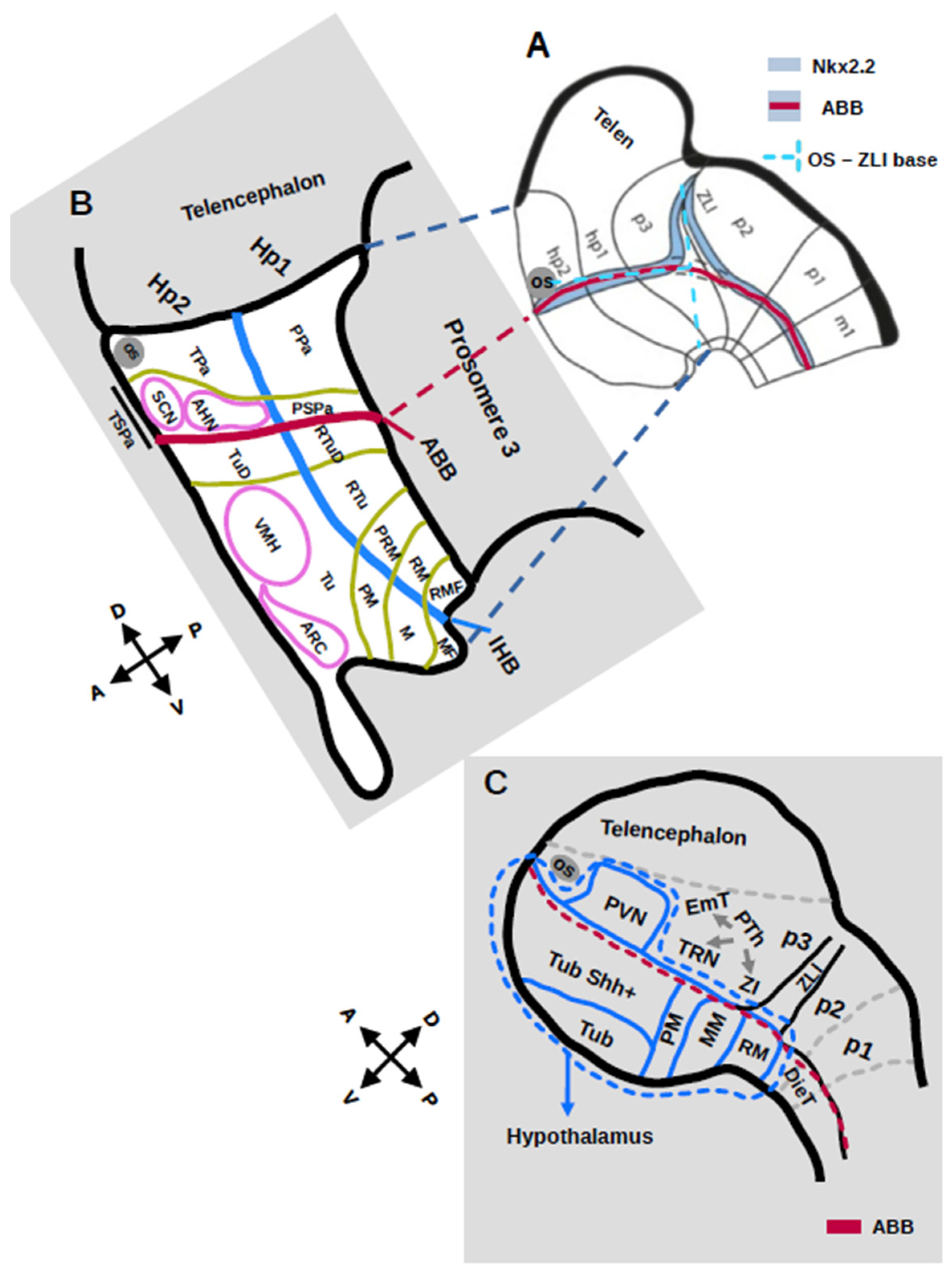
According to the prosomeric model, the hypothalamus is part of the anterior forebrain, ventral to the telencephalon and anterior to the diencephalon. Along the A-P axis, the hypothalamus is divided into two parts: posterior, the peduncular hypothalamus (PHy) belonging to hypothalamic prosomere 1 (Hp1) and anterior, the terminal hypothalamus (THy) belonging to hypothalamic prosomere 2 (Hp2, Figure 2A,B). The terminal hypothalamus contains the singular median acroterminal domain (ATD) [5,47]. Along the alar/basal axis, the hypothalamus is divided into the alar peduncular and terminal paraventricular and subparaventricular areas, the basal retrotuberal and tuberal, and basal retromammillary and mammillary areas in the Hp1 and Hp2, respectively (Figure 2B). Each of the alar and basal areas is further divided into zones depending on the position in the area, e.g., dorsal, central/intermediate, ventral or lateral.
The topographical correspondence between the prosomeric domains and conventional nuclei is as follows (Figure 2B):
-
In the alar plate, the peduncular paraventricular area (PPa) includes the PVN, the terminal paraventricular area (TPa) includes the periventricular hypothalamic nucleus (PeVN), the peduncular subparaventricular area (PSPa) contains the entopeduncular nucleus (EnN), and the terminal subparaventricular area (TSPa) contains the AHN, part of the SON and lateroanterior nucleus (LAN).
-
In the basal plate, the peduncular retrotuberal area (RTu) includes part of the DMH, the posterobasal nucleus (PBN) and part of the LH. The terminal tuberal area (Tu) includes the other part of the DMH, VMH and part of the LH. The Acroterminal domain (ATD) contains the alar lamina terminalis, SON, anterobasal nucleus (ABN), SCN and chiasmatic areas, as well as the ARC, median eminence, and infundibular/neurohypophysial areas. The periretromammillary (PRM) and perimammillary (PM) areas include the dorsal and ventral PMN. The retromammillary area (RM) includes the RMN. The mammillary area (MM) includes the MMN [5,47,57,58,59,60].
These hypothalamic subdivisions are originally defined by the expression of patterning genes [47]. The basal part of the hypothalamus is distinguished from its alar part by Shh expression in the ventricular zone and Nkx2.1 in the mantle layer. Otp and Sim1 are expressed in the alar peduncular dorsal, central, and ventral paraventricular areas and the alar terminal dorsal, central, and ventral paraventricular areas [47,60,61]. Arx is expressed in the alar PSPa and the alar TSPa. Nkx2.1 is expressed in the basal VMH and M and the acroterminal ARC and neurohypophysis. Nkx2.1 and Otp are co-expressed in the basal peduncular PRM, the basal terminal dorsal tuberal area (TuD) and basal terminal PM. Nkx2.1 and Dlx5 are expressed in the basal peduncular dorsal and intermediate RTu and the basal terminal intermediate Tu. Arx, Nk2.1 and Dlx5 are expressed in the basal peduncular ventral RTu and the terminal ventral Tu [47,59]. The hypothalamic floorplate reaches up to the MM and RM, characterized by the epichordal expression of Shh, Ntn1, Lmxb1, Foxa1, and Nr4a2 [47].
Additional gene expression patterns have been analyzed, taking into account the prosomeric model subdivisions. For instance, Erb4, Irx1/3/5, Lmo4, Mfap4, Plagl1, Pmch are expressed in the peduncular hypothalamus (Hp1); Fgf15, Gsc, Nkx6.2, Otx1, Zic1/5 are expressed in the terminal hypothalamus (Hp2); and Fgf8/10/18, Otx2, Pomc, Rx and Six6 are expressed in the ATD which belongs to Hp2 [62].
Figure 2. Schematic comparison of the hypothalamic subdivisions in the prosomeric (A,B) and tripartite model (C). Arrows indicate the A-P and D-V axes and show that the tripartite model (C) is rotated 90° to the prosomeric model (A,B). Nkx2.2 expression, in red, is considered as the Alar/Basal boundary (ABB). In (A), an orthogonal angle is also designed at the base of the ZLI (cyan dotted lines) to facilitate identification of the regional position. In (B), anterior prosomeres are magnified to show hypothalamic subdivisions. The blue line segments the hypothalamus into two prosomeres, Hp1 (peduncular) and Hp2 (terminal), olive green lines segments prosomeric hypothalamic domains/areas, and pink lines mark examples of conventional nuclei. In (C), the blue dotted line delineates the developing hypothalamus, and the small arrows mark the prethalamic (PTh) nuclei. To facilitate the reader, grey dashed lines position potential prosomeric areas (p1–p3) although the tripartite model does not support prosomeric subdivisions. For abbreviations, see Table 1. Adapted from [5,6,59].
Cells 15 01085 g002
The authors of the prosomeric model point out that differential gene expression satisfies the assumption that differential gene expression patterns should characterize each of the anterior prosomeres Hp1 and Hp2 [47,62]. However, when examined in detail, several gene expression patterns question the existence of the aforementioned hypothalamic divisions. Examples of inconsistency of gene expression patterns and the claimed intrahypothalamic boundary are found in Table 2.
Neither signaling pathway molecule nor transcription factor nor neuromodulator/neuropeptide expression support a boundary separating the hypothalamus in Hp1 and Hp2 prosomeres according to these studies (Table 2). The division of the paraventricular, subparaventricular and perimammillary areas in a peduncular and terminal domain are unsupportive of an entire dorso-ventral boundary across the hypothalamus, orthogonal to the alar-basal boundary (ABB). Furthermore, HCR analysis of gene expression shows that Fgf15, Six3 and Zic1/5 expression in the terminal hypothalamus is more medial, that is, in the ventricular zone where progenitors lie, whereas Otp, Sim1, Mfap4, Lmo4 and Rgs4 expression is more lateral where cells start to differentiate, when compared to E13.5 sections from the Allen Mouse Brain Atlas [6,63]. scRNAseq analysis of the pooled E11-E14 mouse hypothalamus is unsupportive of hypothalamic peduncular and terminal subdivisions [8]. In contrast, the TPa Fgf15 and Six3 are expressed in a single progenitor cluster whereas Zic1 expression is found in eight clusters. The PPa markers Mfap4, Rgs4, and Lmo4 are also variably expressed. Altogether, the scRNAseq clusters suggest that TPa and PPa areas reflect differentiating cells remaining apart from progenitors [6].
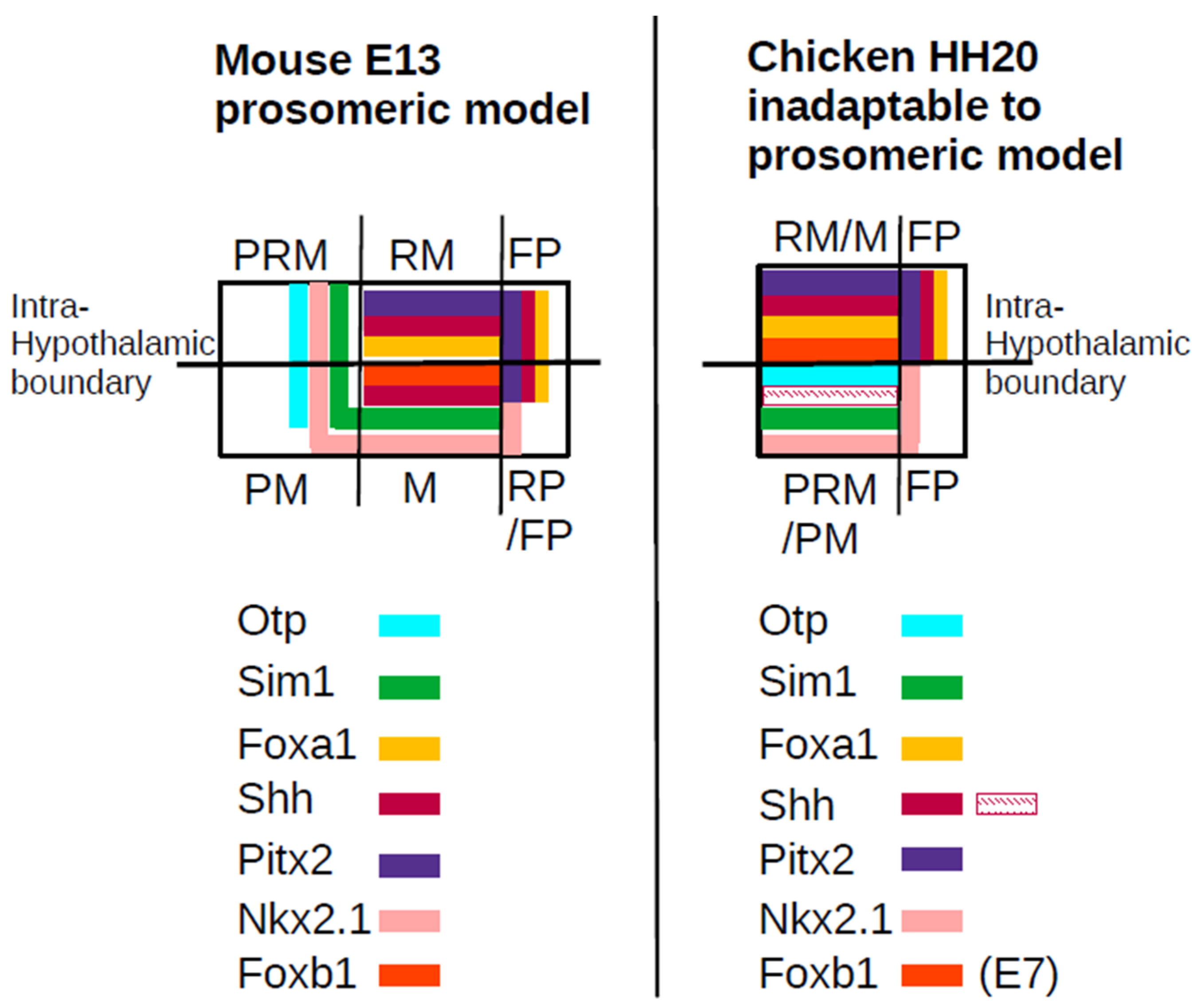
Furthermore, surprisingly, the prosomeric mammillary domain (M) cannot be identified in the chicken hypothalamus [6]. According to the prosomeric model data, the posterior ventral hypothalamus is organized as follows (Figure 3):
A domain expressing Sim1/Otp (PRM/PM) and a domain expressing Nkx2-1/Sim1/Foxb1, that is, M, a domain expressing Pitx2/Foxa1 (RM). Pitx2 expression is found in the RM domain; however, its expression covers the floorplate of both RM and a large part of M domains, and a small Shh/Foxa1 midline region corresponding to the Hp2 floorplate. The Hp1 prosomere floorplate is defined as extending beyond Arx, up to the anterior limit of Shh/Foxa1 expression (Figure 3).
Unexpectedly, the prosomeric model boundaries cannot be found in the HH20 embryonic chicken brain using the same markers: Pitx2 is found in a single territory rather than two distinct RM and M domains, Otp is expressed in an adjacent anterior single territory rather than distinct PRM and PM domains, and Foxb1, at E7, is expressed in a single territory rather than RM and M split fields. In other words, Nkx2.1/Sim1/Foxa1 and Otp/Pitx2 boundaries coincide, instead of intersecting orthogonally, so that Foxa1 and Otp directly abut. Therefore, a Sim1/Nkx2.1 expressing, devoid of Foxa1/Otp expression M domain, could not be identified in the HH20 and E7 chicken (Figure 3) [6].
According to the prosomeric model, the fornix characterizes and belongs to Hp1 since it originates in the hippocampus [5]. It branches in relation to the anterior commissure and extends posterior towards its target, the mammillary hypothalamus, where it bifurcates and innervates part of RM and M. However, this is inconsistent with the existence of a hypothalamic boundary since the tract innervates areas in both sides of the boundary. Furthermore, the fornix bundle develops later than the hypothalamic regionalization takes place, at gestational day 16, whereas hypothalamic regionalization is obvious in E12-E13 mice; thus, it is difficult to conceive that it plays a role in prosomere establishment. Finally, although prosomeric Hp1 and Hp2 regionalization is claimed in teleost fish, the fornix tract is not obvious as the hippocampus does not form [64,65].
Finally, the prosomeric model posits a boundary between prethalamus (p3) and hypothalamus (Hp1 and Hp2). In other words, prosomere 3 (p3) contains the prethalamus but the hypothalamus belongs to a more anterior prosomere [5,47]. However, functional experiments are unsupportive of this boundary. As discussed earlier, hypothalamic and prethalamic formation are tightly bound. HypFP cells recruit prethalamic cells to form the hypothalamus [13]. Nkx2.1 not only promotes and maintains the identity of ventral hypothalamic progenitors but also actively represses prethalamic gene expression [8]. FST from the prethalamus restricts hypothalamic size, and in HH8 chicken, prethalamic and hypothalamic scRNAseq and RNA velocity show a trajectory from prethalamic-like to HypFP clusters [13]. In conclusion, prethalamic and hypothalamic formation share a common territory.

6. The Tripartite Hypothalamic Model

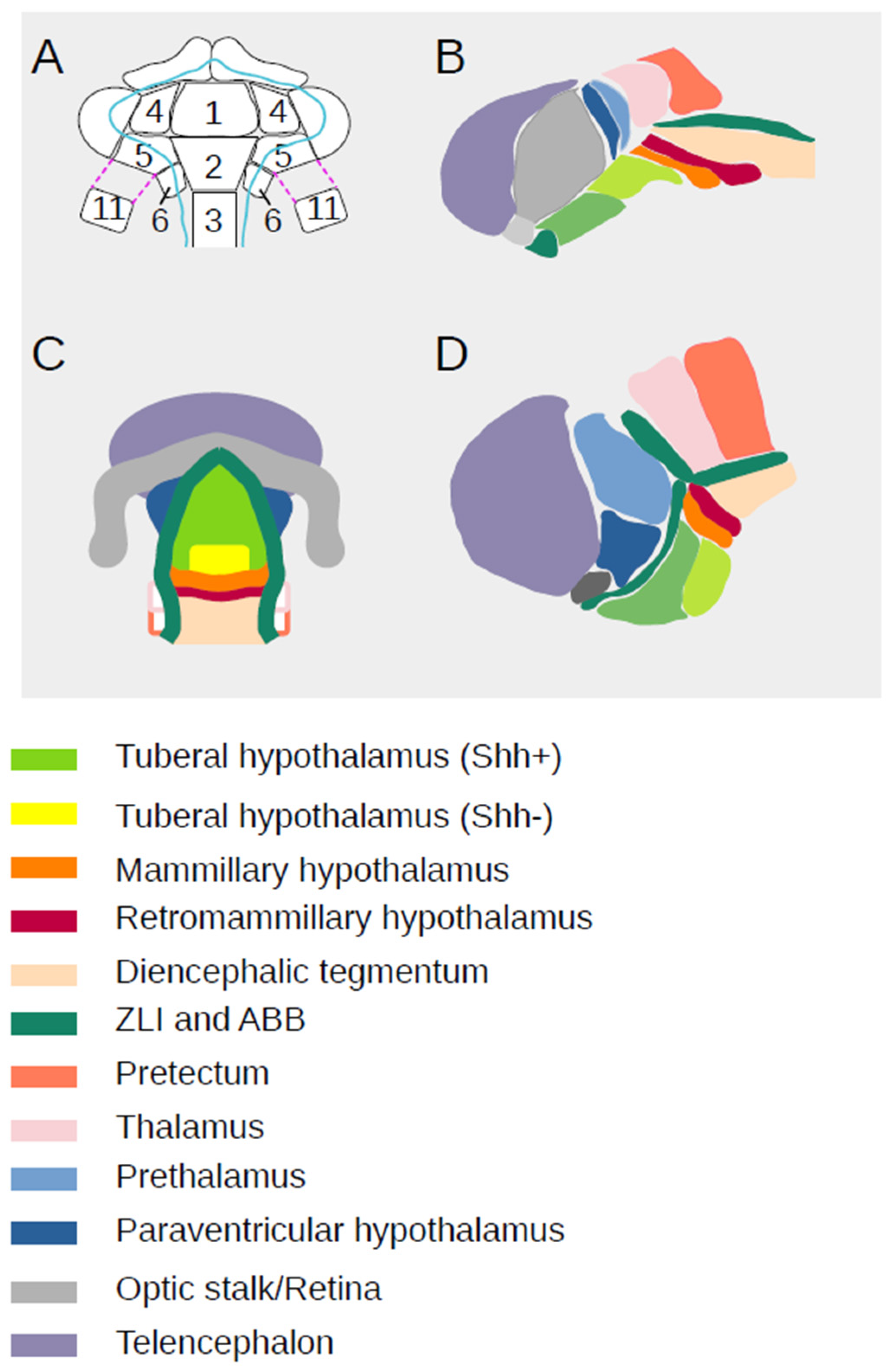
The most complete fate map for the anterior neural tube is provided by (Manning et al., 2025) [6]. Chicken embryos are injected with DiI/DiO in the presumptive forebrain territory at HH10 and analyzed after 48 h (HH17-20) (Figure 4). The A-P axis is considered as parallel to the hypothalamic Nkx2.2 expression domain and the D-V axis as orthogonal to it. Alternatively, because Nkx2.2 expression is not always available, the A-P axis can also be defined as parallel to the Shh expression stripes in the ventral hypothalamus or as a line drawn perpendicular to ZLI from the ZLI base to the optic stalk opening (Figure 2A–C).
The DiI/DiO injection label territories known as growth lines and show how specific areas, by maintaining their relative topology, grow differentially and directionally to generate the chicken forebrain. Key growth patterns are confirmed by genetic clonal analysis using the Cre-Cytbow transgenic line [66] and in Rfp-electroporated embryos [6]. Dye targeting of HypFP at two ventral midline positions along the anterior ventral midline, namely zones 1 and 2, label the ventral hypothalamus (Figure 4A). Zone 3, further posterior, gives rise to a non-hypothalamic, diencephalic floorplate (Figure 4A–D). Wider midline injections in zones 1–3 form an arrowhead shape in the A-P axis, showing that midline cells are displaced anterior relative to their more lateral counterparts from HH10 to HH20 (Figure 4A–D) [6]. This is in agreement with previous fate mapping at HH10 where tuberal Six3 and Fgf10 expressing cells generate anterior hypothalamic cells [41].
Medial zone 4 and 5 injections extend along the A-P axis in the Shh expressing hypothalamus. The lateral parts of these lines stretch from the posterior Shh-non-expressing but Foxd1 expressing hypothalamic region, across the alar-basal boundary (ABB), towards the ventral optic stalk and temporal retina. Zone 6 results in a tricorn-shaped domain, ventral to Nkx2.2 expression and just posterior to the ZLI base (Figure 4A–D). When zones 2 and 6 are targeted simultaneously, an integral-shaped line (∫) pattern extends anteroventral from this tricorn domain through the Pitx2 expressing domain, towards the tuberal non-Shh-expressing hypothalamic midline. Finally, zone 11 is dorsal to zone 5 and expands isometrically to form the PVN hypothalamus and prethalamus. Therefore, fate mapping formally shows a common origin of prethalamic and hypothalamic cells (Figure 4A,D) [6].
This fate mapping analysis shows that at HH20, the hypothalamus comprises the PVN hypothalamus dorsally and the basal hypothalamic area ventrally, containing the cells born from ventral midline and lateral areas. The hypothalamus is segregated anterior and dorsal from the telencephalon by the optic stalk and eye field, dorsal and posterior from the prethalamus, and ventral and posterior by the adjacent diencephalic tegmentum and the beginning of the Irx3 and Arx expressing floorplate. Only the anterior tuberal hypothalamus originates ventral to the telencephalon. The posteroventral hypothalamus arises posterior to the telencephalon and ventral to areas that will give rise to the PVN hypothalamus, prethalamus and thalamus. This is the tripartite hypothalamus model of hypothalamic organization (Figure 2C) [6].
HCR reveals the RMN developmental identity. RMN consists of the Lfng non-expressing, Pitx2, Lmx1b and Shh expressing domain, partially overlapping anterior with Barhl2 expression and lying ventral to the ZLI, posterior to the prethalamus. Anterior to Pitx2 hypothalamic and floorplate expression lies the domain of Emx2 expression (MMN), and posterior to it lies the Arx expressing floorplate in the diencephalic tegmentum. The Pitx2 hypothalamic domain is straddled by and partially overlaps with Sim1, EphA7 and Dbx1 expression, which further reach dorsal and encompass the ZLI. This posterior character is further enhanced by RMN being the anterior domain of Foxa1 and Irx1/Irx3 expression immediately posterior to the Six/Fez expressing hypothalamus. As a result, the RMN hypothalamus is part of a ventral boundary region associated with lineage restriction. The hypothalamic RMN identity is conserved in the mouse [6].
The tripartite hypothalamic model is further supported by fate conversion. Prospective telencephalon and anterior retina at HH10 express Foxg1, prospective prethalamus/PVN hypothalamus expresses Pax6/Arx, and optic stalk expresses Pax2. When chicken hypothalamus is dorsalized by SHH inhibitors, with or without FST (which restricts hypothalamic size) at a neural tube stage, a small basal territory with Shh and decreased Nkx2.1 expression is retained. Fate mapping confirms that cell movements are not altered. Pax2 expression is preserved. Foxg1 is ectopically expressed, but only in part of the anterior-most tuberal hypothalamus, suggesting that this hypothalamic area formation is linked with the telencephalon. More posterior within the tuberal hypothalamus, Pax6 is ectopically expressed and Arx is significantly expanded, showing conversion of the tuberal hypothalamic territory to prethalamus/PVN. Similar results are obtained when HH10 zones 1, 2, 4 and 5 are isolated and cultured in the presence or absence of SHH inhibitors. Olig2 remains confined to the prethalamus, posterior tuberal and MMN hypothalamus. As a result, the anterior-most tuberal hypothalamus is topologically ventral to the telencephalon, in partial agreement with the prosomere model, but the remainder of the tuberal region is ventral to the PVN hypothalamus and prethalamus and is part of the diencephalon. Fate mapping shows a major association between PVN progenitors and the prethalamus [6].
Furthermore, when PVN, telencephalic and perthalamic cells are isolated in vitro, PVN hypothalamic cells intermingle with prethalamic cells but segregate from telencephalic cells, suggesting that the PVN hypothalamus groups with the prethalamus as part of the diencephalon, posterior to the telencephalon. Basal plate cells taken anterior and posterior to the RM hypothalamus segregate in culture while those from posterior and anterior or ventral positions within the basal hypothalamus intermingle. These results suggest that lineage restriction may operate ventral to the ZLI at the RMN.
Altogether, gene expression, fate mapping, fate conversion and cell intermingling support the tripartite model. According to it, (1) the hypothalamus lies ventral to the telencephalon and to two parts of the diencephalon, the prethalamus and the ZLI; (2) the D-V and A-P axes are rotated 90 degrees, relative to the prosomeric model (Figure 2B,C), so that the tuberal hypothalamus is a ventral structure; (3) the anterior forebrain is non-segmented, i.e., does not have an Hp1-Hp2 boundary; and (4) the retromammillary hypothalamus has a more posterior forebrain identity to the rest of the hypothalamus [6].

7. Genetic Mutations and Regulatory Networks Support the Tripartite Model

Additional functional studies provide evidence for the regionalization of the hypothalamus. In particular, genetic mutations and GRN provide robust evidence for the regional boundaries in the forebrain.
The apposition of Six3 and Irx3 expression domains corresponds to the zona limitans intrathalamica (ZLI), the boundary-cell population between prethalamus and thalamus. Removal of Six3 activity in Six3-/- mice results in severe forebrain truncations anterior to the ZLI in the alar plate. In Six3-/- mice, telencephalic Foxg1 and retinal/anterior hypothalamus Rx expression are absent. Nkx2.1 expression is absent in the floor of the truncated forebrain and the anterior and tuberal hypothalamus. Shh expression in the forebrain basal plate is reduced in length but extended into the rostral end of the truncated forebrain. Shh expression is detectable in the ZLI of the Six3−/− embryos and Pax6, a prethalamus marker in the alar plate, extends to the anterior end of the truncated forebrain. These results contribute to the idea that there is a boundary between the hypothalamus anterior and diencephalic prosomere 3 posterior and that the hypothalamus belongs to an anterior prosomere, distinct from p3 [67].
However, a posterior hypothalamic area persists. Nkx2.1 is expressed in a small area of the basal plate in the posterior hypothalamus or posterior to it, and Shh overlaps with the residual domain of Nkx2.1 expression [67]. This domain expresses Irx3; therefore, a ventral Shh expressing domain, at the limits of Nkx2.1 expression, depends on posteriorizing Irx3 activity. Within this domain, ventral to the ZLI lies the Pitx2 expressing domain that corresponds to the RMN. These results suggest that the RMN is the ventral A-P boundary of the ventral forebrain [6].
Both prosomeric and tripartite models agree in the presence of the alar basal hypothalamic boundary along Nkx2.2 expression [5,6]. Nkx2.2 is a Shh-responsive TF expressed in the dorsal tuberal, ventral prethalamus, and ID. GRN analysis of Nkx2.2 null mice at E12.5 predicts that Nkx2.2 directly activates TFs specific to the tuberal hypothalamus, ID and prethalamus and represses RMN-specific TFs. In agreement with this, scRNA-seq of homozygous Nkx2.2 mutants at E12.5 shows reduced numbers of ID and prethalamus and increased numbers of RMN cells. A mutant-specific cell cluster is observed co-expressing prethalamus and RMN/MMN genes such as Sp9 and Meis2, and Pitx2 and Irx5, respectively. These data infer that Nkx2.2 expression maintains regional boundaries by promoting prethalamic and tuberal fates while repressing posterior hypothalamic regions, such as the RMN [8].
Nkx2.1 expression, as discussed earlier, is necessary to promote and maintain mediobasal hypothalamic identity and repress prethalamic fate [9,14]. Therefore, the formations of the prethalamus and hypothalamus are tightly linked.
Isl1 is expressed in both neurogenic progenitors and postmitotic precursors across a domain extending from the prethalamus, through the ID, and into the tuberal and PMN regions. RNAseq analysis predicts that Isl1 activates TFs in the prethalamus, ID, tuberal and PMN and represses those in the RMN, MMN, and EmT, an area of the anterior prethalamus. Complementary to this, in Isl1-null mice, prethalamus and PMN cell populations are reduced and a mutant-specific cluster co-expressing EmT-like genes (Tbr1, Slc17a6) and RMN/MMN genes emerges [8]. Therefore, this analysis postulates that the hypothalamus and prethalamus share a common domain/pool of progenitor cells.
GRN predict that Dlx1/2 activate Ascl1 and multiple genes specifying prethalamus, ID, and PMN while repressing Neurog2 and genes driving RMN, MMN and some tuberal cell types. scRNA-seq and scATAC-seq analyses of conditional mutants Foxd1-Cre;Dlx1/2lox/lox show reduction in Ascl1 expressing neural progenitors and loss of prethalamus/ID and PMN neural precursors. GABAergic gene expression (e.g.,Slc32a1) is also downregulated. Conversely, RMN-like cells expand, with ectopic expression of Nr4a2 and Barhl2 in regions typically producing prethalamic neurons. Histological analysis of Dlx1/2 mutants at E13.5 confirms the reduced prethalamic/ID expression of Dlx2, Dlx5, Isl1, Arx, Hmx2, Pax6, Meis2, Sp8, Sp9 and Gad2 along with a dorsal expansion of the RMN Foxa1 and Pitx2 expression. These data show that Dlx1/2 promotes prethalamic identity and GABAergic differentiation and represses RMN identity [8]. Therefore, the most posterior hypothalamic area, RMN formation, relies on the restriction of prethalamic territory which lies anterior and dorsal to RMN.
Lhx2 expression maintains optic identity by continuously suppressing alternative fates corresponding to EmT and the antero-dorsal hypothalamus. Expression pattern analysis in tissue and microarrays of E13.5 Chx10-Cre;Lhx2lox/lox mice retinae and control littermates show that mutant retinae have substantially reduced expression of Chx10, Foxn4, Rax, and Rorb, Prdm1 and Crx. In contrast, genes selectively expressed in the EmT and anterodorsal hypothalamus, such as Lhx1, Lhx5, Tbr1, Sim1 and Otp, are dramatically upregulated so that their expression is greatly expanded and shifted to enter into the telencephalic caudal ganglionic eminence. The posteroventral hypothalamus shows similar patterns of Shh and Nkx2.1 expression in mutant and control brains. In conclusion, Lhx2 functions to suppress alternative regional fates in the optic vesicle and to pattern the structures surrounding the optic vesicle [68].
In conclusion, a pool of TFs shape the hypothalamus ventral to the telencephalon, optic stalk, prethalamus and ZLI. Lhx2 activity is required for the telencephalic and anterodorsal hypothalamus. Nkx2.1 is required for the formation of the mediobasal hypothalamus. Disrupting Isl1, Nkx2.2 or Dlx1/2 activity results in loss of prethalamic identity and ectopic induction of genes specific to the retromammillary hypothalamus, strong evidence for the interlinked formation of these two areas and absence of a boundary between them.

8. Hypothalamic Regionalization in Zebrafish

The prosomeric model has been applied in the zebrafish hypothalamus, but fate mapping is missing. However, in mice, gene expression patterns are analyzed during relatively late hypothalamic development. For example, in Figures 3a and 4, in the study by Schredelseker and colleagues [65], the prosomeric model is taken for given and hypothalamic subdivisions are applied according to it rather than borders of gene expression taken into account to attribute the underlying pattern.
In other words, prosomeric boundaries for M, RM, PM, and PRM do not coincide with distinct gene expression limits. In addition, the floorplate cannot be faithfully distinguished at 48 hpf in the hypothalamus nor in the posterior diencephalon. Nevertheless, all the genes expressed in the boundary area of the tripartite hypothalamic development model are available in zebrafish, including Pitx2, Emx2, Arx, Wnt8b, Lmx1b, Barhl2 and Irx3 [27,69,70,71,72,73,74,75,76], and their expression profile could be analyzed with respect to hypothalamic/floorplate development at the right developmental period. For instance, Arx expression in the posterior diencephalic floorplate is shown in Figure 4J in [74] at 40 hpf, suggesting that a similar boundary domain is also formed at about 30–45 hpf.

9. Conclusions

Through thorough fate mapping, gene expression pattern, scRNAseq analysis and functional assays, Manning and colleagues [6] show that the major part of the hypothalamus develops ventral to the PVN hypothalamus, prethalamus and ZLI. The retromammillary region characterized by Lnfg non-expressing and Pitx2, Irx3, Foxa1 expressing cells has a posterior identity and likely resides in a boundary region between the hypothalamus and diencephalic tegmentum, ventral to ZLI another diencephalic thalamus/prethalamus boundary. Genetic and GRN analysis support the tripartite model. The ZLI and RM diencephalic continuum is likely to be conserved in all vertebrate species.

Funding

Marika Kapsimali is an INSERM Research fellow. This paper received no external funding.

Institutional Review Board Statement

Not applicable.

Informed Consent Statement

Not applicable.

Data Availability Statement

Data is contained within the article. The original contributions presented in this study are included in the article. Further inquiries can be directed to the corresponding author.

Conflicts of Interest

The author declares no conflicts of interest.

References

  1. Fong, H.; Zheng, J.; Kurrasch, D. The structural and functional complexity of the integrative hypothalamus. Science 2023, 382, 388–394. [Google Scholar] [CrossRef] [Scilit] [PubMed]
  2. Placzek, M.; Chinnaiya, K.; Kim, D.W.; Blackshaw, S. Control of tuberal hypothalamic development and its implications in metabolic disorders. Nat. Rev. Endocrinol. 2025, 21, 118–130. [Google Scholar] [CrossRef] [Scilit]
  3. Ono, D.; Weaver, D.R.; Hastings, M.H.; Honma, K.-I.; Honma, S.; Silver, R. The Suprachiasmatic Nucleus at 50: Looking Back, Then Looking Forward. J. Biol. Rhythm. 2024, 39, 135–165. [Google Scholar] [CrossRef] [Scilit]
  4. Sen, A.; Hoffmann, H.M. Role of core circadian clock genes in hormone release and target tissue sensitivity in the reproductive axis. Mol. Cell. Endocrinol. 2020, 501, 110655. [Google Scholar] [CrossRef] [Scilit] [PubMed]
  5. Puelles, L.; Rubenstein, J.L.R. A new scenario of hypothalamic organization: Rationale of new hypotheses introduced in the updated prosomeric model. Front. Neuroanat. 2015, 9, 27. [Google Scholar] [CrossRef] [Scilit]
  6. Manning, E.; Chinnaiya, K.; Furley, C.; Kim, D.W.; Blackshaw, S.; Placzek, M.; Place, E. Resolving forebrain developmental organisation by analysis of differential growth patterns. Nat. Commun. 2025, 17, 901. [Google Scholar] [CrossRef] [Scilit]
  7. Schambra, U.; Lauder, J.; Silver, J. Atlas of the Prenatal Mouse Brain; Academic Press, Inc.: Cambridge, MA, USA, 1992. [Google Scholar]
  8. Kim, D.W.; Duncan, L.H.; Xu, J.; Chang, M.; Sørensen, S.S.; Terrillion, C.E.; Kanold, P.O.; Place, E.; Blackshaw, S. Decoding Gene Networks Controlling Hypothalamic and Prethalamic Neuron Development. Cell Rep. 2025, 44, 115858. [Google Scholar] [CrossRef] [Scilit]
  9. Kim, D.W.; Washington, P.W.; Wang, Z.Q.; Lin, S.H.; Sun, C.; Ismail, B.T.; Wang, H.; Jiang, L.; Blackshaw, S. The cellular and molecular landscape of hypothalamic patterning and differentiation from embryonic to late postnatal development. Nat. Commun. 2020, 11, 4360. [Google Scholar] [CrossRef] [Scilit]
  10. Chen, Z.; Chen, Z.; Pan, T.; Zhang, Y.; Wang, B.; Sun, X.; Gao, M.; Sun, Y.; Xu, M.; Han, S.; et al. Transcriptional conservation and evolutionary divergence of cell types across mammalian hypothalamus development. Dev. Cell 2025, 60, 1916–1930. [Google Scholar] [CrossRef] [Scilit]
  11. Zhou, X.; Lu, Y.; Zhao, F.; Dong, J.; Ma, W.; Zhong, S.; Wang, M.; Zhao, Y.; Shi, Y.; Ma, Q.; et al. Deciphering the spatial-temporal transcriptional landscape of human hypothalamus development. Cell Stem Cell 2022, 29, 328–343. [Google Scholar] [CrossRef] [Scilit] [PubMed]
  12. Steuernagel, L.; Lam, B.Y.H.; Klemm, P.; Dowsett, G.K.C.; Bauder, C.A.; Tadross, J.A.; Hitschfeld, T.S.; Del Rio Martin, A.; Chen, W.; De Solis, A.J.; et al. HypoMap—A unified single-cell gene expression atlas of the murine hypothalamus. Nat. Metab. 2022, 4, 1402–1419. [Google Scholar] [CrossRef] [Scilit] [PubMed]
  13. Kim, D.W.; Place, E.; Chinnaiya, K.; Manning, E.; Sun, C.; Dai, W.; Groves, I.; Ohyama, K.; Burbridge, S.; Placzek, M.; et al. Single-cell analysis of early chick hypothalamic development reveals that hypothalamic cells are induced from prethalamic-like progenitors. Cell Rep. 2022, 38, 110251. [Google Scholar] [CrossRef] [Scilit]
  14. Shimogori, T.; Lee, D.A.; Miranda-Angulo, A.; Yang, Y.; Wang, H.; Jiang, L.; Yoshida, A.C.; Kataoka, A.; Mashiko, H.; Avetisyan, M.; et al. A genomic atlas of mouse hypothalamic development. Nat. Neurosci. 2010, 13, 767–775. [Google Scholar] [CrossRef] [Scilit]
  15. Dale, K.; Sattar, N.; Heemskerk, J.; Clarke, J.; Placzek, M.; Dodd, J. Differential patterning of ventral midline cells by axial mesoderm is regulated by BMP7 and chordin. Development 1999, 126, 397–408. [Google Scholar] [CrossRef] [Scilit]
  16. Dale, J.K.; Vesque, C.; Lints, T.J.; Sampath, T.K.; Furley, A.; Dodd, J.; Placzek, M. Cooperation of BMP7 and SHH in the Induction of Forebrain Ventral Midline Cells by Prechordal Mesoderm. Cell 1997, 90, 257–269. [Google Scholar] [CrossRef] [Scilit]
  17. Manning, L.; Ohyama, K.; Saeger, B.; Hatano, O.; Wilson, S.A.; Logan, M.; Placzek, M. Regional Morphogenesis in the Hypothalamus: A BMP-Tbx2 Pathway Coordinates Fate and Proliferation through Shh Downregulation. Dev. Cell 2006, 11, 873–885. [Google Scholar] [CrossRef] [Scilit]
  18. Ohyama, K.; Ellis, P.; Kimura, S.; Placzek, M. Directed differentiation of neural cells to hypothalamic dopaminergic neurons. Development 2005, 132, 5185–5197. [Google Scholar] [CrossRef] [Scilit]
  19. Qiu, M.; Shimamura, K.; Sussel, L.; Chen, S.; Rubenstein, J.L.R. Control of anteroposterior and dorsoventral domains of Nkx-6.1 gene expression relative to other Nkx genes during vertebrate CNS development. Mech. Dev. 1998, 72, 77–88. [Google Scholar] [CrossRef] [Scilit]
  20. Rohr, K.B.; Barth, K.A.; Varga, Z.M.; Wilson, S.W. The Nodal Pathway Acts Upstream of Hedgehog Signaling to Specify Ventral Telencephalic Identity. Neuron 2001, 29, 341–351. [Google Scholar] [CrossRef] [Scilit] [PubMed]
  21. Patten, I.; Kulesa, P.; Shen, M.; Fraser, S.; Placzek, M. Distinct modes of floor plate induction in the chick embryo. Development 2003, 130, 4809–4821. [Google Scholar] [CrossRef] [Scilit] [PubMed]
  22. Pera, E.M.; Kessel, M. Demarcation of ventral territories by the homeobox gene NKX2.1 during early chick development. Dev. Gene Evol. 1998, 208, 168–171. [Google Scholar] [CrossRef] [Scilit]
  23. Mathieu, J.; Griffin, K.; Herbomel, P.; Dickmeis, T.; Strahle, U.; Kimelman, D.; Rosa, F.; Peyriéras, N. Nodal and Fgf pathways interact through a positive regulatory loop and synergize to maintain mesodermal cell populations. Development 2004, 131, 629–641. [Google Scholar] [CrossRef] [Scilit]
  24. Müller, F.; Albert, S.; Blader, P.; Fischer, N.; Hallonet, M.; Strahle, U. Direct action of the nodal-related signal cyclops in induction of sonic hedgehog in the ventral midline of the CNS. Development 2000, 127, 3889–3897. [Google Scholar] [CrossRef] [Scilit]
  25. La Manno, G.; Soldatov, R.; Zeisel, A.; Braun, E.; Hochgerner, H.; Petukhov, V.; Lidschreiber, K.; Kastriti, M.E.; Lönnerberg, P.; Furlan, A.; et al. RNA velocity of single cells. Nature 2018, 560, 494–498. [Google Scholar] [CrossRef] [Scilit]
  26. Chinnaiya, K.; Burbridge, S.; Jones, A.; Kim, D.W.; Place, E.; Manning, E.; Groves, I.; Sun, C.; Towers, M.; Blackshaw, S.; et al. A neuroepithelial wave of BMP signalling drives anteroposterior specification of the tuberal hypothalamus. eLife 2023, 12, e83133. [Google Scholar] [CrossRef] [Scilit]
  27. Kapsimali, M.; Caneparo, L.; Houart, C.; Wilson, S.W. Inhibition of Wnt/Axin/β-catenin pathway activity promotes ventral CNS midline tissue to adopt hypothalamic rather than floorplate identity. Development 2004, 131, 5923–5933. [Google Scholar] [CrossRef] [Scilit] [PubMed]
  28. Liu, W.; Rui, H.; Wang, J.; Lin, S.; He, Y.; Chen, M.; Li, Q.; Ye, Z.; Zhang, S.; Chan, S.C.; et al. Axin is a scaffold protein in TGF-b signaling that promotes degradation of Smad7 by Arkadia. EMBO J. 2006, 25, 1646–1658. [Google Scholar] [CrossRef] [Scilit]
  29. Trowe, M.-O.; Zhao, L.; Weiss, A.-C.; Christoffels, V.; Epstein, D.J.; Kispert, A. Inhibition of Sox2-dependent activation of Shh in the ventral diencephalon by Tbx3 is required for formation of the neurohypophysis. Development 2013, 140, 2299–2309. [Google Scholar] [CrossRef] [Scilit] [PubMed]
  30. Dassule, H.; Lewis, P.; Bei, M.; Maas, R.; McMahon, A. Sonic hedgehog regulates growth and morphogenesis of the tooth. Development 2000, 127, 4775–4785. [Google Scholar] [CrossRef] [Scilit] [PubMed]
  31. Xu, Q.; Tam, M.; Anderson, S.A. Fate mapping Nkx2.1-lineage cells in the mouse telencephalon. J. Comp. Neurol. 2008, 506, 16–29. [Google Scholar] [CrossRef] [Scilit]
  32. Echelard, Y.; Epstein, D.; St-Jacques, B.; Shen, L.; Mohler, J.; McMahon, J.; McMahon, A. Sonic hedgehog, a member of a family of putative signaling molecules, is implicated in the regulation of CNS polarity. Cell 1993, 75, 1417–1430. [Google Scholar] [CrossRef] [Scilit]
  33. Corman, T.S.; Bergendahl, S.E.; Epstein, D.J. Distinct temporal requirements for Sonic hedgehog signaling in development of the tuberal hypothalamus. Development 2018, 145, dev167379. [Google Scholar] [CrossRef] [Scilit]
  34. Hayashi, S.; McMahon, A.P. Efficient Recombination in Diverse Tissues by a Tamoxifen-Inducible Form of Cre: A Tool for Temporally Regulated Gene Activation/Inactivation in the Mouse. Dev. Biol. 2002, 244, 305–318. [Google Scholar] [CrossRef] [Scilit] [PubMed]
  35. Voronina, V.A.; Kozlov, S.; Mathers, P.; Lewandoski, M. Conditional alleles for activation and inactivation of the mouse Rx homeobox gene. Genesis 2005, 41, 160–164. [Google Scholar] [CrossRef] [Scilit] [PubMed]
  36. Orquera, D.P.; Nasif, S.; Low, M.J.; Rubinstein, M.; De Souza, F.S.J. Essential function of the transcription factor Rax in the early patterning of the mammalian hypothalamus. Dev. Biol. 2016, 416, 212–224. [Google Scholar] [CrossRef] [Scilit]
  37. Ma, T.; Wong, S.Z.H.; Lee, B.; Ming, G.; Song, H. Decoding neuronal composition and ontogeny of individual hypothalamic nuclei. Neuron 2021, 109, 1150–1167.e6. [Google Scholar] [CrossRef] [Scilit] [PubMed]
  38. Pak, T.; Yoo, S.; Miranda-Angulo, A.M.; Wang, H.; Blackshaw, S. Rax-CreERT2 Knock-In Mice: A Tool for Selective and Conditional Gene Deletion in Progenitor Cells and Radial Glia of the Retina and Hypothalamus. PLoS ONE 2014, 9, e90381. [Google Scholar] [CrossRef] [Scilit]
  39. Muthu, V.; Eachus, H.; Ellis, P.; Brown, S.; Placzek, M. Rx3 and Shh direct anisotropic growth and specification in the zebrafish tuberal/anterior hypothalamus. Development 2016, 143, 2651–2663. [Google Scholar] [CrossRef] [Scilit]
  40. Fu, T.; Pearson, C.; Towers, M.; Placzek, M. Development of the basal hypothalamus through anisotropic growth. J. Neuroendocrinol. 2019, 31, e12727. [Google Scholar] [CrossRef] [Scilit]
  41. Fu, T.; Towers, M.; Placzek, M. Fgf10 + progenitors give rise to the chick hypothalamus by rostral and caudal growth and differentiation. Development 2017, 144, 3278–3288. [Google Scholar] [CrossRef] [Scilit]
  42. Bravo González-Blas, C.; De Winter, S.; Hulselmans, G.; Hecker, N.; Matetovici, I.; Christiaens, V.; Poovathingal, S.; Wouters, J.; Aibar, S.; Aerts, S. SCENIC+: Single-cell multiomic inference of enhancers and gene regulatory networks. Nat. Methods 2023, 20, 1355–1367. [Google Scholar] [CrossRef] [Scilit]
  43. Caqueret, A.; Boucher, F.; Michaud, J.L. Laminar organization of the early developing anterior hypothalamus. Dev. Biol. 2006, 298, 95–106. [Google Scholar] [CrossRef] [Scilit]
  44. Michaud, J.L.; Rosenquist, T.; May, N.R.; Fan, C.-M. Development of neuroendocrine lineages requires the bHLH–PAS transcription factor SIM1. Genes Dev. 1998, 12, 3264–3275. [Google Scholar] [CrossRef] [Scilit]
  45. Monory, K.; Massa, F.; Egertová, M.; Eder, M.; Blaudzun, H.; Westenbroek, R.; Kelsch, W.; Jacob, W.; Marsch, R.; Ekker, M.; et al. The Endocannabinoid System Controls Key Epileptogenic Circuits in the Hippocampus. Neuron 2006, 51, 455–466. [Google Scholar] [CrossRef] [Scilit] [PubMed]
  46. Lu, F.; Kar, D.; Gruenig, N.; Zhang, Z.W.; Cousins, N.; Rodgers, H.M.; Swindell, E.C.; Jamrich, M.; Schuurmans, C.; Mathers, P.H.; et al. Rax Is a Selector Gene for Mediobasal Hypothalamic Cell Types. J. Neurosci. 2013, 33, 259–272. [Google Scholar] [CrossRef] [Scilit] [PubMed]
  47. Puelles, L.; Martinez-de-la-Torre, M.; Bardet, S.; Rubenstein, J.L.R. Hypothalamus. In the Mouse Nervous System; Elsevier: Amsterdam, The Netherlands, 2012; pp. 221–312. [Google Scholar]
  48. Wang, W.; Lufkin, T. The Murine Otp Homeobox Gene Plays an Essential Role in the Specification of Neuronal Cell Lineages in the Developing Hypothalamus. Dev. Biol. 2000, 227, 432–449. [Google Scholar] [CrossRef] [Scilit] [PubMed]
  49. Kim, D.W.; Liu, K.; Wang, Z.Q.; Zhang, Y.S.; Bathini, A.; Brown, M.P.; Lin, S.H.; Washington, P.W.; Sun, C.; Lindtner, S.; et al. Gene regulatory networks controlling differentiation, survival, and diversification of hypothalamic Lhx6-expressing GABAergic neurons. Commun. Biol. 2021, 4, 95. [Google Scholar] [CrossRef] [Scilit]
  50. Liu, K.; Kim, J.; Kim, D.W.; Zhang, Y.S.; Bao, H.; Denaxa, M.; Lim, S.-A.; Kim, E.; Liu, C.; Wickersham, I.R.; et al. Lhx6-positive GABA-releasing neurons of the zona incerta promote sleep. Nature 2017, 548, 582–587. [Google Scholar] [CrossRef] [Scilit]
  51. Pontecorvi, M.; Goding, C.R.; Richardson, W.D.; Kessaris, N. Expression of Tbx2 and Tbx3 in the developing hypothalamic-pituitary axis. Gene Expr. Patterns 2008, 8, 411–417. [Google Scholar] [CrossRef] [Scilit]
  52. Quarta, C.; Fisette, A.; Xu, Y.; Colldén, G.; Legutko, B.; Tseng, Y.-T.; Reim, A.; Wierer, M.; De Rosa, M.C.; Klaus, V.; et al. Functional identity of hypothalamic melanocortin neurons depends on Tbx3. Nat. Metab. 2019, 1, 222–235. [Google Scholar] [CrossRef] [Scilit]
  53. Eriksson, K.S.; Mignot, E. T-box 3 is expressed in the adult mouse hypothalamus and medulla. Brain Res. 2009, 1302, 233–239. [Google Scholar] [CrossRef] [Scilit]
  54. Sanz, E.; Quintana, A.; Deem, J.D.; Steiner, R.A.; Palmiter, R.D.; McKnight, G.S. Fertility-Regulating Kiss1 Neurons Arise from Hypothalamic Pomc -Expressing Progenitors. J. Neurosci. 2015, 35, 5549–5556. [Google Scholar] [CrossRef] [Scilit]
  55. Campbell, J.N.; Macosko, E.Z.; Fenselau, H.; Pers, T.H.; Lyubetskaya, A.; Tenen, D.; Goldman, M.; Verstegen, A.M.J.; Resch, J.M.; McCarroll, S.A.; et al. A molecular census of arcuate hypothalamus and median eminence cell types. Nat. Neurosci. 2017, 20, 484–496. [Google Scholar] [CrossRef] [Scilit]
  56. Lee, B.; Kim, J.; An, T.; Kim, S.; Patel, E.M.; Raber, J.; Lee, S.-K.; Lee, S.; Lee, J.W. Dlx1/2 and Otp coordinate the production of hypothalamic GHRH- and AgRP-neurons. Nat. Commun. 2018, 9, 2026. [Google Scholar] [CrossRef] [Scilit]
  57. Bilbao, M.G.; Garrigos, D.; Martinez-Morga, M.; Toval, A.; Kutsenko, Y.; Bautista, R.; Barreda, A.; Ribeiro Do-Couto, B.; Puelles, L.; Ferran, J.L. Prosomeric Hypothalamic Distribution of Tyrosine Hydroxylase Positive Cells in Adolescent Rats. Front. Neuroanat. 2022, 16, 868345. [Google Scholar] [CrossRef] [Scilit]
  58. Diaz, C.; De La Torre, M.M.; Rubenstein, J.L.R.; Puelles, L. Dorsoventral Arrangement of Lateral Hypothalamus Populations in the Mouse Hypothalamus: A Prosomeric Genoarchitectonic Analysis. Mol. Neurobiol. 2023, 60, 687–731. [Google Scholar] [CrossRef] [Scilit]
  59. Morales-Delgado, N.; Castro-Robles, B.; Ferrán, J.L.; Martinez-de-la-Torre, M.; Puelles, L.; Díaz, C. Regionalized differentiation of CRH, TRH, and GHRH peptidergic neurons in the mouse hypothalamus. Brain Struct. Funct. 2014, 219, 1083–1111. [Google Scholar] [CrossRef] [Scilit]
  60. Morales-Delgado, N.N.; Merchan, P.; Bardet, S.; Ferran, J.L.; Puelles, L.; Diaz, C. Topography of somatostatin gene expression relative to molecular progenitor domains during ontogeny of the mouse hypothalamus. Front. Neuroanat. 2011, 5, 10. [Google Scholar] [CrossRef] [Scilit]
  61. Puelles, L.; Rubenstein, J.L.R. Forebrain gene expression domains and the evolving prosomeric model. Trends Neurosci. 2003, 26, 469–476. [Google Scholar] [CrossRef] [Scilit]
  62. Ferran, J.L.; Puelles, L.; Rubenstein, J.L.R. Molecular codes defining rostrocaudal domains in the embryonic mouse hypothalamus. Front. Neuroanat. 2015, 9, 46. [Google Scholar] [CrossRef] [Scilit]
  63. Allen Institute for Brain Science. Allen Mouse Brain Atlas. 2004. Available online: https://mouse.brain-map.org/static/atlas (accessed on 1 February 2026).
  64. Butler, A.B.; Hodos, W. Comparative Vertebrate Neuroanatomy: Evolution and Adaptation; Wiley-Interscience: Hoboken, NJ, USA, 2005. [Google Scholar]
  65. Schredelseker, T.; Driever, W. Conserved Genoarchitecture of the Basal Hypothalamus in Zebrafish Embryos. Front. Neuroanat. 2020, 14, 3. [Google Scholar] [CrossRef] [Scilit]
  66. Oh, J.D.H.; Freem, L.; Saunders, D.D.Z.; McTeir, L.; Gilhooley, H.; Jackson, M.; Glover, J.D.; Smith, J.; Schoenebeck, J.J.; Lettice, L.A.; et al. Insights into digit evolution from a fate map study of the forearm using Chameleon, a new transgenic chicken line. Development 2024, 151, dev202340. [Google Scholar] [CrossRef] [Scilit]
  67. Lagutin, O.V.; Zhu, C.C.; Kobayashi, D.; Topczewski, J.; Shimamura, K.; Puelles, L.; Russell, H.R.C.; McKinnon, P.J.; Solnica-Krezel, L.; Oliver, G. Six3 repression of Wnt signaling in the anterior neuroectoderm is essential for vertebrate forebrain development. Genes Dev. 2003, 17, 368–379. [Google Scholar] [CrossRef] [Scilit]
  68. Roy, A.; De Melo, J.; Chaturvedi, D.; Thein, T.; Cabrera-Socorro, A.; Houart, C.; Meyer, G.; Blackshaw, S.; Tole, S. LHX2 Is Necessary for the Maintenance of Optic Identity and for the Progression of Optic Morphogenesis. J. Neurosci. 2013, 33, 6877–6884. [Google Scholar] [CrossRef] [Scilit]
  69. Duncan, R.N.; Panahi, S.; Piotrowski, T.; Dorsky, R.I. Identification of Wnt Genes Expressed in Neural Progenitor Zones during Zebrafish Brain Development. PLoS ONE 2015, 10, e0145810. [Google Scholar] [CrossRef] [Scilit]
  70. Filippi, A.; Dürr, K.; Ryu, S.; Willaredt, M.; Holzschuh, J.; Driever, W. Expression and function of nr4a2, lmx1b, and pitx3in zebrafish dopaminergic and noradrenergic neuronal development. BMC Dev. Biol. 2007, 7, 135. [Google Scholar] [CrossRef] [Scilit]
  71. Filippi, A.; Jainok, C.; Driever, W. Analysis of transcriptional codes for zebrafish dopaminergic neurons reveals essential functions of Arx and Isl1 in prethalamic dopaminergic neuron development. Dev. Biol. 2012, 369, 133–149. [Google Scholar] [CrossRef] [Scilit] [PubMed]
  72. Filippi, A.; Mahler, J.; Schweitzer, J.; Driever, W. Expression of the paralogous tyrosine hydroxylase encoding genes th1 and th2 reveals the full complement of dopaminergic and noradrenergic neurons in zebrafish larval and juvenile brain. J. Comp. Neurol. 2010, 518, 423–438. [Google Scholar] [CrossRef] [Scilit]
  73. McMahon, C.; Gestri, G.; Wilson, S.W.; Link, B.A. Lmx1b is essential for survival of periocular mesenchymal cells and influences Fgf-mediated retinal patterning in zebrafish. Dev. Biol. 2009, 332, 287–298. [Google Scholar] [CrossRef] [Scilit] [PubMed]
  74. Miura, H.; Yanazawa, M.; Kato, K.; Kitamura, K. Expression of a novel aristaless related homeobox gene ‘Arx’ in the vertebrate telencephalon, diencephalon and floor plate. Mech. Dev. 1997, 65, 99–109. [Google Scholar] [CrossRef] [Scilit] [PubMed]
  75. Scholpp, S.; Foucher, I.; Staudt, N.; Peukert, D.; Lumsden, A.; Houart, C. Otx1l, Otx2 and Irx1b establish and position the ZLI in the diencephalon. Development 2007, 134, 3167–3176. [Google Scholar] [CrossRef] [Scilit]
  76. Thisse, C.; Thisse, B. High Throughput Expression Analysis of ZF-Models Consortium Clones. ZFIN Direct Data Submission. 2005. Available online: https://zfin.org/ (accessed on 1 February 2026).
Figure 3. Attempt to transfer gene expression patterns from the mouse hypothalamic prosomeric model to the chicken hypothalamus using colors to code gene expression. The subdivisions claimed by the mouse E13 hypothalamus prosomeric model are unidentifiable in the chick HH20 hypothalamus.
Cells 15 01085 g003
Figure 4. The tripartite hypothalamic model. (A,B): Cartoons showing a ventral view outline and a mid-sagittal section of the chicken anterior brain at HH10. (C,D) Cartoons showing a ventral and a lateral view of the HH20 chicken anterior brain. The numbers in (A) correspond to the sites of dye injection performed to mark growth lines. Area 11 is localized dorsally to area 5 (pink dotted lines). Only the areas generating the hypothalamic subdivisions are numerically marked according to [6]. In (BD), the color code corresponds to the forming hypothalamic and neighboring forebrain areas. Adapted from [6].
Cells 15 01085 g004
Table 1. Abbreviations.
ABBalar-basal boundary
ABNanterobasal nucleus
AHNanterior hypothalamic nucleus
A-Panteroposterior
ARCarcuate nucleus
ATDacroterminal domain
DMHdorsomedial hypothalamic nucleus
D-Vdorsoventral
EmTthalamic eminentia
EnNentopeduncular nucleus
FPfloorplate
GRNgene regulatory networks
HH1Hamburger–Hamilton stage 1
HP1hypothalamic prosomere 1
HP2hypothalamic prosomere 2
HypFPhypothalamic floorplate-like cells
IDintrahypothalamic diagonal
LANlateroanterior nucleus
LHlateral hypothalamic area
M/MMmammillary area
MMNmammillary hypothalamic nucleus
osoptic stalk
p1, p2, p3Prosomere 1, 2, 3
PBNposterobasal nucleus
PeVNperiventricular hypothalamic nucleus
PHNposterior hypothalamic nucleus
PHypeduncular hypothalamus
PMperimammillary area
PMNpremammillary hypothalamic nucleus
Ppapeduncular paraventricular area
PSPapeduncular subparaventricular area
Pthprethalamus
PVNparaventricular hypothalamic nucleus
RMretromammillary area
RMNretromammillary hypothalamic nucleus
Rturetrotuberal area
SCNsuprachiasmatic nucleus
scRNAseqsingle cell RNA sequencing
SONsupraoptic nucleus
THyterminal hypothalamus
Tpaterminal paraventricular area
TRNthalamic reticular nucleus (prethalamus)
TSPaterminal subparaventricular area
TTtuberomammillary terminal
Tutuberal domain
TuDdorsal tuberal domain
TuNtuberal nucleus (migrated from VMH)
VMHventromedial hypothalamic nucleus
ZIzona incerta
Table 2. Examples of gene expression domains unsupportive of the hypothalamic prosomeres 1 and 2.
GeneExpression DomainSupporting ConceptExperimental Evidence
OtpPrethalamus and Hypothalamus (E13.5)Continuity p3 and hypothalamusFigure 3E in [62]
OtpPeduncular Ppa and Terminal TpaAbsence of intrahypothalamic boundaryFigure 3E in [62]
Figures 2A and 3L,O in [59]
OtpPeriretromammillary PRM and Absence of intrahypothalamic boundaryFigure 3E in [62]
Perimammillary PM areas
Dlx5Prethalamus and Hypothalamus (E10.5-13.5)Continuity p3 and hypothalamusFigure 2A in [60]
Figures 3I and 4F,H in [59]
Sim1Peduncular Ppa and Terminal Tpa (E13.5)Absence of intrahypothalamic boundaryFigure 2D in [59]
Plagl1Peduncular DM-P, PRM, Terminal HypothalamusAbsence of intrahypothalamic boundaryFigures 3R and 5A–D in [62]
(E15.5-P4)
Rgs4Nuclei throughout Hypothalamus (E13.5-P4)Absence of intrahypothalamic boundaryFigure 3I–L in [62]
(If the ventral premammillary nucleus migrates from RM area, the idea of boundary collapses)
Zic5, Zic1Peduncular Ppa and Terminal Tpa (E13.5-P4)Absence of intrahypothalamic boundaryFigure 3A–D,F (compare Figure 3A,B,F ventricle with Figure 3C,D lateral) in [62]
Meis2Peduncular PSPa and Terminal TSPa (E13.5-18.5)Absence of intrahypothalamic boundaryFigure 4A,B in [62]
TH immuno-Peduncular Ppa and Terminal Tpa (adolescence)Absence of intrahypothalamic boundaryFigures 2A,B and 5A–D in [57]
reactivityPRM,RM, PM, M (adolescence)Absence of intrahypothalamic boundaryFigures 7 and 8 in [57]
Disclaimer/Publisher’s Note: The statements, opinions and data contained in all publications are solely those of the individual author(s) and contributor(s) and not of MDPI and/or the editor(s). MDPI and/or the editor(s) disclaim responsibility for any injury to people or property resulting from any ideas, methods, instructions or products referred to in the content.