Investigating the Effects of UVC Exposure at the Limbus

 , , and
Highlights
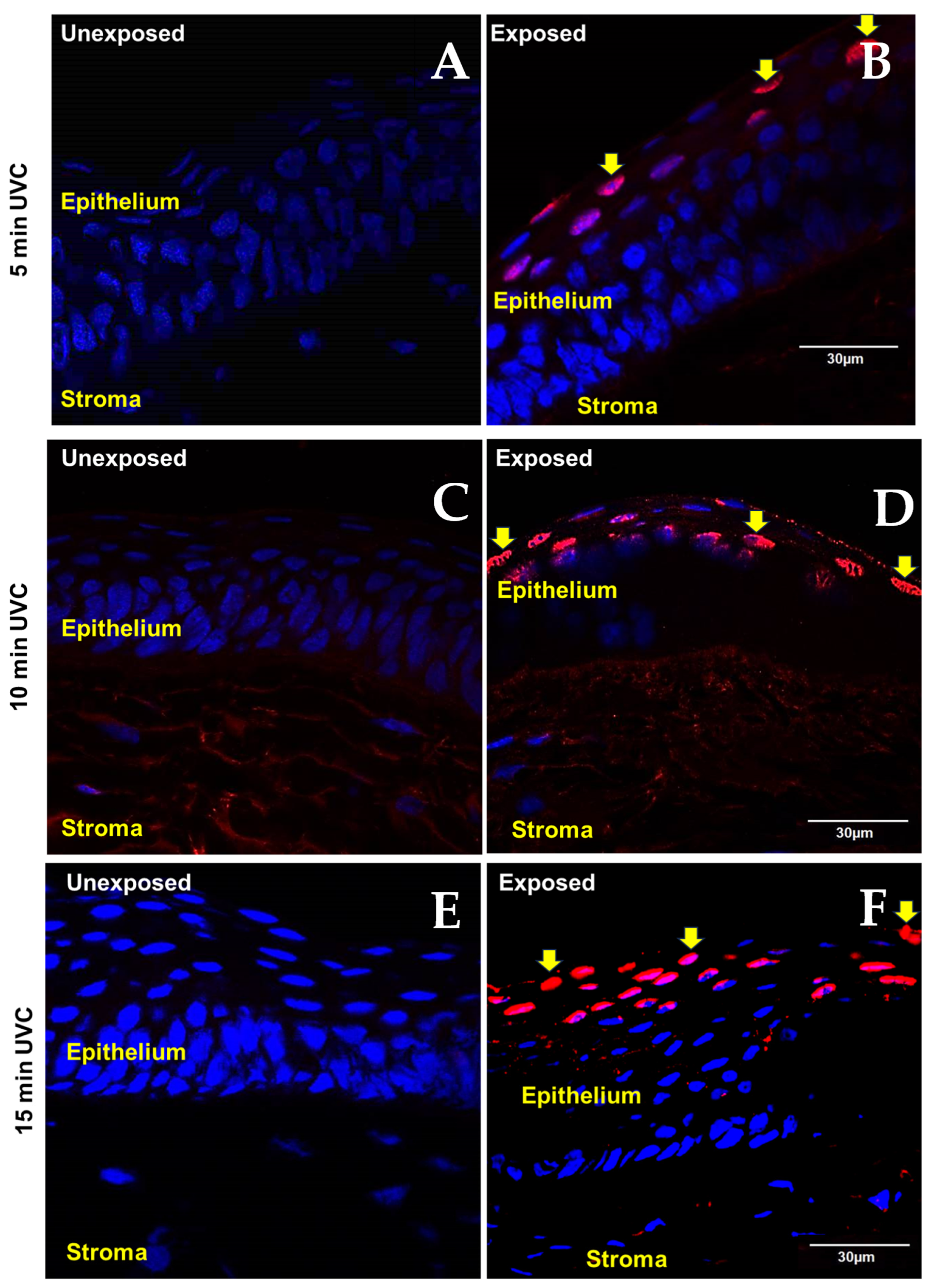
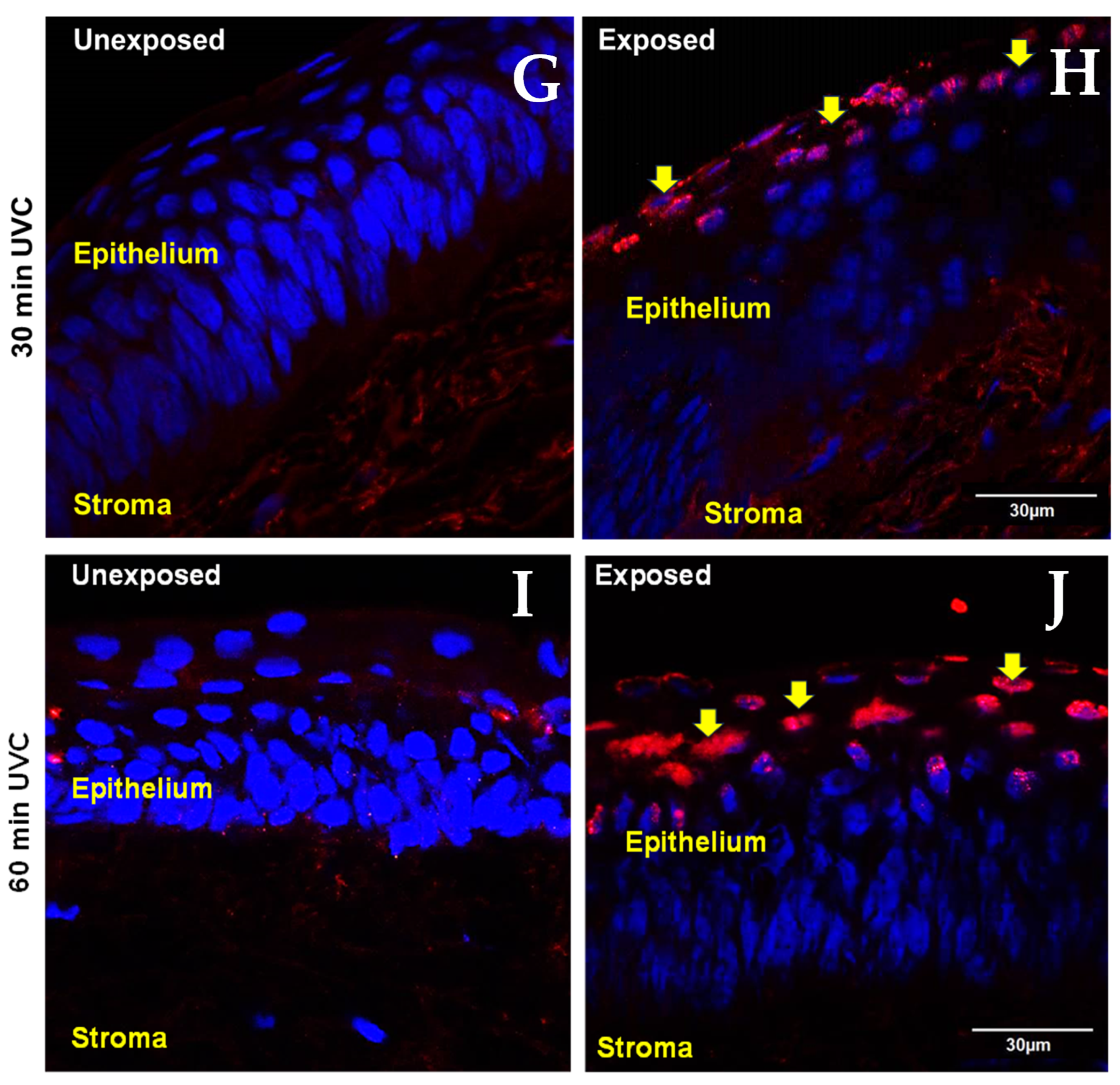
- Low-intensity UVC effects on DNA at the limbus remain limited to superficial epithelial corneal layers, irrespective of dose.
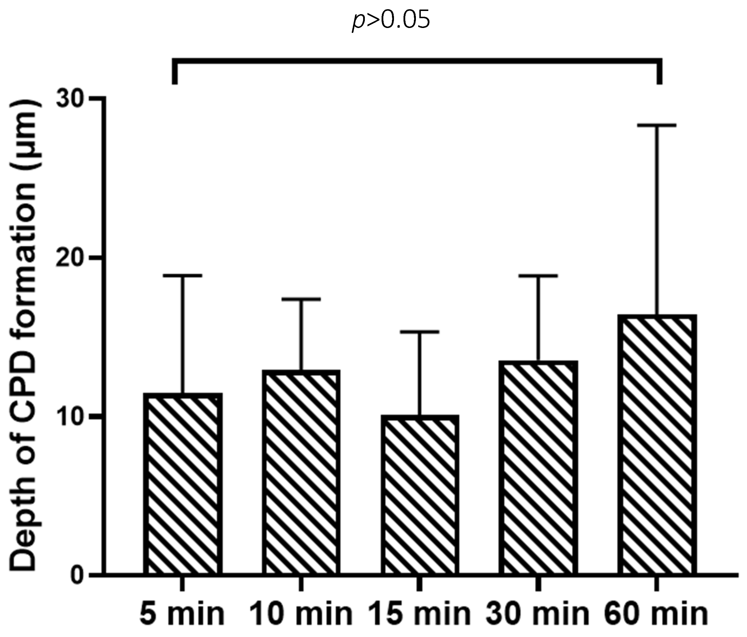
- Limited UVC penetration based on depth of detectable CPD formation suggests sparing of limbal stem cells from DNA damage.
- Demonstrates potential safety of therapeutic low-intensity corneal UVC application in terms of CPD formation.
Abstract
1. Introduction
2. Methods
2.1. Light Source
2.2. Assessment of the Depth of UVC Penetration Through Porcine Cornea
2.3. Immunohistochemistry Analysis
2.4. Assessment of the Depth of UVC Penetration Through Human Donor Cornea
2.5. ImageJ Quantification Within Corneal Epithelium
2.6. Statistical Analysis
3. Results
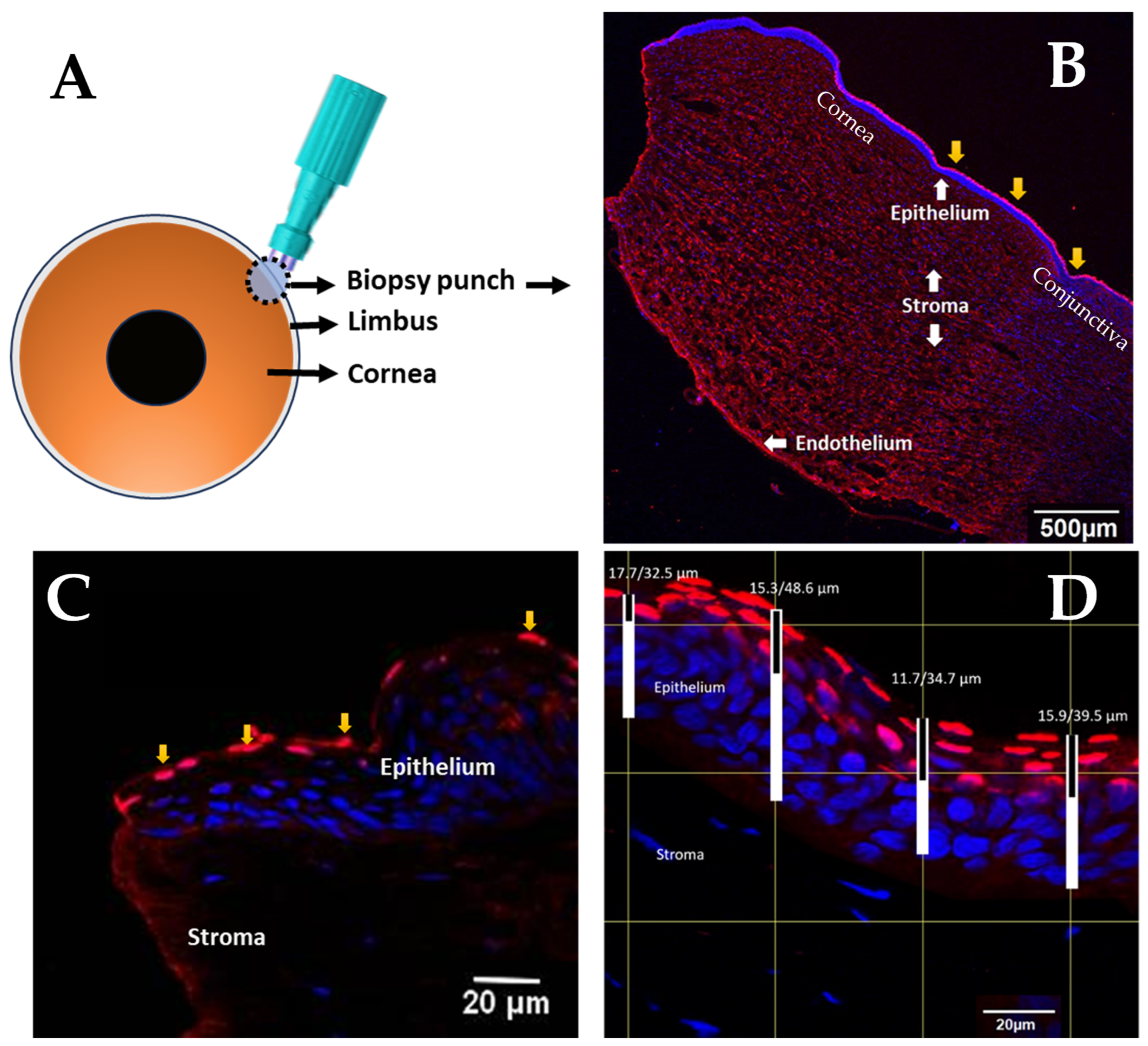
3.1. Depth of CPD Formation in the Porcine Corneal Epithelium
3.2. CPD Distribution in the Human Cornea
4. Discussion
5. Conclusions
Author Contributions
Funding
Data Availability Statement
Conflicts of Interest
References
- Marasini, S.; Dean, S.J.; Swift, S.; Perera, J.; Rupenthal, I.D.; Wang, T.; Read, H.; Craig, J.P. Preclinical confirmation of UVC efficacy in treating infectious keratitis. Ocul. Surf. 2022, 25, 76–86. [Google Scholar] [CrossRef]
- Marasini, S.; Mugisho, O.O.; Swift, S.; Read, H.; Rupenthal, I.D.; Dean, S.J.; Craig, J.P. Effect of therapeutic UVC on corneal DNA: Safety assessment for potential keratitis treatment. Ocul. Surf. 2021, 20, 130–138. [Google Scholar] [CrossRef]
- Dai, T.; Garcia, B.; Murray, C.K.; Vrahas, M.S.; Hamblin, M.R. UVC light prophylaxis for cutaneous wound infections in mice. Antimicrob. Agents Chemother. 2012, 56, 3841–3848. [Google Scholar] [CrossRef] [PubMed]
- Dai, T.; Murray, C.K.; Vrahas, M.S.; Baer, D.G.; Tegos, G.P.; Hamblin, M.R. Ultraviolet C light for Acinetobacter baumannii wound infections in mice: Potential use for battlefield wound decontamination? J. Trauma Acute Care Surg. 2012, 73, 661–667. [Google Scholar] [CrossRef]
- Dai, T.; Kharkwal, G.B.; Zhao, J.; Denis, T.G.S.; Wu, Q.; Xia, Y.; Huang, L.; Sharma, S.K.; d’Enfert, C.; Hamblin, M.R. Ultraviolet-C light for treatment of Candida albicans burn infection in mice. J. Photochem. Photobiol. B 2011, 87, 342–349. [Google Scholar] [CrossRef]
- Sprogyte, L.; Park, M.; Di Girolamo, N. Pathogenesis of alkali injury-induced limbal stem cell deficiency: A literature survey of animal models. Cells 2023, 12, 1294. [Google Scholar] [CrossRef]
- Kaidzu, S.; Sugihara, K.; Sasaki, M.; Nishiaki, A.; Ohashi, H.; Igarashi, T.; Tanito, M. Safety Evaluation of Far-UV-C Irradiation to Epithelial Basal Cells in the Corneal Limbus. Photochem. Photobiol. 2023, 99, 1142–1148. [Google Scholar] [CrossRef]
- Du, Y.; Funderburgh, M.L.; Mann, M.M.; SundarRaj, N.; Funderburgh, J.L. Multipotent stem cells in human corneal stroma. Stem Cells 2005, 23, 1266–1275. [Google Scholar] [CrossRef] [PubMed]
- Sun, T.-T.; Tseng, S.C.; Lavker, R.M. Location of corneal epithelial stem cells. Nature 2010, 463, E10–E11. [Google Scholar] [CrossRef]
- Ljubimov, A.V.; Saghizadeh, M. Progress in corneal wound healing. Prog. Retin. Eye Res. 2015, 49, 17–45. [Google Scholar] [CrossRef] [PubMed]
- Rao, B.K.; Kumar, P.; Rao, S.; Gurung, B. Bactericidal effect of ultraviolet C (UVC), direct and filtered through transparent plastic, on gram-positive cocci: An in vitro study. Ostomy Wound Manag. 2011, 57, 46. [Google Scholar]
- Agarwal, P.; Rupenthal, I.D. In vitro and ex vivo corneal penetration and absorption models. Drug Deliv. Transl. 2016, 6, 634–647. [Google Scholar] [CrossRef]
- Mann, B.K.; Stirland, D.L.; Lee, H.-K.; Wirostko, B.M. Ocular translational science: A review of development steps and paths. Adv. Drug Deliv. Rev. 2018, 126, 195–203. [Google Scholar] [CrossRef]
- Dua, H.S.; Gomes, J.; Singh, A. Corneal epithelial wound healing. Br. J. Ophthalmol. 1994, 78, 401. [Google Scholar] [CrossRef]
- Marasini, S.; Craig, J.P.; Dean, S.J.; Leanse, L.G. Managing Corneal Infections: Out with the old, in with the new? Antibiotics 2023, 12, 1334. [Google Scholar] [CrossRef]
- Dua, H.S.; Azuara-Blanco, A. Limbal stem cells of the corneal epithelium. Surv. Ophthalmol. 2000, 44, 415–425. [Google Scholar] [CrossRef]
- Altshuler, A.; Amitai-Lange, A.; Tarazi, N.; Dey, S.; Strinkovsky, L.; Hadad-Porat, S.; Bhattacharya, S.; Nasser, W.; Imeri, J.; Ben-David, G. Discrete limbal epithelial stem cell populations mediate corneal homeostasis and wound healing. Cell Stem Cell 2021, 28, 1248–1261. e1248. [Google Scholar] [CrossRef] [PubMed]
- Lee, G.A.; Hirst, L.W. Incidence of ocular surface epithelial dysplasia in metropolitan Brisbane: A 10-year survey. Arch. Ophthalmol. 1992, 110, 525–527. [Google Scholar] [CrossRef]
- Yoon, J.J.; Ismail, S.; Sherwin, T. Limbal stem cells: Central concepts of corneal epithelial homeostasis. World J. Stem Cells 2014, 6, 391. [Google Scholar] [CrossRef] [PubMed]
- Setlow, R. Cyclobutane-type pyrimidine dimers in polynucleotides. Science 1966, 153, 379–386. [Google Scholar] [CrossRef] [PubMed]
- Hockberger, P.E. A history of ultraviolet photobiology for humans, animals and microorganisms. Photochem. Photobiol. 2002, 76, 561–579. [Google Scholar] [CrossRef] [PubMed]
- Nishigori, C. Cellular aspects of photocarcinogenesis. Photochem. Photobiol. Sci. 2006, 5, 208–214. [Google Scholar] [CrossRef]
- Sosnin, E.A.; Stoffels, E.; Erofeev, M.V.; Kieft, I.E.; Kunts, S.E. The effects of UV irradiation and gas plasma treatment on living mammalian cells and bacteria: A comparative approach. IEEE Trans. Plasma Sci. 2004, 32, 1544–1550. [Google Scholar] [CrossRef]
- Mallet, J.D.; Rochette, P.J. Wavelength-dependent ultraviolet induction of cyclobutane pyrimidine dimers in the human cornea. Photochem. Photobiol. Sci. 2013, 12, 1310–1318. [Google Scholar] [CrossRef]
- D’Errico, M.; Teson, M.; Calcagnile, A.; De Santis, L.P.; Nikaido, O.; Botta, E.; Zambruno, G.; Stefanini, M.; Dogliotti, E. Apoptosis and efficient repair of DNA damage protect human keratinocytes against UVB. Cell Death Differ. 2003, 10, 754–756. [Google Scholar] [CrossRef]
- Cenedella, R.J.; Fleschner, C.R. Kinetics of corneal epithelium turnover in vivo. Studies of lovastatin. Investig. Ophthalmol. Vis. Sci. 1990, 31, 1957–1962. [Google Scholar]
- Chan, J.Y.; Chow, V.W.; Chan, C.K.; Chan, E.Y.; Lau, J.S.; Lai, T.Y.; Young, A.L.; Tham, C.C. Photokeratitis in outdoor event participants exposed to UV radiation display. JAMA Ophthalmol. 2024, 142, 568–571. [Google Scholar] [CrossRef]
- Crespo-Moral, M.; García-Posadas, L.; López-García, A.; Diebold, Y. Histological and immunohistochemical characterization of the porcine ocular surface. PLoS ONE 2020, 15, e0227732. [Google Scholar] [CrossRef] [PubMed]
- Doughty, M.J.; Zaman, M.L. Human corneal thickness and its impact on intraocular pressure measures: A review and meta-analysis approach. Surv. Ophthalmol. 2000, 44, 367–408. [Google Scholar] [CrossRef]
- Marasini, S.; Dean, S.J.; Swift, S.; Craig, J.P. Comparison of antimicrobial efficacy and safety of pulsed versus continuous wave UVC. Contact Lens Anterior Eye 2025, 48, 102437. [Google Scholar] [CrossRef]
- Davies, K.; Asana, U.; Nku, C.; Osim, E. Ocular effects of chronic exposure to welding light on Calabar welders. Niger. J. Physiol. Sci. 2007, 22, 55–58. [Google Scholar] [CrossRef] [PubMed]
- Estey, T.; Cantore, M.; Weston, P.A.; Carpenter, J.F.; Petrash, J.M.; Vasiliou, V. Mechanisms involved in the protection of UV-induced protein inactivation by the corneal crystallin ALDH3A1. J. Biol. Chem. 2007, 282, 4382–4392. [Google Scholar] [CrossRef]
- Jester, J.V.; Moller-Pedersen, T.; Huang, J.; Sax, C.M.; Kays, W.T.; Cavangh, H.D.; Petroll, W.M.; Piatigorsky, J. The cellular basis of corneal transparency: Evidence for ‘corneal crystallins’. J. Cell Sci. 1999, 112, 613–622. [Google Scholar] [CrossRef] [PubMed]
- Marasini, S. Exploring the Potential of UVC in Treating Superficial Corneal Infections. Ph.D. Thesis, University of Auckland, Auckland, New Zealand, 2019. Available online: https://hdl.handle.net/2292/46388 (accessed on 10 March 2026).
- Tomita, M.; Mita, M.; Huseynova, T. Accelerated versus conventional corneal collagen crosslinking. J. Cataract Refract. Surg. 2014, 40, 1013–1020. [Google Scholar] [CrossRef] [PubMed]



| UVC Exposure Durations | Epithelial Thickness (µm) | ||
|---|---|---|---|
| Control Group | Exposed Group | p-Values (t-Tests) | |
| 5 min | 50.07 ± 19.13 (35.37–64.77) | 45.90 ± 16.03 (37.92–53.87) | 0.58 |
| 10 min | 38.28 ± 11.02 (30.40–46.16) | 28.75 ± 9.95 (22.06–35.43) | 0.05 |
| 15 min | 42.09 ± 10.37 (36.10–48.07) | 34.71 ± 11.35 (28.16–41.26) | 0.08 |
| 30 min | 45.33 ± 12.58 (39.26–51.39) | 38.23 ± 10.87 (29.14–47.31) | 0.15 |
| 60 min | 50.61 ± 21.96 (39.70–61.53) | 44.94 ± 22.52 (35.42–54.45) | 0.41 |
| UVC Exposure Durations | Proportions of CPD (%) | ||
|---|---|---|---|
| Control Group (sample size: 9–12, mean ± SD) | Exposed Group (sample size: 10–12, mean ± SD) | Exposure Comparison p-value (t-test) | |
| 1 min | 0.0 ± 0.0 | 37.2 ± 13.4 (28.75–45. 78) | 0.0001 |
| 5 min | 0.0 ± 0.0 | 39.05 ± 8.6 (32.90–45.21) | 0.0001 |
| Time comparison p-value (t-test) | - | 0.70 | - |
Disclaimer/Publisher’s Note: The statements, opinions and data contained in all publications are solely those of the individual author(s) and contributor(s) and not of MDPI and/or the editor(s). MDPI and/or the editor(s) disclaim responsibility for any injury to people or property resulting from any ideas, methods, instructions or products referred to in the content. |
© 2026 by the authors. Licensee MDPI, Basel, Switzerland. This article is an open access article distributed under the terms and conditions of the Creative Commons Attribution (CC BY) license.
Share and Cite
Torr, B.P.; Craig, J.P.; Dean, S.J.; Sherwin, T.; Marasini, S. Investigating the Effects of UVC Exposure at the Limbus. Cells 2026, 15, 967. https://doi.org/10.3390/cells15110967
Torr BP, Craig JP, Dean SJ, Sherwin T, Marasini S. Investigating the Effects of UVC Exposure at the Limbus. Cells. 2026; 15(11):967. https://doi.org/10.3390/cells15110967
Chicago/Turabian StyleTorr, Bethany P., Jennifer P. Craig, Simon J. Dean, Trevor Sherwin, and Sanjay Marasini. 2026. "Investigating the Effects of UVC Exposure at the Limbus" Cells 15, no. 11: 967. https://doi.org/10.3390/cells15110967
APA StyleTorr, B. P., Craig, J. P., Dean, S. J., Sherwin, T., & Marasini, S. (2026). Investigating the Effects of UVC Exposure at the Limbus. Cells, 15(11), 967. https://doi.org/10.3390/cells15110967