PPARα: Linking Cardiac Metabolism to Therapeutic Opportunities in Cardiovascular Diseases


Abstract
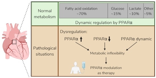
1. Introduction
2. Structure and Activation of PPARα
3. PPARα Is a Crucial Transcription Factor for Heart Metabolism, Maturation and Immunity
3.1. PPARα as Master Regulator of Cardiac Fatty Acid Metabolism
3.2. PPARα as Regulator of Postnatal Cardiac Maturation
3.3. PPARα as Anti-Inflammatory Modulator
4. PPARα Is Implicated in Several Pathological Heart Conditions
4.1. Reduced PPARα Signaling in Cardiovascular Pathology
4.2. Upregulated PPARα in Cardiovascular Diseases
4.3. Dynamic Regulation of PPARα in Cardiovascular Diseases
5. Challenges and Future Perspectives for PPARα Modulation in Cardiovascular Diseases
5.1. Clinical Trials with Fibrates
5.2. Challenges for PPARα Modulation in Cardiovascular Diseases
5.2.1. Disease- and Stage-Dependent Effects
5.2.2. Limited Translation from Animal Models
5.2.3. Selectivity of PPARα Modulation
6. Conclusions
Author Contributions
Funding
Data Availability Statement
Acknowledgments
Conflicts of Interest
References
- Roth, G.A.; Mensah, G.A.; Fuster, V. The Global Burden of Cardiovascular Diseases and Risks. J. Am. Coll. Cardiol. 2020, 76, 2980–2981. [Google Scholar] [CrossRef]
- Tsao, C.W.; Aday, A.W.; Almarzooq, Z.I.; Anderson, C.A.; Arora, P.; Avery, C.L.; Baker-Smith, C.M.; Beaton, A.Z.; Boehme, A.K.; Buxton, A.E.; et al. Heart Disease and Stroke Statistics—2023 Update: A Report From the American Heart Association. Circulation 2023, 147, E93–E621. [Google Scholar] [CrossRef] [PubMed]
- Stanley, W.C.; Recchia, F.A.; Lopaschuk, G.D. Myocardial Substrate Metabolism in the Normal and Failing Heart. Physiol. Rev. 2005, 85, 1093–1129. [Google Scholar] [CrossRef]
- Karwi, Q.G.; Uddin, G.M.; Ho, K.L.; Lopaschuk, G.D. Loss of metabolic flexibility in the failing heart. Front. Cardiovasc. Med. 2018, 5, 68. [Google Scholar] [CrossRef] [PubMed]
- Braissant, O.; Foufelle, F.; Scotto, C.; Dauça, M.; Wahli, W. Differential Expression of Peroxisome Proliferator-Activated Receptors (PPARs): Tissue Distribution of PPAR-a, $3, and-y in the Adult Rat*. Available online: https://academic.oup.com/endo/article/137/1/354/3037365 (accessed on 15 May 2026).
- Pu, Y.; Cheng, C.K.; Zhang, H.; Luo, J.; Wang, L.; Tomlinson, B.; Huang, Y. Molecular mechanisms and therapeutic perspectives of peroxisome proliferator-activated receptor α agonists in cardiovascular health and disease. Med. Res. Rev. 2023, 43, 2086–2114. [Google Scholar] [CrossRef]
- Pawlak, M.; Lefebvre, P.; Staels, B. Molecular mechanism of PPARα action and its impact on lipid metabolism, inflammation and fibrosis in non-alcoholic fatty liver disease. J. Hepatol. 2015, 62, 720–733. [Google Scholar] [CrossRef] [PubMed]
- Lin, Y.; Wang, Y.; Li, P.F. PPARα: An emerging target of metabolic syndrome, neurodegenerative and cardiovascular diseases. Front. Endocrinol. 2022, 13, 1074911. [Google Scholar] [CrossRef]
- Gearing, K.L.; Crickmore, A.; Gustafsson, J.A. Structure of the mouse peroxisome proliferator activated receptor α gene. Biochem. Biophys. Res. Commun. 1994, 199, 255–263. [Google Scholar] [CrossRef]
- Cronet, P.; Petersen, J.F.; Folmer, R.; Blomberg, N.; Sjöblom, K.; Karlsson, U.; Lindstedt, E.-L.; Bamberg, K. Structure of the PPARα and -γ Ligand Binding Domain in Complex with AZ 242; Ligand Selectivity and Agonist Activation in the PPAR Family. Structure 2001, 9, 699–706. [Google Scholar] [CrossRef]
- Kawasaki, M.; Kambe, A.; Yamamoto, Y.; Arulmozhiraja, S.; Ito, S.; Nakagawa, Y.; Tokiwa, H.; Nakano, S.; Shimano, H. Elucidation of Molecular Mechanism of a Selective PPARα Modulator, Pemafibrate, through Combinational Approaches of X-ray Crystallography, Thermodynamic Analysis, and First-Principle Calculations. Int. J. Mol. Sci. 2020, 21, 361. [Google Scholar] [CrossRef]
- Barger, P.M.; Kelly, D.P. PPAR Signaling in the Control of Cardiac Energy Metabolism–Cardiac Energy Production via Mitochondrial Fatty Acid-Oxidation. Circ. Res. 2000, 86, 596–607. [Google Scholar]
- Madrazo, J.A.; Kelly, D.P. The PPAR trio: Regulators of myocardial energy metabolism in health and disease. J. Mol. Cell. Cardiol. 2008, 44, 968–975. [Google Scholar] [CrossRef]
- Delerive, P.; De Bosscher, K.; Besnard, S.; Berghe, W.V.; Peters, J.M.; Gonzalez, F.J.; Fruchart, J.-C.; Tedgui, A.; Haegeman, G.; Staels, B. Peroxisome Proliferator-activated Receptor α Negatively Regulates the Vascular Inflammatory Gene Response by Negative Cross-talk with Transcription Factors NF-κB and AP-1. J. Biol. Chem. 1999, 274, 32048–32054. [Google Scholar] [CrossRef] [PubMed]
- Kim, D.; Ha, S.K.; Gonzalez, F.J. CBFA2T3 Is PPARA Sensitive and Attenuates Fasting-Induced Lipid Accumulation in Mouse Liver. Cells 2024, 13, 831. [Google Scholar] [CrossRef]
- Leuenberger, N.; Pradervand, S.; Wahli, W. Sumoylated PPARα mediates sex-specific gene repression and protects the liver from estrogen-induced toxicity in mice. J. Clin. Investig. 2009, 119, 3138–3148. [Google Scholar] [CrossRef]
- Finck, B.N. The PPAR regulatory system in cardiac physiology and disease. Cardiovasc. Res. 2007, 73, 269–277. [Google Scholar] [CrossRef]
- Bertero, E.; Maack, C. Metabolic remodelling in heart failure. Nat. Rev. Cardiol. 2018, 15, 457–470. [Google Scholar] [CrossRef] [PubMed]
- Fillmore, N.; Hou, V.; Sun, J.; Springer, D.; Murphy, E. Cardiac specific knock-down of peroxisome proliferator activated receptor α prevents fasting-induced cardiac lipid accumulation and reduces perilipin 2. PLoS ONE 2022, 17, e0265007. [Google Scholar] [CrossRef] [PubMed]
- Leone, T.C.; Weinheimer, C.J.; Kelly, D.P. A critical role for the peroxisome proliferator-activated receptor (PPAR) in the cellular fasting response: The PPAR-null mouse as a model of fatty acid oxidation disorders (nuclear hormone receptorslipid metabolismtranscriptional controldietary regulation). Proc. Natl. Acad. Sci. USA 1999, 96, 7473–7478. [Google Scholar] [CrossRef]
- Watanabe, K.; Fujii, H.; Takahashi, T.; Kodama, M.; Aizawa, Y.; Ohta, Y.; Ono, T.; Hasegawa, G.; Naito, M.; Nakajima, T.; et al. Constitutive Regulation of Cardiac Fatty Acid Metabolism through Peroxisome Proliferator-activated Receptor α Associated with Age-dependent Cardiac Toxicity. J. Biol. Chem. 2000, 275, 22293–22299. [Google Scholar] [CrossRef]
- Loichot, C.; Jesel, L.; Tesse, A.; Tabernero, A.; Schoonjans, K.; Roul, G.; Carpusca, I.; Auwerx, J.; Andriantsitohaina, R. Deletion of peroxisome proliferator-activated receptor-α induces an alteration of cardiac functions. Am. J. Physiol. Circ. Physiol. 2006, 291, H161–H166. [Google Scholar] [CrossRef]
- Gélinas, R.; Labarthe, F.; Bouchard, B.; Mc Duff, J.; Charron, G.; Young, M.E.; Rosiers, C.D. Alterations in carbohydrate metabolism and its regulation in PPARα null mouse hearts. Am. J. Physiol. Circ. Physiol. 2008, 294, H1571–H1580. [Google Scholar] [CrossRef]
- Dodd, M.S.; Ambrose, L.; Ball, V.; Clarke, K.; Carr, C.A.; Tyler, D.J. The age-dependent development of abnormal cardiac metabolism in the peroxisome proliferator-activated receptor α-knockout mouse. Atherosclerosis 2024, 399, 118599. [Google Scholar] [CrossRef]
- Yan, J.; Young, M.E.; Cui, L.; Lopaschuk, G.D.; Liao, R.; Tian, R. Increased glucose uptake and oxidation in mouse hearts prevent high fatty acid oxidation but cause cardiac dysfunction in diet-induced obesity. Circulation 2009, 119, 2818–2828. [Google Scholar] [CrossRef]
- Luptak, I.; Balschi, J.A.; Xing, Y.; Leone, T.C.; Kelly, D.P.; Tian, R. Decreased contractile and metabolic reserve in peroxisome proliferator-activated receptor-α-null hearts can be rescued by increasing glucose transport and utilization. Circulation 2005, 112, 2339–2346. [Google Scholar] [CrossRef]
- Zhu, X.-X.; Jiao, S.-Y.; Qi, D.; Yu, B.-Q.; Xie, G.-M.; Liu, Y.; Song, Y.-T.; Xu, Q.-B.; Gonzalez, F.J.; Du, J.; et al. Cardiomyocyte peroxisome proliferator-activated receptor α is essential for energy metabolism and extracellular matrix homeostasis during pressure overload-induced cardiac remodeling. Acta Pharmacol. Sin. 2021, 43, 1231–1242. [Google Scholar] [CrossRef]
- Montaigne, D.; Butruille, L.; Staels, B. PPAR control of metabolism and cardiovascular functions. Nat. Rev. Cardiol. 2021, 18, 809–823. [Google Scholar] [CrossRef]
- Piquereau, J.; Ventura-Clapier, R. Maturation of cardiac energy metabolism during perinatal development. Front. Physiol. 2018, 9, 959. [Google Scholar] [CrossRef] [PubMed]
- Chong, D.; Gu, Y.; Zhang, T.; Xu, Y.; Bu, D.; Chen, Z.; Xu, N.; Li, L.; Zhu, X.; Wang, H.; et al. Neonatal ketone body elevation regulates postnatal heart development by promoting cardiomyocyte mitochondrial maturation and metabolic reprogramming. Cell Discov. 2022, 8, 1–19. [Google Scholar] [CrossRef] [PubMed]
- Planavila, A.; Iglesias, R.; Giralt, M.; Villarroya, F. Sirt1 acts in association with PPARα to protect the heart from hypertrophy, metabolic dysregulation, and inflammation. Cardiovasc. Res. 2011, 90, 276–284. [Google Scholar] [CrossRef] [PubMed]
- Shinkai, Y.; Sasaki, K.; Tamura, R.; Ike, T.; Takahashi, A.; Osaki, Y.; Ishiuchi, N.; Maeoka, Y.; Nakashima, A.; Masaki, T. Selective activation of PPARα by pemafibrate mitigates peritoneal inflammation and fibrosis through suppression of NLRP3 inflammasome and modulation of inflammation. Sci. Rep. 2024, 14, 1–15. [Google Scholar] [CrossRef]
- Hu, A.; Chen, Y.Y. PPARα modulation of macrophage polarization and inflammatory signaling in mimic periodontitis. J. Appl. Oral Sci. 2025, 33, e20250338. [Google Scholar] [CrossRef]
- Wei, P.; Kou, W.; Fu, J.; Chen, Z.; Pan, F. PPARα knockout in mice increases the Th17 development by facilitating the IKKα/RORγt and IKKα/Foxp3 complexes. Commun. Biol. 2023, 6, 847. [Google Scholar] [CrossRef] [PubMed]
- Briand, F.; Maupoint, J.; Brousseau, E.; Breyner, N.; Bouchet, M.; Costard, C.; Leste-Lasserre, T.; Petitjean, M.; Chen, L.; Chabrat, A.; et al. Elafibranor improves diet-induced nonalcoholic steatohepatitis associated with heart failure with preserved ejection fraction in Golden Syrian hamsters. Metabolism 2021, 117, 154707. [Google Scholar] [CrossRef]
- Han, X.; Zhang, Y.; Qiao, O.; Ji, H.; Zhang, X.; Wang, W.; Li, X.; Wang, J.; Li, D.; Ju, A.; et al. Proteomic Analysis Reveals the Protective Effects of Yiqi Fumai Lyophilized Injection on Chronic Heart Failure by Improving Myocardial Energy Metabolism. Front. Pharmacol. 2021, 12, 719532. [Google Scholar] [CrossRef] [PubMed]
- Zhang, F.; Liu, L.; Xie, Y.; Wang, J.; Chen, X.; Zheng, S.; Li, Y.; Dang, Y. Cardiac contractility modulation ameliorates myocardial metabolic remodeling in a rabbit model of chronic heart failure through activation of AMPK and PPAR-α pathway. Open Med. 2022, 17, 365–374. [Google Scholar] [CrossRef] [PubMed]
- Sunagawa, Y.; Kawaguchi, S.; Miyazaki, Y.; Katanasaka, Y.; Funamoto, M.; Shimizu, K.; Shimizu, S.; Hamabe-Horiike, T.; Kawase, Y.; Komiyama, M.; et al. Auraptene, a citrus peel-derived natural product, prevents myocardial infarction-induced heart failure by activating PPARα in rats. Phytomedicine 2022, 107, 154457. [Google Scholar] [CrossRef]
- Li, C.; Zhang, X.; Li, J.; Liang, L.; Zeng, J.; Wen, M.; Pan, L.; Lv, D.; Liu, M.; Cheng, Y.; et al. Ginsenoside Rb1 promotes the activation of PPARα pathway via inhibiting FADD to ameliorate heart failure. Eur. J. Pharmacol. 2023, 947, 175676. [Google Scholar] [CrossRef]
- Yang, J.; Zhao, M.; Zeng, T.; Ye, L.; Gui, Y.; Wang, L. Shenmai injection improves lipid metabolism in post-myocardial infarction heart failure based on network pharmacology and experimental validation. Heliyon 2024, 10, e38648. [Google Scholar] [CrossRef]
- Wang, M.; Zhou, F.; Luo, Y.; Deng, X.; Chen, X.; Yi, Q. The transcription factor PPARA mediates SIRT1 regulation of NCOR1 to protect damaged heart cells. Cardiovasc. Diagn. Ther. 2024, 14, 832–847. [Google Scholar] [CrossRef]
- Zhang, X.; Zhang, Z.; Wang, P.; Han, Y.; Liu, L.; Li, J.; Chen, Y.; Liu, D.; Wang, J.; Tian, X.; et al. Bawei Chenxiang Wan Ameliorates Cardiac Hypertrophy by Activating AMPK/PPAR-α Signaling Pathway Improving Energy Metabolism. Front. Pharmacol. 2021, 12, 653901. [Google Scholar] [CrossRef] [PubMed]
- Kumari, R.; Ray, A.G.; Mukherjee, D.; Chander, V.; Kar, D.; Kumar, U.S.; Bharadwaj, P.V.P.D.; Banerjee, S.K.; Konar, A.; Bandyopadhyay, A. Downregulation of PTEN Promotes Autophagy via Concurrent Reduction in Apoptosis in Cardiac Hypertrophy in PPAR α−/− Mice. Front. Cardiovasc. Med. 2022, 9, 798639. [Google Scholar] [CrossRef]
- Liu, M.-Y.; Yue, L.-J.; Luo, Y.-C.; Lu, J.; Wu, G.-D.; Sheng, S.-Q.; Shi, Y.-Q.; Dong, Z.-X. SUMOylation of SIRT1 activating PGC-1α/PPARα pathway mediates the protective effect of LncRNA-MHRT in cardiac hypertrophy. Eur. J. Pharmacol. 2022, 930, 175155. [Google Scholar] [CrossRef] [PubMed]
- Gao, H.; Tian, K.; Meng, Y.; Liu, X.; Peng, Y. Salidroside Ameliorates Cardiomyocyte Hypertrophy by Upregulating Peroxisome Proliferator-Activated Receptor-α. Front. Pharmacol. 2022, 13, 865434. [Google Scholar] [CrossRef] [PubMed]
- Bianchi, M.; Panera, N.; Petrillo, S.; Cicolani, N.; De Stefanis, C.; Scarsella, M.; Ciavardelli, D.; Piemonte, F.; Alisi, A.; Pastore, A. Peroxisome Proliferator-Activated Receptor Family of Lipid-Activated Nuclear Receptors Alpha Silencing Promotes Oxidative Stress and Hypertrophic Phenotype in Rat Cardiac Cells. Antioxidants 2025, 14, 1059. [Google Scholar] [CrossRef]
- Wang, F.; Li, P.; Yan, X.; Yue, A.; Xu, J.; Shao, Y.; Zhang, K.; Zhang, Q.; Li, Y.; Sun, K. Novel therapeutic insights into pathological cardiac hypertrophy: tRF-16-R29P4PE regulates PACE4 and metabolic pathways. Biochim. Biophys. Acta (BBA)-Mol. Cell Res. 2025, 1872, 119920. [Google Scholar] [CrossRef]
- Zhao, M.; Liu, J.; Peng, S.; Zheng, Z.; Liu, S.; Wan, J.; Xu, Y.; Wang, M. Leucine-Rich Repeat-Containing G Protein-Coupled Receptor 6 Ameliorates Pressure Overload-Induced Cardiac Hypertrophy by Regulating Cardiomyocyte Metabolic Reprogramming. Adv. Sci. 2025, 12, e2417597. [Google Scholar] [CrossRef]
- Xuan, Q.; Li, J.; Feng, Z.; Zhu, L.; Jiang, T.; Li, H.; Liu, M.; Qian, X.; Ma, X. Modulation of PPAR-α and PPAR-γ Influences Cardiomyocyte Growth and Cardiac Remodeling. IUBMB Life 2025, 77, e70035. [Google Scholar] [CrossRef]
- Wheeler, M.; Renick, J.; Fawbush, R.; McAlpin, E.; Stevens, S.; Sreedevi, K.; Warren, J.; Dennis, M.; Pfleger, J. Cardiac REDD1 alters glucose and fatty acid metabolic gene expression via an mTORC1-independent, PPARα-dependent mechanism and drives hypertrophic growth. J. Clin. Investig. 2026. preprint. [Google Scholar] [CrossRef]
- Alsadder, L.; Hamadah, A. Cardiac Ischaemia–Reperfusion Injury: Pathophysiology, Therapeutic Targets and Future Interventions. Biomedicines 2025, 13, 2084. [Google Scholar] [CrossRef]
- Salari, N.; Morddarvanjoghi, F.; Abdolmaleki, A.; Rasoulpoor, S.; Khaleghi, A.A.; Hezarkhani, L.A.; Shohaimi, S.; Mohammadi, M. The global prevalence of myocardial infarction: A systematic review and meta-analysis. BMC Cardiovasc. Disord. 2023, 23, 1–12. [Google Scholar] [CrossRef]
- Xiang, Q.; Yi, X.; Zhu, X.H.; Wei, X.; Jiang, D.S. Regulated cell death in myocardial ischemia–reperfusion injury. Trends Endocrinol. Metab. 2024, 35, 123–138. [Google Scholar] [CrossRef] [PubMed]
- Tian, H.; Zhao, X.; Zhang, Y.; Xia, Z. Abnormalities of glucose and lipid metabolism in myocardial ischemia-reperfusion injury. Biomed. Pharmacother. 2023, 163, 114827. [Google Scholar] [CrossRef] [PubMed]
- Hadanny, A.; Shouval, R.; Wu, J.; Shlomo, N.; Unger, R.; Zahger, D.; Matetzky, S.; Goldenberg, I.; Beigel, R.; Gale, C.; et al. Predicting 30-day mortality after ST elevation myocardial infarction: Machine learning- based random forest and its external validation using two independent nationwide datasets. J. Cardiol. 2021, 78, 439–446. [Google Scholar] [CrossRef] [PubMed]
- Thrane, P.G.; Olesen, K.K.W.; Thim, T.; Gyldenkerne, C.; Mortensen, M.B.; Kristensen, S.D.; Maeng, M. Mortality Trends After Primary Percutaneous Coronary Intervention for ST-Segment Elevation Myocardial Infarction. JACC 2023, 82, 999–1010. [Google Scholar] [CrossRef]
- Mo, H. PPARα activation by fenofibrate protects against acute myocardial ischemia/reperfusion injury by inhibiting mitochondrial apoptosis. Cell. Physiol. Biochem. 2016, 39, 1027–1038. [Google Scholar] [CrossRef]
- Hu, T.; Yu, W.-P.; Wang, X.-Q.; Wang, Z.-Y.; Xu, Z.-Q.; Hu, F.-J.; Liu, J.-C.; Yu, F.; Wang, L.-J. Activation of PPAR-α attenuates myocardial ischemia/reperfusion injury by inhibiting ferroptosis and mitochondrial injury via upregulating 14-3-3η. Sci. Rep. 2024, 14, 1–15. [Google Scholar] [CrossRef]
- Sambandam, N.; Morabito, D.; Wagg, C.; Finck, B.N.; Kelly, D.P.; Lopaschuk, G.D. Chronic activation of PPARα is detrimental to cardiac recovery after ischemia. Am. J. Physiol. Heart Circ. Physiol. 2006, 290, 87–95. [Google Scholar] [CrossRef]
- Duerr, G.D.; Heinemann, J.C.; Arnoldi, V.; Feisst, A.; Kley, J.; Ghanem, A.; Welz, A.; Dewald, O. Cardiomyocyte specific peroxisome proliferator-activated receptor-α overexpression leads to irreversible damage in ischemic murine heart. Life Sci. 2014, 102, 88–97. [Google Scholar] [CrossRef]
- Dewald, O.; Sharma, S.; Adrogue, J.; Salazar, R.; Duerr, G.D.; Crapo, J.D.; Entman, M.L.; Taegtmeyer, H. Downregulation of Peroxisome Proliferator–Activated Receptor-α Gene Expression in a Mouse Model of Ischemic Cardiomyopathy Is Dependent on Reactive Oxygen Species and Prevents Lipotoxicity. Circulation 2005, 112, 407–415. [Google Scholar] [CrossRef]
- Xu, Y.; Lu, L.; Greyson, C.; Rizeq, M.; Nunley, K.; Wyatt, B.; Bristow, M.R.; Long, C.S.; Schwartz, G.G. The PPAR-α activator fenofibrate fails to provide myocardial protection in ischemia and reperfusion in pigs. Am. J. Physiol. Circ. Physiol. 2006, 290, H1798–H1807. [Google Scholar] [CrossRef] [PubMed]
- de Dios, E.; Forteza, M.J.; Perez-Sole, N.; Molina-Garcia, T.; Gavara, J.; Marcos-Garces, V.; Jimenez-Navarro, M.; Ruiz-Sauri, A.; Rios-Navarro, C.; Bodi, V. Temporal and Spatial Dynamics in the Regulation of Myocardial Metabolism During the Ischemia-Reperfusion Process. Int. J. Mol. Sci. 2025, 26, 8820. [Google Scholar] [CrossRef]
- Liepinsh, E.; Makrecka-Kuka, M.; Kuka, J.; Vilskersts, R.; Makarova, E.; Cirule, H.; Loza, E.; Lola, D.; Grinberga, S.; Pugovics, O.; et al. Inhibition of L-carnitine biosynthesis and transport by methyl-γ-butyrobetaine decreases fatty acid oxidation and protects against myocardial infarction. Br. J. Pharmacol. 2015, 172, 1319–1332. [Google Scholar] [CrossRef]
- Zuurbier, C.J.; Bertrand, L.; Beauloye, C.R.; Andreadou, I.; Ruiz-Meana, M.; Jespersen, N.R.; Kula-Alwar, D.; Prag, H.A.; Eric Botker, H.; Dambrova, M.; et al. Cardiac metabolism as a driver and therapeutic target of myocardial infarction. J. Cell. Mol. Med. 2020, 24, 5937–5954. [Google Scholar] [CrossRef]
- Jia, G.; Hill, M.A.; Sowers, J.R. Diabetic cardiomyopathy: An update of mechanisms contributing to this clinical entity. Circ. Res. 2018, 122, 624–638. [Google Scholar] [CrossRef]
- Finck, B.N.; Lehman, J.J.; Leone, T.C.; Welch, M.J.; Bennett, M.J.; Kovacs, A.; Han, X.; Gross, R.W.; Kozak, R.; Lopaschuk, G.D.; et al. The cardiac phenotype induced by PPARα overexpression mimics that caused by diabetes mellitus. J. Clin. Investig. 2002, 109, 121–130. [Google Scholar] [CrossRef]
- Lee, T.W.; Bai, K.J.; Lee, T.I.; Chao, T.F.; Kao, Y.H.; Chen, Y.J. PPARs modulate cardiac metabolism and mitochondrial function in diabetes. Diabetol. Metab. Syndr. 2017, 9, 5. [Google Scholar] [CrossRef]
- Rijzewijk, L.J.; van der Meer, R.W.; Lamb, H.J.; de Jong, H.W.; Lubberink, M.; Romijn, J.A.; Bax, J.J.; de Roos, A.; Twisk, J.W.; Heine, R.J.; et al. Altered Myocardial Substrate Metabolism and Decreased Diastolic Function in Nonischemic Human Diabetic Cardiomyopathy. JACC 2009, 54, 1524–1532. [Google Scholar] [CrossRef] [PubMed]
- Tanabe, K.; Zheng, Q.; Zhang, X.; Tanaka, N.; Hayashi, C.; Yokota, A.; Otsuka, R.; Katahira, T.; Kohjima, M.; Nakamuta, M. Multi-omics analysis of diabetic cardiomyopathy pathogenesis using a type 2 diabetic Zucker diabetic fatty rat model. Sci. Rep. 2025, 15, 1–16. [Google Scholar] [CrossRef]
- Heinonen, I.; Sorop, O.; van Dalen, B.M.; Wüst, R.C.I.; van de Wouw, J.; de Beer, V.J.; Octavia, Y.; van Duin, R.W.B.; Hoogstrate, Y.; Blonden, L.; et al. Cellular, mitochondrial and molecular alterations associate with early left ventricular diastolic dysfunction in a porcine model of diabetic metabolic derangement. Sci. Rep. 2020, 10, 1–14. [Google Scholar] [CrossRef] [PubMed]
- Wang, L.; Cai, Y.; Jian, L.; Cheung, C.W.; Zhang, L.; Xia, Z. Impact of peroxisome proliferator-activated receptor-α on diabetic cardiomyopathy. Cardiovasc. Diabetol. 2021, 20, 2. [Google Scholar] [CrossRef]
- Yin, Z.; Zhao, Y.; He, M.; Li, H.; Fan, J.; Nie, X.; Yan, M.; Chen, C.; Wang, D.W. MiR-30c/PGC-1β protects against diabetic cardiomyopathy via PPARα. Cardiovasc. Diabetol. 2019, 18, 7. [Google Scholar] [CrossRef] [PubMed]
- Ma, X.; Mei, S.; Wuyun, Q.; Zhou, L.; Cai, Z.; Ding, H.; Yan, J. Super-enhancer-driven LncRNA PPARα-seRNA exacerbates glucolipid metabolism and diabetic cardiomyopathy via recruiting KDM4B. Mol. Metab. 2024, 86, 101978. [Google Scholar] [CrossRef]
- Jayakumari, N.R.; Rajendran, R.S.; Sivasailam, A.; Parambil, S.T.; Reghuvaran, A.C.; Sreelatha, H.V.; Gopala, S. Honokiol regulates mitochondrial substrate utilization and cellular fatty acid metabolism in diabetic mice heart. Eur. J. Pharmacol. 2021, 896, 173918. [Google Scholar] [CrossRef]
- Wang, L.; Bi, X.; Han, J. Silencing of peroxisome proliferator-activated receptor-alpha alleviates myocardial injury in diabetic cardiomyopathy by downregulating 3-hydroxy-3-methylglutaryl-coenzyme A synthase 2 expression. IUBMB Life 2020, 72, 1997–2009. [Google Scholar] [CrossRef]
- Yamamoto, K.; Ohta, Y.; Taguchi, A.; Akiyama, M.; Nakabayashi, H.; Nagao, Y.; Ryoko, H.; Wada, Y.; Yamamoto, T.; Yano, M.; et al. Effects of pemafibrate on left ventricular diastolic function in patients with type 2 diabetes mellitus: A pilot study. Diabetol. Int. 2023, 14, 434–439. [Google Scholar] [CrossRef]
- Rudd, K.E.; Johnson, S.C.; Agesa, K.M.; Shackelford, K.A.; Tsoi, D.; Kievlan, D.R.; Colombara, D.V.; Ikuta, K.S.; Kissoon, P.N.; Finfer, P.S.; et al. Global, regional, and national sepsis incidence and mortality, 1990–2017: Analysis for the Global Burden of Disease Study. Lancet 2020, 395, 200–211. [Google Scholar] [CrossRef]
- Hasegawa, D.; Ishisaka, Y.; Maeda, T.; Prasitlumkum, N.; Nishida, K.; Dugar, S.; Sato, R. Prevalence and Prognosis of Sepsis-Induced Cardiomyopathy: A Systematic Review and Meta-Analysis. J. Intensive Care Med. 2023, 38, 797–808. [Google Scholar] [CrossRef] [PubMed]
- Yu, X.; Gao, J.; Zhang, C. Sepsis-induced cardiac dysfunction: Mitochondria and energy metabolism. J. Intensive Care 2025, 13, 28. [Google Scholar] [CrossRef] [PubMed]
- Sato, R.; Sanfilippo, F.; Hasegawa, D.; Prasitlumkum, N.; Duggal, A.; Dugar, S. Prevalence and prognosis of hyperdynamic left ventricular systolic function in septic patients: A systematic review and meta-analysis. Ann. Intensive Care 2024, 14, 55. [Google Scholar] [CrossRef]
- Standage, S.W.; Bennion, B.G.; Knowles, T.O.; Ledee, D.R.; Portman, M.A.; McGuire, J.K.; Liles, W.C.; Olson, A.K. PPARα augments heart function and cardiac fatty acid oxidation in early experimental polymicrobial sepsis. Am. J. Physiol. Heart Circ. Physiol. 2017, 312, 239–249. [Google Scholar] [CrossRef]
- Van Wyngene, L.; Vanderhaeghen, T.; Timmermans, S.; Vandewalle, J.; Van Looveren, K.; Souffriau, J.; Wallaeys, C.; Eggermont, M.; Ernst, S.; Van Hamme, E.; et al. Hepatic PPARα function and lipid metabolic pathways are dysregulated in polymicrobial sepsis. EMBO Mol. Med. 2020, 12, e11319. [Google Scholar] [CrossRef]
- Standage, S.W.; Caldwell, C.C.; Zingarelli, B.; Wong, H.R. Reduced peroxisome proliferator-activated receptor α expression is associated with decreased survival and increased tissue bacterial load in sepsis. Shock 2012, 37, 164–169. [Google Scholar] [CrossRef] [PubMed]
- Standage, S.W.; Waworuntu, R.L.B.; Delaney, M.A.D.; Maskal, S.M.B.; Bennion, B.G.B.; Duffield, J.S.; Parks, W.C.; Liles, W.C.; McGuire, J.K. Nonhematopoietic Peroxisome Proliferator–Activated Receptor-α Protects Against Cardiac Injury and Enhances Survival in Experimental Polymicrobial Sepsis*. Crit. Care Med. 2016, 44, e594–e603. [Google Scholar] [CrossRef]
- Zhu, X.-X.; Wang, X.; Jiao, S.-Y.; Liu, Y.; Shi, L.; Xu, Q.; Wang, J.-J.; Chen, Y.-E.; Zhang, Q.; Song, Y.-T.; et al. Cardiomyocyte peroxisome proliferator-activated receptor α prevents septic cardiomyopathy via improving mitochondrial function. Acta Pharmacol. Sin. 2023, 44, 2184–2200. [Google Scholar] [CrossRef]
- Cheng, L.; Liu, D.; Gao, S. PPARA ameliorates sepsis-induced myocardial injury via promoting macrophage M2 polarization by interacting with DUSP1. Regen. Ther. 2024, 26, 33–41. [Google Scholar] [CrossRef]
- Jozefowicz, E.; Brisson, H.; Rozenberg, S.; Mebazaa, A.; Gelé, P.; Callebert, J.; Lebuffe, G.; Vallet, B.; Bordet, R.; Tavernier, B. Activation of peroxisome proliferator-activated receptor-α by fenofibrate prevents myocardial dysfunction during endotoxemia in rats*. Crit. Care Med. 2007, 35, 856–863. [Google Scholar] [CrossRef]
- Lv, M.; Xie, D.; Long, X. The effect of fenofibrate, a peroxisome proliferator-activated receptor α agonist, on cardiac damage from sepsis in BALB/c mice. Cell. Mol. Biol. 2021, 67, 260–266. [Google Scholar] [CrossRef] [PubMed]
- Mulvey, C.K.; Ferguson, J.F.; Tabita-Martinez, J.; Kong, S.; Shah, R.Y.; Patel, P.N.; Master, S.R.; Usman, M.H.U.; Propert, K.J.; Shah, R.; et al. Peroxisome Proliferator–Activated Receptor-α Agonism With Fenofibrate Does Not Suppress Inflammatory Responses to Evoked Endotoxemia. J. Am. Hear. Assoc. 2012, 1, e002923. [Google Scholar] [CrossRef] [PubMed]
- Zhou, Y.; Zhu, Y.; Wu, Y.; Xiang, X.; Ouyang, X.; Liu, L.; Li, T. 4-phenylbutyric acid improves sepsis-induced cardiac dysfunction by modulating amino acid metabolism and lipid metabolism via Comt/Ptgs2/Ppara. Metabolomics 2024, 20, 46. [Google Scholar] [CrossRef]
- Xing, G.; Meng, L.; Cao, S.; Liu, S.; Wu, J.; Li, Q.; Huang, W.; Zhang, L. PPARα alleviates iron overload-induced ferroptosis in mouse liver. Embo Rep. 2022, 23, e52280. [Google Scholar] [CrossRef]
- Song, W.; Shen, Q.; Zhang, H.; Bu, X.; Gao, W.; Wang, W. c-MAF Transcriptionally Activates Slc40a1 to Repress Ferroptosis in Sepsis-Associated Encephalopathy. CNS Neurosci. Ther. 2026, 32, e70820. [Google Scholar] [CrossRef]
- Nan, W.; Jiang, X.; Cai, X.; Chen, Y.; Peng, Z. 14,15-EET mitigates sepsis-induced cardiomyopathy by inhibiting cardiomyocyte ferroptosis via PPARα activation. Eur. J. Pharmacol. 2026, 1015, 178608. [Google Scholar] [CrossRef]
- Woods, C.G.; Burns, A.M.; Bradford, B.U.; Ross, P.K.; Kosyk, O.; Swenberg, J.A.; Cunningham, M.L.; Rusyn, I. WY-14,643–Induced Cell Proliferation and Oxidative Stress in Mouse Liver are Independent of NADPH Oxidase. Toxicol. Sci. 2007, 98, 366–374. [Google Scholar] [CrossRef]
- Sánchez-Aguilar, M.; Ibarra-Lara, L.; Cano-Martínez, A.; Soria-Castro, E.; Castrejón-Téllez, V.; Pavón, N.; Osorio-Yáñez, C.; Díaz-Díaz, E.; Rubio-Ruíz, M.E. PPAR Alpha Activation by Clofibrate Alleviates Ischemia/Reperfusion Injury in Metabolic Syndrome Rats by Decreasing Cardiac Inflammation and Remodeling and by Regulating the Atrial Natriuretic Peptide Compensatory Response. Int. J. Mol. Sci. 2023, 24, 5321. [Google Scholar] [CrossRef]
- FIELD Study Investigators. Effects of long-term fenofibrate therapy on cardiovascular events in 9795 people with type 2 diabetes mellitus (the FIELD study): Randomised controlled trial. Lancet 2005, 366, 1849–1861. [Google Scholar] [CrossRef]
- ACCORD Study Group. Effects of Combination Lipid Therapy in Type 2 Diabetes Mellitus. N. Engl. J. Med. 2010, 362, 1563–1574. [Google Scholar] [CrossRef]
- Das Pradhan, A.; Glynn, R.J.; Fruchart, J.-C.; MacFadyen, J.G.; Zaharris, E.S.; Everett, B.M.; Campbell, S.E.; Oshima, R.; Amarenco, P.; Blom, D.J.; et al. Triglyceride Lowering with Pemafibrate to Reduce Cardiovascular Risk. N. Engl. J. Med. 2022, 387, 1923–1934. [Google Scholar] [CrossRef]
- Liu, X.; Zhang, J.; Zhou, W.; Liu, J.; Wang, Y. Effect of fenofibrate on blood lipid, sICAM-1, ET-1 and prognosis in chronic heart failure patients complicated with diabetes. Cell. Mol. Biol. 2023, 69, 110–114. [Google Scholar] [CrossRef]
- Kim, J.Y.; Kim, N.H.; Lee, J.; Kim, D.H.; Kim, S.G. Fenofibrate therapy and risk of heart failure outcomes in patients with Type 2 diabetes: A propensity-matched cohort study. Eur. Heart J. Cardiovasc. Pharmacother. 2025, 11, 620–629. [Google Scholar] [CrossRef]



| Study | Condition | Study Type | Model | Description |
|---|---|---|---|---|
| Briand, 2021 [35] | HFpEF | Therapeutic intervention | In vivo hamster model | The PPARα/δ agonist elafibranor was evaluated in a model of NASH/HFpEF. Hamsters treated with elafibranor had significant improvement in diastolic dysfunction and filling pressure. |
| Han, 2021 [36] | Chronic heart failure | Mechanism of therapeutic | In vivo rat model | Yiqi Fumai lyophilized injection (YQFM) is used as a treatment for chronic heart failure. YQFM is upregulating several proteins involved in mitochondrial function and OXPHOS, including PPARα. In addition, cardiomyocyte apoptosis is reduced. |
| Zhang, 2022 [37] | Chronic heart failure | Therapeutic intervention | In vivo rabbit model | Cardiac contractility modulation is applied to rabbits with heart failure. This ameliorated several aspects of heart failure, including cardiac structure and function. In addition, cardiac metabolism is restored with higher protein levels of PPARα and AMPK. |
| Sunagawa, 2022 [38] | Heart failure | Therapeutic intervention | In vitro cultured cardiomyocytes from neonatal rats and an in vivo rat model | Auraptene, a citrus-peel-derived natural product, was evaluated in in vitro and in vivo models of heart failure. It activated PPARα-dependent expression in vitro, and improved systolic function and fibrosis in rats, accompanied by upregulation of PPARα target genes. |
| Li, 2023 [39] | Heart failure | Mechanistic study/therapeutic intervention | In vivo rat model and in vitro neonatal rat cardiomyocytes | Heart failure increases Fas-associated death domain (FADD) expression, promoting its interaction with PPARα and thereby inhibiting PPARα activity. GRb1, a natural product derived from ginseng, disrupted FADD upregulation and prevents PPARα-FADD binding, leading to improved cardiac function, increased ATP production and restoration of metabolic homeostasis. |
| Yang, 2024 [40] | Heart failure | Mechanistic study | In silico predictions In vivo mouse model | The mechanisms of Shenmai injection (SMI), a Chinese medicine formulation, are investigated in heart failure. In silico analysis identified PPARα as a key target, and active SMI components showed high binding affinity to PPARα. This was associated with improved cardiac function, decreased inflammatory cell infiltration and better mitochondrial function. |
| Wang, 2024 [41] | Heart failure | Mechanistic study | In vitro AV16 and human cardiac microvascular endothelial cells | PPARα is part of a cardioprotective signaling axis involving SIRT1 and NCOR1. Overexpression of SIRT1 increases PPARα activity, which in turn enhances NCOR1 transcription. This pathway leads to reduced ROS production and lipotoxicity, improved mitochondrial metabolism and ATP production, decreased inflammation and apoptosis. |
| Study | Condition | Study Type | Model | Summary |
|---|---|---|---|---|
| Zhang, 2021 [42] | Cardiac hypertrophy | Therapeutic intervention | In vivo rat model | Bawei Chenxiang Wan (BCW), a Chinese medicine formulation, improved cardiac function and structural remodeling during hypertrophy. These effects were associated with metabolic normalization via upregulation of PPARα and AMPK signaling. |
| Kumari, 2022 [43] | Cardiac hypertrophy | Mechanistic study | In vivo mouse model | PPARα-null mice subjected to cardiac hypertrophy exhibited altered cardiac proteomic profiles, including reduced apoptotic markers and increased autophagy-related proteins. This suggests that PPARα may regulate cardiomyocyte apoptosis and modulate autophagic processes during hypertrophic remodeling. |
| Liu, 2022 [44] | Cardiac hypertrophy | Mechanistic study | In vitro cardiomyocytes and an in vivo mouse model | Long non-coding RNA MHRT protects against cardiac hypertrophy by promoting SIRT1 SUMOylation, which activates PPARα signaling. This enhances mitochondrial function and fatty acid oxidation, thereby attenuating hypertrophic remodeling |
| Gao, 2022 [45] | Cardiac hypertrophy | Therapeutic intervention | In vitro H9C2 cell model | Salidroside attenuated hypertrophic markers while increasing PPARα expression. This effect was mediated by ATGL, an upstream activator of PPARα. |
| Zhu, 2021 [27] | Pressure overload cardiac hypertrophy | Mechanistic study | In vivo PPARα-MHC-deficient mouse model | Mice with a cardiomyocyte-specific deletion of PPARα developed accelerated cardiac hypertrophy and increased fibrosis. This indicates that PPARα plays a protective role in hypertrophy by regulating fatty acid oxidation and maintaining extracellular matrix homeostasis. |
| Bianchi, 2025 [46] | Cardiac hypertrophy | Mechanistic study | In vitro cardiomyoblast | Silencing of PPARα led to the induction of a hypertrophic phenotype, with increased NPPB, mitochondrial dysfunction and impaired lipid metabolism. |
| Wang, 2025 [47] | Cardiac hypertrophy | Therapeutic intervention | In vitro cardiomyocyte and in vivo rat model | Transfer RNA-derived small RNAs may have an important role in the development of cardiac hypertrophy. tRF-16-R29P4PE was identified as significantly downregulated in patients with cardiac hypertrophy. Modulation of tRF-16-R29P4PE led to reduced hypertrophic markers and induction of PPARα. |
| Zhao, 2025 [48] | Pressure overload-induced hypertrophy | Mechanistic study | In vivo mouse model and in vitro cardiomyocytes | Lgr6 is a regulator of cardiac hypertrophy. Lgr6 is downregulated in hypertrophy, together with PPARα. Lgr6 overexpression attenuated cardiac hypertrophy and dysfunction by upregulating the expression of PPARα, thereby promoting metabolic reprogramming in cardiomyocytes. |
| Xuan, 2025 [49] | Basal | Mechanistic study | In vitro neonatal rat cardiomyocytes | PPARα expression was modulated in cardiomyocytes using pharmacological activators and inhibitors, with hypertrophic markers as readout. Activation by Wy-14643 promoted cardiomyocyte proliferation and reduced hypertrophy, whereas inhibition induced hypertrophic changes and suppressed proliferation. These findings indicate a protective role for PPARα against hypertrophy. |
| Wheeler, 2026 [50] | Cardiac hypertrophy | Mechanistic study | In vitro cardiomyocytes and an in vivo mouse model | Redd1 is upregulated upon cardiac hypertrophy and is associated with reduced PPARα expression and a decrease in fatty acid oxidation gene levels. Deletion of Redd1 restores PPARα signaling and cardiac metabolic function, suggesting a potential therapeutic target for hypertrophy. |
Disclaimer/Publisher’s Note: The statements, opinions and data contained in all publications are solely those of the individual author(s) and contributor(s) and not of MDPI and/or the editor(s). MDPI and/or the editor(s) disclaim responsibility for any injury to people or property resulting from any ideas, methods, instructions or products referred to in the content. |
© 2026 by the authors. Licensee MDPI, Basel, Switzerland. This article is an open access article distributed under the terms and conditions of the Creative Commons Attribution (CC BY) license.
Share and Cite
Roes, M.; Libert, C.; Vandewalle, J. PPARα: Linking Cardiac Metabolism to Therapeutic Opportunities in Cardiovascular Diseases. Cells 2026, 15, 940. https://doi.org/10.3390/cells15100940
Roes M, Libert C, Vandewalle J. PPARα: Linking Cardiac Metabolism to Therapeutic Opportunities in Cardiovascular Diseases. Cells. 2026; 15(10):940. https://doi.org/10.3390/cells15100940
Chicago/Turabian StyleRoes, Maxime, Claude Libert, and Jolien Vandewalle. 2026. "PPARα: Linking Cardiac Metabolism to Therapeutic Opportunities in Cardiovascular Diseases" Cells 15, no. 10: 940. https://doi.org/10.3390/cells15100940
APA StyleRoes, M., Libert, C., & Vandewalle, J. (2026). PPARα: Linking Cardiac Metabolism to Therapeutic Opportunities in Cardiovascular Diseases. Cells, 15(10), 940. https://doi.org/10.3390/cells15100940