Article

Connexin-43-Mediated Gap Junction Coupling Between Adipocytes Regulates Norepinephrine-Induced Ca2+ Responses in Perivascular Adipose Tissue

by
Ae Ra Kim
1,
Julia Jamka
1,
William F. Jackson
2,
Emma D. Flood
2,
Jonathon L. McClain
1 and
Brian D. Gulbransen
1,*
1
Department of Physiology, College of Natural Science, Michigan State University, 567 Wilson Road, East Lansing, MI 48824, USA
2
Department of Pharmacology and Toxicology, College of Osteopathic Medicine, Michigan State University, East Lansing, MI 48824, USA
*
Author to whom correspondence should be addressed.
Cells 2026, 15(10), 906; https://doi.org/10.3390/cells15100906
Submission received: 17 April 2026 / Revised: 12 May 2026 / Accepted: 13 May 2026 / Published: 15 May 2026
(This article belongs to the Special Issue Adipose Tissue Functioning in Health and Diseases)

Highlights

What are the main findings?
  • Adipocytes in perivascular adipose tissue (PVAT) express gap junctions composed of connexin-43.
  • Gap junction coupling between PVAT adipocytes regulates responses to norepinephrine.
What are the implications of the main finding?
  • Gap junction coupling between PVAT adipocytes provides a potential explanation for how signals from limited sympathetic innervation are distributed throughout PVAT depots.
  • While adipocyte gap junction coupling could contribute to the mechanism by which PVAT controls blood vessel contractility, the functional relevance of this mechanism remains unclear because adipocyte-specific connexin-43 knockout mice showed no vascular phenotype and upregulated connexin 26 gene expression, which may indicate potential compensatory mechanisms.

Abstract

Anticontractile factors secreted by perivascular adipose tissue (PVAT) play an important role in regulating vascular tone. This process is driven by the neurotransmitter norepinephrine (NE), but recent data show that adrenergic innervation in PVAT is sparse. How limited innervation might initiate broad responses through PVAT depots remains unknown. Here, we used Ca2+ imaging with genetically encoded sensors, selective drugs, immunolabeling and a conditional ablation model to test the hypothesis that gap junction coupling among PVAT adipocytes contributes to how signals initiated by NE are distributed through PVAT depots. Despite exhibiting differing sensitivities to NE, adipocytes in aortic and mesenteric PVAT and in white adipose tissue displayed robust expression of the gap junction protein connexin-43 (Cx43). Blocking gap junction coupling with the drug carbenoxolone (Cbx) limited NE-evoked Ca2+ responses among adipocytes, while blocking Cx43 hemichannels with the mimetic peptide 43Gap26 had no significant effect. Fluorescence recovery after photobleaching (FRAP) in mPVAT was decreased in the presence of Cbx, suggesting impaired gap junction communication. Wire myography recordings of mesenteric arteries showed that the EC50 for NE was higher in samples with intact PVAT than those without; however, this effect was not significantly different in samples from mice that lacked Cx43 in adipocytes. Analysis of multiple connexins showed that adipocytes upregulate Cx26 gene expression when Cx43 is deleted. These observations support the conclusion that Cx43-mediated gap junction coupling among PVAT adipocytes contributes to distributing signals initiated by NE; however, how this mechanism contributes to regulating vessel constriction remains unclear. This, and how potential compensatory mechanisms are enacted in adipocytes lacking Cx43, should be addressed in future work.

1. Introduction

Cardiovascular diseases are the leading cause of death globally and contribute to major personal and societal costs [1]. Hypertension is one of the most common contributors to cardiovascular disease and affects approximately half of Americans [2]. While processes involved in the pathogenesis of hypertension are often considered to work through inside-out signaling mechanisms that produce blood vessel dysfunction, an emerging concept is that outside-in signaling also plays a significant role in vascular pathophysiology [3]. Much of this outside-in signaling is thought to involve a unique type of adipose tissue called perivascular adipose tissue (PVAT) that surrounds most blood vessels and releases diffusible factors that regulate local vascular reactivity and inflammatory status [4]. PVAT exhibits unique qualities depending on its anatomical location that reflect differences in adipocyte progenitors, differentiation, cellular morphology and cellular composition. Adipocytes are the most abundant cell type, but PVAT also contains immune cells, endothelial cells, fibroblasts and adipocyte stem/progenitor cells. Changes in the cellular composition and/or function of PVAT is now considered an important contributor to pathophysiological changes that occur in cardiovascular diseases, diabetes and obesity [5,6,7,8,9,10,11]. Yet how PVAT exerts beneficial and deleterious effects on the vasculature remains poorly understood.
Vasoactive actions of PVAT adipocytes are mediated, in part, by several secreted anticontractile factors that include adiponectin, leptin, nitric oxide (NO), hydrogen sulfide (H2S), and chemerin, among others [12]. Norepinephrine (NE) appears to play an important role in promoting the release of these factors from adipocytes and, hence, stimulating the anticontractile nature of PVAT [13,14]. This has raised the possibility that sympathetic innervation may play dual roles in regulating the vasculature through direct actions that promote vasoconstriction and indirect actions through innervation of PVAT that promote vasodilation. Despite this, emerging data show that adrenergic innervation of PVAT is extremely limited [15,16]. How such sparse innervation could regulate the activity of many adipocytes within PVAT depots remains unknown.
A possible explanation for how limited innervation could produce broader effects through PVAT has emerged from work on mechanisms underlying beiging in white adipose tissue. Here, responses to minimal sympathetic innervation are propagated between adipocytes through cell–cell signaling involving gap junction-coupled networks composed of connexin-43 (Cx43) [17]. Gap junctions provide direct and selective channels between neighboring cells and allow the passage of molecules smaller than 1000 daltons including inorganic ions such as Ca2+ and Na+, second messengers such as cAMP and inositol 1,4,5-trisphosphate, small RNAs and certain metabolites [18,19]. Gap junctions and hemichannels, unpaired connexons that connect the intracellular space with the extracellular environment, are regulated by several factors that include transmembrane voltage [20], cell redox potential [21], phosphorylation [22,23], and cations [24,25] such as intracellular Ca2+ [26,27]. Gap junctions play a vital role in regulating vascular function [28,29] and are implicated in mechanisms involved in the pathophysiology of diabetes and hypertension [30,31,32,33].
Given the important roles of gap junction coupling in white adipose tissue and in vascular function, we reasoned that similar mechanisms may explain how PVAT adipocytes propagate signals initiated by limited sympathetic innervation. We tested potential contributions of gap junction coupling to PVAT adipocyte responses driven by NE in calcium (Ca2+) imaging experiments and conducted immunolabeling experiments to localize Cx43 in PVAT. Functional assessment of gap junction activity was performed using fluorescence recovery after photobleaching (FRAP), and roles of adipocyte Cx43 in controlling small resistance artery contractile properties were evaluated in adipocyte-specific Cx43 (Adipo∆Cx43) knockout mice. Together, these results provide valuable insight into the significance of gap junction coupling in PVAT as a pathway of cellular communication.

2. Results

2.1. Norepinephrine (NE) Stimulates Ca2+ Responses Among Adipocytes in Perivascular Adipose Tissue (PVAT)

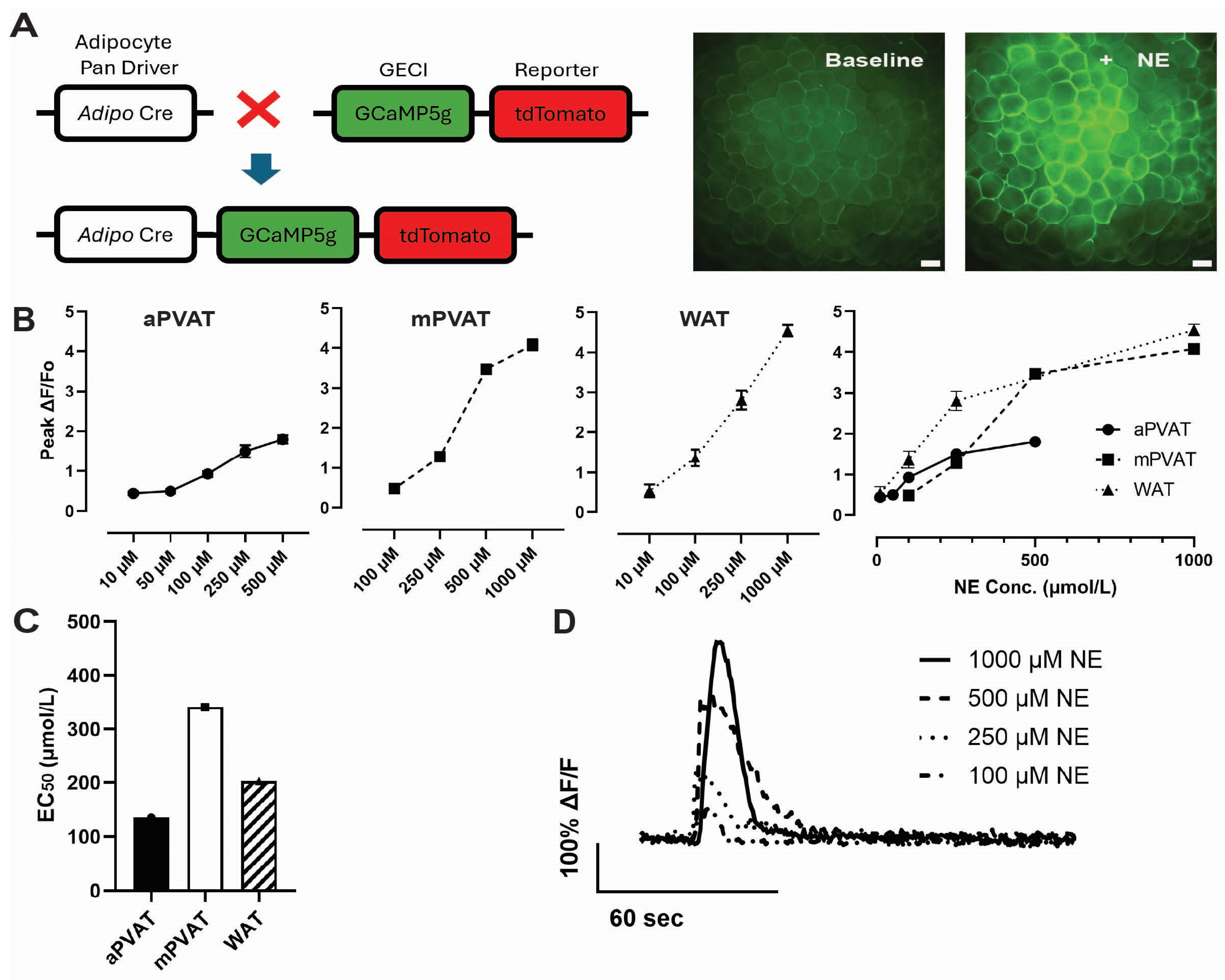
Given the critical role of PVAT in vascular homeostasis and the emerging evidence that NE stimulates the release of anticontractile factors from PVAT [6,7,9], we began by assessing the sensitivity of different PVAT depots to NE using Ca2+ imaging. In prior work, we developed Adipoqcre+;GCaMP5g-tdTf/WT mice (Figure 1A) as a tool to visualize and quantify NE-induced Ca2+ propagation in PVAT adipocytes and showed that exposing samples of PVAT to NE evokes robust Ca2+ responses among adipocytes that are mediated by a1a, b2 and b3 adrenergic receptors and the subsequent release of Ca2+ from intracellular stores, with depot-specific differences [15,34]. However, whether different adipose depots display differing sensitivities to NE has remained unclear. We addressed this issue by conducting ex vivo live-cell Ca2+ imaging in samples of aortic PVAT (aPVAT), mesenteric PVAT (mPVAT) and white adipose tissue (WAT) and challenging the adipocytes with NE concentrations ranging from 10 to 1000 µmol/L.
Adipocytes in aPVAT, mPVAT and WAT were responsive to NE, but sensitivities and maximal responses evoked differed (Figure 1B). Low-amplitude Ca2+ transients were observable in aPVAT in response to NE concentrations as low as 10 µmol/L and NE-driven Ca2+ responses saturated around 250 µmol/L. In contrast, WAT adipocytes exhibited a larger dynamic range and maintained responses up to 1000 µmol/L. mPVAT adipocytes exhibited a response profile that was similar to those in WAT, which agrees with the concept of a common phenotype between adipocytes in these depots [35]. However, mPVAT adipocytes were less sensitive to NE than those in WAT and responses to NE below 100 μmol/L were not observed, which is supported by half-maximal effective concentration (EC50) values 135 µmol/L, 341.1 µmol/L and 202.5 µmol/L at aPVAT, mPVAT and WAT, respectively (Figure 1C). Upon NE stimulation, adipocytes in aPVAT produced Ca2+ responses with average amplitudes of 0.45 ± 0.04, 0.49 ± 0.06, 0.88 ± 0.07, 1.5 ± 0.15, and 1.8 ± 0.10 ΔF/F0 at 10, 50, 100, 250, and 500 μmol/L NE, respectively. In contrast, mPVAT exhibited responses to NE that were 0.48 ± 0.07, 1.28 ± 0.11, 3.47 ± 0.09, and 4.08 ± 0.13 ΔF/F0 at 100, 250, 500 and 1000 μmol/L NE. Adipocytes in WAT exhibited responses of 0.53 ± 0.17, 1.36 ± 0.20, 2.81 ± 0.24, and 4.55 ± 0.15 ΔF/F0 at 10, 100, 250 and 1000 μmol/L NE, respectively. Taken together, these data demonstrate that NE stimulation elicits robust, concentration-dependent Ca2+ responses in adipocytes from aPVAT, mPVAT and WAT (Figure 1B,D). The distinct NE response profiles observed among aPVAT, mPVAT and WAT suggest that PVATs have different sensitivities to NE stimulation that are dependent on their anatomical location and tissue composition [36,37,38].

2.2. Expression of Connexin-43 in PVAT from WT and Adipo∆Cx43 KO Mice

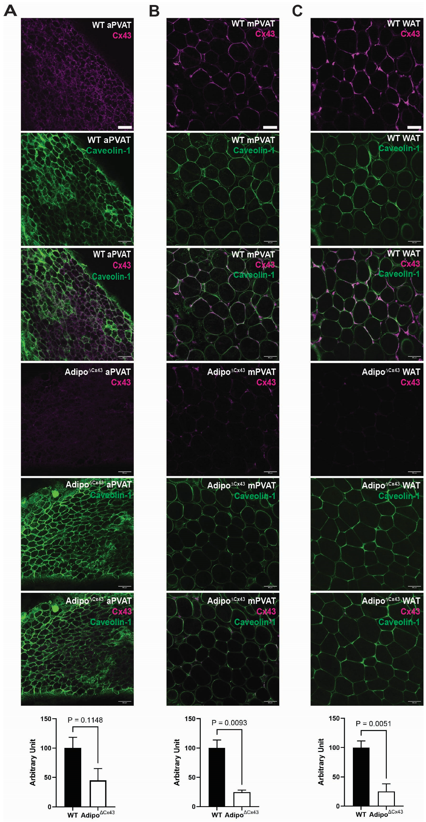
To determine whether the propagation of Ca2+ responses between adipocytes could be functionally regulated by connexin hemichannels, we began assessing the presence of connexin-43 (Cx43) in PVAT using immunofluorescence staining. Cx43 is one of the major connexin subtypes that plays an important role in gap junction communication. Immunolabeling was performed with antibodies against Cx43 and the membrane marker Caveolin-1 [39,40] in WT mice and adipocyte-specific Cx43(f/f) knockout (Adipo∆Cx43 KO) mice.
As shown in Figure 2, Cx43 protein expression was distributed around the nucleus and in large foci at cell–cell borders, corresponding to gap junctions between adjacent cells. No notable alterations or patterns of Cx43 expression were observed between different PVATs. Control experiments showed a loss of Cx43 labeling in samples of PVAT from Adipo∆Cx43 KO mice, confirming the specificity of the antibody.

2.3. Cx43 Gap Junctions, but Not Hemichannels, Modulate NE-Evoked Ca2+ Responses in PVAT

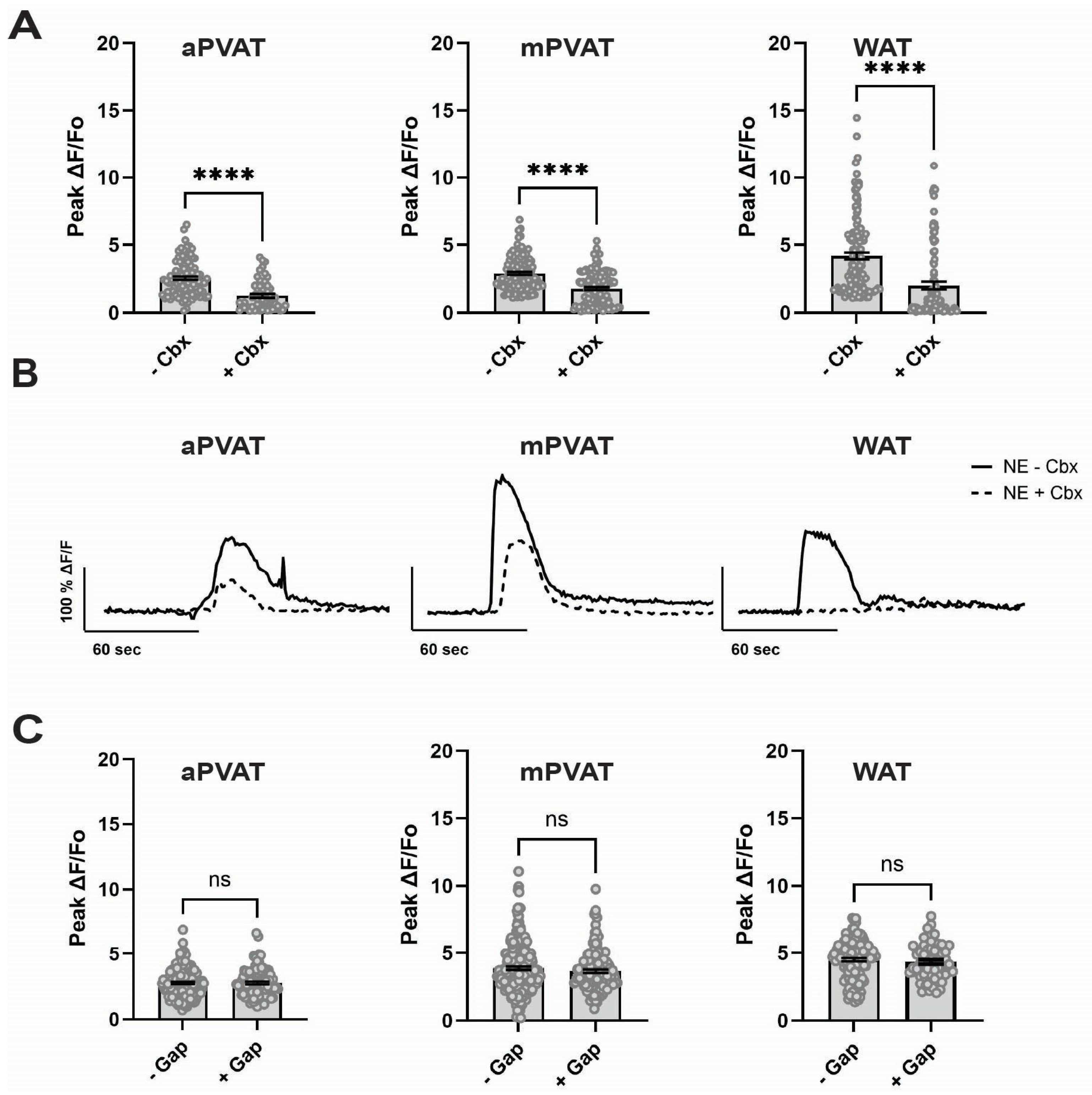
Cx43 can form gap junctions that provide a direct route of cell–cell coupling or can form hemichannels that provide a route to release diffusible factors into the extracellular space. Either of these two mechanisms could affect how adipocytes in PVAT distribute responses to NE among neighboring cells. To differentiate between roles of gap junctions and hemichannels in NE-driven Ca2+ responses in PVAT adipocytes, we conducted experiments in which we blocked gap junctions using the broad antagonist carbenoxolone disodium salt (Sigma, C4790, Cbx) or blocked hemichannels using the Cx43 mimetic peptide 43Gap26 (Anaspec Inc. 62644). Cbx significantly decreased NE-driven Ca2+ responses by 52% in aPVAT (1.56 ± 0.10 ΔF/F0 to 0.82 ± 0.09 ΔF/F0; p < 0.0001, n = 62 cells, three mice), by 64% in mPVAT (2.46 ± 0.11 ΔF/F0 to 1.59 ± 0.09 ΔF/F0; p < 0.0001, n = 161 cells, three mice) and by 48% in WAT (3.68 ± 0.23 ΔF/F0 to 1.75 ± 0.22 ΔF/F0; p < 0.0001, n = 121 cells, three mice) (Figure 3A,B). In contrast, blocking Cx43 hemichannels with 43Gap26 had no significant effect on Ca2+ responses to NE in aPVAT (2.78 ± 0.08 ΔF/F0 to 2.78 ± 0.11 ΔF/F0 with 43Gap26, n = 98 cells, three mice), mPVAT (3.69 ± 0.12 ΔF/F0 to 3.67 ± 0.12 ΔF/F0 with 43Gap26, n = 145 cells, three mice), or WAT adipocytes (4.5 ± 0.12 ΔF/F0 to 4.34 ± 0.16 ΔF/F0 with 43Gap26, n = 67 cells, three mice) (Figure 3C). These data are consistent with the conclusion that gap junction coupling among PVAT adipocytes plays an important role in propagating Ca2+ signals among adipocytes while hemichannels are either not present or play a minor role.

2.4. Gap Junctional Communication in mPVAT

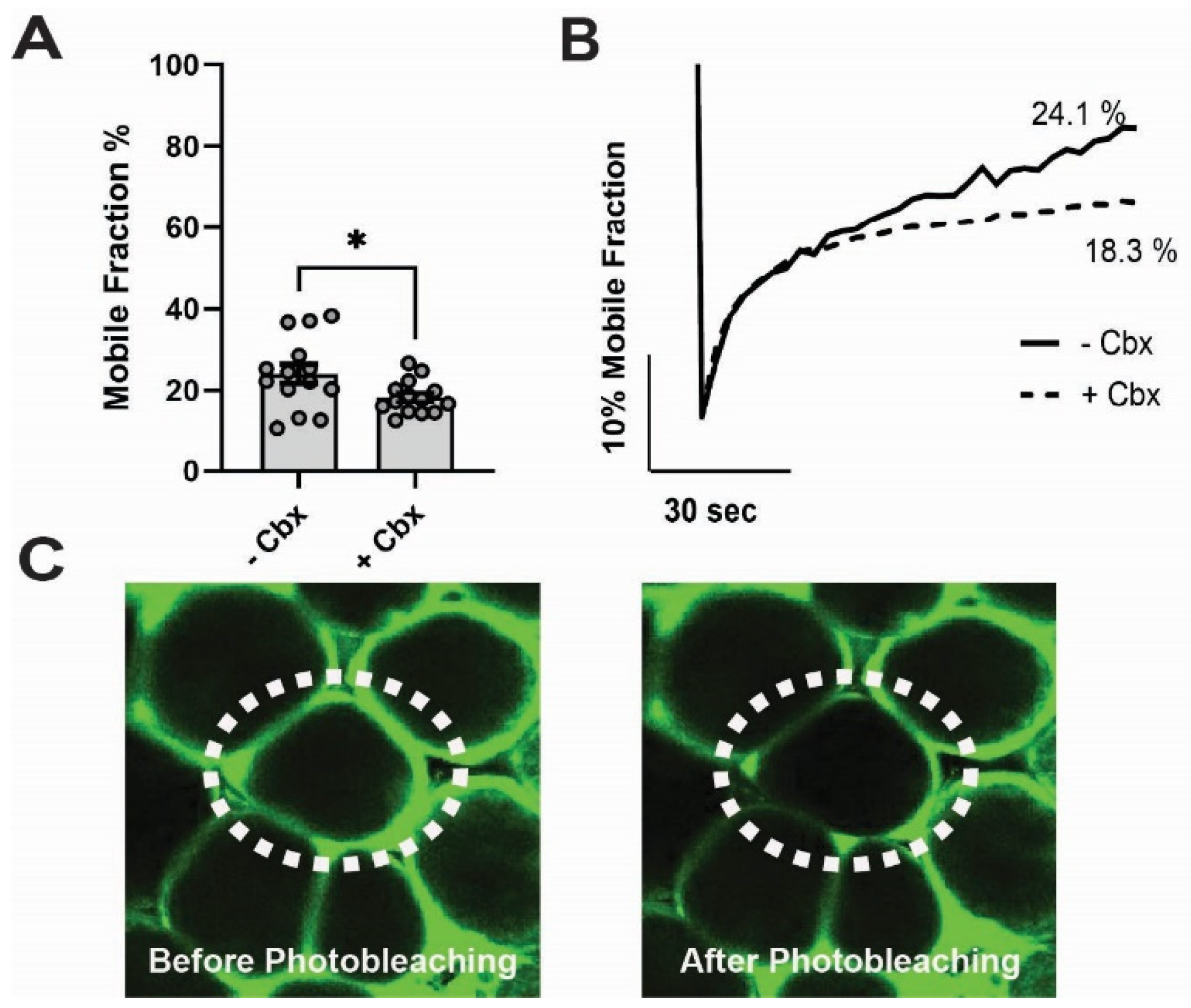
The preceding antagonist data suggest that Cx43 contributes to gap junctional coupling among adipocytes. However, to rule out potential off target effects of Cbx, we conducted more direct assays of functional coupling between mPVAT adipocytes using FRAP [41,42,43,44]. In these experiments, adipocytes were loaded with Calcein AM, which diffuses through gap junctions. Calcein AM fluorescence was photobleached in single adipocytes in the presence or absence of Cbx, and recovery of fluorescence mediated by diffusion from adjacent cells was monitored over 10 min.
On average, normal fluorescence recovery in the absence of Cbx was 24.07 ± 2.36% (n = 14 cells, five mice). In the presence of Cbx, fluorescence recovery was significantly reduced to 18.33 ± 1.09% (p = 0.0036, n = 14 cells, five mice) (Figure 4A–C). These observations suggest that functional gap junctions are present between adipocytes and form a conduit of intercellular communication between neighboring adipocytes in mPVAT. In addition, given the anatomical location of mPVAT next to mesenteric resistance vessels and the function of gap junctions in transferring small molecules between neighboring cells, it is possible that responses evoked in adipocytes distant from vessels could influence mesenteric resistance vascular function through gap junction-coupled networks.

2.5. Effects of Adipocyte-Specific Cx43 Deletion on NE-Induced Mesenteric Resistance Artery Constriction

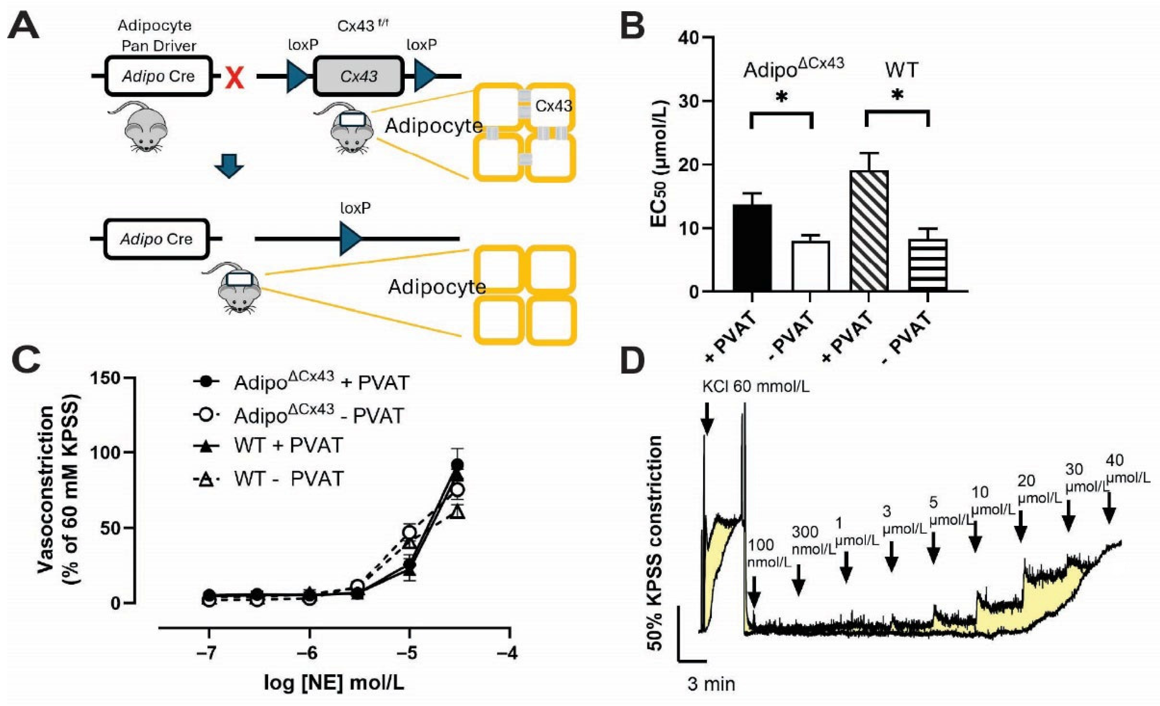
The release of anticontractile factors from PVAT is thought to be driven by NE and our data show that adipocyte responses to NE are regulated by gap junctions that are likely composed of Cx43. Therefore, we tested whether specifically deleting Cx43 from adipocytes would alter the effect of PVAT on vessel function. To do so, we created adipocyte-specific Cx43(f/f) knockout (Adipo∆Cx43 KO) mice (Figure 5A) and carried out wire myography recordings on second- and third-order mesenteric resistance arteries isolated from both Adipo∆Cx43 KO mice and WT mice with or without PVAT [45,46].
In WT mice, the anticontractile effect of PVAT was evident by a rightward shift in the cumulative NE concentration–response curve (Figure 5B,C). The EC50 for NE was 19.13 ± 2.70 µmol/L in arteries with PVAT, compared to 8.30 ± 1.63 µmol/L in arteries without PVAT. Unexpectedly, PVAT retained its anticontractile effect in Adipo∆Cx43 KO mice and EC50 measurements with and without PVAT were comparable to those in WT (13.72 ± 1.75 µmol/L with PVAT and 8.00 ± 0.89 µmol/L without PVAT). These observations were surprising given the multiple other lines of data suggesting a significant role for Cx43 in PVAT responses to NE; however, the experimental conditions tested here in which NE was bath applied may preclude the need for gap junction coupling and limit a clear view of the role of adipocyte gap junction coupling in vessel function.

2.6. mRNA Expression of Connexin Isoforms in Wild Type and Adipo∆Cx43KO Mice

One possibility that could explain why deleting adipocyte Cx43 had no effect on vascular tone is that adipocytes compensate for the loss of Cx43 by upregulating other connexin isoforms that could mask the effects of deleting Cx43. To assess this possibility, we used real-time qPCR analysis to compare the expression of multiple connexin isoforms in mPVAT from Adipo∆Cx43 KO and WT animals. Primer sequences and amplicon sizes are listed in Table 1.
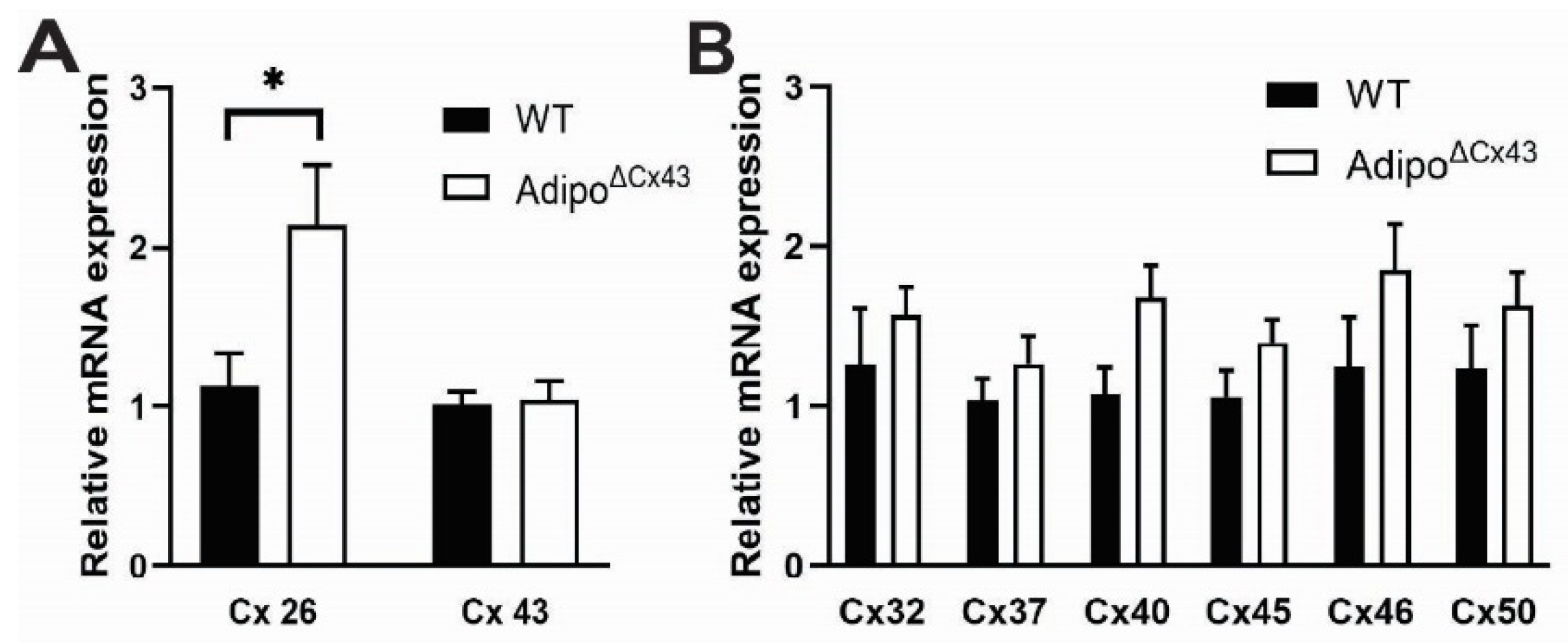
Despite exhibiting reduced Cx43 protein expression in adipocytes (Figure 2), Cx43 mRNA expression was comparable between samples of PVAT from Adipo∆Cx43 and WT mice. This difference is likely due to the fact that Cx43 is expressed by multiple cell types in PVAT in addition to adipocytes such as vascular smooth muscle. Interestingly, Cx26 mRNA increased by two-fold in Adipo∆Cx43 KO mice and ∆∆Ct values demonstrated statistically significant differences in WT (0.00 ± 0.84) and Adipo∆Cx43 KO (−0.95 ± 0.68) mice (n = 6–8, p = 0.048, two-tailed t test) (Figure 6A and Table 2). Expression of other connexin isoforms was not significantly different between Adipo∆Cx43 KO and WT mice, but exhibited several trends that could indicate subtle changes (Figure 6B). These data show that adipocytes exhibit a robust increase in Cx26 mRNA expression that may indicate compensation for the loss of Cx43 in the constitutive ablation model. It is possible that this upregulation of Cx26 masks the effects of deleting Cx43 on vessel function and contributes to the lack of effects observed.

3. Discussion

PVAT regulates vascular tone through mechanisms that involve factors released in response to NE. Here, we show that sensitivity to NE differs between PVAT depots and that responses to NE are influenced by cell–cell coupling among adipocytes. Cx43 is broadly expressed among PVAT adipocytes and blocking gap junctions impair their ability to respond to NE and effectively couple to neighboring cells. Interestingly, deleting Cx43 from adipocytes had no effect on the anticontractile effects of PVAT in vessel contractility experiments and this lack of effect could be due to multiple confounding factors including the experimental paradigm tested here and an upregulation of other connexins such as Cx26. While additional work is needed to understand these compensatory mechanisms and the role of Cx26 in more detail, our results show that gap junction coupling is an important mechanism that regulates how PVAT adipocytes function.
PVAT surrounds most blood vessels and was once considered to act primarily as connective tissue that supports vascular structure. However, contemporary concepts frame PVAT as a physiologically active and independent endocrine tissue that plays a critical role in maintaining vascular homeostasis. Multiple vasomodulatory factors are released by PVAT that exert anticontractile (adiponectin, nitric oxide, omentin, leptin, prostanoids, adipocyte-derived relaxing factor (ADRF)), pro-contractile (angiotensin-II, catecholamines, 5-HT, resistin, visfatin, chemerin, prostaglandin F), anti-inflammatory (IL-10, transforming growth factor (TGF)-β, prostacyclin) and pro-inflammatory (IL-1, IL-6, IL-8, TNF-α) effects [47,48,49,50]. Thus, the balance between these contrasting factors is critical for regulating vascular homeostasis.
The release of anticontractile factors from PVAT is thought to involve mechanisms that are enacted downstream of stimulation by NE [13,14]. Prior work showed that NE drives Ca2+ responses in PVAT adipocytes through mechanisms that involve α1a-, β2-, and β3-adrenergic receptors and downstream pathways that promote Ca2+ release from internal stores [15,34]. Importantly, elevations of intracellular Ca2+ have been implicated in the release of anticontractile factors [9,13,14,51]. In this study, we found that NE stimulation elicited robust adipocyte Ca2+ responses in all tissue types tested, although response profiles differed according to anatomical location. Adipocytes in aPVAT were sensitive to NE between 50 and 500 μmol/L, while those in mPVAT were less sensitive and responded in a range between 100 and 1000 μmol/L. WAT was more sensitive than mPVAT and maintained responsiveness at 1000 μmol/L of NE. It is well known that adipocytes in mPVAT and aPVAT are distinct and that mPVAT displays characteristics of white adipose tissue while aPVAT is more similar to brown adipose tissue [52,53,54]. This makes it likely that the differing sensitivities to NE could reflect their unique phenotypes and developmental origin. However, these differences may also suggest that different types of PVAT have specialized roles in vascular regulation and/or are designed to be recruited at differing times during sympathetic response. Additional work investigating how and why the various PVAT depots would be recruited differently by NE would be beneficial for understanding the physiological relevance of each tissue type.
Our immunolabeling data show that Cx43 is broadly expressed by adipocytes in aPVAT, mPVAT and WAT and Ca2+ imaging and FRAP experiments show that gap junction coupling plays an important role in regulating the breadth and magnitude of responses triggered by NE. Blocking gap junctions with the broad antagonist Cbx effectively reduced Ca2+ responses triggered by NE in every tissue. Likewise, data from FRAP experiments showed that normal dye transfer between adipocytes is impaired in the presence of Cbx. Both of these observations are consistent with the conclusion that PVAT adipocytes are extensively coupled by gap junctions and that gap junction coupling is important for distributing signals between adjacent adipocytes. Although Cbx is a broad gap junction blocker that is not selective for Cx43, these effects appear to be mainly mediated by traditional gap junctions and not hemichannels as the Cx43 hemichannel blocker 43Gap26 had no effect. Extensive gap junction coupling among PVAT adipocytes is interesting because this could explain the apparent mismatch between limited sympathetic innervation and the broad effects of NE on PVAT. Such a mechanism is consistent with what has been described previously in other adipose tissue depots [17]. In future work, it would be interesting to explore if defects in PVAT adipocyte gap junction coupling contribute to the proposed loss of the anticontractile nature of PVAT in conditions associated with hypertension.
As a first step in determining whether Cx43-mediated gap junction coupling plays a significant role in mechanisms by which PVAT regulates vascular tone, we examined mesenteric artery contractile responses to NE in mice lacking adipocyte Cx43 (Adipo∆Cx43 mice). Surprisingly, mesenteric resistance arteries displayed normal contractile responses to NE in mice lacking adipocyte Cx43. It is possible that this result suggests that adipocyte gap junction coupling is dispensable for mechanisms the control vascular tone, but this would be surprising given our data showing that gap junctions play a major role in adipocyte responses to NE and it is known that NE controls the release of PVAT anticontractile factors. It is also possible that technical limitations of the experimental setup limit observing a clear difference in response to NE. In these organ bath experiments, NE is bath applied and would conceivably have access to all adipocytes. This would likely limit the need for gap junction coupling as each adipocyte could be stimulated independently. While we cannot exclude this possibility, it is unlikely to be sufficient to fully explain the lack of effect because diffusion of NE into thick adipose tissue depots would be challenging and we also observed no obvious baseline differences between samples from WT and Adipo∆Cx43 KO mice. It is also possible that other connexin isoforms are upregulated to compensate for the functions of Cx43 when it is deleted. In support of this, prior studies in other tissues have observed compensation by connexin isoforms when Cx43 is deleted developmentally [55,56,57,58]. Our data show that Cx26 mRNA is significantly upregulated in PVAT from Adipo∆Cx43 KO mice. Cx26 and Cx43 share characteristics such as the secretory pathway, types of transport intermediates, turnover dynamics, and they may exist within the same gap junction plaques [59,60]. Therefore, it is possible that the upregulation of Cx26 could functionally compensate for the loss of Cx43 and maintain PVAT functions; however, additional work will be needed to determine whether functional compensation exists beyond changes in gene expression.
As indicated above, there are several limitations to this study. First, while the protein and gene expression data show that Cx43 is expressed by PVAT adipocytes, we cannot be entirely sure that the effects of Cbx are mediated solely by Cx43. Cbx is a broad gap junction inhibitor that also has potential effects on non-gap junction mechanisms. Based on the PCR data, multiple other connexin types are also expressed in PVAT and it remains unclear which of these are expressed by the adipocytes and which are expressed by other cell types that make up PVAT. Data from Ca2+ imaging, FRAP, immunolabeling and gene expression experiments are consistent with the conclusion that adipocytes are gap junction-coupled and that Cx43 is present, but additional work will be needed to determine the composition of the gap junctions and their specific nature. Second, the lack of functional effect in the adipocyte-specific Cx43 knockout model should be interpreted with caution for several reasons. As discussed above, a technical limitation of the experimental paradigm in wire myography recordings is that NE is bath applied and has the potential to influence many adipocytes without the need for gap junction coupling. This is certainly a confounding factor but is unlikely to fully account for the lack of effect given that not all adipocytes would be stimulated simultaneously due to limited diffusion through the thick tissue. Our data suggest that a potential alternate explanation is that adipocytes upregulate Cx26 when Cx43 is deleted developmentally. This observation is important for future work in this area and suggests that inducible, conditional knockouts or knockouts of several connexin genes will be needed to effectively reduce adipocyte gap junction coupling. If functional compensation by Cx26 or other connexin isoforms occurs in the knockout, it would contribute to reasons why no functional phenotype on vessel function was observed in this study. This knowledge would also be important for designing any studies going forward to avoid potential compensatory mechanisms. Lastly, although the gene expression data show a doubling of Cx26 mRNA expression, additional work is needed to determine if Cx26 forms functional gap junctions in PVAT adipocytes and truly compensates for the loss of Cx43. Although such compensation between connexins is common in other contexts, validating the role of Cx26 in PVAT will require additional work.
In conclusion, our data show that PVAT adipocytes express Cx43 and that gap junction coupling among the adipocytes regulates how adrenergic signals are distributed among the adipocytes in PVAT. These mechanisms are likely important for explaining how limited sympathetic innervation exerts broad effects through PVAT depots and possibly contributes to mechanisms that control how PVAT releases anticontractile factors. Given that defects in PVAT’s anticontractile mechanisms have received increased attention as potential contributors to hypertension, additional studies that test how these mechanisms are altered in disease states could produce important new targets to improve cardiovascular health.

4. Materials and Methods

4.1. Animals

Male and female mice aged 13–22 weeks old that expressed the genetically encoded Ca2+ indicator (GECI) GCaMP5g and the tdTomato reporter (tdT) in adipocytes were generated by crossing B6;129s6-Polr2aTm1(CAG-GCaMP5g-tdTomato)Tvrd/J mice (Jackson Laboratory, Bar Harbor, ME, USA; Stock No. 024477; RRID: IMSR_JAX:024477) with B6.FVB-Tg(Adipoq-cre)1Evdr/J mice (Jackson Labotatory, Stock No. 028020; RRID: IMSR_JAX:028020). Offspring, Adipoqcre;GCaMP5g-tdT mice, were used for ex vivo Ca2+ imaging, immunofluorescence studies and fluorescence recovery after photobleaching (FRAP) assays [15,34].
Adipocyte-specific Cx43 knockout mice were generated by breeding B6;129s7-Gja Tm1Dlg/J mice (Jackson Laboratory, Stock No. 008039; RRID: IMSR_JAX:008039) with B6.FVB-Tg(Adipoq-cre)1Evdr/J mice (Jackson Labotatory, Stock No. 028020; RRID: IMSR_JAX:028020). Offspring, referred to as Adipo∆Cx43 KO, were used for experiments. Cre-negative littermates served as controls in immunofluorescence studies and vascular contractility assays.
All mouse genotypes were verified by Transnetyx (Boston, MA, USA). Mice were housed in Optimice cages from Animal Care Systems with bedding from Frontier Distributing, cotton and paper nestlets (NES3600) from Ancare (Bellmore, NY, USA), and red mouse igloos (K3327) from Bio-Serve (Flemington, NJ, USA) in a temperature-controlled environment on a 12 h:12 h light/dark cycle with ad libitum access to food (Teklad Irradiated Global 19% Protein Extruded Rodent Diet, 2919) from Inotiv (Lafayette, IN, USA) and distilled tap water. All works involving animals were conducted following the standards established by the National Institutes of Health (NIH) Guide for the Care and Use of Laboratory Animals, and were approved by the Institutional Animal Care and Use Committee (IACUC) at Michigan State University (AUF# PROTO020400255). All animals were randomly assigned to experimental groups based on age and genotype prior to the initiation of experimental procedures.

4.2. Drugs and Reagents

(±) Norepinephrine (+) bitartrate salt (Sigma, St Louis, MO, USA, A7256, NE) was focally applied to samples of PVAT during Ca2+ imaging experiments using pressure ejection from a glass micropipette and bath applied to vessels in organ baths during wire myography experiments. Carbenoxolone disodium salt (Sigma, C4790, Cbx) was added to imaging chamber baths to achieve a final concentration of 50 µmol/L in modified Krebs buffer during Ca2+ imaging experiments and FRAP experiment. 43Gap26 (Anaspec Inc., Fremont, CA, USA, 62644) was prepared in modified Krebs buffer and applied at 100 µmol/L during Ca2+ imaging experiments. Stock solutions of Calcein acetoxymethylester (Invitrogen, Waltham, MA, USA, C3100MP, Calcein AM) were prepared in dimethyl sulfoxide (DMSO) and diluted to a final concentration of 4 µmol/L in modified Krebs buffer during FRAP experiments.
Modified Krebs buffer was used for Ca2+ imaging and FRAP experiments and contained (in mmol/L) 121 NaCl (Sigma, 746398), 5.9 KCl (Sigma, 74636), 2.5 CaCl2 (Sigma, C1016), 1.2 MgCl2 (Thermo Fisher Scientific, Pittsburgh, PA, USA, 3818), 1.2 NaH2PO4 (Thermo Fisher Scientific, 2444-01), 10 HEPES (Sigma, H3375), 21.2 NaHCO3 (Thermo Fisher Scientific, S233), 1 Sodium pyruvate (Sigma, P5280), 8 glucose (Sigma, G7021). Phosphate-buffered saline (PBS) was used in immunofluorescence studies and contained (in mol/L) 0.137 NaCl, 2.7 KCl, 10 Na2HPO4 (Thermo Fisher Scientific, 3828-01), 0.0018 KH2PO4 (Thermo Fisher Scientific, 3246-01) and 0.02% Sodium azide (Sigma, S2002). DMEM (11039021) and ultrapure water (SH30538.03) was sourced from Thermo Fisher Scientific.
In vessel contractility experiments, a low Ca2+ solution dissection solution used while preparing samples that contained (in mmol/L) 140 NaCl, 5 KCl, 0 CaCl2, 1 MgCl2, 10 HEPES, 10 glucose, 0.01 Sodium nitroprusside (Sigma, 71780), 0.01 Diltiazem (Sigma, D2521) and 1 mg/mL albumin (Thermo Fisher Scientific, J10856-09). During experiments, samples were incubated in buffer that contained (in mmol/L) 135 NaCl, 4 KCl, 1.8 CaCl2, 1 MgCl2, 10 HEPES, 10 glucose. Samples were stimulated with a high potassium 60 mM K solution that contained (in mmol/L) 75 NaCl, 60 KCl, 1.8 CaCl2, 1 MgCl2, 10 HEPES, 10 glucose. The pH of all solutions was adjusted to 7.4 with NaOH.

4.3. Tissue Collection

Aortic and mesenteric PVAT (aPVAT, mPVAT) and white adipose tissue (WAT) were collected from male and female Adipoqcre;GCaMP5g-tdT mice. Following anesthesia and cervical dislocation, the abdominal cavity was opened to access mPVAT and perigonadal WAT and the rib cage was opened to access aPVAT. All tissues were kept in ice-cold DMEM-F12.
For Ca2+ imaging, and FRAP experiments, small sections of mPVAT, aPVAT, and WAT were excised and secured with pins (Thermo Fisher Scientific, 26002-20) in Sylgard (Dow-Corning, Midland, MI, USA)-coated custom rectangular imaging dishes (imaging chamber, 2.1 mm × 1.5 cm) in ice-cold modified Krebs buffer.
For vascular contractility assays, the entire mesentery was removed from male and female Cx43 knockout and control wild type mice. The mesentery was pinned out to facilitate access to the individual vessel branches in ice-cold dissection buffer. Resistance mesenteric arteries with or without mPVAT were carefully isolated from the second- and third-order branches of the mesenteric arterial network in ice-cold dissection buffer [46,61].

4.4. Calcium Imaging

Samples of adipose tissues were visualized using a ×20 widefield water-immersion objective lens (1.0 numerical aperture, XLUMPlanFLN20xW, Olympus, Tokyo, Japan) mounted on an upright BX51WI fixed-stage microscope (Olympus, Tokyo, Japan). Fluorescence illumination was provided by a Lumencor AURA light engine (Lumencor, Beaverton, OR, USA). The excitation light for GCaMP5g was filtered through a 485/20 nm band-pass filter, and emitted fluorescence signal was collected through a 515 nm long-pass filter. The tdTomato signal was excited using light filtered through a 535/20 nm band-pass filter, and emitted fluorescence signal was collected through a 610/75 nm band-pass emission filter before detection. Imaging data were recorded every second using a Photometrics Prime BSI camera (Teledyne, ON, Canada) and NIS-Elements AR 6.02.03 64-bit (Nikon, Tokyo, Japan). Videos and images were saved as .nd2 files for further analysis using FIJI (v.2.14.011.54, National Institutes of Health). Throughout the experiment, 1× modified Krebs buffer was perfused at a rate of 2–4 mL/min (AutoMate Scientific, Berkeley, CA, USA) and maintained at 37 °C with an inline heater (TC-344C, Warner Instruments, Hamden, CT, USA) [15,34].
NE was focally applied to adipocytes using a glass micropipette constructed using a P-87 Flaming-Brown Micropipette Puller (Sutter Instruments, Novato, CA, USA), and backfilled with NE dissolved in modified Krebs buffer. NE was delivered using gentle positive pressure from a 1 mL syringe connected to a pipette holder. This method enabled delivery of small volumes of NE to an isolated patch of adipocytes. Antagonists were bath applied and samples were preincubated for 10 min with either Cbx (50 µmol/L) or 43Gap26 (100 µmol/L) before NE application [15,62,63,64].
Fluorescence changes were quantified as ΔF/F0 using the equation ΔF/F0 = (F(t) − F0)/F0, where F(t) is the fluorescence intensity at each time point (t), and F0 is the baseline fluorescence, defined as the average intensity over the first 20 frames. For each video, the F0 image was calculated, the F0 was subtracted from each frame, which is divided by F0 at each time point. Peak responses were determined for each responding cell in Excel and plotted as scatter plots in Prism.

4.5. Immunofluorescence Staining and Imaging

Immunofluorescence labeling was imaged using a Zeiss LSM 880 NLO system (Carl Zeiss, Oberkochen, Germany) equipped with 594 and 488 nm laser lines. Images were collected with a Plan-Apochromat ×20 objective (NA = 0.8). Acquisition parameters (photomultiplier gain, laser intensity, pin hole size) were optimized using positive samples (i.e., those exposed to both primary and secondary antibodies), across all tissue types to confirm that the fluorescence emission did not saturate the detectors, or produce autofluorescence in the negative control samples. After optimization, the same settings were applied to all samples. Image stacks (approximately 100, three channels, 1024 × 1024 8-bit images, 1 μm step-size) were acquired using sequential line scanning to minimize channel crosstalk. All images were saved in .czi format for analysis in FIJI.
For all incubation and wash steps, tissues were continuously agitated on an orbital shaker at room temperature. Samples were fixed in 4% paraformaldehyde for 2 h, then washed three times for 10 min in PBS. Tissues were blocked for 1 h in blocking buffer [10% normal donkey serum (Jackson Immuno Research, West Grove, PA, USA, 017-000-121), 1% bovine serum albumin (Jackson Immuno Research, 001-000-162, BSA), 0.4% Triton X-100 (Sigma, 93443); in PBS] and then incubated for 3 days in the following primary antibodies: connexin-43 (Sigma, C6219, 1:500), Caveolin-1 (Santa Cruz, Dallas, TX, USA, sc-53564, 1:100). After incubation in the primary antibody, samples were washed 3 times for 1 h in PBS and incubated with the secondary antibodies for 1 day (1:500 dilution; donkey anti-mouse IgG Alexa 488; donkey anti-rabbit IgG Alexa 594, Jackson Immuno Research). Samples were counterstained with 4′,6-diamidino-2-phenylindole dihydrochloride (DAPI, Sigma, D8417, 1:10,000) in PBS for 1 h. Following two additional 1 h rinses in PBS, samples were mounted on slides (VWR, 16004-370) using Coverwell Imaging Chambers (Electron Microscopy Science, Hatfield, PA, USA, 70327-05) and Hydromount (National Diagnostics, Atlanta, GA, USA, H5-106). Mounted slides were stored in the dark at 4 °C before imaging.

4.6. Fluorescence Recovery After Photobleaching

FRAP experiments were performed on a Nikon AXR Confocal system (Nikon Instruments Inc., Melville, NY, USA). Imaging was performed through the ApoLWD 25 × 1.10 WDIC N2 water-immersion objective (numerical aperture, NA = 1.1), providing high resolution and optimal light collection for confocal imaging (resolution 1024 × 1024 with line averaging of 2). Both photobleaching and fluorescence imaging of Calcein AM were performed using an argon-ion laser (488 nm) and a photomultiplier tube for detection of fluorescence in the 500 and 550 nm range. Acquisition was performed at speed 9 (pixel dwell 4 µs), averaging number 1, and 8-bit depth. Photobleaching was carried out at scan speed 3 (pixel dwell 50.42 µs), 50 scans, and 4 iterations.
The adipocytes were loaded with 4 µmol/L of Calcein AM for 15 min and then washed three times with modified Krebs buffer (1 min each) before being subjected to the FRAP method. Gap junction communication in adipocytes was blocked by incubation with 100 µmol/L of Cbx for 15 min. Cbx was present in the modified Krebs buffer during the entire FRAP experiment. To perform precise measurements, three regions of interest (ROIs) were selected. An ROI was manually selected around a cell for photobleaching, a reference ROI within an unbleached area was drawn to correct for fluorescence changes associated with acquisition bleaching, and an ROI was placed outside the cells to measure the background. Prior to photobleaching, initial images were recorded to determine the initial fluorescence intensity. The selected ROI was photobleached at 100% power and time-lapse imaging was then recorded to monitor Calcein AM recovery every 3 s for up to 3 min. The fluorescence intensity of the bleached ROI was normalized to the reference ROI at each time point. Normalized values were used to calculate the percentage mobile fraction of fluorescence using the equation: mobile fraction percentage = [(FFR − FB)/(FI − FB)] × 100, where FFR is the fluorescence intensity in the bleached region after full recovery, FB after bleaching, and FI before bleaching. The image and fluorescence recovery data were exported from Nikon NIS-Elements AR 6.10.01 64-bit to GraphPad Prism (10. 6.1) (GraphPad Software, San Diego, CA, USA) for plotting. Experiments showing considerable fluctuations in fluorescence intensity within reference ROI or base ROI were excluded from the analysis [41,42,43,44,65].

4.7. Vascular Contractility

Small mesenteric artery segments with or without PVAT were isolated from the second- and third-order branches of the mesenteric arterial network and mounted on two 40 µm wires in a DMT Multi-Wire Myograph System, Model 620 M (Danish Myo Technology A/S, Hinnerup, Denmark) [65]. Data were recorded using a PowerLab Data Acquisition unit (ADInstruments, Colorado Springs, CO, USA). The vessels were gassed with clinical blood gas (5% carbon dioxide/21% oxygen/balanced nitrogen) and maintained at 37 °C for 1 h, with washes every 20 min. Wall tension and diameter were then normalized using a standardized procedure. After normalization, the vessels were washed two times for 20 min to achieve a stable baseline. A total of 60 mmol/L high potassium PSS (KPSS) was applied in the chamber for 5 min to establish viability of the vessel and initial maximum contraction. After returning to baseline in PSS for 40 min with washes every 20 min, cumulative concentration–response curves to NE were conducted with 3 min intervals for the vessels without PVAT and 6 min intervals for the vessels with PVAT. Percentage loss of NE-induced tone and potencies (EC50) were calculated for WT and Cx43 KO mesenteric artery [14,46,61,66].

4.8. Real-Time Quantitative Polymerase Chain Reaction

Total mRNA was extracted from frozen samples of mesenteric PVAT. Samples were mechanically lysed using mechanical dissociation (Bead Bug, Stellar Scientific, Baltimore, MD, USA) in lysing Matrix D (MPBio, Irvine, CA, USA). RNA was isolated using miRNAeasy Mini Kit (QGN-217004, Qiagen, Hilden, Germany) and quantified on Nanodrop 1000 spectrophotometer (Thermo Fisher). Purified RNA was stored at −80 °C. cDNA was synthesized from 0.3 µg RNA using Verso cDNA synthesis kit (Thermo Fisher Scientific, AB1453A) and the MiniAmp Thermal Cycler (Thermo Fisher Scientific) with the following parameters: 42 °C for 30 min, 95 °C for 2 min, and held at 4 °C.
Quantitative gene expression analysis was performed on QuantStudio 3 Real-Time PCR system (Applied Biosystems, Carlsbad, CA, USA) with PowerTrackTM SYBR Green Master Mix for qPCR (Applied Biosystems, Carlsbad, CA, USA, A46109): 95 °C for 10 min, 40 cycles of 95 °C for 10 s and 60 °C for 1 min. Relative gene expressions for Adipo∆Cx43 KO and WT mice were normalized to the respective housekeeping gene (RPS6) and were analyzed using the 2−ΔΔCt method. Table 1 shows PCR primers sequences and related key resources.

4.9. Statistical Analysis

All experiments were repeated at least three independent times in separate mice. Statistical analyses were performed using two-tailed unpaired Student’s t-test or one-way ANOVA in GraphPad Prism software (10.6.1). As no sex-dependent differences were observed, data from males and females were combined for analysis. Results are expressed as mean ± standard error of the mean (SEM) and a p-value lower than 0.05 was considered statistically significant.

Author Contributions

Conceptualization, B.D.G. and W.F.J.; methodology, B.D.G., W.F.J., J.J., E.D.F. and J.L.M.; formal analysis, A.R.K. and E.D.F.; investigation, A.R.K., B.D.G., W.F.J. and E.D.F.; resources, B.D.G., W.F.J., E.D.F. and J.L.M.; data curation, A.R.K. and B.D.G.; writing—original draft preparation, A.R.K.; writing—review and editing, B.D.G.; visualization, A.R.K. and E.D.F.; supervision, B.D.G., W.F.J. and J.L.M.; project administration, B.D.G. and W.F.J.; funding acquisition, B.D.G. and W.F.J. All authors have read and agreed to the published version of the manuscript.

Funding

This work was supported by P01 HL152951 to BDG and WFJ from the National Institutes of Health, Heart, Lung and Blood Institute. The content is solely the responsibility of the authors and does not necessarily represent the official views of the NIH.

Institutional Review Board Statement

All work involving animals was performed in accordance with the standards established by the National Institutes of Health (NIH) Guide for the Care and Use of Laboratory Animals and was approved by the Institutional Animal Care and Use Committee (IACUC) at Michigan State University (AUF# PROTO020400255, date of approval 15 December 2025).

Informed Consent Statement

Not applicable.

Data Availability Statement

The original contributions presented in this study are included in the article and data are available upon reasonable request to the corresponding author.

Conflicts of Interest

The authors have no financial, professional, or personal conflicts that are relevant to the manuscript. The main resources generated from this study including mouse lines, analytic methods, study materials, and results will be made available upon request.

Correction Statement

This article has been republished with a minor correction to the Funding statement. This change does not affect the scientific content of the article.

Pre-Print Server

The manuscript has not been shared as a preprint.

Synopsis

This study shows that adipocytes in perivascular adipose tissue are gap junction-coupled and that these gap junctions, which may be composed of connexin-43, play an important role in distributing signals initiated by sensing norepinephrine. These mechanisms could participate in regulating how perivascular adipose tissue influences blood vessel contractility and, hence, susceptibility to diseases such as hypertension.

Abbreviations

PVAT, perivascular adipose tissue; mPVAT, mesenteric perivascular adipose tissue; aPVAT, aortic perivascular adipose tissue; WAT, white adipose tissue; Cx43, connexin-43; Ca2+, calcium; NE, norepinephrine; Cbx, carbenoxolone; FRAP, fluorescence recovery after photobleaching.

References

  1. Palaniappan, L.P.; Allen, N.B.; Almarzooq, Z.I.; Anderson, C.A.M.; Arora, P.; Avery, C.L.; Baker-Smith, C.M.; Bansal, N.; Currie, M.E.; Earlie, R.S.; et al. 2026 Heart Disease and Stroke Statistics: A Report of US and Global Data from the American Heart Association. Circulation 2026, 153, e275–e906. [Google Scholar] [CrossRef]
  2. Earle, W.B.; Watson, N.W.; Juraschek, S.P.; Cluett, J.L.; Krawisz, A.K.; Secemsky, E.A. Contemporary Prevalence and Treatment Patterns Among US Adults with Apparent Treatment-Resistant Hypertension. Hypertension, 2026; online ahead of print. [CrossRef]
  3. Kim, H.W.; Shi, H.; Winkler, M.A.; Lee, R.; Weintraub, N.L. Perivascular Adipose Tissue and Vascular Perturbation/Atherosclerosis. Arterioscler. Thromb. Vasc. Biol. 2020, 40, 2569–2576. [Google Scholar] [CrossRef]
  4. Saxton, S.N.; Withers, S.B.; Nyvad, J.; Mazur, A.; Matchkov, V.; Heagerty, A.M.; Aalkjær, C. Perivascular Adipose Tissue Contributes to the Modulation of Vascular Tone in Vivo. J. Vasc. Res. 2019, 56, 320–332. [Google Scholar] [CrossRef]
  5. Szasz, T.; Bomfim, G.F.; Webb, R.C. The Influence of Perivascular Adipose Tissue on Vascular Homeostasis. Vasc. Health Risk Manag. 2013, 9, 105–116. [Google Scholar] [CrossRef] [PubMed]
  6. Rodríguez, A.; Ezquerro, S.; Méndez-Giménez, L.; Becerril, S.; Frühbeck, G. Revisiting the Adipocyte: A Model for Integration of Cytokine Signaling in the Regulation of Energy Metabolism. Am. J. Physiol. Endocrinol. Metab. 2015, 309, E691–E714. [Google Scholar] [CrossRef] [PubMed]
  7. Ayala-Lopez, N.; Thompson, J.M.; Watts, S.W. Perivascular Adipose Tissue’s Impact on Norepinephrine-Induced Contraction of Mesenteric Resistance Arteries. Front. Physiol. 2017, 8, 37. [Google Scholar] [CrossRef]
  8. Fernández-Alfonso, M.S.; Gil-Ortega, M.; Aranguez, I.; Souza, D.; Dreifaldt, M.; Somoza, B.; Dashwood, M.R. Role of PVAT in Coronary Atherosclerosis and Vein Graft Patency: Friend or Foe? Br. J. Pharmacol. 2017, 174, 3561–3572. [Google Scholar] [CrossRef]
  9. Ramirez, J.G.; O’Malley, E.J.; Ho, W.S.V. Pro-contractile Effects of Perivascular Fat in Health and Disease. Br. J. Pharmacol. 2017, 174, 3482–3495. [Google Scholar] [CrossRef]
  10. Shu, X.; Ruddiman, C.A.; Keller, T.C.S., IV; Keller, A.S.; Yang, Y.; Good, M.E.; Best, A.K.; Columbus, L.; Isakson, B.E. Heterocellular Contact Can Dictate Arterial Function. Circ. Res. 2019, 124, 1473–1481. [Google Scholar] [CrossRef]
  11. Brown, N.K.; Zhou, Z.; Zhang, J.; Zeng, R.; Wu, J.; Eitzman, D.T.; Chen, Y.E.; Chang, L. Perivascular Adipose Tissue in Vascular Function and Disease. Arterioscler. Thromb. Vasc. Biol. 2025, 34, 1621–1630. [Google Scholar] [CrossRef]
  12. Li, X.; Ma, Z.; Zhu, Y.Z. Regional Heterogeneity of Perivascular Adipose Tissue: Morphology, Origin, and Secretome. Front. Pharmacol. 2021, 12, 697720. [Google Scholar] [CrossRef] [PubMed]
  13. Saxton, S.N.; Ryding, K.E.; Aldous, R.G.; Withers, S.B.; Ohanian, J.; Heagerty, A.M. Role of Sympathetic Nerves and Adipocyte Catecholamine Uptake in the Vasorelaxant Function of Perivascular Adipose Tissue. Arterioscler. Thromb. Vasc. Biol. 2018, 38, 880–891. [Google Scholar] [CrossRef]
  14. Bussey, C.E.; Withers, S.B.; Saxton, S.N.; Bodagh, N.; Aldous, R.G.; Heagerty, A.M. β3-Adrenoceptor Stimulation of Perivascular Adipocytes Leads to Increased Fat Cell-Derived NO and Vascular Relaxation in Small Arteries. Br. J. Pharmacol. 2018, 175, 3685–3698. [Google Scholar] [CrossRef]
  15. Hanscom, M.; Morales-Soto, W.; Watts, S.W.; Jackson, W.F.; Gulbransen, B.D. Innervation of Adipocytes Is Limited in Mouse Perivascular Adipose Tissue. Am. J. Physiol. Hear. Circ. Physiol. 2024, 327, H155–H181. [Google Scholar] [CrossRef]
  16. Jackson, W.F.; Flood, E.D.; Lauver, D.A.; Fink, G.D.; Watts, S.W.; Gulbransen, B.D. Sympathetic Nerves Are Sparsely Distributed in Rat Mesenteric Perivascular Adipose Tissue. Front. Physiol. 2025, 16, 1547785. [Google Scholar] [CrossRef] [PubMed]
  17. Zhu, Y.; Gao, Y.; Tao, C.; Shao, M.; Zhao, S.; Huang, W.; Yao, T.; Johnson, J.A.; Liu, T.; Cypess, A.M.; et al. Connexin 43 Mediates White Adipose Tissue Beiging by Facilitating the Propagation of Sympathetic Neuronal Signals. Cell Metab. 2016, 24, 420–433. [Google Scholar] [CrossRef]
  18. Sáez, J.C.; Connor, J.A.; Spray, D.C.; Bennett, M.V. Hepatocyte Gap Junctions Are Permeable to the Second Messenger, Inositol 1,4,5-Trisphosphate, and to Calcium Ions. Proc. Natl. Acad. Sci. USA 1989, 86, 2708–2712. [Google Scholar] [CrossRef]
  19. Harris, A.L. Connexin Channel Permeability to Cytoplasmic Molecules. Prog. Biophys. Mol. Biol. 2007, 94, 120–143. [Google Scholar] [CrossRef]
  20. Valiunas, V.; Weingart, R. Electrical Properties of Gap Junction Hemichannels Identified in Transfected HeLa Cells. Pflügers Arch. 2000, 440, 366–379. [Google Scholar] [CrossRef]
  21. Retamal, M.A. Connexin and Pannexin Hemichannels Are Regulated by Redox Potential. Front. Physiol. 2014, 5, 80. [Google Scholar] [CrossRef]
  22. Li, H.; Liu, T.F.; Lazrak, A.; Peracchia, C.; Goldberg, G.S.; Lampe, P.D.; Johnson, R.G. Properties and Regulation of Gap Junctional Hemichannels in the Plasma Membranes of Cultured Cells. J. Cell Biol. 1996, 134, 1019–1030. [Google Scholar] [CrossRef]
  23. Vuyst, E.D.; Decrock, E.; Bock, M.D.; Yamasaki, H.; Naus, C.C.; Evans, W.H.; Leybaert, L. Connexin Hemichannels and Gap Junction Channels Are Differentially Influenced by Lipopolysaccharide and Basic Fibroblast Growth Factor. Mol. Biol. Cell 2007, 18, 34–46. [Google Scholar] [CrossRef]
  24. Ebihara, L.; Liu, X.; Pal, J.D. Effect of External Magnesium and Calcium on Human Connexin46 Hemichannels. Biophys. J. 2003, 84, 277–286. [Google Scholar] [CrossRef]
  25. Srinivas, M.; Calderon, D.P.; Kronengold, J.; Verselis, V.K. Regulation of Connexin Hemichannels by Monovalent Cations. J. Gen. Physiol. 2006, 127, 67–75. [Google Scholar] [CrossRef] [PubMed]
  26. Vuyst, E.D.; Wang, N.; Decrock, E.; Bock, M.D.; Vinken, M.; Moorhem, M.V.; Lai, C.; Culot, M.; Rogiers, V.; Cecchelli, R.; et al. Ca2+ Regulation of Connexin 43 Hemichannels in C6 Glioma and Glial Cells. Cell Calcium 2009, 46, 176–187. [Google Scholar] [CrossRef] [PubMed]
  27. Carrer, A.; Leparulo, A.; Crispino, G.; Ciubotaru, C.D.; Marin, O.; Zonta, F.; Bortolozzi, M. Cx32 Hemichannel Opening by Cytosolic Ca2+ Is Inhibited by the R220X Mutation That Causes Charcot-Marie-Tooth Disease. Hum. Mol. Genet. 2018, 27, 80–94. [Google Scholar] [CrossRef] [PubMed]
  28. Christ, G.J.; Spray, D.C.; El-Sabban, M.; Moore, L.K.; Brink, P.R. Gap Junctions in Vascular Tissues. Circ. Res. 1996, 79, 631–646. [Google Scholar] [CrossRef]
  29. Evans, W.H.; Martin, P.E.M. Gap Junctions: Structure and Function (Review). Mol. Membr. Biol. 2009, 19, 121–136. [Google Scholar] [CrossRef]
  30. Liao, Y.; Day, K.H.; Damon, D.N.; Duling, B.R. Endothelial Cell-Specific Knockout of Connexin 43 Causes Hypotension and Bradycardia in Mice. Proc. Natl. Acad. Sci. USA 2001, 98, 9989–9994. [Google Scholar] [CrossRef]
  31. Haefliger, J.-A.; Nicod, P.; Meda, P. Contribution of Connexins to the Function of the Vascular Wall. Cardiovasc. Res. 2004, 62, 345–356. [Google Scholar] [CrossRef]
  32. Gandhi, G.K.; Ball, K.K.; Cruz, N.F.; Dienel, G.A. Hyperglycaemia and Diabetes Impair Gap Junctional Communication among Astrocytes. ASN Neuro 2010, 2, AN20090048. [Google Scholar] [CrossRef]
  33. Head, W.S.; Orseth, M.L.; Nunemaker, C.S.; Satin, L.S.; Piston, D.W.; Benninger, R.K.P. Connexin-36 Gap Junctions Regulate In Vivo First- and Second-Phase Insulin Secretion Dynamics and Glucose Tolerance in the Conscious Mouse. Diabetes 2012, 61, 1700–1707. [Google Scholar] [CrossRef] [PubMed]
  34. Jamka, J.R.; Gulbransen, B.D. Mechanisms of Norepinephrine Induced Calcium Responses in Perivascular Adipocytes. Physiology 2025, 40, 0403. [Google Scholar] [CrossRef]
  35. Fernández-Alfonso, M.S.; Somoza, B.; Tsvetkov, D.; Kuczmanski, A.; Dashwood, M.; Gil-Ortega, M. Role of Perivascular Adipose Tissue in Health and Disease. Compr. Physiol. 2018, 8, 23–59. [Google Scholar] [CrossRef]
  36. Berry, D.C.; Stenesen, D.; Zeve, D.; Graff, J.M. The Developmental Origins of Adipose Tissue. Development 2013, 140, 3939–3949. [Google Scholar] [CrossRef]
  37. Villarroya, F.; Cereijo, R.; Villarroya, J.; Giralt, M. Brown Adipose Tissue as a Secretory Organ. Nat. Rev. Endocrinol. 2016, 13, 26–35. [Google Scholar] [CrossRef]
  38. González-Casanova, J.E.; Durán-Agüero, S.; Caro-Fuentes, N.J.; Gamboa-Arancibia, M.E.; Bruna, T.; Bermúdez, V.; Rojas-Gómez, D.M. New Insights on the Role of Connexins and Gap Junctions Channels in Adipose Tissue and Obesity. Int. J. Mol. Sci. 2021, 22, 12145. [Google Scholar] [CrossRef]
  39. Cohen, A.W.; Hnasko, R.; Schubert, W.; Lisanti, M.P. Role of Caveolae and Caveolins in Health and Disease. Physiol. Rev. 2004, 84, 1341–1379. [Google Scholar] [CrossRef]
  40. Fernández-Real, J.M.; Catalán, V.; Moreno-Navarrete, J.M.; Gómez-Ambrosi, J.; Ortega, F.J.; Rodriguez-Hermosa, J.I.; Ricart, W.; Frühbeck, G. Study of Caveolin-1 Gene Expression in Whole Adipose Tissue and Its Subfractions and during Differentiation of Human Adipocytes. Nutr. Metab. 2010, 7, 20. [Google Scholar] [CrossRef]
  41. Cotrina, M.L.; Lin, J.H.-C.; Alves-Rodrigues, A.; Liu, S.; Li, J.; Azmi-Ghadimi, H.; Kang, J.; Naus, C.C.G.; Nedergaard, M. Connexins Regulate Calcium Signaling by Controlling ATP Release. Proc. Natl. Acad. Sci. USA 1998, 95, 15735–15740. [Google Scholar] [CrossRef]
  42. Chi, S.S.; Rattner, J.B.; Sciore, P.; Boorman, R.; Lo, I.K.Y. Gap Junctions of the Medial Collateral Ligament: Structure, Distribution, Associations and Function. J. Anat. 2005, 207, 145–154. [Google Scholar] [CrossRef]
  43. Kuzma-Kuzniarska, M.; Yapp, C.; Pearson-Jones, T.W.; Jones, A.K.; Hulley, P.A. Functional Assessment of Gap Junctions in Monolayer and Three-Dimensional Cultures of Human Tendon Cells Using Fluorescence Recovery after Photobleaching. J. Biomed. Opt. 2014, 19, 015001. [Google Scholar] [CrossRef]
  44. Kuzma-Kuzniarska, M.; Yapp, C.; Hulley, P.A. Gap Junction Protocols. Methods Mol. Biol. 2016, 1437, 171–179. [Google Scholar] [CrossRef]
  45. Almabrouk, T.A.M.; Ugusman, A.B.; Katwan, O.J.; Salt, I.P.; Kennedy, S. Deletion of AMPKα1 Attenuates the Anticontractile Effect of Perivascular Adipose Tissue (PVAT) and Reduces Adiponectin Release. Br. J. Pharmacol. 2017, 174, 3398–3410. [Google Scholar] [CrossRef] [PubMed]
  46. Mulvany, M.J.; Halpern, W. Contractile Properties of Small Arterial Resistance Vessels in Spontaneously Hypertensive and Normotensive Rats. Circ. Res. 1977, 41, 19–26. [Google Scholar] [CrossRef]
  47. Halberg, N.; Wernstedt-Asterholm, I.; Scherer, P.E. The Adipocyte as an Endocrine Cell. Endocrinol. Metab. Clin. N. Am. 2008, 37, 753–768. [Google Scholar] [CrossRef]
  48. Glastonbury, C.A.; Alves, A.C.; Moustafa, J.S.E.-S.; Small, K.S. Cell-Type Heterogeneity in Adipose Tissue Is Associated with Complex Traits and Reveals Disease-Relevant Cell-Specific eQTLs. Am. J. Hum. Genet. 2019, 104, 1013–1024. [Google Scholar] [CrossRef]
  49. Thompson, J.M.; Watts, S.W.; Terrian, L.; Contreras, G.A.; Rockwell, C.; Rendon, C.J.; Wabel, E.; Lockwood, L.; Bhattacharya, S.; Nault, R. A Cell Atlas of Thoracic Aortic Perivascular Adipose Tissue: A Focus on Mechanotransducers. Am. J. Physiol. Heart Circ. Physiol. 2024, 326, H1252–H1265. [Google Scholar] [CrossRef]
  50. Rendon, C.J.; Watts, S.W.; Contreras, G.A. PVAT Adipocyte: Energizing, Modulating, and Structuring Vascular Function during Normotensive and Hypertensive States. Am. J. Physiol. Heart Circ. Physiol. 2025, 328, H1204–H1217. [Google Scholar] [CrossRef] [PubMed]
  51. Ayala-Lopez, N.; Jackson, W.F.; Burnett, R.; Wilson, J.N.; Thompson, J.M.; Watts, S.W. Organic Cation Transporter 3 Contributes to Norepinephrine Uptake into Perivascular Adipose Tissue. Am. J. Physiol. Heart Circ. Physiol. 2015, 309, H1904–H1914. [Google Scholar] [CrossRef]
  52. Frontini, A.; Cinti, S. Distribution and Development of Brown Adipocytes in the Murine and Human Adipose Organ. Cell Metab. 2010, 11, 253–256. [Google Scholar] [CrossRef]
  53. Kim, H.W.; Belin de Chantemèle, E.J.; Weintraub, N.L. Perivascular Adipocytes in Vascular Disease. Arterioscler. Thromb. Vasc. Biol. 2019, 39, 2220–2227. [Google Scholar] [CrossRef]
  54. Diaba-Nuhoho, P.; Mittag, J.; Brunssen, C.; Morawietz, H.; Brendel, H. The Vascular Function of Resistance Arteries Depends on NADPH Oxidase 4 and Is Exacerbated by Perivascular Adipose Tissue. Antioxidants 2024, 13, 503. [Google Scholar] [CrossRef] [PubMed]
  55. Ahmad, S.; Tang, W.; Chang, Q.; Qu, Y.; Hibshman, J.; Li, Y.; Söhl, G.; Willecke, K.; Chen, P.; Lin, X. Restoration of Connexin26 Protein Level in the Cochlea Completely Rescues Hearing in a Mouse Model of Human Connexin30-Linked Deafness. Proc. Natl. Acad. Sci. USA 2007, 104, 1337–1341. [Google Scholar] [CrossRef] [PubMed]
  56. Pfenniger, A.; Chanson, M.; Kwak, B.R. Connexins in Atherosclerosis. Biochim. Biophys. Acta BBA Biomembr. 2013, 1828, 157–166. [Google Scholar] [CrossRef] [PubMed]
  57. Morel, S.; Chanson, M.; Nguyen, T.D.; Glass, A.M.; Sarieddine, M.Z.R.; Meens, M.J.; Burnier, L.; Kwak, B.R.; Taffet, S.M. Titration of the Gap Junction Protein Connexin43 Reduces Atherogenesis. Thromb. Haemost. 2014, 112, 390–401. [Google Scholar] [CrossRef]
  58. Zong, Y.-J.; Liu, X.-Z.; Tu, L.; Sun, Y. Cytomembrane Trafficking Pathways of Connexin 26, 30, and 43. Int. J. Mol. Sci. 2023, 24, 10349. [Google Scholar] [CrossRef]
  59. Falk, M.M. Connexin-Specific Distribution within Gap Junctions Revealed in Living Cells. J. Cell Sci. 2000, 113, 4109–4120. [Google Scholar] [CrossRef]
  60. Thomas, T.; Jordan, K.; Simek, J.; Shao, Q.; Jedeszko, C.; Walton, P.; Laird, D.W. Mechanisms of Cx43 and Cx26 Transport to the Plasma Membrane and Gap Junction Regeneration. J. Cell Sci. 2005, 118, 4451–4462. [Google Scholar] [CrossRef]
  61. Wenceslau, C.F.; McCarthy, C.G.; Earley, S.; England, S.K.; Filosa, J.A.; Goulopoulou, S.; Gutterman, D.D.; Isakson, B.E.; Kanagy, N.L.; Martinez-Lemus, L.A.; et al. Guidelines for the Measurement of Vascular Function and Structure in Isolated Arteries and Veins. Am. J. Physiol. Heart Circ. Physiol. 2021, 321, H77–H111. [Google Scholar] [CrossRef] [PubMed]
  62. Ahmadzai, M.M.; Seguella, L.; Gulbransen, B.D. Circuit-Specific Enteric Glia Regulate Intestinal Motor Neurocircuits. Proc. Natl. Acad. Sci. USA 2021, 118, e2025938118. [Google Scholar] [CrossRef] [PubMed]
  63. Ahmadzai, M.M.; McClain, J.L.; Dharshika, C.; Seguella, L.; Giancola, F.; Giorgio, R.D.; Gulbransen, B.D. LPAR1 Regulates Enteric Nervous System Function through Glial Signaling and Contributes to Chronic Intestinal Pseudo-Obstruction. J. Clin. Investig. 2022, 132, e149464. [Google Scholar] [CrossRef] [PubMed]
  64. Morales-Soto, W.; Gonzales, J.; Jackson, W.F.; Gulbransen, B.D. Enteric Glia Promote Visceral Hypersensitivity during Inflammation through Intercellular Signaling with Gut Nociceptors. Sci. Signal. 2023, 16, eadg1668. [Google Scholar] [CrossRef]
  65. Bol, M.; Wang, N.; De Bock, M.; Wacquier, B.; Decrock, E.; Gadicherla, A.; Decaluwe, K.; Vanheel, B.; van Rijen, H.V.M.; Krysko, D.V.; et al. At the Cross-Point of Connexins, Calcium, and ATP: Blocking Hemichannels Inhibits Vasoconstriction of Rat Small Mesenteric Arteries. Cardiovasc. Res. 2017, 113, 195–206. [Google Scholar] [CrossRef]
  66. Outzen, E.M.; Zaki, M.; Abdolalizadeh, B.; Sams, A.; Boonen, H.C.M.; Sheykhzade, M. Translational Value of Mechanical and Vasomotor Properties of Mouse Isolated Mesenteric Resistance-sized Arteries. Pharmacol. Res. Perspect. 2015, 3, e00200. [Google Scholar] [CrossRef]
Figure 1. Norepinephrine (NE) stimulates Ca2+ responses among adipocytes in perivascular adipose tissue (PVAT). (A) Model depicting AdipoqCre+;GCaMPtdtf/Wt mice expressing the genetically encoded calcium indicator, GCaMP5g, and reporter protein, tdTomato, under control of the adipocyte-selective driver, adiponectin. (B) Quantification of adipocyte Ca2+ response amplitude (peak ∆F/Fo) following focal stimulation with NE in aortic perivascular adipose tissue (aPVAT), mesenteric perivascular adipose tissue (mPVAT) and white adipose tissue (WAT) (n = 30–280 from 3 animals per experimental group). Data are presented as mean ± SEM. (C) EC50 values calculated from NE concentration–response curves. (D) Representative traces of NE-evoked Ca2+ responses in mPVAT. Scale bar = 50 µm.
Cells 15 00906 g001
Figure 2. Expression of connexin-43 (Cx43) in PVAT from WT and Adipo∆Cx43 KO mice. Immunofluorescent labeling and quantification of Cx43 (magenta) and Caveolin-1 (green) in aPVAT (A), mPVAT (B), WAT (C). Images are representative of labeling in n = 3–4 mice. Scale bar = 50 µm.
Cells 15 00906 g002
Figure 3. Gap junctions, but not Cx43 hemichannels, modulate NE-evoked Ca2+ responses in PVAT. (A) Effect of carbenoxolone (Cbx; 50 µmol/L) on the amplitude of adipocyte Ca2+ responses to NE in aPVAT, mPVAT, and WAT. NE was applied at 100 µmol/L in aPVAT, 500 µmol/L in mPVAT, and 1000 µmol/L in WAT. These concentrations were determined based on differing sensitivities to NE calculated in earlier experiments. (B) Representative traces of NE-evoked Ca2+ responses in adipocytes in the presence or absence of Cbx. (C) Effect of blocking Cx43 hemichannels with the mimic peptide 43Gap26 (100 µmol/L) on the amplitude of adipocyte Ca2+ responses to NE in aPVAT, mPVAT, and WAT. Data are presented as mean ± SEM, n = 62–161 from 3 animals per experimental group, **** p < 0.0001, two-tailed t test.
Cells 15 00906 g003
Figure 4. The gap junction blocker carbenoxolone (Cbx) reduces dye transfer between adipocytes. (A) The mobile fraction percentage values in mPVAT treated with of 50 µmol/L Cbx or vehicle control (n = 14 from 5 animals, data are presented as mean ± SEM, * p = 0.0363, two-tailed t test). (B) Representative traces of Calcein AM fluorescence recovery after photobleaching in the presence or absence of Cbx. (C) Representative confocal photomicrographs of mPVAT adipocytes. Cell (circle) is shown before photobleaching (left) and immediately after photobleaching (right).
Cells 15 00906 g004
Figure 5. Effects of adipocyte-specific Cx43 deletion on NE-induced mesenteric resistance artery constriction. (A) Model depicting Adipo∆Cx43 KO mice with loxP sites flanking exon 2 of Cx43 (Cx43 f/f) and Cre expression under control of the adipocyte driver, adiponectin. (B) EC50 values calculated from NE concentration–response curves normalized to maximum response from within each curve (n = 4–5 from a minimum of 4 animals per experimental group, data are presented as mean ± SEM, * p < 0.05, two-tailed t test, with PVAT vs. without PVAT). (C) The presence of PVAT significantly attenuated NE-induced constriction in both WT and Adipo∆Cx43 KO mice, indicating preservation of the anticontractile effect. (D) Representative constriction curves in response to potassium physiological salt solution (KPSS, including 60 mmol/L of KCl), and norepinephrine (0.1–40 µmol/L) in samples from WT mice with (bottom) and without (top) PVAT. The net beneficial anticontractile effect of PVAT on NE contraction is represented in yellow.
Cells 15 00906 g005
Figure 6. mRNA expression of connexin isoforms in wild type and Adipo∆Cx43 KO mice. (A) Relative mRNA expression of Cx26 and Cx43 (n = 6–8 animals per experimental group, data are presented as mean ± SEM, * p = 0.05, two-tailed t test). (B) Relative mRNA expression of Cx32, Cx37, Cx40, Cx45 and Cx50 (n = 6–8 animals per experimental group, data are presented as mean ± SEM). Values were normalized to the housekeeping gene RPS6, and relative Cx expression was quantified using the 2−∆∆Ct method.
Cells 15 00906 g006
Table 1. Sequences of primers for real-time PCR analysis. Primers were validated by melt curve analysis and serial dilution standard curve (5-point, 5-fold dilution).
GenesPrimer SequenceProduct Size
Cx26Forward5′-GATGTTGGCCTTTGGGTTATG-3′211
Reverse5′-CGGCATATCCTATCTGTCTCTTAC-3′
Cx32Forward5′-CTATGGTCCCTGCAGCTTATC-3′151
Reverse5′-GCACCTTGTGTCTCTTTACCT-3′
Cx37Forward5′-CACTGGCTGCTTACCAGAAT-3′87
Reverse5′-CGAGGGTTCACAGAACACTTAG-3′
Cx40Forward5′-GCACCAGATACCGAGATTTAC-3′154
Reverse5′-GGCTCTTCTTCACCATTCTATC-3′
Cx43Forward5′-ACAGCGGTTGAGTCAGCTTG-3′106
Reverse5′-GAGAGATGGGGAAGGACTTGT-3′
Cx45Forward5′-GTGAACAGGGCAAACCAATTC-3′153
Reverse5′-GACTCTCCTCCTACAGCAGTTA-3′
Cx46Forward5′-GCGGGCCAGTACTTTCTATAC-3′232
Reverse5′-CATCTGGGTTGAAGTGGTTAGT-3′
Cx50Forward5′-CTTATGCCACTCCATCCTCTTC-3′209
Reverse5′-TCCCTGTCTCGTCTCTCATAAT-3′
RPS6Forward5′-GAAGCGCAAGTCTGTTCGTG-3′237
Reverse5′-GTCCTGGGCTTCTTACCTTCT-3′
Table 2. Relative expression of connexin mRNA in mPVAT.
GeneGroup∆Ct∆∆CtFold Change
Cx26WT 4.49 ± 0.840.00 ± 0.841.13 ± 0.51
Adipo∆Cx433.54 ± 0.68−0.95 ± 0.68 *2.14 ± 1.06
Cx32WT 6.01 ±1.140.00 ± 1.141.26 ± 0.87
Adipo∆Cx435.44 ± 0.54−0.57 ± 0.541.57 ±0.51
Cx37WT 5.56 ± 0.480.00 ± 0.481.04 ± 0.32
Adipo∆Cx435.32 ± 0.60−0.24 ± 0.601.27 ± 0.50
Cx40WT 4.65 ± 0.660.00 ±0.661.08 ± 0.40
Adipo∆Cx433.97 ± 0.48−0.68 ± 0.481.68 ±0.57
Cx43WT 4.18 ± 0.480.00 ± 0.481.04 ± 0.31
Adipo∆Cx434.17 ± 0.61−0.01 ± 0.611.09 ± 0.44
Cx45WT 4.19 ± 0.550.00 ± 0.551.06 ± 0.40
Adipo∆Cx433.75 ± 0.41−0.44 ± 0.411.40 ± 0.39
Cx46WT 5.84 ± 1.160.00 ± 1.161.25 ± 0.75
Adipo∆Cx435.05 ± 0.55−0.79 ± 0.551.85 ± 0.82
Cx50WT 3.86 ± 1.180.00 ± 1.181.23 ± 0.67
Adipo∆Cx433.25 ± 0.59−0.61 ± 0.591.63 ± 0.59
Genes quantified by Real-Time qPCR in mPVAT from Adipo∆Cx43 KO and WT mice. (n = 6–8 animals per experimental group, data are presented as mean ± SEM, * p = 0.048, two-tailed t test). All RT-qPCR reactions were performed in triplet using PowerTrack SYBR Green master mix. Ct values were analyzed using the ∆∆Ct method.
Disclaimer/Publisher’s Note: The statements, opinions and data contained in all publications are solely those of the individual author(s) and contributor(s) and not of MDPI and/or the editor(s). MDPI and/or the editor(s) disclaim responsibility for any injury to people or property resulting from any ideas, methods, instructions or products referred to in the content.