Review

Targeting the FABP Axis: Interplay Between Lipid Metabolism, Neuroinflammation, and Neurodegeneration

1
School of Medicine, Shenzhen Campus of Sun Yat-sen University, Sun Yat-sen University, Shenzhen 518107, China
2
Department of Molecular Genetics, Institute of Biomedical Sciences, Fukushima Medical University Graduate School of Medicine, Hikarigaoka, Fukushima 960-1295, Japan
3
Department of Ophthalmology, School of Medicine, University of California San Francisco (UCSF), San Francisco, CA 94143, USA
*
Author to whom correspondence should be addressed.
These authors contributed equally to this work.
Cells 2025, 14(19), 1502; https://doi.org/10.3390/cells14191502
Submission received: 12 August 2025 / Revised: 21 September 2025 / Accepted: 23 September 2025 / Published: 25 September 2025

Abstract

Fatty acid-binding proteins (FABPs) represent a superfamily of intracellular lipid chaperones essential for the transport of lipids and homeostatic lipid metabolism. Although well-known for their role in systemic metabolic diseases, emerging evidence has identified brain-expressed FABPs as core players in neurodegeneration. This review focuses on brain-expressed FABPs, synthesizing recent advancements that link their role in metabolic dysregulation to neurotoxicity. We present a system that integrates these proteins within a multi-tiered complex pathobiological system that involves: an advanced glial “meta-inflammation” paradigm; a novel view on proteotoxicity via liquid–liquid phase separation (LLPS); changes in the gut–brain axis; and an involvement in the regulation of ferroptosis. Additionally, we also discuss the emerging pharmacological pipeline, highlighting notable preclinical ligands and drawing important lessons from systemic disease first-in-class-targeted FABPs. These first-in-class therapies have successfully validated this target family in systemic diseases. Finally, we explore future therapeutic strategies, where we emphasize the challenges and the precision cell-type-specific delivery approaches to harness the full therapeutic potential of these pivotal proteins.

1. Introduction

Over the past decade, the therapeutic strategies for neurodegenerative diseases (NDDs) have mainly focused on downstream pathological hallmarks, such as β-amyloid plaques and neurofibrillary tangles in Alzheimer’s disease (AD) [1]. However, these approaches have consistently failed in clinical trials [2], causing a shift toward investigating upstream causative mechanisms. There is growing evidence that a chronic, low-grade inflammatory state driven by metabolic imbalance (termed “meta-inflammation”) is a core pathogenic process in many NDDs [3]. The primary purpose of this review is to synthesize the burgeoning evidence positioning FABPs as central mediators of this process, thereby establishing them as high-potential therapeutic targets.
Under this new paradigm, neuroinflammation is no longer viewed as a passive response to neuronal death, but rather as a core process actively driven by fundamental changes in cellular metabolic patterns, particularly lipid metabolism. At the core of this metabolic–inflammatory network is the fatty acid binding protein family. Fatty acid binding proteins (FABPs) are a class of cytoplasmic chaperone proteins with molecular weights of approximately 14–15 kDa that specialize in regulating intracellular lipid transport and metabolic responses [4,5]. They are known to play a central role in a variety of metabolic and cardiovascular diseases, including obesity, diabetes and atherosclerosis [6]. Their three-dimensional structures are very similar: a barrel skeleton consisting of 10 antiparallel β-strands folded into a “helix–corner–helix” lid. By virtue of this conserved β-barrel cavity, FABP isoforms bind fatty acids and other small hydrophobic molecules with varying affinities and selectivity.
The therapeutic potential of targeting the FABP family has been successfully demonstrated outside the central nervous system (CNS). FABP4, also known as adipocyte protein 2, is a well-established target in systemic metabolic diseases. FABP4 is highly expressed in adipocytes and macrophages, modulates insulin signaling and promotes lipolysis by activating lipases in adipose tissue [7]. Elevated plasma FABP4 levels correlate with obesity and type 2 diabetes mellitus (T2DM), while genetic knockout or pharmacological inhibition of FABP4 in animal models attenuates weight gain, improves insulin resistance, and reduces atherosclerosis [8,9]. The oral pharmacological inhibition of FABP4 by BMS309403, enhanced insulin sensitivity and suppressed inflammatory signaling in preclinical models, establishing FABP inhibition as a promising strategy for metabolic disorders [4,10,11].
Based on this precedent, research focus has shifted to FABPs expressed in the CNS: FABP3 (cardiac type), FABP5 (tactile type), and FABP7 (brain type) [12]. CNS has a very high lipid content, with lipids accounting for about half of the dry weight of brain tissue, making it the second most lipid-rich organ after adipose tissue, and a very high lipid diversity. Fatty acids and their metabolites maintain brain homeostasis and regulate number of neurological functions, including cell survival, neural neogenesis, and synapse shaping. Brain neuronal cells and glial cells are heavily dependent on transient changes in fatty acid and lipid metabolism [13]. All three FABPs are expressed in the brain and involved in fatty acid metabolism in neuronal cells, providing energy to neuronal cells and maintaining normal function and structure of neuronal cells. In models of Alzheimer’s disease, Parkinson’s disease, and multiple sclerosis, activated microglia and reactive astrocytes show significant Fabp3/5/7 transcriptional up-regulation [14,15,16]. This upregulation is associated with a shift in glial metabolism toward a Warburg-like phenotype, characterized by enhanced glycolysis and accumulation of lipid peroxidation products. Blockade of these FABP isoforms by genetic intervention or brain-permeable small molecule inhibitors reverses the Warburg-like metabolic phenotype, inhibits the NF-κB/NLRP3 inflammasome cascade, and significantly reduces neuronal apoptosis and axonal degeneration [17].
This review will systematically explore the multifaceted roles of FABPs in NDDs. We will begin by dissecting the mechanisms through which FABPs drive metabolic dysregulation in different CNS cell types, including the “meta-inflammatory” axis in glia and the “metabolic survival” balance in oligodendrocytes. We will then connect FABP-mediated lipid dyshomeostasis to proteotoxicity from a liquid–liquid phase separation (LLPS) perspective, explore the emerging gut–brain-FABP axis, and detail the novel link between FABPs and ferroptosis. Subsequently, we will review the current landscape of pharmacological modulators, categorizing them by their CNS penetrance and therapeutic applications. Finally, we will conclude by summarizing the key findings and offering a forward-looking perspective on the challenges and future directions for developing precision FABP-targeted therapies.

Search Strategy and Selection Criteria

This review was conducted based on a comprehensive literature search of the PubMed, Scopus, and Web of Science databases for articles published up to May 2025. The search strategy employed a combination of keywords and medical subject headings terms, including “Fatty Acid Binding Protein,” “FABP,” “neurodegeneration,” “neuroinflammation,” “Alzheimer’s Disease,” “Parkinson’s Disease,” “Multiple Sclerosis,” “amyotrophic lateral sclerosis,” “glial metabolism,” “microglia,” “astrocyte,” “oligodendrocyte,” “ferroptosis,” “gut–brain axis,” “LLPS,” and the names of specific pharmacological agents such as “BMS309403,” “MF6,” “MF1,” “HY08,” and “ART26.12.” We included peer-reviewed original research articles, meta-analyses, and comprehensive reviews written in English. The selection criteria prioritized studies that elucidated the molecular mechanisms of FABPs in the CNS, their role in the pathophysiology of NDDs, and the development and preclinical validation of FABP-targeted inhibitors.

2. FABP-Mediated Metabolic Dysregulation: A Core Driver of Neurodegeneration

2.1. The FABP-Driven “Meta-Inflammatory” Axis in Glial Cells

Under physiological conditions, neurons and astrocytes consume most of the glucose in the brain [18]. However, in pathological states, activated microglia and astrocytes undergo metabolic reprogramming [19]. This reprogramming elevates glucose uptake and anaerobic glycolysis and activates the pentose phosphate pathway, while often maintaining mitochondrial function [20]. This metabolic shift supports the high energy and biosynthetic demands of activated glial cells and is tightly coupled to the amplification of inflammatory signaling [21].
FABP5 and FABP7 in astrocytes, and FABP4 in microglia, are key drivers of this “meta-inflammatory” axis. In inflammatory states, these glial cells shift from oxidative phosphorylation to glycolysis. FABP5 and FABP7 in astrocytes capture and deliver peripherally and locally sourced long-chain fatty acids (LCFAs) to provide biosynthetic precursors for rapidly proliferating cells. Concurrently, by delivering fatty acids into the nucleus, they bind to receptors such as PPARs, which deregulates their inhibition of NF-κB, and directly drives the transcription and secretion of pro-inflammatory cytokines like IL-1β, TNF-α, and IL-6 [4,22].
In microglia, FABP4 is now recognized as a core driver of neuroinflammation [23]. Its expression is critical for their inflammatory and metabolic responses to stimuli like lipopolysaccharide (LPS) [24]. The mechanism involves a classic signaling cascade where LPS binds to Toll-like receptor 4 (TLR4), leading to the recruitment of the adaptor protein MyD88 [25,26]. This initiates a downstream cascade involving TRAF6 and TAK1, which activates two major inflammatory pathways: the IKK complex, leading to the phosphorylation and degradation of IκB and subsequent nuclear translocation of NF-κB, and the MAP kinase pathway, leading to JNK phosphorylation and AP-1 activation [27]. Both NF-κB and AP-1 drive the transcription of genes encoding reactive oxygen species (ROS) and inflammatory cytokines. Critically, this process creates a self-amplifying positive feedback loop: NF-κB activation upregulates the expression of FABP4 itself, which in turn enhances TLR4 expression and JNK phosphorylation, thus perpetuating the inflammatory state [28].
Furthermore, FABP4 also acts via a FABP4-UCP2 axis, where FABP4 inhibition leads to an increase in uncoupling protein 2 (UCP2), thereby attenuating ROS production and neuroinflammation [29]. Thus, FABP4 serves as a critical bridge connecting systemic metabolic dysfunction to central neuroinflammation, acting as the molecular entity of “meta-inflammation” within the brain’s resident immune cells (Figure 1).

2.2. The FABP-Driven “Metabolic-Survival” Balance in Oligodendrocytes

Myelin, the lipid-rich sheath surrounding neuronal axons, is critical for rapid nerve impulse conduction. Its formation and repair play a crucial role in the normal function of the nervous system, and requires the rapid synthesis of a large number of lipids membranes in a short period of time, involving a variety of key molecules [30], a process heavily dependent on FABP5 and FABP7 [31]. These FABPs facilitate the efficient transport of LCFAs and cholesterol from astrocytes and plasma into oligodendrocytes [32,33].
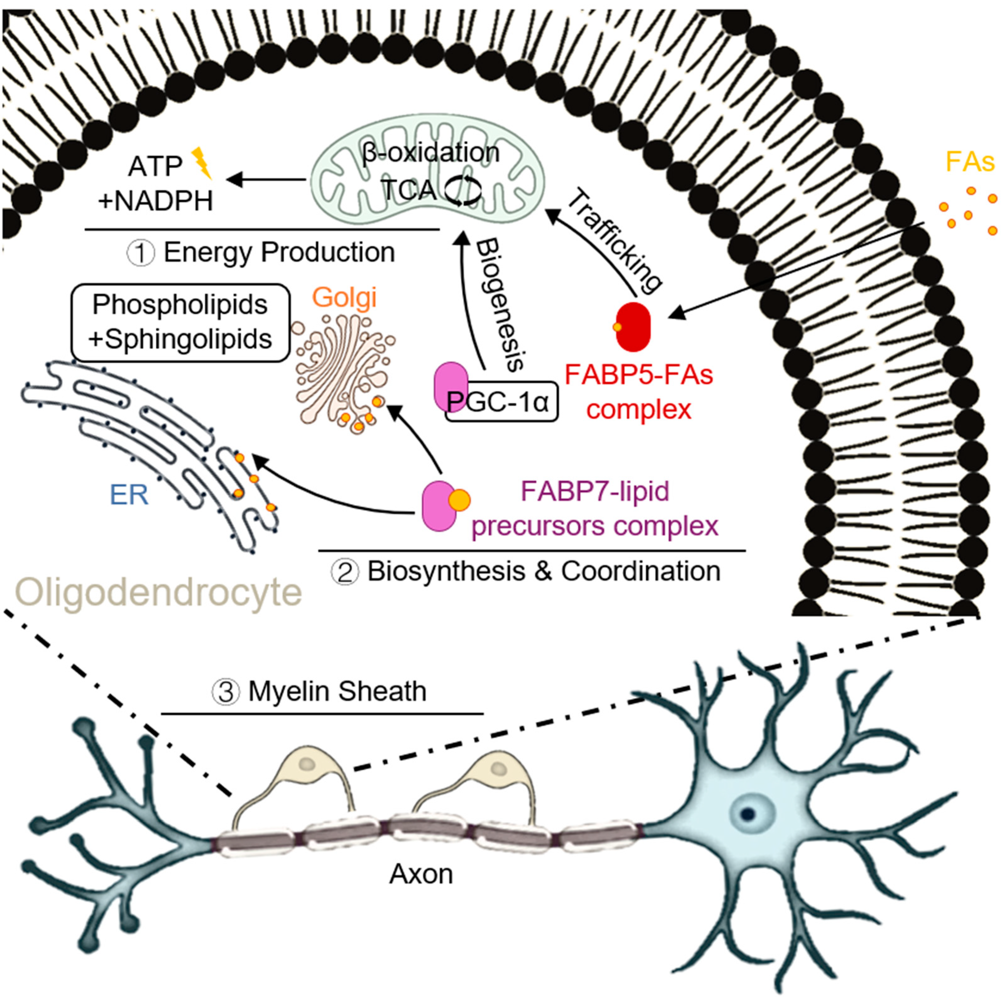
The two isoforms play distinct but complementary roles in this process. FABP5 preferentially delivers saturated/monounsaturated fatty acids to the mitochondrial matrix. There, they undergo β-oxidation to generate acetyl-CoA, which fuels the TCA cycle and oxidative phosphorylation, thereby generating the massive amounts of ATP and NADPH required to power the energetically demanding process of myelin synthesis and to counteract oxidative stress [33]. In contrast, FABP7 is enriched in the myelin precursor membrane system, where it is directly involved in the assembly of phospholipids and sphingolipids and enhances mitochondrial biogenesis by interacting with PGC-1α to ensure that lipid synthesis is synchronized to match the energy requirements [34]. This division of labor—FABP5 for energy production and FABP7 for biosynthesis and coordination—is critical for efficient myelin formation (Figure 2).
When FABP5/7 expression or function is impaired, fatty acids accumulate abnormally in the cytoplasm and mitochondria, leading to mitochondrial lipid overload, decreased respiratory chain activity, and a collapse of the mitochondrial membrane potential, excess lipoyl-CoA further induces a ROS burst and lipid peroxidation, triggering the opening of the mitochondrial permeability transition pore (mPTP), and ultimately triggering apoptosis or iron death in oligodendrocytes [35]. Animal models revealed that mitochondrial swelling, myelin basic protein (MBP) deficiency, and apoptotic markers (cleaved-caspase3) were significantly increased in mitochondria of Fabp5/7 double-knockout mice in Cuprizone-induced demyelination lesions, whereas exogenous FABP5/7 overexpression or administration of the small-molecule inhibitor MF-6 restored mitochondrial homeostasis, reduced ROS, and promoted myelin regeneration [36]. Thus, FABP5/7 plays a role in the transport and metabolism of fatty acids and provides the necessary lipid raw materials for myelination, thereby influencing myelin formation and development.

2.3. The Metabolic Link Between FABPs and Proteotoxicity: A Liquid–Liquid Phase Separation Perspective

The aggregation of misfolded proteins, such as α-synuclein (α-syn), is a central feature of many NDDs like Parkinson’s disease (PD) and Multiple System Atrophy (MSA). α-syn is a 17 kDa protein with three binding structural domains, the N-terminal, hydrophobic, and C-terminal domains, and is enriched in aspartic acid and glutamic acid. α-syn binds to lipids to form a complex and facilitates the binding of α-syn to synaptic membranes that activate platelet-activating factor [37]. Epidemiological evidence of the molecular dynamics of α-syn and in vitro studies have suggested that disorders of lipid homeostatic disturbances may play a role in the pathogenesis of α-syn aggregation [38]. For a long time, FABPs were thought to exacerbate α-syn aggregation by promoting the formation of lipid droplets (LDs), which provide a hydrophobic surface for protein aggregation. However, this description can be deepened with the cutting-edge concept of LLPS, which is now considered a key mechanism driving the formation of membrane less organelles and the initiation of pathological protein aggregation [39,40].
The mechanistic logic is as follows: first, the biogenesis of LDs is itself driven by the LLPS of neutral lipids in the endoplasmic reticulum [41]. Second, and critically, the aggregation of key NDD-related proteins, including α-syn and tau, is now confirmed to be initiated via LLPS [40,42]. In this process, proteins first form liquid-like condensates that subsequently “mature” into irreversible, toxic solid-state aggregates. Therefore, the role of FABPs is far more than providing a passive “hydrophobic surface”. They are very likely active regulators of the LLPS process itself. By precisely controlling the concentration and composition of specific fatty acids within the cell, FABPs directly modulate the thermodynamic environment of the cytoplasm, thereby promoting or inhibiting the phase separation of intrinsically disordered proteins like α-syn. This perspective elevates the role of FABPs from passive “accomplices” to “active regulators” of the first step in proteotoxicity, greatly enhancing their pathological significance.
Beyond generally influencing the lipid environment, specific FABPs can directly participate in the aggregation process [43,44,45]. For instance, recent research has uncovered a direct interaction between FABP7 and α-syn, demonstrating that they form hetero-aggregates which exhibit significantly higher neurotoxicity than α-syn aggregates alone [44]. This finding introduces a novel mechanism where FABP7 acts not just as a lipid transporter but as a direct co-factor in the formation of toxic protein species. Further investigation revealed that these highly toxic hetero-aggregates are selectively propagated into oligodendrocytes and Purkinje cells, the cell types most affected in MSA—via an epsin-2-dependent endocytosis pathway. This work provides a molecular basis for the cell-specific pathology in certain synucleinopathies and establishes FABP7 as a critical mediator linking lipid metabolism directly to the aggregation and cell-to-cell spread of α-syn.

2.4. The New Frontier: The Gut–Brain-FABP Axis and Systemic Inflammation

The long-held view of NDDs as confined to the CNS is being challenged by the concept of the “gut–brain axis” [46]. A central tenet of this model is that pathology can originate in the periphery and spread to the brain [47]. In synucleinopathies like PD, compelling evidence suggests that misfolded α-syn first appears in the enteric nervous system (ENS) and propagates to the brainstem via retrograde axonal transport along the vagus nerve in a prion-like fashion [48].
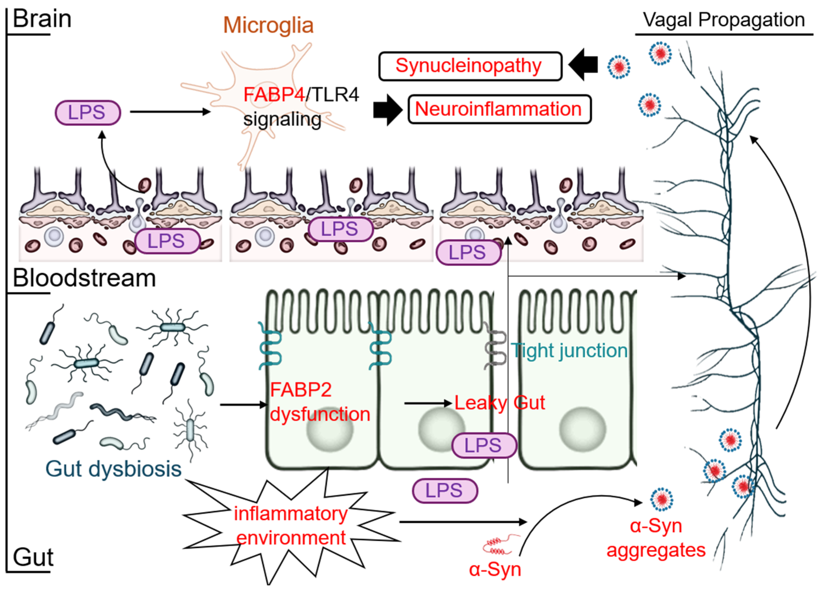
The intestinal protein FABP2 plays a crucial, albeit indirect, role in initiating this cascade. FABP2 is vital for maintaining intestinal barrier integrity by regulating tight junction proteins [49]. In conditions of gut dysbiosis, often driven by poor diet or aging, FABP2 function can be impaired [49,50]. This leads to increased intestinal permeability, a condition known as “leaky gut”. This barrier dysfunction has two critical consequences. First, it is thought to create a local inflammatory environment within the gut that promotes the initial misfolding and aggregation of α-syn in the ENS. Second, it allows bacterial endotoxins, particularly LPS, to leak from the gut lumen into the systemic circulation, causing chronic, low-grade systemic inflammation (Figure 3).
This systemic inflammation represents a “second hit” to the CNS. Circulating inflammatory molecules like LPS can compromise the integrity of the BBB, further promoting neuroinflammation. Once in the brain, LPS directly activates microglia via the FABP4/TLR4 signaling pathway described earlier [24,51], establishing a persistent neuroinflammatory state. This chronic inflammation creates a toxic environment that can exacerbate the neurodegeneration caused by the primary α-syn pathology spreading via the vagus nerve. Thus, the gut–brain-FABP axis represents a multi-faceted pathogenic model where peripheral FABP2 dysfunction initiates events in the gut, leading to both direct prion-like propagation of proteopathy and an indirect, systemic inflammatory response that is mediated centrally by FABP4 [49] (Figure 3). This highlights the utility of peripherally restricted inhibitors, which may succeed not by acting directly on the brain, but by cutting off this peripheral “fuel supply” for central inflammation.

2.5. The FABP-Ferroptosis Link: A New Frontier in Neurotoxic Cell Death

Ferroptosis is a form of regulated, iron-dependent cell death driven by the overwhelming accumulation of lipid peroxides, particularly from polyunsaturated fatty acids (PUFAs) within phospholipids [52]. FABP’s ferroptosis connection is novel, but recent research has unequivocally proven this connection.
FABP5 has been posited as a key driver of ferroptosis especially in the context of cerebral hypoxia, stroke and cancer [53,54]. Under cellular stress, FABP5 is upregulated and acts in a positive feedback loop, facilitating the transport and incorporation of PUFAs into cellular membranes. This enriches the membrane with lipids that are highly susceptible to peroxidation, thereby increasing the cell’s vulnerability to ferroptotic death. In contrast, FABP7 has been identified as a protector against ferroptosis [55]. FABP7 actively sequesters vulnerable PUFAs, transporting them away from the cell membrane and into the relative safety of lipid droplets for storage. This mechanism effectively isolates PUFAs from the iron-dependent peroxidative machinery, shielding cells like astrocytes from ferroptotic death. This dichotomous regulation—FABP5 as a sensitizer and FABP7 as a protector—positions the FABP axis as a critical control point in the balance between cell survival and ferroptotic death (Figure 4 and Table 1).

3. Pharmacological Modulation of FABPs:

3.1. CNS-Penetrant Inhibitors: Resetting Central Metabolism

The clinical application of FABP-targeted therapies is still constrained by the BBB which limits drug access to the CNS and complicates therapeutic delivery. Thus, the design of CNS drugs requires ligands that not only demonstrate high-affinity and isoform-selective FABP binding but also robust BBB permeation [97]. There is now a better ability to overcome these challenges due to advances in medicinal chemistry. These CNS penetrating FABP inhibitors can modulate lipid signaling, glial activation, and the neuroinflammatory processes in NDDs. The following sections discuss preclinical studies of select drugs of this class, and their rationale related to design, pharmacokinetics, as well as preclinical efficacy.

3.1.1. MF6: A CNS-Penetrant Candidate Targeting the Meta-Inflammatory Axis

FABP Ligand 6 (MF6) acts as a selective inhibitor and exhibits high affinity to FABP7 (Kd: 20 nM) [43] and moderate affinity to FABP5 (Kd: 874 nM) [98]. Most notably, its weaker affinity to the cardiac-specific FABP3 (Kd: 1038 nM) [66] decreases the risk for possible cardiotoxic side effects, making it a well-suited candidate for CNS therapies. This favorable selectivity profile allows it to modulate neuroinflammatory pathways while minimizing off-target effects in the periphery.
The therapeutic potential of MF6 is supported by strong preclinical evidence demonstrating both anti-inflammatory and neuroprotective effects. At a cellular level, MF6 demonstrated protective effects on oligodendrocyte precursor cells by stabilizing their mitochondrial membrane potential [80]. In LPS-stimulated primary astrocytes, MF6 provoked a marked reduction in release to pro-inflammatory cytokines IL-1β and TNF-α [84]. These cellular protective benefits translate to clinically meaningful improvements in animal models. In the MOG-induced experimental autoimmune encephalomyelitis (EAE), a model for multiple sclerosis (MS), daily oral MF6 (1 mg/kg for 28 days) significantly delayed disease onset and improved neurological scores alongside reduced spinal cord demyelination and glial activation [84]. Likewise, in the PLP-hα-Syn transgenic mouse model of MSA, treatment with MF6 mitigated cerebellar α-syn inclusions, reduced glial inflammation and oxidative stress, preserved Purkinje neuron morphology, and improved motor coordination [99].
Moreover, MF6 demonstrates CNS drug advantages in pharmacokinetics. Its pharmacokinetics confirmed rapid CNS penetrative capability and therapeutic concentrations following oral dosing. In a model of ischemia and reperfusion, 3 mg/kg administered orally showed significant action with a 30% reduction in infarct volume 30 min post dose highlighting great oral bioavailability and rapid action [100]. This evidence establishes MF6 as a penetrative CNS compound able to influence and modulate glial meta-inflammation and even lessen neurodegenerative pathology in models of MS, MSA and other NDDs (Figure 5).

3.1.2. Targeting FABP3 in Neurotherapeutics: From MF1 to the High-Potency Ligand HY-11-9

MF1 is a pyrazole-type small molecule ligand engineered to selectively target FABP3, possessing a moderate binding affinity of Kd: 302.8 ± 130.3 nM [66]. Its design was based on the changes made to BMS309403 to increase the selectivity for FABP3 while minimizing the unwanted targeting of FABP4 [98]. Preclinical research suggests MF1 could serve as a dual-mechanism therapeutic drug, exhibiting potential both for the treatment of synucleinopathies, such as PD, as well as for seizure disorders, including epilepsy.
The primary mechanism of MF1 in the context of PD is its ability to disrupt pathological protein aggregation [65]. Because FABP3 is highly expressed in dopaminergic neurons and promotes the oligomerization of α-syn, MF1 intervenes by inhibiting this key interaction. This was validated both in vitro, where MF1 prevented α-syn clumping in cells [66], and in vivo in mouse models of PD. In these animal studies, oral administration of MF1 led to significant neuroprotection and functional recovery, including reduced α-syn accumulation, preservation of dopaminergic neurons, and improved motor and cognitive functions [66,101]. These successful outcomes strongly suggest that MF1 has effective CNS penetration and good oral bioavailability.
In addition to its function in neurodegeneration, MF1 has a second, distinct therapeutic action as an anticonvulsant. MF1 has previously shown a considerable degree of seizure suppression in MF1 epilepsy mouse models. This effect is thought to arise from its GABA-A receptor ‘turning on’ action through the benzodiazepine recognition site, a completely different mechanism from its action on FABP3 receptors [102]. While CNS pharmacokinetic data is sparse, pharmacokinetic data from animal models has shown good oral bioavailability and central bioavailability.
Building on this work, HY-11-9, a newly designed and next-generation potent and selective FABP3 ligand [103]. Comparative data show that HY-11-9 has an affinity for FABP3 (Kd≈11.7 nM) that is approximately 25 times higher than that of MF1. More importantly, in a side-by-side comparison made for testing neuroprotection of PD theragnostics in a 1-methyl-4-phenyl-1,2,3,6-tetrahydropyridine (MPTP)-induced PD model, using oral administration of HY-11-9 (0.03 mg/kg), which ameliorated motor deficits, was not observed with MF1 treatment. In further studies, HY-11-9 depletion of pS129-α-syn and reversing MPTP toxicity on dopaminergic neurons was explored. These results establish HY-11-9 as a superior preclinical candidate to MF1, setting a new benchmark for potency and efficacy for FABP3-targeted neuroprotection in PD. While MF1 demonstrated the therapeutic potential of inhibiting the FABP3-α-syn interaction, the development of HY-11-9 provides a more powerful tool and clarifies the path forward for developing novel treatments for PD.

3.1.3. HY08: A FABP3/5 Ligand Targeting Mitochondrial Damage

HY08 is a synthetic small-molecule ligand that selectively inhibits FABP3 and FABP5, which are involved in mitochondria disruption during cerebral ischemia. Hydrophobicity shows high affinity for FABP3 (Kd: 24 ± 7 nM) and moderate affinity for FABP5 (Kd: 410 ± 70 nM), while showing negligible binding to FABP7. By inhibiting FABP- and/or carrier-protein-mediated transport of arachidonic acid HY08 mitigates lipid peroxidation and oxidative stress. In pro-apoptotic signal suppression, pro-apoptotic signal blockade is also observed. With ROTENONE-induced oxidative stress in vitro SH-SY5Y cells demonstrated 4-HNE accumulation suppression, mitochondrial membrane potential preservation, and BAX translocation to mitochondria blockade under pro-oxidative conditions [85].
Pharmacokinetic studies demonstrate and support HY08’s potential for CNS applications, as evidenced in a mouse model of ischemic stroke. Infarct volume was significantly reduced in the stroke model following a single oral dose of 0.3 mg/kg, confirming bioavailability, sufficient therapeutic concentration attainment, and ischemic brain and confirming protective action. These results support its ability to cross the blood–brain barrier and achieve central pharmacological activity following systemic administration [85].
The neuroprotective effect showed in mouse models of transient middle cerebral artery occlusion/reperfusion (tMCAO/R) model. These models received a single oral dose of 0.3 mg/kg 30 min post-reperfusion, significantly reduced infarct volume, improved neurological scores, and increased 7-day survival. Mechanistically, HY08 inhibited the ischemia-induced accumulation of FABP3 and FABP5 in mitochondria, thereby preventing lipid peroxidation (reduced 4-HNE levels) and suppressing BAX-mediated apoptotic signaling. These findings support a role for HY08 in protecting ischemic neurons by disrupting FABP-driven mitochondrial injury pathways [85].

3.2. Peripherally Restricted Inhibitors: Modulating Neuro-Immune Crosstalk

The successful development of ART26.12 provides a new approach for FABP-targeted therapy. ART26.12 is a third-generation selective inhibitor of FABP5 from the truxillic acid monoester series It exhibits high affinity for FABP5 (inhibition constant [Ki]: 0.77 ±0.08 μM) with markedly lower affinity for FABP3 (Ki: 71.32 ± 2.40 μM), FABP4 (Ki: > 100 μM), and FABP7 (Ki: 18.99 ± 1.81 μM), as determined by fluorescence displacement assay. By inhibiting FABP5-mediated trafficking of endocannabinoids such as anandamide, ART26.12 reduces their catabolism and enhances local endocannabinoid tone. This indirectly activates CB1 signaling, with potential contributions from CB2, TRPV1, and PPARα receptors involved in metabolic regulation and anti-inflammatory responses. Transcriptomic profiling in the periaqueductal gray revealed upregulation of ion channel and GABAergic synapse-related genes, suggesting central neurophysiological adaptations relevant to pain processing and lipid signaling [104]. These findings support ART26.12 as a mechanistically distinct FABP5-targeting compound that modulates both metabolic and inflammatory pathways implicated in neurodegenerative disease.
ART26.12 demonstrates favorable oral pharmacokinetics, with rapid absorption (Tmax ≈ 0.5 h at 10 mg/kg) and high bioavailability (163% at 300 mg/kg), suggesting nonlinear uptake or enterohepatic recirculation. [104]. Although CNS penetration is limited (brain-to-plasma ratio ≈ 2.3%), the compound reaches therapeutically relevant concentrations in the spinal cord (0.4 ± 0.1 μM), a key region for neuroimmune and metabolic integration in pain modulation [104,105]. ART26.12 is well tolerated in preclinical species, with a no observed adverse effect level (NOAEL) of 1000 mg/kg/day in both rodents and dogs [104].
ART26.12 has shown efficacy across multiple peripheral neuropathy models, including oxaliplatin-induced peripheral neuropathy (OIPN), paclitaxel-induced neuropathy (PIPN), streptozotocin-induced diabetic neuropathy, and cancer-induced bone pain. It dose-dependently reverses mechanical allodynia and cold hyperalgesia, preserves body weight during treatment [104,105]. In diabetic neuropathy models, ART26.12 outperformed duloxetine in reversing pain behaviors at earlier time points [105]. Mechanistically, its analgesic effects are mediated by FABP5-dependent endocannabinoid pathways, involving CB1 and PPARα signaling, both of which are associated with glial regulation and lipid-driven inflammation [104]. These results highlight ART26.12 as a lipid-targeting analgesic with potential utility in diseases involving neuroimmune dysregulation and metabolic dysfunction and strongly support the aforementioned “gut–brain axis” and systemic inflammation theories: even intervening in peripheral metabolic/inflammatory pathways can have a profound impact on the symptoms of the CNS (Table 2).

4. Conclusions and Future Perspectives

4.1. Summary

This review has emphasized the pivotal position of brain-expressed FABPs as key orchestrators of lipid metabolism, placing them at the nexus of metabolic reprogramming, neuroinflammation, and cell death in NDDs. The evidence provided here supports the conclusion that targeting FABP-mediated metabolic shifts in the brain indicates a primary intervention approach to treating NDDs.
As discussed, meta-inflammatory frameworks in glial cells as well as metabolic survival balance in oligodendrocytes rely heavily on FABP-driven lipid transport. The preclinical successes of the ligands discussed here lend strong support to this hypothesis. Molecules MF6, MF1, and HY08 demonstrate that CNS-penetrant compounds can directly influence critical metabolic pathways to rectify complex demyelinating and proteotoxic as well as ischemic injuries. At the same time, the peripherally restricted inhibitor ART26.12 shows that targeting FABP5 pathways, even outside the CNS, can deeply influence neuroinflammatory processes, underscoring the therapeutic importance of peripheral FABP5 in neuroimmune nexus disordered metabolism.
As a result, disparate FABPs can be regarded as active, multi-faceted therapeutic targets that can be utilized to reshape.

4.2. Outlook: From “What to Inhibit” to “Where and How to Modulate” with Precision

The attractive preclinical rationale for FABP inhibitors supports therapeutic avenues that are yet to be explored. Future FABP drug discovery will likely evolve from broad inhibition toward more precise strategies.
FABP drug discovery is likely to evolve beyond broad inhibition toward isoform-specific and cell-type-specific selectivity. Next-generation inhibitors may aim to differentially modulate FABP5 and FABP7 in disease-relevant glial populations, such as microglia, astrocytes, and oligodendrocytes, allowing cell-type-specific control of lipid metabolism and inflammatory responses. A promising direction involves integrating lipidomic data to guide ligand design. Integrating lipidomic profiling into FABP-targeted drug design may allow for the development of inhibitors that selectively block the trafficking of disease-relevant lipid species, offering a more tailored and mechanism-driven approach to therapy. Such approaches could enhance therapeutic efficacy and minimize off-target effects. Moreover, FABP isoforms and their lipid cargoes may serve as biomarkers for early diagnosis, patient stratification, and treatment monitoring.
FABP inhibitors act upstream of multiple pathological processes and are thus well suited for combination with agents that target downstream consequences. For instance, combining a FABP3 inhibitor like MF1, which suppresses α-syn oligomer formation, with agents that target aggregate clearance could provide a synergistic, dual-pronged therapeutic strategy. In demyelinating disorders, using MF6 in combination with a pro-myelinating compound could halt glial damage while promoting remyelination. Likewise, combining a peripherally restricted inhibitor like ART26.12 with a centrally acting analgesic may enable comprehensive pain control by addressing both peripheral and central sensitization. These combinatorial strategies may amplify therapeutic efficacy and broaden the clinical utility of FABP-targeted approaches.
Aberrant Post-Translational Modifications (PTMs) (e.g., phosphorylation, ubiquitination) are a hallmark of NDDs, altering protein function, localization, and aggregation. The function of FABPs themselves may be subject to such regulations. A critical unanswered question is whether FABPs undergo PTMs in NDDs. For example, could phosphorylation of FABP5 alter its binding to VDAC-1, or could ubiquitination of FABP7 target it for degradation, weakening its neuroprotective role in ferroptosis? Investigating the PTMs of FABPs opens a new research field and suggests novel therapeutic targets (e.g., inhibiting a kinase that phosphorylates a FABP), which could offer a more refined and specific strategy than broad ligand inhibition.
In summary, this review highlights the overlooked potential of FABPs as bridges within the networks of lipid metabolism, neuroinflammation, and cell death. Spanning from basic mechanisms to therapeutics, focusing on FABP-centric pathways presents a single and coherent approach to altering neurodegenerative processes. As the field moves forward, the comprehensive therapeutic prospects of FABP-targeted strategies in NDDs will require lipidomic, structural, and precision pharmacological interventions.

Author Contributions

Conceptualization, A.C. and I.K.; writing—original draft preparation, C.W. and J.L.; visualization, Q.C. and W.Z.; writing—review and editing, A.C., Q.C. and W.Z.; funding acquisition, A.C.; project administration, A.C. All authors have read and agreed to the published version of the manuscript.

Funding

This work was supported by the Start-up Fund from Sun Yat-sen University (Grant No. 59000-12256011).

Institutional Review Board Statement

Not applicable.

Informed Consent Statement

Not applicable.

Data Availability Statement

No new data were created or analyzed in this study.

Conflicts of Interest

The authors declare no conflicts of interest.

Abbreviations

The following abbreviations are used in this manuscript:
4-HNE4-Hydroxynonenal
AAArachidonic Acid
Acetyl-CoAAcetyl-Coenzyme A
ADAlzheimer’s Disease
ALSAmyotrophic lateral sclerosis
BBBBlood–Brain Barrier
CNSCentral Nervous System
EAEExperimental autoimmune encephalomyelitis
ENSEnteric nervous system
FABPsFatty acid-binding proteins
HKHexokinase
LCFAsLong-chain fatty acids
LDLipid droplets
LDHLactate dehydrogenase
LPSLipopolysaccharide
MBPMyelin basic protein
MF6FABP Ligand 6
mPTPmitochondrial permeability transition pore
MPTP1-methyl-4-phenyl-1,2,3,6-tetrahydropyridine
MSMultiple sclerosis
MSAMultiple system atrophy
NDDsNeurodegenerative diseases
NOAELNo observed adverse effect level
OIPNOxaliplatin-induced peripheral neuropathy
OXPHOSOxidative phosphorylation
PDParkinson’s disease
PFKPhosphofructokinase
PIPNPaclitaxel-induced neuropathy
ROSReactive oxygen species
TCATricarboxylic acid cycle
TLR4Toll-like receptor 4
tMCAO/Rtransient middle cerebral artery occlusion/reperfusion
α-synα-synuclein

References

  1. Scheltens, P.; De Strooper, B.; Kivipelto, M.; Holstege, H.; Chételat, G.; Teunissen, C.E.; Cummings, J.; van der Flier, W.M. Alzheimer’s Disease. Lancet 2021, 397, 10284. [Google Scholar] [CrossRef] [PubMed]
  2. Agnello, L.; Gambino, C.M.; Ciaccio, A.M.; Cacciabaudo, F.; Massa, D.; Masucci, A.; Tamburello, M.; Vassallo, R.; Midiri, M.; Scazzone, C.; et al. From Amyloid to Synaptic Dysfunction: Biomarker-Driven Insights into Alzheimer’s Disease. Curr. Issues Mol. Biol. 2025, 47, 580. [Google Scholar] [CrossRef]
  3. Noh, M.Y.; Kwon, H.S.; Kwon, M.S.; Nahm, M.; Jin, H.K.; Bae, J.S.; Kim, S.H. Biomarkers and Therapeutic Strategies Targeting Microglia in Neurodegenerative Diseases: Current Status and Future Directions. Mol. Neurodegener. 2025, 20, 82. [Google Scholar] [CrossRef]
  4. Furuhashi, M.; Hotamisligil, G.S. Fatty Acid-Binding Proteins: Role in Metabolic Diseases and Potential as Drug Targets. Nat. Rev. Drug Discov. 2008, 7, 489–503. [Google Scholar] [CrossRef]
  5. Storch, J.; Corsico, B. The Emerging Functions and Mechanisms of Mammalian Fatty Acid-Binding Proteins. Annu. Rev. Nutr. 2008, 28, 73–95. [Google Scholar] [CrossRef]
  6. Maeda, K.; Uysal, K.T.; Makowski, L.; Görgün, C.Z.; Atsumi, G.; Parker, R.A.; Brüning, J.; Hertzel, A.V.; Bernlohr, D.A.; Hotamisligil, G.S. Role of the Fatty Acid Binding Protein Mal1 in Obesity and Insulin Resistance. Diabetes 2003, 52, 300–307. [Google Scholar] [CrossRef]
  7. Li, B.; Hao, J.; Zeng, J.; Sauter, E.R. SnapShot: FABP Functions. Cell 2020, 182, 1066–1066.e1. [Google Scholar] [CrossRef]
  8. Maeda, K.; Cao, H.; Kono, K.; Gorgun, C.Z.; Furuhashi, M.; Uysal, K.T.; Cao, Q.; Atsumi, G.; Malone, H.; Krishnan, B.; et al. Adipocyte/Macrophage Fatty Acid Binding Proteins Control Integrated Metabolic Responses in Obesity and Diabetes. Cell Metab. 2005, 1, 107–119. [Google Scholar] [CrossRef] [PubMed]
  9. Jin, R.; Hao, J.; Yi, Y.; Sauter, E.; Li, B. Regulation of Macrophage Functions by FABP-Mediated Inflammatory and Metabolic Pathways. Biochim. Biophys. Acta Mol. Cell Biol. Lipids 2021, 1866, 158964. [Google Scholar] [CrossRef] [PubMed]
  10. Furuhashi, M.; Tuncman, G.; Görgün, C.Z.; Makowski, L.; Atsumi, G.; Vaillancourt, E.; Kono, K.; Babaev, V.R.; Fazio, S.; Linton, M.F.; et al. Treatment of Diabetes and Atherosclerosis by Inhibiting Fatty-acid-Binding Protein aP2. Nature 2007, 447, 959–965. [Google Scholar] [CrossRef]
  11. Başarır Sivri, F.N.; Çiftçi, S. A New Insight into Fatty Acid Binding Protein 4 Mechanisms and Therapeutic Implications in Obesity-Associated Diseases: A Mini Review. Mol. Nutr. Food Res. 2024, 68, e2300840. [Google Scholar] [CrossRef] [PubMed]
  12. Powell, A.; Yamaguchi, N.; Lu, H.; Pareek, O.; Elman, I.; Gold, M.S.; Pinhasov, A.; Blum, K.; Thanos, P.K. The Role of Fatty Acid Binding Proteins in Neuropsychiatric Diseases: A Narrative Review. Front. Biosic. 2025, 30, 26812. [Google Scholar] [CrossRef]
  13. Bowers, M.; Liang, T.; Gonzalez-Bohorquez, D.; Zocher, S.; Jaeger, B.N.; Kovacs, W.J.; Röhrl, C.; Cramb, K.M.L.; Winterer, J.; Kruse, M.; et al. FASN-Dependent Lipid Metabolism Links Neurogenic Stem/Progenitor Cell Activity to Learning and Memory Deficits. Cell Stem Cell 2020, 27, 98–109.e11. [Google Scholar] [CrossRef]
  14. Chiasserini, D.; Biscetti, L.; Eusebi, P.; Salvadori, N.; Frattini, G.; Simoni, S.; De Roeck, N.; Tambasco, N.; Stoops, E.; Vanderstichele, H.; et al. Differential Role of CSF Fatty Acid Binding Protein 3, α-Synuclein, and Alzheimer’s Disease Core biomarkers in Lewy Body Disorders and Alzheimer’s Dementia. Alzheimers Res. Ther. 2017, 9, 52. [Google Scholar] [CrossRef]
  15. Rao, E.; Singh, P.; Li, Y.; Zhang, Y.; Chi, Y.I.; Suttles, J.; Li, B. Targeting Epidermal Fatty Acid Binding Protein for Treatment of Experimental Autoimmune Encephalomyelitis. BMC Immunol. 2015, 16, 28. [Google Scholar] [CrossRef]
  16. Teunissen, C.E.; Veerhuis, R.; De Vente, J.; Verhey, F.R.; Vreeling, F.; van Boxtel, M.P.; Glatz, J.F.; Pelsers, M.A. Brain-Specific Fatty Acid-Binding Protein is Elevated in Serum of Patients with Dementia-Related Diseases. Eur. J. Neurol. 2011, 18, 865–871. [Google Scholar] [CrossRef]
  17. Killoy, K.M.; Harlan, B.A.; Pehar, M.; Vargas, M.R. Fabp7 Upregulation Induces a Neurotoxic Phenotype in Astrocytes. Glia 2020, 68, 2693–2704. [Google Scholar] [CrossRef]
  18. Chen, Z.; Yuan, Z.; Yang, S.; Zhu, Y.; Xue, M.; Zhang, J.; Leng, L. Brain Energy Metabolism: Astrocytes in Neurodegenerative Diseases. CNS Neurosci. Ther. 2023, 29, 24–36. [Google Scholar] [CrossRef] [PubMed]
  19. Huang, Q.; Wang, Y.; Chen, S.; Liang, F. Glycometabolic Reprogramming of Microglia in Neurodegenerative Diseases: Insights from Neuroinflammation. Aging Dis. 2024, 15, 1155–1175. [Google Scholar] [CrossRef] [PubMed]
  20. Gimeno-Bayón, J.; López-López, A.; Rodríguez, M.J.; Mahy, N. Glucose Pathways Adaptation Supports Acquisition of Activated Microglia Phenotype. J. Neurosci. Res. 2014, 92, 723–731. [Google Scholar] [CrossRef]
  21. Luo, Z.; Eichinger, K.M.; Zhang, A.; Li, S. Targeting Cancer Metabolic Pathways for Improving Chemotherapy and Immunotherapy. Cancer Lett. 2023, 575, 216396. [Google Scholar] [CrossRef] [PubMed]
  22. Wu, H.; Xing, C.; Yu, B.; Guo, L.; Dou, X.; Gao, L.; Yang, S.; Zhang, Y.; Gao, X.; Li, S.; et al. Metabolic Reprogramming of Neural Stem Cells by Chiral Nanofiber for Spinal Cord Injury. ACS Nano 2025, 19, 4785–4801. [Google Scholar] [CrossRef] [PubMed]
  23. Zhou, X.; Zhou, S.; Tao, J.; Gao, Y.; Meng, G.; Cao, D.; Gao, L. HIV-1 Tat Drives the Fabp4/NF-κB Feedback Loop in Microglia to Mediate Inflammatory Response and Neuronal Apoptosis. J. Neurovirol. 2022, 28, 483–496. [Google Scholar] [CrossRef]
  24. Kagawa, Y.; Low, Y.L.; Pyun, J.; Doglione, U.; Short, J.L.; Pan, Y.; Nicolazzo, J.A. Fatty Acid-Binding Protein 4 is Essential for the Inflammatory and Metabolic Response of Microglia to Lipopolysaccharide. J. Neuroimmune Pharmacol. 2023, 18, 448–461. [Google Scholar] [CrossRef] [PubMed]
  25. Ciesielska, A.; Matyjek, M.; Kwiatkowska, K. TLR4 and CD14 Trafficking and Its Influence on LPS-Induced Pro-Inflammatory Signaling. Cell. Mol. Life Sci. 2021, 78, 1233–1261. [Google Scholar] [CrossRef]
  26. Fu, Y.J.; Xu, B.; Huang, S.W.; Luo, X.; Deng, X.L.; Luo, S.; Liu, C.; Wang, Q.; Chen, J.Y.; Zhou, L. Baicalin Prevents LPS-Induced Activation of TLR4/NF-κB p65 Pathway and Inflammation in Mice via Inhibiting the Expression of CD14. Acta Pharmacol. Sin. 2021, 42, 88–96. [Google Scholar] [CrossRef]
  27. Wang, Y.; Yang, Y.; Meng, T.; Liu, S.; Liu, J.; Liu, D.; Laxman, B.; Chen, S.; Dong, Z. β-Caryophyllene Reduces Inflammation to Protect Against Ischemic Stroke by Suppressing HMGB1 Signaling. Mol. Med. 2025, 31, 112. [Google Scholar] [CrossRef]
  28. Li, H.; Luo, H.Y.; Liu, Q.; Xiao, Y.; Tang, L.; Zhong, F.; Huang, G.; Xu, J.M.; Xu, A.M.; Zhou, Z.G.; et al. Intermittent High Glucose Exacerbates A-FABP Activation and Inflammatory Response through TLR4-JNK Signaling in THP-1 Cells. J. Immunol. Res. 2018, 2018, 1319272. [Google Scholar] [CrossRef]
  29. So, S.W.; Fleming, K.M.; Duffy, C.M.; Nixon, J.P.; Bernlohr, D.A.; Butterick, T.A. Microglial FABP4-UCP2 Axis Modulates Neuroinflammation and Cognitive Decline in Obese Mice. Int. J. Mol. Sci. 2022, 23, 4354. [Google Scholar] [CrossRef]
  30. Yu, Y.Y.; Feng, M.; Chen, Y.; Jia, H.L.; Zhang, Q.; Tong, M.; Li, Y.X.; Zhao, Y.; Liu, X.X.; Cao, S.F.; et al. Asprosin-FABP5 Interaction Modulates Mitochondrial Fatty Acid Oxidation through PPARα Contributing to MASLD Development. Adv. Sci. 2025, 12, e2415846. [Google Scholar] [CrossRef]
  31. Zhou, T.; Zhang, H.; Yu, X.; Pan, K.; Yao, X.; Shen, X.; Lei, H. Fc-Binding Cyclopeptide Induces Allostery from Fc to Fab: Revealed Through in Silico Structural Analysis to Anti-Phenobarbital Antibody. Foods 2025, 14, 1360. [Google Scholar] [CrossRef]
  32. Namiecinska, M.; Piatek, P.; Lewkowicz, P. Nervonic Acid Synthesis Substrates as Essential Components in Profiled Lipid Supplementation for More Effective Central Nervous System Regeneration. Int. J. Mol. Sci. 2024, 25, 3792. [Google Scholar] [CrossRef] [PubMed]
  33. Nsiah-Sefaa, A.; McKenzie, M. Combined Defects in Oxidative Phosphorylation and Fatty Acid β-Oxidation in Mitochondrial Disease. Biosci. Rep. 2016, 36, e00313. [Google Scholar] [CrossRef] [PubMed]
  34. Chen, Y.; Liu, J.; He, Y.; Lü, Y.; Yu, W. The Role of Fatty Acid Binding Protein 7 in Neurological Diseases. Mol. Neurobiol. 2025; ahead of print. [Google Scholar] [CrossRef]
  35. Fang, X.X.; Wei, P.; Zhao, K.; Sheng, Z.C.; Song, B.L.; Yin, L.; Luo, J. Fatty Acid-Binding Proteins 3, 7, and 8 Bind Cholesterol and Facilitate Its Egress from Lysosomes. J. Cell Biol. 2024, 223, e202211062. [Google Scholar] [CrossRef]
  36. Kamizato, K.; Sato, S.; Shil, S.K.; Umaru, B.A.; Kagawa, Y.; Yamamoto, Y.; Ogata, M.; Yasumoto, Y.; Okuyama, Y.; Ishii, N.; et al. The Role of Fatty Acid Binding Protein 7 in Spinal Cord Astrocytes in a Mouse Model of Experimental Autoimmune Encephalomyelitis. Neuroscience 2019, 409, 120–129. [Google Scholar] [CrossRef]
  37. Burré, J.; Sharma, M.; Südhof, T.C. Cell Biology and Pathophysiology of α-Synuclein. Cold Spring Harb. Perspect. Med. 2018, 8, a024091. [Google Scholar] [CrossRef]
  38. Pingale, T.D.; Gupta, G.L. Novel Therapeutic Approaches for Parkinson’s Disease by Targeting Brain Cholesterol Homeostasis. J. Pharm. Pharmacol. 2021, 73, 862–873. [Google Scholar] [CrossRef]
  39. Boyko, S.; Surewicz, W.K. Tau Liquid-Liquid Phase Separation in Neurodegenerative Diseases. Trends Cell Biol. 2022, 32, 611–623. [Google Scholar] [CrossRef] [PubMed]
  40. Ray, S.; Singh, N.; Kumar, R.; Patel, K.; Pandey, S.; Datta, D.; Mahato, J.; Panigrahi, R.; Navalkar, A.; Mehra, S.; et al. α-Synuclein Aggregation Nucleates Through Liquid-Liquid Phase Separation. Nat. Chem. 2020, 12, 705–716. [Google Scholar] [CrossRef]
  41. Zoni, V.; Khaddaj, R.; Campomanes, P.; Thiam, A.R.; Schneiter, R.; Vanni, S. Pre-Existing Bilayer Stresses Modulate Triglyceride Accumulation in the ER Versus Lipid Droplets. Elife 2021, 10, e62886. [Google Scholar] [CrossRef]
  42. Wegmann, S.; Eftekharzadeh, B.; Tepper, K.; Zoltowska, K.M.; Bennett, R.E.; Dujardin, S.; Laskowski, P.R.; MacKenzie, D.; Kamath, T.; Commins, C.; et al. Tau Protein Liquid-Liquid Phase Separation Can Initiate Tau Aggregation. EMBO J. 2018, 37, e98049. [Google Scholar] [CrossRef]
  43. Cheng, A.; Wang, Y.F.; Shinoda, Y.; Kawahata, I.; Yamamoto, T.; Jia, W.B.; Yamamoto, H.; Mizobata, T.; Kawata, Y.; Fukunaga, K. Fatty Acid-Binding Protein 7 Triggers α-Synuclein Oligomerization in Glial Cells and Oligodendrocytes Associated with Oxidative Stress. Acta Pharmacol. Sin. 2022, 43, 552–562. [Google Scholar] [CrossRef]
  44. Cheng, A.; Kawahata, I.; Wang, Y.; Jia, W.; Wang, H.; Sekimori, T.; Chen, Y.; Suzuki, H.; Takeda, A.; Stefanova, N.; et al. Epsin2, a Novel Target for Multiple System Atrophy Therapy via α-Synuclein/FABP7 Propagation. Brain 2023, 146, 3172–3180. [Google Scholar] [CrossRef]
  45. Wang, Y.; Shinoda, Y.; Cheng, A.; Kawahata, I.; Fukunaga, K. Epidermal Fatty Acid-Binding Protein 5 (FABP5) Involvement in Alpha-Synuclein-Induced Mitochondrial Injury under Oxidative Stress. Biomedicines 2021, 9, 110. [Google Scholar] [CrossRef] [PubMed]
  46. Wang, Q.; Yang, Q.; Liu, X. The Microbiota-Gut-Brain Axis and Neurodevelopmental Disorders. Protein Cell 2023, 14, 762–775. [Google Scholar] [CrossRef]
  47. Cryan, J.F.; O’Riordan, K.J.; Sandhu, K.; Peterson, V.; Dinan, T.G. The Gut Microbiome in Neurological Disorders. Lancet Neurol. 2020, 19, 179–194. [Google Scholar] [CrossRef] [PubMed]
  48. Xiang, J.; Tang, J.; Kang, F.; Ye, J.; Cui, Y.; Zhang, Z.; Wang, J.; Wu, S.; Ye, K. Gut-induced alpha-Synuclein and Tau propagation initiate Parkinson’s and Alzheimer’s disease co-pathology and behavior impairments. Neuron 2024, 112, 3585–3601.e5. [Google Scholar] [CrossRef] [PubMed]
  49. Stevens, B.R.; Goel, R.; Seungbum, K.; Richards, E.M.; Holbert, R.C.; Pepine, C.J.; Raizada, M.K. Increased Human Intestinal Barrier Permeability Plasma Biomarkers Zonulin and FABP2 Correlated with Plasma LPS and Altered Gut Microbiome in Anxiety or Depression. Gut 2018, 67, 1555–1557. [Google Scholar] [CrossRef]
  50. Rogers, G.B.; Keating, D.J.; Young, R.L.; Wong, M.L.; Licinio, J.; Wesselingh, S. From Gut Dysbiosis to Altered Brain Function and Mental Illness: Mechanisms and Pathways. Mol. Psychiatry 2016, 21, 738–748. [Google Scholar] [CrossRef]
  51. Keyhanian, K.; Davoudi, V.; Saxena, S.; Kivisakk, P.; Baharnoori, M.; Chitnis, T. Fatty Acid-Binding Protein 4 (FABP4) Enhances TLR2, TNFa and IL23 Expression in CD14+ Monocytes In Vitro. Neurol. J. 2017, 88 (Suppl. S16), P1.395. [Google Scholar] [CrossRef]
  52. Pope, L.E.; Dixon, S.J. Regulation of Ferroptosis by Lipid Metabolism. Trends Cell Biol. 2023, 33, 1077–1087. [Google Scholar] [CrossRef] [PubMed]
  53. Peng, H.; Xin, S.; Pfeiffer, S.; Müller, C.; Merl-Pham, J.; Hauck, S.M.; Harter, P.N.; Spitzer, D.; Devraj, K.; Varynskyi, B.; et al. Fatty Acid-Binding Protein 5 is a Functional Biomarker and Indicator of Ferroptosis in Cerebral Hypoxia. Cell Death Dis. 2024, 15, 286. [Google Scholar] [CrossRef] [PubMed]
  54. Sun, J.; Esplugues, E.; Bort, A.; Cardelo, M.P.; Ruz-Maldonado, I.; Fernández-Tussy, P.; Wong, C.; Wang, H.; Ojima, I.; Kaczocha, M.; et al. Fatty Acid Binding Protein 5 Suppression Attenuates Obesity-Induced Hepatocellular Carcinoma by Promoting Ferroptosis and Intratumoral Immune Rewiring. Nat. Metab. 2024, 6, 741–763. [Google Scholar] [CrossRef]
  55. Freitas-Cortez, M.A.; Masrorpour, F.; Jiang, H.; Mahmud, I.; Lu, Y.; Huang, A.; Duong, L.K.; Wang, Q.; Voss, T.A.; Kettlun Leyton, C.S.; et al. Cancer Cells Avoid Ferroptosis Induced by Immune Cells via Fatty Acid Binding Proteins. Mol. Cancer 2025, 24, 40. [Google Scholar] [CrossRef]
  56. Kocsis, D.; Papp, M.; Tornai, T.; Tulassay, Z.; Herszényi, L.; Tóth, M.; Juhász, M. Intestinal Fatty Acid Binding Protein: Marker of Enterocyte Damage in Acute and Chronic Gastroenterological Diseases. Orvosi Hetil. 2016, 157, 59–64. [Google Scholar] [CrossRef]
  57. Weiss, E.P.; Brown, M.D.; Shuldiner, A.R.; Hagberg, J.M. Fatty Acid Binding Protein-2 Gene Variants and Insulin Resistance: Gene and Gene-Environment Interaction Effects. Physiol. Genom. 2002, 10, 145–157. [Google Scholar] [CrossRef] [PubMed]
  58. Albala, B.C.; Jiménez, R.B.; Pérez, B.F.; Liberman, G.C. Fatty Acid Binding Protein 2 (FABP-2) Polymorphism, Obesity and Insulin Resistance. Rev. Med. Chile 2006, 134, 372–379. [Google Scholar] [CrossRef]
  59. Sekimori, T.; Fukunaga, K.; Oizumi, H.; Baba, T.; Totsune, T.; Takeda, A.; Sasaki, T.; Kawahata, I. FABP2 is Involved in Intestinal α-Synuclein Pathologies. J. Integr. Neurosci. 2024, 23, 44. [Google Scholar] [CrossRef]
  60. Zhu, C.; Hu, D.L.; Liu, Y.Q.; Zhang, Q.J.; Chen, F.K.; Kong, X.Q.; Cao, K.J.; Zhang, J.S.; Qian, L.M. Fabp3 Inhibits Proliferation and Promotes Apoptosis of Embryonic Myocardial Cells. Cell Biochem. Biophys. 2011, 60, 259–266. [Google Scholar] [CrossRef]
  61. Lee, S.M.; Lee, S.H.; Jung, Y.; Lee, Y.; Yoon, J.H.; Choi, J.Y.; Hwang, C.Y.; Son, Y.H.; Park, S.S.; Hwang, G.S.; et al. FABP3-Mediated Membrane Lipid Saturation Alters Fluidity and Induces ER Stress in Skeletal Muscle with Aging. Nat. Commun. 2020, 11, 5661. [Google Scholar] [CrossRef]
  62. Yamamoto, Y.; Shioda, N.; Owada, Y.; Fukunaga, K. Regulation of Dopaminergic Neuronal Activity by Heart-Type Fatty Acid Binding Protein in the Brain. Yakugaku Zasshi 2011, 131, 497–501. [Google Scholar] [CrossRef]
  63. Nakamura, Y.; Sato, T.; Shiimura, Y.; Miura, Y.; Kojima, M. FABP3 and Brown Adipocyte-Characteristic Mitochondrial Fatty Acid Oxidation Enzymes Are Induced in Beige Cells in a Different Pathway from UCP1. Biochem. Biophys. Res. Commun. 2013, 441, 42–46. [Google Scholar] [CrossRef]
  64. Corales, L.G.; Inada, H.; Owada, Y.; Osumi, N. Fatty Acid Preference for Beta-Oxidation in Mitochondria of Murine Cultured Astrocytes. Genes Cells 2024, 29, 757–768. [Google Scholar] [CrossRef]
  65. Shioda, N.; Yabuki, Y.; Kobayashi, Y.; Onozato, M.; Owada, Y.; Fukunaga, K. FABP3 Protein Promotes α-Synuclein Oligomerization Associated with 1-methyl-1,2,3,6-Tetrahydropiridine-Induced Neurotoxicity. J. Biol. Chem. 2014, 289, 18957–18965. [Google Scholar] [CrossRef]
  66. Cheng, A.; Shinoda, Y.; Yamamoto, T.; Miyachi, H.; Fukunaga, K. Development of FABP3 Ligands That Inhibit Arachidonic Acid-Induced α-Synuclein Oligomerization. Brain Res. 2019, 1707, 190–197. [Google Scholar] [CrossRef]
  67. Dulewicz, M.; Kulczyńska-Przybik, A.; Słowik, A.; Borawska, R.; Mroczko, B. Fatty Acid Binding Protein 3 (FABP3) and Apolipoprotein E4 (ApoE4) as Lipid Metabolism-Related Biomarkers of Alzheimer’s Disease. J. Clin. Med. 2021, 10, 3009. [Google Scholar] [CrossRef]
  68. Dhiman, K.; Villemagne, V.L.; Fowler, C.; Bourgeat, P.; Li, Q.X.; Collins, S.; Rowe, C.C.; Masters, C.L.; Ames, D.; Blennow, K.; et al. Cerebrospinal Fluid Levels of Fatty Acid-Binding Protein 3 Are Associated with Likelihood of Amyloidopathy in Cognitively Healthy Individuals. Alzheimer’s Dement. 2022, 14, e12377. [Google Scholar] [CrossRef]
  69. Gargari, P.; Mukhopadhyay, P.; Saboo, B.; Misra, A.; Ghosh, S. Fabkin and Glucose Homeostasis. Diabetes Metab. Syndr. 2022, 16, 102565. [Google Scholar] [CrossRef]
  70. Liu, S.; Wu, D.; Fan, Z.; Yang, J.; Li, Y.; Meng, Y.; Gao, C.; Zhan, H. FABP4 in Obesity-Associated Carcinogenesis: Novel Insights into Mechanisms and Therapeutic Implications. Front. Mol. Biosci. 2022, 9, 973955. [Google Scholar] [CrossRef]
  71. Furuhashi, M.; Saitoh, S.; Shimamoto, K.; Miura, T. Fatty Acid-Binding Protein 4 (FABP4): Pathophysiological Insights and Potent Clinical Biomarker of Metabolic and Cardiovascular Diseases. Clin. Med. Insights Cardiol. 2014, 8, 23–33. [Google Scholar] [CrossRef]
  72. Duffy, C.M.; Xu, H.; Nixon, J.P.; Bernlohr, D.A.; Butterick, T.A. Identification of a Fatty Acid Binding Protein4-UCP2 Axis Regulating Microglial Mediated Neuroinflammation. Mol. Cell. Neurosci. 2017, 80, 52–57. [Google Scholar] [CrossRef]
  73. Wang, B.; Xu, J.; Ren, Q.; Cheng, L.; Guo, F.; Liang, Y.; Yang, L.; Tan, Z.; Fu, P.; Ma, L. Fatty Acid-Binding Protein 4 is a Therapeutic Target for Septic Acute Kidney Injury by Regulating Inflammatory Response and Cell Apoptosis. Cell Death Dis. 2022, 13, 333. [Google Scholar] [CrossRef]
  74. Veerkamp, J.H.; Zimmerman, A.W. Fatty Acid-Binding Proteins of Nervous Tissue. J. Mol. Neurosci. 2001, 16, 133–142; discussion 137–151. [Google Scholar] [CrossRef]
  75. Kawahata, I.; Fukunaga, K. Pathogenic Impact of Fatty Acid-Binding Proteins in Parkinson’s Disease-Potential Biomarkers and Therapeutic Targets. Int. J. Mol. Sci. 2023, 24, 17037. [Google Scholar] [CrossRef]
  76. Oubraim, S.; Fauzan, M.; Studholme, K.; Gordon, C.; Glaser, S.T.; Shen, R.Y.; Ojima, I.; Kaczocha, M.; Haj-Dahmane, S. Astrocytic FABP5 Mediates Retrograde Endocannabinoid Transport at Central Synapses. iScience 2025, 28, 112342. [Google Scholar] [CrossRef] [PubMed]
  77. Haj-Dahmane, S.; Shen, R.Y.; Elmes, M.W.; Studholme, K.; Kanjiya, M.P.; Bogdan, D.; Thanos, P.K.; Miyauchi, J.T.; Tsirka, S.E.; Deutsch, D.G.; et al. Fatty-Acid-Binding Protein 5 Controls Retrograde Endocannabinoid Signaling at Central Glutamate Synapses. Proc. Natl. Acad. Sci. USA 2018, 115, 3482–3487. [Google Scholar] [CrossRef] [PubMed]
  78. Sharifi, K.; Ebrahimi, M.; Kagawa, Y.; Islam, A.; Tuerxun, T.; Yasumoto, Y.; Hara, T.; Yamamoto, Y.; Miyazaki, H.; Tokuda, N.; et al. Differential Expression and Regulatory Roles of FABP5 and FABP7 in Oligodendrocyte Lineage Cells. Cell Tissue Res. 2013, 354, 683–695. [Google Scholar] [CrossRef]
  79. Campbell, W.A.; Tangeman, A.; El-Hodiri, H.M.; Hawthorn, E.C.; Hathoot, M.; Blum, S.; Hoang, T.; Blackshaw, S.; Fischer, A.J. Fatty Acid-Binding Proteins and Fatty Acid Synthase Influence Glial Reactivity and Promote the Formation of Müller Glia-Derived Progenitor Cells in the Chick Retina. Development 2022, 149, dev200127. [Google Scholar] [CrossRef]
  80. Cheng, A.; Kawahata, I.; Fukunaga, K. Fatty Acid Binding Protein 5 Mediates Cell Death by Psychosine Exposure through Mitochondrial Macropores Formation in Oligodendrocytes. Biomedicines 2020, 8, 635. [Google Scholar] [CrossRef] [PubMed]
  81. Kou, F.; Li, X.Y.; Feng, Z.; Hua, J.; Wu, X.; Gao, H.; Lin, J.; Kang, D.; Li, A.; Li, J.; et al. GPR171 Restrains Intestinal Inflammation by Suppressing FABP5-Mediated Th17 Cell Differentiation and Lipid Metabolism. Gut 2025, 74, 1279–1292. [Google Scholar] [CrossRef]
  82. Guo, Y.; Liu, Y.; Zhao, S.; Xu, W.; Li, Y.; Zhao, P.; Wang, D.; Cheng, H.; Ke, Y.; Zhang, X. Oxidative Stress-Induced FABP5 S-Glutathionylation Protects Against acute Lung Injury by Suppressing Inflammation in Macrophages. Nat. Commun. 2021, 12, 7094. [Google Scholar] [CrossRef]
  83. Fabre, T.; Barron, A.M.S.; Christensen, S.M.; Asano, S.; Bound, K.; Lech, M.P.; Wadsworth, M.H., 2nd; Chen, X.; Wang, C.; Wang, J.; et al. Identification of a Broadly Fibrogenic Macrophage Subset Induced by Type 3 Inflammation. Sci. Immunol. 2023, 8, eadd8945. [Google Scholar] [CrossRef]
  84. Cheng, A.; Jia, W.; Kawahata, I.; Fukunaga, K. A Novel Fatty Acid-Binding Protein 5 and 7 Inhibitor Ameliorates Oligodendrocyte Injury in Multiple Sclerosis Mouse Models. EBioMedicine 2021, 72, 103582. [Google Scholar] [CrossRef] [PubMed]
  85. Guo, Q.; Kawahata, I.; Cheng, A.; Wang, H.; Jia, W.; Yoshino, H.; Fukunaga, K. Fatty Acid-Binding Proteins 3 and 5 Are Involved in the Initiation of Mitochondrial Damage in Ischemic Neurons. Redox Biol. 2023, 59, 102547. [Google Scholar] [CrossRef]
  86. Fisher, R.M.; Eriksson, P.; Hoffstedt, J.; Hotamisligil, G.S.; Thörne, A.; Rydén, M.; Hamsten, A.; Arner, P. Fatty acid Binding Protein Expression in Different Adipose Tissue Depots from Lean and Obese Individuals. Diabetologia 2001, 44, 1268–1273. [Google Scholar] [CrossRef] [PubMed]
  87. Hamilton, H.L.; Kinscherf, N.A.; Balmer, G.; Bresque, M.; Salamat, S.M.; Vargas, M.R.; Pehar, M. FABP7 Drives an Inflammatory Response in Human Astrocytes and Is Upregulated in Alzheimer’s Disease. Geroscience 2024, 46, 1607–1625. [Google Scholar] [CrossRef]
  88. Ebrahimi, M.; Yamamoto, Y.; Sharifi, K.; Kida, H.; Kagawa, Y.; Yasumoto, Y.; Islam, A.; Miyazaki, H.; Shimamoto, C.; Maekawa, M.; et al. Astrocyte-Expressed FABP7 Regulates Dendritic Morphology and Excitatory Synaptic Function of Cortical Neurons. Glia 2016, 64, 48–62. [Google Scholar] [CrossRef]
  89. Mita, R.; Coles, J.E.; Glubrecht, D.D.; Sung, R.; Sun, X.; Godbout, R. B-FABP-Expressing Radial Glial Cells: The Malignant Glioma Cell of Origin? Neoplasia 2007, 9, 734–744. [Google Scholar] [CrossRef] [PubMed]
  90. Chen, J.; Zhang, Z.Z.; Luo, B.L.; Yang, Q.G.; Ni, M.Z.; Wu, Q.T.; Li, Y.; Li, X.W.; Chen, G.H. Prenatal Exposure to Inflammation Increases Anxiety-like Behaviors in F1 and F2 Generations: Possible Links to Decreased FABP7 in Hippocampus. Front. Behav. Neurosci. 2022, 16, 973069. [Google Scholar] [CrossRef]
  91. Patel, R.; King, D.; LaBarre, B.; Lokhande, H.; Caefer, D.; Varghese, J.F.; Warner, K.; Bouffard, M.A.; Saxena, S.; Zhirova, A.; et al. FABP7 Is Increased in Progressive Multiple Sclerosis and Induces a Pro-Inflammatory Phenotype in Monocytes Through a Glycolytic Switch. Nat. Commun. 2025, 16, 6049. [Google Scholar] [CrossRef] [PubMed]
  92. Han, X.; He, Y.; Wang, Y.; Hu, W.; Chu, C.; Huang, L.; Hong, Y.; Han, L.; Zhang, X.; Gao, Y.; et al. Deficiency of FABP7 Triggers Premature Neural Differentiation in Idiopathic Normocephalic Autism Organoids. Adv. Sci. 2025, 12, e2406849. [Google Scholar] [CrossRef]
  93. Sharifi, K.; Morihiro, Y.; Maekawa, M.; Yasumoto, Y.; Hoshi, H.; Adachi, Y.; Sawada, T.; Tokuda, N.; Kondo, H.; Yoshikawa, T.; et al. FABP7 Expression in Normal and Stab-Injured Brain Cortex and Its Role in Astrocyte Proliferation. Histochem. Cell Biol. 2011, 136, 501–513. [Google Scholar] [CrossRef] [PubMed]
  94. Yun, S.W.; Leong, C.; Zhai, D.; Tan, Y.L.; Lim, L.; Bi, X.; Lee, J.J.; Kim, H.J.; Kang, N.Y.; Ng, S.H.; et al. Neural Stem Cell Specific Fluorescent Chemical Probe Binding to FABP7. Proc. Natl. Acad. Sci. USA 2012, 109, 10214–10217. [Google Scholar] [CrossRef]
  95. Kipp, M.; Clarner, T.; Gingele, S.; Pott, F.; Amor, S.; van der Valk, P.; Beyer, C. Brain Lipid Binding Protein (FABP7) as Modulator of Astrocyte Function. Physiol. Res. 2011, 60, S49–S60. [Google Scholar] [CrossRef] [PubMed]
  96. Needham, H.; Torpey, G.; Flores, C.C.; Davis, C.J.; Vanderheyden, W.M.; Gerstner, J.R. A Dichotomous Role for FABP7 in Sleep and Alzheimer’s Disease Pathogenesis: A Hypothesis. Front. Neurosci. 2022, 16, 798994. [Google Scholar] [CrossRef]
  97. Pardridge, W.M. The Blood-Brain Barrier: Bottleneck in Brain Drug Development. NeuroRx 2005, 2, 3–14. [Google Scholar] [CrossRef]
  98. Shinoda, Y.; Wang, Y.; Yamamoto, T.; Miyachi, H.; Fukunaga, K. Analysis of Binding Affinity and Docking of Novel Fatty Acid-Binding Protein (FABP) Ligands. J. Pharmacol. Sci. 2020, 143, 264–271. [Google Scholar] [CrossRef]
  99. Cheng, A.; Jia, W.; Finkelstein, D.I.; Stefanova, N.; Wang, H.; Sasaki, T.; Kawahata, I.; Fukunaga, K. Pharmacological Inhibition of FABP7 by MF 6 Counteracts Cerebellum Dysfunction in an Experimental Multiple System Atrophy Mouse Model. Acta Pharmacol. Sin. 2024, 45, 66–75. [Google Scholar] [CrossRef]
  100. Guo, Q.; Kawahata, I.; Degawa, T.; Ikeda-Matsuo, Y.; Sun, M.; Han, F.; Fukunaga, K. Fatty Acid-Binding Proteins Aggravate Cerebral Ischemia-Reperfusion Injury in Mice. Biomedicines 2021, 9, 529. [Google Scholar] [CrossRef]
  101. Matsuo, K.; Cheng, A.; Yabuki, Y.; Takahata, I.; Miyachi, H.; Fukunaga, K. Inhibition of MPTP-Induced α-Synuclein Oligomerization by Fatty Acid-Binding Protein 3 Ligand in MPTP-Treated Mice. Neuropharmacology 2019, 150, 164–174. [Google Scholar] [CrossRef] [PubMed]
  102. Yabuki, Y.; Liu, J.; Kawahata, I.; Izumi, H.; Shinoda, Y.; Koga, K.; Ueno, S.; Shioda, N.; Fukunaga, K. Anti-Epileptic Effects of FABP3 Ligand MF1 through the Benzodiazepine Recognition Site of the GABA(A) Receptor. Int. J. Mol. Sci. 2020, 21, 5525. [Google Scholar] [CrossRef]
  103. Wang, H.; Fukunaga, K.; Cheng, A.; Wang, Y.; Arimura, N.; Yoshino, H.; Sasaki, T.; Kawahata, I. Novel FABP3 Ligand, HY-11-9, Ameliorates Neuropathological Deficits in MPTP-Induced Parkinsonism in Mice. J. Pharmacol. Sci. 2023, 152, 30–38. [Google Scholar] [CrossRef]
  104. Warren, G.; Osborn, M.; Tsantoulas, C.; David-Pereira, A.; Cohn, D.; Duffy, P.; Ruston, L.; Johnson, C.; Bradshaw, H.; Kaczocha, M.; et al. Discovery and Preclinical Evaluation of a Novel Inhibitor of FABP5, ART26.12, Effective in Oxaliplatin-Induced Peripheral Neuropathy. J. Pain 2024, 25, 104470. [Google Scholar] [CrossRef]
  105. Warren, W.G.; Osborn, M.; David-Pereira, A.; Tsantoulas, C.; Xue, W.; Yates, A.; SE, O.S. ART26.12, a Novel Fatty Acid-Binding Protein 5 Inhibitor, Shows Efficacy in Multiple Preclinical Neuropathy Models. Eur. J. Pain 2025, 29, e4718. [Google Scholar] [CrossRef] [PubMed]
  106. Warren, W.G.; Osborn, M.; Yates, A.; O’Sullivan, S.E. ART26.12, an FABP5 Inhibitor, Shows Efficacy in Preclinical Psoriasis Models. J. Investig. Dermatol. 2025, 145, 2510–2523.e1. [Google Scholar] [CrossRef] [PubMed]
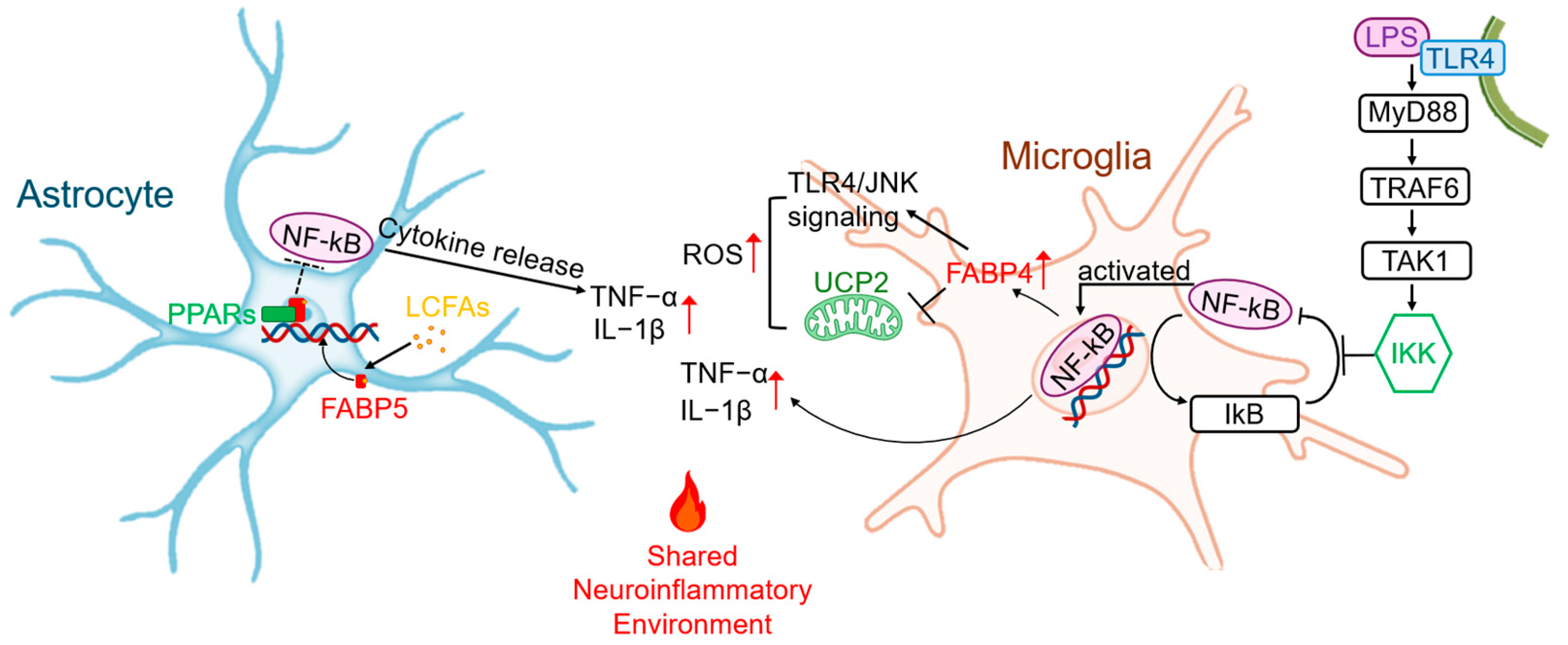
Figure 1. FABP-driven glial “meta-inflammatory” hub. This detailed schematic illustrates the synergistic inflammatory roles of astrocytes and microglia, highlighting specific FABP-mediated signaling pathways. In Microglia: Lipopolysaccharide (LPS) binds to Toll-like receptor 4 (TLR4), recruiting the MyD88 adaptor protein and sequentially activating TRAF6 and TAK1. This initiates the downstream IKK complex, leading to the phosphorylation and degradation of IκB, thereby releasing the NF-κB transcription factor for nuclear translocation. Nuclear NF-κB drives the transcription of pro-inflammatory cytokines (e.g., TNF-α, IL-1β) and, critically, upregulates FABP4 expression. The upregulated FABP4 creates a positive feedback loop that amplifies TLR4/JNK signaling and ROS production while inhibiting the protective UCP2. In Astrocytes: FABP5 transports LCFAs into the nucleus, where they interact with PPARs, de-repressing the inhibition of NF-κB. The activated NF-κB then drives the transcription and release of cytokines such as TNF-α and IL-1β. Together, these glial cells create a Shared Neuroinflammatory Environment that is a key driver of neurodegeneration.
Cells 14 01502 g001
Figure 2. The role of FABP5 and FABP7 in oligodendrocyte lipid metabolism and myelination. This diagram illustrates the complementary functions of FABP5 and FABP7 within a differentiating oligodendrocyte during myelination. External lipids are taken up from the extracellular space. (1) FABP5 Pathway (Energy Production): FABP5 chaperones fatty acids (FAs) to mitochondrion, where they undergo β-oxidation. This process generates acetyl-CoA, which enters the TCA cycle to produce ATP (energy currency) and NADPH (reducing power), both essential for the high energy demands of myelin synthesis. (2) FABP7 Pathway (Biosynthesis and Coordination): FABP7 transports lipid precursors to the endoplasmic reticulum (ER) and Golgi apparatus for the synthesis of core myelin lipids, such as phospholipids and sphingolipids. These lipids are then incorporated into the growing myelin sheath. Additionally, FABP7 interacts with PGC-1α to promote mitochondrial biogenesis, ensuring energy supply matches biosynthetic demand. (3) Final Output: This coordinated action is vital for maintaining oligodendrocyte survival and proper myelin sheath formation around the axon.
Cells 14 01502 g002
Figure 3. The gut–brain-FABP axis in neurodegeneration. This diagram illustrates the multi-stage pathological cascade linking gut health to neurodegeneration. Stage 1 (Gut Initiation): Gut dysbiosis and other insults lead to FABP2 dysfunction, which compromises tight junctions, resulting in a “leaky gut.” This allows bacterial lipopolysaccharide (LPS) to enter the bloodstream and creates a local inflammatory environment that promotes the misfolding of α-syn in the ENS. Stage 2 (Propagation): The pathology spreads to the brain via two parallel pathways. Pathological α-syn “seeds” propagate from the ENS to the brainstem via retrograde transport along the vagus nerve (Vagal Propagation), consistent with the hypothesis. Concurrently, LPS enters the systemic circulation. Stage 3 (Central Pathology and Inflammation): The α-syn pathology spreads trans-neuronally within the brain, causing synucleinopathy. Meanwhile, circulating LPS crosses a compromised blood–brain barrier (BBB) and activates microglia via the FABP4/TLR4 signaling pathway, establishing a chronic neuroinflammatory state that exacerbates the primary proteotoxic damage.
Cells 14 01502 g003
Figure 4. The distinct roles of FABP5 and FABP7 in regulating Ferroptosis. This schematic illustrates the opposing functions of FABP5 and FABP7 in the context of ferroptosis. (1) Pro-ferroptotic pathway (FABP5): FABP5 transports polyunsaturated fatty acids (PUFAs) to the cell membrane, where enzymes like ACSL4 and LPCAT3 incorporate them into phospholipids (PUFA-PLs). In the presence of iron (Fe2+), these PUFA-containing phospholipids are highly susceptible to lipid peroxidation, leading to the accumulation of lipid ROS and execution of ferroptosis. This process is normally counteracted by the antioxidant enzyme GPX4. (2) Anti-ferroptotic pathway (FABP7): FABP7 acts as a protective factor by sequestering PUFAs away from the membrane and trafficking them to LDs for safe storage. This prevents their peroxidation and shields the cell from ferroptotic death. The balance between these two pathways can determine the cell’s fate under oxidative stress.
Cells 14 01502 g004
Figure 5. Chemical structures of key preclinical FABP ligands. The 2D chemical structures of representative CNS-penetrant and peripherally restricted FABP inhibitors are shown.
Cells 14 01502 g005
Table 1. Expression and Roles of Key FABP Isoforms in the Neuro-Gastrointestinal System.
IsoformMajor Peripheral Tissue/Cell TypeMajor CNS Tissue/Cell TypeCore FunctionAssociated Neurodegenerative Disease
FABP2Small intestine epithelial cells [56]Not expressedFatty acid absorption [57,58], maintaining intestinal barrier integrity [49]PD (via gut–brain axis) [59]
FABP3Myocardial cells [60], skeletal muscle cells [61]Neurons (especially dopaminergic neurons) [62]Mitochondrial β-oxidation [63,64], promoting α-syn oligomerization [65,66]PD [65,66], AD [67,68]
FABP4Adipocytes [69], macrophages [70]Microglia [24]Systemic insulin resistance [71], mediating microglial inflammation [72]Obesity-associated cognitive decline [29], Microglia-mediated neuroinflammation [73]
FABP5Epidermal cells [74], macrophages [74]Neurons [75], astrocytes [76,77], oligodendrocytes [78,79,80]Driving inflammation [81,82,83], regulating myelination [84]MS [15,84], Stroke [85]
FABP7Adipocytes [86]Astrocytes [87,88], radial glial cells [89]Driving glial meta-inflammation [90,91], neural stem cell development [92,93,94]MS [84,91,95], MSA [44], AD [87,96], ALS [17]
Table 2. Preclinical Characteristics of Representative FABP Ligands.
LigandTarget Isoform and SelectivityAffinity (Kd/Ki, nM)CNS Penetrance Key Preclinical ModelPrimary Efficacy Endpoint
MF6 [43,66,84,98]FABP7 > FABP5 >> FABP3FABP7: 20; FABP5: 874Good, Plasma Cmax ~522 nM (4 h)EAE (MS), MSAReduced demyelination, inhibited glial activation, improved motor function
MF1 [66,98,101]FABP3302.8Good (confirmed by in vivo efficacy)MPTP (PD), EpilepsyReduced α-syn aggregation, improved motor and cognitive function, anticonvulsant
HY-11-9FABP311.7Good (confirmed by in vivo efficacy)MPTP (PD)Improved motor function, reduced pS129-α-syn aggregation (superior to MF1)
HY08 [85]FABP3 > FABP5FABP3: 24; FABP5: 410Good (confirmed by in vivo efficacy)tMCAO/R (Stroke)Reduced infarct volume, improved neurological function, inhibited mitochondrial damage
ART26.12 [104,106]FABP5 >> FABP7, FABP3, FABP4FABP5: 770 (Ki)Limited (Brain/Plasma Ratio ≈ 2.3%)Neuropathic PainReversed mechanical allodynia and cold hyperalgesia
Disclaimer/Publisher’s Note: The statements, opinions and data contained in all publications are solely those of the individual author(s) and contributor(s) and not of MDPI and/or the editor(s). MDPI and/or the editor(s) disclaim responsibility for any injury to people or property resulting from any ideas, methods, instructions or products referred to in the content.